Article

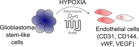
The Adenosine A3 Receptor Regulates Differentiation of Glioblastoma Stem-Like Cells to Endothelial Cells under Hypoxia

1
Laboratorio de Patología Molecular, Instituto de Bioquímica y Microbiología, Facultad de Ciencias, Universidad Austral de Chile, Valdivia 5090000, Chile
2
Instituto de Anatomía, Histología y Patología, Universidad Austral de Chile, Valdivia 5090000, Chile
*
Author to whom correspondence should be addressed.
These authors contributed equally to this work.
Int. J. Mol. Sci. 2018, 19(4), 1228; https://doi.org/10.3390/ijms19041228
Submission received: 14 March 2018 / Revised: 3 April 2018 / Accepted: 3 April 2018 / Published: 18 April 2018

Abstract

:
Glioblastoma (GBM) is a neoplasm characterized by an extensive blood vessel network. Hypoxic niches of GBM can induce tumorigenic properties of a small cell subpopulation called Glioblastoma stem-like cells (GSCs) and can also increase extracellular adenosine generation which activates the A3 adenosine receptor (A3AR). Moreover, GSCs potentiates the persistent neovascularization in GBM. The aim of this study was to determine if A3AR blockade can reduce the vasculogenesis mediated by the differentiation of GSCs to Endothelial Cells (ECs) under hypoxia. We evaluated the expression of endothelial cell markers (CD31, CD34, CD144, and vWF) by fluorescence-activated cell sorting (FACS), and vascular endothelial growth factor (VEGF) secretion by ELISA using MRS1220 (A3AR antagonist) under hypoxia. We validate our results using U87MG-GSCs A3AR knockout (GSCsA3-KO). The effect of MRS1220 on blood vessel formation was evaluated in vivo using a subcutaneous GSCs-tumor model. GSCs increased extracellular adenosine production and A3AR expression under hypoxia. Hypoxia also increased the percentage of GSCs positive for endothelial cell markers and VEGF secretion, which was in turn prevented when using MRS1220 and in GSCsA3-KO. Finally, in vivo treatment with MRS1220 reduced tumor size and blood vessel formation. Blockade of A3AR decreases the differentiation of GSCs to ECs under hypoxia and in vivo blood vessel formation.

Graphical Abstract

1. Introduction

Glioblastoma (GBM) is considered the most common tumor of the central nervous system and one of the most devastating types of cancer. After multimodal therapy, consisting of surgical resection followed by radio- and chemo- therapy with temozolomide, about 99% of cancer cells are eliminated; however, the tumor recurs and, as a result, patients die on average within 15 months [1,2,3]. Therapy failure is mainly attributed to a cell subpopulation called glioblastoma stem-like cells (GSCs) which, like normal neural stem cells (NSCs), have unlimited self-renewal and a multi-lineage differentiation capacity. In addition, GSCs have the potential to form in vivo tumors and exhibit a higher resistance to therapy than differentiated cancer cells [4,5,6,7]. Therefore, strategies are currently aimed at eliminating GSCs or promoting their differentiation to less aggressive phenotypes. A key feature of GBM is the presence of extensive blood vessel networks that support both tumor growth and resistance to treatment [8,9]. Unlike normal vasculature where the endothelium has a low proliferation rate, tumor vasculature is highly proliferative and the formed blood vessels are often disorganized and tortuous [10]. This entails inefficient oxygen supply and as a result, extensive hypoxic areas that promote further vasculogenesis [11]. In fact, the hypoxia inducible factor 1α (HIF-1α) induces the expression of vascular endothelial growth factor (VEGF), which in turn stimulates proliferation and migration of endothelial cells (ECs) [12,13,14,15]. GSCs are enriched in hypoxic areas within the tumor which promote both their stemness and radio- and chemo- resistance [16,17,18,19], therefore, future therapies targeting GSCs should consider this niche. In addition, like GSCs, tumor-derived ECs are found in a greater proportion in internal and hypoxic tumor regions. Importantly, GSCs can differentiate into ECs, a phenomenon where hypoxia appears to be critical [20,21,22]. However, the mechanisms and pathways that control differentiation of GSCs to ECs are not fully understood. Signaling pathways modulated by adenosine are up-regulated under hypoxia and participate in various aspects of cancer, including vasculogenesis [23,24,25,26,27]. This nucleoside is produced mainly by extracellular ATP hydrolysis and signals through its four Adenosine Receptors (ARs): A1 (A1AR), A2A (A2AAR), A2B (A2BAR), and A3 (A3AR) [26]. We showed that GBM cells have increased extracellular adenosine levels and exhibit high expression of A3AR compared to non-tumoral normal cells [28]. Similar results were observed in GSCs, where extracellular adenosine levels and A3AR expression are higher than in differentiated GBM cells [29,30]. However, the role of this receptor on GSCs biology is poorly understood. Therefore, our aim was to determine if A3AR blockade can reduce the vasculogenesis mediated by the differentiation of GSCs to ECs under hypoxia.

2. Results

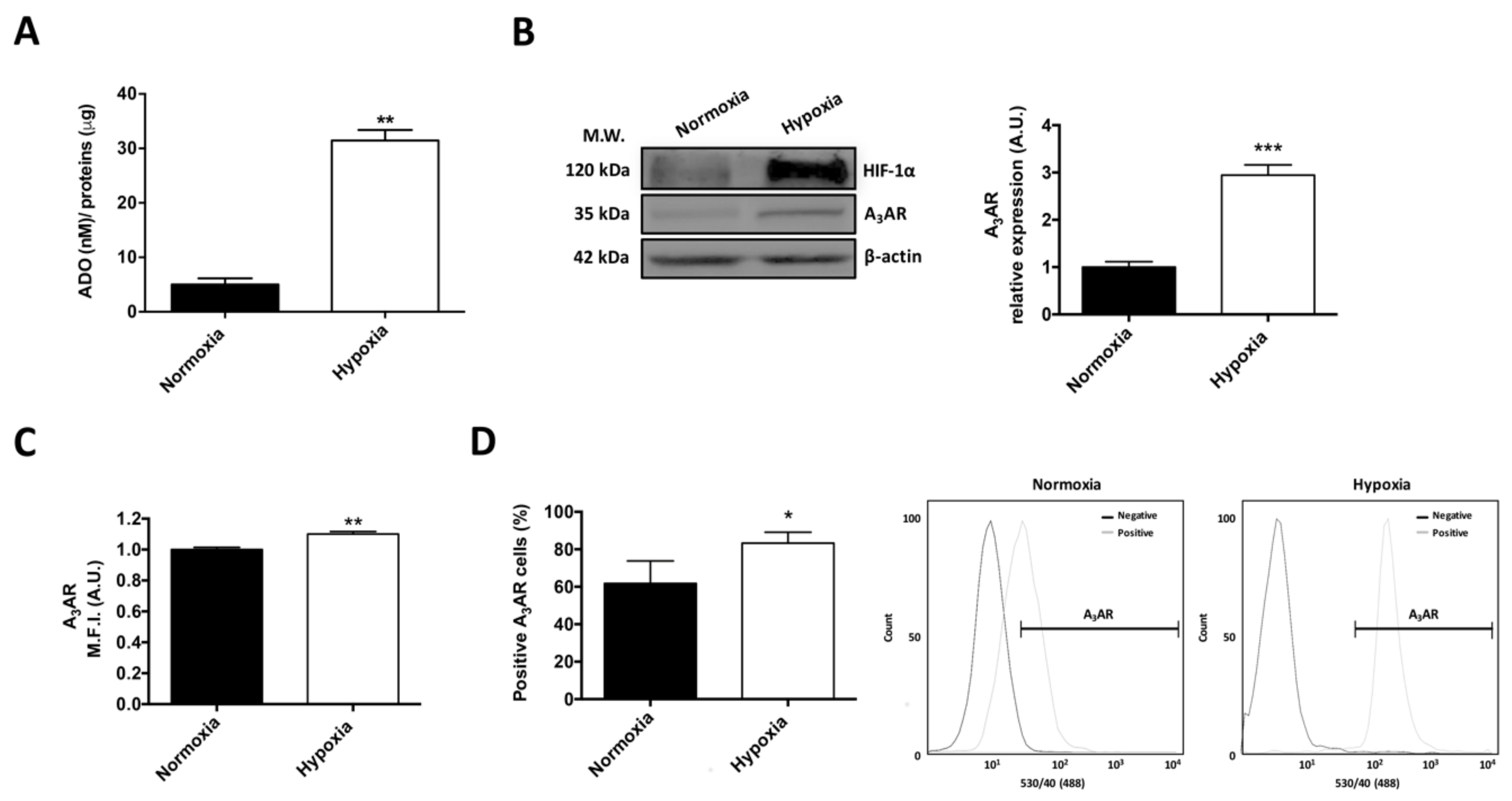
2.1. Extracellular Adenosine Concentration and A3 Adenosine Receptor Expression Increase under Hypoxia

U87MG GSCs were cultured under 0.5% O2 in order to evaluate hypoxia effect on extracellular adenosine generation and A3AR expression. We found that U87MG GSCs had increased extracellular adenosine production (~7 fold) after 24 h of hypoxia (Figure 1A). A3AR expression increased in U87MG GSCs under hypoxia (Figure 1B,C). Similarly, the percentage of A3AR-positive GSCs increased under hypoxic conditions (Figure 1D). These results suggest that the high levels of extracellular adenosine in U87MG GSCs culture could activate the A3AR under hypoxia.

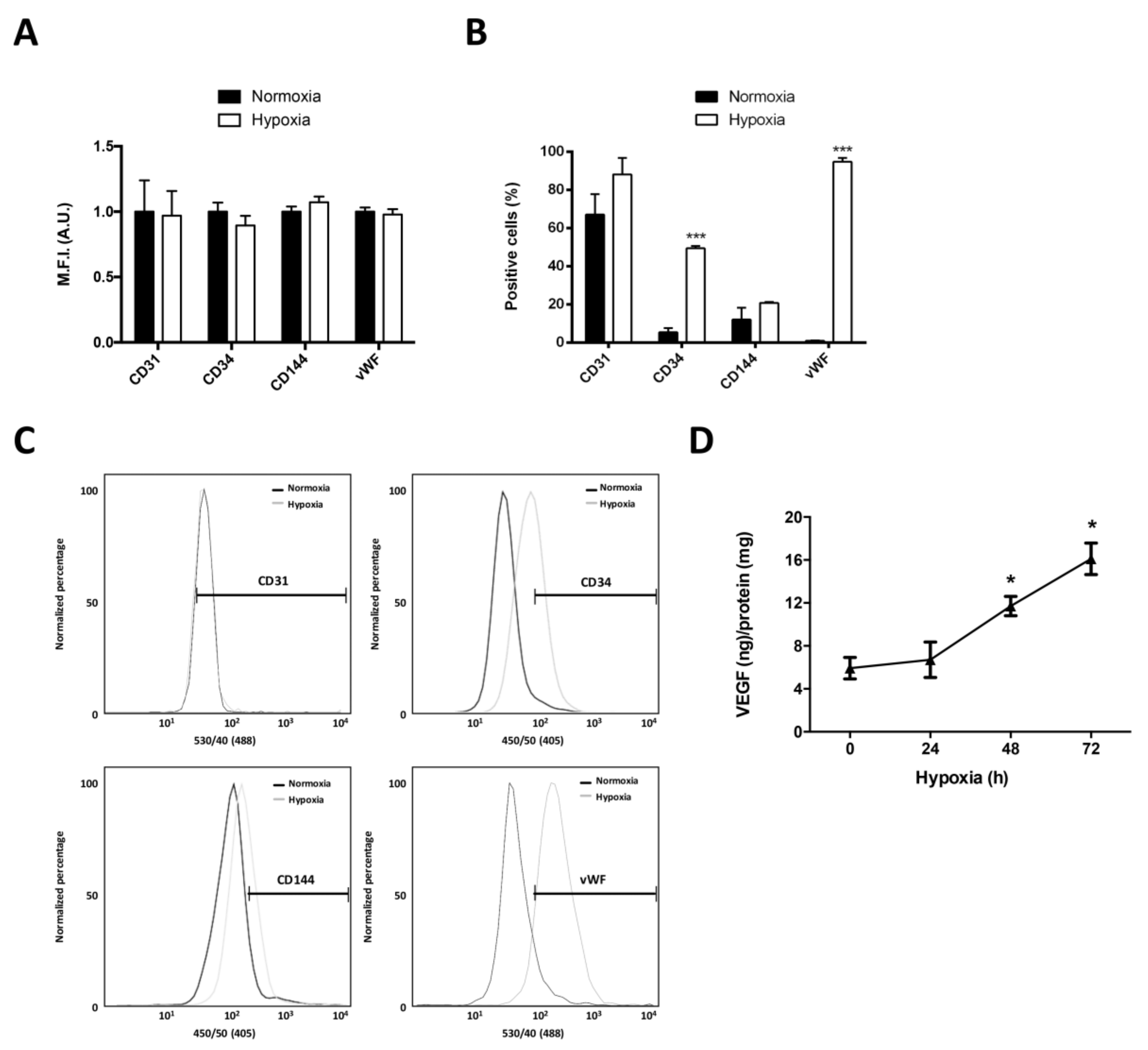
2.2. Differentiation of Glioblastoma Stem-Like Cells to Endothelial Cells Increases under Hypoxia

To evaluate the effect of hypoxia on the differentiation of GSCs to ECs we evaluated the expression of endothelial cell markers (CD31, CD34, CD144, and vWF) and VEGF secretion. No differences were observed in the expression of endothelial markers through Flow Cytometry between U87MG GSCs (Figure 2A). However, the percentage of positive cells for CD34 and vWF increased after 24 h of hypoxia (Figure 2B,C). To evaluate VEGF secretion in GSCs under hypoxia, we evaluated the presence of VEGF-165 in U87MG GSCs medium during 72 h of hypoxia. We observed an increase in VEGF-165 secretion at 48 (~2 fold) and 72 (~2.7 fold) hours under hypoxia (Figure 2D). These results suggest that U87MG GSCs could differentiate into ECs, especially under hypoxia. These results propose that hypoxia promotes the expression of endothelial cell markers and the secretion of VEGF in GSCs.

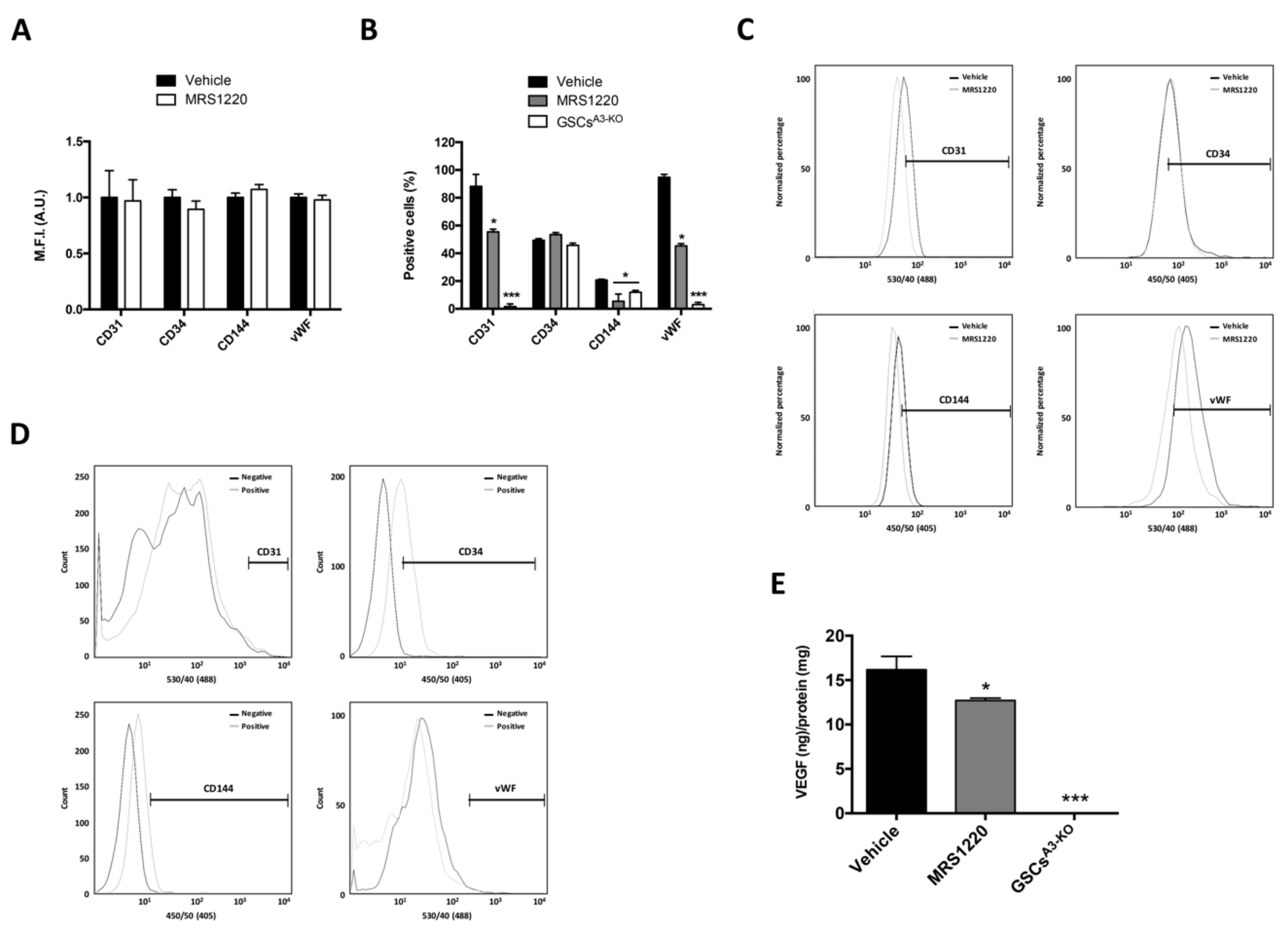
2.3. A3AR Blockade Decreases Differentiation of Glioblastoma Stem-Like Cells to Endothelial Cells under Hypoxia

We explored the effect of A3AR blockade on the differentiation of GSCs to ECs under hypoxia. Cells were treated with MRS1220, a selective A3AR antagonist, under hypoxia and then the expression of endothelial cell markers and VEGF secretion were analyzed. A3AR blockade did not change the expression of endothelial markers (Figure 3A), nevertheless, decreased the percentage of CD31, CD144, and vWF positive GSCs after 24 h under hypoxic conditions (Figure 3B,C). VEGF secretion in U87MG GSCs decreased ~25% with MRS1220 after 72 h of hypoxia (Figure 3E). To validate the effect of MRS1220 in U87MG GSCs differentiation to ECs, we used an A3AR knockout cell line (GSCsA3-KO) to evaluate its intrinsic differentiation ability to ECs under hypoxia. Similarly, we observed a decreased percentage of CD31, CD144, and vWF positive cells (Figure 3B,D), and an almost total decrease in VEGF secretion (Figure 3E) in GSCsA3-KO under hypoxia. These results suggest that the ability of U87MG GSCs to differentiate into ECs could be regulated by A3AR activation under hypoxia.

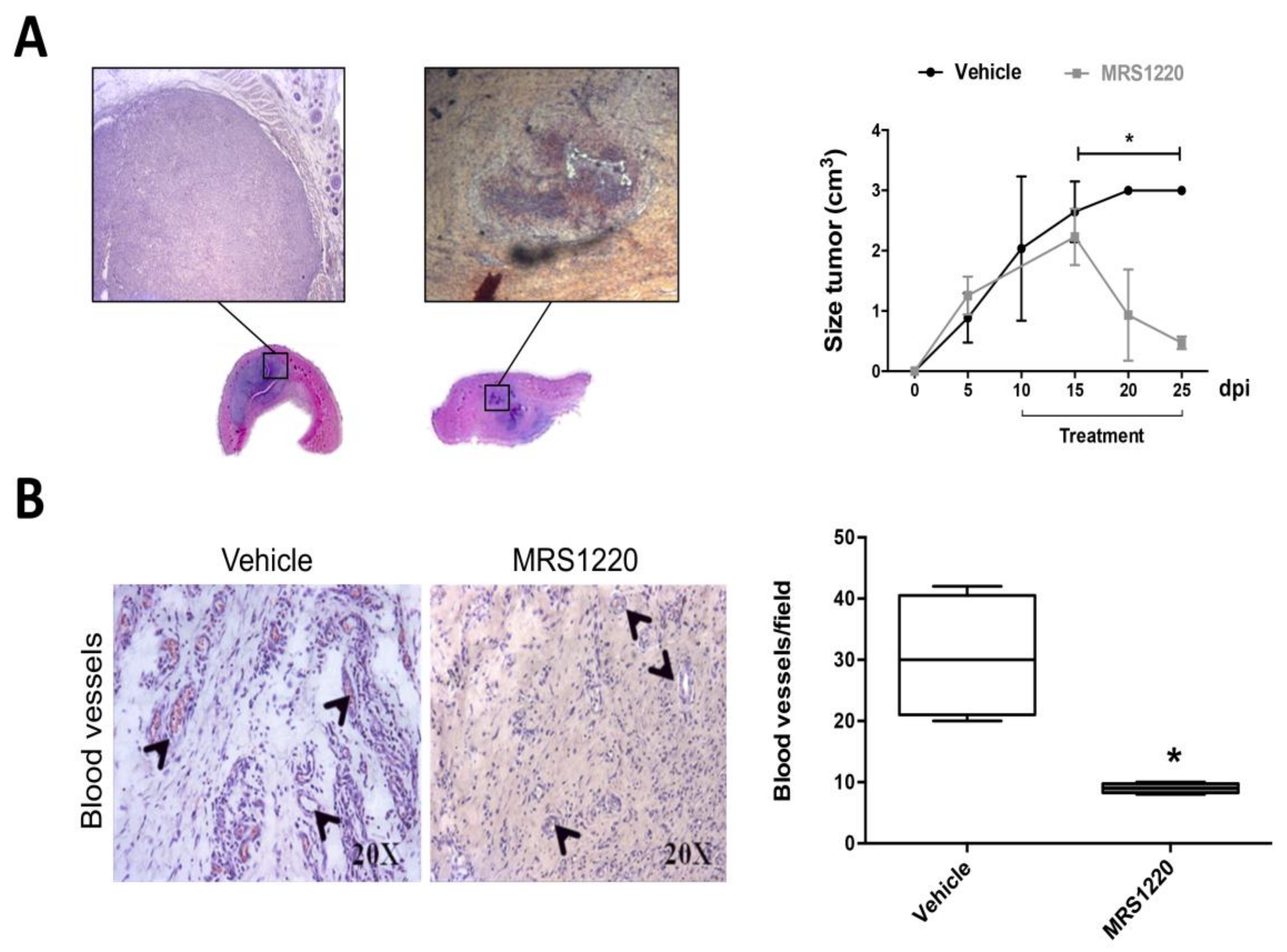
2.4. In Vivo Antagonization of A3AR Decreases Tumor Size and Blood Vessel Formation

To ensure tumor growth, the formation of blood vessels that supply oxygen and nutrients to neoplastic cells is crucial [27]. Increased tumor volume is linked to a larger network of blood vessels, which is why in recent years the generation of new anti-angiogenic therapies has been sought [31]. To evaluate the in vivo effect of A3AR antagonization, we generated an allogeneic rat subcutaneous tumor using GSCs from the rat C6 glioma cell line. At day ten post-inoculation with C6 GSCs, we treated animals with MRS1220 for fifteen days. We observed a reduction close to 80% and 90% in tumor volume compared to the vehicle-treated group at day ten and fifteen post-treatment, respectively (Figure 4A). The histopathological analysis showed extensive necrotic areas with blood vessel formation, which was reverted after pharmacological blockade of A3AR. The number of blood vessels per field was reduced by three times with MRS1220 (Figure 4B), indicating a strong in vivo anti-angiogenic effect.

3. Discussion

The prognosis for GBM treatment is worsened by the presence of GSCs due to their self-renewal and cell differentiation properties, for example to endothelial cells (ECs), promoting angiogenesis and neovascularization [32]. Since GSCs are highly chemo- and radio- resistant, they are maintained in tumor niches even after treatment. This sustained maintenance and increased neovascularization is linked to the high extracellular adenosine concentrations found in tumors and even higher concentrations in hypoxic niches [27]. AR subtypes have different affinities to adenosine; A1 and A2A are high affinity receptors and A2B and A3 are low affinity receptors; therefore, in pathological conditions, such as cancer, extracellular adenosine levels are increased and mainly activate A2BAR and A3AR subtypes enhancing several signaling pathways, such as PI3K/AKT, MAPK, among others [27,29,33].
Knockout of ectonucleotidase CD73, which is important for extracellular adenosine production from AMP, decreased angiogenesis in melanoma models: a process that is reverted when using different AR agonists [34]. The overall effect was greater when inhibiting adenosine production compared to AR blockade, concluding that the AR subtypes have a summatory effect on angiogenesis. Several studies have confirmed that A3AR is important to angiogenesis in different tumors, specifically in the generation of blood vessels and neovascularization [34,35,36]. In this study, we proposed that adenosine regulates the differentiation of GSCs to ECs in vitro through A3AR activation, and that this promotes the formation of new blood vessels in an in vivo GBM tumor model. GSCs produce more adenosine than differentiated cells [29], and their production is enhanced under hypoxia in other tumor models [27]. In this study we showed that GSCs not only enhance adenosine production but also increase A3AR expression, suggesting a loop of positive regulation between the AR and its ligand; probably through expression of the transcription factor HIF-1α, which increases during early hypoxia [32]. Hypoxia-promoted cell differentiation and subsequent expression of HIF-1α have been described in different models [37], however this is poorly understood in GSCs. In this study, we showed for the first time that hypoxia increased A3AR expression, and its blockade decreases the cell population positive to several endothelial cell markers, such as CD34, CD144, and vWF, suggesting that GSCs could be differentiated into ECs, possibly through a mechanism dependent on extracellular adenosine-A3AR axis. In addition, hypoxic conditions increased VEGF secretion, which was previously observed in other tumor models but not in GSCs [38,39]. VEGF-165 secretion, which is the most abundant and potent VEGF isoform in GBM [40], was specifically evaluated. The results suggested that differentiation of GSCs into ECs could promote neovascularization and angiogenesis. These processes are highly relevant to the progression and prognosis of GBM as they support tumor growth and infiltration into surrounding healthy tissue.
In this study we used MRS1220, an A3AR pharmacological antagonist, to demonstrate that this receptor is involved in the differentiation of GSCs to ECs. In addition, our research group previously produced the U87MG A3AR KO cell line, with the ability to differentiate into GSCs, demonstrating their role in chemoresistance [29]. The A3AR antagonist and the KO model showed a decrease in the CD31, CD144, and vWF positive cell population, suggesting that the expression of these markers depends on AR activation; probably due to increased adenosine production in GSCs under hypoxic conditions.
Expression of in vitro markers does not necessarily reflect phenotypic changes in vivo, however, the decrease in blood vessel production in GBM models correlates with the low levels of some markers, such as CD31 [41]. To corroborate whether cell differentiation is directly linked to in vivo neovascularization, a previously validated murine model and an MRS1220 antagonist were used [29], producing a decrease in tumor size and blood vessel formation. The high concentrations of adenosine in the tumor, specifically in hypoxic niches, promotes the expression and over-activation of the A3AR, facilitating neovascularization. This process surely depends on HIF-1α activation; however, the possible signaling pathways involved must still be studied in depth. These results provide adenosine and its signaling with a new and important role in GBM.

4. Materials and Methods

4.1. Cell Culture

Human U87MG GBM and rat C6 glioma cell lines were acquired from the ATCC (HTB-14TM and CCL-107TM, respectively). Cells were grown in DMEM-F12 supplemented with 10% fetal bovine serum and penicillin-streptomycin (Life Technologies, Carlsbad, CA, USA). U87MG knockout for A3AR (GSCsA3-KO) were generated using the CRISPR-Cas9 protocol described by Torres et al., [29]. GSCs were obtained from U87MG and C6 cell lines using the neurosphere formation method [29]. Briefly, GSCs were cultured in Neurobasal and DMEM media for U87MG and C6 cells respectively, supplemented with 20 ng/mL bFGF, 20 ng/mL EGF, Glutamax 1X, B-27 1x, all purchased from Gibco® (Thermo Fisher Scientific Inc., Waltham, MA, USA). For hypoxia experiments, GSCs were cultured in a 0.5% oxygen (O2) atmosphere using a hypoxia chamber (BioSpherix C-274, Oxygen sensor BioSpherix ProOx P110, Parish, NY, USA) that replaces O2 pumping nitrogen. GSCs cultured for 7 days were used to perform all the experiments. A3AR antagonizing was performed with 10 μM MRS1220 (Tocris, Park Ellisville, MI, USA) at different times depending on the experiment.

4.2. High Performance Liquid Chromatography (HPLC)

Quantification of adenosine production of U87MG GSCs was performed by HPLC using the protocol described by Torres et al., [29]. Briefly, GSCs were incubated in 1 mL of Tyrode’s buffer for 1 h at 37 °C. 200 µL of incubation medium was mixed with 100 µL of citrate buffer (pH 6). Adenosine, AMP, ADP, and ATP contents were quantified with 2-chloroacetaldehyde derivatizations by HPLC fractionation in a Chromolith Performance RP-18 column (Merck, Darmstadt, Germany) and by fluorescent detection [42]. Adenosine concentration (nM) was normalized to the total protein concentration (μg).

4.3. Western Blot

Total protein extracts were obtained in 10 mM Tris–HCl buffer, 2% SDS, 10% glycerol, 1 mM PMSF, and protease inhibitors (Complete, Merck). Protein aliquots (50 μg) were separated by 10% SDS-PAGE followed by transfer to PVDF membranes (Bio-Rad, Hercules, CA, USA). Membranes were incubated with anti-HIF-1α (sc-10790) and anti-A3AR (sc-13938) antibodies (Santa Cruz Biotechnology, Dallas, TX, USA) overnight. After washing, the blots were further incubated with HRP-conjugated IgG antibody (DAKO Agilent, Santa Clara, CA, USA) for 1 h at room temperature. Finally, immune staining was visualized by using ECL plus (Amersham Pharmacia, Piscataway, NJ, USA) and the image analysis system syngene G:Box (Synoptics Ltd., Cambridge, UK). The images were analyzed by densitometry (Image J software, NIH, Bethesda, MD, USA) and each membrane was normalized to β-actin (sc-47778-HRP, Santa Cruz Biotechnology) expression.

4.4. Flow Cytometry

To measure endothelial cell marker expression, cells were analyzed by flow cytometry (FACS Jazz; BD Biosciences, Franklin Lakes, NJ, USA). Cells were previously fixed with PFA 3.7% for 15 min at room temperature. Cells were then blocked for 45 min (1xPBS-BSA 0.5% at room temperature) and marked with anti-CD31 (58068), anti-CD34 (562577), or anti-CD144 (561569) antibodies (BD Biosciences, Franklin Lakes). For vWF detection, cells were incubated with anti-vWF (555849) (BD Biosciences, Franklin Lakes) followed by an anti-mouse Alexa 488 (Life Technologies). Lastly, events were acquired through the FL1 filter of the cytometer.

4.5. Enzyme-Linked ImmunoSorbent Assay

VEGF in culture medium was quantified using Human VEGF ELISA Kit (KHG0111, Life Technologies) [40]. For kinetic analysis, a cell density of 104 GSCs/well [40] were incubated for 24, 48, and 72 h under hypoxia conditions. Treatments with 10 μM MRS1220 were carried out for 72 h under hypoxia conditions with the same cell density. VEGF levels (ng) were measured according to the manufacturer’s instructions and normalized to total protein content (mg).

4.6. In Vivo Studies and Histopathological Analysis

A total of eight, 8 week-old male Sprague–Dawley rats were maintained under standard laboratory conditions, approved by the Ethics Committee of Animal Experiments at the Universidad Austral de Chile (Permit Number: 248-2016; date: 23 March, 2016). A density of 2 × 10⁶ GSCs C6 cells were inoculated by subcutaneous injection in previously anesthetized rats (ketamine (100 mg/kg)/xylazine (10 mg/kg) intraperitoneal). At day ten post-inoculation, animals were divided for the following treatments (i) 1xPBS-0.001% DMSO (Vehicle) (Merck), and (ii) MRS1220 (0.15 mg/kg/72 h) administered by intraperitoneal inoculation. Tumor size was measured each five days until 25 days post-inoculation when rats were euthanized by intraperitoneal administration of Sodium Thiopental (120 mg/kg). Subcutaneous tumors were removed, fixed in 3.7% paraformaldehyde, dewaxed with xylol, and rehydrated using alcohols in decreasing concentration. The samples were immersed in hematoxylin and eosin (H&E) for 5 min and finally passed through an ascending alcohol concentration followed by xylol and then mounted (Histomount, Thermo Fisher Scientific Inc.). Tumor preparations were analyzed and the blood vessel count was performed by dividing each sample into ten quadrants where the number of blood vessels was quantified and averaged using ImageJ software (NIH, Bethesda, MD, USA) [29].

4.7. Statistical Analysis and Artwork

Values are expressed as the mean ± Standard Deviation (S.D.), where n indicates number of independent experiments. Statistical analyses were performed using ANOVA, Student’s t-test (unpaired data), and Tukey-test. P values ≤ 0.05 were considered statistically significant. GraphPad Prism 6 (La Jolla, CA, USA) software was used to create all graphs and statistical analyses.

5. Conclusions

In this study, we conclude that A3AR promotes GSCs differentiation to ECs under hypoxia. Expression of endothelial cells markers, such as CD144, CD31, and vWF and VEGF secretion are regulated by adenosine and A3AR activation. Our data suggest that hypoxic niches and the adenosine axis are responsible for neovascularization; proposing GSCs and the adenosine axis as plausible therapeutic targets for GBM (Figure 5).

Acknowledgments

This study was supported by Grants 1160777 (Claudia Quezada ) and 3140217 (René Rocha) from FONDECYT-Chile. Thanks to John James Finnolli for his help in proofreading and editing.

Author Contributions

René Rocha and Claudia Quezada designed the project; René Rocha and Karina Ojeda, performed in vitro experiments; Dellis Rocha and Daniel Uribe performed flow cytometry analyses; Ángelo Torres performed knocking out; Ángelo Torres and José Erices performed in vivo experiments; René Rocha, Ignacio Niechi, Pamela Ehrenfeld and Ángelo Torres analyzed the data; Ángelo Torres and Ignacio Niechi created figures and illustrations; René Rocha, Ángelo Torres, Claudia Quezada, Ignacio Niechi, Rody San Martín, Pamela Ehrenfeld and Daniel Uribe wrote the paper.

Conflicts of Interest

The authors declare no conflict of interest.

Abbreviations

GBMGlioblastoma Multiforme
GSCsGlioblastoma Stem-Like Cells
NSCsNeural Stem Cells
HIF-1αHypoxia Inducible Factor 1
VEGFVascular Endothelial Growth Factor
ECsEndothelial Cells
ARsAdenosine Receptors
A1ARA1 Adenosine Receptor
A2AARA2A Adenosine Receptor
A2BARA2B Adenosine Receptor
A3ARA3 Adenosine Receptor

References

  1. Stupp, R.; Hegi, M.E.; Mason, W.P.; van den Bent, M.J.; Taphoorn, M.J.; Janzer, R.C.; Ludwin, S.K.; Allgeier, A.; Fisher, B.; Belanger, K.; et al. European Organisation for Research and Treatment of Cancer Brain Tumour and Radiation Oncology Groups, National Cancer Institute of Canada Clinical Trials Group. Effects of radiotherapy with concomitant and adjuvant temozolomide versus radiotherapy alone on survival in glioblastoma in a randomised phase III study: 5-year analysis of the EORTC-NCIC trial. Lancet Oncol. 2009, 10, 459–466. [Google Scholar] [CrossRef] [PubMed]
  2. Stupp, R.; Mason, W.P.; van den Bent, M.J.; Weller, M.; Fisher, B.; Taphoorn, M.J.; Belanger, K.; Brandes, A.A.; Marosi, C.; Bogdahn, U.; et al. European Organisation for Research and Treatment of Cancer Brain Tumor and Radiotherapy Groups; National Cancer Institute of Canada Clinical Trials Group. Radiotherapy plus concomitant and adjuvant temozolomide for glioblastoma. N. Engl. J. Med. 2005, 352, 987–996. [Google Scholar] [CrossRef] [PubMed]
  3. Lee, C.Y. Strategies of temozolomide in future glioblastoma treatment. Onco. Targets Ther. 2017, 10, 265–270. [Google Scholar] [CrossRef] [PubMed]
  4. Singh, S.K.; Hawkins, C.; Clarke, I.D.; Squire, J.A.; Bayani, J.; Hide, T.; Henkelman, R.M.; Cusimano, M.D.; Dirks, P.B. Identification of human brain tumour initiating cells. Nature 2004, 432, 396–401. [Google Scholar] [CrossRef] [PubMed]
  5. Galli, R.; Binda, E.; Orfanelli, U.; Cipelletti, B.; Gritti, A.; de Vitis, S.; Fiocco, R.; Foroni, C.; Dimeco, F.; Vescovi, A. Isolation and characterization of tumorigenic, stem-like neural precursors from human glioblastoma. Cancer Res. 2004, 64, 7011–7021. [Google Scholar] [CrossRef] [PubMed]
  6. Bao, S.; Wu, Q.; McLendon, R.E.; Hao, Y.; Shi, Q.; Hjelmeland, A.B.; Dewhirst, M.W.; Bigner, D.D.; Rich, J.N. Glioma stem cells promote radioresistance by preferential activation of the DNA damage response. Nature 2006, 444, 756–760. [Google Scholar] [CrossRef] [PubMed]
  7. Chen, J.; Li, Y.; Yu, T.S.; McKay, R.M.; Burns, D.K.; Kernie, S.G.; Parada, L.F. A restricted cell population propagates glioblastoma growth after chemotherapy. Nature 2012, 488, 522–526. [Google Scholar] [CrossRef] [PubMed]
  8. Shaifer, C.A.; Huang, J.; Lin, P.C. Glioblastoma cells incorporate into tumor vasculature and contribute to vascular radioresistance. Int. J. Cancer 2010, 127, 2063–2075. [Google Scholar] [CrossRef] [PubMed]
  9. Virrey, J.J.; Golden, E.B.; Sivakumar, W.; Wang, W.; Pen, L.; Schönthal, A.H.; Hofman, F.M.; Chen, T.C. Glioma-associated endothelial cells are chemoresistant to temozolomide. J. Neurooncol. 2009, 95, 13–22. [Google Scholar] [CrossRef] [PubMed]
  10. Jain, R.K.; di Tomaso, E.; Duda, D.G.; Loeffler, J.S.; Sorensen, A.G.; Batchelor, T.T. Angiogenesis in brain tumours. Nat. Rev. Neurosci. 2007, 8, 610–622. [Google Scholar] [CrossRef] [PubMed]
  11. Kaur, B.; Khwaja, F.W.; Severson, E.A.; Matheny, S.L.; Brat, D.J.; van Meir, E.G. Hypoxia and the hypoxia-inducible-factor pathway in glioma growth and angiogenesis. Neuro Oncol. 2005, 7, 134–153. [Google Scholar] [CrossRef] [PubMed]
  12. Forsythe, J.A.; Jiang, B.H.; Iyer, N.V.; Agani, F.; Leung, S.W.; Koos, R.D.; Semenza, G.L. Activation of vascular endothelial growth factor gene transcription by hypoxia-inducible factor 1. Mol. Cell. Biol. 1996, 16, 4604–4613. [Google Scholar] [CrossRef] [PubMed]
  13. Plate, K.H.; Breier, G.; Weich, H.A.; Risau, W. Vascular endothelial growth factor is a potential tumour angiogenesis factor in human gliomas in vivo. Nature 1992, 359, 845–848. [Google Scholar] [CrossRef] [PubMed]
  14. Ezhilarasan, R.; Mohanam, I.; Govindarajan, K.; Mohanam, S. Glioma cells suppress hypoxia-induced endothelial cell apoptosis and promote angiogenic process. Int. J. Oncol. 2007, 30, 701–707. [Google Scholar] [CrossRef] [PubMed]
  15. Coma, S.; Shimizu, A.; Klagsbrun, M. Hypoxia induces tumor and endothelial cell migration in a Semaphorin 3F- and VEGF-dependent manner via transcriptional repression of their common receptor Neuropilin 2. Cell Adh. Migr. 2011, 5, 266–275. [Google Scholar] [CrossRef] [PubMed]
  16. Pistollato, F.; Abbadi, S.; Rampazzo, E.; Persano, L.; Della Puppa, A.; Frasson, C.; Sarto, E.; Scienza, R.; D’avella, D.; Basso, G. Intratumoural hypoxic gradient drives stem cells distribution and MGMT expression in glioblastoma. Stem Cells 2010, 28, 851–862. [Google Scholar] [CrossRef] [PubMed]
  17. Mannino, M.; Chalmers, A.J. Radioresistance of glioma stem cells: Intrinsic characteristic or property of the ‘microenvironment-stem cell unit’? Mol. Oncol. 2011, 5, 374–386. [Google Scholar] [CrossRef] [PubMed]
  18. Qiang, L.; Wu, T.; Zhang, H.W.; Lu, N.; Hu, R.; Wang, Y.J.; Zhao, L.; Chen, F.H.; Wang, X.T.; You, Q.D.; et al. HIF-1α is critical for hypoxia-mediated maintenance of glioblastoma stem cells by activating Notch signaling pathway. Cell Death Differ. 2012, 19, 284–294. [Google Scholar] [CrossRef] [PubMed]
  19. Heddleston, J.M.; Hitomi, M.; Venere, M.; Flavahan, W.A.; Yang, K.; Kim, Y.; Minhas, S.; Rich, J.N.; Hjelmeland, A.B. Glioma Stem Cell Maintenance: The Role of the Microenvironment. Curr. Pharm. Des. 2011, 17, 2386–2401. [Google Scholar] [CrossRef] [PubMed]
  20. Soda, Y.; Marumoto, T.; Friedmann-Morvinski, D.; Soda, M.; Liu, F.; Michiue, H.; Pastorino, S.; Yang, M.; Hoffman, R.M.; Kesari, S.; et al. Transdifferentiation of glioblastoma cells into vascular endothelial cells. Proc. Natl. Acad. Sci. USA 2011, 108, 4274–4280. [Google Scholar] [CrossRef] [PubMed]
  21. Wang, R.; Chadalavada, K.; Wilshire, J.; Kowalik, U.; Hovinga, K.E.; Geber, A.; Fligelman, B.; Leversha, M.; Brennan, C.; Tabar, V. Glioblastoma stem-like cells give rise to tumour endothelium. Nature 2010, 468, 829–833. [Google Scholar] [CrossRef] [PubMed]
  22. Ricci-Vitiani, L.; Pallini, R.; Biffoni, M.; Todaro, M.; Invernici, G.; Cenci, T.; Maira, G.; Parati, E.A.; Stassi, G.; Larocca, L.M.; et al. Tumour vascularization via endothelial differentiation of glioblastoma stem-like cells. Nature 2010, 468, 824–828. [Google Scholar] [CrossRef] [PubMed]
  23. Allard, B.; Turcotte, M.; Spring, K.; Pommey, S.; Royal, I.; Stagg, J. Anti-CD73 therapy impairs tumor angiogenesis. Int. J. Cancer 2014, 134, 1466–1473. [Google Scholar] [CrossRef] [PubMed]
  24. Ahmad, A.; Ahmad, S.; Glover, L.; Miller, S.M.; Shannon, J.M.; Guo, X.; Franklin, W.A.; Bridges, J.P.; Schaack, J.B.; Colgan, S.P.; et al. Adenosine A2A receptor is a unique angiogenic target of HIF-2α in pulmonary endothelial cells. Proc. Natl. Acad. Sci. USA 2009, 106, 10684–10689. [Google Scholar] [CrossRef] [PubMed]
  25. Young, A.; Mittal, D.; Stagg, J.; Smyth, M.J. Targeting cancer-derived adenosine: New therapeutic approaches. Cancer Discov. 2014, 4, 879–888. [Google Scholar] [CrossRef] [PubMed]
  26. Gessi, S.; Merighi, S.; Sacchetto, V.; Simioni, C.; Borea, P.A. Adenosine Receptors and Cancer. Biochim. Biophys. Acta 2009, 1808, 1400–1412. [Google Scholar] [CrossRef] [PubMed]
  27. Uribe, D.; Torres, Á.; Rocha, J.D.; Niechi, I.; Oyarzún, C.; Sobrevia, L.; San Martín, R.; Quezada, C. Multidrug resistance in glioblastoma stem-like cells: Role of the hypoxic microenvironment and adenosine signaling. Mol. Asp. Med. 2017, 55, 140–151. [Google Scholar] [CrossRef] [PubMed]
  28. Quezada, C.; Garrido, W.; Oyarzún, C.; Fernández, K.; Segura, R.; Melo, R.; Casanello, P.; Sobrevia, L.; San Martín, R. 5′-ectonucleotidase mediates multiple-drug resistance in glioblastoma multiforme cells. J. Cell. Physiol. 2013, 228, 602–608. [Google Scholar] [CrossRef] [PubMed]
  29. Torres, A.; Vargas, Y.; Uribe, D.; Jaramillo, C.; Gleisner, A.; Salazar-Onfray, F.; López, M.N.; Melo, R.; Oyarzún, C.; San Martín, R.; et al. Adenosine A3 receptor elicits chemoresistance mediated by multiple resistance-associated protein-1 in human glioblastoma stem-like cells. Oncotarget 2016, 7, 67373–67386. [Google Scholar] [CrossRef] [PubMed]
  30. Daniele, S.; Zappelli, E.; Natali, L.; Martini, C.; Trincavelli, M.L. Modulation of A1 and A2B adenosine receptor activity: A new strategy to sensitise glioblastoma stem cells to chemotherapy. Cell Death Dis. 2014, 5, 1539–1546. [Google Scholar] [CrossRef] [PubMed]
  31. Wang, N.; Jain, R.K.; Batchelor, T.T. New Directions in Anti-Angiogenic Therapy for Glioblastoma. Neurotherapeutics 2017, 14, 321–332. [Google Scholar] [CrossRef] [PubMed]
  32. Huang, G.; Tao, L.; Shen, S.; Chen, L. Hypoxia induced CCL28 promotes angiogenesis in lung adenocarcinoma by targeting CCR3 on endothelial cells. Sci. Rep. 2016, 6, 27152. [Google Scholar] [CrossRef] [PubMed]
  33. Fredholm, B.B.; Irenius, E.; Kull, B.; Schulte, G. Comparison of the potency of adenosine as an agonist at human adenosine receptors expressed in Chinese hamster ovary cells. Biochem. Pharmacol. 2001, 61, 443–448. [Google Scholar] [CrossRef]
  34. Koszałka, P.; Gołuńska, M.; Urban, A.; Stasiłojć, G.; Stanisławowski, M.; Majewski, M.; Składanowski, A.C.; Bigda, J. Specific Activation of A3, A2A and A1 Adenosine Receptors in CD73-Knockout Mice Affects B16F10 Melanoma Growth, Neovascularization, Angiogenesis and Macrophage Infiltration. PLoS ONE 2016, 11, e151420. [Google Scholar] [CrossRef] [PubMed]
  35. Madi, L.; Ochaion, A.; Rath-Wolfson, L.; Bar-Yehuda, S.; Erlanger, A.; Ohana, G.; Harish, A.; Merimski, O.; Barer, F.; Fishman, P. The A3 adenosine receptor is highly expressed in tumor versus normal cells: Potential target for tumor growth inhibition. Clin. Cancer Res. 2004, 10, 4472–4479. [Google Scholar] [CrossRef] [PubMed]
  36. Merighi, S.; Benini, A.; Mirandola, P.; Gessi, S.; Varani, K.; Leung, E.; Maclennan, S.; Borea, P.A. Adenosine modulates vascular endothelial growth factor expression via hypoxia-inducible factor-1 in human glioblastoma cells. Biochem. Pharmacol. 2006, 72, 19–31. [Google Scholar] [CrossRef] [PubMed]
  37. Hawkins, K.E.; Sharp, T.V.; McKay, T.R. The role of hypoxia in stem cell potency and differentiation. Regen. Med. 2013, 8, 771–782. [Google Scholar] [CrossRef] [PubMed]
  38. Liang, C.; Guo, S.; Yang, L. Effects of all-trans retinoic acid on VEGF and HIF-1α expression in glioma cells under normoxia and hypoxia and its anti-angiogenic effect in an intracerebral glioma model. Mol. Med. Rep. 2014, 10, 2713–2719. [Google Scholar] [CrossRef] [PubMed]
  39. Xu, H.; Rahimpour, S.; Nesvick, C.L.; Zhang, X.; Ma, J.; Zhang, M.; Zhang, G.; Wang, L.; Yang, C.; Hong, C.S.; et al. Activation of hypoxia signaling induces phenotypic transformation of glioma cells: Implications for bevacizumab antiangiogenic therapy. Oncotarget 2015, 6, 11882–11893. [Google Scholar] [CrossRef] [PubMed]
  40. Ling, G.Q.; Liu, Y.J.; Ke, Y.Q.; Chen, L.; Jiang, X.D.; Jiang, C.L.; Ye, W. All-trans retinoic acid impairs the vasculogenic mimicry formation ability of U87 stem-like cells through promoting differentiation. Mol. Med. Rep. 2015, 12, 165–172. [Google Scholar] [CrossRef] [PubMed]
  41. Gruslova, A.; Cavazos, D.A.; Miller, J.R.; Breitbart, E.; Cohen, Y.C.; Bangio, L.; Yakov, N.; Soundararajan, A.; Floyd, J.R.; Brenner, A.J. VB-111: A novel anti-vascular therapeutic for glioblastoma multiforme. J. Neurooncol. 2015, 124, 365–372. [Google Scholar] [CrossRef] [PubMed]
  42. Roa, H.; Gajardo, C.; Troncoso, E.; Fuentealba, V.; Escudero, C.; Yáñez, A.; Sobrevia, L.; Pastor-Anglada, M.; Quezada, C.; San Martin, R. Adenosine mediates transforming growth factor-beta 1 release in kidney glomeruli of diabetic rats. FEBS Lett. 2009, 583, 3192–3198. [Google Scholar] [CrossRef] [PubMed]
Figure 1. Hypoxia increases the extracellular Adenosine concentration and A3AR expression in glioblastoma stem-like cells. (A) Extracellular adenosine concentration in U87MG glioblastoma stem-like cells (GSCs) under hypoxia. U87MG GSCs were exposed to hypoxia for 24 h. Adenosine concentrations (nM) were normalized to total protein concentration (μg); (B) Western blot of HIF-1α and A3 adenosine receptor (A3AR) expression in U87MG GSCs under normoxia and hypoxia for 24 h; (C) Flow Cytometry analysis of the mean fluorescence intensity (M.F.I.) of A3AR expression in U87MG GSCs under normoxia and hypoxia for 24 h; (D) Flow Cytometry graph of A3AR-positive U87MG GSCs (left panel) and a representative Flow Cytometry histogram (right panel) under normoxia and hypoxia for 24 h. Graphs represent the mean ± standard deviation (S.D.). * p < 0.05; ** p < 0.01; *** p < 0.001 normoxia versus hypoxia (24 h). n = 3.
Ijms 19 01228 g001
Figure 2. Hypoxia increases Cell Differentiation of Glioblastoma Stem-like Cells to Endothelial Cells. (A) Expression of Endothelial cell markers (CD31, CD34, CD144, and vWF) analyzed by Flow Cytometry using the mean fluorescence intensity (M.F.I.) in GSCs under normoxia and hypoxia (24 h); (B) Graphs represent the percentage of positive cells measured by Flow Cytometry for each Endothelial cell marker; (C) Representative Flow Cytometry histograms of (b); (D) VEGF-165 ELISA of the supernatant medium of U87MG GSCs in normoxia and hypoxia by 0, 24, 48 and 72 h. Graphs represent the mean ± S.D. * p < 0.05; *** p < 0.001 normoxia versus hypoxia. n = 3.
Ijms 19 01228 g002
Figure 3. Blockade and absence of A3AR decreases cell differentiation of glioblastoma stem-like cells to endothelial cells under hypoxia. (A) Expression of endothelial cell markers (CD31, CD34, CD144, and vWF) analyzed by Flow Cytometry using the mean fluorescence intensity (M.F.I.) in U87MG GSCs treated with the selective antagonist of A3AR (MRS1220; 10 μM) under hypoxia (24 h); (B) Graphs represent the percentage of positive cells by flow cytometry for each endothelial cell marker in U87MG GSCs treated with MRS1220 (10 μM) and GSCs A3AR knockout (GSCsA3-KO) under hypoxia (24 h); (C) Representative flow cytometry histograms of endothelial cell markers in vehicle vs. MRS1220 treated cells; (D) Representative flow cytometry histograms of endothelial cell markers in GSCsA3-KO; (E) VEGF-165 ELISA of the supernatant medium of U87MG GSCs treated with 0.001% DMSO (vehicle), MRS1220 and GSCs A3AR knockout (GSCsA3-KO) under hypoxia (72 h). Graphs represent the mean ± S.D. * p < 0.05; *** p < 0.001 Vehicle was used as calibrator. n = 3.
Ijms 19 01228 g003
Figure 4. In vivo blockade of A3AR decreases subcutaneous tumor volume and blood vessel formation. (A) Subcutaneous tumors generated by C6 GSCs inoculation in Sprague–Dawley rats. Rats were treated following ten days post-inoculation (dpi) by fifteen days with vehicle (1xPBS-0.001% DMSO) and MRS1220 (0.15 mg/kg) each for 72 h. Representative sections and hematoxylin & eosin (H&E) staining of tumors treated at day twenty post-inoculation are presented (left panel). Tumor size (cm3) was measured each 5 days until day 25 post-inoculation (right panel); (B) Left panel shows the H&E histopathology analysis of tumor sections from treated rats at day twenty post-inoculation. Original magnification ×20 (H&E); Arrows indicate the location of blood vessels. Counting the amount of blood vessels per field in vehicle and MRS1220 treated groups are represented (right panel). Graphs represent the mean ± S.D. * p < 0.05; vehicle versus MRS1220. n = 3.
Ijms 19 01228 g004
Figure 5. Adenosine promotes endothelial cell markers expression and VEGF secretion in GSCs under hypoxia mediated by A3AR activation. Under hypoxic conditions, extracellular adenosine levels and A3 Adenosine Receptor (A3AR) expression are higher than under normoxic conditions, leading to the activation of A3AR. Activation of A3AR triggers the expression of endothelial cell markers (CD31, CD34, and vWF) and VEGF production in GSCs. These processes may be mediated by the transcription factor HIF-1α.
Ijms 19 01228 g005

