Article

Role of Ttca of Citrobacter Werkmanii in Bacterial Growth, Biocides Resistance, Biofilm Formation and Swimming Motility

Guangdong Open Laboratory of Applied Microbiology, Guangdong Provincial Key Laboratory of Microbial Culture Collection and Application, State Key Laboratory of Applied Microbiology Southern China, Guangdong Institute of Microbiology, Guangzhou, Guangdong 510070, China
*
Authors to whom correspondence should be addressed.
These two authors contribute equally to this paper.
Int. J. Mol. Sci. 2018, 19(9), 2644; https://doi.org/10.3390/ijms19092644
Submission received: 14 June 2018 / Revised: 28 August 2018 / Accepted: 30 August 2018 / Published: 6 September 2018
(This article belongs to the Section Biochemistry)

Abstract

:
To screen, identify and study the genes involved in isothiazolone resistance and biofilm formation in Citrobacter werkmanii strain BF-6. A Tn5 transposon library of approximately 900 mutants of C. werkmanii strain BF-6 was generated and screened to isolate 1,2-benzisothiazolin-3-one (BIT) resistant strains. In addition, the tRNA 2-thiocytidine (32) synthetase gene (ttcA) was deleted through homologous recombination and the resulting phenotypic changes of the ΔttcA mutant were studied. A total of 3 genes were successfully identified, among which ΔttcA mutant exhibited a reduction in growth rate and swimming motility. On the other hand, an increase in biofilms formation in ΔttcA were observed but not with a significant resistance enhancement to BIT. This work, for the first time, highlights the role of ttcA gene of C. werkmanii strain BF-6 in BIT resistance and biofilm formation.

1. Introduction

The isothiazolones have been proven for their high efficacy against many fungal and bacterial species [1,2]. Their mechanism of action includes the disruption of the metabolic pathways, inhibition of growth, respiration and energy generation, the destruction of protein thiols and production of free radicals [3]. Our previous study has reported that nitrogen metabolism and oxidative phosphorylation contributed to isothiazolone resistance in Pseudomonas aeruginosa [4]. Likewise, a few outer membrane proteins and genes responsible for peroxide-sensing in Burkholderia cepacia were shown to be involved in the same resistance [5]. 1,2-benzisothiazolin-3-one (BIT), a typical representative of isothiazolones, is an extremely effective biocidal agent often used as a preservative for controlling microbial growth and biofouling in a variety of cosmetic preparations and industrial water treatment applications. However, the complete and precise molecular mechanisms behind the bacterial resistance to isothiazolone, especially for BIT, remain elusive.
The genus Citrobacter, belonging to the family Enterobacteriaceae, is a distinct group of aerobic, Gram-negative, non-spore forming, rod-shaped bacteria [6]. Though commonly found in water, soil, food and intestinal tracts of animals and humans, some strains of this genus can cause serious opportunistic urinary and respiratory tracts infections [7]. In addition, Citrobacter sp. has been associated with extra intestinal disorders and enteric diseases [8,9]. Contrarily, numerous reports have shown the use of biofilm-immobilized Citrobacter sp. to combat environmental pollution or to produce biological metabolites [10,11]. Through our previous study, we have isolated a strain of Citrobacter werkmanii BF-6 from industrial waste in our laboratory, exhibiting a high capacity for biofilm formation. Interestingly, the processes and the resulting biofilms were found to be affected by various antimicrobial agents, especially by isothiazolones [12,13]. However, the underlying molecular mechanisms have not been elucidated.
In the present study, we have constructed a Tn5 insertion library of C. werkmanii BF-6 and successfully screened three BIT-resistant strains. One of the sites inserted by Tn5 transposon was the tRNA 2-thiocytidine (32) synthetase (ttcA) gene. With its deletion, the resulting phenotypic analysis identified the involvement of ttcA gene in the growth rate, BIT resistance, biofilm formation and swimming motility of C. werkmanii BF-6.

2. Results

2.1. Identification of the Insertion Sites of Tn5

To identify the genes contributing to BIT-resistance, a random mutant library of C. werkmanii BF-6 was constructed using the plasmid pRL27 containing a Tn5 transposon. While screening for BIT-resistant clones, 3 out of 900 mutants exhibited significant growths as compared to wild type C. werkmanii BF-6 (Figure 1A). Successfully identified through the method of inverse PCR, the inserted genes were found to be: tRNA 2-thiocytidine (32) synthetase (ttcA), LysR family transcriptional regulator and HNH endonuclease (Table 1). In addition, a ttcA gene deletion strain (ΔttcA) was successfully constructed through homologous recombination and a complementary strain of ΔttcA-com was created using the plasmid pSRK-GM and the recipient ΔttcA. The PCR results for the identification of ΔttcA and ΔttcA-com using primers ttcA-WJ-F/R and ttcA-BS-F/R (Table 2) are shown in Figure 1B.

2.2. Characterization of ttcA Gene

Among the 3 mutants, we found that the mutant numbered 64 on BIT plates displayed the best growth and thereby exhibited the strongest resistance to BIT (Figure 1A). The site of insertion for this mutant was identified to be tRNA 2-thiocytidine (32) synthetase (ttcA, Table 1). In order to elucidate the molecular function of ttcA, we first analyzed the gene structure and constructed a phylogenetic tree using MEGA 4.0. As shown in Figure 2A, there are four typical motifs present in protein TtcA: PP-loop motif (SGGXDS), M3 motif (GHXXDD), two CXXC motifs. Comparing the TtcA protein sequence of C. werkmanii BF-6 with other reported proteins sequences downloaded from NCBI database revealed the presence of the same motifs, thereby suggesting their conservation across the genus Citrobacter (Figure 2B). However, despite the same motifs in their protein sequences, the evolutionary distances of TtcA in the genus Citrobacter were not found to be close (Figure 2C).

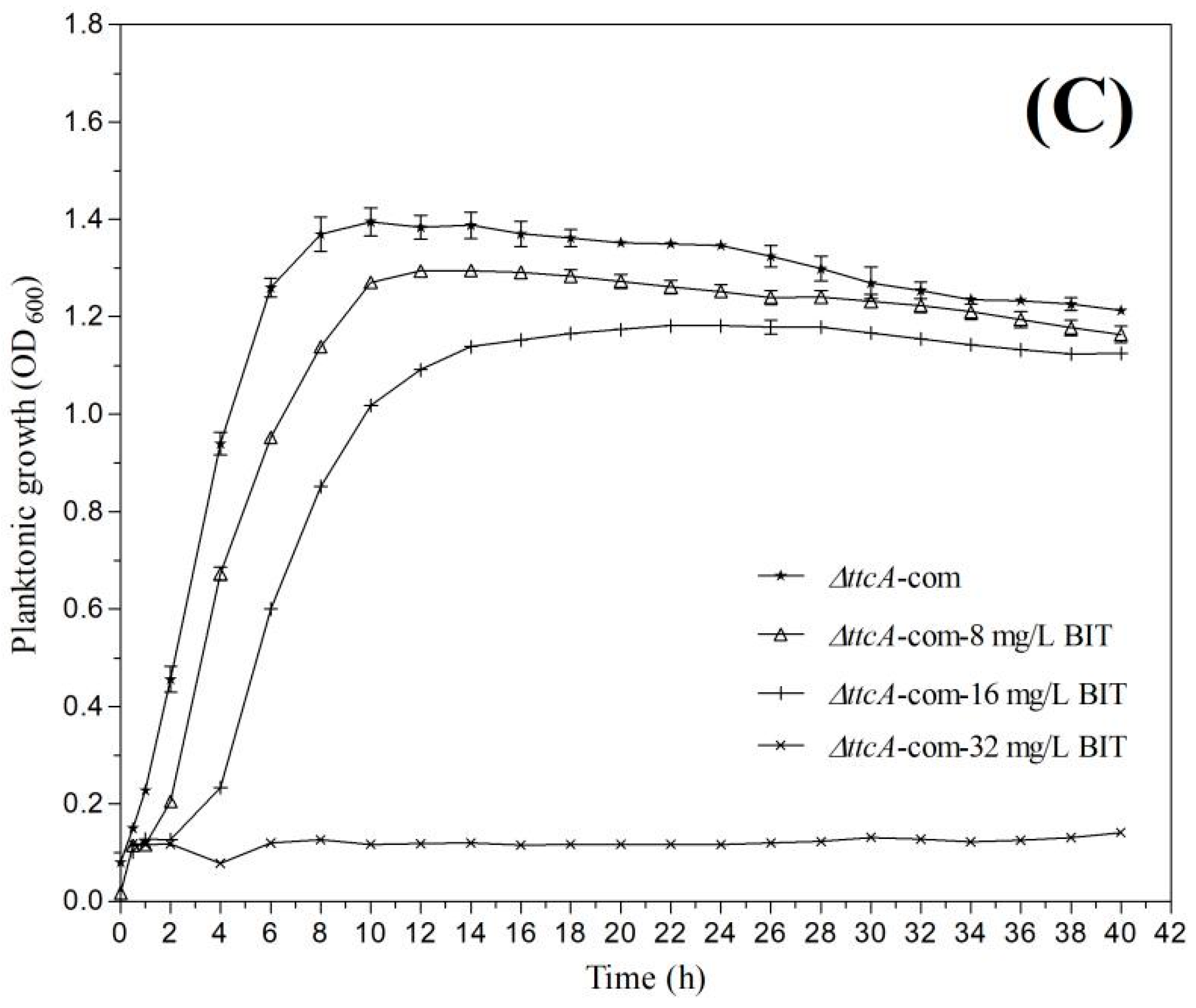
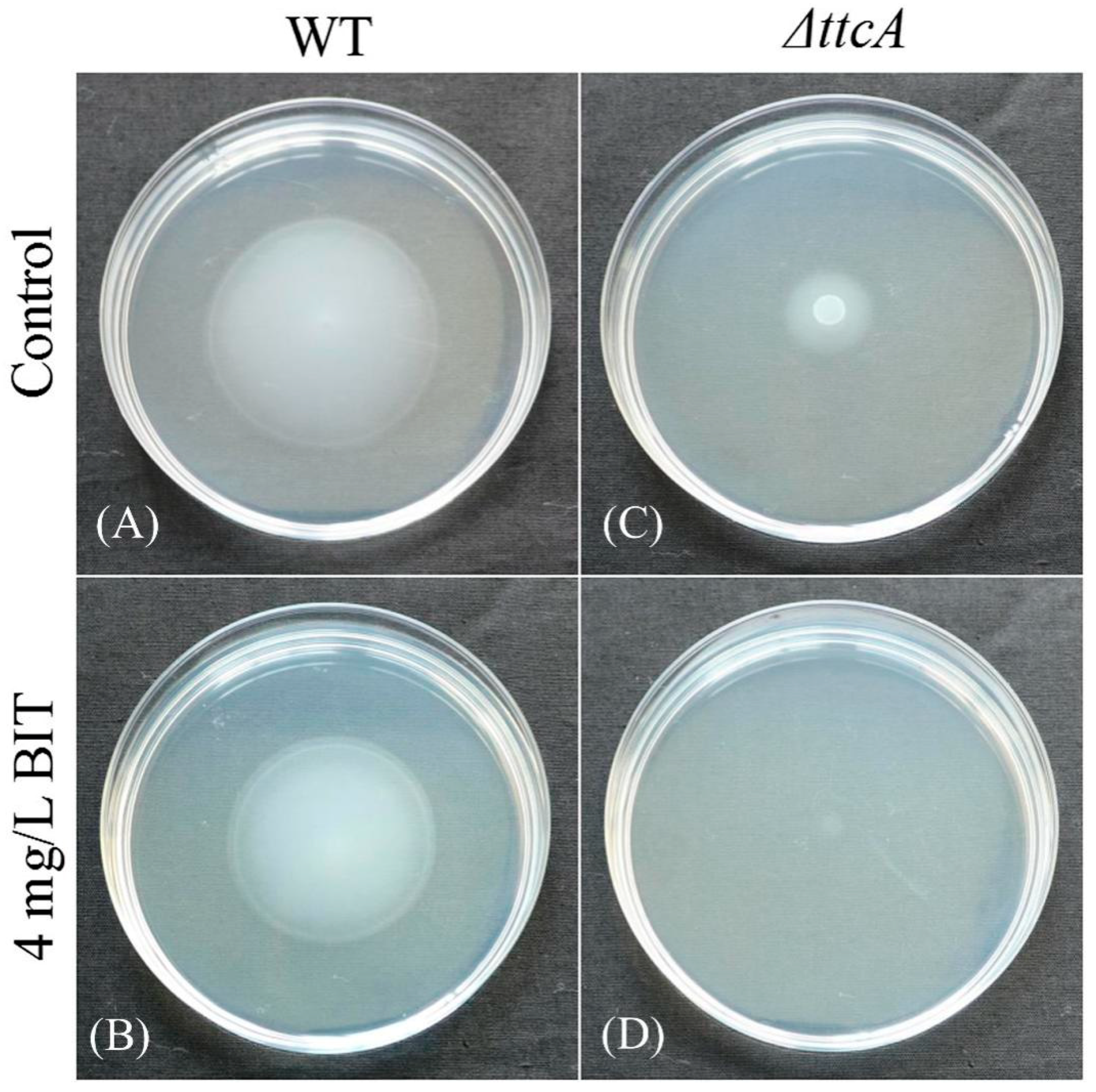
2.3. Effect of BIT on Bacterial Growth

The analysis of growth patterns of C. werkmanii BF-6, ΔttcA and ΔttcA-com in the absence or presence of BIT demonstrated that the growth rates were inhibited in all the three strains with an increase in concentration of BIT (Figure 3). No growth delays were observed in the presence of 8 mg/L BIT except in the case of ΔttcA-com. However, a dosage of 16 mg/L of BIT caused a notable growth delay in all three strains and ΔttcA exhibited the fastest speed to reach the exponential growth phase with a lower plateau growth as compared to wild type C. werkmanii BF-6 (Figure 3B). Further, the growths of C. werkmanii BF-6, ΔttcA and ΔttcA-com were found to be completely repressed by 32 mg/L of BIT (Figure 3).

2.4. Effect of BIT on Planktonic Growth and Initial Biofilm Formation

The formation of bacterial biofilms can induce a declined susceptibility of certain bacteria to antimicrobial agents. Therefore, we detected the difference in the abilities of C. werkmanii BF-6, ΔttcA and ΔttcA-com to form biofilms in the presence of BIT. The results demonstrated that initial biofilm formations (including planktonic growth) were inhibited by BIT in a concentration-dependent manner (Figure 4). For both C. werkmanii BF-6 and ΔttcA, the biofilms formed in the presence of 4, 8 and 16 mg/L BIT were found to be same as their respective controls (Figure 4). However, ΔttcA was found to form more biofilms as compared to either C. werkmanii BF-6 or ΔttcA-com at each detected concentration of BIT (p < 0.05). In addition, the growth of planktonic cells and biofilms of C. werkmanii BF-6, ΔttcA and ΔttcA-com were almost completely inhibited by BIT at concentrations of 32 and 64 mg/L (Figure 4).

2.5. Role of ttcA in Bacterial Motility

Swimming motility is considered as a flagellum-dependent form of movement observed in some Gram-negative bacteria. In the present study, the swimming abilities of C. werkmanii BF-6 and ΔttcA were determined and compared with each other. Results showed that the diameter of ΔttcA colony reduced by about 66.7% as compared with that of C. werkmanii BF-6 (Figure 5C) and the ability to swim by ΔttcA was almost found to be lost in the presence of 4 mg/L BIT (Figure 5D), while BF-6 lost approximately 16.7% of its swimming colony diameter at the same BIT concentration (Figure 5B). In addition, both BF-6 and ΔttcA were found to entirely lose their swimming motility when the BIT concentrations exceeded 8 mg/L (data not shown). Collectively, these results suggest that ttcA gene is necessary for the swimming motility of C. werkmanii while the anti-microbiological agent BIT could inhibit the same.

2.6. Effect of BIT on Biofilm Architecture

Since biofilm formations of C. werkmanii BF-6 and ΔttcA were found to be influenced by BIT, we speculated its effect on biofilm architectures too. In this regard, CLSM images of C. werkmanii BF-6 and ΔttcA biofilms in the absence or presence of BIT were analyzed to quantitatively calculate the parameters of their topographies and architectures. When cultured for 4 days, typical biofilms were formed by C. werkmanii BF-6 and ΔttcA on the surfaces of their respective glass slides in LB medium without BIT (Figure 6A,D). However, ΔttcA formed smoother biofilms and displayed a higher coverage when compared to C. werkmanii BF-6 (Figure 6). When supplemented with 4 and 16 mg/L of BIT, the biofilms of C. werkmanii BF-6 alone were seen to become sparse in nature (Figure 6). Upon further analysis of the CLSM acquired images using the COMSTAT 2.1 software, we noticed that the biofilm parameters comprising of average and maximum thickness, total biomass and average colony size were all higher in ΔttcA than in C. werkmanii BF-6, except for roughness coefficient, in all detected conditions (Table 3). These observations were consistent with the data obtained from CLSM.

3. Discussion

It has been reported that isothiazolones are capable of interacting oxidatively with the accessible thiols, such as glutathione or cysteine, within the microbial cells and thus lead to the inhibition of their metabolism and loss of viability [3,14]. Further, it has been proven that certain growth factor receptors and fibroblast cells along with several intracellular enzymes including transferases, oxidoreductases, isomerases, ligases and hydrolases can be inhibited by these microbiocides [15]. In the present study, we created a Tn5 transposon library of C. werkmanii BF-6 to screen and isolate BIT resistant strains. During this process, the study of three random insertion sites identified from transposon mutants (Table 1) revealed that ttcA as one of the genes that garnered further investigation. It is interesting to note that among the translated protein sequence of ttcA gene, there are two CXXC motifs (Figure 1A), within which the Cys residues coordinate a [4Fe-4S] cluster essential for the thiolation activity [16]. Based on data from the growth curves and resistance determination experiments (Figure 3 and Figure 4), we speculated that either these CXXC motifs or the internal cysteine (C, possesses S-H bond) could be the binding sites for BIT. However, it is important to note that neither the motif nor the protein is a unique binding site of BIT because the deletion of ttcA gene did not lead to the complete absence of BIT action (Figure 3 and Figure 4). Additionally, we found two CXXC motifs (CAYC and CAPC) in the protein sequence of HNH endonuclease (data not shown), which could also play vital roles in the binding of BIT and its subsequent development of resistance. However further study is needed to verify this hypothesis.
The protein tRNA 2-thiocytidine (32) synthetase (TtcA) is required for the thiolation of cytidine at position 32 of tRNA to form 2-thiocytidine (s(2)C32). The TtcA protein family is characterized by the presence of a PP-loop along with a CXXC motif in their central regions (Figure 2). Mutant analysis showed that the cysteines in this centrally conserved CXXC motif are essential for the formation of s(2)C32 [17]. It has been demonstrated that the TtcA enzymes in Escherichia coli contain a redox-active and oxygen-sensitive [4Fe-4S] cluster which are chelated by only three cysteine residues and are absolutely essential for their activity [16]. Meanwhile, it has also been proven that TtcA protein in E. coli is originally a tRNA-thiolating enzyme proceeding via an ATP-dependent pathway depending on a Fe-S cluster, as testified by in vivo and in vitro enzyme activity assays [16]. In another study, it was revealed that the deletion of ttcA gene in the genome of Salmonella enterica did not affect the growth rates of the ΔttcA mutants in comparison to their congenic wild type strains and that the lack of s(2)C32 did not cause any growth disadvantage in a mixed-population experiment [17]. However, we found that the inactivity of ttcA gene in C. werkmanii led to their delayed growth and lowered the growth curves (Figure 3) which suggest that the ttcA gene could be involved in different processes in different bacteria, with respect to their growth. In addition, the mutant strain was not isogenic with wild type C. werkmanii BF-6 and therefore could have different physical properties but the purpose of this manuscript was to determine the effect of the ttcA mutation, including all physical property changes.
Bacterial biofilms represent a life stage different from planktonic cells wherein individual cells are encased in a self-produced matrix of hydrated extracellular polymeric substances (EPS) consisting of polysaccharides, proteins, nucleic acids (eDNA) and lipids [18]. The EPS provide the mechanical stability in biofilms, mediating their adhesion to surfaces and forming a cohesive, three-dimensional polymer network that interconnects and transiently immobilizes biofilm cells [18]. In this study, observations through CLSM demonstrated that both BF-6 and ΔttcA could establish typical biofilms on covers slides (Figure 6). However, with ΔttcA displaying smoother biofilms with higher biomasses as compared to the wild type BF-6 (Table 3 and Figure 6), it can be suggested that ttcA gene is perhaps a negative regulatory factor for both biofilm formation and its structural organization in C. werkmanii.
It is known that the EPS matrix of biofilms can limit and further prevent the transport of antimicrobial agents to the cells by either reacting with the bactericides through either sorption, electrostatic or hydrophobic interactions, size exclusion or through the eventual degradation of biocides [19,20,21,22]. In this study, ΔttcA was seen to form denser biofilms as compared to C. werkmanii BF-6, which indicate a slight increase in resistance to BIT by ΔttcA (Figure 2 and Figure 6, Table 3). However, the barrier of biofilm to BIT only played a role at lower concentration of the drug, as higher dosages could still completely inhibit the growth of planktonic cells and biofilms.
Swimming motility is a flagellum-dependent form of movement observed in Gram-negative bacteria [23,24,25]. Through this study, we found that BIT could inhibit the swimming ability of C. werkmanii. Although the ttcA gene was verified to be essential for the swimming motility in this bacterium, the exact underlying mechanism needs to be elucidated.

4. Materials and Methods

4.1. Bacterial Strains and Chemicals

Wild type C. werkmanii BF-6 (WT) was previously isolated from industrial putrefaction in our laboratory [26] and cultures have been stocked at the Guangdong Culture Collection Center (Guangzhou, Guangdong, China) under the accession number GDMCC 1.1242. Escherichia coli S17-1(λpir)/pRL27 mini-Tn5-Km [27] and pYG4 were kind gifts from professors Lian-hui Zhang of South China Agricultural University, Guangzhou and Yan-guang Cong of Third Military Medical University, Chongqing, China, respectively. The above strains were routinely grown in Luria Bertani (LB) medium with or without the corresponding antibiotics (Kanamycin, 50 mg/L) at 30 °C or 37 °C for necessary durations. All chemicals used in this study were of reagent grade and purchased from Sigma (St. Louis, MO, USA), unless indicated otherwise.

4.2. Construction of Transposon for Mutagenesis

Mutagenesis was performed according to the method described previously, with slight modifications [28]. Both E. coli S17-1(λpir)/pRL27 mini-Tn5-Km and C. werkmanii BF-6 were cultured overnight at 37 °C in LB media in a constant-temperature shaker incubator set to 165 rpm. The donor E. coli S17-1(λpir)/pRL27 and the recipient C. werkmanii BF-6 were mixed in a ratio of 1:1 and then spotted on a LB agar plate which was covered with a 0.45 μm cellulose membrane. The plates were then cultured at 37 °C for at least 12 h to allow conjugational transfer of the plasmid pRL27 from the donor to the recipient cells. Following this, the co-cultivated bacteria on the membrane surface were washed and suspended in 2 mL of 10 mM MgSO4. About 100 μL of C. werkmanii BF-6 clones with the integrated Tn5 cassette were then spread onto LB agar plates supplemented with kanamycin. In addition, all the mutants were transferred onto LB plates supplemented with 32 mg/L BIT to screen for BIT-resistance.

4.3. Identification of Transposon Insertion Site

An inverse PCR method was employed to identify the flanking sequence of the inserted mini-Tn5 transposon in the genomes of the mutants [28]. Briefly, the total genomic DNAs of C. werkmanii BF-6 mutants were extracted from 1 mL of LB medium cultures using the EasyPure Bacteria Genomic DNA Kit (Transgen Biotech, Beijing, China). The isolated genomes were digested with TaqI (NEB, Beijing, China), self-ligated with T4 DNA ligase (NEB) and then used as templates for inverse PCR using the primers TnI-F and TnI-R (Table 2). The amplified fragments were separated by agarose gel electrophoresis, purified using the EasyPure Quick Gel Extraction Kit (Transgen Biotech, Beijing, China) and finally sequenced at the Invitrogen (Guangzhou, China). To verify the mutant genes, the obtained sequences were used to search the nucleotide databases developed by the National Center for Biotechnology Information (NCBI) and the available C. werkmanii BF-6 genome using the BLAST tool.

4.4. Construction of Gene Knockout Mutants and Complementary Strains

Upon analysis, we found that Tn5 was inserted into the tRNA 2-thiocytidine (32) synthetase gene (ttcA) and the resulting mutant exhibited an increased resistance to BIT. In order to testify the molecular role of ttcA in biocides resistance and biofilm formation, we deleted this gene from the genome of C. werkmanii BF-6. The gene knockout and complementation of ttcA was performed according to previously reported methods, with slight modifications [29]. Briefly, the flanking DNA fragments (1059 bp upstream with BglII site; 792 bp downstream with NdeI site) of the gene ttcA (locus_tag = “B2G73_RS05960”) were whole gene synthesized at GENEWIZ Biotechnology Co. Ltd. (Suzhou, China). The resulting fragment was digested with the enzymes BglII and NdeI (NEB) and then cloned into the pYG4 vector digested with the same endonucleases. The resulting plasmid of pYG4-ttcA was used to transform E. coli S17-1 cells and was subsequently mobilized into C. werkmanii BF-6 via biparental mating. The exconjugants were selected on LB medium containing kanamycin (50 mg/L) and rifampicin (100 mg/L) and the deletion mutant candidates were recovered on LB medium containing 5% sucrose and were finally confirmed by PCR using the primers ttcA-QJ-F/R (Table 2). To construct the complementary strain, the genomic DNA was first extracted from C. werkmanii BF-6 by using the EasyPure Genomic DNA Kit (Transgen Biotech, Beijing, China) following the manufacturer’s directions and the full-length ttcA gene with its upstream sequence was amplified using the primers ttcA-Com-F/R (Table 2). The PCR product was cloned into the shuttle vector pSRK-GM between the XbaI and HindIII sites. The recombinant plasmid pSRK-GM-Com was transferred into E. coli S17-1 cells and then subsequently mobilized into C. werkmanii BF-6 deletion strain of ΔttcA. The complemented strain was confirmed by PCR using the primers ttcA-BS-F/R (Table 2) and was designed as ΔttcA-com.

4.5. Determination of the Bacterial Growth Curves

The growth curves of C. werkmanii BF-6, ΔttcA and ΔttcA-com in the absence or presence of 0, 8, 16 and 32 mg/L BIT were determined in a total volume of 200 μL using a Bioscreen C microtiter plate (Labsystems, Helsinki, Finland), starting with an initial bacterial density of approximately 2 × 10⁴ cfu/mL. The cultures were incubated at 30 °C with constant shaking at medium speed for 40 h in a Bioscreen C analyzer (Labsystems, Helsinki, Finland) following the instructions in the manual and the bacterial growth was recorded every 30 min by measuring the optical density (OD) at 600 nm.

4.6. In Vitro Inhibitory Effect of BIT on Initial Biofilm Formation

A column of microtiter plate (Corning Inc., Corning, NY, USA) was inoculated with 100 μL of either BF-6, ΔttcA or ΔttcA-com suspensions (OD600 = 0.10). Dilutions of BIT stock solution in a total volume of 100 μL were added such that the final concentrations of BIT were as follows: 0, 4, 8, 16, 32 and 64 mg/L. Eight replicates were set for each drug concentration and the plates were transferred into a static incubator where they were cultured at 30 °C for 4 days. Following this, they were subsequently managed according to the previously described methods, with minor modifications [12,30]. The growth rates of BF-6, ΔttcA and ΔttcA-com planktonic cells in the cultured plates were measured using a Multiskan GO Reader (Thermo Scientific, Waltham, MA, USA) at 600 nm. The cultivation in each well was discarded gently and washed with 250 μL of sterile water for at least three times to completely remove any of the floating cells. After drying the plate for 30 min at room temperature, each well was filled with 250 μL of 0.1% crystal violet solution (Shanghai Chemical Reagents Co. Ltd., Shanghai, China) to stain the sessile cells attached on the walls of the well for about 40 min. Subsequently, excess of the staining dye was washed away using sterile water for at least three times. After drying the plate at room temperature again, the wells were filled with 250 μL of 95% ethanol (Shanghai Chemical Reagents Co. Ltd., Shanghai, China) to resolubilize the dye for at least 30 min. The OD595 of each well was determined using the same Reader. Meanwhile, the cultures without any BIT were considered as controls.

4.7. Motility Assay

The motility assay for C. werkmanii BF-6 and ΔttcA was determined according to the methods described previously, with slight modifications [24,31]. Briefly, several swimming plates (10 g/L tryptone, 5 g/L NaCl and 0.3% agar) were inoculated with 2 μL of bacterial suspensions (OD600 = 0.05) in the center of the plates, following which, they were transferred into an incubator set to 30 °C. After 24 h of incubation, the plates were taken out and the diameters of the colonies in each plate were measured using a digital Vernier caliper (CD-20CP, Kawasaki, Japan). Additionally, the plates were photographed using a digital camera (Nikon D7200, AF-S DX 18-140 mm; Nikon, Tokyo, Japan).

4.8. Observation of Biofilm Structures with Confocal Laser Scanning Microscopy (CLSM)

Biofilms of C. werkmanii BF-6 and ΔttcA were first allowed to form on pre-sterilized glass microscope slides and then observed with CLSM according to the method reported previously [32]. Briefly, aliquots of 2 mL of BF-6 or ΔttcA suspensions (OD600 = 0.05) supplemented with BIT to final concentrations of 0, 4 or 16 mg/L were added to a well of microtiter plate (12 wells, Corning, NJ, USA) containing a cover slip. Following an incubation under static conditions at 30 °C for 4 days, the glass slips were gently taken out and stained with 5 μM SYTO9 dye (Invitrogen, Carlsbad, CA, USA) and 30 μM propidium iodide (Sigma, St. Louis, MO, USA) in dark for at least 15 min at room temperature. Finally, the stained biofilms were visualized using CLSM (LSM 710 Zeiss, Jena, Germany). In addition, a quantitative analysis of biofilm parameters of the acquired images was analyzed using a COMSAT 2.1 software [33,34].

4.9. Statistical Analysis

All the data collected in this study were recorded as the mean ± standard deviation (SD) and were also subjected to one-way ANOVA test. This was followed by comparison of multiple treatment levels with the controls using Fisher’s LSD test. All the statistical calculations were done using a data processing system (DPS) software [35] and differences were considered statistically significant when p < 0.05.

5. Conclusions

Our results from the present study demonstrate that ttcA gene in C. werkmanii is involved in its planktonic growth, BIT resistance, swimming motility, initial biofilm formation and the underlying structural stability. ttcA plays a significant role in the thiolation of cytidine at position 32 of tRNA. This study, to the best of our knowledge, is the first attempt to construct a relationship between the ttcA gene with isothiazolone resistances, swimming motility and biofilm formation in C. werkmanii. However, the detailed molecular mechanisms of the biological processes demonstrated in this study warrant further research.

Author Contributions

G.Z., Q.-S.S. conceived and designed the experiments. Y.-S.W., H.P. performed the experiments. X.-M.H., X.-B.X. analyzed the data. G.Z., X.-B.X., Q.-S.S. wrote the paper. All authors read and approved the final manuscript.

Funding

This research was funded by the Natural Science Foundation of Guangdong Province, grant number 2015A030313713 and National Natural Science Foundation of China, grant numbers 31500036 and 31770091.

Acknowledgments

We are grateful to Lian-hui Zhang of South China Agricultural University for generously providing us plasmid pRL27. We also want to thank Yan-guang Cong of Third Military Medical University for providing us plasmid pYG4 along with some valuable experimental guidance.

Conflicts of Interest

The authors declare no conflict of interest.

Abbreviations

BIT1,2-benzisothiazolin-3-one
LBLuria Bertani
CLSMConfocal Laser Scanning Microscopy

References

  1. Pucci, M.J.; Podos, S.D.; Thanassi, J.A.; Leggio, M.J.; Bradbury, B.J.; Deshpande, M. In vitro and in vivo profiles of ACH-702, an isothiazoloquinolone, against bacterial pathogens. Antimicrob. Agents Chemother. 2011, 55, 2860–2871. [Google Scholar] [CrossRef] [PubMed]
  2. Vicentini, C.B.; Romagnoli, C.; Manfredini, S.; Rossi, D.; Mares, D. Pyrazolo [3,4-c] isothiazole and isothiazolo [4,3-d] isoxazole derivatives as antifungal agents. Pharm. Biol. 2011, 49, 545–552. [Google Scholar] [CrossRef] [PubMed]
  3. Williams, T.M. The mechanism of action of isothiazolone biocide. Power Plant Chem. 2007, 9, 14–22. [Google Scholar]
  4. Zhou, G.; Shi, Q.S.; Huang, X.M.; Xie, X.B. Comparison of transcriptomes of wild-type and isothiazolone-resistant Pseudomonas aeruginosa by using RNA-seq. Mol. Biol. Rep. 2016, 43, 527–540. [Google Scholar] [CrossRef] [PubMed]
  5. Zhou, G.; Shi, Q.S.; Ouyang, Y.S.; Chen, Y.B. Involvement of outer membrane proteins and peroxide-sensor genes in Burkholderia cepacia resistance to isothiazolone. World J. Microbiol. Biotechnol. 2014, 30, 1251–1260. [Google Scholar] [CrossRef] [PubMed]
  6. Lipsky, B.A.; Hook, E.W.; Smith, A.A.; Plorde, J.J. Citrobacter infections in humans: Experience at the Seattle Veterans Administration Medical Center and a review of the literature. Rev. Infect. Dis. 1980, 2, 746–760. [Google Scholar] [CrossRef] [PubMed]
  7. Arens, S.; Verhaegen, J.; Verbist, L. Differentiation and susceptibility of Citrobacter isolates from patients in a university hospital. Clin. Microbiol. Infect. 1997, 3, 53–57. [Google Scholar] [CrossRef] [PubMed]
  8. Badger, J.L.; Stins, M.F.; Kim, K.S. Citrobacter freundii invades and replicates in human brain microvascular endothelial cells. Infect. Immun. 1999, 67, 4208–4215. [Google Scholar] [PubMed]
  9. Doran, T.I. The role of Citrobacter in clinical disease of children: Review. Clin. Infect. Dis. 1999, 28, 384–394. [Google Scholar] [CrossRef] [PubMed]
  10. Macaskie, L.E.; Empson, R.M.; Lin, F.; Tolley, M.R. Enzymatically-mediated uranium accumulation and uranium recovery using a Citrobacter sp. Immobilised as a biofilm within a plug-flow reactor. J. Chem. Technol. Biotechnol. 1995, 63, 1–16. [Google Scholar] [CrossRef]
  11. Finlay, J.A.; Allan, V.J.M.; Conner, A.; Callow, M.E.; Basnakova, G.; Macaskie, L.E. Phosphate release and heavy metal accumulation by biofilm-immobilized and chemically-coupled cells of a citrobacter sp. pre-grown in continuous culture. Biotechnol. Bioeng. 1999, 63, 87–97. [Google Scholar] [CrossRef]
  12. Zhou, G.; Li, L.J.; Shi, Q.S.; Ouyang, Y.S.; Chen, Y.B.; Hu, W.F. Effects of nutritional and environmental conditions on planktonic growth and biofilm formation for Citrobacter werkmanii BF-6. J. Microbiol. Biotechnol. 2013, 23, 1673–1682. [Google Scholar] [CrossRef] [PubMed]
  13. Zhou, G.; Peng, H.; Wang, Y.S.; Huang, X.M.; Xie, X.B.; Shi, Q.S. Complete genome sequence of Citrobacter werkmanii strain BF-6 isolated from industrial putrefaction. BMC Genom. 2017, 18, 765. [Google Scholar] [CrossRef] [PubMed]
  14. Collier, P.J.; Ramsey, A.; Waigh, R.D.; Douglas, K.T.; Austin, P.; Gilbert, P. Chemical reactivity of some isothiazolone biocides. J. Appl. Microbiol. 1990, 69, 578–584. [Google Scholar] [CrossRef]
  15. Zhou, G.; Shi, Q.S.; Huang, X.M.; Xie, X.B.; Chen, Y.B. Insights into Pseudomonas aeruginosa ATCC9027 resistance to isothiazolones through proteomics. Microb. Drug Resist. 2015, 21, 140–148. [Google Scholar] [CrossRef] [PubMed]
  16. Bouvier, D.; Labessan, N.; Clémancey, M.; Latour, J.-M.; Ravanat, J.-L.; Fontecave, M.; Atta, M. TtcA a new tRNA-thioltransferase with an Fe-S cluster. Nucleic Acids Res. 2014, 42, 7960–7970. [Google Scholar] [CrossRef] [PubMed] [Green Version]
  17. Jäger, G.; Leipuviene, R.; Pollard, M.G.; Qian, Q.; Björk, G.R. The conserved Cys-X1-X2-Cys motif present in the TtcA protein is required for the thiolation of cytidine in position 32 of tRNA from Salmonella enterica serovar Typhimurium. J. Bacteriol. 2004, 186, 750–757. [Google Scholar] [CrossRef] [PubMed]
  18. Flemming, H.C.; Wingender, J. The biofilm matrix. Nat. Rev. Microbiol. 2010, 8, 623–633. [Google Scholar] [CrossRef] [PubMed]
  19. Zhou, G.; Shi, Q.S.; Huang, X.M.; Xie, X.B. The three bacterial lines of defense against antimicrobial agents. Int. J. Mol. Sci. 2015, 16, 21711–21733. [Google Scholar] [CrossRef] [PubMed]
  20. Billings, N.; Millan, M.; Caldara, M.; Rusconi, R.; Tarasova, Y.; Stocker, R.; Ribbeck, K. The extracellular matrix component Psl provides fast-acting antibiotic defense in Pseudomonas aeruginosa biofilms. PLoS Pathog. 2013, 9, e1003526. [Google Scholar] [CrossRef] [PubMed] [Green Version]
  21. Mah, T.F. Biofilm-specific antibiotic resistance. Future Microbiol. 2012, 7, 1061–1072. [Google Scholar] [CrossRef] [PubMed]
  22. Venkatesan, N.; Perumal, G.; Doble, M. Bacterial resistance in biofilm-associated bacteria. Future Microbiol. 2015, 10, 1743–1750. [Google Scholar] [CrossRef] [PubMed]
  23. Ha, D.G.; Kuchma, S.L.; O’Toole, G.A. Plate-based assay for swimming motility in Pseudomonas aeruginosa. Methods Mol. Biol. 2014, 1149, 59–65. [Google Scholar] [PubMed]
  24. Rashid, M.H.; Kornberg, A. Inorganic polyphosphate is needed for swimming, swarming and twitching motilities of Pseudomonas aeruginosa. Proc. Natl. Acad. Sci. USA 2000, 97, 4885–4890. [Google Scholar] [CrossRef] [PubMed]
  25. Murray, T.S.; Kazmierczak, B.I. FlhF is required for swimming and swarming in Pseudomonas aeruginosa. J. Bacteriol. 2006, 188, 6995–7004. [Google Scholar] [CrossRef] [PubMed]
  26. Li, L.J.; Zhou, G.; Shi, Q.S.; Chen, Y.B.; Ouyang, Y.S.; Hu, W.F. Identification and biofilm formation characterization of Citrobacter werkmanii isolated from industrial spoilage. Microbiol. China 2014, 41, 2–7. [Google Scholar]
  27. Larsen, R.A.; Wilson, M.M.; Guss, A.M.; Metcalf, W.W. Genetic analysis of pigment biosynthesis in Xanthobacter autotrophicus Py2 using a new, highly efficient transposon mutagenesis system that is functional in a wide variety of bacteria. Arch. Microbiol. 2002, 178, 193–201. [Google Scholar] [CrossRef] [PubMed]
  28. Cong, Y.; Wang, J.; Chen, Z.; Xiong, K.; Xu, Q.; Hu, F. Characterization of swarming motility in Citrobacter freundii. FEMS Microbiol. Lett. 2011, 317, 160–171. [Google Scholar] [CrossRef] [PubMed]
  29. Ma, J.C.; Wu, Y.Q.; Cao, D.; Zhang, W.B.; Wang, H.H. Only acyl carrier protein 1 (AcpP1) functions in Pseudomonas aeruginosa fatty acid synthesis. Front. Microbiol. 2017, 8, 2186. [Google Scholar] [CrossRef] [PubMed]
  30. Stepanović, S.; Vuković, D.; Dakić, I.; Savić, B.; Švabić-Vlahović, M. A modified microtiter-plate test for quantification of staphylococcal biofilm formation. J. Microbiol. Methods 2000, 40, 175–179. [Google Scholar] [CrossRef]
  31. Huang, Y.H.; Lin, J.S.; Ma, J.C.; Wang, H.H. Functional characterization of triclosan-resistant enoyl-acyl-carrier protein reductase (FabV) in Pseudomonas aeruginosa. Front. Microbiol. 2016, 7, 1903. [Google Scholar] [CrossRef] [PubMed]
  32. Shukla, S.K.; Rao, T.S. Effect of calcium on Staphylococcus aureus biofilm architecture: A confocal laser scanning microscopic study. Colloid Surf. B Biointerfaces 2013, 103, 448–454. [Google Scholar] [CrossRef] [PubMed]
  33. Heydorn, A.; Ersbøll, B.K.; Hentzer, M.; Parsek, M.R.; Givskov, M.; Molin, S. Experimental reproducibility in flow-chamber biofilms. Microbiology 2000, 146, 2409–2415. [Google Scholar] [CrossRef] [PubMed] [Green Version]
  34. Heydorn, A.; Nielsen, A.T.; Hentzer, M.; Sternberg, C.; Givskov, M.; Ersbøll, B.K.; Molin, S. Quantification of biofilm structures by the novel computer program COMSTAT. Microbiology 2000, 146, 2395–2407. [Google Scholar] [CrossRef] [PubMed]
  35. Tang, Q.Y.; Feng, M.G. DPS Data Processing System: Experimental Design, Statistical Analysis and Data Mining; Science Press: Beijing, China, 2007. [Google Scholar]
Figure 1. Screening of random resistant mutants using a LB plate supplemented with 32 mg/L BIT (A) and identification of ΔttcA and ΔttcA-com through PCR (B). Lane 1: C. werkmanii BF-6 (Control); Lane 2: ΔttcA; Lane 3: ΔttcA-com.
Ijms 19 02644 g001
Figure 2. Analysis of the ttcA gene in C. werkmanii BF-6 and encoded protein sequence. Schematic representation of the TtcA protein with its conserved motifs (A). Sequence alignment of reported TtcA proteins representatives in the genus Citrobacter (B). The alignments were conducted with BioEdit software and the conserved amino acid residues and motifs are framed. Phylogenetic tree of TtcA protein of C. werkmanii BF-6 with the species of the same genus (C). This tree was constructed using bootstrapping and Neighbor-Joining methods with MEGA 4.0.
Ijms 19 02644 g002
Figure 3. Growth curves of C. werkmanii BF-6 (A), ΔttcA (B) and ΔttcA-com (C). The strains were cultivated in LB medium supplemented with 0, 8, 16 or 32 mg/L BIT and incubated at 30 °C for 40 h.
Ijms 19 02644 g003aIjms 19 02644 g003b
Figure 4. Planktonic growth and initial biofilm formation of C. werkmanii BF-6 (A,B), ΔttcA (C,D) and ΔttcA-com (E,F), respectively. The strains were grown in LB medium supplemented with indicted concentrations of BIT. All assays were conducted in triplicates and the columns marked with different letters (a, b, c or d) indicate that they are significantly different from each other according to Fisher’s LSD test (p < 0.05), calculated with the DPS data processing software.
Ijms 19 02644 g004
Figure 5. Swimming motility of C. werkmanii BF-6 (A,B) and ΔttcA (C,D) on semisolid agar plates supplemented with 0 or 4 mg/L BIT. Aliquots of 5 μL suspensions were inoculated at the center of the plate with a micropipette and then cultured for 24 h at 30 °C.
Ijms 19 02644 g005
Figure 6. CLSM images of 4 days old biofilms of C. werkmanii BF-6 and ΔttcA in the presence of 0 (A,D), 4 (B,E) or 16 (C,F) mg/L BIT, respectively. The biofilms were cultured on the cover slides and stained with 5 μM SYTO9 dye and 30 μM propidium iodide. Magnification: 400×.
Ijms 19 02644 g006
Table 1. Identification of the random insertion sites of Tn5 transposon.
NumberLocus TagGene NameDescription
64B2G73_RS05960ttcAtRNA 2-thiocytidine(32) synthetase
94B2G73_RS07535-LysR family transcriptional regulator
111B2G73_RS05205-HNH endonuclease
“-”: no gene name.
Table 2. List of primers used for ttcA gene knockout and complementation.
Primer NamesForward Primer Sequences (5′-3′)Reverse Primer Sequences (5′-3′)Application
TnI-F/RGGAGAGGCTATTCGGCTATGGTAAGGTGATCCGGTGGATGIdentification of Tn5 inserted sites
ttcA-WJ-F/RATTATCGCTTCTCACCGACCTGAACGTCCAGCCACGGTAAAATCIdentification of ttcA deletion
ttcAP-F/RGCTCTAGAGTGATGCATACTCTTCTGATCCCAAGCTTTTACTTCACTTCCACAACGTttcA complementation
ttcA-BS-F/RCTTCAATATGATCGAAGACGGCTTGGGTATCATCCTCTTCTGGCPartial sequence of ttcA gene
Table 3. Quantitation of biofilm architecture.
Parameters *WT (Mean ± SD)ΔttcA (Mean ± SD)
0 mg/L BIT4 mg/L BIT16 mg/L BIT0 mg/L BIT4 mg/L BIT16 mg/L BIT
Maximum thickness (μm)12.00 ± 2.0011.33 ± 1.158.00 ± 0.0020.77 ± 2.6918.00 ± 4.0016.67 ± 2.31
Average thickness (μm)8.26 ± 1.405.86 ± 1.173.54 ± 1.2917.33 ± 2.319.58 ± 1.725.79 ± 1.27
Total biomass (μm3/μm2)12.06 ± 0.985.58 ± 1.203.42 ± 1.0614.52 ± 0.149.78 ± 1.636.14 ± 1.27
Average colony size (μm2)195.67 ± 17.65182.18 ± 12.1259.00 ± 6.58213.75 ± 7.93192.55 ± 18.54155.90 ± 17.86
Roughness coefficient0.22 ± 0.020.51 ± 0.020.82 ± 0.210.08 ± 0.020.11 ± 0.010.36 ± 0.06
* Quantitation of biofilm parameters including maximum thickness, average thickness, total biomass, average colony size and roughness coefficient were evaluated using COMSTAT 2.1. The results are means of datasets obtained from the analysis of eight CLSM images acquired at random positions in each of the biofilms. Standard deviations (SD) are indicated along with the means.

