Article

Coexistence of TERT Promoter Mutations and the BRAF V600E Alteration and Its Impact on Histopathological Features of Papillary Thyroid Carcinoma in a Selected Series of Polish Patients

by
Dagmara Rusinek
1,*,
Aleksandra Pfeifer
1,
Jolanta Krajewska
1,
Malgorzata Oczko-Wojciechowska
1,
Daria Handkiewicz-Junak
1,
Agnieszka Pawlaczek
1,
Jadwiga Zebracka-Gala
1,
Malgorzata Kowalska
1,
Renata Cyplinska
1,
Ewa Zembala-Nozynska
2,
Mykola Chekan
2,
Ewa Chmielik
2,
Aleksandra Kropinska
1,
Roman Lamch
2,
Beata Jurecka-Lubieniecka
1,
Barbara Jarzab
1 and
Agnieszka Czarniecka
3
1
Department of Nuclear Medicine and Endocrine Oncology, Maria Sklodowska-Curie Institute, Oncology Center, Gliwice Branch, Wybrzeze Armii Krajowej 15, 44-101 Gliwice, Poland
2
Tumor Pathology Department, Maria Sklodowska-Curie Institute, Oncology Center, Gliwice Branch, Wybrzeze Armii Krajowej 15, 44-101 Gliwice, Poland
3
Department of Oncological and Reconstructive Surgery, Maria Sklodowska-Curie Institute, Oncology Center, Gliwice Branch, Wybrzeze Armii Krajowej 15, 44-101 Gliwice, Poland
*
Author to whom correspondence should be addressed.
Int. J. Mol. Sci. 2018, 19(9), 2647; https://doi.org/10.3390/ijms19092647
Submission received: 27 July 2018 / Revised: 28 August 2018 / Accepted: 30 August 2018 / Published: 6 September 2018
(This article belongs to the Special Issue Cell and Molecular Biology of Thyroid Disorders)

Abstract

:
TERT promoter (TERTp) mutations are important factors in papillary thyroid carcinomas (PTCs). They are associated with tumor aggressiveness, recurrence, and disease-specific mortality and their use in risk stratification of PTC patients has been proposed. In this study we investigated the prevalence of TERTp mutations in a cohort of Polish patients with PTCs and the association of these mutations with histopathological factors, particularly in coexistence with the BRAF V600E mutation. A total of 189 consecutive PTC specimens with known BRAF mutational status were evaluated. TERTp mutations were detected in 8.5% of cases (16/189) with the C228T mutation being the most frequent. In six of the PTC specimens (3.2%), four additional TERTp alterations were found, which included one known polymorphism (rs2735943) and three previously unreported alterations. The association analysis revealed that the TERTp hotspot mutations were highly correlated with the presence of the BRAF V600E mutation and their coexistence was significantly associated with gender, advanced patient age, advanced disease stage, presence of lymph node metastases, larger tumor size, and tumor-capsule infiltration. While correlations were identified, the possibility of TERTp mutations being key molecular modulators responsible for PTC aggressiveness requires further studies.

Graphical Abstract

1. Introduction

Despite excellent overall survival in most cases of papillary thyroid carcinoma (PTC), recurrent disease is diagnosed during the 10-year period following initial treatment in 15%, 22%, 46.4%, and 66.7% of patients with TNM stage I, II, III, and IV, respectively. PTC may be a cause of death, even in patients with clinically indolent tumors [1]. Distant metastases of PTC are a strong predictor of a poor outcome, including disease-specific mortality [2,3]. However, neck lymph node metastases (LNM) are also considered to be a predictor of persistent or recurrent disease [4]. Molecular markers of poor prognosis also exist and include genetic alterations associated with PTC. The BRAF V600E mutation occurs in about 50% of PTC cases and has been identified in a number of studies to be related to an advanced disease stage, extrathyroidal invasion, or the presence of LNM [5]. It is also reported that the presence of BRAF mutation is associated with a decreased expression of thyroid specific genes like NIS and TPO, what results in the loss of sensitivity to radioiodine [6]. However, there are also contradicting studies, in which no significant association between BRAF V600E and poor prognosis is demonstrated [7,8].
Recent reports provide new data on the molecular background of PTC, especially as it relates to its aggressive phenotype. Mutations in the telomerase reverse transcriptase promoter (TERTp) have been detected in a number of solid cancers, including thyroid cancers [9,10,11]. TERT encodes the catalytic subunit of telomerase, the enzyme responsible for extending telomeres and thereby preventing replicative senescence. TERT, together with its RNA partner telomerase TERT component (TERC), demonstrates telomerase reverse transcriptase activity and adds tandem repeats of TTAGGG sequence to the end of chromosomes [12]. As demonstrated in many studies, telomerase is highly expressed in germ cells and stem cells, but is repressed and not transcribed in most somatic cells [13]. However, the reactivation of telomerase has been observed in numerous human cancers, which prevents telomere shortening and thereby provides cancer cells an unlimited replicative potential [14]. Although telomere elongation is the primary function of reactivated telomerase, it has a number of other effects, among them: growing cancer cell proliferation, resistance to apoptosis and antigrowth signals, increased angiogenesis and metastatic potential [15]. Considering all the above-mentioned processes, in which TERT is involved, it has been proposed to treat TERT as an oncogene that is able to act in a telomere-independent manner [15].
In numerous malignancies, including follicular cell derived thyroid cancers (TCs), two hotspot mutations within TERTp have been described, chr5, 1,295,228 C->T (C228T; hg19), and chr5, 1,295,250 C->T (C250T; hg19), which correspond to positions −124 and −146 bp, respectively, and are located upstream from the ATG start codon of TERT [16]. The prevalence of these two mutations in TCs, of which C228T is more frequent, seems to correlate with the degree of aggressiveness in various TC subtypes. They have been shown to be present in 11.3%, 17.1%, 14.6%, 43.2%, and 40.1% of PTC, follicular thyroid carcinoma (FTC), Hürthle cell carcinoma (HCC), poorly differentiated thyroid carcinoma (PDTC), and anaplastic thyroid carcinoma (ATC), respectively [12]. A similar trend in the frequency of TERTp mutations is observed in particular histopathological PTC variants with the highest prevalence being seen in tall-cell PTC variant known for its aggressive phenotype [9]. The presence of these genetic alterations has also been detected in papillary thyroid microcarcinomas (PTMC), but their prevalence does not exceed 4.7% and no relationship to unfavorable clinical features is observed [17]. Interestingly, TERTp mutations were widely reported as associated with the presence of the BRAF V600E mutation in human cancers, mainly in TC and melanoma [11,12,18,19]. Alone, these mutations display a modest effect; however the coexistence of BRAF V600E and TERTp mutations is associated with a higher cancer aggressiveness, which is reflected by lymph node metastases, distant metastases, advanced tumor stage, recurrence, and even disease-specific mortality of PTC patients [20,21]. According to a recent meta-analysis concerning PTCs, the prevalence of the coexistence of the BRAF V600E and TERTp mutations is 7.7% (145/1892) [12]. This frequency corresponds to the percentage of PTCs with poorest clinical outcome, estimated as less than 10%. These data indicate the BRAF V600E and TERTp mutations as an oncogene tandem underlying progression and aggressiveness of thyroid cancers. Similar clinical consequences are observed regarding the coexistence of RAS mutations and TERTp mutations [12,21]. This makes TERT a potential prognostic marker that may be beneficial for risk stratification of patients with PTC. However, several studies investigating the prevalence of TERTp mutations in TCs have shown significant differences in the frequency of TERTp alterations in different countries with rates of 7.5–25.5% and 13.8–36.4% for PTC and FTC, respectively [22].
In the current study we analyzed the prevalence of TERTp mutations as well as their potential associations with the BRAF V600E alteration and histopathological features in selected series of Polish PTC patients. Moreover, the differences between the presence of a single mutation and the coexistence of TERTp and BRAF 600 codon alterations and their relation to particular histopathological PTC features were analyzed.

2. Results

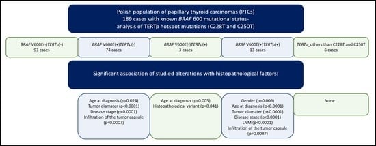
2.1. TERT Promoter Status in PTCs and Its Relation to the Presence of the BRAF V600E Mutation

We evaluated TERTp in 189 consecutive PTC tumor specimens for the presence of two hotspot mutations, C228T and C250T. In total, 16 of these 189 PTC specimens (8.5%) were positive for the C228T or C250T TERTp mutations. Of the specimens analyzed, 13 harbored the C228T mutation (81%; Figure 1A) and the remaining three PTC specimens had the C250T mutation (Figure 1B). In the tumor from one patient, who was positive for the BRAF V600E mutation, a known polymorphism within TERTp was found (rs2735943; −100 C->T; Figure 1C). Three new TERTp mutations were also identified in five of the PTC specimens. None of the TERTp mutations detected coexisted with each other in any of the samples analyzed. The new mutations found in TERTp were located at positions −162 bp, −80 bp, and −77 bp, upstream of the ATG start codon of TERT, and all involved a substitution of cytosine to thymine (Figure 2A–C). None of the new nucleotide substitutions, detected in the specimens from our PTC cohort, have been previously reported. Because of the lack of knowledge regarding whether we were dealing with a mutation or a polymorphism, and since one PTC specimen had the rs2735943 polymorphism, which has an unknown frequency, the new TERTp mutations were analyzed further as a separate set of samples and excluded from the TERTp-positive cohort, which consisted only of PTC specimens harboring the hotspot mutations C228T or C250T.
In order to analyze the relationship of TERTp hotspot mutations with the presence of the BRAF V600E mutation, the cases with TERTp alteration other than C228T or C250T were excluded from the PTC dataset. This resulted in 87 BRAF-positive and 96 BRAF-negative samples being analyzed. A significant association between TERTp hotspot mutations and the presence of BRAF mutation was identified. Thirteen of the 16 TERTp-positive PTC specimens that contained the C228T mutation and all that contained the C250T mutation also harbored the BRAF V600E mutation (81.2%; p = 0.007; Table 1). The remaining three TERTp-positive PTC specimens were BRAF V600E-negative and all contained the C228T mutation. In contrast, the BRAF V600E mutation was found in 74 TERTp-negative cases (74/167; 44.3%; Table 1).

2.2. Association of the BRAF V600E Mutation and TERTp Alterations with Histopathological Factors in the Cohort of Polish Patients with PTC

In order to analyze the cohort of Polish patients with PTC for a possible relationship between each of the investigated molecular events, the samples were divided into four subgroups, (1) no detected mutation within the BRAF 600 codon or TERTp; (2) positive for the BRAF V600E mutation but no TERTp alterations; (3) positive for the BRAF V600E mutation and one of the hotspot TERTp mutations; and (4) a TERTp alterations present other than C228T or C250T. Subgroups 2–4 were compared to subgroup 1 to determine how the presence of the analyzed alterations was related to particular histopathological PTC features, both individually or together (Table 2).
The BRAF V600E mutation alone was significantly associated with more advanced age of the patients (p = 0.024), larger tumor size (p < 0.0001), more advanced disease stage (p = 0.002), and infiltration of the tumor capsule (p = 0.0007). The TERTp hotspot mutations alone were significantly associated only with advanced age of patients (p = 0.005) and histopathological PTC variant (p = 0.041); however, the number of cases in the cohort was too small to interpret these results with a high degree of confidence. Coexistence of the BRAF V600E mutation and TERTp hotspot mutations, demonstrated an association with gender (p = 0.006), advanced age of the patients (p < 0.0001), advanced disease stage (p < 0.0001), the presence of lymph nodes metastases (p < 0.0001), larger tumor size (p < 0.0001), and infiltration of the tumor capsule (p = 0.0007). The results are summarized in Table 2.
With respect to the subgroup of PTC specimens with TERTp alterations other than the hotspot mutations, no association with any of the analyzed factors was identified. However, the small number of samples in that group (n = 6) does not justify drawing any unequivocal conclusions. Also, it was not possible to determine the association of the studied alterations with distant metastases since none of PTC cases in the cohort demonstrated distant metastases at the time of diagnosis.

2.3. Further Analysis of the Presence of TERTp Alterations in a Subgroup of Patients with PTMC

Among 80 PTMC specimens, only one tumor harbored the TERTp hotspot mutation C228T, while two other specimens displayed unknown TERTp alterations −77 C->T and −162 C->T, with the latter one also being positive for the BRAF V600E mutation. In contrast to PTMC specimens containing a single alteration, which was consistent with classical PTC, the single case that harbored the BRAF mutation and the −162 C->T TERTp alteration was diagnosed as a follicular PTC variant. None of the three TERTp-positive PTMC specimens demonstrated angioinvasion, infiltration of tumor capsule, multifocality, or LNM. Twenty of 80 PTMC specimens contained the BRAF V600E mutation. However, infiltration of the tumor capsule was present in only 20% (4/20) of the BRAF-positive cases. These specimens were taken from the oldest patients in the PTMC subgroup. No other histopathological factors related to poor prognosis were observed.

3. Discussion

Since their discovery, somatic TERTp mutations have been extensively studied in a number of cancers, including melanomas, glioblastomas, clear cell renal carcinomas and nonclear cell renal carcinomas, non-small cell lung cancer, and TCs [10,12,23,24,25]. It has been demonstrated that the frequency of TERTp mutations is higher in more aggressive types of TC, as well as in histopathological PTC variants, and the presence of the mutations correlate with a poorer outcome [9,12]. Moreover, several investigators suggest that the presence of these alterations in PTCs is associated with particularly aggressive clinicopathological factors, such as greater tumor size, extrathyroidal invasion, stage III or IV disease, lymph node metastases, and distant metastases [26,27].
Taking into consideration the divergence observed in the frequency of TERTp mutations in PTC between different countries and scarce data concerning the impact of TERTp mutations alone on PTC histopathological features we decided to investigate Polish PTC patients. Regarding the well-documented association between the presence of TERTp and BRAF V600E mutations we analyzed TERTp in PTCs with known BRAF 600 codon status. We also performed a number of comparative analyses to identify a potential role of these two molecular markers alone or their coexistence in PTC carcinogenesis. The TERTp hotspot mutations were detected in 16 of the 189 consecutive analyzed PTC specimens (8.5%). However, if we excluded PTMCs and focused only on PTCs, the frequency of TERTp mutations increased up to 13.8% (15/109). Such observation is in concordance with previous studies [12]. In our study, one patient diagnosed with PTMC harbored the TERTp hotspot mutation C228T but not the BRAF V600E mutation. Consistent with the findings of de Biase and coauthors [17], TERTp mutations were present, but relatively uncommon in PTMC specimens at a frequency of 4.7%, and did not correlate with unfavorable clinical features. Similarly, our only case of PTMC with the C228T TERTp mutation did not demonstrate extrathyroidal invasion, angioinvasion, or lymph node metastases. In addition to the known hotspot mutations in TERTp, we also detected another known alteration, previously reported in dbSNP (rs2735943). Because its frequency is not known it was treated in our study like the three new TERTp mutations we identified, since there was no certainty whether it was a polymorphism or mutation. The analyses revealed that these four TERTp alterations (−100 C->T; −162 C->T; −80 C->T; −77 C->T) were not associated with any of analyzed clinical factors. However, we cannot exclude their impact on PTC carcinogenesis and further studies are needed to determine their role. It is noteworthy that one of the novel nucleotide substitutions (−77 C->T) created a putative consensus binding site (GGAA) for ETS (E26 transformation-specific) transcription factors; the same as that created by C228T and C250T hotspot mutations. It is believed that creating such additional binding sites for transcription factors is a mechanism of action for C228T and C250T, related to increased TERT expression [28].
In the cohort of Polish patients with PTC, analyzed in the current study, the TERTp hotspot mutations were more prevalent in the BRAF V600E-positive specimens compared to BRAF V600E-negative specimens (81.2%; p = 0.007) and the BRAF V600E mutation was less frequent in TERTp-negative PTC specimens. Our findings are consistent with a previous report [12]. However, there are some contradicting data showing no correlation in the occurrence of these two molecular events [29]. Contrary results were obtained by Landa and coworkers [30], who observed that TERTp mutations were less common in well-differentiated PTC specimens harboring BRAF or RAS mutations. However, in PDTC specimens or ATC specimens, BRAF-positive or RAS-positive cases are enriched by TERTp alterations. This observation supports the belief that TERTp mutations may be useful as potential prognostic markers for TC.
We analyzed whether TERTp hotspot mutations were related to any poor prognostic factors in Polish patients with PTC. We determined that these mutations alone were associated only with advanced age at the time of PTC diagnosis and with a histopathological PTC variant (oxyphilic). However, the number of BRAF(−)TERTp(+) samples was small so these data are limited for their interpretative value. On the other hand, similar results were obtained by Gandolfi et al. [31] who described a significant association between TERTp mutations and advanced age, but only in PTC cases with distant metastases. In a study by Shen et al. [21], TERTp mutations analyzed individually failed to show any significant association with clinicopathological outcomes. In contrast, the coexistence of the BRAF V600E mutation and TERTp hotspot mutations is reported to be strongly associated with a number of histopathological factors related to more aggressive clinical course of the disease [21,31]. The same relations were detected in our study with a significant association of the coexistence of BRAF and TERTp mutations with gender, advanced age of patients, T3 and T4 stage of disease, the presence of lymph node metastases, larger tumor size, and infiltration of the tumor capsule. The role of these two genetic alterations in promoting PTC aggressiveness and poor clinical outcome may be explained by the mechanisms observed at the molecular level. Previous studies indicated a possible molecular interaction between BRAF V600E and TERTp mutations via BRAF V600E-dependent constitutive activation of MAPK pathway leading to activation of ETS transcription factors and their binding to binding motifs, created de novo by the hotspot mutations in TERTp, what in consequence would enhance TERT activity. However, Liu et al. study from 2018 showed the exact role of BRAF and TERTp mutations in the expression of TERT through the BRAF V600E-> MAPK pathway->FOS-> GABP-> TERT axis [32]. Moreover, the authors presented that BRAF V600E/MAPK pathway also promoted TERT expression via MYC in TERTp mutation-independent manner, but in a much less degree.
According to the previous studies, TERTp hotspot mutations were found in more aggressive histopathological PTC variants, like tall cell variant [9]. Our PTC cohort was mainly represented by classical PTCs and follicular variant. There was only one case of tall cell PTC and it was positive for the BRAF V600E mutation and TERTp hotspot alterations.
The major limitation of the current study was a small number of cases with more aggressive clinical course of the disease, including no PTC cases with distant metastases. However, while we were not able to identify a possible relation between TERTp mutations and more aggressive PTC subtypes, our data did show a strong effect of coexistence of TERTp hotspot mutations and the BRAF V600E mutation on the development of PTCs with histopathological factors related to poorer outcome. In order to determine the independent effects of TERTp mutations on PTC, an analysis of a larger cohort is needed. There is a single study in which only TERTp mutations, but not the BRAF V600E mutation, were associated with poorer histopathological factors [29]. However, it should be noted that the number of PTC samples with TERTp mutations alone and PTCs with double mutations (TERTp and BRAF mutations) was only four in each group. Thus, the data from such limited sample size should be interpreted with caution.
So far, the detection of BRAF V600E and TERTp mutations in TCs is not a routine diagnostic procedure. These alterations are mainly analyzed in fine needle aspiration biopsy material in order to support the cytological examination. According to the ATA recommendations [33] molecular tests may be used to refine risk prior to surgery, assuming that surgical management would change based on a positive test result. As mentioned earlier, TERTp mutations were shown to be a helpful tool in PTC risk stratification, however, no standardized therapeutic strategies, based on these two molecular markers, were established.

4. Material and Methods

4.1. DNA Isolation

The analyzed group of specimens were collected from 189 consecutive Polish patients with PTC treated at the Maria Sklodowska-Curie Institute—Oncology Center, Gliwice Branch in Gliwice, Poland. Among them, 80 (42%; 80/189) of patients were diagnosed with papillary microcarcinoma. The selection of the patients was based on the known mutation status in BRAF 600 codon. The study cohort included 166 women and 23 men with the median age at the time of PTC diagnosis of 53 years (range 12 to 83 years). The clinical data are summarized in Table 3. All samples were analyzed for the presence of TERTp mutations. The use of human tissue was approved by the institutional Bioethics Committee. Informed written consent was obtained for the use of their tissue for analysis from all patients or their caregivers. All the clinical data were anonymized and de-identified prior to the analysis.

4.2. DNA Isolation

Total DNA was extracted from 10 μm-thick sections of formalin-fixed and paraffin-embedded (FFPE) tumor specimen tissues (5 sections per sample). The DNA isolation was performed using a QIAamp DNA FFPE Tissue Kit (Qiagen GmbH; Hilden, Germany) according to the manufacturer’s protocol after deparaffinization using single xylene extraction and rinsing with ethanol 98%. DNA concentrations were evaluated using a Nanodrop ND-1000 microspectrophotometer (Thermo Scientific, Wilmington, DE, USA).

4.3. Detection of TERTp Mutations

Detection of TERTp mutations was performed by direct sequencing using the Sanger method. Approximately 30 ng of genomic DNA was used as template for polymerase chain reaction (PCR) amplification of 157-bp length fragment of TERTp containing the analyzed hotspot sites (−124 and −146 upstream of the ATG start codon). Primers used in the PCR were F-5′-CCCCTTCACCTTCCAGCTC-3′ and R-5′-CAGCGCTGCCTGAAACTC-3′. The reaction was carried out with an initial polymerase activation step at 95 °C for 15 min, followed by 34 consecutive cycles of denaturation at 95 °C for 30 s, primers annealing at 59.7 °C for 30 s, extension at 72 °C for 30 s, and final elongation at 72 °C for 5 min. The PCR product was analyzed for quality by gel electrophoresis and then sequenced on a 3130xl Genetic Analyzer Applied Biosystems (Life Technologies, Carlsbad CA, USA). Briefly, PCR products were enzyme purified with alkaline phosphatase (SAP) and exonuclease I (Life Technologies) and then subjected to standard sequencing using an ABI PRISM 1.1 BigDye Terminator Cycle Sequencing Ready Reaction Kit (Life Technologies).

4.4. Detection of BRAF V600E Mutation

Information regarding the BRAF 600 codon status was determined using the following methodology. A fragment of exon 15 of BRAF containing codon 600 was amplified by PCR using primers F-5′-TGTTTTCCTTTACTTACTACACCTCA-3′ and R-5′-GCCTCAATTCTTACCATCCA-3′ resulting in amplification of a 160-bp product. The PCR cycling conditions included an initial polymerase activation step at 95 °C for 15 min; 34 cycles of denaturation at 95 °C for 30 s, primer annealing at 56.5 °C for 30 s, extension at 72 °C for 30 s; and the final elongation step at 72 °C for 5 min. After quality confirmation by gel electrophoresis, the PCR products were sequenced as described for detection of the TERTp mutations.

4.5. Statistical Analysis

The association analysis was performed on categorical data that were summarized as numbers and percentages. Continuous data were summarized as medians and interquartile ranges.
In order to test, whether there were significant differences between analyzed groups of tumors, the Fisher’s exact test and the Mann–Whitney U test were applied. The Fisher’s exact test was used for comparisons in case of categorical variables, whereas the Mann–Whitney U test was used in case of continuous variables. All p-values were two sided, and a p-value of 0.05 was considered as statistically significant.
Statistical analyses were performed using the R software version 3.4.1 and Gmisc package version 1.4.1 [34,35].

5. Conclusions

Although most PTCs represent relatively indolent tumors, some cases develop distant metastases or recurrent disease, or may dedifferentiate into PDTCs or ATCs. There is no definitive answer regarding whether TERTp mutations are the key to PTC aggressiveness and dedifferentiation. However, more and more recently published data, including our current study, suggest their association with a poor prognosis. Despite the small number of analyzed PTC samples the frequency of TERTp hotspot mutations as well as the association between the BRAF V600E mutation and some PTC histopathological factors, observed in this preliminary study, were in concordance with the results of previous reports. Moreover, we identified three novel TERTp alterations, which may play a role in PTC aggressiveness. Further functional studies are needed to determine their impact. Although relatively rare, it is clear that TERTp mutations in PTCs are an important molecular event and may influence risk stratification and the clinical management.

Author Contributions

Conceptualization, B.J.; Data curation, J.K.; Formal analysis, A.P. (Aleksandra Pfeifer); Investigation, D.R., D.H.-J., A.P. (Agnieszka Pawlaczek), J.Z.-G., M.K., and R.C.; Methodology, D.R.; Resources, D.H.-J., E.Z.-N., M.C., E.C., A.K., R.L., B.J.-L., and A.C.; Software, A.P. (Aleksandra Pfeifer); Supervision, M.O.-W. and B.J.; Writing—original draft, D.R.; Writing—review & editing, D.R., A.P. (Aleksandra Pfeifer), J.K., B.J. and A.C.

Funding

This work was supported by the National Centre for Research and Development project under the program “Prevention practices and treatment of civilization diseases” STRATEGMED (STRATEGMED2/267398/4/NCBR/2015).

Acknowledgments

We would like to thank Zofia Kolosza for substantive support in statistical analysis of the data.

Conflicts of Interest

The authors declare no conflict of interest.

Abbreviations

ATCAnaplastic thyroid carcinoma
ETSE26 transformation-specific
FTCFollicular thyroid carcinoma
HCCHürthle cell carcinoma
LNMLymph node metastases
PDTCPoorly differentiated thyroid carcinoma
PTCPapillary thyroid carcinoma
PTMCPapillary thyroid microcarcinoma
TCThyroid carcinoma
TERTpTERT promoter

References

  1. Loh, K.C.; Greenspan, F.S.; Gee, L.; Miller, T.R.; Yeo, P.P. Pathological tumor-node-metastasis (pTNM) staging for papillary and follicular thyroid carcinomas: A retrospective analysis of 700 patients. J. Clin. Endocrinol. Metab. 1997, 82, 3553–3562. [Google Scholar] [CrossRef] [PubMed]
  2. Durante, C.; Haddy, N.; Baudin, E.; Leboulleux, S.; Hartl, D.; Travagli, J.P.; Caillou, B.; Ricard, M.; Lumbroso, J.D.; De Vathaire, F.; et al. Long-term outcome of 444 patients with distant metastases from papillary and follicular thyroid carcinoma: Benefits and limits of radioiodine therapy. J. Clin. Endocrinol. Metab. 2006, 91, 2892–2899. [Google Scholar] [CrossRef] [PubMed]
  3. Su, D.-H.; Chang, S.-H.; Chang, T.-C. The impact of locoregional recurrences and distant metastases on the survival of patients with papillary thyroid carcinoma. Clin. Endocrinol. 2015, 82, 286–294. [Google Scholar] [CrossRef] [PubMed]
  4. Machens, A.; Hinze, R.; Thomusch, O.; Dralle, H. Pattern of nodal metastasis for primary and reoperative thyroid cancer. World J. Surg. 2002, 26, 22–28. [Google Scholar] [CrossRef] [PubMed]
  5. Xing, M.; Westra, W.H.; Tufano, R.P.; Cohen, Y.; Rosenbaum, E.; Rhoden, K.J.; Carson, K.A.; Vasko, V.; Larin, A.; Tallini, G.; et al. BRAF mutation predicts a poorer clinical prognosis for papillary thyroid cancer. J. Clin. Endocrinol. Metab. 2005, 90, 6373–6379. [Google Scholar] [CrossRef] [PubMed]
  6. Durante, C.; Puxeddu, E.; Ferretti, E.; Morisi, R.; Moretti, S.; Bruno, R.; Barbi, F.; Avenia, N.; Scipioni, A.; Verrienti, A.; et al. BRAF mutations in papillary thyroid carcinomas inhibit genes involved in iodine metabolism. J. Clin. Endocrinol. Metab. 2007, 92, 2840–2843. [Google Scholar] [CrossRef] [PubMed]
  7. Fugazzola, L.; Mannavola, D.; Cirello, V.; Vannucchi, G.; Muzza, M.; Vicentini, L.; Beck-Peccoz, P. BRAF mutations in an Italian cohort of thyroid cancers. Clin. Endocrinol. 2004, 61, 239–243. [Google Scholar] [CrossRef] [PubMed]
  8. Kim, T.Y.; Kim, W.B.; Rhee, Y.S.; Song, J.Y.; Kim, J.M.; Gong, G.; Lee, S.; Kim, S.Y.; Kim, S.C.; Hong, S.J.; et al. The BRAF mutation is useful for prediction of clinical recurrence in low-risk patients with conventional papillary thyroid carcinoma. Clin. Endocrinol. 2006, 65, 364–368. [Google Scholar] [CrossRef] [PubMed]
  9. Liu, X.; Bishop, J.; Shan, Y.; Pai, S.; Liu, D.; Murugan, A.K.; Sun, H.; El-Naggar, A.K.; Xing, M. Highly prevalent TERT promoter mutations in aggressive thyroid cancers. Endocr. Relat. Cancer 2013, 20, 603–610. [Google Scholar] [CrossRef] [PubMed] [Green Version]
  10. Huang, F.W.; Hodis, E.; Xu, M.J.; Kryukov, G.V.; Chin, L.; Garraway, L.A. Highly Recurrent TERT Promoter Mutations in Human Melanoma. Science 2013, 339, 957–959. [Google Scholar] [CrossRef] [PubMed] [Green Version]
  11. Horn, S.; Figl, A.; Rachakonda, P.S.; Fischer, C.; Sucker, A.; Gast, A.; Kadel, S.; Moll, I.; Nagore, E.; Hemminki, K.; et al. TERT Promoter Mutations in Familial and Sporadic Melanoma. Science 2013, 339, 959–961. [Google Scholar] [CrossRef] [PubMed]
  12. Liu, R.; Xing, M. TERT promoter mutations in thyroid cancer. Endocr. Relat. Cancer 2016, 23, R143–R145. [Google Scholar] [CrossRef] [PubMed]
  13. Blasco, M.A. Telomeres and human disease: Ageing, cancer and beyond. Nat. Rev. Genet. 2005, 6, 611–622. [Google Scholar] [CrossRef] [PubMed]
  14. Hanahan, D.; Weinberg, R.A. Hallmarks of cancer: The next generation. Cell 2011, 144, 646–674. [Google Scholar] [CrossRef] [PubMed]
  15. Low, K.C.; Tergaonkar, V. Telomerase: Central regulator of all of the hallmarks of cancer. Trends Biochem. Sci. 2013, 38, 426–434. [Google Scholar] [CrossRef] [PubMed]
  16. Vinagre, J.; Almeida, A.; Populo, H.; Batista, R.; Lyra, J.; Pinto, V.; Coelho, R.; Celestino, R.; Prazeres, H.; Lima, L.; et al. Frequency of TERT promoter mutations in human cancers. Nat. Commun. 2013, 4, 2185. [Google Scholar] [CrossRef] [PubMed] [Green Version]
  17. De Biase, D.; Gandolfi, G.; Ragazzi, M.; Eszlinger, M.; Sancisi, V.; Gugnoni, M.; Visani, M.; Pession, A.; Casadei, G.; Durante, C.; et al. TERT Promoter Mutations in Papillary Thyroid Microcarcinomas. Thyroid 2015, 25, 1013–1019. [Google Scholar] [CrossRef] [PubMed]
  18. Macerola, E.; Loggini, B.; Giannini, R.; Garavello, G.; Giordano, M.; Proietti, A.; Niccoli, C.; Basolo, F.; Fontanini, G. Coexistence of TERT promoter and BRAF mutations in cutaneous melanoma is associated with more clinicopathological features of aggressiveness. Virchows Arch. 2015, 467, 177–184. [Google Scholar] [CrossRef] [PubMed]
  19. Griewank, K.G.; Murali, R.; Puig-Butille, J.A.; Schilling, B.; Livingstone, E.; Potrony, M.; Carrera, C.; Schimming, T.; Moller, I.; Schwamborn, M.; et al. TERT promoter mutation status as an independent prognostic factor in cutaneous melanoma. J. Natl. Cancer Inst. 2014, 106. [Google Scholar] [CrossRef] [PubMed]
  20. Liu, R.; Bishop, J.; Zhu, G.; Zhang, T.; Ladenson, P.W.; Xing, M. Mortality Risk Stratification by Combining BRAF V600E and TERT Promoter Mutations in Papillary Thyroid Cancer: Genetic Duet of BRAF and TERT Promoter Mutations in Thyroid Cancer Mortality. JAMA Oncol. 2016. [Google Scholar] [CrossRef] [PubMed]
  21. Shen, X.; Liu, R.; Xing, M. A six-genotype genetic prognostic model for papillary thyroid cancer. Endocr. Relat. Cancer 2017, 24, 41–52. [Google Scholar] [CrossRef] [PubMed]
  22. Marques, I.J.; Moura, M.M.; Cabrera, R.; Pinto, A.E.; Simões-Pereira, J.; Santos, C.; Menezes, F.D.; Montezuma, D.; Henrique, R.; Rodrigues Teixeira, M.; et al. Identification of somatic TERT promoter mutations in familial nonmedullary thyroid carcinomas. Clin. Endocrinol. 2017, 87, 394–399. [Google Scholar] [CrossRef] [PubMed]
  23. Nakajima, N.; Nobusawa, S.; Nakata, S.; Nakada, M.; Yamazaki, T.; Matsumura, N.; Harada, K.; Matsuda, H.; Funata, N.; Nagai, S.; et al. BRAF V600E, TERT promoter mutations and CDKN2A/B homozygous deletions are frequent in epithelioid glioblastomas: A histological and molecular analysis focusing on intratumoral heterogeneity. Brain Pathol. 2017. [Google Scholar] [CrossRef] [PubMed]
  24. Casuscelli, J.; Becerra, M.F.; Manley, B.J.; Zabor, E.C.; Reznik, E.; Redzematovic, A.; Arcila, M.E.; Tennenbaum, D.M.; Ghanaat, M.; Kashan, M.; et al. Characterization and Impact of TERT Promoter Region Mutations on Clinical Outcome in Renal Cell Carcinoma. Eur. Urol. Focus 2017. [Google Scholar] [CrossRef] [PubMed]
  25. Jung, S.; Kim, D.; Park, W.; Lee, H.; Choi, I.; Park, J.; Lee, J. Mutation of the TERT promoter leads to poor prognosis of patients with non-small cell lung cancer. Oncol. Lett. 2017, 14, 1609–1614. [Google Scholar] [CrossRef] [PubMed] [Green Version]
  26. Melo, M.; da Rocha, A.G.; Vinagre, J.; Batista, R.; Peixoto, J.; Tavares, C.; Celestino, R.; Almeida, A.; Salgado, C.; Eloy, C.; et al. TERT promoter mutations are a major indicator of poor outcome in differentiated thyroid carcinomas. J. Clin. Endocrinol. Metab. 2014, 99, E754–E765. [Google Scholar] [CrossRef] [PubMed]
  27. Su, X.; Jiang, X.; Wang, W.; Wang, H.; Xu, X.; Lin, A.; Teng, X.; Wu, H.; Teng, L. Association of telomerase reverse transcriptase promoter mutations with clinicopathological features and prognosis of thyroid cancer: A meta-analysis. OncoTargets Ther. 2016, 9, 6965–6976. [Google Scholar] [CrossRef] [PubMed]
  28. Liu, T.; Yuan, X.; Xu, D. Cancer-Specific Telomerase Reverse Transcriptase (TERT) Promoter Mutations: Biological and Clinical Implications. Genes 2016, 7, 38. [Google Scholar] [CrossRef] [PubMed]
  29. Nasirden, A.; Saito, T.; Fukumura, Y.; Hara, K.; Akaike, K.; Kurisaki-Arakawa, A.; Asahina, M.; Yamashita, A.; Tomomasa, R.; Hayashi, T.; et al. In Japanese patients with papillary thyroid carcinoma, TERT promoter mutation is associated with poor prognosis, in contrast to BRAF V600E mutation. Virchows Arch. 2016, 469, 687–696. [Google Scholar] [CrossRef] [PubMed]
  30. Landa, I.; Ganly, I.; Chan, T.A.; Mitsutake, N.; Matsuse, M.; Ibrahimpasic, T.; Ghossein, R.A.; Fagin, J.A. Frequent somatic TERT promoter mutations in thyroid cancer: Higher prevalence in advanced forms of the disease. J. Clin. Endocrinol. Metab. 2013, 98, E1562–E1566. [Google Scholar] [CrossRef] [PubMed]
  31. Gandolfi, G.; Ragazzi, M.; Frasoldati, A.; Piana, S.; Ciarrocchi, A.; Sancisi, V. TERT promoter mutations are associated with distant metastases in papillary thyroid carcinoma. Eur. J. Endocrinol. 2015, 172, 403–413. [Google Scholar] [CrossRef] [PubMed] [Green Version]
  32. Liu, R.; Zhang, T.; Zhu, G.; Xing, M. Regulation of mutant TERT by BRAF V600E/MAP kinase pathway through FOS/GABP in human cancer. Nat. Commun. 2018, 9, 579. [Google Scholar] [CrossRef] [PubMed] [Green Version]
  33. Haugen, B.R.; Alexander, E.K.; Bible, K.C.; Doherty, G.M.; Mandel, S.J.; Nikiforov, Y.E.; Pacini, F.; Randolph, G.W.; Sawka, A.M.; Schlumberger, M.; et al. 2015 American Thyroid Association Management Guidelines for Adult Patients with Thyroid Nodules and Differentiated Thyroid Cancer: The American Thyroid Association Guidelines Task Force on Thyroid Nodules and Differentiated Thyroid Cancer. Thyroid 2016, 26, 1–133. [Google Scholar] [CrossRef] [PubMed] [Green Version]
  34. Team, R.C. A Language and Environment for Statistical Computing. R Foundation for Statistical Computing. Available online: https://www.r-project.org/ (accessed on 15 December 2017).
  35. Gordon, M. Gmisc: Descriptive Statistics, Transition Plots, and More. R Package, Version 1.4.1; 2016. Available online: https://rdrr.io/cran/Gmisc/ (accessed on 5 September 2018).
Figure 1. Known TERT promoter (TERTp) alterations detected in the analyzed papillary thyroid carcinoma (PTC) cohort. (A) Mutation −124 C->T (C228T); (B) mutation −146 C->T (C250T); (C) alteration −100 C->T (rs2735943). The black triangles point to the nucleotide positions of the nucleotide changes.
Ijms 19 02647 g001
Figure 2. New TERT promoter (TERTp) alterations detected in the analyzed papillary thyroid carcinoma (PTC) cohort. (A) Alteration −77 C->T, which created a putative consensus binding site (GGAA) for transcription factor ETS2; (B) alteration −80 C->T; (C) alteration −162 C->T. The black triangles point to the nucleotide positions of the nucleotide substitutions.
Ijms 19 02647 g002
Table 1. Association of TERT promoter (TERTp) hotspot mutations and the BRAF V600E mutation.
TERT/BRAF StatusBRAF V600E-PositiveBRAF V600E-Negativep-Value
(n = 87)(n = 96)
TERTp hotspot mutation 0.007
TERTp-positive13 (14.9%)3 (3.1%)
TERTp-negative74 (85.1%)93 (96.9%)
TERTp-PositiveTERTp-Negativep-Value
(n = 16)(n = 167)
BRAF mutation 0.007
BRAF V600E-positive13 (81.2%)74 (44.3%)
BRAF V600E-negative3 (18.8%)93 (55.7%)
Table 2. Association of TERT promoter (TERTp) hotspot mutations, the BRAF V600E mutation, their co-existence, and other TERTp alterations with histopathological features of the analyzed papillary thyroid carcinoma (PTC) cohort.
Histopathological FeaturesNo MutationBRAF + TERTp−BRAFTERTp+BRAF + TERTp+TERTp_Others
No. (%)No. of Missing CasesNo. (%)No. of Missing Casesp-ValueNo. (%)No. of Missing Casesp-ValueNo. (%)No. of Missing Casesp-ValueNo. (%)No. of Missing Casesp-Value
Total No. Of Cases93 74 3 13 6
Sex 0.3 0.23 0.006 1
Female86 (92.5%) 64 (86.5%) 2 (66.7%) 8 (61.5%) 6 (100.0%)
Male7 (7.5%) 10 (13.5%) 1 (33.3%) 5 (38.5%) 0 (0.0%)
Age at diagnosis (years)50.0 (36.0–58.0) 54.0 (40.2–64.0) 0.02470.0 (68.0–76.5) 0.00571.0 (63.0–75.0) <0.000148.5 (34.0–60.8) 0.94
Tumor diameter (mm)7.0 (4.0–14.0)113.0 (8.0–17.8) <0.000112.0 (11.0–13.0) 0.2125.0 (17.2–31.5)1<0.000110.0 (3.5–18.8) 0.91
Histopathological variant 10 60.97 0.041 20.14 1
Classical (CPTC)34 (41.0%) 29 (42.6%) 1 (33.3%) 8 (72.7%) 3 (50.0%)
Follicular (FVPTC)23 (27.7%) 21 (30.9%) 0 (0.0%) 1 (9.1%) 2 (33.3%)
Classical and follicular16 (19.3%) 12 (17.6%) 0 (0.0%) 0 (0.0%) 1 (16.7%)
Oxyphilic5 (6.0%) 3 (4.4%) 2 (66.7%) 1 (9.1%) 0 (0.0%)
Others5 (6.0%) 3 (4.4%) 0 (0.0%) 1 (9.1%) 0 (0.0%)
pT 1 1<0.0001 0.14 <0.0001 0.14
1a58 (63.0%) 19 (26.0%) 1 (33.3%) 0 (0.0%) 2 (33.3%)
1b12 (13.0%) 23 (31.5%) 2 (66.7%) 3 (23.1%) 2 (33.3%)
29 (9.8%) 5 (6.8%) 0 (0.0%) 1 (7.7%) 0 (0.0%)
313 (14.1%) 24 (32.9%) 0 (0.0%) 7 (53.8%) 2 (33.3%)
4a0 (0.0%) 2 (2.7%) 0 (0.0%) 2 (15.4%) 0 (0.0%)
pT 1 10.002 1 <0.0001 0.23
T1/T279 (85.9%) 47 (64.4%) 3 (100.0%) 4 (30.8%) 4 (66.7%)
T3/T413 (14.1%) 26 (35.6%) 0 (0.0%) 9 (69.2%) 2 (33.3%)
Lymph node metastases 14 210.42 21 6<0.0001 20.16
059 (74.7%) 34 (64.2%) 1 (100.0%) 0 (0.0%) 2 (50.0%)
1a11 (13.9%) 10 (18.9%) 0 (0.0%) 2 (28.6%) 0 (0.0%)
1b9 (11.4%) 9 (17.0%) 0 (0.0%) 5 (71.4%) 2 (50.0%)
Infiltration of the tumor capsule 0.0007 0.51 0.0007 0.6
Yes19 (20.4%) 34 (45.9%) 1 (33.3%) 9 (69.2%) 2 (33.3%)
No74 (79.6%) 40 (54.1%) 2 (66.7%) 4 (30.8%) 4 (66.7%)
Angioinvasion 1 0.26 0.13 1
Yes8 (8.6%) 6 (8.1%) 1 (33.3%) 3 (23.1%) 0 (0.0%)
No85 (91.4%) 68 (91.9%) 2 (66.7%) 10 (76.9%) 6 (100.0%)
Multifocality 0.83 1 0.69 0.59
Yes14 (15.1%) 12 (16.2%) 0 (0.0%) 1 (7.7%) 0 (0.0%)
No79 (84.9%) 62 (83.8%) 3 (100.0%) 12 (92.3%) 6 (100.0%)
“No mutation” refers to PTCs without the BRAF V600E mutation or TERTp alterations.
Table 3. Characteristics of the study cohort.
TERT Status and Histopathological FeaturesTotalBRAF V600E-NegativeBRAF V600E-Positive
No. (%)No. of Missing CasesNo. (%)No. of Missing CasesNo. (%)No. of Missing Cases
Total No. of cases189 100 89
Sex
Female166 (87.8%) 92 (92.0%) 74 (83.1%)
Male23 (12.2%) 8 (8.0%) 15 (16.9%)
Age at diagnosis [years]53.0 (40.0–62.0) 50.5 (37.5–59.0) 57.0 (44.0–66.0)
TERT promoter hotspot mutation (C228T and C250T)
Lack of mutation173 (91.5%) 97 (97.0%) 76 (85.4%)
Mutation16 (8.5%) 3 (3.0%) 13 (14.6%)
Type of TERT promoter alteration
Number of negative samples167 93 74
C228T13 (59.1%) 3 (42.9%) 10 (66.7%)
C250T3 (13.6%) 0 (0.0%) 3 (20.0%)
alteration −100 C → T; rs27359431 (4.5%) 0 (0.0%) 1 (6.7%)
alteration −162 C->T1 (4.5%) 0 (0.0%) 1 (6.7%)
alteration −77 C->T2 (9.1%) 2 (28.6%) 0 (0.0%)
alteration −80 C->T2 (9.1%) 2 (28.6%) 0 (0.0%)
Tumor diameter (mm)10.0 (5.0–17.0)28.0 (4.5–14.0)113.0 (8.8–20.0)1
Histopathological variant 18 10 8
classical (CPTC)75 (43.9%) 37 (41.1%) 38 (46.9%)
follicular (FVPTC)47 (27.5%) 24 (26.7%) 23 (28.4%)
classical and follicular29 (17.0%) 17 (18.9%) 12 (14.8%)
oxyphilic11 (6.4%) 7 (7.8%) 4 (4.9%)
others9 (5.3%) 5 (5.6%) 4 (4.9%)
pT 2 1 1
1a80 (42.8%) 60 (60.6%) 20 (22.7%)
1b42 (22.5%) 16 (16.2%) 26 (29.5%)
215 (8.0%) 9 (9.1%) 6 (6.8%)
346 (24.6%) 14 (14.1%) 32 (36.4%)
4a4 (2.1%) 0 (0.0%) 4 (4.5%)
pT 2 1 1
T1/T2137 (73.3%) 85 (85.9%) 52 (59.1%)
T3/T450 (26.7%) 14 (14.1%) 36 (40.9%)
Lymph node metastases
096 (66.7%) 61 (74.4%) 35 (56.5%)
1a23 (16.0%) 11 (13.4%) 12 (19.4%)
1b25 (17.4%) 10 (12.2%) 15 (24.2%)
Distant metastases 2 2
0187 (100.0%) 98 (100.0%) 89 (100.0%)
Infiltration of the tumor capsule
yes65 (34.4%) 21 (21.0%) 44 (49.4%)
no124 (65.6%) 79 (79.0%) 45 (50.6%)
Angioinvasion
yes18 (9.5%) 9 (9.0%) 9 (10.1%)
no171 (90.5%) 91 (91.0%) 80 (89.9%)
Multifocality
yes27 (14.3%) 14 (14.0%) 13 (14.6%)
no162 (85.7%) 86 (86.0%) 76 (85.4%)

