Article

Shot-Gun Proteomic Analysis on Roots of Arabidopsis pldα1 Mutants Suggesting the Involvement of PLDα1 in Mitochondrial Protein Import, Vesicular Trafficking and Glucosinolate Biosynthesis

1
Centre of the Region Haná for Biotechnological and Agricultural Research, Faculty of Science, Palacký University, Šlechtitelů 27, 78371 Olomouc, Czech Republic
2
Institute for Genomics, Biocomputing & Biotechnology, Mississippi Agricultural and Forestry Experiment Station, Mississippi State University, Starkville, MS 39759, USA
*
Author to whom correspondence should be addressed.
Int. J. Mol. Sci. 2019, 20(1), 82; https://doi.org/10.3390/ijms20010082
Submission received: 19 November 2018 / Revised: 19 December 2018 / Accepted: 21 December 2018 / Published: 26 December 2018
(This article belongs to the Section Molecular Plant Sciences)

Abstract

:
Phospholipase Dα1 (PLDα1) belongs to phospholipases, a large phospholipid hydrolyzing protein family. PLDα1 has a substrate preference for phosphatidylcholine leading to enzymatic production of phosphatidic acid, a lipid second messenger with multiple cellular functions. PLDα1 itself is implicated in biotic and abiotic stress responses. Here, we present a shot-gun differential proteomic analysis on roots of two Arabidopsis pldα1 mutants compared to the wild type. Interestingly, PLDα1 deficiency leads to altered abundances of proteins involved in diverse processes related to membrane transport including endocytosis and endoplasmic reticulum-Golgi transport. PLDα1 may be involved in the stability of attachment sites of endoplasmic reticulum to the plasma membrane as suggested by increased abundance of synaptotagmin 1, which was validated by immunoblotting and whole-mount immunolabelling analyses. Moreover, we noticed a robust abundance alterations of proteins involved in mitochondrial import and electron transport chain. Notably, the abundances of numerous proteins implicated in glucosinolate biosynthesis were also affected in pldα1 mutants. Our results suggest a broader biological involvement of PLDα1 than anticipated thus far, especially in the processes such as endomembrane transport, mitochondrial protein import and protein quality control, as well as glucosinolate biosynthesis.

Graphical Abstract

1. Introduction

Phospholipases are phospholipid hydrolyzing enzymes with multiple roles in biotic and abiotic stress responses of plants as well as in plant growth and development [1]. Phospholipase D (PLD) alpha 1 (PLDα1) is a member of D subfamily of phospholipases and it shows the highest expression levels among all twelve PLD members in Arabidopsis [2]. Total PLD activity is substantially decreased in Arabidopsis pldα1 mutants [3]. PLDs utilize preferentially phosphatidylcholine as a substrate, which they hydrolyze in Ca2+ dependent manner [2]. This hydrolysis is accompanied with the production of phosphatidic acid (PA), a second messenger bearing important signaling functions [4]. The absence of PLDα1 leads to the reduction of cellular PA pool and membrane lipid remodeling [5,6]. This remodeling affects physical and mechanical properties of membranes leading to endomembrane reorganizations and changes in membrane transport [7,8]. PLDα1 is also involved in the regulation of cytoskeletal dynamics and organization, which is either mediated by PA or by direct binding/association of PLDα1 with the cytoskeleton [3,9,10,11,12]. PLDα1 promotes stomata closure and inhibits their opening [13]. At a molecular level, stomatal movements are governed by PLDα1 through interaction of PA with protein phosphatase 2C (ABI1) [5], NADPH oxidase [14], sphingosine kinase [15] and microtubule associated protein 65-1 [3]. In addition, PLDα1 binds and modulates components of G protein complex during stomatal movements [16,17]. These functions render PLDα1 an important regulator of the plant stress response, growth and development. PLDα1 was shown to be involved in plant response to drought [18], cold [19] and salt stress [12]. This protein has promising biotechnological applications, since its genetic manipulation modulates plant response to abiotic stresses [20]. Nevertheless, PLDs usually act cooperatively (including the production of cellular PA pool), as it was previously exemplified in abscisic acid (ABA)-induced stomatal closure [21]. Arabidopsis mutants of PLDα1 exhibit conditional phenotypes, whereas under control conditions they show phenotypes similar to the wild-type plants [3,22]. Recent detailed fluorescent in vivo imaging of PLDα1 fused to YFP and expressed in Arabidopsis PLDα1 t-DNA insertion mutants under its own promoter showed that PLDα1-YFP localized to the cytoplasm in the close vicinity of plasma membrane (PM) and exerted developmentally-dependent and tissue-specific expression [12]. Interestingly, most of PLDα1 functions are assigned to processes occurring in leaves. On the other hand, PLDα1 functions in roots are obscure. Shot gun proteomic analysis on genetically modified plants proved to be very useful tool for elucidation of protein functions. Here, we performed a comparative shot gun proteomic analysis on roots of two t-DNA insertion mutants (pldα1-1 and pldα1-2) as compared to the Col-0 wild type. Our results indicated that PLDα1 is involved in mitochondrial protein import and quality control, glucosinolate biosynthesis and that it controls very specific processes of subcellular vesicular transport.

2. Results

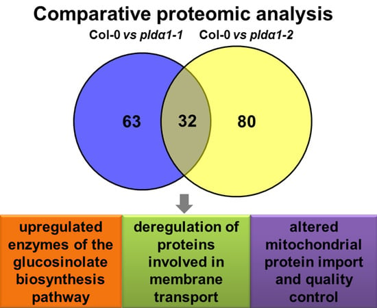
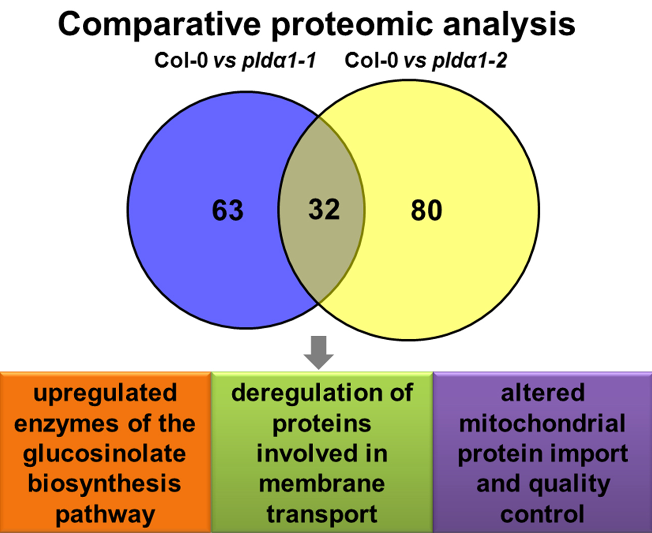
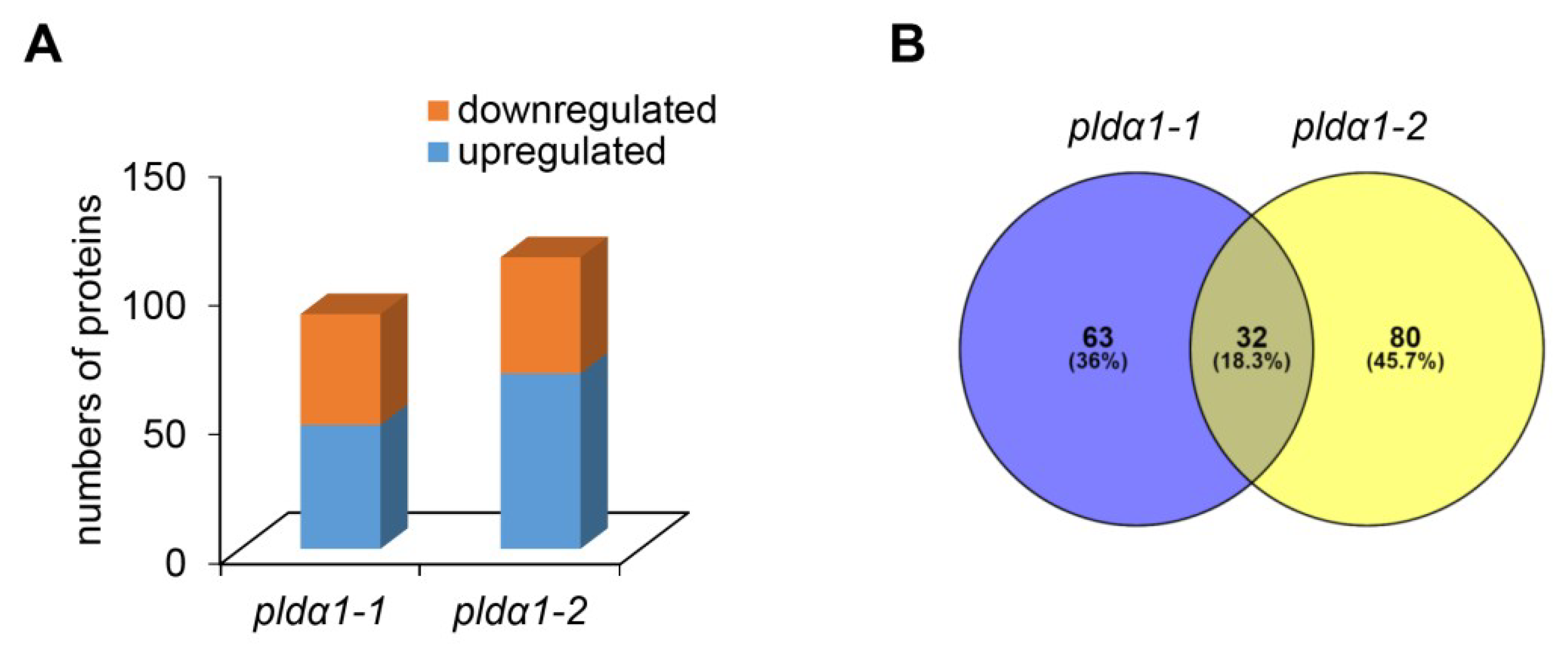
2.1. Overview of Differential Root Proteomes in Two pldα1 Mutants

We carried out a comparative shot-gun proteomic analysis of roots of two pldα1 mutants compared to the Col-0 as a wild type. First, we compared the number of identified proteins (Figure S1A) and peptides (Figure S1B) in Col-0, pldα1-1 and pldα1-2 mutants showing high reproducibility of our analysis (Figure S1A). Considering proteins identified at least in 2 biological replicates, 92%, 82% and 75% of the total proteomes of Col-0, pldα1-1 and pldα1-2 roots were found commonly in all three lines (Figure S1C). In pldα1-1, 92 proteins with changed abundances were found, while 113 were identified in pldα1-2 mutant (Figure 1A). In both mutants, 32 proteins were commonly changed (Figure 1B, Table 1). PLDα1 was identified uniquely in the wild type, while we did not detect this protein in two studied mutants, confirming the reliability of our approach. Similarly, we were unable to detect PLDrp1 (PLD regulated protein 1; At5g39570), a phosphoprotein interacting with PLDα1 [23], in pldα1 mutants. A complete list of all differentially abundant proteins (DAPs) of both mutants is available in Table S1. A detailed outputs of protein identification in all samples is presented in the Supplementary Materials, and deposited in PRoteomics IDEntifications (PRIDE) database (see below).

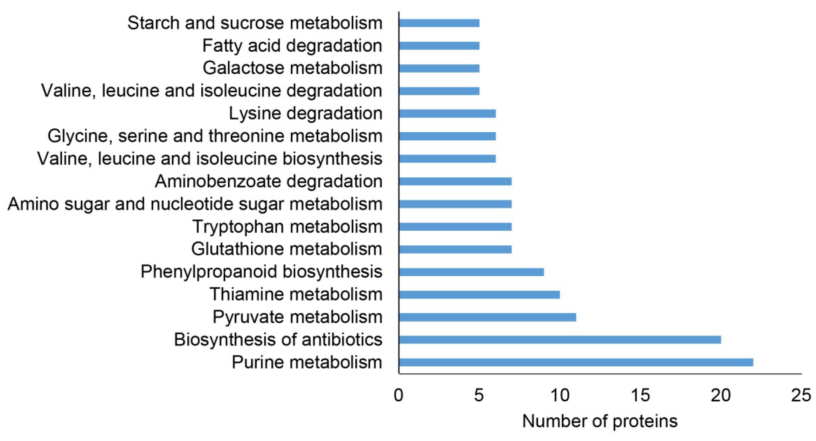
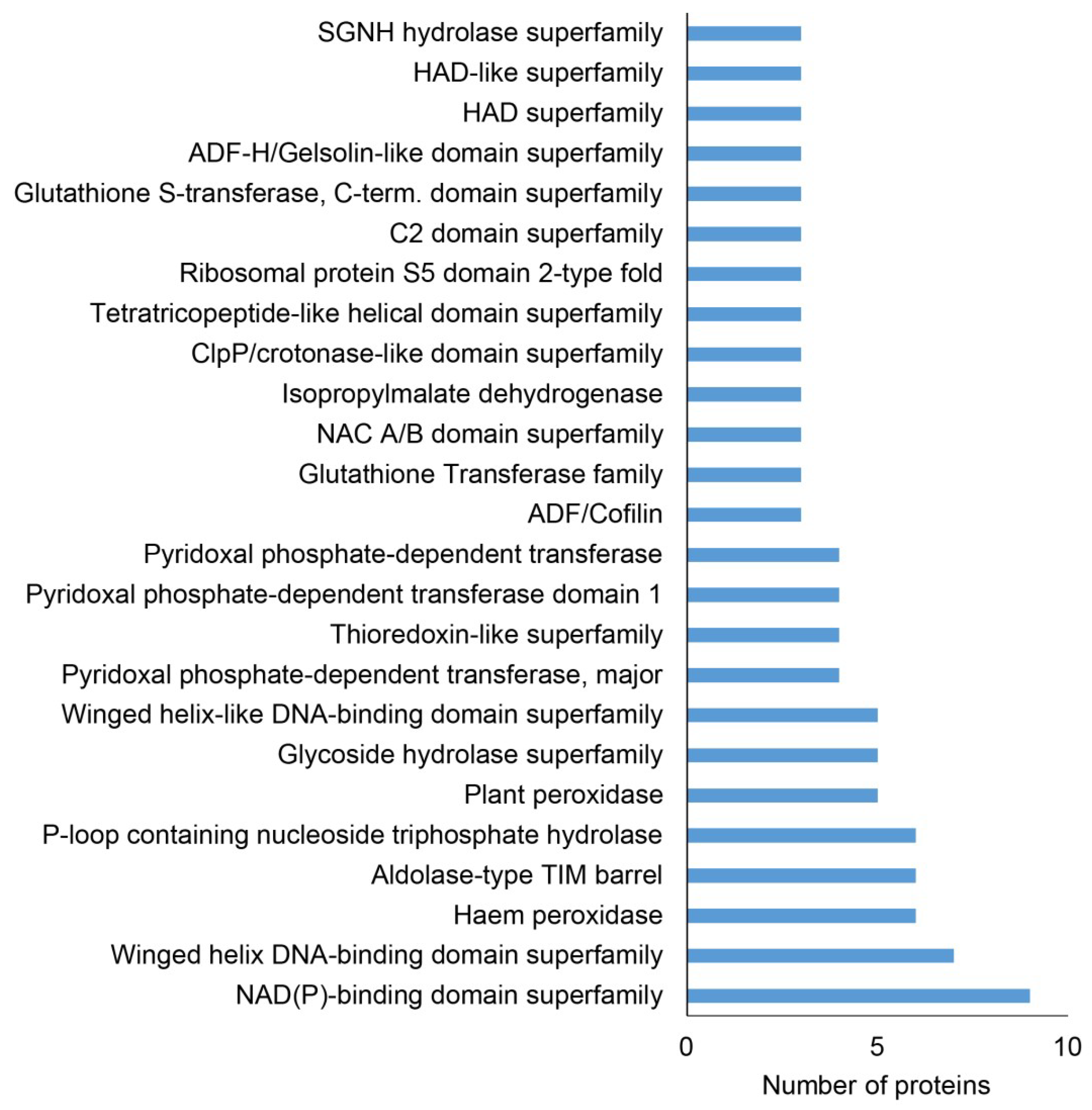
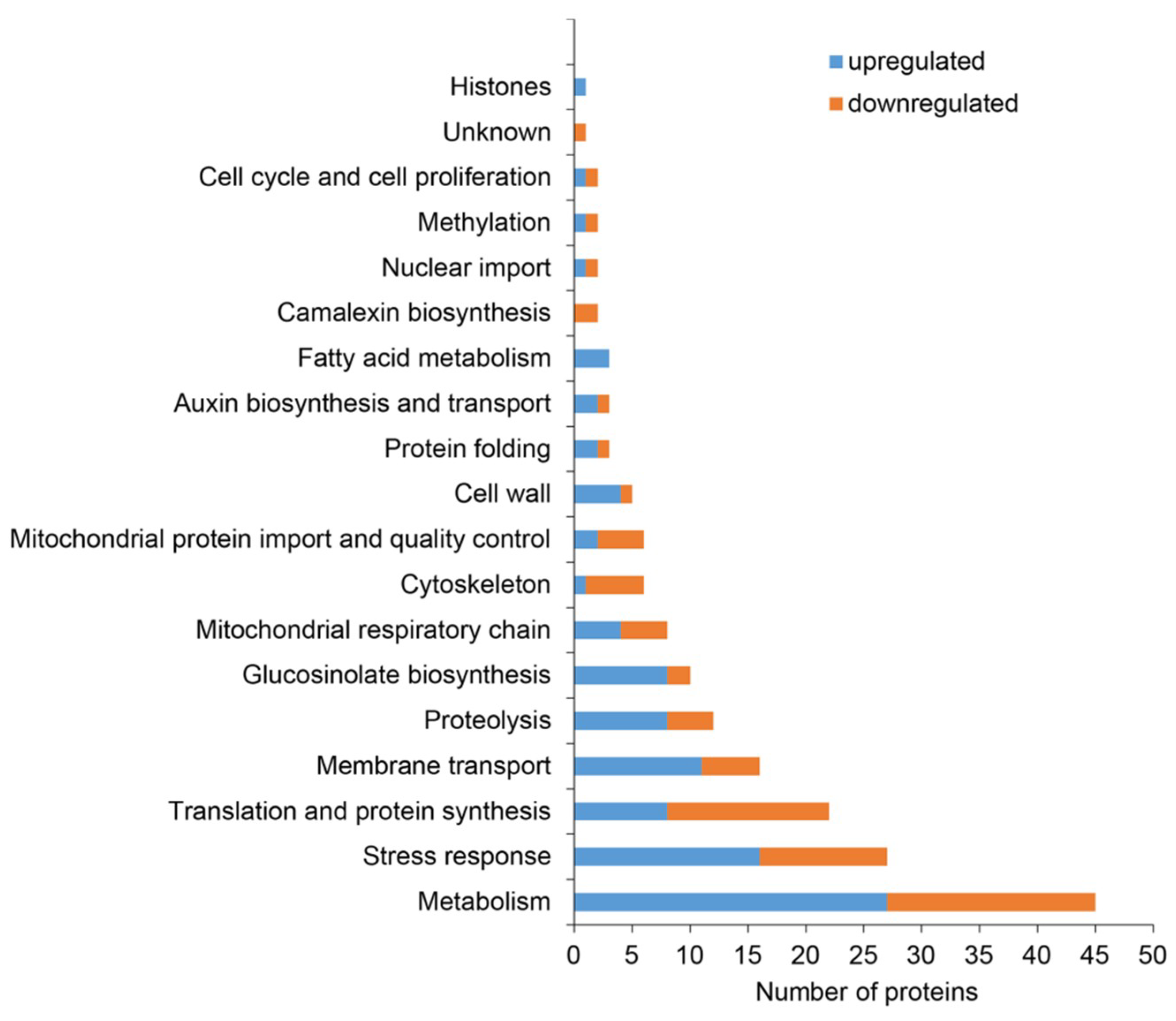
2.2. Classification of Root Differential Proteomes in pldα1 Mutants

A Kyoto encyclopedia of genes and genomes (KEGG) pathways analysis is a reasonable tool for the evaluation of proteins involved in metabolism. The highest number of DAPs was classified into the purine metabolism pathway and biosynthesis of antibiotics. Several proteins affected in both mutants are involved in pyruvate metabolism, amino acid biosynthesis and metabolism and phenylpropanoid biosynthesis (Figure 2).
Additionally, we screened differential proteomes of both mutants for the abundance of protein families, as evaluated by the Blast2Go software using InterPro application (Figure 3, Table S2). We identified nine proteins belonging to the NAD(P) binding protein superfamily, while seven proteins belonged to the Winged helix DNA-binding domain superfamily. Later ones include proteins with different functions (Table S2) and possessed specific DNA binding mechanisms different from sequence specific binding. They display an exposed patch of hydrophobic residues implicated in protein-protein interactions [24]. The peroxidases and aldolase-type TIM (triose phosphate isomerase) barrel protein family represented abundant protein classes found in both pldα1 mutants (Figure 3, Table S2). These proteins might show higher sensitivity to PLDα1 and PA deregulation in Arabidopsis.
Furthermore, we classified differential proteomes of pldα1 mutants (combined) using a gene ontology (GO) annotation analysis. The highest number of the DAPs was assigned to metabolic processes and nitrogen compound metabolic processes. A significant number of DAPs were involved in response to stress as well as establishment of localization (Figure S2A). Higher levels of GO revealed that proteins annotated as involved in stress response belong to GO class called response to osmotic stress (Table S3). Concerning cellular compartment, the GO ontology analysis showed that the highest number of DAPs was assigned to cytosol, followed by plastid, mitochondria, protein complex and the nucleus (Figure S2B).
Since GO ontology analysis does not consider all relevant information about protein functions, we decided to classify the combined differential proteome based on published data (Figure 4, Table 1 and Table S1).
Apart from the high number of DAPs with diverse metabolic functions, proteins related to the stress response were the second most abundant category (Figure 4, Table S1). Notably, PLDα1 deficiency in both mutants negatively affected the abundance of protein C2-domain ABA-related 10 (CAR10), a component of the pyrabactin resistance1/pyrabactin resistance1-like/car (PYR/PYL/CAR) receptors for ABA [25]. Additionally, we noticed the significant disturbance of antioxidant defense and redox homeostasis. This is represented by the increased abundance of ironic superoxide dismutase 1 (FeSOD1), ascorbate peroxidase and peptide methionine sulfoxide reductase B6. Secretory peroxidases exhibited varying changes in protein abundance, while catalase and glutathione S-transferase F7 had a lower abundance in the mutants compared to the wild type. To prove the increased abundance of FeSOD1, we performed an immunoblotting analysis on pldα1 mutants using anti-FeSOD1 polyclonal primary antibody (Figure 5A,B). The Arabidopsis thaliana genome contains three isoforms of FeSOD, out of which FeSOD2 and FeSOD3 are not expressed in the roots. Therefore, anti-FeSOD antibody recognizes FeSOD1 in the Arabidopsis roots. These analyses showed significant upregulation of FeSOD1 abundance in both pldα1 mutants.
Interestingly, PLDα1 deficiency also leads to deregulation of proteins involved in cell wall remodeling (Figure 4, Table S1), which represents one of the primary plant defense responses to pathogens. This is consistent with the known role of PLDα1 in plant biotic stress [26]. Furthermore, we have found several defense related proteins differentially abundant in the pldα1 mutants, including secretory peroxidases, nitrile specifier protein 1 and defensin-like protein 1 (Table S1). The majority of these proteins show increased abundance in the mutants. Notably, proteins involved in glucosinolate biosynthesis (discussed below) are highly represented, showing mostly increased abundances in the mutants (Figure 4).
Additionally, we have found numerous proteins involved in membrane fusion and transport. They are described in detail in the Discussion section. Among others, a PLDα1 deficiency resulted in accumulation of synaptotagmin 1 in the mutants. These proteomic data were successfully validated using immunoblotting analyses (Figure 5C,D) and immunolocalization of the syntaptotagmin 1 (SYT1) protein in intact roots showing an increased accumulation in both pldα1 mutants (Figure 6).
Proteins involved in ribosome biogenesis and translation, mitochondrial respiration, mitochondrial protein import and quality control represented a significant functional classes altered by PLDa1 deficiency (Figure 4, Table 1 and Table S1). These findings might indicate defects of cytosolic translation and mitochondrial protein import resulting in changed abundances of mitochondrial proteins. Therefore we searched for proteins carrying mitochondrial targeting signal among DAPs. We have found 19 proteins with varying changes in their abundance, suggesting an altered homeostasis in the import of mitochondrial proteins (Table S4). One of such proteins, mitochondrial uncoupling protein 1 (UPC1) has increased abundance in the mutants, being in agreement with the immunoblotting analysis (Figure 5E,F) and immunolocalization of uncoupling protein 1 (UCP1) protein (Figure 7). In addition, we observed also decreased levels of MORF8 (multiple site organellar RNA editing factor, designated also as RIP1; Table S1), a protein important for mitochondrial mRNA editing. Finally, absence of PLDα1 in both mutants affects also a cluster of components of the mitochondrial respiratory chain. Thus, PLDα1 is likely required for multiple mitochondrial functions in Arabidopsis (Table S1, Figure 4).
PLDα1 and PA are important regulators of actin and microtubule cytoskeletons in plants [11,27]. As expected, PLDα1 deficiency in both mutants resulted in differential abundances of actin and microtubule associated proteins, including actin1 and actin depolymerizing factors (ADFs) 1, 8 and 10 (showing decreased abundances in pldα1 mutants) (Table 1 and Table S1). Such results indicate possible disturbances in actin monomer turnover and actin polymerization in pldα1 mutants. Additionally, we identified two protein candidates potentially important for microtubule regulation by PLDα1. Both proteins were detected uniquely in pldα1 mutants and are involved in tubulin monomer folding. Tubulin-folding cofactor B is a member of the Arabidopsis pilZ domain proteins [28,29]. It interacts with alpha-tubulin and its overexpression results in reduced number of microtubules [30]. Chaperone prefoldin 6 is required for tubulin monomer abundance, microtubule dynamics and organization [31].

3. Discussion

This differential proteomic analysis on roots of pldα1 mutants revealed that PLDα1 is required for homeostasis of proteins involved in diverse processes. In this study, we focused especially on potential new functions of PLDα1 such as mitochondrial protein import and quality control, vesicular trafficking and glucosinolate biosynthesis. Considering the regulatory and catalytic roles of PLDα1, we assume that besides its lipid hydrolyzing activity, the changes in the proteomes of pldα1 mutants occurred as a consequence of compromised PA, G protein complex and ABA signalling.

3.1. New Insights into ABA Signalling

PLDα1 derived PA is a crucial regulator of stomatal movements, because it targets/binds multiple proteins essential for this process, including ABI1 [5], NADPH oxidase [14], G protein complexes [13] and MAP65-1 [3]. Assuming from our results, there seems to be a broader impact on other components of ABA signalling because PLDα1 deficiency negatively affected the abundance of protein C2-DOMAIN ABA-RELATED 10 (CAR10). CAR10 interacts with PYR/PYL ABA receptors and recruits them transiently into phospholipid vesicles, thus positively regulating ABA signaling [25]. The PYR/PYL/CAR receptors also bind to ABI1 [32]. These data indicate a possible feedback regulation of CAR10 abundance in the absence of PLDα1 and decreased levels of PA. In addition, aquaporins PM intrinsic protein 1-2 (PIP1-2) and PIP2-1 are ABA-inducible proteins, which promote water uptake and transport [33], and they bind PA [34], PLDδ and PLDγ [35]. Our proteomic analysis showed that abundances of these proteins substantially increased in pldα1 mutants.

3.2. Mitochondrial Protein Import and Quality Control

According to our results, PLDα1 deficiency in mutants caused a deregulation of proteins involved in protein import to mitochondria, including mitochondrial import inner membrane translocase subunits TIM23-2 and TIM13, which are downregulated. While TIM23-2 is a translocase responsible for the transport of mitochondrial precursor proteins carrying a cleavable N-terminal pre-sequence [36], TIM13 is a member of small TIM complex delivering client precursors that pass through the TOM (mitochondrial import outer membrane translocase) channel to Tim22 in the mitochondrial intermembrane space [37]. Therefore, the import of nucleus-encoded mitochondrial proteins is altered in pldα1 mutants. Along with altered protein import to mitochondria, PLDα1 deficiency may affect also N-terminal presequence cleavage (inferred by increased abundance of presequence protease 1 in pldα1 mutants) occurring after protein precursor import into mitochondria [38]. Furthermore, we provided experimental evidence on deregulation of prohibitin 6 involved in mitochondrial protein folding [39]. Prohibitins (PHBs) are considered to be structural proteins that form a scaffold-like structure for interacting with a set of proteins involved in various mitochondrial processes [39]. These proteins participate in the assembly of multi-subunit complexes such as mitochondrial respiratory complex [40]. Accordingly, several proteins of the mitochondrial electron transport chain show significant changes in their abundance in both mutants as compared to the wild type. Mitochondrial protein import machinery was also reported to be in close interaction with the organization of respiratory complexes. Tim23-2 is localized also in respiratory complex 1 and its genetic modification leads to altered transcription of mitochondrial proteins and defective mitochondria biogenesis [36]. A similar role in mitochondria biogenesis was found for prohibitins [41]. Thus far, PLDα1 was not linked to these mitochondrial functions, although the ATP synthase subunit gamma and ADP/ATP carrier protein were targeted by PA in Arabidopsis [34].

3.3. Vesicular Transport

PLD-derived PA can regulate membrane transport by direct modification of membrane curvature or by recruiting important regulatory proteins [42]. These proteins positively affect protein internalization [43,44], vesicle fusion and aggregation [45]. In Drosophila, PLD activity couples endocytosis with retromer dependent recycling [46]. Our findings indicate that PLDα1 alters multiple sites of endomembrane system. For example, in both mutants we detected decreased abundances of vacuolar H+ ATPases (subunits D and d2), which control multiple events in endomembrane transport by acidification of endomembrane compartments [47].
In accordance with the known involvement of PLDs in vesicle fusions, we observed an increased abundance of alpha-soluble N-ethylmaleimide-sensitive factor (NSF) attachment protein 2 (Alpha-SNAP2) in the pldα1-1 mutant. Alpha-SNAP proteins bind the soluble NSF attachment protein receptor (SNARE) complex [48] and are required for the vesicle pre-docking, an initial step of the membrane fusion reaction [49,50]. The precise function of alpha-SNAP2 is unknown, though it might require PLDα1. Remarkably, alpha-SNAP2 interacting syntaxin 32 (SYP32), a Golgi localized Qa SNARE [51] was found as upregulated in pldα1 mutants. Thus, PLDα1 might be necessary for SNARE-SNAP protein complexes stability.
We identified several proteins involved in the endocytic pathway as differentially regulated in both pldα1 mutants. These include mainly the probable clathrin assembly protein At4g32285 (not detected in pldα1 mutants), which is involved in clathrin-mediated endocytosis [52]. Clathrin assembly proteins interact directly with proteins of the clathrin coat and are able to bind phospholipids [53]. Two such proteins were identified as PA-binding proteins [34]. Furthermore, PLDα1 localized in the vicinity of clathrin heavy chain and microtubules of Arabidopsis root cells [12] and it may directly bind clathrin in a complex containing adaptor protein-2 (AP-2) [54]. Vacuolar protein sorting 29 (VPS29), a protein found uniquely in the pldα1-1 mutant, is a component of retromer complex. This is a coat complex localized to the cytosolic face of endosomes and involved in intracellular sorting of some transmembrane proteins [55]. VPS29 is important for normal morphology of prevacuolar compartment (PVC) and plays crucial role in recycling vacuolar sorting receptors from the PVC to the trans Golgi network (TGN) during trafficking of soluble proteins to the lytic vacuole [56,57]. These data uncovers new endocytic proteins affected in pldα1 mutants.
PLDα1 deficiency in both mutants altered also abundances of proteins involved in the regulation of endoplasmic reticulum (ER) to Golgi transport. Protein transport protein SEC13 homolog A is upregulated nearly threefold in both pldα1 mutants. Sec13 makes a lattice structure together with Sec31 to form COPII vesicles [58], which are responsible for ER to Golgi transport. According to our results, PLDα1 may have also an impact on the morphology of Golgi apparatus, inferred by the upregulation of Golgin candidate 5 (also known as the TATA element modulatory factor) in the pldα1-2 mutant [59,60]. Another protein important for ER to Golgi trafficking is vesicle-associated protein 1-2 (PVA12, also known as VAP27-3), which is upregulated in the pldα1-1 mutant. This is an ER-localized protein belonging to a VAP27 family [61]. It binds to oxysterol-binding protein-related protein 3B [62], which is also upregulated in the mutants and is proposed to cycle between the ER and the Golgi [62]. Recently, PVA12 was shown to colocalize and interact with Networked 3C (NET3C) at ER–PM contact sites [61]. Considering PLDα1 localization in the PM vicinity, we suggest an involvement of this protein in ER-PM attachment. This is emphasized by an increased abundance of synaptotagmin 1 (SYT1) in pldα1 mutants, representing a protein mediating the ER-PM contacts in Arabidopsis [63].
PLDα1 depletion leads to changed abundance of proteins regulating the membrane transport. Changes in protein level might be a result of deregulation of protein synthesis and proteolysis or transcriptional control. Previously, it was shown that changes in membrane transport might result in changed abundance of proteins. This was exemplified for example in Arabidopsis roots exposed to brefeldin A (BFA), which blocks secretion/exocytosis by aggregation of TGN and PM-derived vesicles surrounded by Golgi stacks into so called BFA-compartments [64]. Altered endocytosis and vacuolar trafficking by wortmannin lead to altered abundances of vacuolar proteases potentially leading to defected protein degradation [65]. Similar downregulation of such protease, subtilisin-like protease SBT1.7 is encountered also in roots of pldα1 mutants. Based on our proteomic data we suggest that this dynamics of membrane transport regulatory processes might result from defected protein degradation and as a feedback mechanism of PLDα1 depletion-induced changes in membrane architecture, membrane transport and PA accumulation.

3.4. Glucosinolate Biosynthesis

PLDs have been shown to crosstalk with hormonal signaling in plants. In addition to their well-known role in ABA signaling, they also participate in salicylic acid signaling by controlling relocation of NPR1, an essential regulator of SA induced gene transcription, into the nucleus [66]. In addition, PLDs might be activated by cytokinins [67] and ethylene [68]. Constitutive triple response 1 (CTR1), a negative regulator of ethylene response is a potential target of PA [69]. PLDs are also involved in auxin distribution. Thus, PLDζ-derived PA is required for protein phosphatase 2Ac (PP2Ac) recruitment to the membrane resulting in altered auxin efflux carrier component 1 (PIN1) phosphorylation and polar distribution [7]. Auxins share an initial steps of biosynthetic pathway with glucosinolates [70,71]. Arabidopsis mutants with reduced glucosinolate contents show severe auxin phenotypes [72]. Generally, glucosinolates are secondary messengers produced in Brassicaceae with important defense and developmental functions [70,73]. PLDα1 deficiency in mutants causes increased abundances of enzymes involved in glucosinolate biosynthesis, including four subunits of 3-isopropylmalate dehydrogenase and methylthioalkylmalate synthase, all involved in the chain elongation machinery. Enzymes involved in the biosynthesis of the core glucosinolate structure, namely cytochrome P450 83B1, glutathione S-transferase F9, indole glucosinolate O-methyltransferase 1 and adenylyl-sulfate kinase 1, showed similar trends in their abundances (Table S1). PLDα1 induced an imbalance of indole glucosinolate o-methyltransferase 1 abundance, which is a glucosinolate modifying enzyme [71]. Glutathione synthase 1 showed an increased abundance in mutants, most likely contributing to the glutathione pool, which serves as a sulfur donor within the second stage of GLS biosynthesis [71]. Such differential regulation of enzymes involved in one metabolic pathway in untargeted proteomic approach is very unusual, suggesting that PLDα1 might be a master regulator of glucosinolate biosynthesis. It is likely that this regulation is mediated via PA, since cytochrome P450 83B1 is a PA-binding protein, as identified in a proteomic screen [34].

4. Materials and Methods

4.1. Plant Material

Seeds of Arabidopsis thaliana wild type (ecotype Col-0) as well as pldα1-1 (SALK_067533) and pldα1-2 (SALK_053785) t-DNA insertion mutants were used in this study. Following ethanol surface-sterilization, they were cultivated vertically on solid half-strength Murashige-Skoog (MS) media at 21 °C under 16/8 light/dark illumination conditions for 14 days. Roots were quickly dissected and harvested for protein extraction. Proteomic analyses were performed in four biological replicates. Roots of 30 seedlings were pooled in one biological replicate.

4.2. Protein Extraction and Trypsin Digestion

Samples were ground in liquid nitrogen and subjected to phenol protein extraction followed by ammonium acetate/methanol precipitation as described by Takáč et al. [74]. Cleaned precipitates were dissolved in 6 M urea in 100 mM Tris (pH 7,8). Prior to trypsin digestion, extracts containing 50 µg of proteins (in volume of 50 µl) were diluted with 100 mM Tris-HCl (pH7,8) to adjust the urea concentration bellow 1 mM. Proteins were digested with trypsin (Promega;1 µg of trypsin to 50 µg of proteins) at 37 °C overnight. Reaction was stopped by addition of 4 µL of acetic acid. Peptide mixtures were cleaned using C18 gravity flow cartridges (Bond Elut C18; Agilent Technologies, Santa Clara, CA, USA) according to manufacturer’s instructions. Peptides eluted by 95% acetonitrile were dried using vacuum concentrator and stored under −80 °C until analysis.

4.3. Liquid Chromatography, Mass Spectrometry, Protein Identification and Relative Quantitative Analysis

Liquid chromatography-MSMS and protein identification was performed as published previously [74] with minor modifications. As target database and decoy databases, the UNIPROT (www.uniprot.org) Arabidopsis genus taxonomy reviewed protein database (17,586 entries as of 31st September 2017), and its reversed copy (created automatically by the software) were used, respectively. The mass spectrometry proteomics data have been deposited to the ProteomeXchange Consortium via the PRIDE partner repository with the dataset identifier PXD011196.
The quantitative analysis was done using the ProteoIQ 2.1 (NuSep Inc., Athens, GA, USA) software as published previously [75]. The ANOVA p ≤ 0.05 was used to filter statistically significant results. Proteins with fold changes higher than 1.5 were considered as differentially abundant. Proteins present at least in two biological replicates and identified by at least two peptide spectral matches were quantified.

4.4. Bioinformatic Analysis

Gene ontology (GO) annotation analysis of DAPs was performed using Blast2Go software [76]. BLAST searching was performed against the Arabidopsis thaliana NCBI database allowing 1 BLAST Hit. The annotation was carried out by using these parameters: E Value Hit filter: 1.0 × 10⁻⁶; Annotation cut off: 55; GO weight: 5. The prediction of presence of mitochondrial targeting pre-sequence in differential proteomes of both mutants was performed using MitoFates [77].

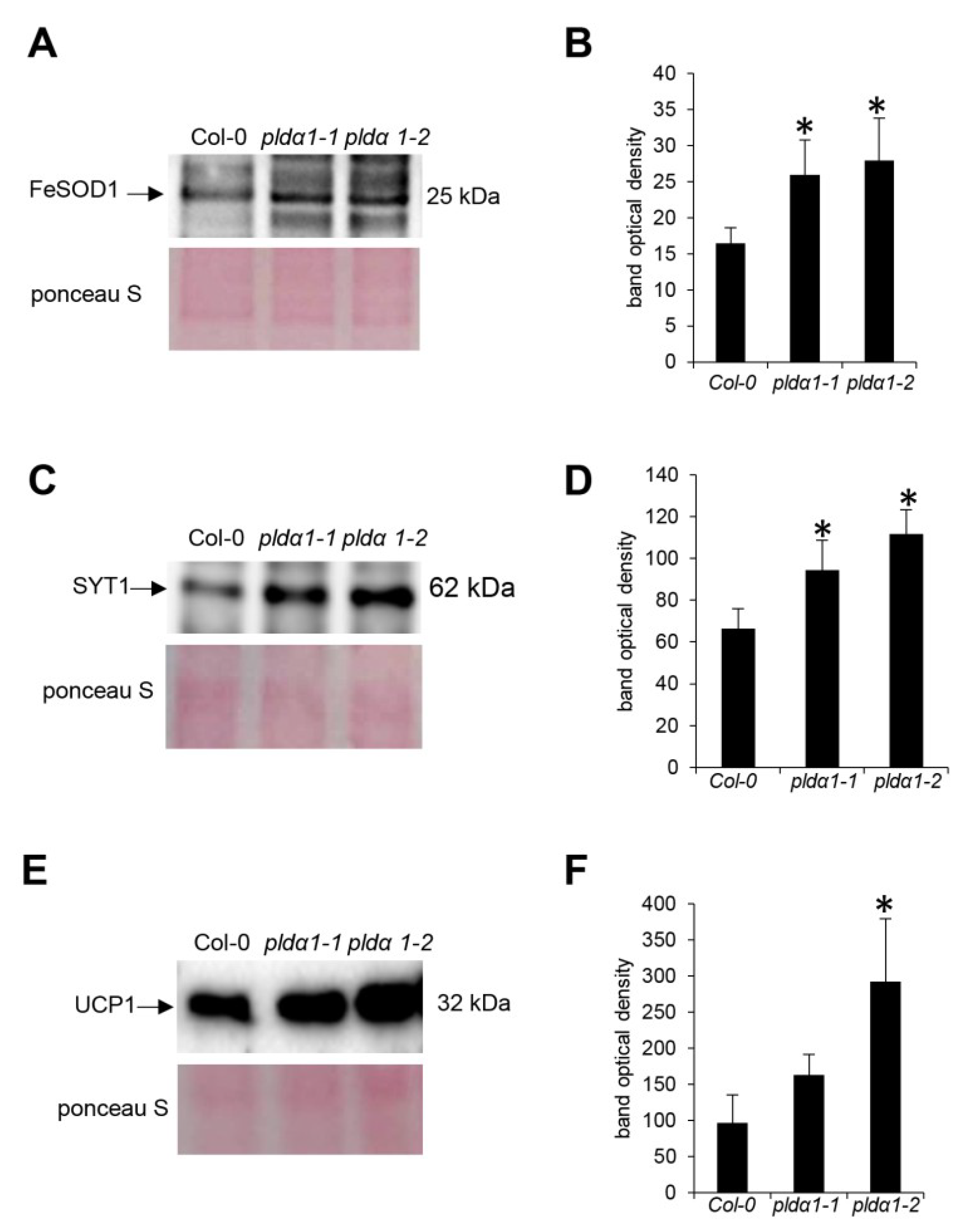
4.5. Immunoblotting Analysis

Immunoblotting analysis was performed on protein extracts derived from roots of 14 day-old plants of wild type, as well as pldα1-1 and pldα1-2 mutants following published procedure [74]. Anti-synaptotagmin (PhytoAb; dilution 1:1000), anti-FeSOD (Agrisera; dilution 1:3000) and anti-UCP1 (Agrisera; dilution 1:1000) primary antibodies were used. Immunoblot analyses were carried out in three biological replicates. Differences in signal intensity between wild type and the mutants were statistically evaluated using Student’s t-test (p < 0.05).

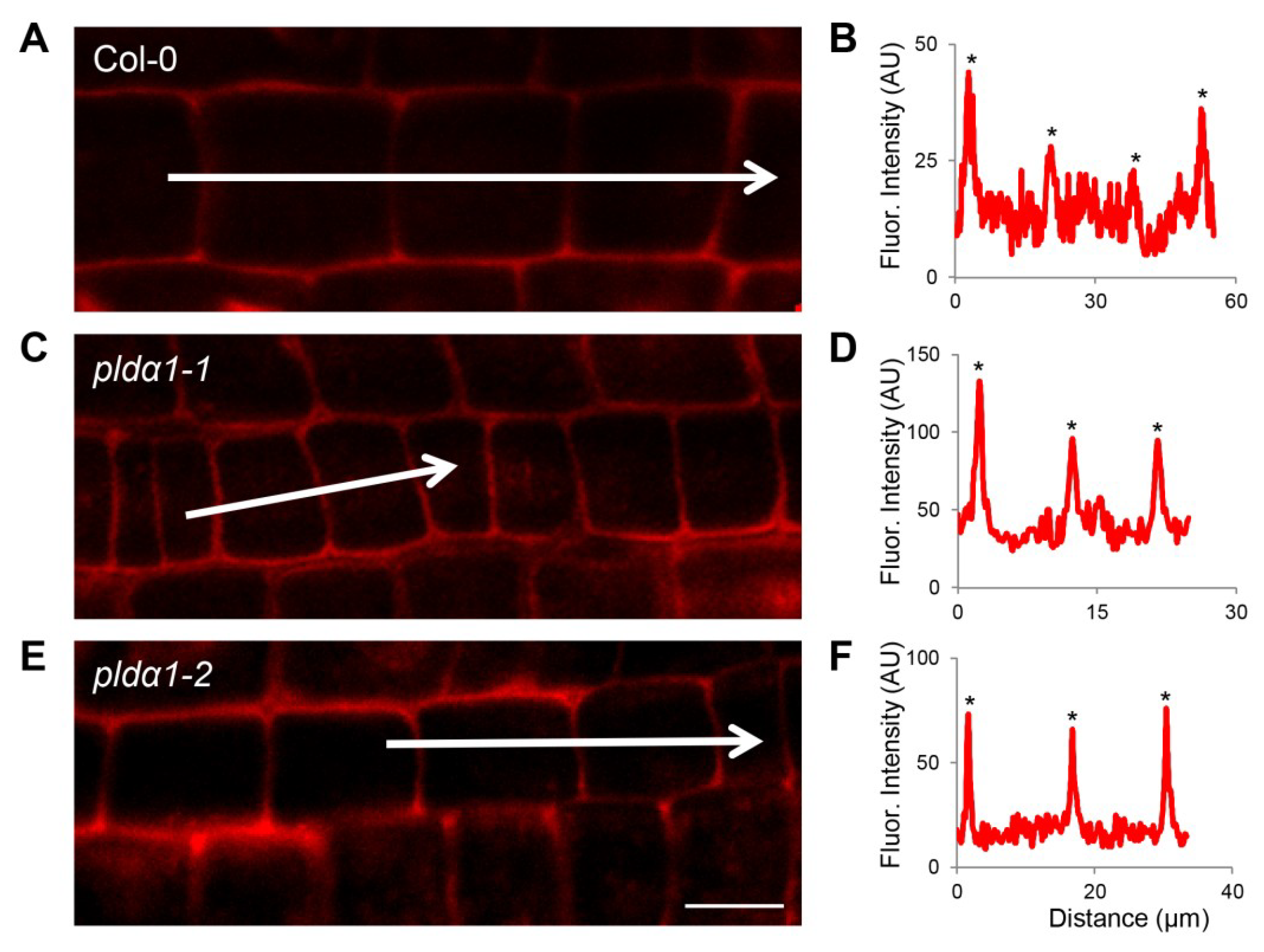
4.6. Whole Mount Immunofluorescence Labelling

Immunolocalization of SYT1 and UCP1 proteins in root wholemounts was carried out as published previously [78]. As primary antibodies, we have used the rabbit anti-synaptotagmin 1 antibody (PhytoAb; 1:200) and anti-UCP1 antibody (Agrisera; 1:200), while Alexa-Fluor 647 goat anti-rabbit IgG was exploited as secondary antibody. Microscopic observations were performed using the Zeiss 710 Confocal Laser Scanning Microscope platform (Carl Zeiss, Jena, Germany), using excitation lines at 405 and 561 nm from argon, HeNe, diode and diode pumped solid-state lasers. ZEN 2010 software (Carl Zeiss) was used for post-processing, default deconvolution and quantification of fluorescence intensity. Additionally, Photoshop 6.0/CS, and Microsoft PowerPoint softwares were used to process the obtained images.

5. Conclusions

Based on this proteomic analysis, PLDα1 is a protein which in addition to its well-known functions in ABA signalling and cytoskeleton organization, important for the homeostasis of proteins involved in mitochondrial protein import, vesicular trafficking and glucosinolate biosynthesis.

Supplementary Materials

Supplementary materials can be found at https://www.mdpi.com/1422-0067/20/1/82/s1.

Author Contributions

J.Š. conceived and coordinated the experiments and helped to evaluate data, T.T., T.P., O.Š. and P.V. made analyses and experiments, T.T. and J.Š. wrote the manuscript. All authors reviewed the manuscript.

Funding

This research was funded by Czech Science Foundation GAČR, grant number 16-22044S; the European Regional Development Fund OPVVV project “Plants as a tool for sustainable development” number CZ.02.1.01/0.0/16_019/0000827 supporting Excellent Research at CRH, USDA NIFA award #58-6066-6-059 and NIH award #USM-GR05802-03.

Acknowledgments

The mass spectrometry proteomics analysis was performed at the Institute for Genomics, Biocomputing and Biotechnology, Mississippi State University, with partial support from the Mississippi Agriculture and Forestry Experimental Station.

Conflicts of Interest

The authors declare no conflict of interest. The funders had no role in the design of the study; in the collection, analyses, or interpretation of data; in the writing of the manuscript, or in the decision to publish the results.

References

  1. Wang, X. (Ed.) Phospholipases in Plant Signaling; Signaling and Communication in Plants; Springer: Berlin/Heidelberg, Germany, 2014; Volume 20, ISBN 978-3-642-42010-8. [Google Scholar]
  2. Hong, Y.; Zhao, J.; Guo, L.; Kim, S.-C.; Deng, X.; Wang, G.; Zhang, G.; Li, M.; Wang, X. Plant phospholipases D and C and their diverse functions in stress responses. Prog. Lipid Res. 2016, 62, 55–74. [Google Scholar] [CrossRef] [PubMed] [Green Version]
  3. Zhang, Q.; Lin, F.; Mao, T.; Nie, J.; Yan, M.; Yuan, M.; Zhang, W. Phosphatidic acid regulates microtubule organization by interacting with MAP65-1 in response to salt stress in Arabidopsis. Plant Cell 2012, 24, 4555–4576. [Google Scholar] [CrossRef] [PubMed]
  4. Testerink, C.; Munnik, T. Molecular, cellular, and physiological responses to phosphatidic acid formation in plants. J. Exp. Bot. 2011, 62, 2349–2361. [Google Scholar] [CrossRef] [PubMed] [Green Version]
  5. Zhang, W.; Qin, C.; Zhao, J.; Wang, X. Phospholipase D alpha 1-derived phosphatidic acid interacts with ABI1 phosphatase 2C and regulates abscisic acid signaling. Proc. Natl. Acad. Sci. USA 2004, 101, 9508–9513. [Google Scholar] [CrossRef] [PubMed]
  6. Devaiah, S.P.; Roth, M.R.; Baughman, E.; Li, M.; Tamura, P.; Jeannotte, R.; Welti, R.; Wang, X. Quantitative profiling of polar glycerolipid species from organs of wild-type Arabidopsis and a PHOSPHOLIPASE Dα1 knockout mutant. Phytochemistry 2006, 67, 1907–1924. [Google Scholar] [CrossRef] [PubMed]
  7. Gao, H.-B.; Chu, Y.-J.; Xue, H.-W. Phosphatidic Acid (PA) Binds PP2AA1 to Regulate PP2A Activity and PIN1 Polar Localization. Mol. Plant 2013, 6, 1692–1702. [Google Scholar] [CrossRef] [PubMed]
  8. Boutté, Y.; Moreau, P. Modulation of endomembranes morphodynamics in the secretory/retrograde pathways depends on lipid diversity. Curr. Opin. Plant Biol. 2014, 22, 22–29. [Google Scholar] [CrossRef]
  9. Dhonukshe, P.; Laxalt, A.M.; Goedhart, J.; Gadella, T.W.J.; Munnik, T. Phospholipase d activation correlates with microtubule reorganization in living plant cells. Plant Cell 2003, 15, 2666–2679. [Google Scholar] [CrossRef]
  10. Pleskot, R.; Potocký, M.; Pejchar, P.; Linek, J.; Bezvoda, R.; Martinec, J.; Valentová, O.; Novotná, Z.; Zárský, V. Mutual regulation of plant phospholipase D and the actin cytoskeleton. Plant J. 2010, 62, 494–507. [Google Scholar] [CrossRef] [Green Version]
  11. Pleskot, R.; Li, J.; Žárský, V.; Potocký, M.; Staiger, C.J. Regulation of cytoskeletal dynamics by phospholipase D and phosphatidic acid. Trends Plant Sci. 2013, 18, 496–504. [Google Scholar] [CrossRef]
  12. Novák, D.; Vadovič, P.; Ovečka, M.; Šamajová, O.; Komis, G.; Colcombet, J.; Šamaj, J. Gene expression pattern and protein localization of Arabidopsis Phospholipase D Alpha 1 revealed by advanced light-sheet and super-resolution microscopy. Front. Plant Sci. 2018, 9, 371. [Google Scholar] [CrossRef] [PubMed]
  13. Mishra, G.; Zhang, W.; Deng, F.; Zhao, J.; Wang, X. A bifurcating pathway directs abscisic acid effects on stomatal closure and opening in Arabidopsis. Science 2006, 312, 264–266. [Google Scholar] [CrossRef] [PubMed]
  14. Zhang, Y.; Zhu, H.; Zhang, Q.; Li, M.; Yan, M.; Wang, R.; Wang, L.; Welti, R.; Zhang, W.; Wang, X. Phospholipase Dα1 and Phosphatidic Acid Regulate NADPH Oxidase Activity and Production of Reactive Oxygen Species in ABA-Mediated Stomatal Closure in Arabidopsis. Plant Cell 2009, 21, 2357–2377. [Google Scholar] [CrossRef] [PubMed]
  15. Guo, L.; Mishra, G.; Markham, J.E.; Li, M.; Tawfall, A.; Welti, R.; Wang, X. Connections between sphingosine kinase and phospholipase D in the abscisic acid signaling pathway in Arabidopsis. J. Biol. Chem. 2012, 287, 8286–8296. [Google Scholar] [CrossRef] [PubMed]
  16. Roy Choudhury, S.; Pandey, S. The role of PLDα1 in providing specificity to signal-response coupling by heterotrimeric G-protein components in Arabidopsis. Plant J. 2016, 86, 50–61. [Google Scholar] [CrossRef] [PubMed] [Green Version]
  17. Roy Choudhury, S.; Pandey, S. Phosphatidic acid binding inhibits RGS1 activity to affect specific signaling pathways in Arabidopsis. Plant J. 2017, 90, 466–477. [Google Scholar] [CrossRef] [PubMed]
  18. Hong, Y.; Zheng, S.; Wang, X. Dual functions of phospholipase Dalpha1 in plant response to drought. Mol. Plant 2008, 1, 262–269. [Google Scholar] [CrossRef]
  19. Huo, C.; Zhang, B.; Wang, H.; Wang, F.; Liu, M.; Gao, Y.; Zhang, W.; Deng, Z.; Sun, D.; Tang, W. Comparative Study of Early Cold-Regulated Proteins by Two-Dimensional Difference Gel Electrophoresis Reveals a Key Role for Phospholipase Dα1 in Mediating Cold Acclimation Signaling Pathway in Rice. Mol. Cell. Proteom. 2016, 15, 1397–1411. [Google Scholar] [CrossRef]
  20. Lu, S.; Bahn, S.C.; Qu, G.; Qin, H.; Hong, Y.; Xu, Q.; Zhou, Y.; Hong, Y.; Wang, X. Increased expression of phospholipase Dα1 in guard cells decreases water loss with improved seed production under drought in Brassica napus. Plant Biotechnol. J. 2013, 11, 380–389. [Google Scholar] [CrossRef]
  21. Uraji, M.; Katagiri, T.; Okuma, E.; Ye, W.; Hossain, M.A.; Masuda, C.; Miura, A.; Nakamura, Y.; Mori, I.C.; Shinozaki, K.; et al. Cooperative function of PLDδ and PLDα1 in abscisic acid-induced stomatal closure in Arabidopsis. Plant Physiol. 2012, 159, 450–460. [Google Scholar] [CrossRef]
  22. Devaiah, S.P.; Pan, X.; Hong, Y.; Roth, M.; Welti, R.; Wang, X. Enhancing seed quality and viability by suppressing phospholipase D in Arabidopsis: Phospholipase D in seed aging. Plant J. 2007, 50, 950–957. [Google Scholar] [CrossRef] [PubMed]
  23. Ufer, G.; Gertzmann, A.; Gasulla, F.; Röhrig, H.; Bartels, D. Identification and characterization of the phosphatidic acid-binding A. thaliana phosphoprotein PLDrp1 that is regulated by PLDα1 in a stress-dependent manner. Plant J. 2017, 92, 276–290. [Google Scholar] [CrossRef]
  24. Gajiwala, K.S.; Burley, S.K. Winged helix proteins. Curr. Opin. Struct. Biol. 2000, 10, 110–116. [Google Scholar] [CrossRef]
  25. Rodriguez, L.; Gonzalez-Guzman, M.; Diaz, M.; Rodrigues, A.; Izquierdo-Garcia, A.C.; Peirats-Llobet, M.; Fernandez, M.A.; Antoni, R.; Fernandez, D.; Marquez, J.A.; et al. C2-domain abscisic acid-related proteins mediate the interaction of PYR/PYL/RCAR abscisic acid receptors with the plasma membrane and regulate abscisic acid sensitivity in Arabidopsis. Plant Cell 2014, 26, 4802–4820. [Google Scholar] [CrossRef] [PubMed]
  26. Zhang, Q.; Berkey, R.; Blakeslee, J.J.; Lin, J.; Ma, X.; King, H.; Liddle, A.; Guo, L.; Munnik, T.; Wang, X.; et al. Arabidopsis phospholipase Dα1 and Dδ oppositely modulate EDS1- and SA-independent basal resistance against adapted powdery mildew. J. Exp. Bot. 2018, 69, 3675–3688. [Google Scholar] [CrossRef] [PubMed]
  27. Pleskot, R.; Pejchar, P.; Staiger, C.J.; Potocký, M. When fat is not bad: The regulation of actin dynamics by phospholipid signaling molecules. Front. Plant Sci. 2014, 5, 5. [Google Scholar] [CrossRef] [PubMed]
  28. Steinborn, K. The Arabidopsis PILZ group genes encode tubulin-folding cofactor orthologs required for cell division but not cell growth. Genes Dev. 2002, 16, 959–971. [Google Scholar] [CrossRef] [PubMed]
  29. Du, Y.; Cui, M.; Qian, D.; Zhu, L.; Wei, C.; Yuan, M.; Zhang, Z.; Li, Y. AtTFC B is involved in control of cell division. Front. Biosci. Elite Ed. 2010, 2, 752–763. [Google Scholar]
  30. Dhonukshe, P.; Bargmann, B.O.R.; Gadella, T.W.J. Arabidopsis Tubulin Folding Cofactor B Interacts with α-Tubulin In Vivo. Plant Cell Physiol. 2006, 47, 1406–1411. [Google Scholar] [CrossRef] [Green Version]
  31. Gu, Y.; Deng, Z.; Paredez, A.R.; DeBolt, S.; Wang, Z.-Y.; Somerville, C. Prefoldin 6 is required for normal microtubule dynamics and organization in Arabidopsis. Proc. Natl. Acad. Sci. USA 2008, 105, 18064–18069. [Google Scholar] [CrossRef] [Green Version]
  32. Nishimura, N.; Sarkeshik, A.; Nito, K.; Park, S.-Y.; Wang, A.; Carvalho, P.C.; Lee, S.; Caddell, D.F.; Cutler, S.R.; Chory, J.; et al. PYR/PYL/RCAR family members are major in-vivo ABI1 protein phosphatase 2C-interacting proteins in Arabidopsis. Plant J. Cell Mol. Biol. 2010, 61, 290–299. [Google Scholar] [CrossRef] [PubMed]
  33. Postaire, O.; Tournaire-Roux, C.; Grondin, A.; Boursiac, Y.; Morillon, R.; Schäffner, A.R.; Maurel, C. A PIP1 aquaporin contributes to hydrostatic pressure-induced water transport in both the root and rosette of Arabidopsis. Plant Physiol. 2010, 152, 1418–1430. [Google Scholar] [CrossRef] [PubMed]
  34. McLoughlin, F.; Arisz, S.A.; Dekker, H.L.; Kramer, G.; de Koster, C.G.; Haring, M.A.; Munnik, T.; Testerink, C. Identification of novel candidate phosphatidic acid-binding proteins involved in the salt-stress response of Arabidopsis thaliana roots. Biochem. J. 2013, 450, 573–581. [Google Scholar] [CrossRef] [PubMed]
  35. Bellati, J.; Champeyroux, C.; Hem, S.; Rofidal, V.; Krouk, G.; Maurel, C.; Santoni, V. Novel Aquaporin Regulatory Mechanisms Revealed by Interactomics. Mol. Cell. Proteom. 2016, 15, 3473–3487. [Google Scholar] [CrossRef] [PubMed] [Green Version]
  36. Wang, Y.; Carrie, C.; Giraud, E.; Elhafez, D.; Narsai, R.; Duncan, O.; Whelan, J.; Murcha, M.W. Dual location of the mitochondrial preprotein transporters B14.7 and Tim23-2 in complex I and the TIM17:23 complex in Arabidopsis links mitochondrial activity and biogenesis. Plant Cell 2012, 24, 2675–2695. [Google Scholar] [CrossRef] [PubMed]
  37. Neupert, W. A Perspective on Transport of Proteins into Mitochondria: A Myriad of Open Questions. J. Mol. Biol. 2015, 427, 1135–1158. [Google Scholar] [CrossRef] [PubMed]
  38. Teixeira, P.F.; Glaser, E. Processing peptidases in mitochondria and chloroplasts. Biochim. Biophys. Acta Mol. Cell Res. 2013, 1833, 360–370. [Google Scholar] [CrossRef] [PubMed] [Green Version]
  39. Van Aken, O.; Whelan, J.; Van Breusegem, F. Prohibitins: Mitochondrial partners in development and stress response. Trends Plant Sci. 2010, 15, 275–282. [Google Scholar] [CrossRef]
  40. Piechota, J.; Bereza, M.; Sokołowska, A.; Suszyński, K.; Lech, K.; Jańska, H. Unraveling the functions of type II-prohibitins in Arabidopsis mitochondria. Plant Mol. Biol. 2015, 88, 249–267. [Google Scholar] [CrossRef]
  41. Ahn, C.S.; Lee, J.H.; Reum Hwang, A.; Kim, W.T.; Pai, H.-S. Prohibitin is involved in mitochondrial biogenesis in plants. Plant J. 2006, 46, 658–667. [Google Scholar] [CrossRef] [Green Version]
  42. Donaldson, J.G. Phospholipase D in endocytosis and endosomal recycling pathways. Biochim. Biophys. Acta 2009, 1791, 845–849. [Google Scholar] [CrossRef] [PubMed] [Green Version]
  43. Antonescu, C.N.; Danuser, G.; Schmid, S.L. Phosphatidic Acid Plays a Regulatory Role in Clathrin-mediated Endocytosis. Mol. Biol. Cell 2010, 21, 2944–2952. [Google Scholar] [CrossRef] [Green Version]
  44. Li, G.; Xue, H.-W. Arabidopsis PLD 2 Regulates Vesicle Trafficking and Is Required for Auxin Response. Plant Cell 2007, 19, 281–295. [Google Scholar] [CrossRef] [PubMed] [Green Version]
  45. Roth, M.G. Molecular mechanisms of PLD function in membrane traffic. Traffic 2008, 9, 1233–1239. [Google Scholar] [CrossRef] [PubMed]
  46. Thakur, R.; Panda, A.; Coessens, E.; Raj, N.; Yadav, S.; Balakrishnan, S.; Zhang, Q.; Georgiev, P.; Basak, B.; Pasricha, R.; et al. Phospholipase D activity couples plasma membrane endocytosis with retromer dependent recycling. eLife 2016, 5, e18515. [Google Scholar] [CrossRef] [PubMed]
  47. Schumacher, K.; Krebs, M. The V-ATPase: Small cargo, large effects. Curr. Opin. Plant Biol. 2010, 13, 724–730. [Google Scholar] [CrossRef] [PubMed]
  48. Fujiwara, M.; Uemura, T.; Ebine, K.; Nishimori, Y.; Ueda, T.; Nakano, A.; Sato, M.H.; Fukao, Y. Interactomics of Qa-SNARE in Arabidopsis thaliana. Plant Cell Physiol. 2014, 55, 781–789. [Google Scholar] [CrossRef] [Green Version]
  49. Mayer, A.; Wickner, W.; Haas, A. Sec18p (NSF)-Driven Release of Sec17p (α-SNAP) Can Precede Docking and Fusion of Yeast Vacuoles. Cell 1996, 85, 83–94. [Google Scholar] [CrossRef]
  50. Wang, T.; Li, L.; Hong, W. SNARE proteins in membrane trafficking. Traffic 2017, 18, 767–775. [Google Scholar] [CrossRef]
  51. Uemura, T.; Ueda, T.; Ohniwa, R.L.; Nakano, A.; Takeyasu, K.; Sato, M.H. Systematic analysis of SNARE molecules in Arabidopsis: Dissection of the post-Golgi network in plant cells. Cell Struct. Funct. 2004, 29, 49–65. [Google Scholar] [CrossRef]
  52. Wang, C.; Yan, X.; Chen, Q.; Jiang, N.; Fu, W.; Ma, B.; Liu, J.; Li, C.; Bednarek, S.Y.; Pan, J. Clathrin Light Chains Regulate Clathrin-Mediated Trafficking, Auxin Signaling, and Development in Arabidopsis. Plant Cell 2013, 25, 499–516. [Google Scholar] [CrossRef] [PubMed] [Green Version]
  53. Fan, L.; Li, R.; Pan, J.; Ding, Z.; Lin, J. Endocytosis and its regulation in plants. Trends Plant Sci. 2015, 20, 388–397. [Google Scholar] [CrossRef] [PubMed]
  54. Yamaoka, S.; Shimono, Y.; Shirakawa, M.; Fukao, Y.; Kawase, T.; Hatsugai, N.; Tamura, K.; Shimada, T.; Hara-Nishimura, I. Identification and dynamics of Arabidopsis adaptor protein-2 complex and its involvement in floral organ development. Plant Cell 2013, 25, 2958–2969. [Google Scholar] [CrossRef] [PubMed]
  55. Zelazny, E.; Santambrogio, M.; Pourcher, M.; Chambrier, P.; Berne-Dedieu, A.; Fobis-Loisy, I.; Miège, C.; Jaillais, Y.; Gaude, T. Mechanisms Governing the Endosomal Membrane Recruitment of the Core Retromer in Arabidopsis. J. Biol. Chem. 2013, 288, 8815–8825. [Google Scholar] [CrossRef] [PubMed]
  56. Kang, H.; Kim, S.Y.; Song, K.; Sohn, E.J.; Lee, Y.; Lee, D.W.; Hara-Nishimura, I.; Hwang, I. Trafficking of Vacuolar Proteins: The Crucial Role of Arabidopsis Vacuolar Protein Sorting 29 in Recycling Vacuolar Sorting Receptor. Plant Cell 2012, 24, 5058–5073. [Google Scholar] [CrossRef] [PubMed] [Green Version]
  57. Nodzyński, T.; Feraru, M.I.; Hirsch, S.; De Rycke, R.; Niculaes, C.; Boerjan, W.; Van Leene, J.; De Jaeger, G.; Vanneste, S.; Friml, J. Retromer Subunits VPS35A and VPS29 Mediate Prevacuolar Compartment (PVC) Function in Arabidopsis. Mol. Plant 2013, 6, 1849–1862. [Google Scholar] [CrossRef]
  58. Hino, T.; Tanaka, Y.; Kawamukai, M.; Nishimura, K.; Mano, S.; Nakagawa, T. Two Sec13p Homologs, AtSec13A and AtSec13B, Redundantly Contribute to the Formation of COPII Transport Vesicles in Arabidopsis thaliana. Biosci. Biotechnol. Biochem. 2011, 75, 1848–1852. [Google Scholar] [CrossRef]
  59. Fridmann-Sirkis, Y.; Siniossoglou, S.; Pelham, H.R.B. TMF is a golgin that binds Rab6 and influences Golgi morphology. BMC Cell Biol. 2004, 5, 18. [Google Scholar] [CrossRef]
  60. Latijnhouwers, M.; Gillespie, T.; Boevink, P.; Kriechbaumer, V.; Hawes, C.; Carvalho, C.M. Localization and domain characterization of Arabidopsis golgin candidates. J. Exp. Bot. 2007, 58, 4373–4386. [Google Scholar] [CrossRef] [Green Version]
  61. Wang, P.; Richardson, C.; Hawkins, T.J.; Sparkes, I.; Hawes, C.; Hussey, P.J. Plant VAP27 proteins: Domain characterization, intracellular localization and role in plant development. New Phytol. 2016, 210, 1311–1326. [Google Scholar] [CrossRef]
  62. Saravanan, R.S.; Slabaugh, E.; Singh, V.R.; Lapidus, L.J.; Haas, T.; Brandizzi, F. The targeting of the oxysterol-binding protein ORP3a to the endoplasmic reticulum relies on the plant VAP33 homolog PVA12. Plant J. Cell Mol. Biol. 2009, 58, 817–830. [Google Scholar] [CrossRef] [PubMed]
  63. Siao, W.; Wang, P.; Voigt, B.; Hussey, P.J.; Baluska, F. Arabidopsis SYT1 maintains stability of cortical endoplasmic reticulum networks and VAP27-1-enriched endoplasmic reticulum–plasma membrane contact sites. J. Exp. Bot. 2016, 67, 6161–6171. [Google Scholar] [CrossRef] [PubMed] [Green Version]
  64. Takáč, T.; Pechan, T.; Richter, H.; Müller, J.; Eck, C.; Böhm, N.; Obert, B.; Ren, H.; Niehaus, K.; Šamaj, J. Proteomics on brefeldin A-treated Arabidopsis roots reveals profilin 2 as a new protein involved in the cross-talk between vesicular trafficking and the actin cytoskeleton. J. Proteome Res. 2011, 10, 488–501. [Google Scholar] [CrossRef]
  65. Takáč, T.; Pechan, T.; Šamajová, O.; Ovečka, M.; Richter, H.; Eck, C.; Niehaus, K.; Šamaj, J. Wortmannin treatment induces changes in Arabidopsis root proteome and post-Golgi compartments. J. Proteome Res. 2012, 11, 3127–3142. [Google Scholar] [CrossRef] [PubMed]
  66. Janda, M.; Šašek, V.; Chmelařová, H.; Andrejch, J.; Nováková, M.; Hajšlová, J.; Burketová, L.; Valentová, O. Phospholipase D affects translocation of NPR1 to the nucleus in Arabidopsis thaliana. Front. Plant Sci. 2015, 6, 59. [Google Scholar] [CrossRef]
  67. Kravets, V.S.; Kretinin, S.V.; Kolesnikov, Y.S.; Getman, I.A.; Romanov, G.A. Cytokinins evoke rapid activation of phospholipase D in sensitive plant tissues. Dokl. Biochem. Biophys. 2009, 428, 264–267. [Google Scholar] [CrossRef] [PubMed]
  68. Fan, L.; Zheng, S.; Wang, X. Antisense suppression of phospholipase D alpha retards abscisic acid- and ethylene-promoted senescence of postharvest Arabidopsis leaves. Plant Cell 1997, 9, 2183. [Google Scholar] [CrossRef]
  69. Testerink, C.; Larsen, P.B.; van der Does, D.; van Himbergen, J.A.J.; Munnik, T. Phosphatidic acid binds to and inhibits the activity of Arabidopsis CTR1. J. Exp. Bot. 2007, 58, 3905–3914. [Google Scholar] [CrossRef] [Green Version]
  70. Malka, S.K.; Cheng, Y. Possible Interactions between the Biosynthetic Pathways of Indole Glucosinolate and Auxin. Front. Plant Sci. 2017, 8, 2131. [Google Scholar] [CrossRef]
  71. Sønderby, I.E.; Geu-Flores, F.; Halkier, B.A. Biosynthesis of glucosinolates--gene discovery and beyond. Trends Plant Sci. 2010, 15, 283–290. [Google Scholar] [CrossRef]
  72. Skirycz, A.; Reichelt, M.; Burow, M.; Birkemeyer, C.; Rolcik, J.; Kopka, J.; Zanor, M.I.; Gershenzon, J.; Strnad, M.; Szopa, J.; et al. DOF transcription factor AtDof1.1 (OBP2) is part of a regulatory network controlling glucosinolate biosynthesis in Arabidopsis. Plant J. Cell Mol. Biol. 2006, 47, 10–24. [Google Scholar] [CrossRef] [PubMed] [Green Version]
  73. Petersen, A.; Wang, C.; Crocoll, C.; Halkier, B.A. Biotechnological approaches in glucosinolate production. J. Integr. Plant Biol. 2018, 60, 1231–1248. [Google Scholar] [CrossRef] [PubMed]
  74. Takáč, T.; Šamajová, O.; Pechan, T.; Luptovčiak, I.; Šamaj, J. Feedback microtubule control and microtubule-actin cross-talk in arabidopsis revealed by integrative proteomic and cell biology analysis of KATANIN 1 mutants. Mol. Cell. Proteom. 2017, 16, 1591–1609. [Google Scholar] [CrossRef] [PubMed]
  75. Takáč, T.; Vadovič, P.; Pechan, T.; Luptovčiak, I.; Šamajová, O.; Šamaj, J. Comparative proteomic study of Arabidopsis mutants mpk4 and mpk6. Sci. Rep. 2016, 6, 28306. [Google Scholar] [CrossRef] [PubMed] [Green Version]
  76. Conesa, A.; Götz, S. Blast2GO: A comprehensive suite for functional analysis in plant genomics. Int. J. Plant Genom. 2008, 2008, 619832. [Google Scholar] [CrossRef]
  77. Fukasawa, Y.; Tsuji, J.; Fu, S.-C.; Tomii, K.; Horton, P.; Imai, K. MitoFates: Improved prediction of mitochondrial targeting sequences and their cleavage sites. Mol. Cell. Proteom. 2015, 14, 1113–1126. [Google Scholar] [CrossRef] [PubMed]
  78. Šamajová, O.; Komis, G.; Šamaj, J. Immunofluorescent Localization of MAPKs and Colocalization with Microtubules in Arabidopsis Seedling Whole-Mount Probes. In Plant MAP Kinases: Methods and Protocols; Komis, G., Šamaj, J., Eds.; Springer: New York, NY, USA, 2014; pp. 107–115. ISBN 978-1-4939-0922-3. [Google Scholar]
Figure 1. Overview of differential root proteomes of pldα1 mutants. (A) Numbers of proteins with increased and decreased abundances in pldα1-1 and pldα1-2 mutant. (B) Venn diagram showing difference between differential proteomes the pldα1-1 and pldα1-2.
Ijms 20 00082 g001
Figure 2. Functional classification of differentially abundant proteins found collectively in roots of pldα1-1 and pldα1-2 mutants using Kyoto Encyclopedia of Genes and Genomes (KEGG) pathways analysis.
Ijms 20 00082 g002
Figure 3. Distribution of protein families, in differentially abundant proteins found collectively in roots of pldα1-1 and pldα1-2 mutants, as evaluated by InterPro application of Blast2Go software. HAD = haloacid dehydrogenase; ADF = actin depolymerization factor; TIM = mitochondrial import inner membrane translocase; NAC = nascent polypeptide-associated complex; SGNH = serin, glycin, asparagine, histidin.
Ijms 20 00082 g003
Figure 4. Functional classification of differentially abundant proteins found collectively in roots of pldα1-1 and pldα1-2 mutants based on published information, as presented in Table S1.
Ijms 20 00082 g004
Figure 5. Immunoblotting analysis of ironic superoxide dismutase 1 (FeSOD1), syntaptotagmin 1 (SYT1) and mitochondrial uncoupling protein 1 (UCP1) in the roots of the Arabidopsis wild type and pldα1 mutants. (A,C,E) Immunoblots probed with anti-FeSOD (A), anti-SYT1 (B) and anti-UCP1 (C) antibodies and visualization of proteins transferred on nitrocellulose membranes using Ponceau S. (B,D,F) Optical density quantification of the respective bands in (A,C,E). Stars indicate significant differences between mutants and wild type at p ≤ 0.05 according to the Student t-test. Error bars represent standard deviations.
Ijms 20 00082 g005
Figure 6. Immunolocalization of synaptotagmin (SYT1) in root epidermal cells of wild type (A), pldα1-1 (C) and pldα1-2 (E). (B,D,F) Fluorescence intensity profiles of immunolabeled synaptotagmin distributions in wild type (B), pldα1-1 (D) and pldα1-2 (F). Arrows indicate positions of measured cells for fluorescence intensity profiles. Asterisks indicate peaks of highest fluorescence intensities in measured cells. Note that fluorescence intensities in pldα1 mutants are much higher in comparison to the wild type, indicating overabundance of SYT1 in these mutants. Scale bar = 10 μm.
Ijms 20 00082 g006
Figure 7. Immunolocalization of mitochondrial uncoupling protein 1 (UCP1) in root epidermal cells of wild type (A), pldα1-1 (C) and pldα1-2 (E). (B,D,F) Fluorescence intensity profiles of immunolabeled synaptotagmin distributions in wild type (B), pldα1-1 (D) and pldα1-2 (F). Arrows indicate positions of measured cells for fluorescence intensity profiles. Asterisks indicate peaks of highest fluorescence intensities in measured cells. Note that fluorescence intensities in pldα1 mutants are much higher in comparison to the wild type, indicating an overabundance of UCP1 in these mutants. Scale bar = 10 μm.
Ijms 20 00082 g007
Table 1. List of differentially abundant proteins found commonly in roots of both pldα1-1 and pldα1-2 mutants as compared to the wild type (WT). n.a. = not applicable.
TAIR Accession NumberUNIPROT Accession NumberSequence Namepldα1-1/Col-0 Ratiopldα1-2/Col-0 Ratiopldα1-1/Col-0
p Value
pldα1-2/Col-0
p Value
Translation
Q8LD46At2g3946060S ribosomal protein L23a-120.827.620.010.012
Q9LHG9At3g12390Nascent polypeptide-associated complex subunit alpha-like protein 11.821.910.0520.029
Q9FJH6At5g60790ABC transporter F family member 1Unique in WT0.38n.a.0.03
Stress response
P50700At4g11650Osmotin-like protein OSM340.420.260.0480.039
Q9LYW9At5g03160DnaJ protein P58IPK homolog4.034.110.0040.026
P24102At2g38380Peroxidase 221.791.990.0310.005
Q9LSY7At3g21770Peroxidase 30Unique in mutantUnique in mutantn.a.n.a.
P42760At1g02930Glutathione S-transferase F60.290.260.0490.029
Q9SRY5At1g02920Glutathione S-transferase F70.310.250.0530.036
Q38882At3g15730Phospholipase D alpha 1Unique in WTUnique in WTn.a.n.a.
Q9FKA5At5g39570Uncharacterized protein At5g39570 (PLD regulated protein1, PLDRP1)Unique in WTUnique in WTn.a.n.a.
P32961At3g44310Nitrilase 11.791.790.0180.023
Membrane transport
Q9SRI1At3g01340Protein transport protein SEC13 homolog A2.552.970.0010.011
Q8S9J8At4g32285Probable clathrin assembly protein At4g32285Unique in WTUnique in WTn.a.n.a.
Mitochondrial respiratory chain
Q9FT52At3g52300ATP synthase subunit d, mitochondrial1.691.570.0470.046
O81845At3g54110Mitochondrial uncoupling protein 11.672.140.020.01
P93306AtMg00510NADH dehydrogenase [ubiquinone] iron-sulfur protein 20.500.560.0280.054
Q9S7L9At1g22450Cytochrome c oxidase subunit 6b-1Unique in mutantUnique in mutantn.a.n.a.
Glucosinolate biosynthesis
O49340At2g30750Cytochrome P450 71A12Unique in WTUnique in WTn.a.n.a.
Q9FG67At5g23010Methylthioalkylmalate synthase 1, chloroplastic1.131.740.010.036
Other functions
Q9LSB4At3g15950TSA1-like protein1.331.710.0490.003
Q9SP02At5g58710Peptidyl-prolyl cis-trans isomerase CYP20-11.131.560.0040.013
Q8VYV7At5g661203-dehydroquinate synthase, chloroplastic0.390.440.0460.01
Q9AV97At1g795002-dehydro-3-deoxyphosphooctonate aldolase 1Unique in mutantUnique in mutantn.a.n.a.
Q9FHR8At5g43280Delta(3,5)-Delta(2,4)-dienoyl-CoA isomerase, peroxisomal0.440.300.0110.003
Q9FIK7At5g47720Probable acetyl-CoA acetyltransferase, cytosolic 20.591.710.0420.055
Q9FLQ4At5g55070Dihydrolipoyllysine-residue succinyltransferase component of 2-oxoglutarate dehydrogenase complex 1, mitochondrial8.285.540.0080.029
Q9FMT1At5g142003-isopropylmalate dehydrogenase 3, chloroplastic1.491.610.0020.01
Q9LQ04At1g63000Bifunctional dTDP-4-dehydrorhamnose 3,5-epimerase/dTDP-4-dehydrorhamnose reductase1.381.600.0380.029
Q9SA14At1g311803-isopropylmalate dehydrogenase 1, chloroplastic1.521.550.0190.02
Q9SIU0At2g13560NAD-dependent malic enzyme 1, mitochondrial4.992.100.0110.009

