Review

Perspectives for Applying G-Quadruplex Structures in Neurobiology and Neuropharmacology

1
Department of Genomic Neurology, Institute of Molecular Embryology and Genetics, Kumamoto University, 2-2-1 Honjo, Chuo-ku, Kumamoto 860-0811, Japan
2
Universal Corporation Co., Ltd., 2-13-6 Noritake-Higashi, Gifu 502-0929, Japan
3
Department of Neurology, Graduate School of Medical Sciences, Kumamoto University, 1-1-1 Honjo, Chuo-ku, Kumamoto 860-8556, Japan
*
Author to whom correspondence should be addressed.
Int. J. Mol. Sci. 2019, 20(12), 2884; https://doi.org/10.3390/ijms20122884
Submission received: 17 May 2019 / Revised: 7 June 2019 / Accepted: 11 June 2019 / Published: 13 June 2019

Abstract

:
The most common form of DNA is a right-handed helix or the B-form DNA. DNA can also adopt a variety of alternative conformations, non-B-form DNA secondary structures, including the DNA G-quadruplex (DNA-G4). Furthermore, besides stem-loops that yield A-form double-stranded RNA, non-canonical RNA G-quadruplex (RNA-G4) secondary structures are also observed. Recent bioinformatics analysis of the whole-genome and transcriptome obtained using G-quadruplex–specific antibodies and ligands, revealed genomic positions of G-quadruplexes. In addition, accumulating evidence pointed to the existence of these structures under physiologically- and pathologically-relevant conditions, with functional roles in vivo. In this review, we focused on DNA-G4 and RNA-G4, which may have important roles in neuronal function, and reveal mechanisms underlying neurological disorders related to synaptic dysfunction. In addition, we mention the potential of G-quadruplexes as therapeutic targets for neurological diseases.

1. Introduction

The most studied form of DNA is the right-handed helix, termed the B-form. In 1953, based on X-ray crystallography data, Watson and Crick proposed the B-form DNA model, a double helical structure [1]. However, it is becoming evident that non-Watson–Crick base pairing, resulting in the formation of alternative DNA secondary structures, also occurs in the genome. These non-canonical structures, termed non-B form DNA, include the left-handed helix Z-form [2], i-motif [3] and G-quadruplex [4]. The non-B form DNA structures have been shown to influence critical genetic processes such as DNA replication, transcription, recombination, and repair [5].
G-quadruplexes are stacked nucleic acid structures that form within G-rich DNA or RNA sequences. In 1910, it was demonstrated for the first time that a concentrated solution of guanylic acid could form a gel, suggesting that G-rich sequences may form higher-order structures [6]. Fifty years later, the characteristics of that gel have been explored in detail. X-ray diffraction analysis revealed that four guanine molecules form a square planar arrangement, so that each guanine is hydrogen-bonded to the two adjacent guanines [7], called the G-quartet (Figure 1a). Later, it was shown that single-stranded guanine-rich DNA could form a four-stranded structure in the presence of physiological salt concentrations, and that the G-quartets were stacked on top of each other to form the G-quadruplex structure. G-quadruplex can also form in the presence of four tracts with at least two adjacent guanines separated by three loop-regions with variable nucleotide composition and length within one strand. The G-rich sequence and loop size in the G-quadruplex regions vary (1–7 nucleotides) (Figure 1b) [4,8,9].
In recent years, interest in the G-quadruplex structures in vivo has increased because of their unique properties, such as high thermal stability and the presence of G-rich sequences in biologically functional regions of the genome. G-quadruplex formation may be associated with various biological functions, including DNA replication, transcription, epigenetic modification, and RNA metabolism. For example, DNA G-quadruplexes (DNA-G4) play critical roles at the telomeres, mitotic and meiotic double-strand break sites, transcription initiation sites, and replication origins [10]. In addition, RNA G-quadruplexes (RNA-G4) coordinate many steps of RNA metabolism ranging from splicing, RNA processing and transport, to mRNA translation [11]. Excellent reviews of the most up to date information regarding the roles of DNA-G4 and RNA-G4 in biological processes have been published [12,13,14,15].
In the current review, we have summarized the state of knowledge on G-quadruplexes derived from the available topological evidence and bioinformatics analysis. We have also discussed the putative roles of DNA-G4 and RNA-G4 in neurobiology, and their potential as novel therapeutic targets in neurological disorders.

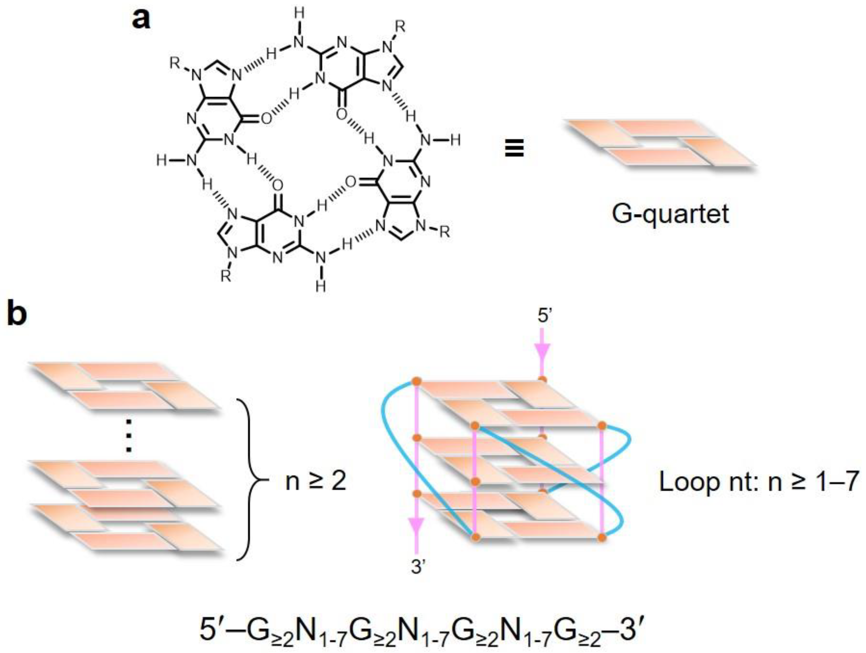
2. Topological Analysis of G-Quadruplex

The formation of G-quadruplex is influenced by several factors, including the presence of binding cations, temperature, strand stoichiometry, orientation, and loop size [16,17]. The general trend of DNA-G4 stabilization by monovalent cations is K+ >Na+ > Li+; these cations are located in the center of the G-quartet [18]. Similar to DNA-G4, the central building block of RNA-G4 is the G-quartet. The stability of RNA-G4 depends on the presence of monovalent cations, mainly K+, e.g., in the telomeric repeat-containing RNA and neuroblastoma RAS viral oncogene homolog RNA [19].
Numerous studies have addressed the effect of loop length and sequence on the DNA-G4 structure; adaptation of the glycosidic bond conformation (syn or anti); the number of molecules of nucleic acid involved in the structure formation, e.g., intramolecular/unimolecular (Figure 2a), bimolecular (Figure 2b), or tetramolecular structures (Figure 2c); and the relative orientation of the strands resulting in parallel (Figure 2d), antiparallel (Figure 2e), and higher-order G-quartets (Figure 2f) [20,21,22]. RNA-G4 is thermodynamically more stable and less polymorphic than DNA-G4 [23]. RNA-G4 topology is predominantly parallel and independent of the loop length, as determined by using RNA oligonucleotide libraries representing individual loop lengths of 1 to 5 nucleotides and total loop lengths of 3 to 15 nucleotides [24]. Telomere DNA-G4 and RNA-G4 structures are shown in Figure 2g. While DNA-G4 may form an equilibrium towards the Watson–Crick double-stranded conformation and is bound to histones in the nucleus, RNA-G4 may form an equilibrium towards various secondary structures (hairpins, loops, bulges, and pseudoknots) with many RNA-binding proteins. RNA may be more prone to form G-quadruplex structures in vivo than DNA, because it lacks a complementary strand. As described above, the basis of the physical topology of G-quadruplex structures has been determined by in vitro analysis. However, it should be noted that it is not yet clear how the in vitro topology translates into in vivo physiology and pathology.

3. Genome-Wide Analysis of G-Quadruplexes

The distribution of G-quadruplexes in the human genome is an important question that may be addressed by a genome-wide study, on the basis of (1) in silico prediction, (2) polymerase stalling analysis, and (3) antibody-mediated pull-down approach.
Many algorithms have been developed for the computational prediction of G-quadruplexes. These include Quad-Parser [25], QGRS Mapper [26], G4P Calculator [27], QuadBase [28], cGcC score [29], and G4Hunter [30]. The cGcC score may be used to predict RNA-G4 [29]. G4Hunter has been designed for DNA analysis but was also shown to be applicable for RNA analysis [30]. These algorithms may be used to predict the number of putative G-quadruplex sequences (PQS) randomly formed within a genome. The in-silico analysis has revealed that the PQS are enriched at promoters, CpG islands, 5′-untranslated regions (5′-UTRs), first exons, first exon/intron junctions, and nuclease-hypersensitive sites [25,27,30,31]. Computational predictions using Quad-Parser indicate that among 38,915 5′-UTRs of all the known Homo sapiens genes annotated in the National Center for Biotechnology Information database, 2922 contained one or more G-quadruplex motif [25,32]. Using the same computational prediction method, it was shown that the G-quadruplex motifs were highly correlated with the position of replication origins, 5′-UTRs, nucleosome-free regions, and CpG islands in human cells, including HeLa cells, primary fibroblasts, human embryonic stem cells, and human induced pluripotent stem cells [31]. As a limitation, these tools predict the potential formation of G-quadruplex directly from the primary DNA sequence, i.e., “G3 + N1–7G3 + N1–7G3 + N1–7G3 (where N = A, C, G, or T)”, with four stretches of at least three guanines separated by short stretches of other bases. Hence, the consensus sequence cannot be used to predict all DNA-G4 structures in a genome, e.g., motifs with non-guanine bulges (with a non-guanine base interrupting a three-guanine track sequence) [33].
Recently, a tool for the identification of potential RNA-G4s, named G4RNA screener, was reported, which was not limited by a canonical G-quadruplex motif definition [34]. G4RNA screener combined the previously established cGcC score [28], the newly described G4Hunter [29], and G4NN into a single tool. G4NN, one of the tools included in the G4RNA screener, is a novel machine learning approach trained on sequences that were investigated experimentally in previous studies. G4NN provides a score based on abstract sequence similarity, computed by a simple artificial neural network that was trained on the sequences available in the G4RNA database [35]. G4NN learns from available examples and considers both irregular and canonical G-quadruplexes. The artificial neural network takes the trinucleotide composition of a window as its input, which translates to an abstract representation of the sequence. By providing the composition of all trinucleotides, G4NN avoids bias by the classifier in considering specific trinucleotides as more important than others.
As yet another genome-wide approach for the identification of G-quadruplex sites, polymerase stop assay was combined with next-generation sequencing [36]. The sequencing was performed in the presence or absence of G-quadruplex stabilizers, pyridostatin or K+. Consequently, 716,310 G-quadruplex-forming sequences stabilized by pyridostatin and 525,890 G-quadruplex-forming sequences stabilized by K+ were identified in primary human B lymphocytes [36]. G-quadruplex formation was also significantly associated with cancer-related genes, such as BRCA1, BRCA2, and MAP3K8; and oncogenes and tumor suppressors, such as CUL7, FOXA1, TUSC2, and HOXB13 [34]. In another study involving the G-quadruplex stabilizer L1H1-7OTD, 9651 G-quadruplex clusters were identified by high-throughput sequencing of human genomic DNA [37]. However, these methods did not capture G-quadruplex structure formation within endogenous chromatin.
To capture G-quadruplex structure formation within endogenous chromatin, G-quadruplex chromatin immunoprecipitation-sequencing (G4 ChIP-seq) with a G-quadruplex–recognizing antibody BG4 may be used [38], e.g., to analyze the human epidermal keratinocyte HaCaT cell line [39]. In HaCaT chromatin, 10,560 G4 ChIP-seq peaks were identified with high confidence, including in cancer-related genes, such as MYC, TP53, JUN, HOXA9, FOXA1, and RAC1. Importantly, the G4 ChIP-seq peak analysis identified nucleosome-depleted regions in euchromatin, and a positive and dynamic relationship between G-quadruplex structure and transcriptional activity was noted, independent of the degree of chromatin accessibility [39]. Based on the data from genome-wide approaches for G-quadruplex identification, the number of peaks detected by G4 ChIP-seq is lower (approximately 10,000) [39] than that detected by PQS determined in silico (approximately 300,000) [25] and that identified by the polymerase stop assay (approximately 700,000) [36].
Two types of high-throughput transcriptome-wide analysis of RNA-G4 have been reported in studies analyzing in vitro transcripts extracted from cells: A reverse transcriptase (RT) stalling sequencing approach, termed rG4-sequencing [40], and RT stop profiling [41]. The two methods are conceptually similar (the differences in the experimental workflows have been summarized in a recent review paper [42]). Both methods were used to identify thousands of RNA-G4 structures in vitro. The rG4-sequencing approach revealed a cluster of G-quadruplex sequences that were conserved among eukaryotes [40]. The RT stop profiling was used to identify 4935 overlapping and 7852 non-overlapping RNA-G4 regions in two human cell lines, HEK293T and HeLa cells [41]. While RNA-G4 readily assembles in vitro, the structure may be unfolded in vivo. In vivo RNA-G4 genome-wide analysis involving a chemical probing sequencing approach, the dimethyl sulfate sequencing, followed by RT stop profiling in the presence of K+, was reported [41]. The approach was used to analyze mouse, human, and yeast cells, suggesting that most RNA-G4 regions were unfolded [41]. However, since this method was used to analyze the whole-cell ensemble RNA structural conformations within the reaction time frame, it may not have reflected the structural conformation of individual RNAs, the dynamic structural interconversions, molecule subpopulations, and heterogeneity [42]. To confirm the folding of RNA-G4 in vivo, it will be necessary to analyze further using different methods.

4. DNA-G4 and Neurological Diseases

Dysregulation of DNA-G4 is associated with human disorders, including neurological dysfunction, accelerated ageing, and increased risk of cancer development [43]. Although the role of DNA-G4 in neurophysiology has not yet been fully elucidated, the involvement of DNA-G4 in some neurological diseases has been demonstrated. As an example of the role in a human genetic disease, G-quadruplex structures of GGGGCC (G4C2) repeats form 800 to >4000 large hexanucleotide repeat expansions (HRE) in the C9orf72 gene implicated in the pathogenesis of C9orf72 amyotrophic lateral sclerosis and frontotemporal dementia (C9ALS/FTD) [44,45]. After the HREs are transcribed, the resulting RNA forms nuclear foci that can be translated in all reading frames into dipeptide repeat proteins via a repeat associated non-AUG (RAN) translation [46,47]. Both repeat-derived RNA foci and RAN translation products have been proposed to drive the pathogenesis [48,49,50]. Further, G4C2 RNA can fold to form a highly stable G-quadruplex conformation [51], which may play important roles in RNA foci and/or RAN translation toxicity. Interestingly, G4C2 repeat-derived RNA drives phase separation in G-quadruplex conformation and assembles into RNA granules containing RNA-binding proteins (RBPs) in vitro and in U2OS cells, indicating that RNA-mediated perturbations in granule dynamics may underline cellular toxicity, possibly contributing to C9ALS/FTD pathogenesis [52].
Some classes of DNA-G4–binding helicases are involved in the unfolding of G-quadruplex. Mutations in many of these helicases causes human diseases associated with genomic instability, and transcriptional and epigenetic regulation. These include RecQ-like helicases (BLM and WRN), iron–sulfur (Fe–S) helicases (RTEL1, DDX11, FANCJ, and XPD), (Asp-Glu-Ala-His) DEAH-box helicases (DHX36 and DHX9), and switch/sucrose nonfermentable (SWI/SNF)-like helicase ATRX [53,54]. Among the helicases mentioned, ATRX is involved in brain function in humans. Males inheriting germline mutations in ATRX develop a rare, congenital neurodevelopmental condition associated with impaired intellectual ability, the X-linked alpha thalassemia intellectual disability (ATR-X) syndrome [55,56].
Considering germline mutations of ATRX, ATR-X syndrome is characterized by various clinical manifestations, including severe intellectual disability, facial dysmorphism, genital abnormalities, and epileptic seizures [57]. ATR-X syndrome is a very rare intellectual disability syndrome, with an incidence of approximately 1/100,000 [57]. The causative gene, ATRX, encodes the SWI/SNF-like helicase protein ATRX, which contains two signature motifs: (1) The ATRX–DNMT3–DNMT3L domain, which binds to histone H3 tails and the modified histone H3K9me3 [58,59,60] and (2) helicase subdomains that confer ATPase activity [61,62]. In a cell, ATRX is localized to pericentric heterochromatin via the interaction between the ATRX–DNMT3–DNMT3L domain and histone H3K9me3 [59,60]. As determined by genome-wide analysis, ATRX is detected at a wide range of tandem repeats throughout the genome, including rDNA repeats, telomeric repeats, pericentric repeats, minisatellites, and endogenous retroviral sequences [63,64]. Interestingly, ATRX is also enriched at G-rich variable-number tandem repeats, including DNA-G4, as determined by ChIP-seq analysis of primary human erythroid cells and mouse embryonic stem cells [64]. Recombinant ATRX protein binds to Oxytricha telomeric repeat DNA [64]. Furthermore, ATRX functions as a high-affinity RBP that directly interacts with Xist A-repeat RNA [65] and telomeric repeat-containing RNAs [66] in vitro. However, the exact domain of ATRX that binds G-quadruplex has not yet been identified.
ATRX function in the brain remains unclear. To address that issue, some researchers have analyzed brain samples of genetically modified Atrx mice. Some imprinted genes are up-regulated in the forebrain of an Atrx conditional knockout mouse [67,68], suggesting that ATRX silences the active allele by an unknown mechanism. In an AtrxΔE2 mouse engineered to lack the Atrx exon 2 [69,70], the imprinted gene Xlr3b was significantly overexpressed. ATRX and DNA methyltransferases (DNMTs) are enriched at G-quadruplex in the Xlr3b CpG island, and ATRX binds to parallel G-quadruplex in the Xlr3b CpG island, where it regulates the Xlr3b expression. Conversely, reduced ATRX levels at certain sites are accompanied by reduced DNMT levels and substantial DNA demethylation, suggesting that ATRX regulates epigenetics, DNA methylation, by DNMT recruitment to G-quadruplexes [71]. Interestingly, Xlr3b protein is a component of RNA granules, which inhibit mRNA transport into the neuronal dendrite. In addition, neuron-specific Xlr3b transgenic mouse exhibits memory deficits, indicating that aberrant neuronal Xlr3b expression partially affects dendritic mRNA transport and learning behavior in the AtrxΔE2 mouse [71]. In addition to the role of ATRX in these epigenetic modifications, especially ones of the imprinted genes in the brain, several studies on the additional roles of the protein have been reported, e.g., telomere maintenance, DNA replication, and heterochromatin silencing in mammals [72]. Further studies are required to reveal the role of ATRX interaction with regions bearing the G-quadruplex motifs in the brain.

5. RNA-G4 and Neurological Diseases

It has been suggested that RNA-G4 plays a critical role in the translocation and translation of neuronal mRNAs in the axon, dendrite, and dendritic spine. Neuronal mRNAs are transported to the axon and dendrite, and are locally translated at the synapse. The mRNA transport is related to synaptic plasticity, and underlies learning and memory, with deregulation of this process greatly impacting neural function [73,74,75,76]. Dendritic mRNA forms a complex with RBPs and some motor proteins along the microtubules, in the form of RNA granules, transported to the dendrite. Furthermore, local protein translation of specific mRNAs takes place at the synapse. QGRS mapper, a bioinformatics tool for G-quadruplex analysis [25], revealed that approximately 30% of the best-characterized dendritic RNAs harbor the G-quadruplex motif in the 3′-UTR [77]. Representative dendritic mRNAs, i.e., those of the postsynaptic localization protein PSD-95 and calcium calmodulin-dependent protein kinase IIα (CaMKIIα), harbor a G-quadruplex structure in the 3′-UTR, and structural RNA-G4 mutations of these mRNAs inhibit delivery to the dendrite [77]. Interestingly, in a mouse harboring CaMKIIα with a deficient 3′-UTR, CaMKIIα mRNA was not transferred to the dendrite and the animal exhibited memory-learning disorder [78].
Transport of mRNA in the axon and dendrite is controlled by RBPs that act in trans on RNA-G4. It has been speculated that mutations in RBPs with G-quadruplex–binding ability cause neurological diseases, with a high incidence, including amyotrophic lateral sclerosis (ALS), the fragile X syndrome (FXS), and fragile X-associated tremor/ataxia syndrome. Bioinformatics analysis of G-quadruplex–binding proteins from the G4IPDB database, which contains over 200 molecules from various organisms, has been conducted [79]. Comparison of the amino acid composition of all (77) described G-quadruplex–binding proteins of H. sapiens revealed that they share a 20-amino acid motif of glycine and arginine residues (RGRGR GRGGG SGGSG GRGRG). Interestingly, the motif is similar to the arginine-glycine-rich (RGG) box found in the causative RBPs of neurological disorders, such as TDP-43, FUS, and FMRP [80,81]. Below, we have focused on the well-studied protein FMRP encoded by the fragile X mental retardation gene 1 (FMR1), the causative gene of FXS and fragile X-associated tremor/ataxia syndrome [82].
Individuals with FXS exhibit an intellectual disability of variable severity and display a wide array of behavioral alterations, such as hyperactivity, attention deficit, anxiety, and epilepsy. The incidence of FXS is 1 in 5000 males [83]. Most cases of FXS are caused by a large expansion of CGG repeats in the 5′-UTR of FMR1, and subsequent hyper-methylation of the FMR1 gene promoter, leading to the loss of FMRP expression [84]. In the neuron, FMRP binds to synaptic polyribosomes and is an RNA granule component [85]. FMRP is a sequence-specific mRNA-binding protein that represses translation of a subset of dendritically localized mRNAs, corresponding to approximately 4% of all mRNAs in the brain [86]. FMRP binds predominantly in the coding regions of mRNA, as determined by high-throughput sequencing of RNAs isolated by crosslinking immunoprecipitation, and stalls polyribosomes on mRNAs encoding presynaptic and postsynaptic proteins implicated in psychiatric disorders [87].
Interestingly, FMRP recognizes RNA-G4 in target mRNA [88,89]. In fact, 432 mRNAs were co-immunoprecipitated with FMRP from the mouse brain, nearly 70% of which contained a G-quadruplex sequence [90]. FMRP contains four RNA-binding motifs: Three K-homology domains and one RGG box [86,91,92,93]. The RGG box is positioned in the major groove of the G-rich RNA sequence, binding it with nanomolar affinity in vitro [94]. In addition, X-ray crystallography analysis revealed that the RGG peptide stabilized the G-tetrads and facilitated G-quadruplex formation [95].
The ability of FMRP to bind G-quadruplex is probably linked to FXS, leading to alterations in protein translation and RNA localization. The absence of FMRP changes the production of targeting proteins that are essential for neural development and whose encoding mRNAs harbor G-quadruplex structures. Biochemical assays confirmed that FMRP functioned as a repressor of translation of their own mRNA by binding to Fmr1 G-quadruplex in exon 13 [89,96]. The G-quadruplex–binding ability of FMRP also represses the translation of some mRNAs, e.g., MAP1B, APP, and PP2Ac [97,98,99,100]. FMRP binding to MAP1B mRNA represses translation during synaptogenesis in neonatal brain development. Elevated MAP1B protein levels lead to abnormally increased microtubule stability in the brain of an Fmr1 knockout mouse [97]. FMRP interaction with a G-quadruplex structure located in the 5′-UTR of Map1B favors ribosomal stalling [98]. In addition, FMRP binds to a G-rich element in APP mRNA, and represses its translation [99]. Furthermore, FMRP controls the levels of PP2A by modulating the expression of its catalytic subunit, PP2Ac. FMRP appears to be a negative regulator of PP2Ac mRNA translation, by binding with high affinity to the 5′-UTR of the PP2Ac mRNA, which harbors four G-quadruplex structures. In the absence of FMRP, increased expression of PP2Ac alters actin remodeling [100]. These observations suggested the role of FMRP as a translational repressor when it binds and stabilizes a G-quadruplex structure located in the 5′-UTR of an mRNA, by preventing ribosome scanning. Ribosome profiling of mouse adult neural stem cells revealed that FMRP mainly acts as a translational repressor [101]. It has been speculated that the reduction of gene expression correlates with the stability of the corresponding RNA-G4 [102,103], indicating that RNA-G4 in the 5′-UTR of mRNAs may be a negative modulator of gene expression at the translation stage. Further, it has been recently demonstrated, using a ribosome profiling strategy, that the folding of RNA-G4 structures hinders the translation of protein-coding sequences in HeLa cells by stimulating the translation of repressive upstream open reading frames (uORFs) [104]. Indeed, depletion of the DEAH-box helicases DHX36 and DHX9, which unwind RNA-G4s, shift translation towards RNA-G4s containing uORFs, thus disturbing the translation of selected transcripts for proto-oncogenes, transcription factors, and epigenetic regulators [104].
On the other hand, some mRNA targets of FMRP, such as Semaphorin 3F (Sem3F) and arginine vasopressin receptor V1a mRNAs, harbor a G-quadruplex structure in the 3′-UTRs [88,105]. In Fmr1 knockout cells, which lack FMRP, the association of Sem3F and V1a receptor mRNAs with polyribosomes is reduced [88], suggesting that in this case, FMRP could act as a translational activator or could impact the stability of these mRNAs.

6. G-Quadruplex Is a Therapeutic Target for Neurological Diseases

Small molecules binding to G-quadruplex have been identified [106,107], and these molecules have therapeutic potential. G-quadruplex–binding small molecules, such as porphyrin, function as transcriptional repressors [106,107,108]. Porphyrins, PPIX and hemin, are potential candidate drugs for treating the ATR-X syndrome [71]. Importantly, PPIX and hemin are produced from 5-aminolevulinic acid (5-ALA) in vivo. Intracellular generation of porphyrins following the administration of 5-ALA significantly rescued neuronal phenotypes seen in AtrxΔE2 mice [71]. Although potential off-target effects remain to be investigated, a novel therapeutic strategy targeting DNA-G4 in patients with the ATR-X syndrome is conceivable. Further, 5-ALA has been clinically approved as a fluorescent reagent for glioblastoma resection in Europe, Canada, and Japan [109]. Consequently, data on 5-ALA safety and pharmacology in humans are available.
In C9ALS/FTD, molecules targeting G4C2-repeat RNA have therapeutic potential by preventing pathogenic interactions of the expanded RNA with RBPs, and/or by interfering with RAN translation. Several research groups have reported that small-molecule compounds or antisense oligonucleotides targeting the G4C2-repeat RNA are effective [110,111,112]. DNA and RNA C9orf72 HREs exist in equilibrium between two folded states, as a hairpin and G-quadruplex structure [51,110]. The small molecule 1a binds the hairpin structure of G4C2-repeat RNA and inhibits C9ALS/FTD-associated defects, RAN translation, and foci formation in induced neurons [110]. Antisense oligonucleotides targeting C9ORF72 mRNA improve ALS-associated defects in some mouse models [111,112]. Collectively, these observations serve as a proof of principle for the development of drugs that target RNA-G4 in C9ALS/FTD.

7. Concluding Remarks and Future Perspectives

Genome-wide approaches for detecting DNA-G4 and RNA-G4 revealed the various regulatory functions of G-quadruplex structures, but the dynamic control of the folded and unfolded states data, especially in RNA-G4 analysis, require careful interpretation. For DNA-G4, some questions remain: (1) Is DNA-G4 formation different at different cellular developmental stages? (2) How and does DNA-G4 formation link to epigenetic markers? (3) How and does DNA-G4 formation regulate G4-binding proteins? In addition, it is necessary to elucidate whether DNA-G4 is involved in transcriptional modifications, telomere maintenance, and epigenetic modifications in the neuron (Figure 3a). Understanding the G-quadruplex structure and function is required for the advancement of drug development. G-quadruplex stabilizers can block the interactions between pathogenic RNA and RBPs, block abnormal phase separations, and inhibit RAN translation (Figure 3b). The effect of G-quadruplex stabilizers should also be evaluated by studying the metabolism of dendritic (Figure 3c) and axonal (Figure 3d) mRNAs, including their stability, translation, and subcellular localization. If treatments with such stabilizers could restore the normal RNA metabolism, they could represent a new avenue for the therapeutic approach to neurology in the future. Alternatively, pharmacological approaches can be used to facilitate the understanding of some roles of G-quadruplex structures in the neuron. The current knowledge represents only the tip of an iceberg, with many exciting discoveries still waiting in the “G-quadruplex World”.

Author Contributions

Conceptualization, N.S.; writing—original draft preparation, N.S. and S.A.; critical advice, M.T., S.I., Y.I. and H.K.

Funding

This research was funded by the Ministry of Education, Culture, Sports, Science and Technology (MEXT), grant number 19H05221 (Grant-in Scientific Research on Innovation Areas “Constructive understanding of multi-scale dynamism of neuropsychiatric disorders”), to Norifumi Shioda.

Conflicts of Interest

The authors declare no conflicts of interest. The sponsors had no role in the design, execution, interpretation, or writing up of the study.

Abbreviations

DNA-G4DNA G-quadruplex
RNA-G4RNA G-quadruplex
PQSputative G-quadruplex sequences
UTRuntranslated region
ChIP-seqchromatin immunoprecipitation-sequencing
RTreverse transcriptase
HREhexanucleotide repeat expansion
RBPRNA-binding protein
C9ALS/FTDC9orf72 amyotrophic lateral sclerosis and frontotemporal dementia
RANrepeat associated non-AUG
ATR-XX-linked alpha thalassemia intellectual disability
DNMTDNA methyltransferase
CaMKIIαcalcium calmodulin-dependent protein kinase IIα
FXSfragile X syndrome
5-ALA5-aminolevulinic acid

References

  1. Watson, J.D.; Crick, F.H. Molecular structure of nucleic acids; A structure for deoxyribose nucleic acid. Nature 1953, 171, 737–738. [Google Scholar] [CrossRef] [PubMed]
  2. Wang, A.H.; Quigley, G.J.; Kolpak, F.J.; Crawford, J.L.; van Boom, J.H.; van der Marel, G.; Rich, A. Molecular structure of a left-handed double helical DNA fragment at atomic resolution. Nature 1979, 282, 680–686. [Google Scholar] [CrossRef] [PubMed]
  3. Gehring, K.; Leroy, J.L.; Gueron, M. A tetrameric DNA-structure with protonated cytosine–cytosine base-pairs. Nature 1993, 363, 561–565. [Google Scholar] [CrossRef] [PubMed]
  4. Sen, D.; Gilbert, W. Formation of parallel 4-stranded complexes by guanine-rich motifs in DNA and its implications for meiosis. Nature 1988, 334, 364–366. [Google Scholar] [CrossRef] [PubMed]
  5. Zhao, J.; Bacolla, A.; Wang, G.; Vasquez, K.M. Non-B DNA structure-induced genetic instability and evolution. Cell. Mol. Life Sci. 2010, 67, 43–62. [Google Scholar] [CrossRef] [PubMed]
  6. Bang, I. Untersuchungen über die Guanylsäure. Biochem. Z. 1910, 26, 293–311. [Google Scholar]
  7. Gellert, M.; Lipsett, M.N.; Davies, D.R. Helix formation by guanylic acid. Proc. Natl. Acad. Sci. USA 1962, 48, 2013–2018. [Google Scholar] [CrossRef] [PubMed]
  8. Sundquist, W.I.; Klug, A. Telomeric DNA dimerizes by formation of guanine tetrads between hairpin loops. Nature 1989, 342, 825–829. [Google Scholar] [CrossRef]
  9. Williamson, J.R.; Raghuraman, M.K.; Cech, T.R. Monovalent cation-induced structure of telomeric DNA: The G-Quartet model. Cell 1989, 59, 871–880. [Google Scholar] [CrossRef]
  10. Bochman, M.L.; Paeschke, K.; Zakian, V.A. DNA secondary structures: Stability and function of G-quadruplex structures. Nat. Rev. Genet. 2012, 13, 770–780. [Google Scholar] [CrossRef] [PubMed]
  11. Song, J.; Perreault, J.P.; Topisirovic, I.; Richard, S. RNA G-quadruplexes and their potential regulatory roles in translation. Translation 2016, 4, e1244031. [Google Scholar] [CrossRef] [PubMed] [Green Version]
  12. Hänsel-Hertsch, R.; Di Antonio, M.; Balasubramanian, S. DNA G-quadruplexes in the human genome: Detection, functions and therapeutic potential. Nat. Rev. Mol. Cell Biol. 2017, 18, 279–284. [Google Scholar] [CrossRef] [PubMed]
  13. Fay, M.M.; Lyons, S.M.; Ivanov, P. RNA G-quadruplexes in biology: Principles and molecular mechanisms. J. Mol. Biol. 2017, 429, 2127–2147. [Google Scholar] [CrossRef] [PubMed]
  14. Kwok, C.K.; Merrick, C.J. G-Quadruplexes: Prediction, characterization, and biological application. Trends Biotechnol. 2017, 35, 997–1013. [Google Scholar] [CrossRef] [PubMed]
  15. Mukherjee, A.K.; Sharma, S.; Chowdhury, S. Non-duplex G-quadruplex structures emerge as mediators of epigenetic modifications. Trends Genet. 2019, 35, 129–144. [Google Scholar] [CrossRef] [PubMed]
  16. Deng, H.; Braunlin, W.H. Duplex to quadruplex equilibrium of the self-complementary oligonucleotide d(GGGGCCCC). Biopolymers 1995, 35, 677–681. [Google Scholar] [CrossRef]
  17. Kumar, N.; Sahoo, B.; Varun, K.A.; Maiti, S.; Maiti, S. Effect of loop length variation on quadruplex-Watson Crick duplex competition. Nucleic Acids Res. 2008, 36, 4433–4442. [Google Scholar] [CrossRef] [PubMed] [Green Version]
  18. Bhattacharyya, D.; Mirihana-Arachchilage, G.; Basu, S. Metal Cations in G-Quadruplex Folding and Stability. Front Chem. 2016, 4, 38. [Google Scholar] [CrossRef] [PubMed] [Green Version]
  19. Guiset Miserachs, H.; Donghi, D.; Börner, R.; Johannsen, S.; Sigel, R.K. Distinct differences in metal ion specificity of RNA and DNA G-quadruplexes. J. Biol. Inorg. Chem. 2016, 21, 975–986. [Google Scholar] [CrossRef] [PubMed]
  20. Keniry, M.A. Quadruplex structures in nucleic acids. Biopolymers 2000, 56, 123–146. [Google Scholar] [CrossRef]
  21. Yaku, H.; Fujimoto, T.; Murashima, T.; Miyoshi, D.; Sugimoto, N. Phthalocyanines: A new class of G-quadruplex-ligands with many potential applications. Chem. Commun. 2012, 48, 6203–6216. [Google Scholar] [CrossRef] [PubMed]
  22. Patel, D.J.; Phan, A.T.; Kuryavyi, V. Human telomere, oncogenic promoter and 5′-UTR G-quadruplexes: diverse higher order DNA and RNA targets for cancer therapeutics. Nucleic Acids Res. 2007, 35, 7429–7455. [Google Scholar] [CrossRef]
  23. Joachimi, A.; Benz, A.; Hartig, J.S. A comparison of DNA and RNA quadruplex structures and stabilities. Bioorg. Med. Chem. 2009, 17, 6811–6815. [Google Scholar] [CrossRef] [PubMed] [Green Version]
  24. Zhang, A.Y.; Bugaut, A.; Balasubramanian, S. A sequence-independent analysis of the loop length dependence of intramolecular RNA G-quadruplex stability and topology. Biochemistry 2011, 50, 7251–7258. [Google Scholar] [CrossRef] [PubMed]
  25. Huppert, J.L.; Balasubramanian, S. Prevalence of quadruplexes in the human genome. Nucleic Acids Res. 2005, 33, 2908–2916. [Google Scholar] [CrossRef] [PubMed] [Green Version]
  26. Kikin, O.; D’Antonio, L.; Bagga, P.S. QGRS Mapper: A web-based server for predicting G-quadruplexes in nucleotide sequences. Nucleic Acids Res. 2006, 34, W676–W682. [Google Scholar] [CrossRef]
  27. Eddy, J.; Maizels, N. Gene function correlates with potential for G4 DNA formation in the human genome. Nucleic Acids Res. 2006, 34, 3887–3896. [Google Scholar] [CrossRef] [Green Version]
  28. Yadav, V.K.; Abraham, J.K.; Mani, P.; Kulshrestha, R.; Chowdhury, S. QuadBase: Genome-wide database of G4 DNA--occurrence and conservation in human, chimpanzee, mouse and rat promoters and 146 microbes. Nucleic Acids Res. 2008, 36, D381–D385. [Google Scholar] [CrossRef]
  29. Beaudoin, J.D.; Jodoin, R.; Perreault, J.P. New scoring system to identify RNA G-quadruplex folding. Nucleic Acids Res. 2014, 42, 1209–1223. [Google Scholar] [CrossRef]
  30. Bedrat, A.; Lacroix, L.; Mergny, J.L. Re-evaluation of G-quadruplex propensity with G4Hunter. Nucleic Acids Res. 2016, 44, 1746–1759. [Google Scholar] [CrossRef]
  31. Besnard, E.; Babled, A.; Lapasset, L.; Milhavet, O.; Parrinello, H.; Dantec, C.; Marin, J.M.; Lemaitre, J.M. Unraveling cell type-specific and reprogrammable human replication origin signatures associated with G-quadruplex consensus motifs. Nat. Struct. Mol. Biol. 2012, 19, 837–844. [Google Scholar] [CrossRef] [PubMed]
  32. Kumari, S.; Bugaut, A.; Huppert, J.L.; Balasubramanian, S. An RNA G-quadruplex in the 5′ UTR of the NRAS proto-oncogene modulates translation. Nat. Chem. Biol. 2007, 3, 218–221. [Google Scholar] [CrossRef] [PubMed]
  33. Mukundan, V.T.; Phan, A.T. Bulges in G-quadruplexes: Broadening the definition of G-quadruplex-forming sequences. J. Am. Chem. Soc. 2013, 135, 5017–5028. [Google Scholar] [CrossRef] [PubMed]
  34. Garant, J.M.; Perreault, J.P.; Scott, M.S. Motif independent identification of potential RNA G-quadruplexes by G4RNA screener. Bioinformatics 2017, 33, 3532–3537. [Google Scholar] [CrossRef] [PubMed] [Green Version]
  35. Garant, J.M.; Luce, M.J.; Scott, M.S.; Perreault, J.P. G4RNA: An RNA G-quadruplex database. Database 2015. [Google Scholar] [CrossRef] [PubMed]
  36. Chambers, V.S.; Marsico, G.; Boutell, J.M.; Di Antonio, M.; Smith, G.P.; Balasubramanian, S. High-throughput sequencing of DNA G-quadruplex structures in the human genome. Nat. Biotechnol. 2015, 8, 877–881. [Google Scholar] [CrossRef]
  37. Yoshida, W.; Saikyo, H.; Nakabayashi, K.; Yoshioka, H.; Bay, D.H.; Iida, K.; Kawai, T.; Hata, K.; Ikebukuro, K.; Nagasawa, K.; et al. Identification of G-quadruplex clusters by high-throughput sequencing of whole-genome amplified products with a G-quadruplex ligand. Sci. Rep. 2018, 8, 3116. [Google Scholar] [CrossRef] [PubMed]
  38. Biffi, G.; Tannahill, D.; McCafferty, J.; Balasubramanian, S. Quantitative visualization of DNA G-quadruplex structures in human cells. Nat. Chem. 2013, 5, 182–186. [Google Scholar] [CrossRef]
  39. Hänsel-Hertsch, R.; Beraldi, D.; Lensing, S.V.; Marsico, G.; Zyner, K.; Parry, A.; Di Antonio, M.; Pike, J.; Kimura, H.; Narita, M.; et al. G-quadruplex structures mark human regulatory chromatin. Nat. Genet. 2016, 10, 1267–1272. [Google Scholar] [CrossRef]
  40. Kwok, C.K.; Marsico, G.; Sahakyan, A.B.; Chambers, V.S.; Balasubramanian, S. rG4-seq reveals widespread formation of G-quadruplex structures in the human transcriptome. Nat. Methods 2016, 10, 841–844. [Google Scholar] [CrossRef]
  41. Guo, J.U.; Bartel, D.P. RNA G-quadruplexes are globally unfolded in eukaryotic cells and depleted in bacteria. Science 2016, 353, aaf5371. [Google Scholar] [CrossRef] [PubMed]
  42. Kwok, C.K.; Marsico, G.; Balasubramanian, S. Detecting RNA G-uadruplexes (rG4s) in the Transcriptome. Cold Spring Harb. Perspect. Biol. 2018, 10, a032284. [Google Scholar] [CrossRef] [PubMed]
  43. Maizels, N. G4-associated human diseases. EMBO Rep. 2015, 16, 910–922. [Google Scholar] [CrossRef] [PubMed]
  44. DeJesus-Hernandez, M.; Mackenzie, I.R.; Boeve, B.F.; Boxer, A.L.; Baker, M.; Rutherford, N.J.; Nicholson, A.M.; Finch, N.A.; Flynn, H.; Adamson, J.; et al. Expanded GGGGCC hexanucleotide repeat in noncoding region of C9ORF72 causes chromosome 9p-linked FTD and ALS. Neuron 2011, 72, 245–256. [Google Scholar] [CrossRef] [PubMed]
  45. Renton, A.E.; Majounie, E.; Waite, A.; Simón-Sánchez, J.; Rollinson, S.; Gibbs, J.R.; Schymick, J.C.; Laaksovirta, H.; van Swieten, J.C.; Myllykangas, L.; et al. A hexanucleotide repeat expansion in C9ORF72 is the cause of chromosome 9p21-linked ALS-FTD. Neuron 2011, 72, 257–268. [Google Scholar] [CrossRef] [PubMed]
  46. Ash, P.E.A.; Bieniek, K.F.; Gendron, T.F.; Caulfield, T.; Lin, W.L.; DeJesus-Hernandez, M.; van Blitterswijk, M.M.; Jansen-West, K.; Paul, J.W., 3rd; Rademakers, R.; et al. Unconventional translation of C9ORF72 GGGGCC expansion generates insoluble polypeptides specific to c9FTD/ALS 77. Neuron 2013, 77, 639–646. [Google Scholar] [CrossRef] [PubMed]
  47. Zu, T.; Liu, Y.; Bañez-Coronel, M.; Reid, T.; Pletnikova, O.; Lewis, J.; Miller, T.M.; Harms, M.B.; Falchook, A.E.; Subramony, S.H.; et al. RAN proteins and RNA foci from antisense transcripts in C9ORF72 ALS and frontotemporal dementia. Proc. Natl. Acad. Sci. USA 2013, 110, E4968–E4977. [Google Scholar] [CrossRef] [PubMed]
  48. Haeusler, A.R.; Donnelly, C.J.; Periz, G.; Simko, E.A.J.; Shaw, P.G.; Kim, M.S.; Maragakis, N.J.; Troncoso, J.C.; Pandey, A.; Sattler, R.; et al. C9orf72 nucleotide repeat structures initiate molecular cascades of disease. Nature 2014, 507, 195–200. [Google Scholar] [CrossRef] [PubMed]
  49. Wen, X.; Tan, W.; Westergard, T.; Krishnamurthy, K.; Markandaiah, S.S.; Shi, Y.; Lin, S.; Shneider, N.A.; Monaghan, J.; Pandey, U.B.; et al. Antisense proline-arginine RAN dipeptides linked to C9ORF72-ALS/FTD form toxic nuclear aggregates that initiate in vitro and in vivo neuronal death. Neuron 2014, 84, 1213–1225. [Google Scholar] [CrossRef]
  50. Zhang, K.; Donnelly, C.J.; Haeusler, A.R.; Grima, J.C.; Machamer, J.B.; Steinwald, P.; Daley, E.L.; Miller, S.J.; Cunningham, K.M.; Vidensky, S.; et al. The C9orf72 repeat expansion disrupts nucleocytoplasmic transport. Nature 2015, 525, 56–61. [Google Scholar] [CrossRef] [Green Version]
  51. Fratta, P.; Mizielinska, S.; Nicoll, A.J.; Zloh, M.; Fisher, E.M.C.; Parkinson, G.; Isaacs, A.M. C9orf72 hexanucleotide repeat associated with amyotrophic lateral sclerosis and frontotemporal dementia forms RNA G-quadruplexes. Sci. Rep. 2012, 2, 1016. [Google Scholar] [CrossRef] [PubMed]
  52. Fay, M.M.; Anderson, P.J.; Ivanov, P. ALS/FTD-associated C9ORF72 repeat RNA promotes phase transitions in vitro and in cells. Cell Rep. 2017, 21, 3573–3584. [Google Scholar] [CrossRef] [PubMed]
  53. Singleton, M.R.; Dillingham, M.S.; Wigley, D.B. Structure and mechanism of helicases and nucleic acid translocases. Annu. Rev. Biochem. 2007, 76, 23–50. [Google Scholar] [CrossRef]
  54. Valton, A.L.; Prioleau, M.N. G-quadruplexes in DNA replication: A problem or a necessity? Trends Genet. 2016, 32, 697–706. [Google Scholar] [CrossRef] [PubMed]
  55. Gibbons, R.J.; Suthers, G.K.; Wilkie, A.O.; Buckle, V.J.; Higgs, D.R. X-linked alpha-thalassemia/mental retardation (ATR-X) syndrome: Localization to Xq12-q21.31 by X inactivation and linkage analysis. Am. J. Hum. Genet. 1992, 51, 1136–1149. [Google Scholar] [PubMed]
  56. Gibbons, R.J.; Picketts, D.J.; Villard, L.; Higgs, D.R. Mutations in a putative global transcriptional regulator cause X-linked mental retardation with alpha-thalassemia (ATR-X syndrome). Cell 1995, 80, 837–845. [Google Scholar] [CrossRef]
  57. Gibbons, R.J.; Wada, T.; Fisher, C.A.; Malik, N.; Mitson, M.J.; Steensma, D.P.; Fryer, A.; Goudie, D.R.; Krantz, I.D.; Traeger-Synodinos, J. Mutations in the chromatin-associated protein ATRX. Hum. Mutat. 2008, 29, 796–802. [Google Scholar] [CrossRef] [PubMed]
  58. Argentaro, A.; Yang, J.C.; Chapman, L.; Kowalczyk, M.S.; Gibbons, R.J.; Higgs, D.R.; Neuhaus, D.; Rhodes, D. Structural consequences of disease-causing mutations in the ATRX-DNMT3-DNMT3L (ADD) domain of the chromatin-associated protein ATRX. Proc. Natl. Acad. Sci. USA 2007, 104, 11939–11944. [Google Scholar] [CrossRef] [Green Version]
  59. Dhayalan, A.; Tamas, R.; Bock, I.; Tattermusch, A.; Dimitrova, E.; Kudithipudi, S.; Ragozin, S.; Jeltsch, A. The ATRX–ADD domain binds to H3 tail peptides and reads the combined methylation state of K4 and K9. Hum. Mol. Genet. 2011, 20, 2195–2203. [Google Scholar] [CrossRef]
  60. Iwase, S.; Xiang, B.; Ghosh, S.; Ren, T.; Lewis, P.W.; Cochrane, J.C.; Allis, C.D.; Picketts, D.J.; Patel, D.J.; Li, H.; et al. ATRX ADD domain links an atypical histone methylation recognition mechanism to human mental-retardation syndrome. Nat. Struct. Mol. Biol. 2011, 18, 769–776. [Google Scholar] [CrossRef] [PubMed]
  61. Picketts, D.J.; Higgs, D.R.; Bachoo, S.; Blake, D.J.; Quarrell, O.W.; Gibbons, R.J. ATRX encodes a novel member of the SNF2 family of proteins: Mutations point a common mechanism underlying the ATR-X syndrome. Hum. Mol. Genet. 1996, 5, 1899–1907. [Google Scholar] [CrossRef] [PubMed]
  62. Mitson, M.; Kelley, L.A.; Sternberg, M.J.; Higgs, D.R.; Gibbons, R.J. Functional significance of mutations in the Snf2 domain of ATRX. Hum. Mol. Genet. 2011, 20, 2603–2610. [Google Scholar] [CrossRef] [PubMed] [Green Version]
  63. Gibbons, R.J.; McDowell, T.L.; Raman, S.; O’Rourke, D.M.; Garrick, D.; Ayyub, H.; Higgs, D.R. Mutations in ATRX, encoding a SWI/SNF-like protein, cause diverse changes in the pattern of DNA methylation. Nat. Genet. 2000, 24, 368–371. [Google Scholar] [CrossRef] [PubMed] [Green Version]
  64. Law, M.J.; Lower, K.M.; Voon, H.P.; Hughes, J.R.; Garrick, D.; Viprakasit, V.; Mitson, M.; De Gobbi, M.; Marra, M.; Morris, A.; et al. ATR-X syndrome protein targets tandem repeats and influences allele-specific expression in a size-dependent manner. Cell 2010, 143, 367–378. [Google Scholar] [CrossRef] [PubMed]
  65. Sarma, K.; Cifuentes-Rojas, C.; Ergun, A.; Del Rosario, A.; Jeon, Y.; White, F.; Sadreyev, R.; Lee, J.T. ATRX directs binding of PRC2 to Xist RNA and Polycomb targets. Cell 2014, 159, 869–883. [Google Scholar] [CrossRef] [PubMed]
  66. Chu, H.P.; Cifuentes-Rojas, C.; Kesner, B.; Aeby, E.; Lee, H.G.; Wei, C.; Oh, H.J.; Boukhali, M.; Haas, W.; Lee, J.T. TERRA RNA antagonizes ATRX and protects telomeres. Cell 2017, 170, 86–101. [Google Scholar] [CrossRef]
  67. Kernohan, K.D.; Jiang, Y.; Tremblay, D.C.; Bonvissuto, A.C.; Eubanks, J.H.; Mann, M.R.; Bérubé, N.G. ATRX partners with cohesin and MeCP2 and contributes to developmental silencing of imprinted genes in the brain. Dev. Cell 2010, 18, 191–202. [Google Scholar] [CrossRef]
  68. Kernohan, K.D.; Vernimmen, D.; Gloor, G.B.; Bérubé, N.G. Analysis of neonatal brain lacking ATRX or MeCP2 reveals changes in nucleosome density, CTCF binding and chromatin looping. Nucleic Acids Res. 2014, 42, 8356–8368. [Google Scholar] [CrossRef]
  69. Shioda, N.; Beppu, H.; Fukuda, T.; Li, E.; Kitajima, I.; Fukunaga, K. Aberrant calcium/calmodulin-dependent protein kinase II (CaMKII) activity is associated with abnormal dendritic spine morphology in the ATRX mutant mouse brain. J. Neurosci. 2011, 31, 346–358. [Google Scholar] [CrossRef]
  70. Nogami, T.; Beppu, H.; Tokoro, T.; Moriguchi, S.; Shioda, N.; Fukunaga, K.; Ohtsuka, T.; Ishii, Y.; Sasahara, M.; Shimada, Y.; et al. Reduced expression of the ATRX gene, a chromatin-remodeling factor, causes hippocampal dysfunction in mice. Hippocampus 2011, 21, 678–687. [Google Scholar] [CrossRef]
  71. Shioda, N.; Yabuki, Y.; Yamaguchi, K.; Onozato, M.; Li, Y.; Kurosawa, K.; Tanabe, H.; Okamoto, N.; Era, T.; Sugiyama, H.; et al. Targeting G-quadruplex DNA as cognitive function therapy for ATR-X syndrome. Nat. Med. 2018, 24, 802–813. [Google Scholar] [CrossRef] [PubMed]
  72. Clynes, D.; Higgs, D.R.; Gibbons, R.J. The chromatin remodeller ATRX: A repeat offender in human disease. Trends Biochem. Sci. 2013, 38, 461–466. [Google Scholar] [CrossRef] [PubMed]
  73. Tiedge, H. RNA reigns in neurons. Neurons 2005, 48, 1–6. [Google Scholar] [CrossRef]
  74. Sutton, M.A.; Schuman, E.M. Dendritic protein synthesis, synaptic plasticity, and memory. Cell 2006, 127, 49–58. [Google Scholar] [CrossRef] [PubMed]
  75. Bramham, C.R.; Wells, D.G. Dendritic mRNA: Transport, translation and function. Nat. Rev. Neurosci. 2007, 8, 776–789. [Google Scholar] [CrossRef] [PubMed]
  76. Martin, K.C.; Ephrussi, A. mRNA localization: Gene expression in the spatial dimension. Cell 2009, 136, 719–730. [Google Scholar] [CrossRef]
  77. Subramanian, M.; Rage, F.; Tabet, R.; Flatter, E.; Mandel, J.L.; Moine, H. G–quadruplex RNA structure as a signal for neurite mRNA targeting. EMBO Rep. 2011, 12, 697–704. [Google Scholar] [CrossRef]
  78. Miller, S.; Yasuda, M.; Coats, J.K.; Jones, Y.; Martone, M.E.; Mayford, M. Disruption of dendritic translation of CaMKIIα impairs stabilization of synaptic plasticity and memory consolidation. Neuron 2002, 36, 507–519. [Google Scholar] [CrossRef]
  79. Mishra, S.K.; Tawani, A.; Mishra, A.; Kumar, A. G4IPDB: A database for G-quadruplex structure forming nucleic acid interacting proteins. Sci. Rep. 2016, 6, 38144. [Google Scholar] [CrossRef] [Green Version]
  80. Ule, J. Ribonucleoprotein complexes in neurological diseases. Curr. Opin. Neurobiol. 2008, 18, 516–523. [Google Scholar] [CrossRef]
  81. Gerstberger, S.; Hafner, M.; Tuschl, T. A census of human RNA-binding proteins. Nat. Rev. Genet. 2014, 15, 829–845. [Google Scholar] [CrossRef] [PubMed]
  82. Verkerk, A.J.; Pieretti, M.; Sutcliffe, J.S.; Fu, Y.H.; Kuhl, D.P.; Pizzuti, A.; Reiner, O.; Richards, S.; Victoria, M.F.; Zhang, F.P. Identification of a gene (FMR-1) containing a CGG repeat coincident with a breakpoint cluster region exhibiting length variation in fragile X syndrome. Cell 1991, 65, 905–914. [Google Scholar] [CrossRef]
  83. Coffee, B.; Keith, K.; Albizua, I.; Malone, T.; Mowrey, J.; Sherman, S.L.; Warren, S.T. Incidence of fragile X syndrome by newborn screening for methylated FMR1 DNA. Am. J. Hum. Genet. 2009, 85, 503–514. [Google Scholar] [CrossRef] [PubMed]
  84. Pieretti, M.; Zhang, F.P.; Fu, Y.H.; Warren, S.T.; Oostra, B.A.; Caskey, C.T.; Nelson, D.L. Absence of expression of the FMR-1 gene in fragile X syndrome. Cell 1991, 66, 817–822. [Google Scholar] [CrossRef]
  85. Bassell, G.J. Fragile balance: RNA editing tunes the synapse. Nat. Neurosci. 2011, 14, 1492–1494. [Google Scholar] [CrossRef] [PubMed]
  86. Ashley, C.T.; Wilkinson, K.D.; Reines, D.; Warren, S.T. FMR1 protein: Conserved RNP family domains and selective RNA binding. Science 1993, 262, 563–566. [Google Scholar] [CrossRef] [PubMed]
  87. Darnell, J.C.; Van Driesche, S.J.; Zhang, C.; Hung, K.Y.; Mele, A.; Fraser, C.E.; Stone, E.F.; Chen, C.; Fak, J.J.; Chi, S.W.; et al. FMRP stalls ribosomal translocation on mRNAs linked to synaptic function and autism. Cell 2011, 146, 247–261. [Google Scholar] [CrossRef] [PubMed]
  88. Darnell, J.C.; Jensen, K.B.; Jin, P.; Brown, V.; Warren, S.T.; Darnell, R.B. Fragile X mental retardation protein targets G quartet mRNAs important for neuronal function. Cell 2001, 107, 489–499. [Google Scholar] [CrossRef]
  89. Schaeffer, C.; Bardoni, B.; Mandel, J.L.; Ehresmann, B.; Ehresmann, C.; Moine, H. The fragile X mental retardation protein binds specifically to its mRNA via a purine quartet motif. EMBO J. 2001, 20, 4803–4813. [Google Scholar] [CrossRef]
  90. Brown, V.; Jin, P.; Ceman, S.; Darnell, J.C.; O’Donnell, W.T.; Tenenbaum, S.A.; Jin, X.; Feng, Y.; Wilkinson, K.D.; Keene, J.D.; et al. Microarray identification of FMRP-associated brain mRNAs and altered mRNA translational profiles in fragile X syndrome. Cell 2001, 107, 477–487. [Google Scholar] [CrossRef]
  91. Siomi, H.; Siomi, M.C.; Nussbaum, R.L.; Dreyfuss, G. The protein product of the fragile X gene, FMR1, has characteristics of an RNA-binding protein. Cell 1993, 74, 291–298. [Google Scholar] [CrossRef]
  92. Siomi, M.C.; Zhang, Y.; Siomi, H.; Dreyfuss, G. Specific sequences in the fragile X syndrome protein FMR1 and the FXR proteins mediate their binding to 60S ribosomal subunits and the interactions among them. Mol. Cell. Biol. 1996, 16, 3825–3832. [Google Scholar] [CrossRef] [PubMed] [Green Version]
  93. Myrick, L.K.; Hashimoto, H.; Cheng, X.; Warren, S.T. Human FMRP contains an integral tandem Agenet (Tudor) and KH motif in the amino terminal domain. Hum. Mol. Genet. 2015, 24, 1733–1740. [Google Scholar] [CrossRef] [PubMed]
  94. Phan, A.T.; Kuryavyi, V.; Darnell, J.C.; Serganov, A.; Majumdar, A.; Ilin, S.; Raslin, T.; Polonskaia, A.; Chen, C.; Clain, D.; et al. Structure-function studies of FMRP RGG peptide recognition of an RNA duplex-quadruplex junction. Nat. Struct. Mol. Biol. 2011, 18, 796–804. [Google Scholar] [CrossRef] [PubMed]
  95. Vasilyev, N.; Polonskaia, A.; Darnell, J.C.; Darnell, R.B.; Patel, D.J.; Serganov, A. Crystal structure reveals specific recognition of a G-quadruplex RNA by a β-turn in the RGG motif of FMRP. Proc. Natl. Acad. Sci. USA 2015, 112, E5391–E5400. [Google Scholar] [CrossRef] [PubMed]
  96. Laggerbauer, B.; Ostareck, D.; Keidel, E.M.; Ostareck-Lederer, A.; Fischer, U. Evidence that fragile X mental retardation protein is a negative regulator of translation. Hum. Mol. Genet. 2001, 10, 329–338. [Google Scholar] [CrossRef] [PubMed]
  97. Lu, R.; Wang, H.; Liang, Z.; Ku, L.; O’Donnell, W.T.; Li, W.; Warren, S.T.; Feng, Y. The fragile X protein controls microtubule-associated protein 1B translation and microtubule stability in brain neuron development. Proc. Natl. Acad. Sci. USA 2004, 101, 15201–15206. [Google Scholar] [CrossRef] [Green Version]
  98. Menon, L.; Mader, S.A.; Mihailescu, M.R. Fragile X mental retardation protein interactions with the microtubule associated protein 1B RNA. RNA 2008, 14, 1644–1655. [Google Scholar] [CrossRef] [PubMed] [Green Version]
  99. Westmark, C.J.; Malter, J.S. FMRP mediates mGluR5-dependent translation of amyloid precursor protein. PLoS Biol. 2007, 5, e52. [Google Scholar] [CrossRef]
  100. Castets, M.; Schaeffer, C.; Bechara, E.; Schenck, A.; Khandjian, E.W.; Luche, S.; Moine, H.; Rabilloud, T.; Mandel, J.L.; Bardoni, B. FMRP interferes with the Rac1 pathway and controls actin cytoskeleton dynamics in murine fibroblasts. Hum. Mol. Genet. 2005, 14, 835–844. [Google Scholar] [CrossRef] [Green Version]
  101. Liu, B.; Li, Y.; Stackpole, E.E.; Novak, A.; Gao, Y.; Zhao, Y.; Zhao, X.; Richter, J.D. Regulatory discrimination of mRNAs by FMRP controls mouse adult neural stem cell differentiation. Proc. Natl. Acad. Sci. USA 2018, 115, E11397–E11405. [Google Scholar] [CrossRef] [PubMed]
  102. Wieland, M.; Hartig, J.S. RNA quadruplex-based modulation of gene expression. Chem. Biol. 2007, 14, 757–763. [Google Scholar] [CrossRef] [PubMed]
  103. Halder, K.; Wieland, M.; Hartig, J.S. Predictable suppression of gene expression by 5′-UTR-based RNA quadruplexes. Nucleic Acids Res. 2009, 37, 6811–6817. [Google Scholar] [CrossRef] [PubMed]
  104. Murat, P.; Marsico, G.; Herdy, B.; Ghanbarian, A.T.; Portella, G.; Balasubramanian, S. RNA G-quadruplexes at upstream open reading frames cause DHX36- and DHX9-dependent translation of human mRNAs. Genome Biol. 2018, 19, 229. [Google Scholar] [CrossRef] [PubMed]
  105. Menon, L.; Mihailescu, M.R. Interactions of the G quartet forming semaphorin 3F RNA with the RGG box domain of the fragile X protein family. Nucleic Acids Res. 2007, 35, 5379–5392. [Google Scholar] [CrossRef] [Green Version]
  106. Asamitsu, S.; Obata, S.; Yu, Z.; Bando, T.; Sugiyama, H. Recent progress of targeted G-quadruplex-preferred ligands toward cancer therapy. Molecules 2019, 24, 429. [Google Scholar] [CrossRef] [PubMed]
  107. Asamitsu, S.; Bando, T.; Sugiyama, H. Ligand design to acquire specificity to intended G-quadruplex structures. Chemistry 2019, 25, 417–430. [Google Scholar] [CrossRef] [PubMed]
  108. Balasubramanian, S.; Hurley, L.H.; Neidle, S. Targeting G-quadruplexes in gene promoters: A novel anticancer strategy? Nat. Rev. Drug Discov. 2011, 10, 261–275. [Google Scholar] [CrossRef]
  109. Roberts, D.W.; Valdés, P.A.; Harris, B.T.; Hartov, A.; Fan, X.; Ji, S.; Leblond, F.; Tosteson, T.D.; Wilson, B.C.; Paulsen, K.D. Glioblastoma multiforme treatment with clinical trials for surgical resection (aminolevulinic acid). Neurosurg. Clin. N. Am. 2012, 23, 371–377. [Google Scholar] [CrossRef]
  110. Su, Z.; Zhang, Y.; Gendron, T.F.; Bauer, P.O.; Chew, J.; Yang, W.Y.; Fostvedt, E.; Jansen-West, K.; Belzil, V.V.; Desaro, P.; et al. Discovery of a biomarker and lead small molecules to target r(GGGGCC)-associated defects in c9FTD/ALS. Neuron 2014, 83, 1043–1050. [Google Scholar] [CrossRef]
  111. Jiang, J.; Zhu, Q.; Gendron, T.F.; Saberi, S.; McAlonis-Downes, M.; Seelman, A.; Stauffer, J.E.; Jafar-Nejad, P.; Drenner, K.; Schulte, D.; et al. Gain of toxicity from ALS/FTD-linked repeat expansions in C9ORF72 is alleviated by antisense oligonucleotides targeting GGGGCC-containing RNAs. Neuron 2016, 90, 535–550. [Google Scholar] [CrossRef] [PubMed]
  112. Gendron, T.F.; Chew, J.; Stankowski, J.N.; Hayes, L.R.; Zhang, Y.J.; Prudencio, M.; Carlomagno, Y.; Daughrity, L.M.; Jansen-West, K.; Perkerson, E.A.; et al. Poly (GP) proteins are a useful pharmacodynamic marker for C9ORF72-associated amyotrophic lateral sclerosis. Sci. Transl. Med. 2017, 9. [Google Scholar] [CrossRef] [PubMed]
Figure 1. Characteristics of G-quadruplexes. (a) Chemical structure and schematic illustration of the G-quartet. (b) Schematic illustration of G-quadruplex structures. The G-quadruplex has at least two or three G-quartets, and three loops of varied length (1–7 nucleotides).
Ijms 20 02884 g001
Figure 2. The topological variety of G-quadruplex structures. Schematic illustration of intramolecular/unimolecular (a), bimolecular (b), tetramolecular (c), parallel (d), antiparallel (e), and higher-order (f) G-quadruplexes. (g) Schematic illustration of telomere DNA-G4 (left) and RNA-G4 (right) in a potassium-containing solution. DNA-G4 is arranged in three G-quartet layers composed of 2′-deoxyguanosines and three TTA loops, while RNA-G4 is arranged in three G-quartet layers composed of guanosines and three UUA loops. The 2′-hydroxyl group is highlighted in red for clarity.
Ijms 20 02884 g002
Figure 3. Multiple roles and pharmacological targets of G-quadruplex in the neuron. (a) In the nucleus, DNA-G4 may be involved in transcriptional modifications, telomere maintenance, and epigenetic modifications, together with DNA-G4 binding proteins (BPs). (b) In the cytoplasm, RNA-G4 is involved in the metabolism of RNA granules, including interactions with RNA-G4 BPs and phase separations. (c,d) RNA-G4 is also involved in the transport, stability, translation, and subcellular localization of dendritic (c) and axonal (d) mRNAs. A detailed understanding of these processes will inform a new therapeutic strategy for neurological diseases.
Ijms 20 02884 g003

