Article

Role of CpxR in Biofilm Development: Expression of Key Fimbrial, O-Antigen and Virulence Operons of Salmonella Enteritidis

1
Department of Food and Bioproduct Sciences, University of Saskatchewan, Saskatoon, SK S7N 5A8, Canada
2
University of Minnesota Informatics Institute, University of Minnesota, Minneapolis, MN 55455, USA
3
Department of Veterinary and Biomedical Sciences, University of Minnesota, Saint Paul, MN 55108, USA
*
Author to whom correspondence should be addressed.
Int. J. Mol. Sci. 2019, 20(20), 5146; https://doi.org/10.3390/ijms20205146
Submission received: 5 September 2019 / Revised: 6 October 2019 / Accepted: 15 October 2019 / Published: 17 October 2019
(This article belongs to the Special Issue Biofilms and Bacterial Virulence)

Abstract

:
Salmonella Enteritidis is a non-typhoidal serovar of great public health significance worldwide. The RpoE sigma factor and CpxRA two-component system are the major regulators of the extracytoplasmic stress response. In this study, we found that the CpxR has highly significant, but opposite effects on the auto-aggregation and swarming motility of S. Enteritidis. Auto-aggregation was negatively affected in the ∆cpxR mutant, whereas the same mutant significantly out-performed its wild-type counterpart with respect to swarming motility, indicating that the CpxR plays a role in biofilm-associated phenotypes. Indeed, biofilm-related assays showed that the CpxR is of critical importance in biofilm development under both static (microtiter plate) and dynamic (flow cell) media flow conditions. In contrast, the RpoE sigma factor showed no significant role in biofilm development under dynamic conditions. Transcriptomic analysis revealed that the cpxR mutation negatively affected the constitutive expression of the operons critical for biosynthesis of O-antigen and adherence, but positively affected the expression of virulence genes critical for Salmonella-mediated endocytosis. Conversely, CpxR induced the expression of curli csgAB and fimbrial stdAC operons only during biofilm development and flagellar motAB and fliL operons exclusively during the planktonic phase, indicating a responsive biofilm-associated loop of the CpxR regulator.

1. Introduction

Non-typhoidal Salmonella (NTS) are the leading cause of food-borne gastroenteritis worldwide [1]. Among NTS, Salmonella enterica subspecies enterica serovar Enteritidis (hereafter referred to as S. Enteritidis) is one of the most common serovars associated with human salmonellosis [1]. This zoonotic pathogen owes its great public health importance to an unusually broad host range [2]. According to the World Health Organization, Salmonella enterica serotypes Enteritidis and Typhimurium are the two most important serovars of Salmonella transmitted from animals to humans [3]. In addition to a large natural reservoir, S. Enteritidis has the ability to form robust biofilms on various surfaces, including hydrophobic (e.g., wood and plastic) and hydrophilic surfaces (e.g., glass) under different conditions (e.g., at room temperature under dynamic culture conditions, or at 35 °C under static culture conditions) [4,5], further enhancing its persistence in the environment.
To respond to a wide range of external conditions bacteria must quickly process an external stimulus and adjust their physiology/life style to new conditions or environmental niches. The RpoE sigma factor (σE) and two-component signal transduction system, CpxRA, play crucial roles in maintaining homeostasis of the bacterial envelope [6]. The RpoE regulon consists of a conserved group of functionally coherent genes involved in the synthesis, assembly, and homeostasis of lipopolysaccharides and outer membrane porins, along with a variable group of genes associated with pathogenesis [7]. On the other hand, the Cpx regulon is involved in protein translocation across the inner membrane [8] along with the biogenesis of bacterial appendages [9]. Consequently, these two extracytoplasmic stress-response regulators are involved in the most important biological processes where bacterial cells interact with their surroundings; survival [10,11] and pathogenicity [12,13]. The presence of a wide range of unfolded outer membrane proteins (i.e., any denatured protein) in the periplasmic space of Gram-negative bacteria leads to a cascade of proteolytic steps that results in degradation of the membrane-bound anti-sigma factor, RseA, and release of σE into the cytosol [14]. Once released into the cytosol, σE-directs RNA polymerase to σE-dependent promoters, thereby inducing the expression of a set of genes (i.e., the RpoE regulon) to protect the cell from the extracytoplasmic stress [7]. The CpxRA extracytoplasmic stress response system includes an inner membrane sensory histidine kinase, CpxA, and a DNA-binding response regulator, CpxR [15]. The presence of misfolded inner membrane proteins leads to autophosphorylation of the cytoplasmic domain of CpxA, which induces a phosphotransfer to the receiver domain of CpxR [16]. Once phosphorylated, CpxR binds cognate DNA, which further results in an expression of the Cpx regulon [16].
It has been shown that the RpoE and Cpx regulators share multiple linkages for inter-extracytoplasmic stress communication in the prokaryotic stress response [6]. Several findings indicate that the Cpx response acts antagonistically to RpoE; specifically, repressing the production of outer membrane β-barrel proteins [17,18] and the σE-induced chaperone Skp [19]. It has been established that the CpxR extracytoplasmic stress response regulator is important in the development of Actinobacillus pleuropneumoniae biofilms [20]. In this study, we examined individual and combined contributions of Cpx and RpoE to biofilm-associated phenotypes (i.e., auto-aggregation, swarming motility) as well as biofilm development in S. Enteritidis. In addition, we analyzed the transcriptomic responses of cpxR mutant and wild-type S. Enteritidis cells grown under planktonic and biofilm conditions, enhancing our understanding of the regulatory role of the Cpx regulon during biofilm development.

2. Results

2.1. Extracytoplasmic Stress-Response Regulators, RpoE and CpxR, Differentially Affect the Growth Rate of S. Enteritidis

To investigate the role of the extracytoplasmic regulators, RpoE sigma factor and CpxR system, on the growth rate of S. Enteritidis, we measured the growth kinetics of the isogenic ΔcpxR and ΔrpoE mutants as well as a double ΔcpxRrpoE mutant. The growth assay revealed no significant differences in growth kinetics between the wild type and ΔcpxR mutant strains (Figure 1).
However, the ΔrpoE strain showed an extended lag phase with a final optical density (OD) value approximately 20% (p < 0.2) that of the wild-type and ΔcpxR mutant strains (specific growth rate ranging from 0.6 [wild type] to 0.48 [ΔrpoE] h−1; Figure 1). A large, statistically-significant (p < 0.05) growth deficiency was observed in the case of the ΔrpoEcpxR mutant strain. Deletion of both genes, rpoE and cpxR, had a much stronger impact on the fitness of S. Enteritidis compared to that of the individual deletion mutations (Figure 1).

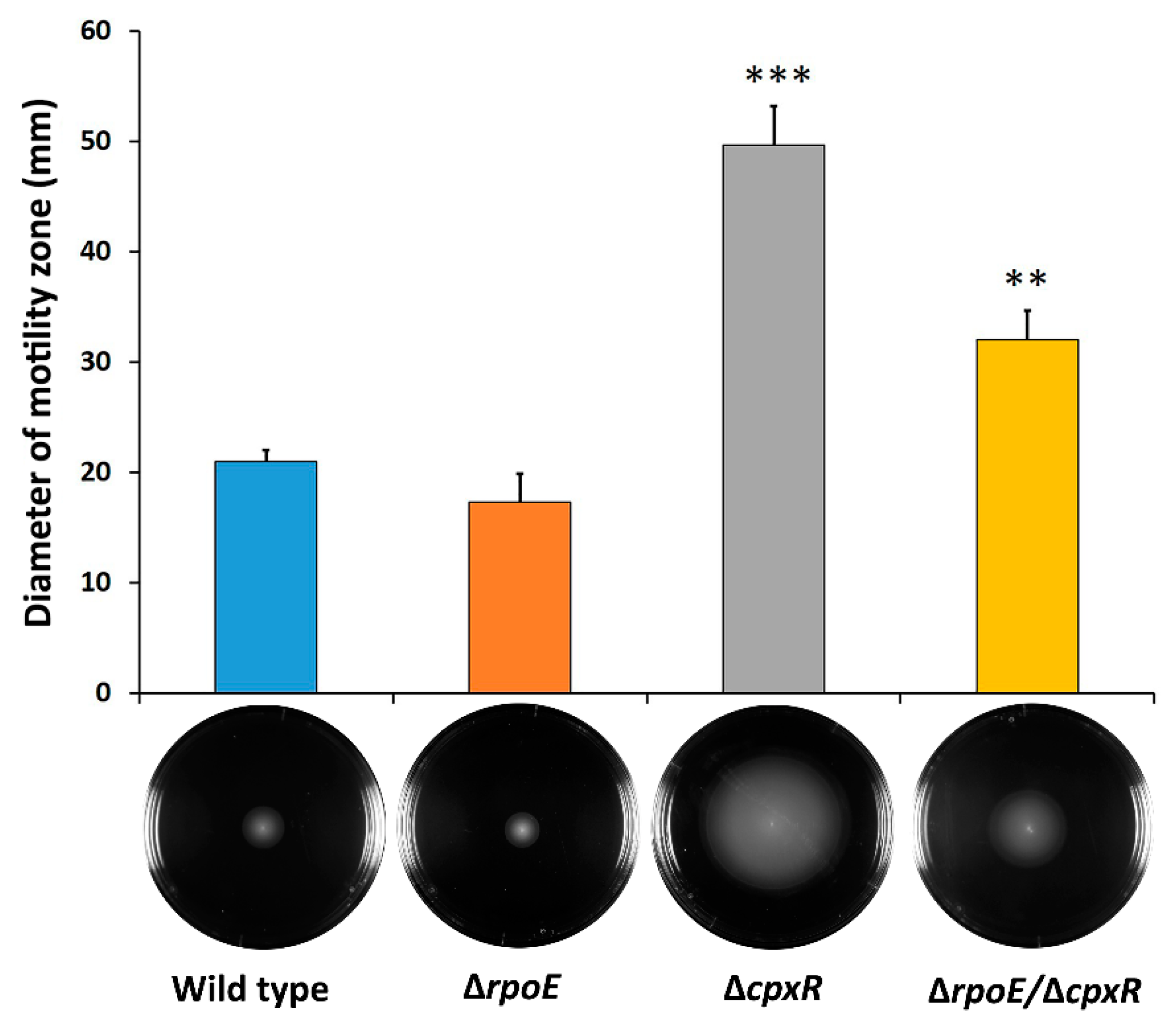
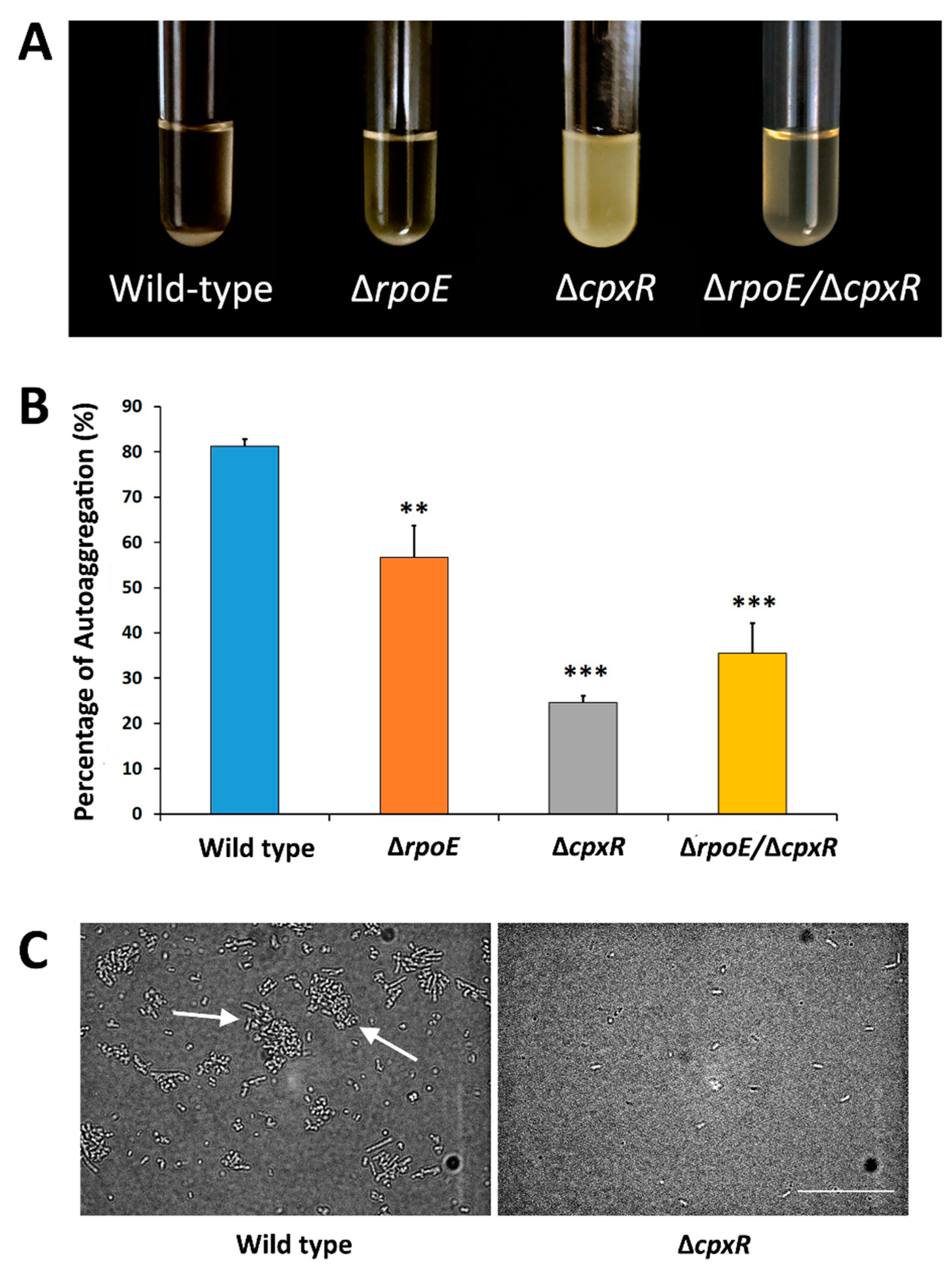
2.2. CpxR Impacts Motility and Auto-Aggregation of S. Enteritidis

As the RpoE and CpxR regulate the biogenesis of various outer membrane proteins, as well as the outer membrane itself, we hypothesized that these two regulators would play a role in swarming motility and auto-aggregation, important phenotypic characteristics related to the sessile lifestyle of various bacterial species. Swarming motility of the ΔrpoE mutant (17.5 mm in diameter) was slightly-reduced compared to that of the wild-type strain (21 mm in diameter) (Figure 2).
In contrast to the ΔrpoE strain, the ΔcpxR mutant exhibited more than two times increased swarming (50 mm in diameter) compared to the wild type, while the double ΔrpoEcpxR mutant showed a reduced motility trend (32 mm in diameter) compared to the ΔcpxR, but increased motility relative to the wild-type strain (Figure 2). Deletion of the cpxR gene resulted in a highly-significant (p < 0.001) increase in swarming motility of S. Enteritidis; whereas, the rpoE deletion did not significantly (p = 0.3) affect motility of the same organism (Figure 2).
We subsequently sought to determine the effect of single gene deletions (rpoE and cpxR), as well as the effect of the double rpoE/cpxR deletion, on the ability of S. Enteritidis cells to auto-aggregate. Among these four organisms, the wild-type exhibited the greatest ability to auto-aggregate (80%), followed by the ΔrpoE mutant (60%) (Figure 3A,B).
In contrast to the wild type, ΔcpxR mutant cells showed the weakest auto-aggregation tendencies (22%), virtually resulting in an inability of cells to form aggregates (which subsequently would sediment out) with cells consequently remaining mainly in suspension (Figure 3A,B). Interestingly, the double rpoE/cpxR deletion mutant resulted in an intermediate phenotype (i.e., a phenotype between the rpoE and cpxR phenotypes) (Figure 3A). The cpxR gene deletion caused a highly-significant (p < 0.001) reduction in auto-aggregation, while the rpoE gene deletion showed a significant (p < 0.01) but less-profound effect compared to deletion of the cpxR gene (Figure 3B). In agreement with the auto-aggregation results, direct observation using phase contrast microscopy confirmed the ability of the wild-type strain to form aggregates; whereas, the ΔcpxR mutant cells remained mainly in a state of planktonic suspension (Figure 3C). Together, these are compelling data providing initial insights into individual and combined effects of the RpoE and CpxR extracytoplasmic stress response regulators relevant to the sessile lifestyle in S. Enteritidis.

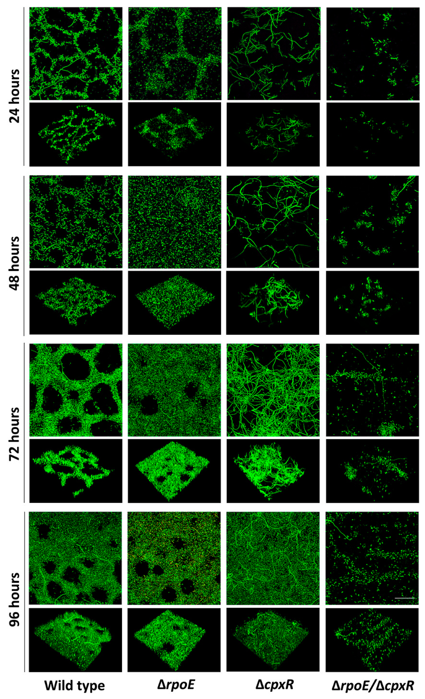
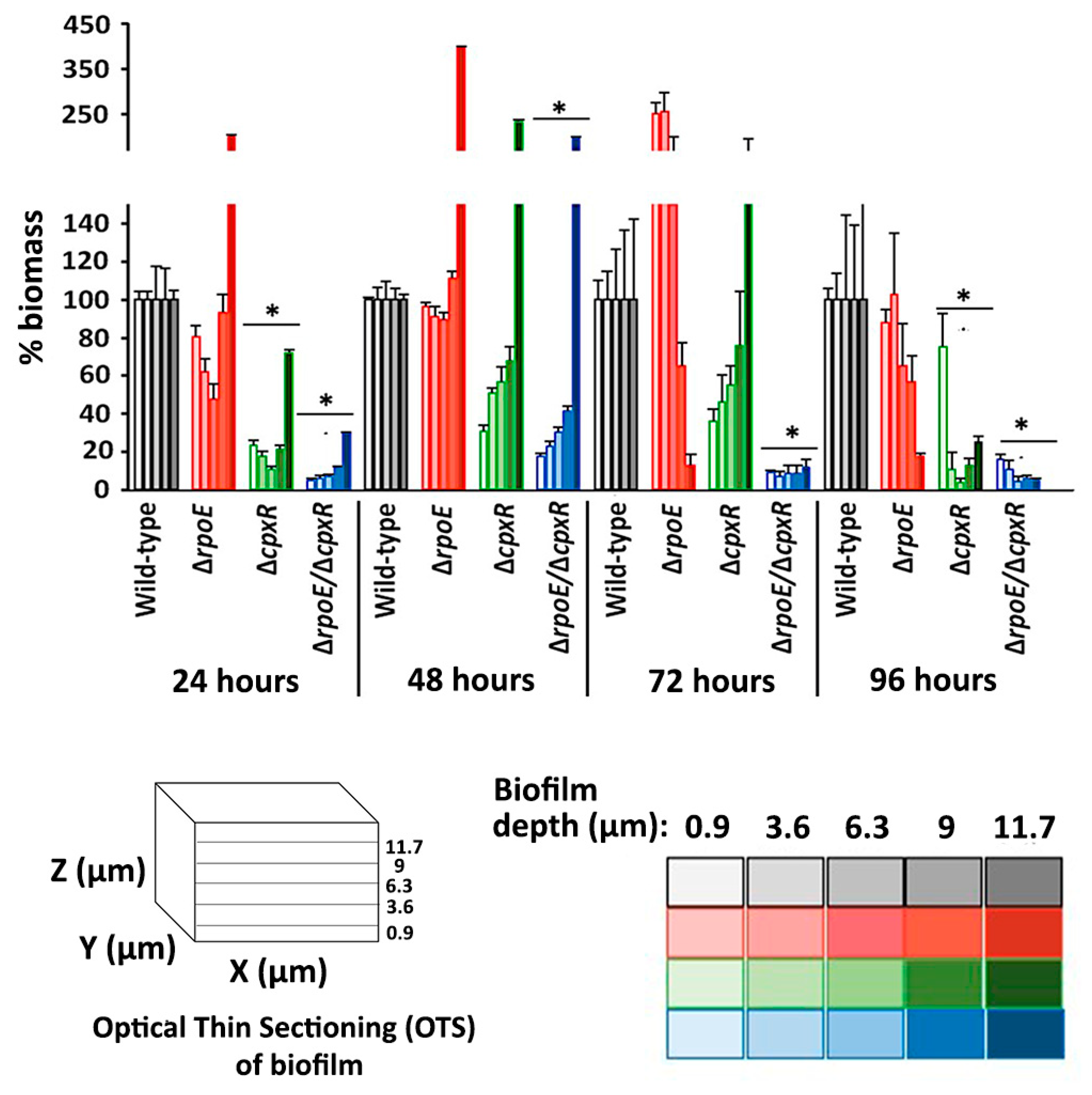
2.3. CpxR and CpxR/RpoE Have Profound Effects on Biofilm Formation

Based on the swarming motility and auto-aggregation data, we hypothesized that the cpxR and double cpxR/rpoE deletions would affect biofilm formation in S. Enteritidis. To test this hypothesis, we first carried out a microtiter plate biofilm assay, determining the abilities of all three mutants and wild type to form biofilm over a 48 h period. The ΔcpxR mutant showed the most significant (p < 0.001) reduction in crystal violet uptake (87.7%) compared to the wild type (Figure 4). The double ΔcpxR/ΔrpoE mutant responded similarly to the ΔcpxR mutant, exhibiting a highly-significant (p < 0.001) decrease in crystal violet uptake (86.2%), whereas the ΔrpoE mutant was slightly less-affected, with a 78.5% decrease in crystal violet uptake compared to the wild type (Figure 4).
To observe biofilm formation in situ over a longer period of time, and under continuous flow, fully hydrated conditions more representative to real-world settings, we carried out a series of experiments where biofilms were grown in flow cells and then analyzed using non-destructive, confocal laser scanning microscopy (CLSM) over a 96-h time course. After 24 h of incubation, cells of wild-type and the ΔrpoE mutant strains were able to attach and develop nascent biofilms, qualitatively characterized as a network of tightly packed microcolonies (Figure 5).
In contrast, the ΔcpxR mutant followed a distinct development pathway, forming biofilm consisting of individual, unusually-elongated cells (filaments) that were loosely-attached to the surface, but which didn’t form significant cohesive interactions or inter-cell packing tendencies (Figure 5). The double ΔrpoE/ΔcpxR mutant resembled the phenotype of biofilms formed by the ΔcpxR mutant strain, except that the cells were not elongated (Figure 5). Quantitative biofilm measurements showed that the mean thickness of the ΔrpoE/ΔcpxR mutant (8 μm) was significantly (p < 0.05) reduced from that of the ΔrpoE (19 μm), wild-type (18 μm) and the ΔcpxR (16 μm) strains (Figure S1). However, the biofilm biomass was significantly-reduced for both the ΔrpoE/ΔcpxR (p < 0.001) and ΔcpxR (p < 0.005) mutant strains compared to the wild-type (Figure 6).
After 48 h of incubation, the wild type and the ΔrpoE mutant formed biofilms where cell aggregates, or microcolonies, merged to become confluent; whereas, the ΔcpxR and ΔrpoE/ΔcpxR mutants retained their initial phenotypes (Figure 5). During this period of time, the thickness of the ΔcpxR mutant (35 μm) biofilm significantly (p < 0.005) increased compared to the previous day (16 μm); whereas, the thicknesses of biofilms formed by the other three strains only increased slightly (Supplementary Figure S1). However, the biofilm biomass of the ΔcpxR mutant was significantly-reduced (p < 0.005) compared to that of the wild-type and the ΔrpoE mutant strains (Figure 6), indicating an inability of the ΔcpxR mutant to form a “mature” fully-developed biofilm, as seen in the wild-type strain after 48 h of incubation. By 72 h, all four organisms maintained their previous biofilm phenotypes (Figure 5). After 96 h of incubation, the ΔcpxR mutant exhibited an additional loss of biofilm biomass resulting in a highly significant (p < 0.001) decrease in this parameter compared to the wild type (Figure 6), clearly showing a deleterious effect of the single cpxR deletion and double cpxR and rpoE deletions on the ability of S. Enteritidis to form extensive, well-developed biofilms as seen in the wild-type organism. The double ΔrpoE/ΔcpxR mutant showed significantly-less (p < 0.001) biofilm biomass compared to that of the wild type during each of the four days of the experiment (Figure 6).

2.4. Effect of cpxR Deletion on the Biofilm Transcriptome

During the biofilm formation assays, it was shown that the cpxR gene alone, or in a combination with the rpoE gene, plays a significant role in biofilm development of S. Enteritidis; whereas, the rpoE gene alone did not affect biofilm formation of this zoonotic pathogen under dynamic media flow conditions. To further investigate the role of the cpxR deletion on biofilm formation, we examined the transcriptional changes that occurred between biofilms of the isogenic ΔcpxR mutant and parental wild-type strains. Comparative analysis of the RNA-Seq data showed that 673 genes were significantly differentially expressed by at least 2-fold in all three biological replications of the ΔcpxR mutant compared to that of the wild-type. Out of 673 differently-expressed genes in the ΔcpxR genetic background, 461 genes were down-regulated (Table S1) and 212 genes were up-regulated (Table S2).
The most notable change caused by deletion of cpxR was the down-regulation of entire operons or genes encoding for cell adherence, O-antigen biosynthesis, and anabolic-associated processes (Figure S2 and Figure 7).
The ΔcpxR mutant strain manifested a significant decrease in the expression of the safABCD operon encoding fimbrial pili, pegABCD operon encoding fimbria, csgAB operon encoding curli fibers, and stfAGFE, as well as stdAC encoding fimbrial proteins (Figure 7). Another two operons, gtrABC and rfbVXES, encoding proteins involved in O-antigen biosynthesis, underwent significant down-regulation in biofilms of the ΔcpxR strain compared to wild-type strain biofilms (Figure 7). Besides genes associated with cell appendages and O-antigen biosynthesis, the ΔcpxR mutant underwent significant down-regulation of genes encoding for proteins involved in carbohydrate uptake (malEFKPQSMZG—maltose metabolic processes; frwBCD—phosphotransferase system (PTS fructose-specific transporter), peptide uptake (oppABCDF—oligopeptide ATP-binding cassette transporters (ABC transporter); dppABCDF – dipeptide ABC transporter), thiamine synthesis (thiEGHF), glycolysis (yihPR) and leucine synthesis (leuA) (Figure 7), clearly indicating that the cpxR deletion significantly affected anabolic processes during sessile growth of S. Enteritidis.
In contrast to the down-regulation of key genes associated with bacterial appendages, O-antigen biosynthesis and anabolic processes, ΔcpxR mutant biofilms caused the up-regulation of numerous virulence-associated genes, including those linked with the type III secretion system (T3SS). Comparative transcriptomics analysis revealed that the ΔcpxR biofilm-associated cells significantly up-regulated genes encoding the T3SS apparatus, specifically the needle complex (invG, prgKHIJ, orgA), an ATPase of T3SS (invC) and translocon (invABEFIJH, spaPQROS) (Figure 7) (Supplementary Table S2). In addition to the T3SS apparatus, the ΔcpxR mutant up-regulated a wide range of virulence genes, encoding effectors on both the Salmonella pathogenicity island I (SPI1) (sipABCD, sicAP, sopBEE2, hilACD, iagB, SEN4028, SEN4029) and SPI2 (ssaIJL, pipC) (Figure 7), suggesting an increased virulence potential of the ΔcpxR mutant strain associated with the biofilm phenotype. Besides the large up-regulation of virulence-associated genes, the ΔcpxR mutant significantly up-regulated genes associated with stress response (hdeB, cspBH, SEN0663, osmY, emrD, dinI, yebG, yeaQ), nitrate respiration (narJKIHG) and cytochrome biosynthesis (SEN3633, ccmABDEF, SEN3628, napBC) (Figure 7), demonstrating that ΔcpxR biofilm-associated cells modify their metabolism and adapt to stress as a consequence of cpxR gene-deletion. The results of RNA-Seq were validated by real-time polymerase chain reaction (PCR), presented in Figure S3.

2.5. Effect of cpxR Deletion on the Transcriptome of Planktonic Cells

In total, 805 genes showed significant (p < 0.05) differences in expression between the ΔcpxR mutant and wild-type S. Enteritidis strains during planktonic growth. The full list of down-regulated and up-regulated genes in the cpxR genetic background, along with their gene names, descriptions, biological replications gene expression value, mean gene expression fold-change and false discovery rate p-value, are presented in Supplementary Tables S3 and S5, respectively.
In general, the transcriptome of the ΔcpxR mutant planktonic cells resembled the transcriptome of the ΔcpxR mutant biofilm-associated cells. Comparative expression analysis showed that genes associated with adherence (safABCD, SEN4247, SEN4249, pegABCD, SEN4251, stfGF, SEN4248, SEN4250, SEN1978, bcfG, yddX, SEN2794), O-antigen synthesis (rfbEVSX, gtrABC) and anabolic processes (malEFGKMPQSTZ, lamB, frwBCD, oppABCDF, dppABCDF, thiEGHF, gltBDP) were significantly down-regulated in planktonic cells of the ΔcpxR mutant compared to the wild type (Table S3), further suggesting that down-regulation of these genes was constitutively-regulated due to cpxR deletion, but not exclusively linked to the planktonic/sessile mode of life. However, two fimbrial/curli operons, stdAC and csgAB, exhibited no difference in gene expression between the ΔcpxR mutant and wild type during planktonic growth; whereas, these two operons showed significant down-regulation in biofilm-associated ΔcpxR mutant cells relative to the wild type. This finding clearly indicates that the stdAC and csgAB operons are not constitutively-governed by the CpxR regulator, but rather induced in response to the sessile life style of S. Enteritidis.
Similarly, the set of up-regulated genes in ΔcpxR mutant planktonic cells resembled their respective biofilm transcriptome. In the ΔcpxR genetic background during planktonic growth, the genes encoding the T3SS apparatus (invG, prgHIJK, orgA), T3SS specific ATPase (invC), translocons (invABCEFIJH, spaOPQRS), SPI1 effectors (sipABCD, sicAP, sopBDEE2, hilACD, iagB) and SPI2 effectors (pipBC, ssaBDGHIJKL, srfABC, sseA, avrA) were significantly up-regulated, showing their constitutive regulation by CpxR. Unique features of the ΔcpxR planktonic transcriptome included the significant up-regulation of genes involved in flagellar biosynthesis (flgABCDEFGHIJKLMN, motAB, fliAEFGHIJKLMNOPSZ, flhBE) (Table S4). The highly-significant increase (p < 0.005) in swarming motility of planktonic ΔcpxR cells compared to planktonic wild-type cells could be explained by this unique transcriptional feature of the cpxR mutant cells. The results of RNA-Seq were validated by real-time PCR and are presented in Figure S3.

3. Discussion

Aggregation, adherence and formation of biofilms in vitro represent important features in the environmental persistence of S. Enteritidis [21,22], and consequently influence dissemination of this global pathogen [23,24]. In this study, we found that auto-aggregation was most significantly affected by the cpxR deletion. This mutation made it virtually impossible for ΔcpxR mutant cells to auto-aggregate and sediment, resulting in the cells remaining suspended in broth media during the auto-aggregation assay. Surprisingly, the same cpxR deletion mutation caused the opposite effect with respect to the swarming motility of S. Enteritidis. In this case, the isogenic ΔcpxR mutant was able to outperform its wild-type parent by more than two times. Thus, our results reveal that the cpxR deletion has the highly significant, but opposite effect, on auto-aggregation (i.e., negative) and swarming motility (i.e., positive) in S. Enteritidis. Interestingly, the ΔrpoE mutant possessed both auto-aggregation and swarming motility phenotypes similar to its wild-type parent, whereas the double ΔcpxR/ΔrpoE mutant yielded an intermediate phenotype (i.e., phenotypes between the ΔrpoE and ΔcpxR mutant) with respect to these traits.
Previously, it has been observed that biofilm formation by the same bacterial strain can be influenced by growth under static or dynamic media conditions [25]. To determine the contribution of the RpoE and CpxR, the two major extra cytoplasmic stress response regulators [6], on the formation of S. Enteritidis biofilms under static and dynamic conditions, separately we analyzed biofilms grown in microtiter plate (i.e., static condition) and flow cells (i.e., dynamic conditions). Under the static media growth condition all three mutants (ΔcpxR, ΔrpoE, and ΔcpxR/ΔrpoE) were significantly affected in their ability to form biofilms compared to that of the wild-type strain. Under flowing (dynamic) conditions, the ΔrpoE mutant showed no significant difference in biofilm formation tendencies from the wild-type parent; whereas the double mutant ΔcpxR/ΔrpoE and ΔcpxR mutant exhibited significantly reduced abilities to form biofilms. Our flow cell biofilm assay in conjunction with CLSM imaging and analysis showed that the cpxR mutant strain attached poorly to the substratum compared to both the wild-type and the ΔrpoE mutant strains. The same mutation caused elongation of cells, which subsequently resulted in formation of a thick, but porous biofilm (i.e., biofilm with low biomass). The double mutation, rpoE/cpxR, reversed the elongated cell phenotype to the normal wild-type phenotype, albeit with a low number of attached cells, one of the hallmarks of ΔcpxR biofilms [26]. Taken together, our study provides evidence that the rpoE mutation affects the biofilm formation of S. Enteritidis only under the static media condition; whereas, the cpxR mutation affects it under both, static and dynamic media conditions. These results can be explained by the fact that under the static condition metabolic wastes accumulate and impose an additional extracytoplasmic stress on the ΔrpoE mutant [27], with indirect effect on the ability of this strain to form biofilm. In contrast to the RpoE sigma factor, the CpxR regulator showed its importance in biofilm development under both static and dynamic conditions. This finding clearly indicates that the CpxR does not affect biofilm development via bacterial fitness, but rather directly, by interfering with some steps in the process of biofilm development. It is important to mention that in the all examined biofilm-associated assays (i.e., swarming motility, auto-aggregation and biofilm development under static and dynamic media conditions) the same pattern emerged. The rpoE mutant phenotypes were most closely related to the wild-type phenotypes, whereas the cpxR mutant showed the most distantly related phenotypes compared to its parental wild type strain. It is unlikely that the growth rate indirectly affected the biofilm-associated phenotypes of the rpoE and double mutant. First, the inocula for all four organisms across all assays were standardized. Second, during the swarming motility assay, the double cpxR/rpoE mutant, despite its growth deficiency, significantly outperformed its parental wild type strain, clearly indicating that this phenotype was not affected by its growth rate but rather by the absence of the rpoE and cpxR genes.
A number of studies have documented the regulatory role of the CpxR in the synthesis of various bacterial appendages, including Pap pili [28], type IV bundle-forming pili [29,30], P pili [16], Longus [31], and flagella [32], all of which may subsequently affect the biofilm formation. Indeed, several studies have shown that CpxR has a direct [20,33] or indirect [34] role in the biofilm development. Although these studies have given us valuable information on the role of the CpxR on production of various appendages and/or provided information of the involvement of certain genes under CpxR control during biofilm formation, they still haven’t demonstrated an overall regulatory function of CpxR (i.e., regulon) associated with biofilm formation. Accordingly, we first identified the CpxR regulon of S. Enteritidis by performing a global transcriptomic analysis of the wild-type planktonic cells compared with their isogenic cpxR mutant counterparts. Then, we specifically determined the CpxR biofilm-associated regulon using biofilm transcriptomes of the wild type and the cpxR mutant grown under dynamic media conditions.
Most notably, evidence generated through comparison of planktonic and biofilm transcriptomes suggested that the cpxR deletion constitutively-affected the expression of operons that are critical for biosynthesis of O-antigen, various adhesins, a large pool of anabolic-associated genes, as well as virulence and stress response genes. The CpxR regulatory circuit also showed a clear division, based on biological process ontology, negatively affecting the expression of adherence- (i.e., O-antigen and adhesins), and anabolic-associated genes; whereas, it positively impacted the expression of the core virulence (i.e., genes encoded on SPI 1 and SPI 2) and stress response genes. It is important to emphasize that the CpxR regulator had a highly-significant and extensive effect on adherence- and virulence-associated genes. For instance, a significant down-regulation (i.e., over 100-fold) of the entire peg, saf, SEN4247-SEN4251 fimbrial operons, gtr, and rfb, as well as O-antigen operons was observed for the isogenic ΔcpxR mutant. On the other hand, the ΔcpxR mutant showed an extensive up-regulation of salmonellae-essential virulence genes, involving not only genes encoding T3SS, but also genes encoding effector proteins, SopB, SopE2, SipA, SipC, all essential for the invasion of epithelial cells [35]. The extensive up-regulation of essential salmonellae virulence genes in the cpxR mutant may be related to the virulence function of some of the down-regulated fimbrial operons. It has been reported that the S. Enteritidis pegD mutant was reduced significantly in invasiveness in chicken Leghom Male Hepatoma (LMH) cells [36], while host recognition in salmonellae was completely lost without a functional saf operon [37]. We hypothesize that by significant down-regulation of fimbrial operons in the ΔcpxR background, which are not only involved in adherence, but also in host recognition and invasion, the ΔcpxR mutant up-regulates virulence genes essential for the host invasion to compensate for its lack of invasiveness and host recognition.
Besides a large pool of constitutively expressed genes, the CpxR regulator showed responsive control over the flagellar and fimbrial operons, including fli, flg, mot, flh; curli csg and fimbrial std operons. A unique transcriptional feature of the planktonic cpxR mutant cells was a significant up-regulation of the flagellar operons, involving genes that encode proteins, MotAB and FliL, which play an essential role in Salmonella swarming motility over agar surfaces [38]. Biofilm transcriptomics of the cpxR mutant cells showed that the flagellar operons were down-regulated compared to their planktonic counterparts, which is in agreement with the ‘swim-or-stick’ theory. According to this theory, motility and biofilm development are mutually-exclusive processes [39], which can explain down-regulation of the flagellar operons in the cpxR biofilm-associated cells. A possible explanation for the up-regulation of the flagellar operons in the cpxR mutant planktonic cells compared to the wild-type planktonic cells could be associated with the role of flagella in mechanosensing of surface and initial surface adherence [40]. In another words, the cpxR deletion could increase the importance of flagella during surface adherence in the ∆cpxR mutant due to inactivation of numerous, normally-functional adhesin operons.
Another unique responsive transcriptional feature, the significant down-regulation of the curli csgAB and fimbrial stdAC operons, was observed in cpxR biofilm-associated cells, suggesting that CpxR regulates expression of these two operons specifically during biofilm development. Curli is the major proteinaceous component of the extracellular matrix in S. enterica and plays a very important role in biofilm development during the attachment phase [41], while Std fimbriae play a role in the adhesion of S. enterica to specific intestinal receptors of the host [42].
In conclusion, our findings offer new insights into the individual and combined contributions of the major extracytoplasmic stress-response regulators, RpoE and CpxR, during auto-aggregation, swarming motility and biofilm development of S. Enteritidis. Although both of these regulators are involved in the extracytoplasmic stress response, our biofilm-related experiments showed the limited importance of the RpoE regulator; whereas, the CpxR protein showed a consistent and significant importance in the all biofilm-related assays. Furthermore, our transcriptomic analyses provided a unique understanding into the regulatory role of the Cpx regulator during biofilm development, indicating that this regulator controls the expression of the most critical fimbrial and O-antigen operons. In addition, this study revealed the global genetic basis underlying CpxR biofilm regulation, which may point to a valuable target(s) for the development of measures to control biofilms generated by this zoonotic pathogen.

4. Experimental Procedures

4.1. Bacterial Strains, Plasmids and Growth Conditions

Salmonella enterica subsp. enterica serovar Enteritidis strain ATCC 13076 served as the wild-type organism. Plasmid pKD3 was used as template for amplification of the Cm resistance cassette. Plasmids pKD46 and pCP20 were used during the Red Lambda procedure (see below). Growth media was supplemented with ampicillin (100 μg/mL), chloramphenicol (30 μg/mL) or arabinose 10 mM (Sigma Chemical Co., St. Louis, MO, USA) for maintenance of plasmids and selection of bacterial strains, as required. Bacterial strains were routinely grown in Luria–Bertani (LB) broth with shaking (190 ± 5 rpm) at either 37 or 28 °C, as required.

4.2. Construction of ΔcpxR, ΔrpoE and ΔcpxR/ΔrpoE Salmonella Enteritidis Mutant Strains

Construction of chromosomal deletions was performed using the Red Lambda recombination system, as previously described [43]. Briefly, a chloramphenicol resistance cassette, cat, flanked by Flp recognition sites, was amplified using the pKD3 plasmid as the DNA template. All primers used for the construction of mutants are listed in Table 1.
Amplified cat cassettes were used to transform the wild-type strain harbouring the Red recombination plasmid, pKD46. Introduction of desirable mutations was verified by PCR and DNA Sanger sequencing. To excise the cat cassette, a temperature-sensitive, Flp recombinase-expressing vector, pCP20, was introduced via electroporation. Subsequently, the pCP20 plasmid was cured by growing the mutants at elevated temperature (42 °C). All mutants were verified using PCR and Sanger sequencing and later used for functional analyses.

4.3. Growth Assay

The differences in the growth kinetics between the wild-type, ΔcpxR, ΔrpoE and ΔcpxR/ΔrpoE strains were determined by measuring optical density (OD) at 600 nm over a 10 h time course. Overnight cultures were diluted 100-fold into 100 mL of LB followed by incubation at room temperature (22 °C) with continuous shaking at 190 ± 5 rpm. The specific growth rate (µ) was calculated based on the increase in OD600 over the optical density interval 0.025 to 0.3 for the wild-type and normalized to the growth rate of the corresponding ΔcpxR, ΔrpoE and ΔcpxR/ΔrpoE mutant strains. The formula used to calculate the specific growth rate was µ = (ln [OD2-OD1])/(T2-T1) [44].

4.4. Swarming-Motility Assay

The swarming motility assay was carried out as described by Legendre et al. [45]. Soft agar (0.25% LB agar) was freshly prepared (i.e., one day prior to the experiment) and used to determine motility of the wild-type and mutant strains. Accordingly, standardized cultures of the wild-type and three mutant strains were stabbed into motility soft agar. Motility diameter was measured as distance in millimeters after incubation for 24 h at room temperature (22 °C).

4.5. Auto-Aggregation Assay

Auto-aggregation assays were performed as described by Shanks et al. [46], with minor modifications. Briefly, an aliquot (5 mL) of overnight culture was incubated for 24 h at 22 °C after which the upper 1 mL fraction of the above overnight culture was carefully removed to measure its optical density (OD600) (recorded as OD600 pre-vortex). The remaining culture in the test tube was then mixed by brief vortexing, followed by optical density measurements (recorded as OD600 post-vortex). The “percent aggregation” was calculated using the formula: 100 × (OD600 post-vortex − OD600 pre-vortex)/OD600 post-vortex. Meanwhile, light microscopy digital photographs of the wild-type and ΔcpxR strains after auto-aggregation were obtained using a Carl Zeiss Axiokop2 phase contrast microscope equipped with a Carl Zeiss AxioCam ICc1 camera (Zeiss, Jena, Germany) using a 100 times 1.4 N.A. oil immersion lens.

4.6. Microtiter-Plate Biofilm Formation Assay

The ability of the wild-type, ΔrpoE, ΔcpxR and ΔcpxR/ΔrpoE strains to form biofilms were assessed as previously described [47]. Briefly, overnight cultures of tested strains were diluted 1:100 in tryptic soy broth (TSB) medium, and then used to inoculate the wells of 96-well polyvinyl chloride microtiter plates (Costar 2797, Corning, NY, USA), followed by incubation at 37 °C for 48 h. After incubation, the medium was removed and the wells were washed with 250 μL of sterile distilled water. Subsequently, each well was stained with 250 μL of 0.5% (w/v) crystal violet for 10 min. After incubation at room temperature, the dye was removed, and the wells washed thoroughly with distilled water followed by air drying. Biofilm accumulation was quantified by solubilizing the bound crystal violet with 250 μL of 30% (v/v) acetic acid solution, and the absorbance was measured at 600 nm using an automated microtiter plate reader (Packard SpectraCount BS10000 absorbance microplate reader, Cole-Parmer Canada Company, Montreal, QC, Canada).

4.7. Flow-Cell Biofilm Formation Assay

Multi-channel flow cells were constructed using polycarbonate sheets into which channels were milled, as described previously [48]. The reactor system consisted a reservoir of sterile medium [10% (w/v) TSB] connected via silicone tubing to a bubble trap, and subsequently to the flow cell followed by the effluent reservoir. The entire reactor system was sterilized by flushing 5.25% (w/v) sodium hypochlorite solution for a period of 15 min. Sterile medium was pumped through the flow cell channels, in a once-through fashion, using a Watson–Marlow peristaltic pump (Model 202U; Watson–Marlow, Cornwall, UK). Each flow cell channel was inoculated with 0.5 mL of bacterial culture of an OD600 of 0.5 (i.e., mid-log growth phase). The inoculum was retained in the flow cell channel for 30 min. at room temperature (22 °C) to facilitate adhesion of bacterial cells to the flow cell channel surfaces. Flow was then resumed, with biofilms thereafter grown under a continuous nutrient laminar flow velocity of 0.2 cm sec−1 at room temperature for the duration of the assay. Biofilms were non-destructively analyzed using a Nikon C2 confocal scanning laser microscope (CSLM; Nikon, Mississauga, ON, Canada) at 24, 48, 72 and 96 h.

4.8. Confocal Laser Scanning Microscopy (CLSM) and Biofilm Quantification

Biofilm development was quantified using CLSM (Nikon C2, Mississauga, ON, Canada) and SYTO 9 (Molecular Probes, Life Technologies, Burlington, ON). Biofilm images, corresponding to fluorescence emission in the green (excitation/emission 488/522 nm) (SYTO 9) wavelengths, were acquired in the horizontal (xy) and vertical (xz) planes. Optical thin section (OTS) stacks were collected over the thickness of the biofilm using a 60 times Plan Apo VC (N.A. 1.4, Nikon) objective lens. Biofilm biomass estimation was carried out by performing image analysis on OTSs using a z-step increment of 0.9 µm from the attachment surface (i.e., 0.9, 3.6, 6.3, 9 and 11.7 µm) at five randomly chosen locations per biofilm. The analysis of biofilm biomass (whereby pixels were converted to µm2 of cell material) in each OTS was carried out using the Nikon NIS-Elements Confocal Microscope Imaging Software (version 4.10). The biomass percentage at each OTS depth was measured relative to the wild-type using the following formula (mutant/wild-type × 100). The reported mean total biomass was the result of three independent experiments. Biofilm thickness was measured in micrometers (μm) using a computer-controlled, motorized z-axis stepper motor [48,49]. Fifteen random fields were assessed for each biofilm with five separate thickness values obtained per field (n = 75). These values were averaged to obtain the thickness data for each biofilm.

4.9. Preparation of Planktonic and Biofilm Samples for RNA Extraction

Biomass was collected from biofilms formed by the wild-type and ΔcpxR strains after 48 h of incubation using the continuous-flow method described above (e.g., flow cell biofilm assay). Planktonic cells were grown in 10% Tryptic soy broth with shaking (190 ± 5 rpm) at 22 °C until the OD reached 0.5 at 600 nm. Approximately 108 colony-forming units of the wild-type and ΔcpxR planktonic cells were accordingly obtained and then centrifuged at 1844× g for 10 min. All bacterial samples were re-suspended in RNAprotect bacterial agent (Qiagen, Valencia, CA, USA) and stored at −20 °C until RNA extraction. Total RNA was extracted using the RNeasy mini kit (Qiagen, Valencia, CA, USA) following the manufacturer’s instructions. Sample quality was assessed using capillary electrophoresis (i.e., using an Agilent BioAnalyzer 2100, Santa Clara, CA, USA), generating an RNA Integrity Number (RIN). A RIN of 6 or greater was required to pass initial quality control for sequencing.

4.10. RNA-Seq Analysis

After quality control, RNA samples were converted to Illumina sequencing libraries using the KAPA rRNA-depleted (bacteria) stranded library preparation kit (KAPA Biosystems, Wilmington, MA, USA) at the Génome Québec Innovation Centre (Montreal, Quebec, Canada). Approximately 500 nanograms of total RNA was rRNA-depleted using sequence-specific Ribozero capture probes. The mRNA was then fragmented and reverse-transcribed into cDNA. The cDNA fragments were blunt-ended and ligated to indexed (barcoded) adaptors and then amplified using 15 cycles of PCR. Truseq libraries were hybridized to a single-read flow cell and individual fragments were clonally-amplified by bridge amplification on the Illumina cBot. Once clustering was complete, the flow cell was loaded onto a HiSeq 4000 PE100 (Illumina, San Diego, CA, USA) and sequenced using Illumina’s SBS chemistry. The raw reads were first quality-checked with FastQC_v 0.11.7 (http://www.bioinformatics.babraham.ac.uk/projects/fastqc) and then filtered using Trimmomatic v 0.33 [50] to remove low-quality sequences in order to eliminate failed reads. Read mapping was performed via Hisat2 (v2.1.0) [51] using Salmonella serovar Enteritidis strain P125109 as reference. Gene quantification was carried out using Cuffquant [52] and Subreads feature counts v1.4.6 [53]. Differentially-expressed genes were identified using the edgeR (negative binominal) feature in CLCGWB (Qiagen, Valencia, CA, USA), followed by filtration based on a minimum 2 times fold-change and false discovery rate (FDR) corrected (p < 0.05).

4.11. Validation of RNA-Seq Data Using Real-Time Polymerase Chain Reaction (PCR)

The RNA-seq data were validated by quantitative real-time PCR (qRT-PCR). Synthesis of cDNA was carried out using SuperScript™ III Reverse Transcriptase Kit (Invitrogen, Life Technologies, Carlsbad, CA, USA). qRT-PCR was performed on a MiniOpticonTM Real-Time PCR Detection System (Bio-Rad Laboratories Inc., Mississauga, CA, USA) with Quantabio Perfecta SYBR Green FastMix (Qiagen, Valencia, USA). Gene gyrA, encoding for DNA gyrase, was selected as an internal reference gene. The expression of gyrA in the wild-type and isogenic cpxR mutant was not affected by biofilm formation or cpxR mutation. Primer sequences for the genes selected for qRT-PCR are listed in Table S5. Results were analyzed using the relative quantification (ΔΔCt) method and expressed as fold-change ± standard error of the mean (SEM) [54].

4.12. Experimental Replications and Bioinformatics

All experimental data represent the arithmetic mean of at least three independent experiments. Biofilm thicknesses were analyzed using SAS statistical software (version 9.4, SAS Institute Inc., Cary, NC, USA), and the PROC t-test was used to test for significant (p < 0.05) differences. Gene Ontology (GO) analysis was conducted using the Database for Annotation, Visualization and Integrated Discovery (DAVID) [55]. To illustrate highly-significant differential gene expression, volcano plots were constructed using R (v 3.4.3) and Plot.ly (v 4.8.0) [56].

Supplementary Materials

Supplementary materials can be found at https://www.mdpi.com/1422-0067/20/20/5146/s1.

Author Contributions

Conceptualization, S.V. and D.R.K.; Methodology, D.S., S.V., S.M.C. and X.W.; Formal Analysis, J.E.A., D.S. and S.V.; Interpretation of the data and writing—original draft, S.V.

Funding

This research was supported by a Natural Sciences and Engineering Research Council of Canada (NSERC) Discovery Grant to DRK and a start-up fund from the University of Minnesota to S.V.

Acknowledgments

The authors gratefully acknowledge the technical support from Daniela Vidovic.

Conflicts of Interest

The authors declare no conflict of interest.

References

  1. Majowicz, S.E.; Musto, J.; Scallan, E.; Angulo, F.J.; Kirk, M.; O’Brien, S.J.; Jones, T.F.; Fazil, A.; Hoekstra, R.M.; for the international Collaboration on Enteric Disease “Burden of Illness” Studies. The global burden of nontyphoidal Salmonella gastroenteritis. Clin. Infect. Dis. 2010, 50, 882–889. [Google Scholar] [CrossRef] [PubMed]
  2. Langridge, G.C.; Fookes, M.; Connor, T.R.; Feltwell, T.; Feasey, N.; Persons, B.N.; Seth-Smith, H.M.B.; Barquist, L.; Stedman, A.; Humphrey, T.; et al. Patterns of genome evolution that have accompanied host adaptation in Salmonella. Proc. Natl. Acad. Sci. USA 2015, 112, 863–868. [Google Scholar] [CrossRef]
  3. World Health Organization. Available online: https://www.who.int/news-room/fact-sheets/detail/salmonella-(non-typhoidal) (accessed on 2 October 2019).
  4. Korber, D.R.; Choi, A.; Wolfaardt, G.M.; Ingham, S.C.; Caldwell, D.E. Substratum topography influences susceptibility of Salmonella enteritidis biofilms to trisodium phosphate. Appl. Env. Microbiol. 1997, 63, 3352–3358. [Google Scholar]
  5. Dantas, S.T.A.; Rossi, B.F.; Bonsaglia, E.C.R.; Castilho, I.G.; Hernandes, R.T.; Fernandes, A.; Rall, V.L.M. Cross-contamination and biofilm formation by Salmonella enterica serovar Enteritidis on various cutting boards. Foodborne Pathog Dis. 2018, 15, 81–85. [Google Scholar] [CrossRef] [PubMed]
  6. Grabowicz, M.; Silhavy, T.J. Envelope stress responses: An interconnected safety net. Trends Biochem Sci. 2017, 42, 232–242. [Google Scholar] [CrossRef] [PubMed]
  7. Rhodius, V.A.; Suh, W.C.; Nonaka, G.; West, J.; Gross, C.A. Conserved and variable functions of the sigma E stress response in related genomes. PLoS Biol. 2006, 4, e2. [Google Scholar]
  8. Ravio, T.L. Everything old is new again: An update on current research on the Cpx envelope stress response. Biochim. Biophys Acta. 2014, 1843, 1529–1541. [Google Scholar] [CrossRef] [Green Version]
  9. Hung, D.L.; Raivio, T.L.; Jones, C.H.; Silhavy, T.J.; Hultgren, S.J. Cpx signalling pathway monitors biogenesis and affects assembly and expression of P pili. EMBO J. 2001, 20, 1508–1518. [Google Scholar] [CrossRef]
  10. Vidovic, S.; Elder, J.; Medihala, P.; Lawrence, J.R.; Predicala, B.; Zhang, H.; Korber, D.R. ZnO nanoparticles impose a panmetabolic toxic effect along with strong necrosis, inducing activation of the envelope stress response in Salmonella enterica serovar Enteritidis. Antimicrob Agents Chemother 2015, 59, 3317–3328. [Google Scholar] [CrossRef]
  11. Vidovic, S.; Medihala, P.; Dynes, J.J.; Daida, P.; Vujanovic, V.; Hitchcock, A.P.; Shetty, D.; Zhang, H.; Brown, D.R.; Lawrence, J.R.; et al. Importance of the RpoE regulon in maintaining the lipid bilayer during antimicrobial treatment with the polycationic agent, chlorhexidine. Proteomics 2018, 18. [Google Scholar] [CrossRef]
  12. Rattanama, P.; Thompson, J.R.; Kongkerd, M.; Srinitiwarawong, K.; Vuddhakul, V.; Mekalanos, J.J. Sigma E regulators control haemolytic activity and virulence in a shrimp pathogenic Vibrio harveyi. PLoS ONE 2012, 7, e32523. [Google Scholar] [CrossRef]
  13. Matter, L.B.; Ares, M.A.; Abundes-Gallegos, J.; Cedillo, M.L.; Yáñez, J.A.; Martinez-Laguna, Y.; De la Cruz, M.A.; Girón, J.A. The CpxRA stress response system regulates virulence features of avian pathogenic Escherichia coli. Env. Microbiol. 2018, 20, 3363–3377. [Google Scholar] [CrossRef] [PubMed]
  14. Barchinger, S.E.; Ades, S.E. Regulated proteolysis: Control of the Escherichia coli σE-dependent cell envelope stress response. Subcell Biochem. 2013, 66, 129–160. [Google Scholar] [PubMed]
  15. DiGiuseppe, P.A.; Silhavy, T.J. Signal detection and target gene induction by the CpxRA two-component system. J. Bacteriol. 2003, 185, 2432–2440. [Google Scholar] [CrossRef] [PubMed]
  16. Raivio, T.L.; Silhavy, T.J. Transduction of envelope stress in Escherichia coli by the Cpx two-component system. J. Bacteriol. 1997, 17, 7724–7733. [Google Scholar] [CrossRef]
  17. Batchelor, E.; Walthers, D.; Kenney, L.J.; Goulian, M. The Escherichia coli CpxA-CpxR envelope stress response system regulates expression of the porins OmpF and OmpC. J. Bacteriol. 2005, 187, 5723–5731. [Google Scholar] [CrossRef]
  18. Gerken, H.; Charlson, E.S.; Cicirelli, E.M.; Kenney, L.J.; Misra, R. MzrA: A novel modulator of the EnvZ/OmpR two-component regulon. Mol. Microbiol. 2009, 72, 1408–1422. [Google Scholar] [CrossRef]
  19. Grabowicz, M.; Koren, D.; Silhavy, T.J. The CpxQ sRNA negatively regulates Skp to prevent mistargeting of β-barrel outer membrane proteins into the cytoplasmic membrane. mBio 2016, 7, e00312-16. [Google Scholar] [CrossRef]
  20. Li, H.; Liu, F.; Peng, W.; Yan, K.; Zhao, H.; Liu, T.; Cheng, H.; Chang, P.; Yuan, F.; Chen, H.; et al. The CpxA/CpxR two-component system affects biofilm formation and virulence in Actinobacillus pleuropmeumoniae. Front Cell Infect Microbiol. 2018, 8, 72. [Google Scholar] [CrossRef]
  21. Vestby, L.K.; Møretrø, T.; Balance, S.; Langsrud, S.; Nesse, L.L. Survival potential of wild type cellulose deficient Salmonella from the feed industry. BMC Vet. Res. 2009, 5, 43. [Google Scholar] [CrossRef]
  22. Yaron, S.; Romling, U. Biofilm formation by enteric pathogens and its role in plant colonization and persistence. Microb Biotechnol. 2014, 7, 496–516. [Google Scholar] [CrossRef] [PubMed]
  23. Baloda, S.B.; Christensen, L.; Trajcevska, S. Persistence of a Salmonella enterica serovar Typhimurium DT12 clone in a piggery and in agriculture soil amended with Salmonella-contaminated slurry. Appl. Env. Microbiol. 2001, 67, 2859–2862. [Google Scholar] [CrossRef] [PubMed]
  24. Greene, S.K.; Daly, E.R.; Talbot, E.A.; Demma, L.J.; Holzbauer, S.; Patel, N.J.; Hill, T.A.; Walderhaug, M.O.; Hoekstra, R.M.; Lynch, M.F.; et al. Recurrent multistate outbreak of Salmonella Newport associated with tomatoes from contaminated fields, 2005. Epidemiol. Infect. 2008, 136, 157–165. [Google Scholar] [CrossRef] [PubMed]
  25. Tremblay, Y.D.N.; Vogeleer, P.; Jacques, M.; Harel, J. High-throughput microfluidic method to study biofilm formation and host-pathogen interactions in pathogenic Escherichia coli. Appl. Env. Microbiol. 2015, 81, 2827–2840. [Google Scholar] [CrossRef] [PubMed]
  26. Otto, K.; Silhavy, T.J. Surface sensing and adhesion of Escherichia coli controlled by the Cpx-signalling pathway. Proc. Natl. Acad. Sci. USA 2002, 99, 2287–2292. [Google Scholar] [CrossRef] [PubMed]
  27. Raivio, T.L. Envelope stress response and Gram-negative bacterial pathogenesis. Mol. Microbiol. 2005, 56, 1119–1128. [Google Scholar] [CrossRef]
  28. Hernday, A.D.; Braaten, B.A.; Broitman-Maduro, G.; Engelberts, P.; Low, D.A. Regulation of the Pap epigenetic switch by CpxAR: Phosphorylated CpxR inhibits transition to the phase ON state by competition with Lrp. Mol. Cell 2004, 16, 537–547. [Google Scholar]
  29. Nevesinjac, A.Z.; Raivio, T.L. The Cpx envolope stress response affects expression of the type IV bundle-forming pili of enteropathogenic Escherichia coli. J. Bacteriol. 2005, 187, 672–686. [Google Scholar] [CrossRef]
  30. Vogt, S.L.; Nevesinjac, A.Z.; Humphries, R.M.; Donnenberg, M.S.; Armstrong, G.D.; Raivio, T.L. The Cpx envelope stress response both facilitates and inhibits elaboration of the enteropathogenic Escherichia coli bundle-forming pilus. Mol. Microbiol. 2010, 76, 1095–1110. [Google Scholar] [CrossRef]
  31. De la Cruz, M.A.; Ruiz-Tagle, A.; Ares, M.A.; Pacheco, S.; Yanez, J.A.; Cedillo, L.; Torres, J.; Giron, J.A. The expression of Longus type 4 pilus of enterotoxigenic Escherichia coli is regulated by LngR and LngS and by H-NS, CpxR and CRP global regulators. Environ. Microbiol. 2017, 19, 1761–1775. [Google Scholar] [CrossRef]
  32. Dudin, O.; Geiselmann, J.; Ogasawara, H.; Ishihama, A.; Lacour, S. Repression of flagellar genes in exponential phase by CsgD and CpxR, two crucial modulators of Escherichia coli biofilm formation. J. Bacteriol. 2014, 196, 707–715. [Google Scholar] [CrossRef] [PubMed]
  33. Dorel, C.; Vidal, O.; Prigent-Combaret, C.; Vallet, I.; Lejeune, P. Involvement of the Cpx signal transduction pathway of E. coli in biofilm formation. FEMS Microbiol Lett. 1999, 178, 169–175. [Google Scholar] [CrossRef] [PubMed]
  34. Ma, Q.; Wood, T.K. OmpA influences Escherichia coli biofilm formation by repressing cellulose production through the CpxRA two-component system. Env. Microbiol. 2009, 11, 2735–2746. [Google Scholar] [CrossRef] [PubMed]
  35. Haraga, A.; Ohlson, M.B.; Miller, S.I. Salmonellae interplay with host cells. Nat. Rev. Microbiol. 2008, 6, 53–66. [Google Scholar] [CrossRef] [PubMed]
  36. Shah, D.H.; Zhou, X.; Kim, H.Y.; Call, D.R.; Guard, J. Transposon mutagenesis of Salmonella enterica serovar Enteritidis identifies genes that contribute to invasiveness in human and chicken cells and survival in egg albumen. Infect. Immun. 2012, 80, 4203–4215. [Google Scholar] [CrossRef] [PubMed]
  37. Zeng, L.; Zhang, L.; Wang, P.; Meng, G. Structural basis of host recognition and biofilm formation by Salmonella Saf pili. eLife 2017, 6, e28619. [Google Scholar] [CrossRef]
  38. Partridge, J.D.; Harshey, R.M. More than motility: Salmonella flagella contribute to overriding friction and facilitating colony hydration during swarming. J. Bacteriol. 2013, 195, 919–929. [Google Scholar] [CrossRef]
  39. Ditmarsch, D.; Boyle, K.E.; Sakhtah, H.; Oyler, J.E.; Nadell, C.D.; Déziel, É.; Dietrich, L.E.P.; Xavier, J.B. Convergent evolution of hyperswarming leads to impaired biofilm formation in pathogenic bacteria. Cell Rep. 2013, 4, 697–708. [Google Scholar] [CrossRef]
  40. Belas, P. Biofilms, flagella, and mechanosensing of surfaces by bacteria. Trends Microbiol. 2014, 22, 517–527. [Google Scholar] [CrossRef]
  41. Barnhart, M.M.; Chapman, M.R. Curli biogenesis and function. Annu Rev. Microbiol. 2006, 60, 131–147. [Google Scholar] [CrossRef]
  42. Weening, E.H.; Barker, J.D.; Laarakker, M.C.; Humphries, A.D.; Tsolis, R.M.; Bäumler, A.J. The Salmonella enterica serotype Typhimurium lpf, bcf, stb, stc, std, and sth fimbrial operons are required for intestinal persistence in mice. Infect. Immun. 2005, 73, 3358–3366. [Google Scholar] [CrossRef] [PubMed]
  43. Datsenko, K.A.; Wanner, B.L. One-step inactivation of chromosomal genes in Escherichia coli K-12 using PCR products. Proc. Natl. Acad. Sci. USA 2000, 97, 6640–6645. [Google Scholar] [CrossRef] [PubMed]
  44. Widdel, F. Theory and measurement of bacterial growth. Di dalam Grundpraktikum Mikrobiologie 2017, 4, 1–11. [Google Scholar]
  45. Legendre, G.; Fay, F.; Linossier, I.; Vallee-Rehel, K. Evaluation of antibacterial activity against Salmonella Enteritidis. J. Microbiol. 2011, 49, 349–354. [Google Scholar] [CrossRef] [PubMed]
  46. Shanks, R.M.Q.; Meehl, M.A.; Brothers, K.M.; Martinez, R.M.; Donegan, N.P.; Graber, M.; Cheung, A.L.; O’Toole, G.A. Genetic evidence for an alternative citrate-dependent biofilm formation pathway in Staphylococcus aureus that is dependent on fibronectin binding proteins and the GraRS two-component regulatory system. Infect. Immun. 2008, 76, 2469–2477. [Google Scholar] [CrossRef] [PubMed]
  47. O’Toole, G.A.; Kolter, R. The initiation of biofilm formation in Pseudomonas fluorescens WCS365 proceeds with multiple, convergent signaling pathways: A genetic analysis. Mol. Microbiol. 1998, 28, 449–461. [Google Scholar] [CrossRef]
  48. Korber, D.R.; Greer, G.G.; Wolfaardt, G.M.; Kohlman, S. Efficacy enhancement of trisodium phosphate against spoilage and pathogenic bacteria in model biofilms and on adipose tissue. J. Food Prot. 2002, 65, 627–635. [Google Scholar] [CrossRef]
  49. Mangalappalli-Illathu, A.K.; Vidović, S.; Korber, D.R. Differential adaptive response and survival of Salmonella enterica serovar enteritidis planktonic and biofilm cells exposed to benzalkonium chloride. Antimicrob Agents Chemother. 2008, 52, 3669–3680. [Google Scholar] [CrossRef]
  50. Bolger, A.M.; Lohse, M.; Usadel, B. Trimmomatic: A flexible trimmer for Illumina sequence data. Bioinformatics 2014, 30, 2114–2120. [Google Scholar] [CrossRef]
  51. Kim, D.; Langmead, B.; Salzberg, S.L. HISAT: A fast spliced aligner with low memory requirements. Nat. Methods 2015, 12, 357–360. [Google Scholar] [CrossRef]
  52. Trapnell, C.; Roberts, A.; Goff, L.; Pertea, G.; Kim, D.; Kelley, D.R.; Pimentel, H.; Salzberg, S.L.; Rinn, J.L.; Pachter, L. Differential gene and transcript expression analysis of RNA-seq experiments with TopHat and Cufflinks. Nat. Protoc. 2012, 7, 562–578. [Google Scholar] [CrossRef] [PubMed] [Green Version]
  53. Liao, Y.; Smyth, G.K.; Shi, W. The Subread aligner: Fast, accurate and scalable read mapping by seed-and-vote. Nucleic Acids Res. 2013, 41, e108. [Google Scholar] [CrossRef] [PubMed]
  54. Winer, J.; Jung, C.K.; Shackel, I.; Williams, P.M. Development and validation of real-time quantitative reverse transcriptase- polymerase chain reaction for monitoring gene expression in cardiac myocytes in vitro. Anal. Biochem. 1999, 270, 41–49. [Google Scholar] [CrossRef] [PubMed]
  55. Huang, D.W.; Sherman, B.T.; Lempicki, R.A. Systematic and integrative analysis of large gene lists using DAVID bioinformatics resources. Nat. Protoc. 2009, 4, 44–57. [Google Scholar] [CrossRef] [PubMed]
  56. Sievert, C.; Parmer, C.; Hocking, T.; Chamberlain, S.; Ram, K.; Corvellec, M.; Despouy, P. Plotly for R: Create Interactive Web Graphics via ‘plotly.js’. R Package Version 4.1. 2017, p. 110. Available online: https://CRAN.R-project.org/package=plotly (accessed on 15 May 2019).
Figure 1. Effect of deletion of the rpoE, cpxR and rpoE/cpxR genes on the growth of S. Enteritidis in Luria–Bertani (LB) broth at 22 °C (room temperature). A significant decrease in growth was seen in ΔrpoE as well as for the ΔrpoEcpxR strain when compared with the wild type and ΔcpxR strains (p < 0.001). Error bars represent the standard deviation as determined by t-test in comparison with the wild-type and ΔcpxR strains.
Ijms 20 05146 g001
Figure 2. Effect of deletion of the rpoE, cpxR and rpoE/cpxR genes on the swarming motility, as determined by swimming distance (diameter in mm) of cells in motility agar. Quantification of swarm ring diameter of the wild-type and the isogenic ΔrpoE, ΔcpxR and ΔrpoE/cpxR mutant strains was performed after 24 h of incubation at room temperature. Each data point represents the average of at least three independent experiments. Error bars represent the standard deviation as determined by t-test in comparison to the wild-type strain. ** p < 0.01, *** p < 0.001.
Ijms 20 05146 g002
Figure 3. Effect of deletion of the rpoE, cpxR and rpoE/cpxR genes on the auto-aggregation. (A) The ΔcpxR cells remained in suspension, while the wild-type cells auto-aggregated and settled to the bottom of the tubes. The other two mutants, the ΔrpoE and ΔrpoE/cpxR, showed intermediate auto-aggregation phenotypes compared to the wild-type and ΔcpxR mutant. (B) Quantitative measurements of sedimentation-based auto-aggregation assay of the wild-type and mutant strains. Each data point represents the average of at least three independent experiments. The error bars represent the standard deviation determined by t-test on comparison with wild-type. ** p < 0.01, *** p < 0.001. (C) Digital microscope photographs of the wild-type and ΔcpxR cells from the auto-aggregation assay, as obtained using phase contrast microscopy and a digital camera. Arrows depicts cell aggregates. Scale bar indicates 20 µm.
Ijms 20 05146 g003
Figure 4. Effect of deletion of the rpoE, cpxR and rpoE/cpxR genes on the biofilm of S. Enteritidis formation under static media condition. Quantification of biofilm production by S. Enteritidis wild-type and mutant strains after 48 h growth in tryptic soy broth (TSB) media at 37 °C was determined by measuring adsorption of crystal violet at optical density (OD600). *** p < 0.001.
Ijms 20 05146 g004
Figure 5. Effect of deletion of the rpoE, cpxR and rpoE/cpxR genes on the formation of S. Enteritidis biofilms under dynamic (flowing) media conditions. Biofilms were grown for 96 h under a continuous nutrient (10% TSB) laminar flow velocity of 0.2 cm sec−1 at room temperature. Wild-type and mutant strain biofilms were stained with SYTO 9 and a series of xy CLSM optical thin section (OTS) were obtained at a 0.9 μm interval along the Z-axis. Three-dimensional presentations highlighting differences in biofilm architecture were constructed using a NIS Elements Confocal Microscope Imaging Software (version 4.10). Scale bar indicates 20 µm.
Ijms 20 05146 g005
Figure 6. Effect of deletion of the rpoE, cpxR and rpoE/cpxR genes on the biofilm biomass, as measured by optical thin sectioning (OTS). Biomass of the wild-type, ΔrpoE, ΔcpxR and ΔrpoEcpxR biofilms was measured at 24 h intervals over the duration of the experiment (e.g., 96 h) under conditions reported above (Figure 5 legend). Each measurement was carried out at five OTS depths, including 0.9, 3.6, 6.3, 9 and 11.7 µm, where 0.9 µm sectioning depth represents the biofilm-substratum interface and the 11.7 depth µm represents that region closer to the biofilm-liquid interface (nearest to the center of the flow cell channel). The color-coded table indicates the percentage total biomass relative to the wild-type control biofilm at 0.9, 3.6, 6.3, 9 and 11.7 µm OTS depths, respectively. * p < 0.05.
Ijms 20 05146 g006
Figure 7. Transcriptional profile of the ΔcpxR biofilm relative to its wild-type parental counterpart. RNA was isolated from the 48 h wild-type and ΔcpxR mutant strains biofilms. The transcriptomic profiles were determined using a HiSeq 4000 PE100 Illumina platform. A heat map depicting the most impacted (i.e., based on number of genes, level of expression and functional significance to sessile lifestyle) functionally-associated groups of significantly differentially expressed genes (SDEGs) is provided. Each SDEG had to satisfy two criteria: (1) a minimum 2- fold change, and (2) a false discovery rate (FDR) of p < 0.05. The intensity of the red and blue colors indicates the fold change in gene expression. Alongside the heat maps are included color-coded lines, further indicating affiliation of SDEGs to different functional groups (e.g., biological processes).
Ijms 20 05146 g007
Table 1. Primers used for construction of mutants.
Primer NameSequence (5′-3′)
Primers for rpoE and cpxR deletion
rpoE - ForwardATG AGC GAG CAG TTA ACG GAC CAG GTC CTG GTT GAA CGG TGT AGG CTG GAG CTG CTT CG
rpoE - ReverseTCA ACG CCT GAT AAG CGG TTG AAC TTT ATT ATC AAT AGC CAT ATG AAT ATC CTC CTT AG
cpxR - ForwardATT AGC GAC GCC TGA TGA CGT AAT TTC TGC CTC GGA GGT ACG TAA ACA TGT AGG CTG GAG CTG CTT CG
cpxR - ReverseCCA GCG TCA ACC AGA AGA TGG CGA AGA TGC GCG CGG TTA AAC TTC CTA CAT ATG AAT ATC CTC CTT AG
Primers for confirmation of rpoE and cpxR gene deletions
rpoE mutant FGAC CTG TCT ACA ACA TGA CAA ACA
rpoE mutant RCGG ATC AGG TGA TAA CTC TCC CAG
cpxR mutant FCGC TTG CTC CCA AAA TCT TTT CTG
cpxR mutant RGTT GCT CTA TCA TCA ATC CCT GGC

