Review

Cellular Stress-Modulating Drugs Can Potentially Be Identified by in Silico Screening with Connectivity Map (CMap)

1
Department of New Biology, Daegu Gyeongbuk Institute of Science and Technology (DGIST), Daegu 42988, Korea
2
Well Aging Research Center, Daegu Gyeongbuk Institute of Science and Technology (DGIST), Daegu 42988, Korea
*
Authors to whom correspondence should be addressed.
These authors contributed equally to this work.
Int. J. Mol. Sci. 2019, 20(22), 5601; https://doi.org/10.3390/ijms20225601
Submission received: 1 October 2019 / Revised: 5 November 2019 / Accepted: 6 November 2019 / Published: 9 November 2019
(This article belongs to the Special Issue Chemopreventive Activities of Phytochemicals)

Abstract

:
Accompanied by increased life span, aging-associated diseases, such as metabolic diseases and cancers, have become serious health threats. Recent studies have documented that aging-associated diseases are caused by prolonged cellular stresses such as endoplasmic reticulum (ER) stress, mitochondrial stress, and oxidative stress. Thus, ameliorating cellular stresses could be an effective approach to treat aging-associated diseases and, more importantly, to prevent such diseases from happening. However, cellular stresses and their molecular responses within the cell are typically mediated by a variety of factors encompassing different signaling pathways. Therefore, a target-based drug discovery method currently being used widely (reverse pharmacology) may not be adequate to uncover novel drugs targeting cellular stresses and related diseases. The connectivity map (CMap) is an online pharmacogenomic database cataloging gene expression data from cultured cells treated individually with various chemicals, including a variety of phytochemicals. Moreover, by querying through CMap, researchers may screen registered chemicals in silico and obtain the likelihood of drugs showing a similar gene expression profile with desired and chemopreventive conditions. Thus, CMap is an effective genome-based tool to discover novel chemopreventive drugs.

1. Introduction

Recent progresses in public health, the health care system, and medicine have greatly helped to extend our life span [1]. However, extended life span inevitably increases the risk of aging-associated diseases including cardiovascular diseases and cancers. Furthermore, a surplus of food consumption and lack of physical activity from a sedentary lifestyle has led to the drastic increase of obesity and its associated metabolic disorders such as type 2 diabetes [2,3]. Recent studies have demonstrated that aging-associated diseases, metabolic disorders, and cancers are caused by prolonged exposure to cellular stresses such as endoplasmic reticulum (ER) stress, mitochondrial stress, heat shock stress, and oxidative stress [4,5]. For example, the development of leptin resistance and insulin resistance leads to obesity and type 2 diabetes, respectively, and chronic inflammation and cellular stresses, including ER stress, oxidative stress, and mitochondrial stress have been reported to contribute to leptin and insulin resistance [4,5,6,7]. Furthermore, metabolic and cellular stresses also play a crucial role in the development of cancer and its pathophysiology [4,8,9]. Chronic exposure of cells to cellular stresses such as oxidative stress may lead to tumorigenesis; however, elevated cellular stresses such as hypoxia and ER stress may kill cancer cells [4,8,9]. Indeed, cancer cells have been shown to actively employ stress responses (e.g., unfolded protein response (UPR) against ER stress) to survive from excess cellular stresses [4,8,9]. Therefore, alleviating certain cellular stresses may prevent the development of cancer, whereas suppressing adaptive responses and escalating stresses can be useful in removing existing cancer cells [4,8,9].
Therefore, developing chemopreventive ways to target appropriate cellular stresses could be an effective prevention and therapeutic treatment toward various aging-associated disorders [10]. However, cellular stresses and related molecular responses are mediated by a myriad of molecules encompassing multiple signaling pathways [4,5]. In addition, categorized cellular stresses do not take place solely inside of cells; instead, several stresses appear altogether [4,5]. For this reason, a target-based drug discovery process (reverse pharmacology) may not be adequate to discover novel chemicals that can address cellular stresses and associated disorders, although this is currently being used widely in academia and pharmaceutical companies (Figure 1A).
Phenotypic drug discovery (forward pharmacology) started to regain interest recently due to its potential usefulness in finding novel drugs to target complex diseases wherein the mechanism needs to be understood further, thanks to recent technological advances in cell-based phenotypic screening and analysis of vast genomic data (Figure 1B) [11]. The connectivity map (CMap) is an online genome-based database established by Todd R. Golub’s group at the Broad Institute (Boston, MA, USA), and catalogs transcriptome data from cultured cells treated individually with small molecules (Figure 1C) [12,13]. By searching on CMap, researchers can screen registered chemicals in silico and obtain the list of drugs displaying a similar gene expression profile with the desired biological or pathological conditions as a rank. Numerous studies have successfully demonstrated CMap’s potential as an effective pharmacogenomic drug discovery tool. In this article, we review the current understanding of cellular stresses and signaling responses, and discuss CMap as a potentially useful in silico drug screening tool to unearth novel drugs and phytochemicals to address cellular stresses and their related disorders.

2. Cellular Stresses

2.1. Heat Shock Stress and Heat Shock Response

Newly synthesized proteins form their native tertiary structure primarily based on their thermodynamic stability [14]. However, certain environmental conditions (e.g., heat, over-nutrition) and mutations within proteins often disturb proper protein folding and lead them to form aggregates [14]. Studies have shown that accumulated misfolded proteins and their aggregates cause many debilitating diseases, notably neurodegenerative diseases such as Alzheimer’s disease, Huntington’s disease, and Parkinson’s disease [15]. In order to facilitate appropriate protein folding and to prevent misfolded protein from forming aggregates, cells produce chaperone proteins such as cytoplasmic heat shock proteins (HSPs) and ER chaperones [15,16,17,18].
Heat shock response was initially reported from the observation in which active transcription (chromosomal puffs) was induced by heat treatment in the saliva gland of a fruit fly, Drosophila busckii [19]. Many of these loci have been identified to encode HSPs which are categorized and named based on their molecular weights—small HSPs, HSP40, HSP60, HSP70, HSP90, and HSP110 [20]. Although the specific role and mechanism of each HSP still needs to be investigated, HSPs generally function cytoprotectively [21,22,23]. One of the widely studied roles of HSPs is to function as molecular chaperones. They bind to misfolded and unfolded proteins, thus helping in folding and preventing them from forming aggregates [21,22,23]. Additionally, HSPs have been shown to modulate protein localization inside of cells and to promote antigen presentation [24].
Heat shock response including HSP expression is induced not only by heat but also by other cellular stresses such as oxidative stress, osmotic stress, and exposure to heavy metals [21,22,23]. Subsequently, these stresses activate heat shock transcription factors (HSFs), a major transcription factor family mediating heat shock response. However, it is not understood clearly how HSFs sense cellular stresses. There are several isomers of HSFs (6 isoforms were identified in human—HSF1, HSF2, HSF4, HSF5, HSFX, and HSFY), and HSF1 is the most extensively studied among HSF isomers [22,25]. HSF1 exists as an inactive monomer in cytosol under normal conditions. In response to various stressors, HSF1 becomes an active transcription factor by forming a homotrimer and translocates to the nucleus [26,27,28]. Although the details of how the structure and activity of HSF1 are regulated are still under investigation, it has been suggested that physical interaction between HSPs and HSF1 leads to HSF1’s monomerization and cytosol localization under unstressed state, and in turn inhibits HSF1’s activity [29,30]. Under heat shock stress, HSPs are released from HSF1 probably by recruiting to unfolded or misfolded proteins, which subsequently allows HSF1 to form a homotrimer, to translocate to the nucleus, and to transcribe its target genes with unique HSF1 binding promoter (heat shock element) (Figure 2A) [22,25,26,27,28,29,30]. The changes in HSF1’s intrinsic structure itself during environmental stress, especially heat, have been shown to promote HSF1’s homotrimerization and nuclear translocation (Figure 2A) [31]. Furthermore, various post-translational modifications such as acetylation and phosphorylation have been shown to modulate HSF1’s activity [22,25].
Several studies have documented the role of heat shock response in aging, decreased and impaired function of HSF1 and other protein quality control machinery during aging have been reported, and further HSF1 activation was shown to increase the life span in a worm, Caenorhabditis elegans [22,32,33]. In addition, increased expression of HSF1 and HSP70 helps to ameliorate pathologies of neurodegenerative diseases such as Huntington’s disease, Parkinson’s disease, and amyotrophic lateral sclerosis (ALS) in mouse and fly models [34,35,36,37]. Moreover, mice deficient of HSF1 are resistant to form tumors under oncogenic conditions, suggesting that heat shock response protects tumor cells from cellular stresses and promotes their survival and proliferation [38].

2.2. Endoplasmic Reticulum (ER) Stress and Unfolded Protein Response (UPR)

The ER is an intracellular organelle that can be found in all eukaryotic cells. The ER bound with ribosomes (rough endoplasmic reticulum (RER)) is the major place to synthesize secretory and membrane proteins. The ER also produces lipids and stores intracellular calcium [5,39]. Newly translated proteins are moved into the ER lumen where they are folded into their native structure and also modified post-translationally by disulfide bond formation and glycosylation. Within the ER lumen, the quality control machinery such as ER chaperones helps to ensure proper protein folding [16]. However, when the ER fails to secure proper folding of ER proteins, protein homeostasis (proteostasis) is perturbed, and such a condition is referred to as ER stress [5,40]. Although the accumulation of unfolded or misfolded proteins beyond ER’s folding capacity is a primary cause of ER stress, metabolic stress, over-nutritional condition, and other cellular stresses also induce ER stress [5,40]. Under ER stress, cells employ an adaptive mechanism, UPR, to reestablish the ER homeostasis. The initial goal of the UPR signaling is to restore the ER proteostasis by increasing the expression of genes which promote protein folding and attenuating general protein translation which reduces additional protein load into the ER [5,40]. In addition, terminally misfolded proteins in the ER are translocated to the cytoplasm and degraded by the 26S proteasome, which is known as ER-associated degradation (ERAD) [41]. However, when ER proteostasis is not restored after these initial responses, UPR signaling launches cell death pathways [5,40].
The UPR signaling is initiated by three ER-located transmembrane proteins in metazoans, namely, inositol requiring protein-1 (IRE1), protein kinase RNA-like ER kinase (PERK), and activating transcription factor-6 (ATF6) (Figure 2B). IRE1 (yeast) or IRE1α (mammal) is a type I transmembrane protein residing in the ER and consists of an ER-lumenal domain and a cytoplasmic region with serine/threonine kinase domain and a ribonuclease (RNase) domain [42,43]. Under normal conditions, IRE1/IRE1α exists as a monomer by physical association of its ER-lumenal domain with an ER chaperone, glucose-regulated protein 78 kDa (GRP78). However, under ER stress which demands more ER chaperones to help the folding of unfolded or misfolded proteins, GRP78 is released from IRE1/IRE1α, which then triggers the dimerization/oligomerization of IRE1/IRE1α. Dimerization/oligomerization in turn leads to auto-transphosphorylation of IRE1/IRE1α at multiple sites including Ser724 of mammalian IRE1α [44,45], which ultimately activates the RNase domain of IRE1/IRE1α [46]. The RNase domain of IRE1/IRE1α selectively excises a 252-base intron of HAC1 mRNA by IRE1 (yeast) and a 26-base fragment from XBP1 (X-box binding protein 1) mRNA (XBP1u) by IRE1α (mammal) [47,48,49]. Spliced HAC1 and XBP1 (XBP1s) mRNA generate functional transcription factors, Hac1p and XBP1s protein, which translocate to the nucleus and transcribe their target genes which are generally involved in protein folding, ER biogenesis, and ERAD to restore ER proteostasis (Figure 2B) [47,48,49]. In addition to the splicing of HAC1 and XBP1 mRNA, IRE1/IRE1α cleaves and downregulates miRNAs, mRNAs, and other ER-associated RNAs, which is referred to as regulated IRE1-dependent decay (RIDD) [50,51,52,53]. XBP1s protein also shows various crosstalks with other signaling molecules including p38 MAPK, IKKβ, p85α/β, BRD7, PGC-1α, and FOXO1, which regulate XBP1s activity and its intracellular localization, and also modulate systemic glucose and lipid metabolism [54,55,56,57,58,59].
PERK is an ER-residing type I transmembrane protein composed of an ER-lumenal domain and a cytoplasmic serine/threonine kinase domain. The ER-lumenal domain of PERK is structurally homologous with the one of IRE1α, thus the dissociation of GRP78 from PERK monomer upon ER stress prompts homodimerization, auto-transphosphorylation, and activation of the kinase domain of PERK (Figure 2B) [60]. The activated PERK subsequently phosphorylates eukaryotic translation initiation factor 2 subunit alpha (eIF2α) at Ser51, resulting in the suppression of the assembly of ribosomal complex and global protein translation [61]. Despite the suppressed protein translation by eIF2α phosphorylation, certain transcription factors such as ATF4 and ATF5 can be actively translated due to multiple upstream open reading frames (uORFs) in their mRNA [62]. ATF4 then induces the expression of the proapoptotic transcription factor, C/EBP homologous protein (CHOP), which has been proposed as a major mediator of ER stress-induced apoptosis (Figure 2B) [63,64].
ATF6 is a type II ER transmembrane protein consisting with an ER-lumenal domain sensing the ER stress and a cytoplasmic domain that is a bZIP transcription factor [65]. The transcriptional activity of ATF6 remains inhibited without ER stress due to its retention in the ER via its physical association with GRP78. The dissociation of GRP78 from ATF6 under ER stress allows ATF6 to translocate to the Golgi where it is cleaved by site-1 protease (S1P) and S2P (Figure 2B) [66]. After cleavage, the cytoplasmic domain of ATF6 (N-terminal ATF6, ATF6N), an active transcription factor, translocates to the nucleus and transcribes its target genes in order to restore ER proteostasis (Figure 2B) [65].
ER stress and UPR signaling play a critical role in metabolic regulation and diseases [5]. Increased ER stress has been reported in several metabolically important tissues such as the liver, hypothalamus, and white adipose tissues of obese animal models [67,68]. Furthermore, the treatment of chemical chaperones alleviating ER stress, such as 4-phenylbutyric acid (4-PBA), and tauroursodeoxycholic acid (TUDCA) reduces ER stress and restores insulin and leptin sensitivity in animal models and human subjects, which suggests that modulating ER stress and its associated signaling pathways can be a useful therapeutic treatment to various metabolic diseases [67,69,70,71]. Additionally, several recent efforts have identified numerous novel chemicals as specific modulators of the individual UPR factors such as IRE1α, PERK, and ATF6 [72,73,74].
Cancer cells are constantly exposed to elevated ER stress and thus employ UPR and other signaling responses to ensure their survival from ER stress. Increased expression of UPR signaling factors such as XBP1s correlates with poor prognosis of several cancers such as glioblastoma, breast cancer and leukemia, and pharmacological or genetic inhibition of UPR responses demonstrates varying degrees of tumor-suppressing effects [75,76]. In addition, one of the mechanisms of Food and Drug Administration (FDA)-approved bortezomib, a proteasome inhibitor against multiple myeloma and mantle cell lymphoma, is to trigger ER stress-induced cell death in these cancer cells [77].

2.3. Mitochondrial Stress

Mitochondria are organelles derived from alphaproteobacteria that were engulfed by a eukaryotic progenitor before evolving as endosymbionts between 1 to 2 billion years ago [78,79]. Mitochondria form a highly dynamic network and continually undergo fusion and fission [80]. Mitochondria primarily function as a powerhouse of eukaryotic cells with oxidative phosphorylation protein complexes that are involved in electron transport and ATP synthesis. Mitochondria also perform crucial functions in many essential metabolisms and signaling pathways including iron–sulfur cluster synthesis, calcium buffering, and stress responses such as autophagy and apoptosis [7,81,82,83]. It is therefore not surprising that their dysfunction has been associated with a variety of diseases such as neurodegeneration, metabolic disease, heart failure, and cancer [7,81,82,83].
Eukaryotic cells have evolved multiple stress responses and adaptations to recognize and resolve mitochondrial dysfunctions. Protease-mediated mitochondrial protein quality control has been known for many years as the first line of defense against mitochondrial damage through the degradation of non-assembled proteins and misfolded proteins. The main ATP-dependent proteases performing protein surveillance are the Lon protease homologue (LONP), Clp protease proteolytic subunit (CLPP), intermembrane AAA protease (Yme1), and matrix AAA protease (AFG3L2/SPG7). Two ATP-independent proteases participate as well in mitochondrial protein quality control—mitochondrial inner membrane protease Atp23 homologue (ATP23) and intermembrane Ser protease (HTRA2) [84].
A recent series of studies has revealed that mitochondrial unfolded protein response (UPRmt) counteracts mitochondrial damage. Mitochondrial proteotoxic stress activates the UPRmt, which results in increased transcription of mitochondrial chaperones to help mitochondrial protein folding and proteases to degrade misfolded proteins. The mechanistic understanding of how the UPRmt regulates the transcription has been extensively studied in C. elegans, in which the matrix protease CLPP digests unfolded or unassembled mitochondrial proteins into peptides. These peptides are transported to the cytoplasm and induce a transcriptional response in the nucleus via activating transcription factor associated with stress-1 (ATFS‑1). ATFS-1 is normally imported into mitochondria where it is degraded by the LONP. However, in response to mitochondrial stress, ATFS-1 accumulates in the cytosol and subsequently traffics to the nucleus (Figure 2C) [85]. In addition to ATFS-1, mitochondrial stress also induces ubiquitin-like 5 (UBL‑5) expression and UBL-5 protein forms a complex with defective proventriculus (Drosophila) homolog-1 (DVE-1), a transcription factor, which translocates into the nucleus (Figure 2C) [86]. In the nucleus, ATFS‑1 and DVE‑1-UBL‑5 induce the transcription of mitochondrial chaperones and proteases [85,86]. However, in mammals, the understanding of UPRmt is not clear. It has been reported that the transcription factor ATF5 regulates a mammalian UPRmt and appears to function as mammalian orthologs of ATFS-1 [87]. Another study found that mammalian UPRmt altered the expression of nuclear genes including mitochondrial chaperonins that is involved in protein folding, concurrently with reduced protein synthesis in the matrix via rapid but reversible translational inhibition. Functional studies also revealed that transcriptional repression and LON protease-mediated degradation of mitochondrial pre-RNA processing nuclease MRPP3 lead to defects in pre-RNA processing within the mitochondria, which in turn suppresses the translation of mtDNA-encoded proteins, thereby reducing protein folding load in the mitochondrial matrix [88]. Another study demonstrated that ATF4 is a main player in the mitochondrial stress response in mammals, which acts downstream of the integrated stress response (Figure 2C). ATF4 promotes the expression of various cytoprotective genes, some of which reprogram cellular metabolism toward the synthesis of key metabolites, especially serine. Newly produced serine may promote lipid and phospholipid synthesis which have been known to be critical in mitochondrial stress [89]. Moreover, UPRmt attenuates mitochondrial translation by decreasing the levels of mitochondrial ribosomal proteins independently of ATF4 [89].
Mitophagy is selective autophagy which degrades damaged mitochondria, thereby maintaining a healthy mitochondrial population. Mitophagy requires PINK1, a kinase that is imported into mitochondria under normal conditions and subsequently degraded by proteolysis. When mitochondria are depolarized and dysfunctional, PINK1 is stabilized on the outer mitochondrial membrane, and recruits Parkin, a ubiquitin ligase, on the damaged mitochondria. The outer membrane on the mitochondria is then ubiquitylated by Parkin. Consequently, the poly-ubiquitinated mitochondria are selectively recognized and bound by autophagy machinery, triggering the selected degradation of mitochondria (Figure 2C) [90,91].
Aging accompanies the accumulation of dysfunctional mitochondria and mutations in genes involved in mitochondrial function, which affect life span [92,93]. In addition, mitochondrial dysfunction is linked to various metabolic diseases such as obesity, type 2 diabetes, hypertension, and non-alcoholic fatty liver disease [83,94,95]. Moreover, as exemplified by Parkinson’s disease, impaired mitophagy and mitochondrial dysfunction have been suggested to cause various neurodegenerative diseases [96,97]. Mutations in PINK1, PARK2 (Parkin), ATP13A2, and DJ-1 impair mitophagy and elicit mitochondrial dysfunction, in turn leading to autosomal recessive Parkinson’s disease [98,99]. Additionally, mitochondrial dysfunction is observed in Alzheimer’s disease, Huntington’s disease, ALS, and other neuropathies, but its causal role in these neurodegenerative diseases has not yet been established [96,100]. Since Otto Warburg discovered that cancer cells mainly utilize aerobic glycolysis to produce lactate from glucose in the presence of oxygen (Warburg effect), the defects of oxidative phosphorylation in mitochondria was believed to produce this Warburg effect in cancer cells [101]. However, recent studies document that cancers alter the mitochondrial function instead of inactivating it to produce metabolite needed by cancer cells [101,102]. Mutations in tricarboxylic acid (TCA) cycle enzymes such as isocitrate dehydrogenase 2 (IDH2), succinate dehydrogenase, and fumarate hydratase are frequently found in human cancers [101,102], and the inhibitor of mutant IDH2, enasidenib, was approved by the FDA to treat acute myeloid leukemia [103,104].

2.4. Hypoxia

Molecular oxygen (O2) is a critical substrate for mitochondrial ATP production, signaling, and numerous cellular metabolisms. The maintenance of O2 homeostasis is, therefore, essential for the development of multicellular animal life. O2 deprivation (hypoxia) is the condition in which cellular O2 delivery does not meet the demand. Hypoxia is one of defining features of solid tumors associated with increased therapeutic resistance [105,106,107]. The central mediators of cellular adaptation to hypoxia are hypoxia-inducible factors (HIFs), a family of heterodimeric basic helix-loop-helix transcription factors composed of an oxygen-sensitive HIFα subunit and a constitutively expressed HIF1β subunit. Three HIFα subunits are identified in mammals—HIF1α, HIF2α, and HIF3α. In the presence of oxygen, HIFα subunits are rapidly hydroxylated on proline residues by a group of prolyl hydroxylase domain (PHD) enzymes. Once hydroxylated, HIFα binds to the von Hippel–Lindau (VHL) protein, an E3 ubiquitin ligase targeting HIFα for proteasomal degradation (Figure 2D). In another mode of HIFα regulation, HIFα undergoes asparaginyl hydroxylation by factor inhibiting HIF1 (FIH1), which inactivates HIFα transcriptional activity by preventing its interaction with the transcriptional co-activator CREB-binding protein (CBP) and histone acetyltransferase p300 (Figure 2D). Thus, PHDs and FIH1 function as O2-dependent oxygenases to post-translationally modify HIFs to suppress their transcriptional activity [108]. Conversely, during hypoxia, PHD and FIH activity is suppressed, resulting in HIFα stabilization and dimerization with HIF1β. Subsequently, the HIF dimer translocates to the nucleus and transcribes its target genes with hypoxia-responsive elements (HREs), HIF-binding promoters (Figure 2D) [109,110,111]. HIF target genes generally stimulate vascularization (VEGF), raise the blood’s oxygen carrying capacity (erythropoietin), and modulate mitochondrial metabolism.
The largest group of genes regulated by HIF1 are associated with glucose metabolism. HIF1 can increase the rate of glucose uptake through the upregulation of the glucose transporters, GLUT1 and GLUT3. Furthermore, HIF1 stimulates enzymes responsible for the glycolytic breakdown of intracellular glucose to pyruvate. HIF1 also upregulates lactate dehydrogenase A (LDHA) which converts pyruvate to lactate. The lactate can then be transported out of the cell through the action of the HIF-inducible cell surface monocarboxylate transporter 4 (MCT4) [112,113]. Thus, HIF1 activation leads to an increase in glycolysis.
Mitochondria and O2 are inextricably intertwined. There are several mechanisms by which HIF signaling can affect mitochondrial function. HIF1 induces the expression of pyruvate dehydrogenase kinase 1 (PDK1), which phosphorylates and inactivates the mitochondrial pyruvate dehydrogenase (PDH) and blocks the conversion of pyruvate to acetyl-CoA, thereby suppressing the TCA cycle and attenuating oxidative phosphorylation and excessive toxic reactive oxygen species (ROS) production [114]. HIF1 also modulates mitochondrial metabolism by replacing the cytochrome c oxidase subunit COX4‑1 with COX4‑2, in which HIF1 increases the transcription of COX4-2 while downregulating COX4-1 protein levels by augmenting the expression of Lon protease. COX4-2 is more efficient at facilitating the electron transfer to O2, and thereby lowers ROS levels [115]. In addition, HIF1 upregulates BNIP3 and BNIP3L, which promote mitophagy [105]. In another report, the researchers suggest chronic hypoxia could be used as an unexpected treatment for defects in the mitochondrial respiratory chain [116].
Regulation of hypoxic responses via the HIFs is well established, but growing evidence also indicates that HIF-independent mechanisms are also involved. In one study, hypoxic response depends on the accumulation of lactate which binds to the NDRG3 protein and stabilizes it. NDRG3 is an oxygen-regulated protein and also a substrate of the PHD2/VHL system. The stabilized NDRG3 mediates hypoxia-induced activation of the Raf-ERK pathway and promotes angiogenesis and cell growth [110]. In another study, hypoxia promotes survival of in vitro and in vivo models of Friedreich’s ataxia by restoring the steady-state levels of Fe–S clusters independently of HIF. Mitochondrial protein frataxin (FXN) participates in the biosynthesis of Fe–S clusters, and FXN-deficient yeast, human cells, and C. elegans, which cannot survive under normoxia, were able to grow continuously in ambient 1% O2, a hypoxic condition. This indicates that hypoxia somehow could directly promote Fe–S synthesis bypassing the requirement of FXN [117].
Cancer cells are constantly exposed to the hypoxic condition, thus cancer cells frequently employ various responses ameliorating hypoxic stress. Increased expression of HIF1α and HIF2α correlates with negative outcome of human tumors, and HIFs in cancer cells promote glucose metabolism and angiogenesis to help tumor proliferation and survival [118]. In addition to cancer metabolism, hypoxia signaling contributes to systemic glucose and lipid metabolism; depletion of HIF1α in pancreas β-cells causes glucose intolerance due to impaired insulin secretion [119]. Additionally, genetic and pharmacological suppression of HIF2α activity in the intestine alleviates hepatic steatosis of obese mice, whereas activation of HIF2α in the liver improves glucose metabolism and ameliorates type 2 diabetes [120,121,122].

2.5. Oxidative Stress

Oxidation–reduction (redox) homeostasis is crucial to maintaining nearly all principal cellular processes. During the redox reaction, various oxidants and antioxidants are generated endogenously, and when oxidants are produced or obtained beyond the balancing redox capacity of cells, it leads to oxidative stress. ROS, which causes oxidative stress, includes not only narrowly defined ROS but also various other kinds of chemicals such as reactive nitrogen species, reactive chlorine/bromine species, reactive sulfur species, reactive carbonyl species, and reactive selenium species [123,124]. ROS is continuously generated during metabolism, which has been considered to facilitate accumulated DNA damages and ultimately lead to the development of cancers and cellular aging. ROS and accompanying oxidative stress also have been demonstrated to contribute to the pathophysiologies of various chronic diseases such as cardiovascular diseases, obesity, diabetes, and neurodegenerative diseases [125]. However, recent studies also show that ROS plays a beneficial role in many cellular functions. For example, ROS generated from phagocytes constitutes a pathogen-killing mechanism during phagocytosis. Furthermore, some ROS such as hydrogen peroxide (H2O2) and nitric oxide (NO) play a role in cellular signaling and in post-translational modifications of proteins such as sulfenylation and S-nitrosylation [126,127,128,129,130]. The majority of ROS is produced from the electron transport chain of mitochondria as a superoxide anion radical, O2, and most of O2 is converted to H2O2 by manganese superoxide dismutase (MnSOD) [131,132,133]. Additionally, NADPH oxidases, which are activated by growth factors, also generate H2O2 [134], whereas NO synthases produce NO [135].
Because oxidative stress can be produced at every cellular metabolic process, a myriad of signaling responses even in other stress responses are employed to curb oxidative stresses, which include NRF2-KEAP1, p53, MAPKs (JNK, p38 MAPK, ERK), PI3K/Akt, NF-κB, heat shock response, and UPR [125,136]. In general, the majority of these pathways exercise pro-survival responses, whereas some responses from JNK, p38, p53, and UPR pathways (e.g., CHOP) exert cell death [5,125]. Among these oxidative stress responses, NRF2-KEAP1 is regarded as one of the main regulators of the cellular antioxidant responses. NRF2 is a transcription factor and its protein levels are maintained at low under unstressed conditions by three E3 ubiquitin ligase complexes—KEAP1-CUL3-RBX1, β-TrCP-SKP1-CUL1-RBX1, and HRD1 [137]. However, KEAP1-CUL3-RBX1 is considered as a principal negative regulator responding to the changes of redox condition [136,137]. KEAP1 is a substrate adaptor protein of the CUL3-RBX1 E3 ligase complex and binds to NRF2 to prompt NRF2 ubiquitylation and its subsequent degradation during unstressed conditions. Under oxidative stress condition, excessive ROS reacts with cysteines (especially Cys151) at the N-terminal part of KEAP1, leading to its conformational changes and subsequent loss of affinity to NRF2. In turn, NRF2 translocates to the nucleus, forms a heterodimer with sMAF, and then transcribes its target genes with antioxidant response element, many of which contribute to antioxidant responses (e.g., glucose 6-phosphate dehydrogenase, 6-phosphogluconate dehydrogenase, malic enzyme 1, and isocitrate dehydrogenase 1, which are involved in NADPH production) [136,137].
Many studies have suggested that ROS and oxidative stress contribute to cellular senescence, aging, and aging-associated diseases [125]. During the progression of type 2 diabetes, pancreatic β-cell dysfunction is caused by increased ER stress, mitochondrial stress, and oxidative stress [138]. Moreover, ROS-induced DNA damage, in addition to the chemical modifications of macromolecules such as lipids and proteins, is considered to lead to the development of cancer, whereas many chemotherapy and radiation therapy treatments induce excessive oxidative stress to kill cancer cells [139,140]. Furthermore, genes in the KEAP1-NRF2 pathway are frequently mutated in certain cancers such as squamous cell carcinoma and lung adenocarcinoma showing the strong resistance to chemotherapy and radiation therapy [141,142].

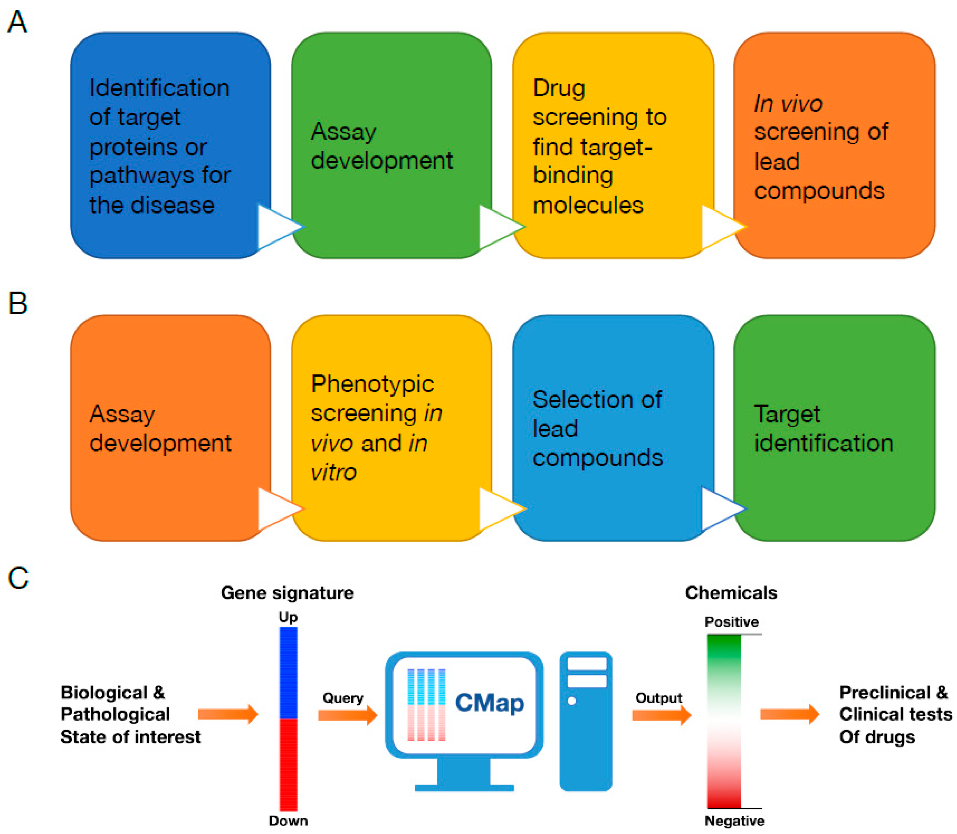
3. Connectivity Map (CMap)

As we explained above, cellular stresses and their related signaling responses are mediated by various environmental factors (e.g., heat, oxidants, osmotic stress, and over-nutrition) and a plethora of signaling molecules. Therefore, finding chemopreventive and even therapeutic chemicals targeting cellular stresses and their associated diseases is challenging and has proven to be difficult with target-based drug discovery. Because of its potential advantage to address complex diseases that require more understanding of their mechanisms, and also recent advances in screening methods for phenotypic drug discovery such as cell-based phenotypic screening and pharmacogenomic analysis, phenotypic drug discovery has started to regain its interest and usage in drug screening.
Recently, the connectivity map (CMap) and the upgraded CMap (L1000) have been demonstrated as useful in silico drug screening tools to target cellular stresses and their related disorders [12,13,143,144]. The CMap (https://portals.broadinstitute.org/cmap/) is a gene expression compendium archiving gene expression data from cultured cells, treated with individual chemical perturbagens and whose ≈22,000 gene expression levels were analyzed with microarray (CMap Build 2 stores results from over 1300 chemical treatments that include a variety of phytochemicals).
Importantly, researchers can query the CMap with their gene expression signatures that consist of a list of genes upregulated and downregulated in the biological or pathological states of interest (Figure 1C). In order to compare the query signature to the entire microarray data in the CMap, which were generated from different cell lines and batches as well as with various doses and treatment time, CMap uses a nonparametric, rank-based pattern-matching analysis. In the end, CMap presents its query result as a list of drugs with a “connectivity score” ranging from +1 (positive connectivity) to -1 (negative connectivity) (Figure 1C). Drugs with a positive connectivity score may generate similar gene expression outcomes with the state of interest (query state), whereas ones with a negative score produce reverse gene expression patterns with the query. Additionally, drugs with a near zero score are unlikely to induce any related responses with the query state. Therefore, the CMap potentially provides a list of candidate chemicals which may mimic or reverse the biological or pathological state of interest [12,13].
Recently, increasing numbers of studies have used CMap to uncover promising small molecules to address various diseases. For example, searching on CMap with gene expression data from tissues (liver and hypothalamus) showing diminished ER stress and improved leptin/insulin receptor signaling as query signatures successfully identified celastrol as an effective leptin sensitizer and chemical chaperone ameliorating obesity in the leptin-resistant mouse model [145]. Celastrol is a phytochemical originally extracted from the root of the thunder god vine, Tripterygium wilfordii, which has been used as a medicinal plant in China and other East Asian countries as a treatment of inflammatory diseases such as rheumatoid arthritis [145,146]. Furthermore, using the gene expression signature of celastrol as a query on CMap uncovered that withaferin A is also a chemical chaperone and a leptin sensitizer, and significantly ameliorates obesity [147]. Similar to celastrol, withaferin A is also a phytochemical originally extracted from leaves, berries, and roots of Withania somnifera, a winter cherry (also called Ashwagandha in India), which has been used as a medicinal plant in India as a treatment of various disorders including inflammation, autoimmune diseases, tumors, stress, anxiety, and aging [148,149,150]. Besides the discovery of celastrol and withaferin A as chemical chaperones and anti-obesity drugs, CMap has been successfully utilized to uncover numerous chemicals with potentials to treat various other diseases, for example, COX2- and ADRA2A-targeting chemicals to treat type 1 and type 2 diabetes [151], tomatidine (a phytochemical from tomato plants) for skeletal muscle atrophy [152], anisomycin for spinal muscle atrophy [153], topiramate for inflammatory bowel disease [154], kaempferol (a phytochemical) for cigarette smoke-induced inflammation [155], pyrvinium for obesity [156], cannabidiol (a phytochemical from Cannabis sativa, marijuana plant) for diabetic cardiomyopathy [157], piperazine for central nervous system injury [158], and many other chemicals for cancers such as medulloblastoma [159], breast cancers [160,161], lung cancers [162,163], glioblastoma [164], ovarian cancer [165], prostate cancers [166], myeloma [167], atypical meningioma [168], leukemia [169,170,171,172], and many others [173,174,175].
Despite CMap’s promising potential as a genome-based and phenotypic drug discovery tool, CMap has limitations. First, constructing and expanding reference profiles of CMap database are time consuming and expensive because CMap is built upon full transcriptome analysis. Second, CMap is still built on data from limited numbers of small molecules and cell lines. In addition, gene expression data from cultured cells may not be appropriate to address the diseases happening in our body or in specific organs. Third, CMap results may not provide enough information about direct drug targets because CMap is a phenotypic drug discovery tool. To overcome these shortcomings, the same team at the Broad Institute who created the original CMap has developed the “next generation connectivity map” or L1000 (https://clue.io/) as part of the National Institutes of Health (NIH) Library of Integrated Network-Based Cellular Signatures (LINCS) initiative [144]. The current L1000 expands the original CMap by using nearly 28,000 perturbagens including over 19,000 small molecules and ≈7000 genetic modulations using knockdown with shRNAs and over-expression with cDNAs. L1000 also includes more cell lines (nine core cell lines) to test perturbagens or uses 3 to 77 variable cell lines for chemicals without characterized mode of action. Moreover, in order to build the new CMap through high-throughput screening at lower costs, L1000 uses only 1000 landmark transcripts as references instead of the full transcriptome, which the authors claim addresses ≈80% of the information in the entire transcriptome [144]. Collectively, L1000 alongside with the original CMap provide powerful in silico pharmacogenomic ways for researchers to discover novel small molecules targeting various diseases that currently do not have effective therapeutics due to their complicated pathophysiologies.

4. Conclusions and Future Perspectives

As our modern society enters the state of population aging, aging-associated diseases such as cardiovascular diseases, obesity, diabetes, neurodegenerative diseases, and cancers have become a major health threat as well as a serious economic and social burden. Even though tremendous efforts have expanded our understanding of the pathophysiologies of these disorders and have also developed numerous medications against them, such efforts still fell short to alleviate significantly chronic and aging-associated disorders. This is partly due to their compounding nature in which a myriad of genetic and environmental factors are interwoven with each other. Recently, a growing number of studies have documented that cellular stresses caused by the disruption of homeostasis within the cell contribute to the development of aging-associated diseases and have suggested that ameliorating these cellular stresses could be an effective prevention and therapeutic treatment. There are several uniquely categorized cellular stresses such as heat shock stress, ER stress, mitochondrial stress, oxidative stress, and hypoxia. However, it should be noted that cellular stresses do not occur individually but frequently happen together. Due to these complexities, developing chemopreventive and therapeutic treatments against cellular stresses and their associated diseases has not yet achieved any significant progress. However, recent technological and genomic advances bring new opportunities to tackle many debilitating chronic disorders. Among them, CMap and its upgraded L1000 are potentially powerful genome-based in silico drug discovery methods based on phenotypic drug discovery, and many studies have successfully used CMap to uncover novel chemicals to alleviate cellular stresses and aging-associated diseases. Furthermore, there are still many possibilities to expand CMap and L1000 in the future in order to be more effective as follows. (1) The numbers of perturbagens (small chemicals and genetic modulations) in CMap/L1000 could increase further, including numerous phytochemicals available currently, and CRISPR/cas9 could be also utilized as genetic perturbation. (2) CMap/L1000 could include more cell types, especially induced pluripotent stem cells (iPSC) and tissue-specific organoids. Additionally, (3) future CMap/L1000 or other pharmacogenomic tools could include more phenotypic information, including proteomic and epigenetic data and also high-content imaging profiles. Furthermore, (4) recent advances in machine learning could empower future genome-based in silico drug discovery tools by potentially providing the information about probable modes of action and target proteins of small molecules.
Historically, phytochemicals have provided huge medical benefits to humankind, as famously shown by salicin (from the willow tree and modified to aspirin®), morphine (from the opium poppy), cocaine (from coca leaves), guanidine (from the French lilac and modified to metformin), and many other examples. However, many phytochemicals’ potential medical benefits are still unknown. However, with recent phenotypic drug discovery tools such as CMap, we can finally be in the position to uncover novel functions of phytochemicals which could be both chemopreventive and therapeutic toward many chronic diseases caused by cellular stresses.

Author Contributions

All authors contributed to researching the literature and writing the article.

Funding

This work was supported by DGIST R&D and the Basic Science Research Program through the National Research Foundation of Korea (NRF) funded by the Ministry of Science and ICT (MSIT) or the Ministry of Education of the Republic of Korea to S.K. (2018R1A6A3A01013000), Y.-I.L. (18-LC-01, 19-BT-01), and J.L. (18-LC-01, 2017R1A2B4006200). J.L. also received funding from the Korea Institute of Oriental Medicine (KIOM) provided by the MSIT (KSN1812160).

Conflicts of Interest

The authors declare no conflict of interest.

Abbreviations

β-TrCPBeta-transducin repeat containing E3 ubiquitin protein ligase
AAAATPases associated with diverse cellular activities
AFG3L2AFG3-like protein 2
ALSAmyotrophic lateral sclerosis
AMPKAMP-activated protein kinase
BNIP3LBCL2 interacting protein 3 like
BRD7Bromodomain-containing protein 7
CLPPClp protease proteolytic subunit
CUL1Cullin 1
CUL3Cullin 3
ERKExtracellular signal-regulated kinase
FDAFood and Drug Administration, USA
HRD1HMG-CoA reductase degradation 1 homolog
IDH2Isocitrate dehydrogenase 2
IKKβInhibitor of nuclear factor kappa-B kinase subunit beta
JNKc-Jun N-terminal kinase
KEAP1Kelch-like ECH-associated protein 1
MAPKMitogen-activated protein kinase
NADPHNicotinamide adenine dinucleotide phosphate, reduced form
NF-κBNuclear factor kappa-light-chain-enhancer of activated B cells
NRF2Nuclear factor erythroid 2-related factor 2
PGC-1αPeroxisome proliferator-activated receptor gamma coactivator-1 alpha
PI3KPhosphoinositide 3-kinase
PINK1PTEN-induced kinase 1
RBX1Ring box 1
SKP1S-phase kinase-associated protein 1
sMAFSmall musculoaponeurotic fibrosarcoma
SPG7Spastic paraplegia 7
TCATricarboxylic acid
VEGF-BVascular endothelial growth factor B

References

  1. Vijg, J.; Le Bourg, E. Aging and the Inevitable Limit to Human Life Span. Gerontology 2017, 63, 432–434. [Google Scholar] [CrossRef] [PubMed] [Green Version]
  2. Campisi, J.; Kapahi, P.; Lithgow, G.J.; Melov, S.; Newman, J.C.; Verdin, E. From discoveries in ageing research to therapeutics for healthy ageing. Nature 2019, 571, 183–192. [Google Scholar] [CrossRef] [PubMed] [Green Version]
  3. Franceschi, C.; Garagnani, P.; Parini, P.; Giuliani, C.; Santoro, A. Inflammaging: A new immune-metabolic viewpoint for age-related diseases. Nat. Rev. Endocrinol. 2018, 14, 576–590. [Google Scholar] [CrossRef] [PubMed]
  4. Galluzzi, L.; Yamazaki, T.; Kroemer, G. Linking cellular stress responses to systemic homeostasis. Nat. Rev. Mol. Cell Biol. 2018, 19, 731–745. [Google Scholar] [CrossRef]
  5. Lee, J.; Ozcan, U. Unfolded protein response signaling and metabolic diseases. J. Biol. Chem. 2014, 289, 1203–1211. [Google Scholar] [CrossRef]
  6. Jovaisaite, V.; Mouchiroud, L.; Auwerx, J. The mitochondrial unfolded protein response, a conserved stress response pathway with implications in health and disease. J. Exp. Biol. 2014, 217, 137–143. [Google Scholar] [CrossRef] [Green Version]
  7. Andreux, P.A.; Houtkooper, R.H.; Auwerx, J. Pharmacological approaches to restore mitochondrial function. Nat. Rev. Drug Discov. 2013, 12, 465–483. [Google Scholar] [CrossRef] [Green Version]
  8. Joshi, S.; Wang, T.; Araujo, T.L.S.; Sharma, S.; Brodsky, J.L.; Chiosis, G. Adapting to stress—Chaperome networks in cancer. Nat. Rev. Cancer 2018, 18, 562–575. [Google Scholar] [CrossRef]
  9. Buono, R.; Longo, V.D. Starvation, Stress Resistance, and Cancer. Trends Endocrinol. Metab. 2018, 29, 271–280. [Google Scholar] [CrossRef]
  10. Steward, W.P.; Brown, K. Cancer chemoprevention: A rapidly evolving field. Br. J. Cancer 2013, 109, 1–7. [Google Scholar] [CrossRef]
  11. Moffat, J.G.; Vincent, F.; Lee, J.A.; Eder, J.; Prunotto, M. Opportunities and challenges in phenotypic drug discovery: An industry perspective. Nat. Rev. Drug Discov. 2017, 16, 531–543. [Google Scholar] [CrossRef]
  12. Lamb, J. The Connectivity Map: A new tool for biomedical research. Nat. Rev. Cancer 2007, 7, 54–60. [Google Scholar] [CrossRef] [PubMed]
  13. Lamb, J.; Crawford, E.D.; Peck, D.; Modell, J.W.; Blat, I.C.; Wrobel, M.J.; Lerner, J.; Brunet, J.P.; Subramanian, A.; Ross, K.N.; et al. The Connectivity Map: Using gene-expression signatures to connect small molecules, genes, and disease. Science 2006, 313, 1929–1935. [Google Scholar] [CrossRef] [PubMed]
  14. Moran Luengo, T.; Mayer, M.P.; Rudiger, S.G.D. The Hsp70-Hsp90 Chaperone Cascade in Protein Folding. Trends Cell Biol. 2019, 29, 164–177. [Google Scholar] [CrossRef] [PubMed]
  15. Hipp, M.S.; Park, S.H.; Hartl, F.U. Proteostasis impairment in protein-misfolding and -aggregation diseases. Trends Cell Biol. 2014, 24, 506–514. [Google Scholar] [CrossRef] [PubMed]
  16. Sun, Z.; Brodsky, J.L. Protein quality control in the secretory pathway. J. Cell Biol. 2019, 218, 3171–3187. [Google Scholar] [CrossRef] [Green Version]
  17. Kaushik, S.; Cuervo, A.M. Proteostasis and aging. Nat. Med. 2015, 21, 1406–1415. [Google Scholar] [CrossRef]
  18. Hipp, M.S.; Kasturi, P.; Hartl, F.U. The proteostasis network and its decline in ageing. Nat. Rev. Mol. Cell Biol. 2019, 20, 421–435. [Google Scholar] [CrossRef]
  19. Ritossa, F. A new puffing pattern induced by temperature shock and DNP in drosophila. Experientia 1962, 18, 571–573. [Google Scholar] [CrossRef]
  20. Kregel, K.C. Heat shock proteins: Modifying factors in physiological stress responses and acquired thermotolerance. J. Appl. Physiol. 1985 2002, 92, 2177–2186. [Google Scholar] [CrossRef]
  21. Chatterjee, S.; Burns, T.F. Targeting Heat Shock Proteins in Cancer: A Promising Therapeutic Approach. Int. J. Mol. Sci. 2017, 18, 1978. [Google Scholar] [CrossRef] [PubMed]
  22. Gomez-Pastor, R.; Burchfiel, E.T.; Thiele, D.J. Regulation of heat shock transcription factors and their roles in physiology and disease. Nat. Rev. Mol. Cell Biol. 2018, 19, 4–19. [Google Scholar] [CrossRef] [PubMed]
  23. Wu, J.; Liu, T.; Rios, Z.; Mei, Q.; Lin, X.; Cao, S. Heat Shock Proteins and Cancer. Trends Pharmacol. Sci. 2017, 38, 226–256. [Google Scholar] [CrossRef] [PubMed]
  24. Javid, B.; MacAry, P.A.; Lehner, P.J. Structure and function: Heat shock proteins and adaptive immunity. J. Immunol. 2007, 179, 2035–2040. [Google Scholar] [CrossRef] [PubMed]
  25. Akerfelt, M.; Morimoto, R.I.; Sistonen, L. Heat shock factors: Integrators of cell stress, development and lifespan. Nat. Rev. Mol. Cell Biol. 2010, 11, 545–555. [Google Scholar] [CrossRef] [PubMed]
  26. Mercier, P.A.; Winegarden, N.A.; Westwood, J.T. Human heat shock factor 1 is predominantly a nuclear protein before and after heat stress. J. Cell Sci. 1999, 112, 2765–2774. [Google Scholar] [PubMed]
  27. Baler, R.; Dahl, G.; Voellmy, R. Activation of human heat shock genes is accompanied by oligomerization, modification, and rapid translocation of heat shock transcription factor HSF1. Mol. Cell. Biol. 1993, 13, 2486–2496. [Google Scholar] [CrossRef]
  28. Sarge, K.D.; Murphy, S.P.; Morimoto, R.I. Activation of heat shock gene transcription by heat shock factor 1 involves oligomerization, acquisition of DNA-binding activity, and nuclear localization and can occur in the absence of stress. Mol. Cell. Biol. 1993, 13, 1392–1407. [Google Scholar] [CrossRef]
  29. Ali, A.; Bharadwaj, S.; O’Carroll, R.; Ovsenek, N. HSP90 interacts with and regulates the activity of heat shock factor 1 in Xenopus oocytes. Mol. Cell. Biol. 1998, 18, 4949–4960. [Google Scholar] [CrossRef]
  30. Zou, J.; Guo, Y.; Guettouche, T.; Smith, D.F.; Voellmy, R. Repression of heat shock transcription factor HSF1 activation by HSP90 (HSP90 complex) that forms a stress-sensitive complex with HSF1. Cell 1998, 94, 471–480. [Google Scholar] [CrossRef]
  31. Hentze, N.; Le Breton, L.; Wiesner, J.; Kempf, G.; Mayer, M.P. Molecular mechanism of thermosensory function of human heat shock transcription factor Hsf1. Elife 2016, 5, e11576. [Google Scholar] [CrossRef] [PubMed]
  32. Morley, J.F.; Morimoto, R.I. Regulation of longevity in Caenorhabditis elegans by heat shock factor and molecular chaperones. Mol. Biol. Cell 2004, 15, 657–664. [Google Scholar] [CrossRef] [PubMed]
  33. Hsu, A.L.; Murphy, C.T.; Kenyon, C. Regulation of aging and age-related disease by DAF-16 and heat-shock factor. Science 2003, 300, 1142–1145. [Google Scholar] [CrossRef] [PubMed]
  34. Fujimoto, M.; Takaki, E.; Hayashi, T.; Kitaura, Y.; Tanaka, Y.; Inouye, S.; Nakai, A. Active HSF1 significantly suppresses polyglutamine aggregate formation in cellular and mouse models. J. Biol. Chem. 2005, 280, 34908–34916. [Google Scholar] [CrossRef] [PubMed]
  35. Hayashida, N.; Fujimoto, M.; Tan, K.; Prakasam, R.; Shinkawa, T.; Li, L.; Ichikawa, H.; Takii, R.; Nakai, A. Heat shock factor 1 ameliorates proteotoxicity in cooperation with the transcription factor NFAT. EMBO J. 2010, 29, 3459–3469. [Google Scholar] [CrossRef] [PubMed] [Green Version]
  36. Chen, H.J.; Mitchell, J.C.; Novoselov, S.; Miller, J.; Nishimura, A.L.; Scotter, E.L.; Vance, C.A.; Cheetham, M.E.; Shaw, C.E. The heat shock response plays an important role in TDP-43 clearance: Evidence for dysfunction in amyotrophic lateral sclerosis. Brain 2016, 139, 1417–1432. [Google Scholar] [CrossRef] [PubMed]
  37. Auluck, P.K.; Chan, H.Y.; Trojanowski, J.Q.; Lee, V.M.; Bonini, N.M. Chaperone suppression of alpha-synuclein toxicity in a Drosophila model for Parkinson’s disease. Science 2002, 295, 865–868. [Google Scholar] [CrossRef]
  38. Dai, C.; Whitesell, L.; Rogers, A.B.; Lindquist, S. Heat shock factor 1 is a powerful multifaceted modifier of carcinogenesis. Cell 2007, 130, 1005–1018. [Google Scholar] [CrossRef]
  39. Voeltz, G.K.; Rolls, M.M.; Rapoport, T.A. Structural organization of the endoplasmic reticulum. EMBO Rep. 2002, 3, 944–950. [Google Scholar] [CrossRef]
  40. Hetz, C.; Papa, F.R. The Unfolded Protein Response and Cell Fate Control. Mol. Cell 2018, 69, 169–181. [Google Scholar] [CrossRef] [Green Version]
  41. Qi, L.; Tsai, B.; Arvan, P. New Insights into the Physiological Role of Endoplasmic Reticulum-Associated Degradation. Trends Cell Biol. 2017, 27, 430–440. [Google Scholar] [CrossRef] [PubMed]
  42. Cox, J.S.; Shamu, C.E.; Walter, P. Transcriptional induction of genes encoding endoplasmic reticulum resident proteins requires a transmembrane protein kinase. Cell 1993, 73, 1197–1206. [Google Scholar] [CrossRef]
  43. Mori, K.; Ma, W.; Gething, M.J.; Sambrook, J. A transmembrane protein with a cdc2+/CDC28-related kinase activity is required for signaling from the ER to the nucleus. Cell 1993, 74, 743–756. [Google Scholar] [PubMed]
  44. Ali, M.M.; Bagratuni, T.; Davenport, E.L.; Nowak, P.R.; Silva-Santisteban, M.C.; Hardcastle, A.; McAndrews, C.; Rowlands, M.G.; Morgan, G.J.; Aherne, W.; et al. Structure of the Ire1 autophosphorylation complex and implications for the unfolded protein response. EMBO J. 2011, 30, 894–905. [Google Scholar] [CrossRef] [PubMed] [Green Version]
  45. Lee, K.P.; Dey, M.; Neculai, D.; Cao, C.; Dever, T.E.; Sicheri, F. Structure of the dual enzyme Ire1 reveals the basis for catalysis and regulation in nonconventional RNA splicing. Cell 2008, 132, 89–100. [Google Scholar] [CrossRef] [PubMed]
  46. Cox, J.S.; Walter, P. A novel mechanism for regulating activity of a transcription factor that controls the unfolded protein response. Cell 1996, 87, 391–404. [Google Scholar] [CrossRef]
  47. Yoshida, H.; Matsui, T.; Yamamoto, A.; Okada, T.; Mori, K. XBP1 mRNA is induced by ATF6 and spliced by IRE1 in response to ER stress to produce a highly active transcription factor. Cell 2001, 107, 881–891. [Google Scholar] [CrossRef]
  48. Chapman, R.E.; Walter, P. Translational attenuation mediated by an mRNA intron. Curr. Biol. 1997, 7, 850–859. [Google Scholar] [CrossRef] [Green Version]
  49. Kawahara, T.; Yanagi, H.; Yura, T.; Mori, K. Endoplasmic reticulum stress-induced mRNA splicing permits synthesis of transcription factor Hac1p/Ern4p that activates the unfolded protein response. Mol. Biol. Cell 1997, 8, 1845–1862. [Google Scholar] [CrossRef]
  50. Hollien, J.; Lin, J.H.; Li, H.; Stevens, N.; Walter, P.; Weissman, J.S. Regulated Ire1-dependent decay of messenger RNAs in mammalian cells. J. Cell Biol. 2009, 186, 323–331. [Google Scholar] [CrossRef] [Green Version]
  51. Han, D.; Lerner, A.G.; Vande Walle, L.; Upton, J.P.; Xu, W.; Hagen, A.; Backes, B.J.; Oakes, S.A.; Papa, F.R. IRE1alpha kinase activation modes control alternate endoribonuclease outputs to determine divergent cell fates. Cell 2009, 138, 562–575. [Google Scholar] [CrossRef] [PubMed]
  52. Upton, J.P.; Wang, L.; Han, D.; Wang, E.S.; Huskey, N.E.; Lim, L.; Truitt, M.; McManus, M.T.; Ruggero, D.; Goga, A.; et al. IRE1alpha cleaves select microRNAs during ER stress to derepress translation of proapoptotic Caspase-2. Science 2012, 338, 818–822. [Google Scholar] [CrossRef] [PubMed]
  53. Maurel, M.; Chevet, E.; Tavernier, J.; Gerlo, S. Getting RIDD of RNA: IRE1 in cell fate regulation. Trends Biochem. Sci. 2014, 39, 245–254. [Google Scholar] [CrossRef] [PubMed]
  54. Lee, J.; Salazar Hernandez, M.A.; Auen, T.; Mucka, P.; Lee, J.; Ozcan, U. PGC-1alpha functions as a co-suppressor of XBP1s to regulate glucose metabolism. Mol. Metab. 2018, 7, 119–131. [Google Scholar] [CrossRef] [PubMed]
  55. Lee, J.; Sun, C.; Zhou, Y.; Gokalp, D.; Herrema, H.; Park, S.W.; Davis, R.J.; Ozcan, U. p38 MAPK-mediated regulation of Xbp1s is crucial for glucose homeostasis. Nat. Med. 2011, 17, 1251–1260. [Google Scholar] [CrossRef]
  56. Park, S.W.; Herrema, H.; Salazar, M.; Cakir, I.; Cabi, S.; Basibuyuk Sahin, F.; Chiu, Y.H.; Cantley, L.C.; Ozcan, U. BRD7 regulates XBP1s’ activity and glucose homeostasis through its interaction with the regulatory subunits of PI3K. Cell Metab. 2014, 20, 73–84. [Google Scholar] [CrossRef]
  57. Park, S.W.; Zhou, Y.; Lee, J.; Lu, A.; Sun, C.; Chung, J.; Ueki, K.; Ozcan, U. The regulatory subunits of PI3K, p85alpha and p85beta, interact with XBP-1 and increase its nuclear translocation. Nat. Med. 2010, 16, 429–437. [Google Scholar] [CrossRef]
  58. Zhou, Y.; Lee, J.; Reno, C.M.; Sun, C.; Park, S.W.; Chung, J.; Fisher, S.J.; White, M.F.; Biddinger, S.B.; Ozcan, U. Regulation of glucose homeostasis through a XBP-1-FoxO1 interaction. Nat. Med. 2011, 17, 356–365. [Google Scholar] [CrossRef]
  59. Liu, J.; Ibi, D.; Taniguchi, K.; Lee, J.; Herrema, H.; Akosman, B.; Mucka, P.; Salazar Hernandez, M.A.; Uyar, M.F.; Park, S.W.; et al. Inflammation Improves Glucose Homeostasis through IKKbeta-XBP1s Interaction. Cell 2016, 167, 1052–1066. [Google Scholar] [CrossRef]
  60. Harding, H.P.; Zhang, Y.; Ron, D. Protein translation and folding are coupled by an endoplasmic-reticulum-resident kinase. Nature 1999, 397, 271–274. [Google Scholar] [CrossRef]
  61. Ma, K.; Vattem, K.M.; Wek, R.C. Dimerization and release of molecular chaperone inhibition facilitate activation of eukaryotic initiation factor-2 kinase in response to endoplasmic reticulum stress. J. Biol. Chem. 2002, 277, 18728–18735. [Google Scholar] [CrossRef] [PubMed]
  62. Wek, R.C.; Cavener, D.R. Translational control and the unfolded protein response. Antioxid. Redox Signal. 2007, 9, 2357–2371. [Google Scholar] [CrossRef] [PubMed]
  63. Zinszner, H.; Kuroda, M.; Wang, X.; Batchvarova, N.; Lightfoot, R.T.; Remotti, H.; Stevens, J.L.; Ron, D. CHOP is implicated in programmed cell death in response to impaired function of the endoplasmic reticulum. Genes Dev. 1998, 12, 982–995. [Google Scholar] [CrossRef] [PubMed]
  64. Harding, H.P.; Novoa, I.; Zhang, Y.; Zeng, H.; Wek, R.; Schapira, M.; Ron, D. Regulated translation initiation controls stress-induced gene expression in mammalian cells. Mol. Cell 2000, 6, 1099–1108. [Google Scholar] [CrossRef]
  65. Haze, K.; Yoshida, H.; Yanagi, H.; Yura, T.; Mori, K. Mammalian transcription factor ATF6 is synthesized as a transmembrane protein and activated by proteolysis in response to endoplasmic reticulum stress. Mol. Biol. Cell 1999, 10, 3787–3799. [Google Scholar] [CrossRef]
  66. Shen, J.; Chen, X.; Hendershot, L.; Prywes, R. ER stress regulation of ATF6 localization by dissociation of BiP/GRP78 binding and unmasking of Golgi localization signals. Dev. Cell 2002, 3, 99–111. [Google Scholar] [CrossRef]
  67. Ozcan, L.; Ergin, A.S.; Lu, A.; Chung, J.; Sarkar, S.; Nie, D.; Myers, M.G., Jr.; Ozcan, U. Endoplasmic reticulum stress plays a central role in development of leptin resistance. Cell Metab. 2009, 9, 35–51. [Google Scholar] [CrossRef]
  68. Ozcan, U.; Cao, Q.; Yilmaz, E.; Lee, A.H.; Iwakoshi, N.N.; Ozdelen, E.; Tuncman, G.; Gorgun, C.; Glimcher, L.H.; Hotamisligil, G.S. Endoplasmic reticulum stress links obesity, insulin action, and type 2 diabetes. Science 2004, 306, 457–461. [Google Scholar] [CrossRef]
  69. Kars, M.; Yang, L.; Gregor, M.F.; Mohammed, B.S.; Pietka, T.A.; Finck, B.N.; Patterson, B.W.; Horton, J.D.; Mittendorfer, B.; Hotamisligil, G.S.; et al. Tauroursodeoxycholic Acid may improve liver and muscle but not adipose tissue insulin sensitivity in obese men and women. Diabetes 2010, 59, 1899–1905. [Google Scholar] [CrossRef]
  70. Ozcan, U.; Yilmaz, E.; Ozcan, L.; Furuhashi, M.; Vaillancourt, E.; Smith, R.O.; Gorgun, C.Z.; Hotamisligil, G.S. Chemical chaperones reduce ER stress and restore glucose homeostasis in a mouse model of type 2 diabetes. Science 2006, 313, 1137–1140. [Google Scholar] [CrossRef]
  71. Xiao, C.; Giacca, A.; Lewis, G.F. Sodium phenylbutyrate, a drug with known capacity to reduce endoplasmic reticulum stress, partially alleviates lipid-induced insulin resistance and beta-cell dysfunction in humans. Diabetes 2011, 60, 918–924. [Google Scholar] [CrossRef] [PubMed]
  72. Gonzalez-Teuber, V.; Albert-Gasco, H.; Auyeung, V.C.; Papa, F.R.; Mallucci, G.R.; Hetz, C. Small Molecules to Improve ER Proteostasis in Disease. Trends Pharmacol. Sci. 2019, 40, 684–695. [Google Scholar] [CrossRef] [PubMed]
  73. Hetz, C.; Axten, J.M.; Patterson, J.B. Pharmacological targeting of the unfolded protein response for disease intervention. Nat. Chem. Biol. 2019, 15, 764–775. [Google Scholar] [CrossRef] [PubMed]
  74. Hetz, C.; Chevet, E.; Harding, H.P. Targeting the unfolded protein response in disease. Nat. Rev. Drug Discov. 2013, 12, 703–719. [Google Scholar] [CrossRef] [PubMed]
  75. Urra, H.; Dufey, E.; Avril, T.; Chevet, E.; Hetz, C. Endoplasmic Reticulum Stress and the Hallmarks of Cancer. Trends Cancer 2016, 2, 252–262. [Google Scholar] [CrossRef] [PubMed]
  76. Cubillos-Ruiz, J.R.; Bettigole, S.E.; Glimcher, L.H. Tumorigenic and Immunosuppressive Effects of Endoplasmic Reticulum Stress in Cancer. Cell 2017, 168, 692–706. [Google Scholar] [CrossRef] [Green Version]
  77. Nikesitch, N.; Lee, J.M.; Ling, S.; Roberts, T.L. Endoplasmic reticulum stress in the development of multiple myeloma and drug resistance. Clin. Transl. Immunol. 2018, 7, e1007. [Google Scholar] [CrossRef] [Green Version]
  78. Lane, N.; Martin, W. The energetics of genome complexity. Nature 2010, 467, 929–934. [Google Scholar] [CrossRef]
  79. Youle, R.J. Mitochondria-Striking a balance between host and endosymbiont. Science 2019, 365, eaaw9855. [Google Scholar] [CrossRef]
  80. Van der Bliek, A.M.; Shen, Q.; Kawajiri, S. Mechanisms of mitochondrial fission and fusion. Cold Spring Harb. Perspect. Biol. 2013, 5, a011072. [Google Scholar] [CrossRef]
  81. Pfanner, N.; Warscheid, B.; Wiedemann, N. Mitochondrial proteins: From biogenesis to functional networks. Nat. Rev. Mol. Cell Biol. 2019, 20, 267–284. [Google Scholar] [CrossRef] [PubMed]
  82. Nunnari, J.; Suomalainen, A. Mitochondria: In sickness and in health. Cell 2012, 148, 1145–1159. [Google Scholar] [CrossRef] [PubMed]
  83. Murphy, M.P.; Hartley, R.C. Mitochondria as a therapeutic target for common pathologies. Nat. Rev. Drug Discov. 2018, 17, 865–886. [Google Scholar] [CrossRef] [PubMed] [Green Version]
  84. Quiros, P.M.; Langer, T.; Lopez-Otin, C. New roles for mitochondrial proteases in health, ageing and disease. Nat. Rev. Mol. Cell Biol. 2015, 16, 345–359. [Google Scholar] [CrossRef] [PubMed]
  85. Nargund, A.M.; Pellegrino, M.W.; Fiorese, C.J.; Baker, B.M.; Haynes, C.M. Mitochondrial import efficiency of ATFS-1 regulates mitochondrial UPR activation. Science 2012, 337, 587–590. [Google Scholar] [CrossRef] [PubMed]
  86. Benedetti, C.; Haynes, C.M.; Yang, Y.; Harding, H.P.; Ron, D. Ubiquitin-like protein 5 positively regulates chaperone gene expression in the mitochondrial unfolded protein response. Genetics 2006, 174, 229–239. [Google Scholar] [CrossRef] [PubMed]
  87. Fiorese, C.J.; Schulz, A.M.; Lin, Y.F.; Rosin, N.; Pellegrino, M.W.; Haynes, C.M. The Transcription Factor ATF5 Mediates a Mammalian Mitochondrial UPR. Curr. Biol. 2016, 26, 2037–2043. [Google Scholar] [CrossRef] [Green Version]
  88. Munch, C.; Harper, J.W. Mitochondrial unfolded protein response controls matrix pre-RNA processing and translation. Nature 2016, 534, 710–713. [Google Scholar] [CrossRef] [Green Version]
  89. Quiros, P.M.; Prado, M.A.; Zamboni, N.; D’Amico, D.; Williams, R.W.; Finley, D.; Gygi, S.P.; Auwerx, J. Multi-omics analysis identifies ATF4 as a key regulator of the mitochondrial stress response in mammals. J. Cell Biol. 2017, 216, 2027–2045. [Google Scholar] [CrossRef]
  90. Jin, S.M.; Youle, R.J. The accumulation of misfolded proteins in the mitochondrial matrix is sensed by PINK1 to induce PARK2/Parkin-mediated mitophagy of polarized mitochondria. Autophagy 2013, 9, 1750–1757. [Google Scholar] [CrossRef] [Green Version]
  91. Youle, R.J.; Narendra, D.P. Mechanisms of mitophagy. Nat. Rev. Mol. Cell Biol. 2011, 12, 9–14. [Google Scholar] [CrossRef] [PubMed]
  92. Wang, Y.; Hekimi, S. Mitochondrial dysfunction and longevity in animals: Untangling the knot. Science 2015, 350, 1204–1207. [Google Scholar] [CrossRef] [PubMed]
  93. Lee, S.S.; Lee, R.Y.; Fraser, A.G.; Kamath, R.S.; Ahringer, J.; Ruvkun, G. A systematic RNAi screen identifies a critical role for mitochondria in C. elegans longevity. Nat. Genet. 2003, 33, 40–48. [Google Scholar] [CrossRef] [PubMed]
  94. Nassir, F.; Ibdah, J.A. Role of mitochondria in nonalcoholic fatty liver disease. Int. J. Mol. Sci. 2014, 15, 8713–8742. [Google Scholar] [CrossRef]
  95. Szendroedi, J.; Phielix, E.; Roden, M. The role of mitochondria in insulin resistance and type 2 diabetes mellitus. Nat. Rev. Endocrinol. 2011, 8, 92–103. [Google Scholar] [CrossRef]
  96. Hudson, G. The Ageing Brain, Mitochondria and Neurodegeneration. In Mitochondrial Dysfunction in Neurodegenerative Disorders; Reeve, A.K., Simcox, E.M., Duchen, M.R., Turnbull, D.M., Eds.; Springer International Publishing: Cham, Switzerland, 2016; pp. 59–80. [Google Scholar]
  97. Cho, B.; Kim, T.; Huh, Y.J.; Lee, J.; Lee, Y.I. Amelioration of Mitochondrial Quality Control and Proteostasis by Natural Compounds in Parkinson’s Disease Models. Int. J. Mol. Sci. 2019, 20, 5208. [Google Scholar] [CrossRef]
  98. Blesa, J.; Trigo-Damas, I.; Quiroga-Varela, A.; del Rey, N.L.-G. Parkinson’s Disease-Associated Mutations Affect Mitochondrial Function. In Mitochondrial Mechanisms of Degeneration and Repair in Parkinson’s Disease; Buhlman, L.M., Ed.; Springer International Publishing: Cham, Switzerland, 2016; pp. 139–158. [Google Scholar]
  99. Chen, C.; Turnbull, D.M.; Reeve, A.K. Mitochondrial Dysfunction in Parkinson’s Disease-Cause or Consequence? Biology 2019, 8, 38. [Google Scholar] [CrossRef]
  100. Wu, Y.; Chen, M.; Jiang, J. Mitochondrial dysfunction in neurodegenerative diseases and drug targets via apoptotic signaling. Mitochondrion 2019, 49, 35–45. [Google Scholar] [CrossRef]
  101. Zong, W.X.; Rabinowitz, J.D.; White, E. Mitochondria and Cancer. Mol. Cell 2016, 61, 667–676. [Google Scholar] [CrossRef] [Green Version]
  102. Vyas, S.; Zaganjor, E.; Haigis, M.C. Mitochondria and Cancer. Cell 2016, 166, 555–566. [Google Scholar] [CrossRef]
  103. Mullard, A. FDA approves first-in-class cancer metabolism drug. Nat. Rev. Drug Discov. 2017, 16, 593. [Google Scholar] [CrossRef] [PubMed]
  104. Pollyea, D.A.; Tallman, M.S.; de Botton, S.; Kantarjian, H.M.; Collins, R.; Stein, A.S.; Frattini, M.G.; Xu, Q.; Tosolini, A.; See, W.L.; et al. Enasidenib, an inhibitor of mutant IDH2 proteins, induces durable remissions in older patients with newly diagnosed acute myeloid leukemia. Leukemia 2019, 33, 2575–2584. [Google Scholar] [CrossRef] [PubMed] [Green Version]
  105. Nakazawa, M.S.; Keith, B.; Simon, M.C. Oxygen availability and metabolic adaptations. Nat. Rev. Cancer 2016, 16, 663–673. [Google Scholar] [CrossRef] [PubMed] [Green Version]
  106. Nobre, A.R.; Entenberg, D.; Wang, Y.; Condeelis, J.; Aguirre-Ghiso, J.A. The Different Routes to Metastasis via Hypoxia-Regulated Programs. Trends Cell Biol. 2018, 28, 941–956. [Google Scholar] [CrossRef] [PubMed]
  107. Xie, H.; Simon, M.C. Oxygen availability and metabolic reprogramming in cancer. J. Biol. Chem. 2017, 292, 16825–16832. [Google Scholar] [CrossRef] [Green Version]
  108. Hewitson, K.S.; McNeill, L.A.; Elkins, J.M.; Schofield, C.J. The role of iron and 2-oxoglutarate oxygenases in signalling. Biochem. Soc. Trans. 2003, 31, 510–515. [Google Scholar] [CrossRef] [PubMed]
  109. Gonzalez, F.J.; Xie, C.; Jiang, C. The role of hypoxia-inducible factors in metabolic diseases. Nat. Rev. Endocrinol. 2018, 15, 21–32. [Google Scholar] [CrossRef]
  110. Lee, K.E.; Simon, M.C. SnapShot: Hypoxia-Inducible Factors. Cell 2015, 163, 1288. [Google Scholar] [CrossRef]
  111. Thompson, C.B. Into Thin Air: How We Sense and Respond to Hypoxia. Cell 2016, 167, 9–11. [Google Scholar] [CrossRef] [Green Version]
  112. Denko, N.C. Hypoxia, HIF1 and glucose metabolism in the solid tumour. Nat. Rev. Cancer 2008, 8, 705–713. [Google Scholar] [CrossRef]
  113. Ullah, M.S.; Davies, A.J.; Halestrap, A.P. The plasma membrane lactate transporter MCT4, but not MCT1, is up-regulated by hypoxia through a HIF-1alpha-dependent mechanism. J. Biol. Chem. 2006, 281, 9030–9037. [Google Scholar] [CrossRef] [PubMed]
  114. Kim, J.W.; Tchernyshyov, I.; Semenza, G.L.; Dang, C.V. HIF-1-mediated expression of pyruvate dehydrogenase kinase: A metabolic switch required for cellular adaptation to hypoxia. Cell Metab. 2006, 3, 177–185. [Google Scholar] [CrossRef] [PubMed] [Green Version]
  115. Fukuda, R.; Zhang, H.; Kim, J.W.; Shimoda, L.; Dang, C.V.; Semenza, G.L. HIF-1 regulates cytochrome oxidase subunits to optimize efficiency of respiration in hypoxic cells. Cell 2007, 129, 111–122. [Google Scholar] [CrossRef] [PubMed]
  116. Jain, I.H.; Zazzeron, L.; Goli, R.; Alexa, K.; Schatzman-Bone, S.; Dhillon, H.; Goldberger, O.; Peng, J.; Shalem, O.; Sanjana, N.E.; et al. Hypoxia as a therapy for mitochondrial disease. Science 2016, 352, 54–61. [Google Scholar] [CrossRef] [Green Version]
  117. Ast, T.; Meisel, J.D.; Patra, S.; Wang, H.; Grange, R.M.H.; Kim, S.H.; Calvo, S.E.; Orefice, L.L.; Nagashima, F.; Ichinose, F.; et al. Hypoxia Rescues Frataxin Loss by Restoring Iron Sulfur Cluster Biogenesis. Cell 2019, 177, 1507–1521. [Google Scholar] [CrossRef]
  118. Semenza, G.L. Oxygen sensing, hypoxia-inducible factors, and disease pathophysiology. Annu. Rev. Pathol. 2014, 9, 47–71. [Google Scholar] [CrossRef]
  119. Cheng, K.; Ho, K.; Stokes, R.; Scott, C.; Lau, S.M.; Hawthorne, W.J.; O’Connell, P.J.; Loudovaris, T.; Kay, T.W.; Kulkarni, R.N.; et al. Hypoxia-inducible factor-1alpha regulates beta cell function in mouse and human islets. J. Clin. Investig. 2010, 120, 2171–2183. [Google Scholar] [CrossRef]
  120. Taniguchi, C.M.; Finger, E.C.; Krieg, A.J.; Wu, C.; Diep, A.N.; LaGory, E.L.; Wei, K.; McGinnis, L.M.; Yuan, J.; Kuo, C.J.; et al. Cross-talk between hypoxia and insulin signaling through Phd3 regulates hepatic glucose and lipid metabolism and ameliorates diabetes. Nat. Med. 2013, 19, 1325–1330. [Google Scholar] [CrossRef] [Green Version]
  121. Wei, K.; Piecewicz, S.M.; McGinnis, L.M.; Taniguchi, C.M.; Wiegand, S.J.; Anderson, K.; Chan, C.W.; Mulligan, K.X.; Kuo, D.; Yuan, J.; et al. A liver Hif-2alpha-Irs2 pathway sensitizes hepatic insulin signaling and is modulated by Vegf inhibition. Nat. Med. 2013, 19, 1331–1337. [Google Scholar] [CrossRef]
  122. Xie, C.; Yagai, T.; Luo, Y.; Liang, X.; Chen, T.; Wang, Q.; Sun, D.; Zhao, J.; Ramakrishnan, S.K.; Sun, L.; et al. Activation of intestinal hypoxia-inducible factor 2alpha during obesity contributes to hepatic steatosis. Nat. Med. 2017, 23, 1298–1308. [Google Scholar] [CrossRef]
  123. Sies, H.; Berndt, C.; Jones, D.P. Oxidative Stress. Annu. Rev. Biochem. 2017, 86, 715–748. [Google Scholar] [CrossRef] [PubMed]
  124. Luo, H.; Chiang, H.H.; Louw, M.; Susanto, A.; Chen, D. Nutrient Sensing and the Oxidative Stress Response. Trends Endocrinol. Metab. 2017, 28, 449–460. [Google Scholar] [CrossRef] [PubMed]
  125. Finkel, T.; Holbrook, N.J. Oxidants, oxidative stress and the biology of ageing. Nature 2000, 408, 239–247. [Google Scholar] [CrossRef] [PubMed]
  126. Collins, Y.; Chouchani, E.T.; James, A.M.; Menger, K.E.; Cocheme, H.M.; Murphy, M.P. Mitochondrial redox signalling at a glance. J. Cell Sci. 2012, 125, 801–806. [Google Scholar] [CrossRef] [PubMed] [Green Version]
  127. Chouchani, E.T.; Kazak, L.; Jedrychowski, M.P.; Lu, G.Z.; Erickson, B.K.; Szpyt, J.; Pierce, K.A.; Laznik-Bogoslavski, D.; Vetrivelan, R.; Clish, C.B.; et al. Mitochondrial ROS regulate thermogenic energy expenditure and sulfenylation of UCP1. Nature 2016, 532, 112–116. [Google Scholar] [CrossRef] [PubMed] [Green Version]
  128. Dupre-Crochet, S.; Erard, M.; Nubetae, O. ROS production in phagocytes: Why, when, and where? J. Leukoc. Biol. 2013, 94, 657–670. [Google Scholar] [CrossRef]
  129. Mullebner, A.; Dorighello, G.G.; Kozlov, A.V.; Duvigneau, J.C. Interaction between Mitochondrial Reactive Oxygen Species, Heme Oxygenase, and Nitric Oxide Synthase Stimulates Phagocytosis in Macrophages. Front. Med. 2017, 4, 252. [Google Scholar] [CrossRef]
  130. Iovine, N.M.; Pursnani, S.; Voldman, A.; Wasserman, G.; Blaser, M.J.; Weinrauch, Y. Reactive nitrogen species contribute to innate host defense against Campylobacter jejuni. Infect. Immun. 2008, 76, 986–993. [Google Scholar] [CrossRef]
  131. Murphy, M.P. How mitochondria produce reactive oxygen species. Biochem. J. 2009, 417, 1–13. [Google Scholar] [CrossRef]
  132. Powers, S.K.; Ji, L.L.; Kavazis, A.N.; Jackson, M.J. Reactive oxygen species: Impact on skeletal muscle. Compr. Physiol. 2011, 1, 941–969. [Google Scholar]
  133. Finkel, T. Signal transduction by mitochondrial oxidants. J. Biol. Chem. 2012, 287, 4434–4440. [Google Scholar] [CrossRef] [PubMed]
  134. Meitzler, J.L.; Antony, S.; Wu, Y.; Juhasz, A.; Liu, H.; Jiang, G.; Lu, J.; Roy, K.; Doroshow, J.H. NADPH oxidases: A perspective on reactive oxygen species production in tumor biology. Antioxid. Redox Signal. 2014, 20, 2873–2889. [Google Scholar] [CrossRef] [PubMed]
  135. Nisoli, E.; Carruba, M.O. Nitric oxide and mitochondrial biogenesis. J. Cell Sci. 2006, 119, 2855–2862. [Google Scholar] [CrossRef] [PubMed] [Green Version]
  136. Cuadrado, A.; Rojo, A.I.; Wells, G.; Hayes, J.D.; Cousin, S.P.; Rumsey, W.L.; Attucks, O.C.; Franklin, S.; Levonen, A.L.; Kensler, T.W.; et al. Therapeutic targeting of the NRF2 and KEAP1 partnership in chronic diseases. Nat. Rev. Drug Discov. 2019, 18, 295–317. [Google Scholar] [CrossRef] [PubMed] [Green Version]
  137. Rojo de la Vega, M.; Chapman, E.; Zhang, D.D. NRF2 and the Hallmarks of Cancer. Cancer Cell 2018, 34, 21–43. [Google Scholar] [CrossRef]
  138. Prentki, M.; Nolan, C.J. Islet beta cell failure in type 2 diabetes. J. Clin. Investig. 2006, 116, 1802–1812. [Google Scholar] [CrossRef]
  139. Campbell, A.; Solaimani, P. Oxidative and Inflammatory Pathways in Age-Related Chronic Disease Processes. In Inflammation, Aging, and Oxidative Stress; Bondy, S.C., Campbell, A., Eds.; Springer International Publishing: Cham, Switzerland, 2016; pp. 95–106. [Google Scholar]
  140. Dasgupta, A.; Klein, K. Antioxidants in Food, Vitamins and Supplements; Elsevier: San Diego, CA, USA, 2014; pp. 129–150. [Google Scholar]
  141. Jeong, Y.; Hellyer, J.A.; Stehr, H.; Hoang, N.T.; Niu, X.; Das, M.; Padda, S.K.; Ramchandran, K.; Neal, J.W.; Wakelee, H.A.; et al. Role of KEAP1/NFE2L2 mutations in the chemotherapeutic response of non-small cell lung cancer patients. Clin. Cancer Res. 2019. [Google Scholar] [CrossRef]
  142. Jeong, Y.; Hoang, N.T.; Lovejoy, A.; Stehr, H.; Newman, A.M.; Gentles, A.J.; Kong, W.; Truong, D.; Martin, S.; Chaudhuri, A.; et al. Role of KEAP1/NRF2 and TP53 Mutations in Lung Squamous Cell Carcinoma Development and Radiation Resistance. Cancer Discov. 2017, 7, 86–101. [Google Scholar] [CrossRef]
  143. Musa, A.; Ghoraie, L.S.; Zhang, S.D.; Glazko, G.; Yli-Harja, O.; Dehmer, M.; Haibe-Kains, B.; Emmert-Streib, F. A review of connectivity map and computational approaches in pharmacogenomics. Brief. Bioinform. 2018, 19, 506–523. [Google Scholar]
  144. Subramanian, A.; Narayan, R.; Corsello, S.M.; Peck, D.D.; Natoli, T.E.; Lu, X.; Gould, J.; Davis, J.F.; Tubelli, A.A.; Asiedu, J.K.; et al. A Next Generation Connectivity Map: L1000 Platform and the First 1,000,000 Profiles. Cell 2017, 171, 1437–1452. [Google Scholar] [CrossRef]
  145. Liu, J.; Lee, J.; Salazar Hernandez, M.A.; Mazitschek, R.; Ozcan, U. Treatment of obesity with celastrol. Cell 2015, 161, 999–1011. [Google Scholar] [CrossRef] [PubMed]
  146. Zhang, C.; Sun, P.P.; Guo, H.T.; Liu, Y.; Li, J.; He, X.J.; Lu, A.P. Safety Profiles of Tripterygium wilfordii Hook F: A Systematic Review and Meta-Analysis. Front. Pharmacol. 2016, 7, 402. [Google Scholar] [CrossRef] [PubMed] [Green Version]
  147. Lee, J.; Liu, J.; Feng, X.; Salazar Hernandez, M.A.; Mucka, P.; Ibi, D.; Choi, J.W.; Ozcan, U. Withaferin A is a leptin sensitizer with strong antidiabetic properties in mice. Nat. Med. 2016, 22, 1023–1032. [Google Scholar] [CrossRef] [PubMed] [Green Version]
  148. Mirjalili, M.H.; Moyano, E.; Bonfill, M.; Cusido, R.M.; Palazon, J. Steroidal lactones from Withania somnifera, an ancient plant for novel medicine. Molecules 2009, 14, 2373–2393. [Google Scholar] [CrossRef] [PubMed]
  149. Winters, M. Ancient medicine, modern use: Withania somnifera and its potential role in integrative oncology. Altern. Med. Rev. 2006, 11, 269–277. [Google Scholar] [PubMed]
  150. Mishra, L.C.; Singh, B.B.; Dagenais, S. Scientific basis for the therapeutic use of Withania somnifera (ashwagandha): A review. Altern. Med. Rev. 2000, 5, 334–346. [Google Scholar] [PubMed]
  151. Zhang, M.; Luo, H.; Xi, Z.; Rogaeva, E. Drug repositioning for diabetes based on ‘omics’ data mining. PLoS ONE 2015, 10, e0126082. [Google Scholar] [CrossRef]
  152. Dyle, M.C.; Ebert, S.M.; Cook, D.P.; Kunkel, S.D.; Fox, D.K.; Bongers, K.S.; Bullard, S.A.; Dierdorff, J.M.; Adams, C.M. Systems-based discovery of tomatidine as a natural small molecule inhibitor of skeletal muscle atrophy. J. Biol. Chem. 2014, 289, 14913–14924. [Google Scholar] [CrossRef]
  153. Farooq, F.; Balabanian, S.; Liu, X.; Holcik, M.; MacKenzie, A. p38 Mitogen-activated protein kinase stabilizes SMN mRNA through RNA binding protein HuR. Hum. Mol. Genet. 2009, 18, 4035–4045. [Google Scholar] [CrossRef] [Green Version]
  154. Dudley, J.T.; Sirota, M.; Shenoy, M.; Pai, R.K.; Roedder, S.; Chiang, A.P.; Morgan, A.A.; Sarwal, M.M.; Pasricha, P.J.; Butte, A.J. Computational repositioning of the anticonvulsant topiramate for inflammatory bowel disease. Sci. Transl. Med. 2011, 3, 96ra76. [Google Scholar] [CrossRef]
  155. Vanderstocken, G.; Dvorkin-Gheva, A.; Shen, P.; Brandsma, C.A.; Obeidat, M.; Bosse, Y.; Hassell, J.A.; Stampfli, M.R. Identification of Drug Candidates to Suppress Cigarette Smoke-induced Inflammation via Connectivity Map Analyses. Am. J. Respir. Cell Mol. Biol. 2018, 58, 727–735. [Google Scholar] [CrossRef] [PubMed]
  156. Wang, Z.; Dai, Z.; Luo, Z.; Zuo, C. Identification of Pyrvinium, an Anthelmintic Drug, as a Novel Anti-Adipogenic Compound Based on the Gene Expression Microarray and Connectivity Map. Molecules 2019, 24, 2391. [Google Scholar] [CrossRef] [PubMed]
  157. Rajesh, M.; Mukhopadhyay, P.; Batkai, S.; Patel, V.; Saito, K.; Matsumoto, S.; Kashiwaya, Y.; Horvath, B.; Mukhopadhyay, B.; Becker, L.; et al. Cannabidiol attenuates cardiac dysfunction, oxidative stress, fibrosis, and inflammatory and cell death signaling pathways in diabetic cardiomyopathy. J. Am. Coll. Cardiol. 2010, 56, 2115–2125. [Google Scholar] [CrossRef] [PubMed]
  158. Johnstone, A.L.; Reierson, G.W.; Smith, R.P.; Goldberg, J.L.; Lemmon, V.P.; Bixby, J.L. A chemical genetic approach identifies piperazine antipsychotics as promoters of CNS neurite growth on inhibitory substrates. Mol. Cell. Neurosci. 2012, 50, 125–135. [Google Scholar] [CrossRef] [PubMed] [Green Version]
  159. Singh, A.R.; Joshi, S.; Zulcic, M.; Alcaraz, M.; Garlich, J.R.; Morales, G.A.; Cho, Y.J.; Bao, L.; Levy, M.L.; Newbury, R.; et al. PI-3K Inhibitors Preferentially Target CD15+ Cancer Stem Cell Population in SHH Driven Medulloblastoma. PLoS ONE 2016, 11, e0150836. [Google Scholar] [CrossRef] [PubMed]
  160. Muthuswami, M.; Ramesh, V.; Banerjee, S.; Viveka Thangaraj, S.; Periasamy, J.; Bhaskar Rao, D.; Barnabas, G.D.; Raghavan, S.; Ganesan, K. Breast tumors with elevated expression of 1q candidate genes confer poor clinical outcome and sensitivity to Ras/PI3K inhibition. PLoS ONE 2013, 8, e77553. [Google Scholar] [CrossRef] [PubMed]
  161. Wang, S.E.; Xiang, B.; Guix, M.; Olivares, M.G.; Parker, J.; Chung, C.H.; Pandiella, A.; Arteaga, C.L. Transforming growth factor beta engages TACE and ErbB3 to activate phosphatidylinositol-3 kinase/Akt in ErbB2-overexpressing breast cancer and desensitizes cells to trastuzumab. Mol. Cell. Biol. 2008, 28, 5605–5620. [Google Scholar] [CrossRef]
  162. Wang, G.; Ye, Y.; Yang, X.; Liao, H.; Zhao, C.; Liang, S. Expression-based in silico screening of candidate therapeutic compounds for lung adenocarcinoma. PLoS ONE 2011, 6, e14573. [Google Scholar] [CrossRef]
  163. Fortney, K.; Griesman, J.; Kotlyar, M.; Pastrello, C.; Angeli, M.; Sound-Tsao, M.; Jurisica, I. Prioritizing therapeutics for lung cancer: An integrative meta-analysis of cancer gene signatures and chemogenomic data. PLoS Comput. Biol. 2015, 11, e1004068. [Google Scholar] [CrossRef]
  164. Cheng, H.W.; Liang, Y.H.; Kuo, Y.L.; Chuu, C.P.; Lin, C.Y.; Lee, M.H.; Wu, A.T.; Yeh, C.T.; Chen, E.I.; Whang-Peng, J.; et al. Identification of thioridazine, an antipsychotic drug, as an antiglioblastoma and anticancer stem cell agent using public gene expression data. Cell Death Dis. 2015, 6, e1753. [Google Scholar] [CrossRef]
  165. Rho, S.B.; Kim, B.R.; Kang, S. A gene signature-based approach identifies thioridazine as an inhibitor of phosphatidylinositol-3’-kinase (PI3K)/AKT pathway in ovarian cancer cells. Gynecol. Oncol. 2011, 120, 121–127. [Google Scholar] [CrossRef] [PubMed]
  166. Hieronymus, H.; Lamb, J.; Ross, K.N.; Peng, X.P.; Clement, C.; Rodina, A.; Nieto, M.; Du, J.; Stegmaier, K.; Raj, S.M.; et al. Gene expression signature-based chemical genomic prediction identifies a novel class of HSP90 pathway modulators. Cancer Cell 2006, 10, 321–330. [Google Scholar] [CrossRef] [PubMed] [Green Version]
  167. Tiedemann, R.E.; Schmidt, J.; Keats, J.J.; Shi, C.X.; Zhu, Y.X.; Palmer, S.E.; Mao, X.; Schimmer, A.D.; Stewart, A.K. Identification of a potent natural triterpenoid inhibitor of proteosome chymotrypsin-like activity and NF-kappaB with antimyeloma activity in vitro and in vivo. Blood 2009, 113, 4027–4037. [Google Scholar] [CrossRef] [PubMed]
  168. Zador, Z.; King, A.T.; Geifman, N. New drug candidates for treatment of atypical meningiomas: An integrated approach using gene expression signatures for drug repurposing. PLoS ONE 2018, 13, e0194701. [Google Scholar] [CrossRef] [PubMed]
  169. Sanda, T.; Li, X.; Gutierrez, A.; Ahn, Y.; Neuberg, D.S.; O’Neil, J.; Strack, P.R.; Winter, C.G.; Winter, S.S.; Larson, R.S.; et al. Interconnecting molecular pathways in the pathogenesis and drug sensitivity of T-cell acute lymphoblastic leukemia. Blood 2010, 115, 1735–1745. [Google Scholar] [CrossRef] [PubMed] [Green Version]
  170. Hassane, D.C.; Guzman, M.L.; Corbett, C.; Li, X.; Abboud, R.; Young, F.; Liesveld, J.L.; Carroll, M.; Jordan, C.T. Discovery of agents that eradicate leukemia stem cells using an in silico screen of public gene expression data. Blood 2008, 111, 5654–5662. [Google Scholar] [CrossRef] [PubMed] [Green Version]
  171. Schnell, S.A.; Ambesi-Impiombato, A.; Sanchez-Martin, M.; Belver, L.; Xu, L.; Qin, Y.; Kageyama, R.; Ferrando, A.A. Therapeutic targeting of HES1 transcriptional programs in T-ALL. Blood 2015, 125, 2806–2814. [Google Scholar] [CrossRef] [Green Version]
  172. Churchman, M.L.; Low, J.; Qu, C.; Paietta, E.M.; Kasper, L.H.; Chang, Y.; Payne-Turner, D.; Althoff, M.J.; Song, G.; Chen, S.C.; et al. Efficacy of Retinoids in IKZF1-Mutated BCR-ABL1 Acute Lymphoblastic Leukemia. Cancer Cell 2015, 28, 343–356. [Google Scholar] [CrossRef]
  173. Rosenbluth, J.M.; Mays, D.J.; Pino, M.F.; Tang, L.J.; Pietenpol, J.A. A gene signature-based approach identifies mTOR as a regulator of p73. Mol. Cell. Biol. 2008, 28, 5951–5964. [Google Scholar] [CrossRef]
  174. Saito, S.; Furuno, A.; Sakurai, J.; Sakamoto, A.; Park, H.R.; Shin-Ya, K.; Tsuruo, T.; Tomida, A. Chemical genomics identifies the unfolded protein response as a target for selective cancer cell killing during glucose deprivation. Cancer Res. 2009, 69, 4225–4234. [Google Scholar] [CrossRef]
  175. Stockwell, S.R.; Platt, G.; Barrie, S.E.; Zoumpoulidou, G.; Te Poele, R.H.; Aherne, G.W.; Wilson, S.C.; Sheldrake, P.; McDonald, E.; Venet, M.; et al. Mechanism-based screen for G1/S checkpoint activators identifies a selective activator of EIF2AK3/PERK signalling. PLoS ONE 2012, 7, e28568. [Google Scholar] [CrossRef] [PubMed]
Figure 1. Drug discovery using the connectivity map (CMap). (A) Target-based drug discovery. (B) Phenotypic drug discovery. (C) CMap-based drug discovery. Gene signature of the biological or pathological state of interest can be used as a query to search through CMap. CMap provides the search result as a list of small molecules scored to predict their probability to mimic or reverse gene expression profiles of the state of interest. Candidate chemicals can be further tested in in vitro cell culture and in vivo animal experiments before proceeding with clinical trials to human subjects.
Ijms 20 05601 g001
Figure 2. Cellular stresses and signaling responses. (A) Heat shock stress and heat shock response. The stressors such as heat lead to releasing of heat shock proteins (HSPs) from heat shock factor 1 (HSF1) or directly changing the conformation of HSF1 resulting in its trimerization, nuclear translocation, and target gene transcription. (B) Endoplasmic reticulum (ER) stress and unfolded protein response (UPR). The accumulation of unfolded or misfolded proteins activates three ER transmembrane proteins—activating transcription factor-6 (ATF6), inositol requiring protein-1 (IRE1), and protein kinase RNA-like ER kinase (PERK). ATF6 and IRE1 generate the functional transcription factors, ATF6N and spliced form of X-box binding protein 1 (XBP1s), which translocate to the nucleus and transcribe their target genes, whereas PERK suppresses protein translation and thus reduces protein load into the ER. (C) Mitochondrial stress and mitochondrial unfolded protein response (UPRmt). Mitochondrial stress activates several transcription factors, activating transcription factor associated with stress-1 (ATFS-1) and defective proventriculus (Drosophila) homolog-1/ubiquitin-like 5 (DVE-1/UBL-5) (Caenorhabditis elegans) and ATF4 (mammals), which promote their target gene expression to restore mitochondrial homeostasis. Mitochondrial stress also triggers autophagy (mitophagy) via Parkin and Pink1. (D) Hypoxia and hypoxia-induced factor. Under normoxia, hypoxia-inducible factor α (HIFα) is hydroxylated on proline by prolyl hydroxylase domain enzymes (PHDs) or on asparagine by factor inhibiting HIF1 (FIH1), and the activity of HIFα is suppressed by its von Hippel–Lindau (VHL)-mediated ubiquitylation and degradation or its loss of the interaction with p300/CREB-binding protein (CBP).
Ijms 20 05601 g002

