Communication

Integrin β1-Mediated Cell–Cell Adhesion Augments Metformin-Induced Anoikis

Center of Growth, Metabolism and Aging, Key Laboratory of Bio-Resource and Eco-Environment, Ministry of Education, College of Life Sciences, Sichuan University, Chengdu 610064, China
*
Author to whom correspondence should be addressed.
Int. J. Mol. Sci. 2019, 20(5), 1161; https://doi.org/10.3390/ijms20051161
Submission received: 18 February 2019 / Revised: 27 February 2019 / Accepted: 4 March 2019 / Published: 7 March 2019
(This article belongs to the Section Molecular Oncology)

Abstract

:
Cell–cell adhesion plays an important role in regulation of cell proliferation, migration, survival, and drug sensitivity. Metformin, a first line drug for type 2 diabetes, has been shown to possess anti-cancer activities. However, whether cell–cell adhesion affects metformin anti-cancer activity is unknown. In this study, Microscopic and FACS analyses showed that metformin induced cancer cell–cell adhesion exemplified by cell aggregation and anoikis under glucose restriction. Furthermore, western blot and QPCR analyses revealed that metformin dramatically upregulated integrin β1 expression. Silencing of integrin β1 significantly disrupted cell aggregation and reduced anoikis induced by metformin. Moreover, we showed that p53 family member ΔNp63α transcriptionally suppressed integrin β1 expression and is responsible for metformin-mediated upregulation of integrin β1. In summary, this study reveals a novel mechanism for metformin anticancer activity and demonstrates that cell–cell adhesion mediated by integrin β1 plays a critical role in metformin-induced anoikis.

Graphical Abstract

1. Introduction

Metformin has been widely used to treat type 2 diabetes for many years. In recent years, clinical and basic evidence indicates that metformin has biological activities beyond the treatment of diabetes, such as anti-aging [1,2] and anti-obesity effects [3,4]. Metformin was the first FDA-approved drug in a clinical anti-aging trial [5]. Moreover, accumulating evidence demonstrates that metformin has anti-cancer activities [6]. Metformin can inhibit cancer cell growth, survival, and metastasis [7,8]. In addition, latest studies demonstrate that metformin can improve tumor immune response [9] and modulation of cancer-related epigenetic modification [10]. Lots of researches demonstrate that activation of AMPK is the major mechanism of metformin anti-cancer [9,10,11]. Our previous study indicates that metformin-induced cancer cell anoikis is an AMPK-independent manner [12]. Currently, there are more than 300 FDA-approved clinical trials in assessment of metformin effects on cancer therapy (www.clinicaltrials.gov).
p63 is a member of p53 family proteins involved in multiple biological processes, including embryonic development, cell differentiation, proliferation, survival, and aging [13]. Due to alternative transcription starting from two sites and the alternative spicing at the C-termini, there are at least six major p63 protein isoforms [13]. ΔNp63α is the predominant isoform of p63 protein expresses in epithelial cells and is critical for epithelial development [13]. It is reported that ΔNp63α is frequently overexpressed in squamous cell carcinoma and promotes cell proliferation and tumor growth [14]. On the other hand, ΔNp63α is a common inhibitory target in oncogenic PI3K/Her2/Ras-induced cell migration and cancer metastasis [15]. Moreover, ΔNp63α is a master regulator of cell adhesion program, which regulates several adhesion molecules, including fibronectin 1, integrin α6, and integrin β4 [16]. Our previous study has demonstrated that metformin destabilizes ΔNp63α protein and inhibits cancer cell viability [12].
Cell–cell adhesion and cell-extracellular matrix (ECM) interaction are essential signals in regulation of cell proliferation, cell migration, survival and drug sensitivity [17,18]. When anchorage-dependent cells detach from the surrounding ECM, they often undergo anoikis, a form of programmed cell death [19]. Integrin β1 is a family member of integrins, playing an important role in maintaining cell-matrix adhesion [16,20]. Accumulating evidence indicates that integrin β1 is involved in tumor growth and cancer metastasis [21]. However, whether integrin β1 is involved in cell–cell adhesion or apoptosis/anoikis is unclear.
In this study, we showed that integrin β1-mediated cell–cell adhesion enhances metformin-mediated cell aggregation and anoikis under glucose restriction. ΔNp63α is a negative regulator of integrin β1, which plays a pivotal role in metformin-mediated upregulation of integrin β1. Taken together, this study demonstrates that ΔNp63α-integrin β1 axis is critical in metformin anticancer process and reveals a novel mechanism for metformin anticancer action.

2. Results

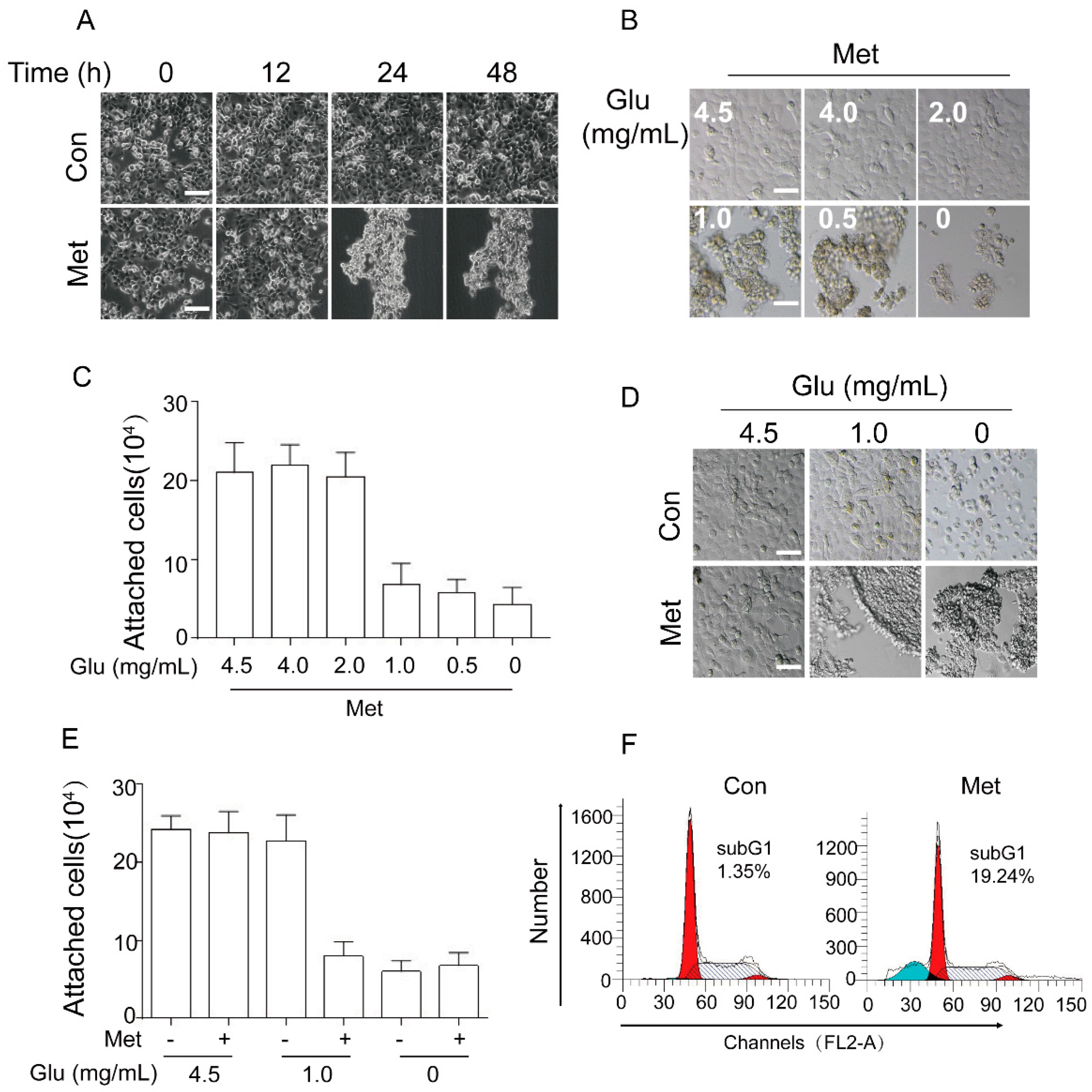
2.1. Metformin Induces Cancer Cell Aggregation/Detachment and Anoikis under Glucose Restriction

We have previously shown that metformin inhibits expression of ΔNp63α resulting in human head and neck squamous cell carcinoma (HNSCC) FaDu cell anoikis under glucose restriction [12]. During the course of the study, we constantly noticed that metformin significantly induced cell aggregation prior to detachment under glucose restriction (DMEM containing 1.0 mg/mL glucose). As shown in Figure 1A, under glucose restriction, FaDu cells showed little cell detachment, whereas treatment with 20 mM metformin led to dramatic Cell Aggregation and Detachment (CAD), consistent with our previous report [12]. We then examined the impact of glucose concentration on metformin-induced CAD. As shown in Figure 1B,C, metformin-induced CAD only occurred at the condition of restricted glucose.
It has been reported that human hepatoma HepG2 cells can undergo detachment in the absence of glucose [22], we therefore examined the effects of no glucose on cell detachment in comparison to the effects of metformin. As shown in Figure 1D,E, cells grown in DMEM containing 1.0 mg/mL glucose exhibited no detachment, whereas cells grown in DMEM containing 0 mg/mL glucose exhibited cell detachment without cell aggregation. By contrast, cells grown in DMEM containing 0 mg/mL glucose in the presence of metformin exhibited dramatic CAD. These results indicate that metformin specifically induces CAD.
We next examined the effects of metformin on cell viability. As shown in Figure 1F, metformin significantly induced FaDu cell apoptosis under glucose restriction, consistent with our previous study [12]. Taken together, these results demonstrate that metformin can induce cancer cell detachment and apoptosis, a well-known phenomenon characterized as anoikis.

2.2. Metformin Induces Expression of Integrin β1 Resulting in Cell Aggregation and Promoting Anoikis

To dissect the molecular basis by which metformin induces CAD, we hypothesized that metformin may upregulate subset of cell adhesion molecules, such as integrin β1, integrin β4, N-cadherin, and E-cadherin. We first examined the impact of metformin on the protein levels of several cell-adhesion molecules. As shown in Figure 2A,B, metformin treatment led to a significant decrease in integrin β4 and E-cadherin at both protein and mRNA levels. Surprisingly, metformin dramatically upregulated expression of integrin β1 at both protein and mRNA levels (Figure 2A,B). Moreover, consistent with our previous data [12], metformin significantly inhibited ΔNp63α protein expression (Figure 2A).
Next, we examined whether integrin β1 plays a role in metformin-induced cell aggregation. As shown in Figure 2C,D, again, metformin significantly upregulated expression of integrin β1, concomitant with CAD. Silencing of integrin β1 completely eliminated metformin-induced cell aggregation. Importantly, ablation of integrin β1 clearly reduced metformin-induced apoptosis (Figure 2E). Taken together, these results indicate that integrin β1 plays an important role in metformin-induced cell aggregation and anoikis.

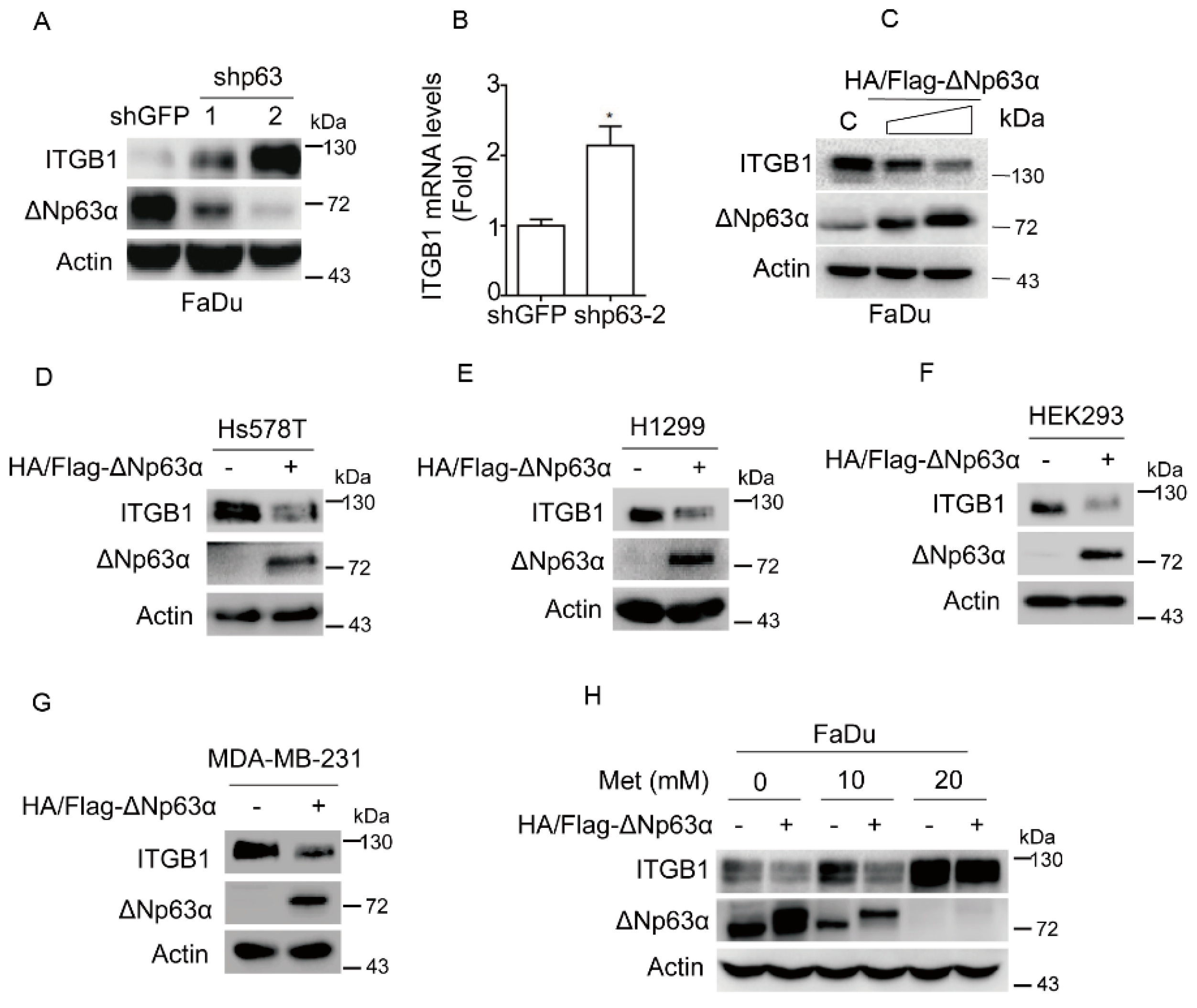
2.3. Metformin Upregulates Integrin β1 Expression via Inhibiting ΔNp63α
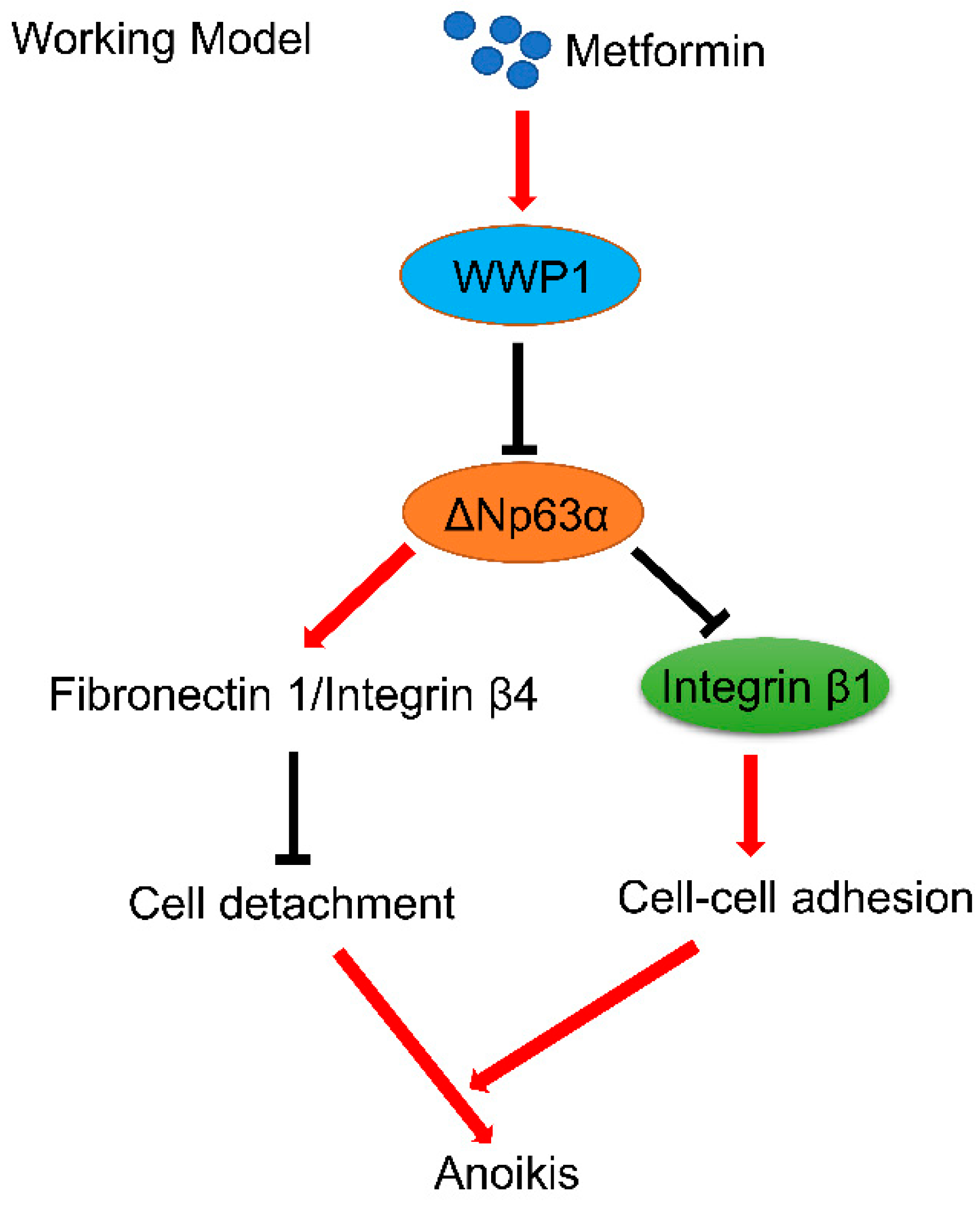
We then investigated the molecular mechanism by which metformin induces expression of integrin β1. We have previously shown that metformin can significantly inhibit ΔNp63α expression. Since p63 is a master regulator to transcriptionally regulate expression of cell adhesion molecules [16], we therefore examined the effects of ΔNp63α on integrin β1 expression. As shown in Figure 3A,B, silencing of ΔNp63α dramatically upregulated integrin β1 expression at both protein and mRNA levels. On the other hand, ectopic expression of ΔNp63α significantly downregulated integrin β1 protein expression in a dose-dependent manner (Figure 3C). Similar results are observed in other cell lines, including human breast cancer Hs578T and MDA-MB-231, human non-small cell lung cancer (NSCLC) H1299 and human embryonic kidney HEK293, all of which express high levels of endogenous integrin β1 (Figure 3D–G). Importantly, restoration of ΔNp63α expression effectively reversed metformin-induced upregulation of integrin β1 expression (Figure 3H). Together, these results demonstrate that ΔNp63α is a critical negative regulator of integrin β1 and is most likely responsible for metformin-mediated upregulation of integrin β1.

3. Discussion

Until now, it has been well established that metformin possesses anti-cancer activities. Currently, there are FDA-approved more than 300 clinical trials to assess effects of metformin on cancer therapy (www.clinicaltrials.gov.13/01/2019). In this study, we showed that metformin induces cancer cell anoikis. Furthermore, we found that integrin β1 is pivotal in both metformin-induced cell aggregation and anoikis. Moreover, we demonstrated that ΔNp63α is a negative regulator of integrin β1 and is responsible for metformin-mediated upregulation of integrin β1.
How does metformin upregulate integrin β1 expression? Our results clearly indicate that ΔNp63α is a pivotal negative regulator of integrin β1 expression and is responsible to metformin-mediated upregulation of integrin β1. However, in contrast to a possible positive role for p63 in regulation of integrin β1 as shown in MCF 10A cells [16], our data reproducibly show that expression of ΔNp63α leads to downregulation of integrin β1 in FaDu, Hs578T, H1299, HEK 293, and MDA-MB-231 cells. The reason for the discrepancy is not yet clear although it might be possible that there are some unknown factors affecting expression of integrin β1 in MCF 10A cells.
Cell–cell adhesion is fundamentally important for cell–cell communication involved in multiple facets of biological processes, including cell proliferation, migration, and survival [23]. Our previous study demonstrates that metformin destabilizes ΔNp63α via upregulating ubiquitin E3 ligase WWP1, leading to downregulation of proteins involved in cell-matrix adhesion including integrin β4 and fibronectin 1, which in turn results in cell detachment [12]. In this study, we further showed that metformin not only induces cell detachment but also induces cell aggregation, both of which significantly contribute to metformin-mediated anoikis (Figure 4).
E-cadherin is an adherent junction marker, critically important in maintaining epithelial cell–cell adhesion [24]. However, we found that metformin significantly inhibits E-cadherin expression at both mRNA and protein levels, indicating that E-cadherin is unlikely to be responsible for metformin-mediated cell aggregation. On the other hand, integrin β1 is a transmembrane receptor which has been shown as a critical protein in maintaining cell-extracellular matrix (ECM) adhesion [16,20]. Most interestingly, we found that metformin can transcriptionally upregulate integrin β1 expression, which in turn plays a causative role in both metformin-mediated cell aggregation and anoikis. Together, our findings demonstrate that integrin β1 is not only important in maintaining cell-matrix adhesion but also is critical in cell–cell adhesion. Furthermore, our data clearly indicates that integrin β1-mediated cell–cell adhesion plays an important role in metformin-induced anoikis. It would be interesting to further investigate whether cell–cell adhesion plays a role in cell viability under pathophysiological conditions in vivo.
Most notably, it is reported that circulating tumor cell clusters exhibit enhanced metastatic potential due to increased cancer cell stemness and/or increased anoikis-resistance [25,26]. Therefore, it is conceivable that circulating tumor cell clusters might be more sensitive to metformin. Conceptually, integrin β1 could be a potential target for cancer therapy.

4. Materials and Methods

4.1. Cell Culture and Drug Treatment

Human head and neck squamous cell carcinoma (HNSCC) FaDu cells, HEK293 cells, HEK293T cells, human breast cancer MDA-MB-231 and Hs578T cells, and human non-small cell lung cancer (NSCLC) H1299 cells were purchased from ATCC and maintained in DMEM medium (GIBCO, Rockville, MD, USA) containing 10% fetal bovine serum (FBS; Hyclone, Logan, UT, USA), 100 units/mL penicillin (GIBCO) and 100 µg/mL streptomycin (GIBCO). Cells were grown in a humidified 37 °C incubator under a 5% CO2 atmosphere. Cells at 75–85% confluence were treated with an indicated chemical compound. Metformin (PHR1084) was purchased from Sigma-Aldrich (St Louis, MO).

4.2. Plasmids Transfection, Lentiviral Infection, and RNA Interference

Cells at 80% confluence were transfected using Lipofectamine 2000 transfection reagent (Invitrogen, Carlsbad, CA, USA). Recombinant lentiviruses were generated by transfecting HEK293T cells with psPAX2 and pMD2.G packaging plasmids and lentiviral expression plasmid human HA/Flag-ΔNp63α using Lipofectamine 2000. Virus were collected at 60 h post-transfection. Cells at 30% confluence supplemented with 10 µg/mL polybrene were infected with recombinant lentivirus encoding an empty vector or expression plasmid, followed by 12 h incubation at 37 °C with 5% CO2. Lentiviral-based shRNAs specific for green fluorescent protein (GFP, GAAGCAGCACGACTTCTTC), integrin β1(GGAATGCCTACTTCTGCAC) and human p63 (GAGTGGAATGACTTCAACTTT; CCGTTTCGTCAGAACACACAT) were constructed as described [27].

4.3. Western Blot Analyses

Cells were lysed in EBC250 lysis buffer (250 mM NaCl, 50 mM Tris pH 8.0, 0.5% Nonidet p-40, 50 mM NaF, 2 µg/mL aprotinin, 1 mM phenylmethylsulfonyl fluoride and 2 µg/mL leupeptin). Whole cell lysates (50 µg) were separated by SDS-PAGE, and then transferred to PVDF membranes (Millipore, Darmstadt, Germany). Membranes were blocked in 5% milk in TBS-T buffer and then hybridized to an appropriate primary antibody and HRP-conjugated goat anti-mouse IgG (1:3000, Santa Cruz Biotech (Santa Cruz, CA, USA), sc-2005) or goat anti-rabbit IgG (1:3000, Santa Cruz Biotech, sc-2004) for subsequent detection by enhanced chemiluminescence kit (WBKLS0500, Millipore). Specific antibodies for p63 (sc-8431,1:200) or actin (sc-1615, 1:1000) were purchased from Santa Cruz Biotech. Antibodies for N-cadherin (14215, 1:1000) or Integrin β4 (4704, 1:1000) were purchased from Cell Signaling Technology (Danvers, MA, USA). Antibodies for Integrin β1 (ab52971, 1:1000) or E-cadherin (ab40772, 1:10000) were purchased from Abcam (Cambridge, MA, USA).

4.4. Quantitative PCR

Total RNA was extracted from cells using RNeasy plus Mini Kit (cat# 74134, QIAGEN) and reverse-transcribed. Q-PCR was performed for Integrin β1 (F: GCCTTACATTAGCACAACACC; R: CATCTCCAGCAAAGTGAAAC), Integrin β4 (F: CACCGCGTGCTAAGCACAT; R: TGTGGTCGAGTGAGTGTTCTG), E-cadherin (F: GGATGTGCTGGATGTGAATG; R: CACATCAGACAGGATCAGCAGAA), N-cadherin (F: CTCCTATGAGTGGAACAGGAACG; R: TTGGATCAATGTCATAATCAAGTGCTGTA) and GAPDH (F: GGGGAGCCAAAAAGGGTCATCATCT; R: GAGGGGCCATCCACAGTCTTCT). The Q-PCR reactions were carried out in CFX-960 Real time PCR System (Bio-Rad, Saint-Laurent, QC, Canada) and using Bio-Rad SoFast Eva-Green Supermix (Bio-Rad) according to the manufacturer’s instructions. Relative quantitation values were calculated using the ΔΔCt method.

4.5. FACS Assay

Cells were grown in 6-well plates to approximately 80% confluence prior to treatment with metformin. Both floating and adherent cells were collected and trypsinized to obtain individual cell, which were then fixed in 75% ethanol at 4 °C overnight. Cells were stained with 50 µg/mL propidium iodide (PI)/80 µg/mL RNase A at 37 °C for 1 h. Cells were then subjected to FACS analysis by FACScalibur flow cytometer (Becton Dickson, Bedford, MA, USA).

4.6. Statistical Analysis

Quantitative data were analyzed statistically using Student’s t-test to assess significance. All experiments were performed three times in triplicates. Data are presented as means ± s.d.

Author Contributions

Conceived and designed the experiments: Y.Y. and Z.-X.X. Performed the experiments: T.A., Z.Z., Y.L., Y.Y., D.C., J.A., W.Z., J.Y. Analyzed the data: Y.Y. and Z.-X.X. Wrote the paper: Y.Y. and Z.-X.X.

Funding

This work was supported by the China Postdoctoral Science Foundation (2018M631081) and Postdoctoral Fellowship of Sichuan University (2018SCU12054) to Y.Y., National Natural Science Foundation of China (81802951) to Y.Y.; (81830108, 81861148031, 81330054, and 81520108020) to Z.-X.X., and Sichuan Department of Science and Technology International Cooperation and Exchange Program (2017HH0057) to Z.-X.X.

Acknowledgments

We thank Zhonghan Li, Qintong Li, and Chenhua Li for helpful discussions. We thank Han Kang for technical assistance on FACS analyses.

Conflicts of Interest

We declare that we have no conflict of interest.

Abbreviations

ECMCell-extracellular matrix
CADCell Aggregation and Detachment
HNSCCHuman head and neck squamous cell carcinoma
NSCLCHuman non-small cell lung cancer

References

  1. Cabreiro, F.; Au, C.; Leung, K.Y.; Vergara-Irigaray, N.; Cocheme, H.M.; Noori, T.; Weinkove, D.; Schuster, E.; Greene, N.D.; Gems, D. Metformin retards aging in C. elegans by altering microbial folate and methionine metabolism. Cell 2013, 153, 228–239. [Google Scholar] [CrossRef] [PubMed]
  2. Barzilai, N.; Crandall, J.P.; Kritchevsky, S.B.; Espeland, M.A. Metformin as a Tool to Target Aging. Cell Metab. 2016, 23, 1060–1065. [Google Scholar] [CrossRef] [PubMed]
  3. Seifarth, C.; Schehler, B.; Schneider, H.J. Effectiveness of metformin on weight loss in non-diabetic individuals with obesity. Exp. Clin. Endocrinol. Diabetes 2013, 121, 27–31. [Google Scholar] [CrossRef] [PubMed]
  4. Desilets, A.R.; Dhakal-Karki, S.; Dunican, K.C. Role of metformin for weight management in patients without type 2 diabetes. Ann. Pharm. 2008, 42, 817–826. [Google Scholar] [CrossRef] [PubMed]
  5. Hall, S.S. A Trial for the ages. Science 2015, 349, 1274–1278. [Google Scholar] [CrossRef] [PubMed]
  6. Pernicova, I.; Korbonits, M. Metformin—Mode of action and clinical implications for diabetes and cancer. Nat. Rev. Endocrinol. 2014, 10, 143–156. [Google Scholar] [CrossRef] [PubMed]
  7. Menendez, J.A.; Cufi, S.; Oliveras-Ferraros, C.; Martin-Castillo, B.; Joven, J.; Vellon, L.; Vazquez-Martin, A. Metformin and the ATM DNA damage response (DDR): Accelerating the onset of stress-induced senescence to boost protection against cancer. Aging 2011, 3, 1063–1077. [Google Scholar] [CrossRef] [PubMed] [Green Version]
  8. Kisfalvi, K.; Eibl, G.; Sinnett-Smith, J.; Rozengurt, E. Metformin disrupts crosstalk between G protein-coupled receptor and insulin receptor signaling systems and inhibits pancreatic cancer growth. Cancer Res. 2009, 69, 6539–6545. [Google Scholar] [CrossRef] [PubMed]
  9. Cha, J.H.; Yang, W.H.; Xia, W.; Wei, Y.; Chan, L.C.; Lim, S.O.; Li, C.W.; Kim, T.; Chang, S.S.; Lee, H.H.; et al. Metformin Promotes Antitumor Immunity via Endoplasmic-Reticulum-Associated Degradation of PD-L1. Mol. Cell 2018, 71, 606–620. [Google Scholar] [CrossRef] [PubMed]
  10. Wu, D.; Hu, D.; Chen, H.; Shi, G.; Fetahu, I.S.; Wu, F.; Rabidou, K.; Fang, R.; Tan, L.; Xu, S.; et al. Glucose-regulated phosphorylation of TET2 by AMPK reveals a pathway linking diabetes to cancer. Nature 2018, 559, 637–641. [Google Scholar] [CrossRef] [PubMed]
  11. Zhang, C.S.; Li, M.; Ma, T.; Zong, Y.; Cui, J.; Feng, J.W.; Wu, Y.Q.; Lin, S.Y.; Lin, S.C. Metformin Activates AMPK through the Lysosomal Pathway. Cell Metab. 2016, 24, 521–522. [Google Scholar] [CrossRef] [PubMed]
  12. Yi, Y.; Chen, D.; Ao, J.; Sun, S.; Wu, M.; Li, X.; Bergholz, J.; Zhang, Y.; Xiao, Z.X. Metformin Promotes AMP-activated Protein Kinase-independent Suppression of DeltaNp63alpha Protein Expression and Inhibits Cancer Cell Viability. J. Biol. Chem. 2017, 292, 5253–5261. [Google Scholar] [CrossRef] [PubMed]
  13. Bergholz, J.; Xiao, Z.X. Role of p63 in Development, Tumorigenesis and Cancer Progression. Cancer Microenviron. 2012, 5, 311–322. [Google Scholar] [CrossRef] [PubMed]
  14. Li, C.; Chang, D.L.; Yang, Z.; Qi, J.; Liu, R.; He, H.; Li, D.; Xiao, Z.X. Pin1 modulates p63alpha protein stability in regulation of cell survival, proliferation and tumor formation. Cell Death Dis. 2013, 4, e943. [Google Scholar] [CrossRef] [PubMed]
  15. Hu, L.; Liang, S.; Chen, H.; Lv, T.; Wu, J.; Chen, D.; Wu, M.; Sun, S.; Zhang, H.; You, H.; et al. DeltaNp63alpha is a common inhibitory target in oncogenic PI3K/Ras/Her2-induced cell motility and tumor metastasis. Proc. Natl. Acad. Sci. USA 2017, 114, E3964–E3973. [Google Scholar] [CrossRef] [PubMed]
  16. Carroll, D.K.; Carroll, J.S.; Leong, C.O.; Cheng, F.; Brown, M.; Mills, A.A.; Brugge, J.S.; Ellisen, L.W. p63 regulates an adhesion programme and cell survival in epithelial cells. Nat. Cell Biol. 2006, 8, 551–561. [Google Scholar] [CrossRef] [PubMed]
  17. Damiano, J.S.; Cress, A.E.; Hazlehurst, L.A.; Shtil, A.A.; Dalton, W.S. Cell adhesion mediated drug resistance (CAM-DR): Role of integrins and resistance to apoptosis in human myeloma cell lines. Blood 1999, 93, 1658–1667. [Google Scholar] [PubMed]
  18. Albelda, S.M. Role of integrins and other cell adhesion molecules in tumor progression and metastasis. Lab. Investig. 1993, 68, 4–17. [Google Scholar] [PubMed]
  19. Frisch, S.M.; Francis, H. Disruption of epithelial cell-matrix interactions induces apoptosis. J. Cell Biol. 1994, 124, 619–626. [Google Scholar] [CrossRef] [PubMed] [Green Version]
  20. Burridge, K.; Chrzanowska-Wodnicka, M. Focal adhesions, contractility, and signaling. Annu. Rev. Cell Dev. Biol. 1996, 12, 463–518. [Google Scholar] [CrossRef] [PubMed]
  21. Muller, P.A.; Caswell, P.T.; Doyle, B.; Iwanicki, M.P.; Tan, E.H.; Karim, S.; Lukashchuk, N.; Gillespie, D.A.; Ludwig, R.L.; Gosselin, P.; et al. Mutant p53 drives invasion by promoting integrin recycling. Cell 2009, 139, 1327–1341. [Google Scholar] [CrossRef] [PubMed]
  22. Suzuki, A.; Kusakai, G.; Kishimoto, A.; Minegichi, Y.; Ogura, T.; Esumi, H. Induction of cell-cell detachment during glucose starvation through F-actin conversion by SNARK, the fourth member of the AMP-activated protein kinase catalytic subunit family. Biochem. Biophys. Res. Commun. 2003, 311, 156–161. [Google Scholar] [CrossRef] [PubMed]
  23. Okegawa, T.; Pong, R.C.; Li, Y.; Hsieh, J.T. The role of cell adhesion molecule in cancer progression and its application in cancer therapy. Acta Biochim. Pol. 2004, 51, 445–457. [Google Scholar] [PubMed]
  24. Beavon, I.R. The E-cadherin-catenin complex in tumour metastasis: Structure, function and regulation. Eur. J. Cancer 2000, 36, 1607–1620. [Google Scholar] [CrossRef]
  25. Gkountela, S.; Castro-Giner, F.; Szczerba, B.M.; Vetter, M.; Landin, J.; Scherrer, R.; Krol, I.; Scheidmann, M.C.; Beisel, C.; Stirnimann, C.U.; et al. Circulating Tumor Cell Clustering Shapes DNA Methylation to Enable Metastasis Seeding. Cell 2019, 176, 98–112. [Google Scholar] [CrossRef] [PubMed]
  26. Aceto, N.; Toner, M.; Maheswaran, S.; Haber, D.A. En Route to Metastasis: Circulating Tumor Cell Clusters and Epithelial-to-Mesenchymal Transition. Trends Cancer 2015, 1, 44–52. [Google Scholar] [CrossRef] [PubMed]
  27. Bergholz, J.; Zhang, Y.; Wu, J.; Meng, L.; Walsh, E.M.; Rai, A.; Sherman, M.Y.; Xiao, Z.X. DeltaNp63alpha regulates Erk signaling via MKP3 to inhibit cancer metastasis. Oncogene 2014, 33, 212–224. [Google Scholar] [CrossRef] [PubMed]
Figure 1. Metformin induces cell aggregation/detachment and anoikis under glucose restriction. (A) FaDu cells were treated with metformin (20 mM, and thereafter) for an indicated time (0, 12, 24, or 48 h) in DMEM containing 1.0 mg/mL glucose condition (DMEM-LG). Cell morphology was captured by phase contrast microscopy. (B,C) FaDu cells were treated with metformin for 24 h in DMEM containing an indicated concentration of glucose (4.5, 4.0, 2.0, 1.0, 0.5, or 0 mg/mL). Cell morphology was captured by phase contrast microscopy and representative pictures were shown (B). Attached cells were quantitated and presented (C). (D,E) FaDu cells were treated with or without metformin for 24 h in DMEM containing an indicated dose of glucose (4.5, 1.0, or 0 mg/mL) respectively. Representative images were shown (D). Attached cell were quantitated and presented (E). (F) FaDu cells were treated with or without metformin for 48 h in DMEM-LG. Both adherent and floating cells were subjected to FACS analyses. All experiments were performed three times in triplicates. Data were presented as means ± s.d. Scale bar = 50 µm.
Ijms 20 01161 g001
Figure 2. Metformin induces expression of integrin β1 resulting in cell aggregation and promoting anoikis. (A,B) FaDu cells were treated with metformin (20 mM, and thereafter) for as an indicated time (0, 12, or 24 h) in DMEM-LG. Cells were subjected to western blot analyses (A) or subjected to Q-PCR analyses (cells were collected at 18 h) (B). Statistical analyses were based on Student’s t-test. Data were presented as means ± s.d. **. p < 0.05; *, p < 0.05. (C) FaDu cells stably expressing shITGB1 or shGFP were treated with metformin (0, 10, or 20 mM) in DMEM-LG for 24 h. Cells were subjected to western blot analyses. (D,E) FaDu cells stably expressing shITGB1 or shGFP were treated with metformin in DMEM-LG for an indicated time (0, 24, 48, or 60 h). Representative images were shown (D) and cells were subjected to FACS analyses (E). Scale bars = 50 µm. For western blotting, all of PVDF membranes derived from the transfers were cut in appropriate areas and reacted with an indicated primary antibody.
Ijms 20 01161 g002
Figure 3. Metformin suppresses expression of ΔNp63α resulting in upregulation of integrin β1. (A,B) FaDu cells stably expressing shp63-1, shp63-2 or shGFP were subjected to western blot analyses (A) or subjected to Q-PCR analyses (B). Data are presented as means ± s.d. *, p < 0.05. (C) FaDu cells stably overexpressing HA/Flag-ΔNp63α or vector were subjected to western blot analyses. (D,G) Ectopic expression of HA/Flag-ΔNp63 in Hs578T, H1299, HEK293, and MDA-MB-231 cells were subjected to western blot analyses. (H) FaDu cells stably overexpressing HA/Flag-ΔNp63α or vector were treated with metformin (0, 10, or 20 mM) for 24 h in DMEM-LG. Cells were subjected to western blot analyses. All western blot membranes were cut in appropriate areas and reacted with an indicated primary antibody.
Ijms 20 01161 g003
Figure 4. A working model of metformin-induced cancer cell anoikis. Metformin upregulates ubiquitin E3 ligase WWP1, leading to destabilization of ΔNp63α and downregulation of proteins involved in cell-matrix adhesion including integrin β4 and fibronectin 1, which in turn results in cell detachment [12]. On the other hand, metformin inhibits ΔNp63α expression resulting in upregulation of integrin β1, which in turn promotes cell–cell adhesion and cell aggregation. Both disruption of cell-matrix adhesion and enhanced cell–cell adhesion contribute to metformin-induced anoikis.
Ijms 20 01161 g004

