Article

In Silico Analysis of Homologous Heterodimers of Cruzipain-Chagasin from Structural Models Built by Homology

by
Francisco Reyes-Espinosa
1,*,
Alfredo Juárez-Saldivar
1,
Isidro Palos
2,
Verónica Herrera-Mayorga
1,3,
Carlos García-Pérez
4 and
Gildardo Rivera
1,*
1
Laboratorio de Biotecnología Farmacéutica, Centro de Biotecnología Genómica, Instituto Politécnico Nacional, Boulevard del Maestro s/n esq. Elías Piña, Col. Narciso Mendoza, Reynosa 88710, Mexico
2
Unidad Académica Multidisciplinaria Reynosa-Rodhe, Universidad Autónoma Tamaulipas, Carr. Reynosa-San Fernando, Reynosa 88779, Mexico
3
Departamento de Ingeniería Bioquímica, Unidad Académica Multidisciplinaria Mante, Universidad Autónoma Tamaulipas, Blvd. Enrique Cárdenas González 1201, Mante 89840, Mexico
4
Scientific Computing Research Unit, Helmholtz Zentrum München, 85764 Munich, Germany
*
Authors to whom correspondence should be addressed.
Int. J. Mol. Sci. 2019, 20(6), 1320; https://doi.org/10.3390/ijms20061320
Submission received: 12 February 2019 / Revised: 8 March 2019 / Accepted: 11 March 2019 / Published: 15 March 2019
(This article belongs to the Special Issue New Avenues in Molecular Docking for Drug Design)

Abstract

:
The present study gives an overview of the binding energetics of the homologous heterodimers of cruzipain−chagasin based on the binding energy (ΔGb) prediction obtained with FoldX. This analysis involves a total of 70 homologous models of the cruzipain−chagasin complex which were constructed by homology from the combinatory variation of nine papain-like cysteine peptidase structures and seven cysteine protease inhibitor structures (as chagasin-like and cystatin-like inhibitors). Only 32 systems have been evaluated experimentally, ΔGbexperimental values previously reported. Therefore, the result of the multiple analysis in terms of the thermodynamic parameters, are shown as relative energy |ΔΔG| = |ΔGbfrom FoldX − ΔGbexperimental|. Nine models were identified that recorded |ΔΔG| < 1.3, five models to 2.8 > |ΔΔG| > 1.3 and the other 18 models, values of |ΔΔG| > 2.8. The energetic analysis of the contribution of ΔH and ΔS to ΔGb to the 14-molecular model presents a ΔGb mostly ΔH-driven at neutral pH and at an ionic strength (I) of 0.15 M. The dependence of ΔGb(I,pH) at 298 K to the cruzipain−chagasin complex predicts a linear dependence of ΔGb(I). The computational protocol allowed the identification and prediction of thermodynamics binding energy parameters for cruzipain−chagasin-like heterodimers.

Graphical Abstract

1. Introduction

The protozoan Trypanosoma cruzi (T. cruzi) is known as the etiologic agent of Chagas disease (CD) which is also termed American trypanosomiasis [1,2]. In endemic areas, the main vector of transmission of CD that has its natural ecosystem on the American continent is triatomine bugs (of the genus Triatoma). Other forms of CD transmission are congenital, by blood transfusion, by organ transplantation, orally (by food contaminated with parasites) or by laboratory accident [3,4]. With the purpose of treating CD, the scientific community has directed its efforts mainly in three lines of research: the search and design of new drugs, the design of biomarkers to test the effectiveness of drugs and techniques to diagnose the disease. To fulfil this purpose, a series of pharmacological targets have been identified (e.g., cruzipain, trans-sialidase, trypanothione reductase, triosephosphate isomerase, sterol 14-demethylase and tubulin, among others) as potential trypanocidal drugs.
Cruzipain (papain-like enzyme) is the major cysteine proteinase (CP) of T. cruzi. It belongs to the group of papain-like enzymes known as clan CA which is the most studied group of CP of parasitic protozoa [5,6]. The subcellular localization of cruzipain varies in different stages of the parasite’s biological cycle and is involved in penetration of the parasite and evasion of the host´s immune response. The parasitic protozoa contain cysteine peptidases (CP) that are crucial for a range of important biological processes [5]. These are involved in several stages of the parasite life cycle and thus exert substantial influence over host-parasite relationships, including metamorphosis, immune evasion and adaptation to specific hosts [6].
Natural protein structure inhibitors, known as cysteine protease inhibitors (CPI), regulate the catalytic activity of CP. These inhibitory proteins can be cystatin-like or chagasin-like. A complete description and classification of CP and CPI can be found in the MEROPS database (https://www.ebi.ac.uk/merops/ (accessed on February 2019)). A growing effort is currently being to design CPI from their endogenous and exogenous natural protein inhibitor (e.g., peptide derivatives as prodrugs and peptide mimetics) because of their high affinity [7,8] and specific CP inhibition of parasitic protozoa [9,10,11].
It is been nearly two decades since the first rationally designed small molecule drug (dorzolamide, an anti-glaucoma agent) was marketed [12]. Since then, the scientific community has made great efforts in the development of computational methods that allow the design of small molecules to produce biotherapeutics. This class of therapeutics includes proteins, peptides and nucleic acids that could more effectively combat drug resistance and even act when the disease is caused by a molecular deficiency [12].
On the other hand, in the field of bioinformatics, significant achievements have been made in the development of computational methods for modelling the protein−protein interaction [13,14], mainly in cases where a crystallographic complex is available with a potential to biologically bind to its target. Some methods such as molecular dynamics (MD) and other related methods can be used as routine computational tools for drug discovery. Limitations in the experimental techniques used to solve 3D structures make recurrence to theoretical molecular models necessary. When the appropriate templates are available, useful models can be built using some of the several software available today for this purpose. Homology modelling is a method of protein structure modelling. It is also called comparative modelling or sometimes template-based modelling (THM) [15,16]. Another technique used is the ligand-based methodology [17]. In recent decades, several 3D models of proteins have been constructed with these techniques. Some of these models have allowed a better understanding of function and structure and some are used as a guide for experimental works. They are also used in a wide variety of structural biology studies and in some cases provide successful molecular models for the design and discovery of new drugs [15,16,18].
The first complete in silico study of these systems, in the particular cysteine protease complex by homology modelling, was reported in 2011 by Tastan Bishop and Kroon [19]. Recent computational approaches are directed at understanding enzyme evolution from the perspective of protein structure, dynamics and promiscuity [20] as well as enzyme stability [21]. Currently, the FoldX program [22], which uses the empirical force field FoldX, is very useful for exploring point mutations in protein residues, in the design of high affinity binding proteins or in the study of recombinant proteins with potential therapeutic, diagnostic, industrial and basic science applications [23]. Therefore, we are interested in the study of these systems, mainly in the development of protein structure inhibitors of CP in pathogenic protozoa. Our objective is to elucidate the mechanism of PPIs using as a study method, the interaction of CP and CPI, proteins present in both protozoan pathogens and humans. In this context, we will refer to the CP−CPI complex as heterodimers formed by CP bound to the natural inhibitor with a protein structure. We carried out a literature search for homologous heterodimers of cruzipain−chagasin with inhibition constants (Ki) reported in in vitro studies. These are showed in Table 1. These systems have a high sequence identity as well as a high structural similarity and share a common inhibition mechanism [19] and a Ki with a magnitude in the order of nM or less (Table 1). Therefore, their high specificity and the structural study of their binding make them attractive potentials for the natural design of inhibitors of the peptide structure of proteases, as a protein or as peptide inhibitors for use as drugs in the treatment of diseases caused by pathogenic protozoa.
In order to design new CPI against Chagas disease, we studied homologous heterodimers of cruzipain−chagasin; both proteins occur in T. cruzi protozoa. This analysis involved a total of 70 homologous heterodimers that were constructed from the combination of nine CP structures and seven natural CPI structures. The CP structures occur in protozoan pathogens (T. cruzi: cruzipain; T. congolense: congopain; P. falciparum: falcipain 2; and L. mexicana: LMCP and LMCP B) as well as in human (cath_B, cath_H, cath_L and cath_V) and plant (papain) organisms. The inhibitor structures involve natural CPI of protozoan pathogens (T. cruzi: chagasin; and L. mexicana: LEIME) as well as cystatin of human (cyst_A, cyst_B and cyst_C), coturnix (Cj_cyst) and gallus (Gg_cyst) organisms. We analysed papain-like structures such as cystein-protease (CP) biding to the natural inhibitor cystein-protease (CPI) and performed an in silico analysis of their dependence of ΔGb with the variation of pH, temperature and ionic strength.

2. Results

There is little published information on Ki values determined by spectroscopic methods or other instrumental analysis methods of homologs of the cuzipain−chagasin complex (Table 1). These originate from different sources and time periods, have been normalized and can be truly compared. Only 32 systems have been evaluated experimentally, ΔGbexperimental values previously reported. However, using the information obtained from ΔGb prediction of the systems under study and the knowledge of the structural levels (mainly residue sequence information and crystallographic structures of free and complexed protein) of the homologous heterodimers of cuzipain−chagasin, it was feasible to carry out the present study. In order to obtain the optimal visual appreciation of Ki values obtained in in vitro experiments previously reported, we present in Figure 1 the (Ki)−1 profile of homologs of the cuzipain−chagasin complex.

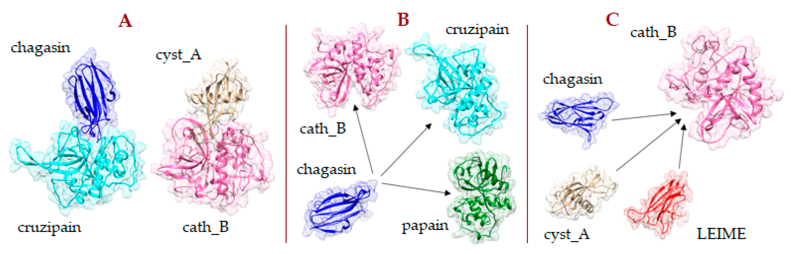
2.1. Heterodimer Models Constructed by Homology Modeling

Seventy molecular models, homologous cruzipain-chagasin heterodimers (e.g., Figure 2A), were constructed by homology modelling using the SWISS-MODEL server [34]. The details of the templates used are presented in Table 2. Two model quality evaluation parameters are showed. The first score refers to the global model quality estimation (QMQE) from the model building which is presented in a SWISS-MODEL homology modelling report [34] (e.g., Figure 2D) and the second, the protein-protein complex evaluation that is calculated by the PROCOS program [35]. All constructed models that registered a QMQE score > 0.72 or a PROCOS score > 0.75 were optimized. These models were refined with the GalaxyRefineComplex program by interface repacking, which successfully improved homology model structures. This refinement method allows flexibility at the protein interface and in the overall structure to capture conformational changes that occur upon binding [36]. ΔGb was calculated by FoldX, which uses the empirical force field FoldX [22]. Previously, to evaluate energy, the Repair-FoldX tool was used to avoid the presence of incompatibilities between the CP and CPI structure.
Validation of the computational methodology was carried out by evaluating the RMSD parameter of the nine molecular models obtained by homology, using as a reference their respective crystal complex heterodimer structures obtained from the Protein Data Bank as well as the PROCOS score [35]. This comparison of results is showed in Table 3. A more detailed analysis of the QMQE score and the PROCOS score of the 70 molecular models constructed by homology is presented in Table 2. We also performed an analysis of the composition of the residues in the interface of the PDB structures. The corresponding sequence alignment and the conserved residues identified in the interface of homologous heterodimers of cuzipain−chagasin are presented in Appendix A.

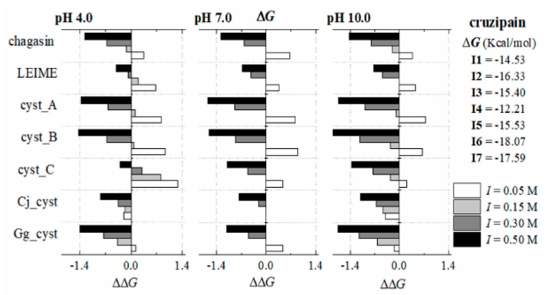
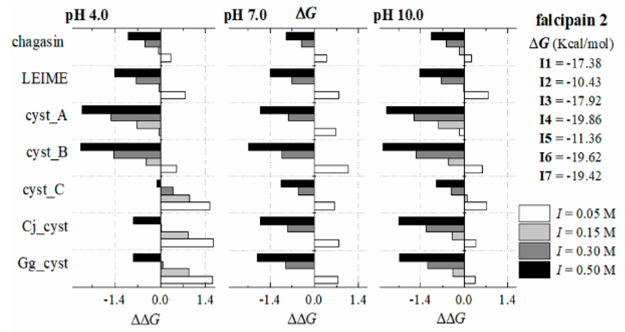
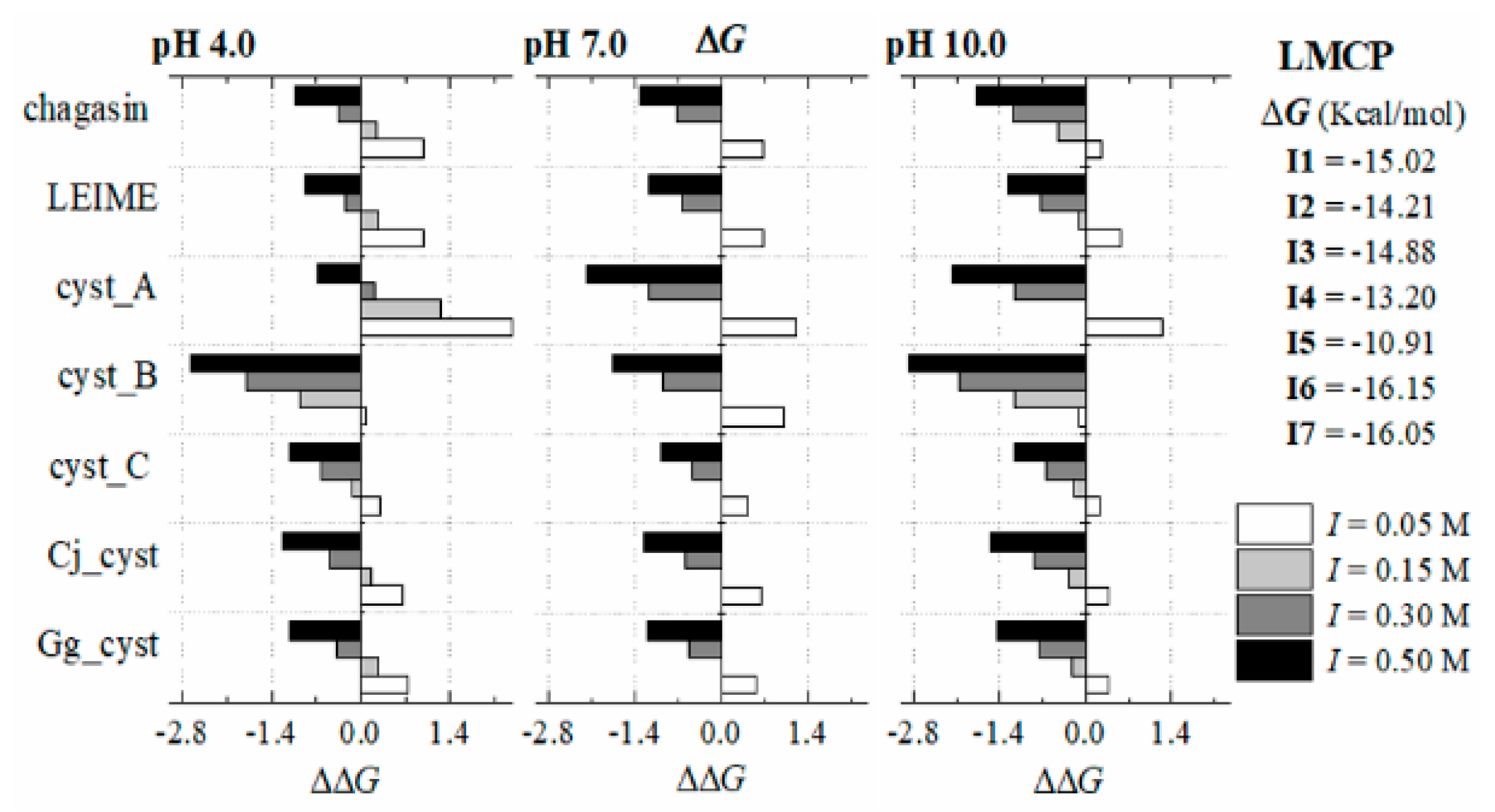
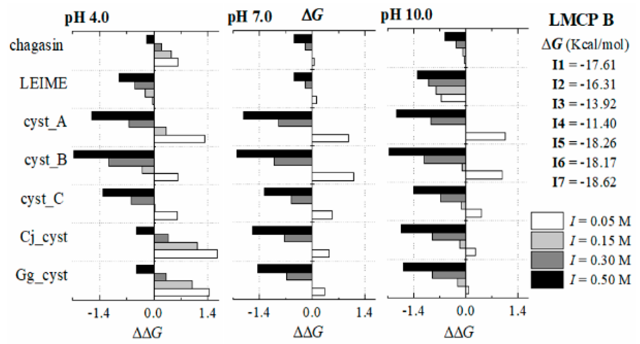
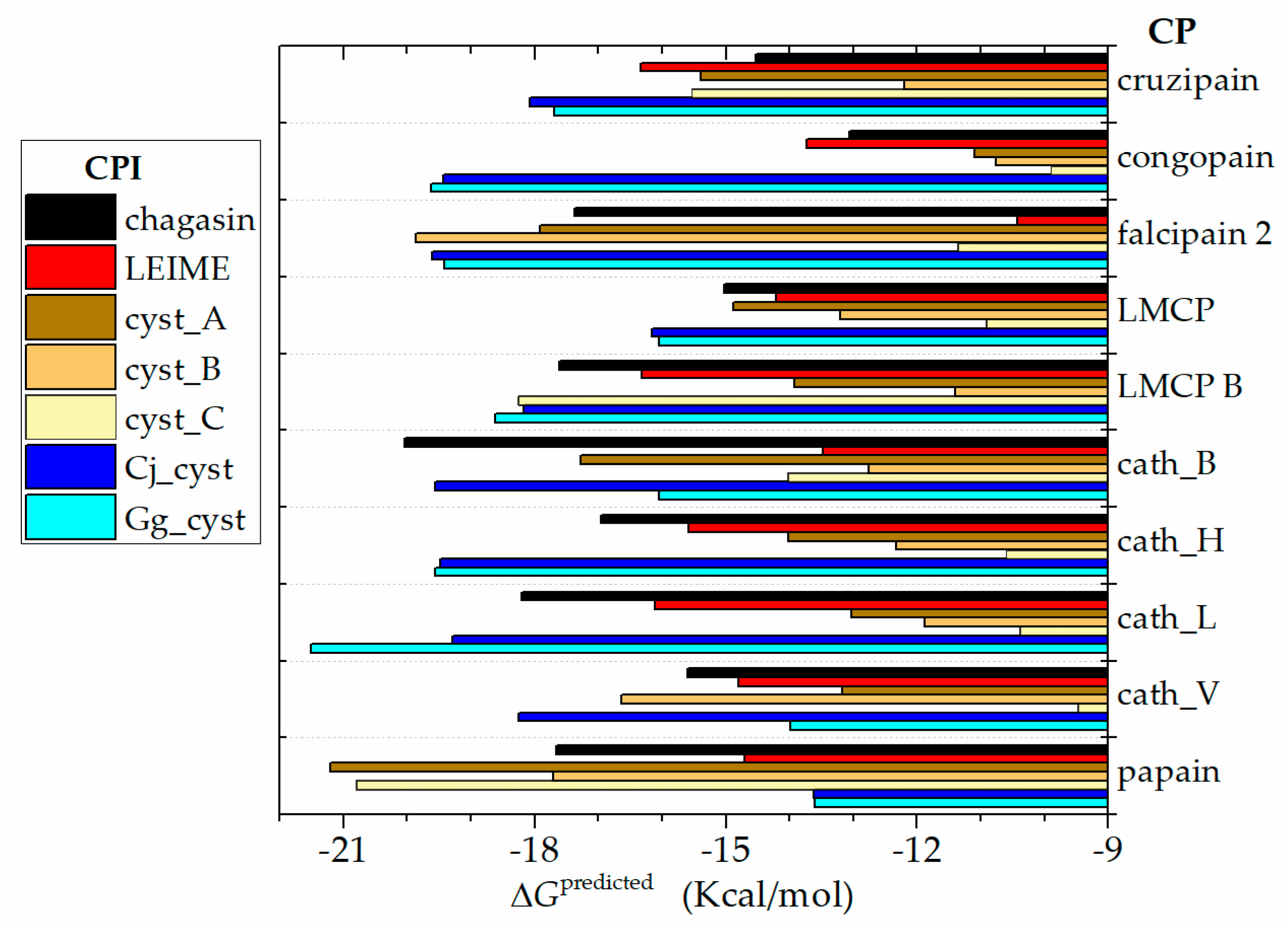
2.2. ΔGb Prediction of Homologs of the Cruzipain−Chagasin Complex

The predicted ΔGb values obtained from the 70 models built by homology for the different systems studied are showed in a range of −21.5 to −9.5 Kcal/mol. In terms of affinity as Ka = (Kd)−1 (see Materials and Methods), these results recorded a moderate affinity (107 to 1012 M−1) and a high affinity (1012 to 1015 M−1). The (Kd)−1 data profile is shown in Figure 3.

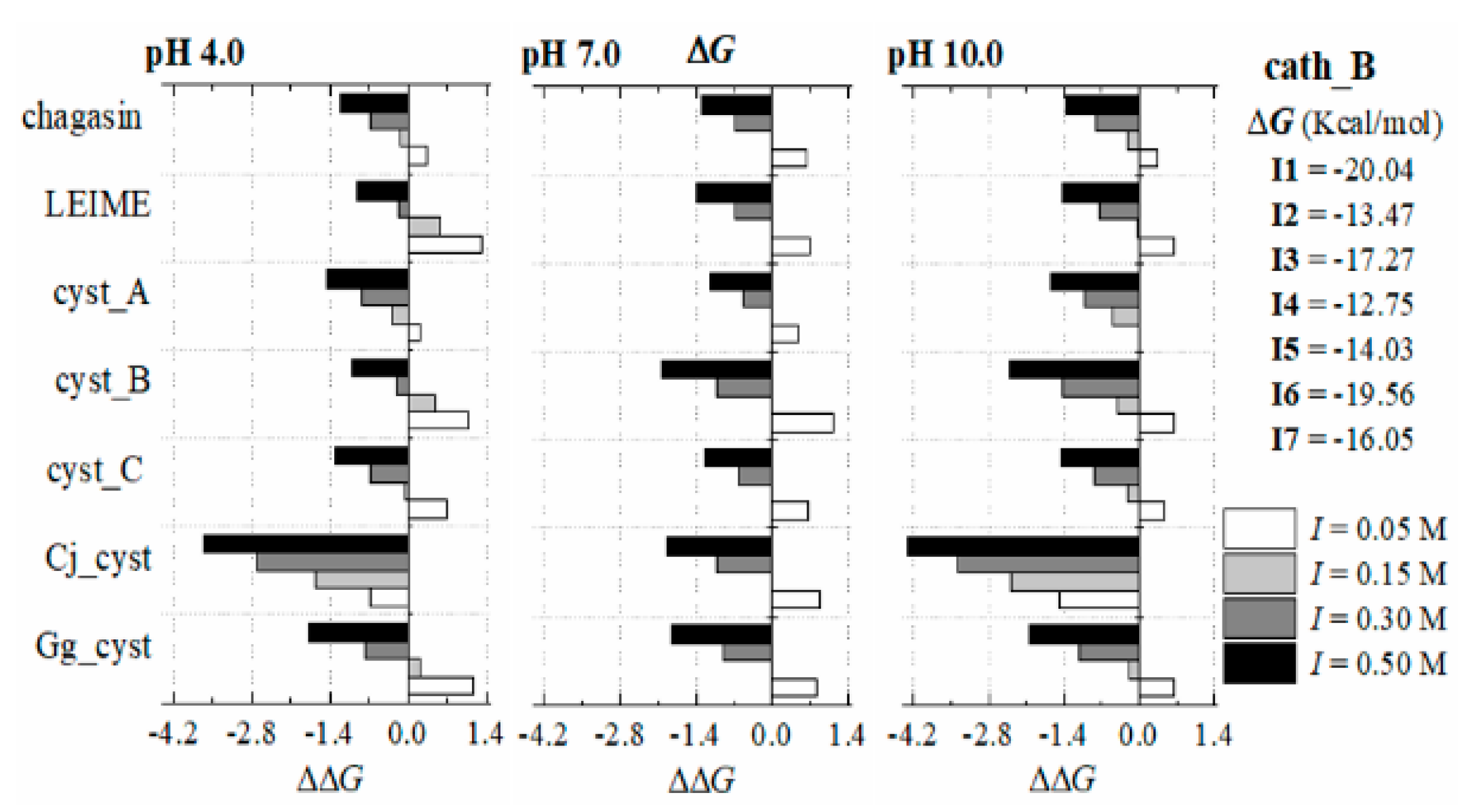
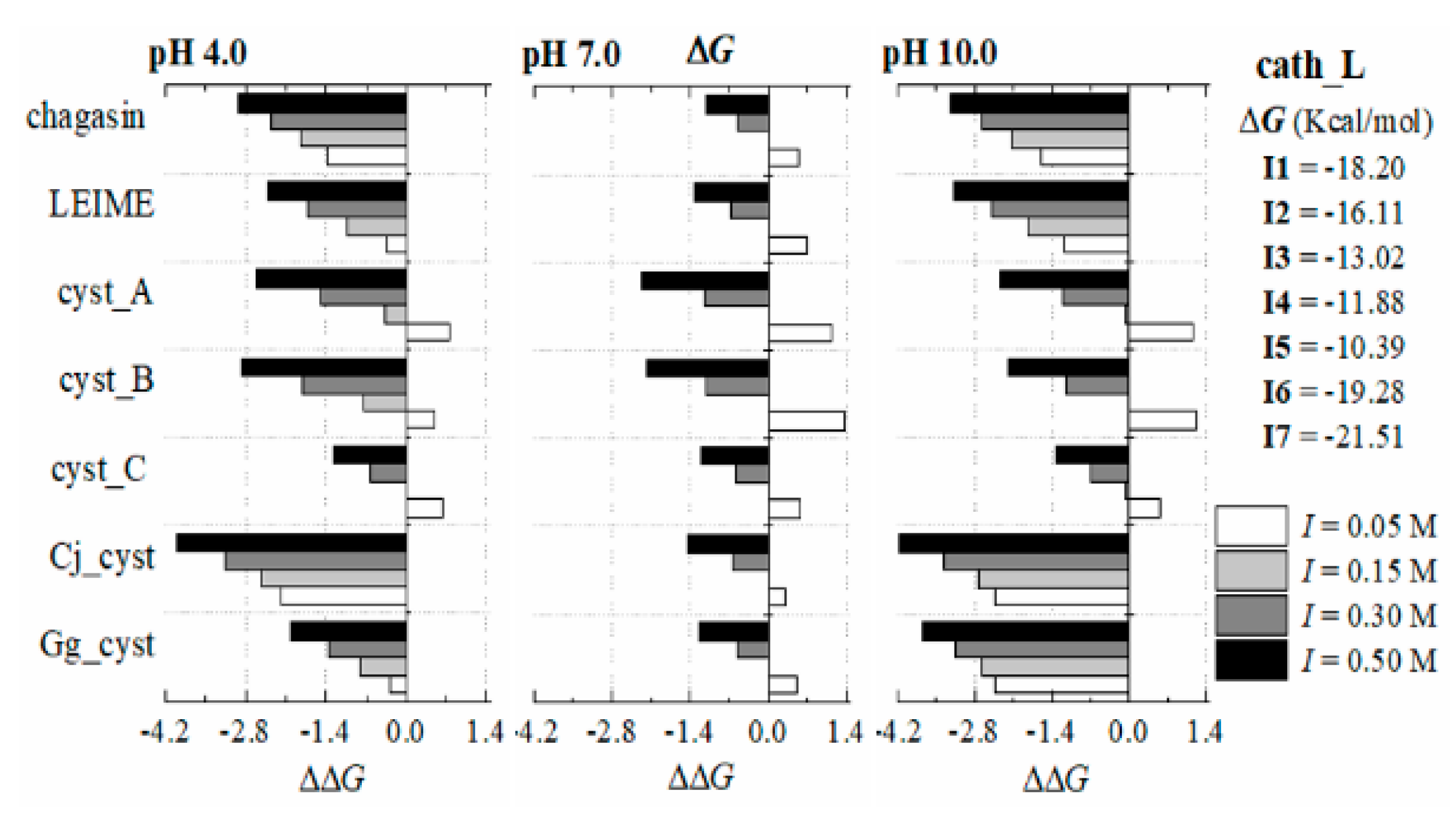
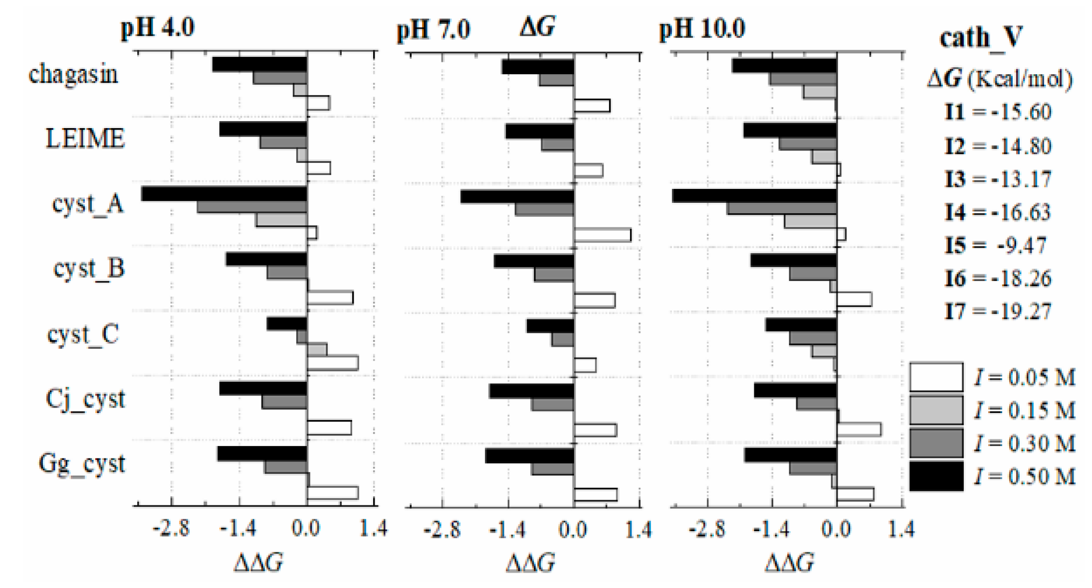
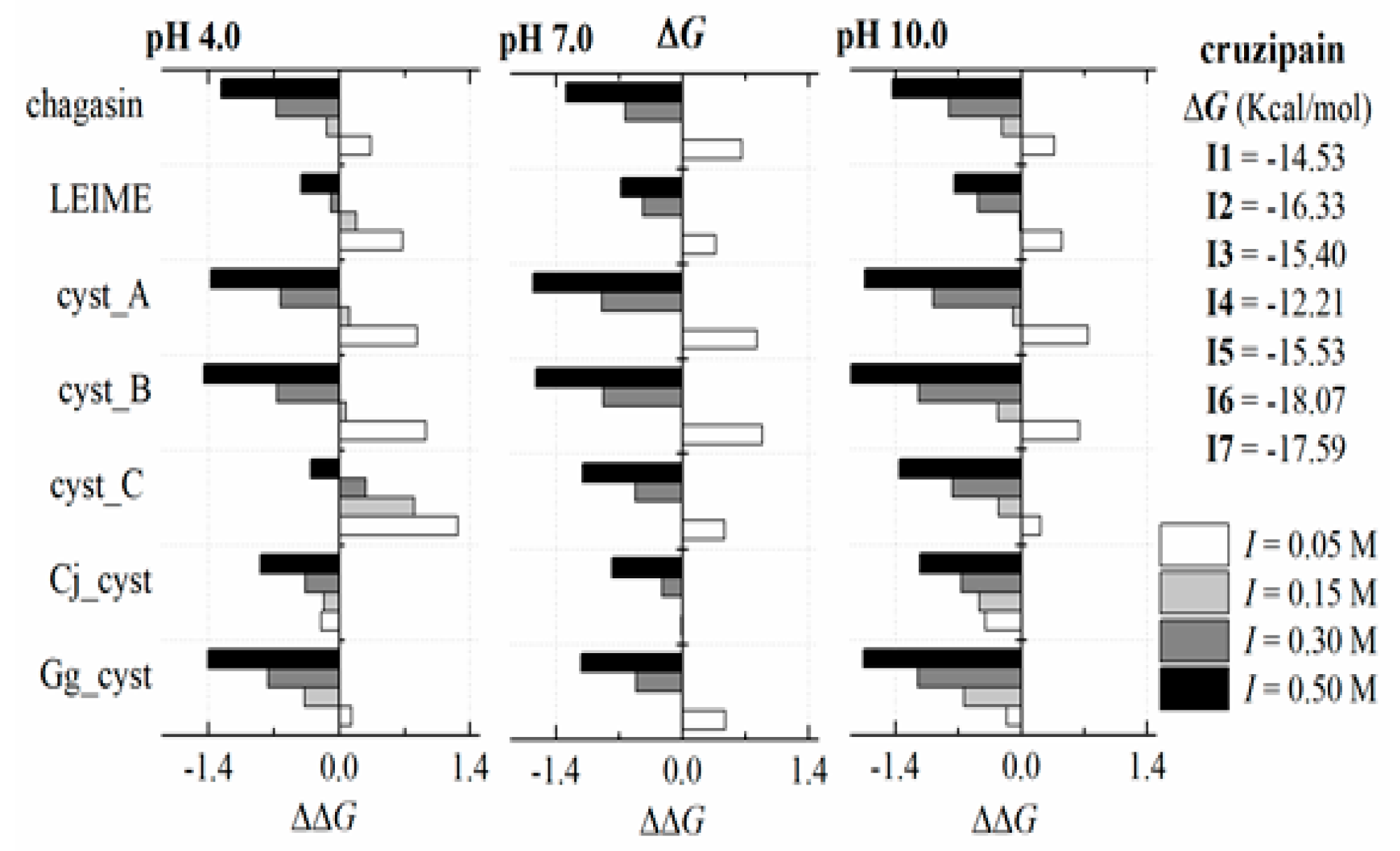
2.3. Energetic Analysis

In order to carry out a multiple analysis of the 70 models of homologous heterodimers of cuzipain−chagasin, we calculated ΔΔG and considered the presence of two ligands that bind to the same receptor. Therefore, the multiple analysis in terms of thermodynamic parameters was performed as |ΔΔG| = |ΔGbfrom FoldX − ΔGb from Ki| in absolute value (see Section 4). In this analysis, nine models were identified that recorded |ΔΔG| < 1.3 (dimer systems as cruzipain in complex with chagasin, cyst_A, cyst_C and Gg_cyst and also cath_H−LEIME, cath_H−cyst_A, cath_L−cyst_A, cath_L−cyst_B and cath_B−cyst_C) and five models to 2.8 > |ΔΔG| > 1.3 (systems as cruzipain−cyst_B, papain−cyst_C, LMCP−LEIME, papain−Cj_cyst and papain−Gg_cyst) (see Figure 4). The other twenty models for comparison showed a tendency or profile according to the behaviour between the systems analyzed but not in magnitude (|ΔΔG| > 2.8).
The Gibbs equation is defined as ΔGb = ΔHTΔS and involves two components that contribute energy to the equilibrium, enthalpy (ΔH) and entropy (ΔS). The predicted ΔGb was obtained at the temperatures of 298, 313 and 323 K. Then, a van‘t Hoff plots analysis was performed to obtain the thermodynamic terms (ΔH and ΔS). Thus, the ΔGb expressed in terms of the contribution of ΔH and ΔS is showed in Figure 5. The magnitude of the contribution of ΔH and ΔS to ΔG are presented in Appendix B.
Finally, we present the results of a multiple analysis of the 14 models of homologous heterodimers of cuzipain−chagasin and their ΔGb(I, pH) profiles based on ΔΔGb (relative binding energy). These studies are present in Appendix B. The typical result of this analysis for the particular case of cruzipain in interaction with the seven inhibitors is shown in Figure 6.

3. Discussion

3.1. Analysis of Ki Values Obtained in In Vitro Experiments (Reported in the Literature)

Homologous cruzipain-chagasin complexes previously reported, this shows Ki records in the range of nM to fM (Table 1). The cruzipain-chagasin and cruzipain-cyst_B complexes recorded a Ki of around 0.045 nM. In reference to the cruzipain-cyst_A, cruzipain-cyst_C cruzipain-Gg_cyst complexes have an order of magnitude in Ki less than that registered in the cruzipain-chagasin complex. It highlights a particular interest other systems such as cath_L-cyst_C, papain-cyst_C and papain-Gg_cyst complex due to its low registered Ki.

3.2. Molecular Models Constructed by Homology Modeling and Methodology Validation

The methodology implemented in the development of this work was validated with satisfactory results. The PROCOS results showed a probability-like measure to be a native composition interface for the 70 molecular models analysed. The PROCOS scores collected from the nine PDB structure analysis were values of 0.47 to 1.0 (66.7% of values between 0.8 and 1.0) and 0.59 to 0.99 for the respective molecular models constructed by homology (Table 2). Values with the lowest probability may be an implication that shows very high van der Waals energies [36] such as those of the 3CBK, 3KSE and 3KFQ complex. The global analysis of the molecular model constructed by homology showed a PROCOS score of 75% for values of 0.75 to 1.0. We considered, according to the validation results, that in low scores there is a presence of strong nonspecific interactions due to the presence of aromatic residues at the interface; only 33.3% of models constructed were refined with the GalaxyRefineComplex program (see Table 2). An extensive analysis of the interface of cysteine protease−cystatin structures, such as crystal structures (PDB ID: 1STF, 3K9M, 1NB5, 3KSE and 3KFQ) and two molecular models (chymo−Gg_cyst and papain−Gg_cyst complex) has been presented previously [38].

3.3. Analysis of ΔGb Prediction Values

The analysis of ΔGb from cruzipain-CPIs complex (Figure 7) reveals that CPI proteins from protozoan pathogens both cruzipain-chagasin and cruzipain-LEIME complex (−14.5 and −16.3 Kcal/mol, respectively) register a ΔGb magnitude close to cruzipain-cyst_A and cruzipain-cyst_C (both CPIs of human) of −15.4 Kcal/mol. While the cruzipain-cyst_B complex recorded a higher ΔGb value (−12.2 Kcal/mol) compared to that obtained for cruzipain-chagasin complex. Whereas the inhibitors Cj_cyst and Gg_cyst register a considerably lower ΔGb (−18.1 and −17.7 Kcal/mol, respectively) than cruzipain- chagasin complex. Other important correlations in the CP-CPI complex study may emerge from a more detailed analysis of the predictions presented in Figure 7. Of our particular interest is the prediction of a high affinity of chagasin for cathepsins human -cath_B, cath_H, cath_L and cath_V- (ΔGb of −15.6 to −20.0 Kcal/mol) as well as bird cystatins (Cj_cyst and Gg_cyst) for cathepsins human and CP from protozoan pathogens (e.g., cruzipain, congopain, falcipain 2, LMCP and LMCP B). Plant CPs such as papain-chagasin complex showed interesting lower ΔGb magnitudes (−17.3 Kcal/mol) compared to cruzipain-chagasin complex.
The results presented in the FoldX predictions (Figure 3) with the experimental data (Figure 1) are not comparable; conceptually Ki is different from Kd. Therefore, the presented magnitudes of Ki as experimental data do not need to be of the same order of magnitude as Kd obtained by the predictions of FoldX. In a reversible bimolecular mechanism of a simple one-step model, the Kd and the equilibrium association constant (Ka) are reciprocally related (Ka = Kd−1) [38]. On the other hand, the inhibition constant (Ki) can be determined or approximated (Kiapp) with various enzymatic kinetics from data obtained in an enzyme inhibition experiment (for example, methods that use a nonlinear expression more accurately estimate Ki as well as values for KM and VMAX or the linearized approach of Dixon [39]). In ligand binding studies and under controlled conditions, Ki can be calculated with the Cheng and Proof equation, where it is assumed that KiKd = (Ka−1) [40].
Performing a global analysis of results in terms of ΔGb, there is no clear trend in the behaviour of the energy magnitudes obtained. This variability of magnitudes recorded for ΔGb in the structural homologs of the cruzipain−chagasin complex is inherent in the composition of the residues that constitute their respective interfaces. The energy analysis of the contribution of ΔH and ΔS to ΔGb of the 70-molecular model of homologs of the cruzipain−chagasin complex presents a ΔGb is mostly ΔH-driven (Figure 5) at neutral pH and at an ionic strength (I) of 0.15 M.
The dependence of ΔGb (I, pH) at 298 K to the cruzipain−chagasin complex at three pH values (pH 4.0, 7.0 and 10.0) predicts a linear dependence of ΔGb (I) with R2 values equal to 0.9977 (at acid pH), 0.9999 (at neutral pH) and 0.9997 (at alkali pH). Similar results (R2 values > 0.99) were recorded in systems such as cyst_A, cyst_B, cyst_C and Gg_cyst in complex with cruzipain. Cruzipain−LEIME and cruzipain−Cj_cyst systems recorded R2 values > 0.77 and >0.98, respectively (Figure 6). Other systems of homologous heterodimers of cruzipain−chagasin presented similar results (see Appendix B).
Papain-like CP play an important role in the regulation of biological cycles of all organisms. Endogenous protein structure inhibitors such as chagasin-like and cystatin-like regulate its biological function. We can appreciate in Figure 1 that cysteine proteases of the protozoan pathogen have greater specificity and a high affinity for their human endogen inhibitors (cystatin-like) compared to their natural inhibitor (chagasin). Regarding the natural chagasin-like inhibitors such as chagasin and LEIME, the predicted ΔGb reveals a high affinity for human cathepsins much better than human cystatins (Figure 7).
Considering the results analyzed, it is possible to formulate two hypothetical events that could occur simultaneously in the invasive process of the pathogenic protozoan (Figure 8). The first favours the metabolism of the pathogenic protozoan where endogenous CPI of the human (CPI_hum) is not able to inhibit the CP of the protozoan pathogen (CP_pp); that is, ΔGbCP_pp-CPI_pp << ΔGb CP_pp-CPI_hum. The second event occurs in favour of the parasite defense mechanism due to the presence of endogenous CPI inhibitors of pathogenic protozoan (CPI_pp) with a high affinity that can inactivate human CP (CP_hum); that is ΔGbCP_hum-CPI_pp << ΔGbCP_hum-CPI_hum. At this moment, the putative situation that we have cited is of great interest to the scientific community and innovative research is being developed in this respect to continue discovering and characterizing new natural inhibitors of this protein structure [7].
The agonist interactions between CP and CPI allow correct metabolic function of the living organism. In the infective process of a human organism by a pathogenic protozoan, the presence of both CP and exogenous PCI (from the protozoan pathogen) is a determinant that causes the disease because the defense mechanism of the human organism is affected or interrupted. A potential solution to these antagonistic interactions that could be applied in the pharmacological treatment of the disease caused by pathogenic protozoa is the exogenous insertion of natural plant CPI or CP [7].
In agreement with the previously reported information about the experimental Ki heterodimer homologs of cruzipain−chagasin (Table 1) and the respective analysis presented in this work, we suggest that the presence of highly specific cross-linkers and chagasin for homologous agonists (cathepsins and cystatins) in humans is a key point of the evolutionary persistence of Chagas disease. The development of new CPIs with a protein structure and peptide inhibitors derived from these protein structures is based on the study of the interaction of natural PCI such as chagasin-like and cystatin-like inhibitors in order to develop new antiparasitic drugs that have non-toxic properties, high specificity, biological selectivity and potent and reversible inhibition.

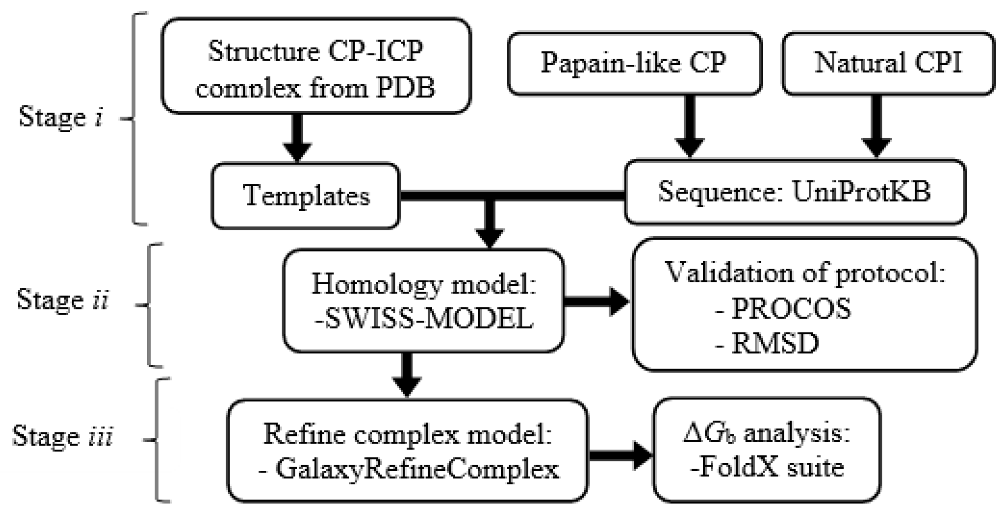
4. Materials and Methods

The protocol used to study the 70 homologous heterodimers of cruzipain−chagasin was divided into three stages (the methodology is presented in Figure 9): (i) Identification templates in RCSB PDB and alignment of sequences obtained from UniProtKB; (ii) Model building with the SWISS MODEL [34] and refinement of model interfaces with GalaxyRefineComplex [36] and stage (iii) Analysis of binding energy (ΔGb) with the FoldX suite [22].

4.1. Preparation and Analysis of Sequences

Ten crystal complex structures (ID PDB: 1YVB, 2OUL, 3CBK, 3CBK, 3K9M, 2NQD, 3KSE, 3KFQ and 1STF) and five free protein structures (ID PDB: 1ME3, 1YAL, 1PPO, 3GAX and 2C34) were selected from the RCSB PDB (www.rcsb.org (accessed on February 2019)) [37] as templates (Table 4) for the study of homologous heterodimers of the cruzipain−chagasin complex. All structures were refined by subtracting water molecules and external chains. Energy minimization was performed using the YASARA force field [41]. These PDB structures include CP and CPI that occur in organisms such as protozoan pathogens, plants and humans. The respective sequences were recovered with their accession number from UniProt [42]. The sequences were aligned with those reported in the crystal structures and cut to the same length; these protein sequences were used to build the structure models.

4.2. Structural Modeling by Homology

Structural models of homologous heterodimers were generated with the SWISS-MODEL server [34], an automated protein homology-modelling server. Each pair of the protein sequences that form the heterodimer (previously prepared) was loaded into the SWISS-MODEL server using the template mode [34]. The interfaces of all the structures generated were refined with the GalaxyRefineComplex program [36] installed in a computer with a 3.4 GHz Intel Core i7 processor and 23.5 GB RAM and with Linux Mint 17.3 as the operating system. The generated structural models were validated by measuring two parameters. The first was the RMSD (root-mean-square deviation) of the models obtained with respect to the crystallographic structures reported. The second was the evaluation of the probability of amino acids in the interface of the models. This measurement was made with the PROCOS server [35].

4.3. Evaluation of Heterodimer Models and ΔGb Analysis

ΔGb was evaluated in all models built with the FoldX program [22]. FoldX uses the 3D structure to calculate the energy. The empirical force field algorithm is based on free energy (ΔG) terms aiming to calculate the ΔG in Kcal/mol. The FoldX energy function includes terms as van der Waals, solvation (polar and hydrophobic), electrostatic, hydrogen bonds and so forth [43,44]. Other relevant aspects about FoldX program are brief described in official website. Available online: http://foldxsuite.crg.eu/products (accessed on February 2019) [45]. We did not perform a test case of the modelling/energetics from FoldX program against MD simulation [43].
In order to identify a modulation of ΔGb(pH) due to changes in the electrostatic energy [38], the dependence of ΔGb was studied with a pH variation of 3.0 (acid pH), 7.0 (neutral pH) and 10.0 (alkali pH), at temperature of 298 K and at an ionic strength (I) of 0.15 M. The dependence of ΔGb(I, pH) (at ionic strengths of 0.05, 0.15, 3.0 and 5.0 M) was studied to three pH values (pHs of 3.0, 7.0 and 10.0), at temperature of 298 K. Also, the dependence of ΔGb(T) was studied at temperatures of 298, 313 and 323 K, pH 7.0 (at neutral pH) and at an ionic strength of 0.15 M. Prior to the execution of the simulation, the variations of the parameters of pH, temperature and ionic strength were established in the commands used to operate FoldX, these are RepairPDB command and AnalyseComplex command [45].
In order to obtain the thermodynamic terms of Gibbs equation (ΔG = ΔH − TΔS), enthalpy (ΔH) and entropy (ΔS), we used the method based on ΔGb prediction at different temperatures combined with van‘t Hoff plot analyses (lnK = −ΔH/(RT) + ΔS/R; K, equilibrium constant and R, ideal gas constant) to obtain the thermodynamic terms of Gibbs equation (ΔG = ΔH − TΔS), enthalpy (ΔH) and entropy (ΔS). Therefore, we proceeded to generate a typical graph 1/T versus lnK, the slope = −ΔH/R and intercept = ΔS/R. Solving the linear equation resulting from the linear fitting, we obtained ΔH = (slope) (−R) and ΔS = (intercept) (R).
According to the equation in the equilibrium system, we can define free energy, ΔGb = −RTlnKa = RTlnKd; where R is a gas constant, T is temperature and Ka is a binding constant (Ka = 1/Kd, where Kd is dissociation constant) [12,38,40]. ΔΔG is defined as ΔΔG = ΔGbII − ΔGbI = RT ln(KdII/KdI). The term ΔΔG is a thermodynamic parameter that allows us to identify the ΔGb difference present in a system under study (ΔGbI) in relation to a reference system (ΔGbII). It is well known that if it is present, a ten-fold difference in Kds at 298 K corresponds to a ΔΔG difference of 1.34 Kcal/mol. Therefore, a ΔΔG difference of 1 Kcal/mol at 298 K corresponds to a 5.4-fold difference in Kds. In both hypothetical situations the binding of ligand 2 is weaker than that of ligand 1. In the present work, we use ΔΔG to compare the ΔGb recorded in in silico study with data reported previously.

5. Conclusions

In the present in silico study, we can suggest that from the scaffolding of the interaction of bird cystatins (Cj_cyst and Gg_cyst), it is possible to design or propose natural inhibitors with a high affinity for protozoan proteases. Therefore, it is possible to rationally design both highly selective antagonistic proteins to CP and CPI from protozoan pathogens. The interface composition is decisive to modulate ΔGb. Chagasin-like and cystatin-like structures in interaction with cruzipain homologs showed that ΔGb is mostly ΔH-driven at neutral pH and at an ionic strength (I) of 0.15 M. Because of their wide structural stability to pH, ionic strength (I) and temperature changes (previously reported in in vitro studies by other researchers) and according to the results presented, we propose Cj_cyst and Gg_cyst as excellent candidates and desirable scaffolding for the design of new cruzipain inhibitors derived from peptide structure. The computational protocol based on Homology Modelling and FoldX predictions allowed the identification and prediction of thermodynamics parameters of binding energy for cruzipain−chagasin-like heterodimers.

Author Contributions

Formal analysis, F.R.E., I.P. and G.R.; Investigation, F.R.E., A.J.S., V.H.-M. and C.G.P.; Methodology, F.R.E.; Supervision, G.R.; Writing—original draft, F.R.E. and G.R. Writing—review & editing, F.R.E. and G.R.

Funding

This research received no external funding.

Acknowledgments

This work was supported by the National Council of Science and Technology (CONACyT, Mexico), by grant number Postdoc, CVU 204984 to F.R.E. Gildardo Rivera holds a scholarship from the “Comisión de Operación y Fomento de Actividades Académicas” (COFAA-IPN) and the “Programa de Estímulos al Desempeño de los Investigadores” (EDI-IPN). Publication was supported by the Universidad Autonoma de Tamaulipas. We wish to express our gratitude to the Consejo Nacional de Ciencia y Tecnología, México (Proyecto Apoyado por el Fondo Sectorial de Investigación para la Educación, CB-2014-01, 241615) and Secretaría de Investigación y Posgrado del Instituto Politécnico Nacional (SIP-20180306) for their financial support.

Conflicts of Interest

The authors declare no conflict of interest.

Abbreviations

ΔGbbinding energy
CPcysteine proteinase
ICPcysteine protease inhibitors
cathcathepsin
QMQEthe global model quality estimation
cystciystatin
Iionic strength

Appendix A

Residues Present in the Interaction of CP to Natural ICP from Crystal Structures

The search for the interaction of the residues present in the interaction of CP to natural ICP from crystal structures (Figure A1) was made with the computational tool Find Interaction Partners and Binding Sites [46,47]. Protein structure in bold and underlined indicates the protein that was searched for in the database; those in bold represent the structures found in the templates registered in the database. The information contained in Table A1 allowed the identification of residues located in the protein−protein interaction site before constructing molecular models. Once the models were constructed, a 3D alignment of the homologous structures was carried out and the corresponding residues were located (e.g., Figure 2B,C).
Figure A1. Identification of residues present in the CP to CPI interaction. Query: Cruzipain (PDB 1ME3), chagasin, LEIME (PDB 2C34) and cystatins. *, residues at binding site; **, numbering; i, cruzipain; ii, chagasin; iii, LEIME; iv, cyst_A; v, Gg_cyst. PDB: A, 3E1Z; B, 2OUL; C 2NQD; D, 3K9M; E, 1YVB; F, 3KSE; G, 3KFQ; H, 3CBK; and I, 1STF.
Ijms 20 01320 g0a1

Appendix B

Appendix B.1. Contribution of ΔH y ΔS to ΔG

ΔGb(T) were calculated respectively at 273, 313 and 323 K, I 0.15 M and pH 7.0 (see Materials and Methods). Then, Table A1 shows the result of the analysis carried out at 273 K, pH 7.0 and I 0.15 M.
Table A1. Profile of the contribution of enthalpy (ΔH) y entropy (−TΔS) to ΔG (Kcal/mol) at 273 K, pH 7.0 and I 0.15 M.
ICPchagasinLEIMEcyst_Acyst_Bcyst_CCj_cystGg_cyst
CPΔH−TΔSΔH−TΔSΔH−TΔSΔH−TΔSΔH−TΔSΔH−TΔSΔH−TΔS
E1−20.35.8−17.00.6−19.03.5−23.211.0−18.52.9−22.64.5−22.85.1
E2−16.63.6−15.31.5−18.47.3−18.37.5−12.52.6−28.28.7−26.87.2
E3−22.14.8−10.80.4−20.22.3−27.98.1−13.32.0−21.21.5−20.91.5
E4−21.76.7−16.52.4−24.19.2−21.78.5−15.74.8−23.87.7−23.57.5
E5−18.10.5−17.31.0−21.67.7−21.510.1−23.75.5−21.93.7−22.03.4
E6−21.61.5−15.72.3−22.04.8−21.78.9−16.42.4−23.23.6−23.07.0
E7−22.95.9−22.87.3−22.58.4−19.57.1−14.03.4−28.89.3−23.94.3
E8−19.81.6−18.11.9−24.511.4−26.414.6−14.64.3−34.815.6−31.810.2
E9−19.84.2−19.44.6−26.313.1−22.96.2−13.13.6−25.87.6−27.78.4
E10−22.85.1−21.26.5−30.08.7−24.16.4−26.55.7−21.17.5−22.69.0
CP, cysteine proteases: E1, cruzipain; E2, congopain; E3, falcipain 2; E4, LMCP; E5, LMCP B; E6, cath_B; E7, cath_H; E8, cath_L; E9, cath_V; E10, papain; ICP, cysteine protease inhibitors: chagasin, LEIME, cyst_A, cyst_B, cyst_C, Cj_cyst and Gg_cyst.

Appendix B.2. Dependence of ΔGb (I, pH) at 298 K

Analysis of ΔGb (I, pH) was performed on the variation of I (values of 0.05, 0.15, 3.0 and 5.0 M) and pH (values of 3.0, 7.0 and 10.0) at 298 K. In Figure A2, Figure A3, Figure A4, Figure A5, Figure A6, Figure A7, Figure A8, Figure A9, Figure A10 and Figure A11 we show the ΔΔG calculated as ΔΔG = (ΔGb − ΔG), where ΔGb (I, pH) at 273 K and ΔG reference were calculated respectively at 273 K, I 0.15 M and pH 7.0; data showed in box.
Figure A2, Figure A3, Figure A4, Figure A5, Figure A6, Figure A7, Figure A8, Figure A9, Figure A10 and Figure A11. Profile of ΔGb(I,pH). ΔGb, free energy; ΔΔG, relative binding energy obtained from ΔGb (binding energy) which was predicted at 278 K, pH 7.0 and I = 0.15 M. Binding inhibitor: I1, chagasin; I2, LEIME; I3, cyst_A; I4, cyst_B; I5, cyst_C; I6, Cj_cyst; I7, Gg_cyst.
Figure A2. cruzipain−CPIs complex.
Ijms 20 01320 g0a2
Figure A3. congopain−CPIs complex.
Ijms 20 01320 g0a3
Figure A4. falcipain 2−CPIs complex.
Ijms 20 01320 g0a4
Figure A5. LMCP−CPIs complex.
Ijms 20 01320 g0a5
Figure A6. LMCP B−CPIs complex.
Ijms 20 01320 g0a6
Figure A7. cath B−CPIs complex.
Ijms 20 01320 g0a7
Figure A8. cath H−CPIs complex.
Ijms 20 01320 g0a8
Figure A9. cath L−CPIs complex.
Ijms 20 01320 g0a9
Figure A10. cath V−CPIs complex.
Ijms 20 01320 g0a10
Figure A11. papain−CPIs complex.
Ijms 20 01320 g0a11

References

  1. Sueth-Santiago, V.; Decote-Ricardo, D.; Morrot, A.; Freire-de-Lima, C.G.; Freire Lima, M.E. Challenges in the chemotherapy of Chagas disease: Looking for possibilities related to the differences and similarities between the parasite and host. World J. Biol. Chem. 2017, 26, 57–80. [Google Scholar] [CrossRef] [PubMed]
  2. Lugo-Caballero, C.I.; Dzul-Rosado, K.; Dzul-Tut, I.; Balam-May, I.; Zavala-Castro, J. Knowledge of vector-borne diseases (dengue, rickettsiosis and Chagas disease) in physicians. Gaceta Médica de México 2017, 153, 321–328. [Google Scholar] [PubMed]
  3. Liu, Q.; Zhou, X.N. Preventing the transmission of American trypanosomiasis and its spread into non-endemic countries. Infect. Dis. Poverty 2015, 4, 1–11. [Google Scholar] [CrossRef] [PubMed]
  4. Roca, C.S.; Soriano-Arandes, A.; Solsona, D.L.; Gascón, B.J. Grupo de consenso Chagas-APS. Documento de consenso sobre el abordaje de la enfermedad de Chagas en atención primaria de salud de áreas no endémicas. Atención Primaria 2015, 475, 308–317. [Google Scholar] [CrossRef] [PubMed]
  5. Mottram, J.C.; Helms, M.J.; Coombs, G.H.; Sajid, M. Clan CD cysteine peptidases of parasitic protozoa. TRENDS Parasitol. 2003, 19, 182–187. [Google Scholar] [CrossRef]
  6. Joon-Yong, C.; Young-An, B.; Byoung-Kuk, N.; Yoon, K. Cysteine protease inhibitors as potential antiparasitic agents. Expert Opin. Ther. Pat. 2005, 15, 995–1007. [Google Scholar] [CrossRef]
  7. Van Wyk, S.G.; Kunert, K.J.; Cullis, C.A.; Pillay, P.; Makgopa, M.E.; Schlüter, U.; Vorster, B.J. Review: The future of cystatin engineering. Plant Sci. 2016, 246, 119–127. [Google Scholar] [CrossRef] [Green Version]
  8. Leto, G.; Crescimanno, M.; Flandina, C. On the role of cystatin C in cancer progression. Life Sci. 2018, 202, 152–160. [Google Scholar] [CrossRef]
  9. Costa, F.R.; Lima Ana, C.A. Natural cysteine protease inhibitors in protozoa: Fifteen years of the chagasin family. Biochimie 2016, 122, 197–207. [Google Scholar] [CrossRef]
  10. Monteiro, A.C.; Abrahamson, M.; Lima, A.P.; Vannier-Santos, M.A.; Scharfstein, J. Identification, characterization and localization of chagasin, a tight-binding cysteine protease inhibitor in Trypanosoma cruzi. J. Cell Sci. 2002, 114, 3933–3942. [Google Scholar]
  11. Smith, B.O.; Picken, N.C.; Westrop, G.D.; Bromek, K.; Mottram, J.C.; Coombs, G.H. The structure of leishmania mexicana ICP provides evidence for convergent evolution of cysteine peptidase inhibitors. J. Biol. Chem. 2006, 281, 5821–5828. [Google Scholar] [CrossRef] [PubMed]
  12. Roy, A.; Nair, S.; Sen, N.; Soni, N.; Madhusudhant, M.S. In silico methods for design of biological therapeutics. Methods 2017, 131, 33–65. [Google Scholar] [CrossRef] [PubMed]
  13. Dourado, D.F.; Flores, S.C. Modeling and fitting protein-protein complexes to predict change of binding energy. Sci. Rep. 2016, 6, 25406. [Google Scholar] [CrossRef] [PubMed]
  14. De Vivo, M.; Masetti, M.; Bottegoni, G.; Cavalli, A. Role of molecular dynamics and related methods in drug discovery. J. Med. Chem. 2016, 59, 4035–4061. [Google Scholar] [CrossRef] [PubMed]
  15. França, T.C. Homology modeling: An important tool for the drug discovery. J. Biomol. Struct. Dyn. 2015, 33, 1780–1793. [Google Scholar] [CrossRef] [PubMed]
  16. Cichero, E.; D’Ursi, P.; Moscatelli, M.; Bruno, O.; Orro, A.; Rotolo, C.; Milanesi, L.; Fossa, P. Homology modeling, docking studies and molecular dynamic simulations using graphical processing unit architecture to probe the type-11 phosphodiesterase catalytic site: A computational approach for the rational design of selective inhibitors. Chem. Biol. Drug 2013, 82, 718–731. [Google Scholar] [CrossRef] [PubMed]
  17. Moro, S.; Deflorian, F.; Bacilieri, M.; Spalluto, G. Ligand-based homology modeling as attractive tool to inspect GPCR structural plasticity. Curr. Pharm. Des. 2006, 12, 2175–2185. [Google Scholar] [CrossRef]
  18. Cichero, E.; Espinoza, S.; Tonelli, M.; Franchini, S.; Gerasimo, A.S.; Sorbi, C.; Gainetdinov, R.R.; Brasili, L.; Fossa, P. A homology modelling-driven study leading to the discovery of the first mouse trace amine-associated receptor 5 (TAAR5) antagonists. Med. Chem. Commun. 2016, 7, 353–364. [Google Scholar] [CrossRef]
  19. Tastan Bishop, O.; Kroon, M. Study of protein complexes via homology modeling, applied to cysteine proteases and their protein inhibitors. J. Mol. Model. 2011, 17, 3163–3172. [Google Scholar] [CrossRef] [PubMed]
  20. Tyzack, J.D.; Furnham, N.; Sillitoe, I.; Orengo, C.M.; Thornton, J.M. Understanding enzyme function evolution from a computational perspective. Curr. Opin. Struct. Biol. 2017, 47, 131–139. [Google Scholar] [CrossRef] [PubMed]
  21. Silva, C.; Martins, M.; Jing, S.; Fu, J.; Cavaco-Paulo, A. Practical insights on enzyme stabilization. Crit. Rev. Biotechnol. 2018, 38, 335–350. [Google Scholar] [CrossRef] [PubMed]
  22. Schymkowitz, J.; Borg, J.; Stricher, F.; Nys, R.; Rousseau, F.; Serrano, L. The FoldX web server: An online force field. Nucleic Acids Res. 2005, 33, W382–W388. [Google Scholar] [CrossRef] [PubMed]
  23. Van Durme, J.; Delgado, J.; Stricher, F.; Serrano, L.; Schymkowitz, J.; Rousseau, F. A graphical interface for the FoldX force field. Bioinformatics 2011, 27, 1711–1712. [Google Scholar] [CrossRef] [PubMed]
  24. Turk, V.; Stoka, V.; Vasiljeva, O.; Renko, M.; Sun, T.; Turk, B.; Turk, D. Cysteine cathepsins: From structure, function and regulation to new frontiers. Biochim. Biophys. Acta 2012, 1824, 68–88. [Google Scholar] [CrossRef] [PubMed] [Green Version]
  25. Redzynia, I.; Ljunggren, A.; Bujacz, A.; Abrahamson, M.; Jaskolski, M.; Bujacz, G. Crystal structure of the parasite inhibitor chagasin in complex with papain allows identification of structural requirements for broad reactivity and specificity determinants for target proteases. FEBS J. 2009, 276, 793–806. [Google Scholar] [CrossRef] [PubMed] [Green Version]
  26. Wang, S.X.; Pandey, K.C.; Scharfstein, J.; Whisstock, J.; Huang, R.K.; Jacobelli, J.; Fletterick, R.J.; Rosenthal, P.J.; Abrahamson, M.; Brinen, L.S.; et al. The structure of chagasin in complex with a cysteine protease clarifies the binding mode and evolution of an inhibitor family. Structure 2007, 15, 535–543. [Google Scholar] [CrossRef]
  27. Ljunggren, A.; Redzynia, I.; Alvarez-Fernandez, M.; Abrahamson, M.; Mort, J.S.; Krupa, J.C.; Jaskolski, M.; Bujacz, G. Crystal structure of the parasite protease inhibitor chagasin in complex with a host target cysteine protease. J. Mol. Biol. 2007, 371, 137–153. [Google Scholar] [CrossRef]
  28. Rowan, A.D.; Brzin, J.; Buttle, D.J.; Barrett, A.J. Inhibition of cysteine proteinases by a protein inhibitor from patato. FEBS Lett. 1990, 269, 328–330. [Google Scholar] [CrossRef]
  29. Chagas, J.R.; Authie, E.; Serveau, C.; Lalmanach, G.; Juliano, L.; Gauthier, F. A comparison of the enzymatic properties of the major cysteine proteinases from Trypanosoma congolense and Trypanosoma cruzi. Mol. Biochem. Parasitol. 1997, 88, 85–94. [Google Scholar] [CrossRef]
  30. Gerhartz, B.; Engh, R.A.; Mentele, R.; Eckerskorn, C.; Torquato, R.; Wittmann, J.; Kolb, H.J.; Machleidt, W.; Fritz, H.; Auerswald, E.A. Quail cystatin: Isolation and characterisation of a new member of the cystatin family and its hypothetical interaction with cathepsin B. FEBS Lett. 1997, 412, 551–558. [Google Scholar] [CrossRef] [Green Version]
  31. Lima, A.P.; dos Reis, F.C.; Serveau, C.; Lalmanach, G.; Juliano, L.; Ménard, R.; Vernet, T.; Thomas, D.Y.; Storer, A.C.; Scharfstein, J. Cysteine protease isoforms from Trypanosoma cruzi, cruzipain 2 and cruzipain, present different substrate preference and susceptibility to inhibitors. Mol. Biochem. Parasitol. 2001, 114, 41–52. [Google Scholar] [CrossRef]
  32. Dos Reis, F.C.; Smith, B.O.; Santos, C.C.; Costa, T.F.; Scharfstein, J.; Coombs, G.H.; Mottram, J.C.; Lima, A.P. The role of conserved residues of chagasin in the inhibition of cysteine peptidases. FEBS Lett. 2008, 582, 485–490. [Google Scholar] [CrossRef] [PubMed] [Green Version]
  33. Stoka, V.; Nycander, M.; Lenarčič, B.; Labriola, C.; Cazzulo, J.J.; Björk, I.; Turk, V. Inhibition of cruzipain, the major cysteine proteinase of the protozoan parasite, Trypanosoma cruzi, by proteinase inhibitors of the cystatin superfamily. FEBS Lett. 1995, 370, 101–104. [Google Scholar] [CrossRef]
  34. Biasini, M.; Bienert, S.; Waterhouse, A.; Arnold, K.; Studer, G.; Schmidt, T.; Kiefer, F.; Cassarino, T.G.; Bertoni, M.; Bordoli, L.; et al. SWISS-MODEL: Modelling protein tertiary and quaternary structure using evolutionary information. Nucleic Acids Res. 2014, 42, W252–W258. [Google Scholar] [CrossRef] [PubMed]
  35. Fink, F.; Hochrein, J.; Wolowski, V.; Merkl, R.; Gronwald, W. PROCOS: Computational analysis of protein–protein complexes. J. Comput. Chem. 2011, 32, 2575–2586. [Google Scholar] [CrossRef] [PubMed]
  36. Heo, L.; Lee, H.; Seok, C. GalaxyRefineComplex: Refinement of protein-protein complex model structures driven by interface repacking. Sci. Rep. 2016, 6, 32153. [Google Scholar] [CrossRef] [PubMed] [Green Version]
  37. Berman, H.M.; Westbrook, J.; Feng, Z.; Gilliland, G.; Bhat, T.N.; Weissig, H.; Shindyalov, I.N.; Bourne, P.E. The Protein Data Bank. Nucleic Acids Res. 2000, 28, 235–242. [Google Scholar] [CrossRef] [Green Version]
  38. Reyes-Espinosa, F.; Arroyo-Reyna, A.; García-Gutiérrez, P.; Serratos, I.N.; Zubillaga, R.A. Effects of pH on the association between the inhibitor cystatin and the proteinase chymopapain. Protein Pept. Lett. 2015, 22, 239–247. [Google Scholar] [CrossRef]
  39. Kakkar, T.; Boxenbaum, H.; Mayersohn, M. Estimation of Ki in a competitive enzyme-inhibition model: Comparisons among three methods of data analysis. Drug Metab. Dispos. 1999, 27, 756–762. [Google Scholar]
  40. Borea, P.A.; Varani, K.; Gessi, S.; Gilli, P.; Dalpiaz, A. Receptor binding thermodynamics as a tool for linking drug efficacy and affinity. Farmaco 1998, 53, 249–254. [Google Scholar] [CrossRef]
  41. Krieger, E.; Joo, K.; Lee, J.; Raman, S.; Thompson, J.; Tyka, M.; Baker, D.; Karplus, K. Improving physical realism, stereochemistry, and side-chain accuracy in homology modeling: Four approaches that performed well in CASP8. Proteins 2009, 77, 114–122. [Google Scholar] [CrossRef] [Green Version]
  42. The UniProt Consortium. UniProt: The universal protein knowledgebase. Nucleic Acids Res. 2017, 45, D158–D169. [Google Scholar] [CrossRef]
  43. Guerois, R.; Nielsen, J.E.; Serrano, L. Predicting changes in the stability of proteins and protein complexes: A study of more than 1000 mutations. J. Mol. Biol. 2002, 320, 369–387. [Google Scholar] [CrossRef]
  44. Buß, O.; Rudat, J.; Ochsenreither, K. FoldX as protein engineering tool: Better than random based approaches? Comput. Struct. Biotechnol. J. 2018, 16, 25–33. [Google Scholar] [CrossRef]
  45. The FoldX Suite. Available online: URL http://foldxsuite.crg.eu/products (accessed on 1 February 2019).
  46. Shoemaker, B.A.; Zhang, D.; Tyagi, M.; Thangudu, R.R.; Fong, J.H.; Marchler-Bauer, A.; Bryant, S.H.; Madej, T.; Panchenko, A.R. IBIS (Inferred Biomolecular Interaction Server) reports, predicts and integrates multiple types of conserved interactions for proteins. Nucleic Acids Res. 2012, 40, D834–D840. [Google Scholar] [CrossRef]
  47. Shoemaker, B.A.; Zhang, D.; Thangudu, R.R.; Tyagi, M.; Fong, J.H.; Marchler-Bauer, A.; Bryant, S.H.; Madej, T.; Panchenko, A.R. Inferred Biomolecular Interaction Server—A web server to analyze and predict protein interacting partners and binding sites. Nucleic Acids Res. 2010, 38, D518–D524. [Google Scholar] [CrossRef]
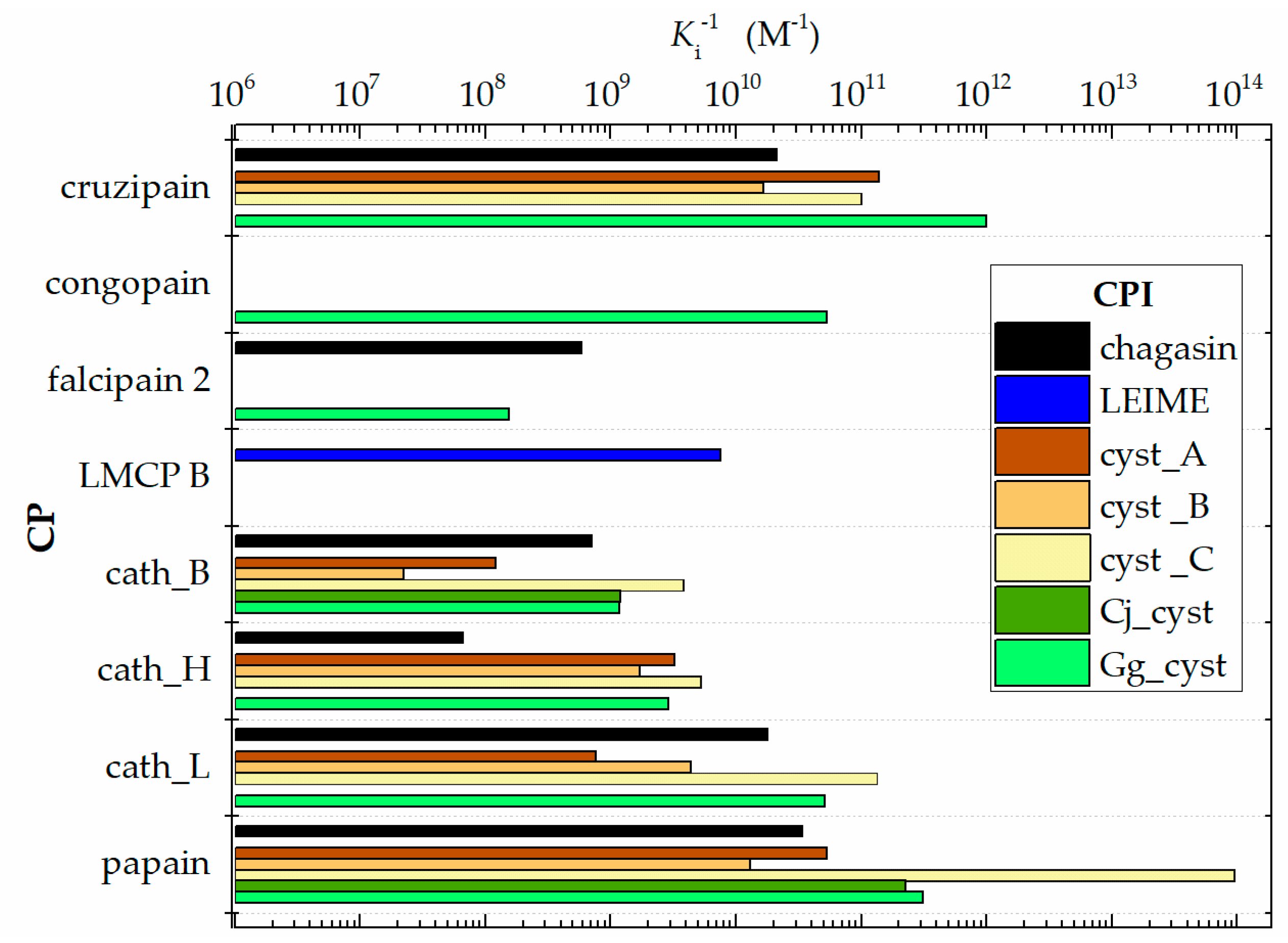
Figure 1. Graphical comparison of (Ki)−1 of homologous heterodimers of cruzipain−chagasin. (Data obtained from Table 1).
Ijms 20 01320 g001
Figure 2. Molecular models constructed by homology. (A) cruzipain−chagasin complex: Chain in red, chagasin structure; chain in grey, cruzipain structure. (B,C) 3D alignment of molecular models of LEIME (chain in green) and Gg_cystain (chain in magenta) on chagasin (chain in red) -numbering of chagasin structure-. (D) Typical SWISS-MODEL homology modelling report (e.g., cruzipain-chagasin complex).
Ijms 20 01320 g002
Figure 3. Data profile of affinity (Ka = Kd−1) from analysis of the 70 homologous heterodimers of cruzipain−chagasin, which were constructed by homology; (Kd)−1 data grouped by CP. Ka was calculated from ΔG. ΔG was evaluated with the FoldX program: pH 7.0 at T 298 K and I = 0.15 M.
Ijms 20 01320 g003
Figure 4. Analysis in terms of relative binding energy. ΔGbpredicted, obtained from FoldX, ΔGbexperimental, obtained from Ki (Table 1). A Graph representation of competitive inhibition of cruzipain with chagasin inhibitor homologs (|ΔΔGi| = |ΔGbfrom FoldX − ΔGbexperimental|). B Graphical record of energy ΔG.
Ijms 20 01320 g004
Figure 5. Data profile of the contribution of enthalpy (ΔH) and entropy (−TΔS) to ΔGb.
Ijms 20 01320 g005
Figure 6. Cruzipain−CPIs complex. ΔG, free energy; ΔΔG = ΔGb − ΔG relative binding energy, where ΔGb (I, pH) at 273 K and ΔG reference was predicted at 278 K, pH 7.0 and I 0.15 M. Inhibitors: I1, chagasin; I2, LEIME; I3, cyst_A; I4, cyst_B; I5, cyst_C; I6, Cj_cyst; I7, Gg_cyst.
Ijms 20 01320 g006
Figure 7. Data profile of ΔGb prediction of the 70 homologous heterodimer of cruzipain−chagasin that were constructed by homology. ΔG was evaluated with the FoldX program: pH 7.0 at T 298 K and I = 0.15 M.
Ijms 20 01320 g007
Figure 8. Putative enzyme−inhibitor interaction between agonists−antagonists in three possible scenarios that could occur in the treatment of disease from pathogenic protozoa. (A) Enzyme−natural inhibitor as a like-agonist interaction; (B,C) Enzyme−natural inhibitor as a like-antagonist interaction. Alternative interaction to regulate the biological metabolism in the human organism by an exogenous agent such as an enzyme or natural inhibitor.
Ijms 20 01320 g008
Figure 9. Methodology developed to predict the binding energy (ΔGb) of heterodimers modelled by structural homology.
Ijms 20 01320 g009
Table 1. Experimental inhibition constant experimental (Ki, nM) of homologous heterodimers of cruzipain−chagasin.
EnzymeNatural Inhibitor Cystein-Protease
ChagasinLEIMEcyst_Acyst _Bcyst _CCj_cystGg_cyst
cruzipain0.018 d,b 0.007 a,k0.060 a0.01 a,h 0.001 a,k
0.076 i 0.005 d
congopain 0.019 f
falcipain 21.70 b,c 6.50 c
LMCP B 0.133 j
cath_B0.93 b 8.20 a73.00 a0.27 a,d0.828 g0.81 e
1.90 d 16.00 b0.26 e0.047 g
cath_H15.00 c 0.31 a0.58 a0.28 a 0.06 a
0.10 e 0.63 c
cath_L0.039 d,b 1.30 a0.23 a<0.005 a,d 0.019 a,e
papain0.036 b 0.019 a0.12 a0.00001 a,d0.004 g0.005 a,e
0.023 d 0.03 b0.0014 g
a Turk et al. [24]; b Redzynia et al. [25]; c Wang et al. [26]; d Ljunggren et al. [27]; e Rowan et al. [28]; f Chagas et al. [29]; g Gerhartz et al. [30]; h Lima et al. [31]; i Dos Reis et al. [32]; j Smith et al. [11]; k Stoka et al. [33].
Table 2. Model quality estimation.
ModelE1_I1E2_I1E3_I1E4_I1E5_I1E6_I1E7_I1E8_I1E9_I1E10_I1
Template a2NQD2NQD2NQD2NQD2NQD3CBK2NQD2NQD2NQD3E1Z
GMQE b0.840.830.780.840.840.990.840.980.970.99
PROCOS c0.910.970.971.000.970.780.970.991.000.69
ModelE1_I2E2_I2E3_I2E4_I2E5_I2E6_I2E7_I2E8_I2E9_I2E10_I2
Template a2NQD2NQD3PNR2NQD2NQD3CBK2NQD2NQD2NQD3E1Z
GMQE b0.730.730.840.730.730.890.730.870.860.87
PROCOS c0.970.970.610.990.970.620.990.971.00.97
ModelE1_I3E2_I3E3_I3E4_I3E5_I3E6_I3E7_I3E8_I3E9_I3E10_I3
Template a3KSE3KSE3K9M3KSE3KSE3K9M1NB53KSE3KSE3K9M
GMQE b0.830.830.710.830.830.990.980.980.970.75
PROCOS c0.590.860.970.700.820.970.970.630.590.67
ModelE1_I4E2_I4E3_I4E4_I4E5_I4E6_I4E7_I4E8_I4E9_I4E10_I4
Template a3KSE3KSE3K9M3KSE3KSE3K9M1NB53KSE3KSE3K9M
GMQE b0.770.770.660.760.770.930.920.920.910.72
PROCOS c0.860.970.970.910.980.780.970.640.820.82
ModelE1_I5E2_I5E3_I5E4_I5E5_I5E6_I5E7_I5E8_I5E9_I5E10_I5
Template a1YVB1YVB1YVB1YVB1YVB1YVB1YVB1YVB1YVB1YVB
GMQE b0.740.750.920.730.740.640.740.740.740.74
PROCOS c0.940.970.860.670.690.910.970.910.690.67
ModelE1_I6E2_I6E3_I6E4_I6E5_I6E6_I6E7_I6E8_I6E9_I6E10_I6
Template a1YVB1YVB1YVB1YVB1YVB1YVB1YVB1YVB1YVB1YVB
GMQE b0.820.820.980.800.810.710.820.820.820.82
PROCOS c0.970.971.000.970.970.970.970.970.990.82
ModelE1_I7E2_I7E3_I7E4_I7E5_I7E6_I7E7_I7E8_I7E9_I7E10_I7
Template a1YVB1YVB1YVB1YVB1YVB1YVB1YVB1YVB1YVB1YVB
GMQE b0.810.810.990.810.810.820.820.710.820.82
PROCOS c0.970.970.980.970.970.910.990.970.990.94
a ID complex from Protein Data Bank. b Global model quality estimation [1]. c Interface evaluation of protein–protein complex [2]. Protease structures: E1, cruzipain; E2, congopain; E3, falcipain 2; E4, LMCP; E5, LMCP B; E6, cath_B; E7, cath_H; E8, cath_L; E9, cath_V; E10, papain. Inhibitor structures: I1, chagasin; I2, LEIME; I3, cyst_A; I4, cyst_B; I5, cyst_C; I6, Cj_cyst; I7, Gg_cyst.
Table 3. Validation dataset protocol for building models.
PDB ID aSM_HMRMSD (Å)Sequence Identity (%)Scores PROCOS
PDBSM HM
2OULfalcipain−chagasin1.22100.00.970.97
1YVBfalcipain−Gg_cyst0.42100.00.980.99
3CBKcath_B−chagasin0.4299.20.390.78
3K9Mcath_B−cyst_A0.45100.00.970.97
2NQDcath_L−chagasin0.3899.60.970.99
3KSEcath_L−cyst_A0.3499.40.630.63
3KFQcath_V−cyst_A0.8999.70.630.59
3E1Zpapain−chagasin0.35100.00.820.69
1STFpapain−cyst_B1.2096.50.970.82
a Code identifier in the Protein Data Bank [37], SM_HM molecular models built by homology modelling using SWISS-MODEL [34]; RMSD, Root Mean Square Deviation computed from superpose evaluation of model SM_HM on PDB structure.
Table 4. Reference data of papain-like cysteine proteinases and their natural inhibitor in this study.
ECProteaseOrganismIDPDB a InhibitorOrganismID
3.4.22.51cruzipainT. cruziP257791ME3 b
congopainT. congolenseQ26909
3.4.22.B69falcipain 2P. falciparumQ9N6S81YVB cGg_cystG. gallusP01038
falcipain 2P. falciparumQ8I6U42OUL cchagasinT. cruziQ966X9
LMCPL. mexicanaQ7JMY2
3.4.22.B5LMCP BL. mexicanaP36400
3.4.22.1cath_BH. sapiensP078583CBK cchagasinT. cruziQ966X9
3.4.22.1cath_BH. sapiensP078583K9M ccyst_AH. sapiensP01040
3.4.22.16cath_HH. sapiensP09668
3.4.22.15cath_LH. sapiensP077112NQD cchagasinT. cruziQ966X9
3.4.22.15cath_LH. sapiensP077113KSE ccyst_AH. sapiensP01040
3.4.22.43cath_VH. sapiensO609113KFQ ccyst_AH. sapiensP01040
3.4.22.2papainC. papayaP007843E1Z cchagasinT. cruziQ966X9
3.4.22.2papainC. papayaP007841STF ccyst_BH. sapiensP04080
3.4.22.6chymoC. papayaP140801YAL b
3.4.22.30caricainC. papayaP100561PPO b
Cj_cystC. japonicaP81061
3GAX bcyst_CH. sapiensP01034
2C34 bLEIMEL. mexicanaQ868H1
EC, Enzyme Commission numbers; a structures obtained from RCSB PDB [37]; b crystal free protein structures; c crystal complex structures; ID, accession number on UniProt [42].

