Article

Stage-Specific Genetic Interaction between FgYCK1 and FgBNI4 during Vegetative Growth and Conidiation in Fusarium graminearum

1
State Key Laboratory of Crop Stress Biology for Arid Areas, College of Plant Protection, Northwest A&F University, Yangling, Xianyang 712100, China
2
College of Forestry, Guizhou University, Guiyang 550025, China
3
Department of Botany and Plant Pathology, Purdue University, West Lafayette, IN 47907, USA
*
Authors to whom correspondence should be addressed.
Int. J. Mol. Sci. 2022, 23(16), 9106; https://doi.org/10.3390/ijms23169106
Submission received: 4 July 2022 / Revised: 9 August 2022 / Accepted: 12 August 2022 / Published: 14 August 2022
(This article belongs to the Special Issue Fungal Diseases in Crops)

Abstract

:
CK1 casein kinases are well conserved in filamentous fungi. However, their functions are not well characterized in plant pathogens. In Fusarium graminearum, deletion of FgYCK1 caused severe growth defects and loss of conidiation, fertility, and pathogenicity. Interestingly, the Fgyck1 mutant was not stable and often produced fast-growing spontaneous suppressors. Suppressor mutations were frequently identified in the FgBNI4 gene by sequencing analyses. Deletion of the entire FgBNI4 or disruptions of its conserved C-terminal region could suppress the defects of Fgyck1 in hyphal growth and conidiation, indicating the genetic relationship between FgYCK1 and FgBNI4. Furthermore, the Fgyck1 mutant showed defects in polarized growth, cell wall integrity, internalization of FgRho1 and vacuole fusion, which were all partially suppressed by deletion of FgBNI4. Overall, our results indicate a stage-specific functional relationship between FgYCK1 and FgBNI4, possibly via FgRho1 signaling for regulating polarized hyphal growth and cell wall integrity.

1. Introduction

The homothallic ascomycete Fusarium graminearum is one major causal agent of the destructive Fusarium head blight (FHB) disease on wheat and barley [1,2]. Ascospores released from perithecia that overwinter on infected plant debris are the primary inoculum of FHB [3,4]. This important fungal pathogen not only causes severe yield loss but also produces harmful mycotoxins in infected kernels, including deoxynivalenol (DON) and zearalenone (ZEA) [5,6]. As a potent protein biosynthesis inhibitor, DON is also an important virulence factor during plant infection.
In a previous study, we systematically characterized the 116 protein kinases in F. graminearum [7]. Among the 96 protein kinase genes with knockout mutants identified and characterized in F. graminearum, several are single copy genes that are orthologous to two paralogs in the budding yeast Saccharomyces cerevisiae. In yeast, deletion of these paralogous kinase genes individually, such as YCK1 and YCK2 or TOR1 and TOR2, is not lethal but the double mutants are inviable. In F. graminearum, deletion of the only TOR kinase appears to be lethal but deletion of the only ortholog of YCK1 and YCK2 is viable; however, the Fgyck1 deletion mutant generated in the systematic characterization of the F. graminearum kinome has severe growth defects [7]. Moreover, FgYck1 also interacts with the HOPS tethering complex subunit FgVps41 and regulates vacuole membrane fusion [8].
Casein kinases are highly conserved serine/threonine protein kinases that were firstly characterized in rat livers and named for their preferential utilization of casein and other acidic proteins [9]. Casein kinases can be divided into two subtypes: casein kinase I (CK1) and casein kinase II (CK2). CK2 isoforms are usually found as a heterotetramer, whereas CK1 isoforms are functional as a monomeric enzyme [10,11]. CK1 is ubiquitously expressed in all eukaryotic organisms. In mammals, at least seven CK1 isoforms (α, β, γ1, γ2, γ3, δ, and ε) have been identified [12]. In S. cerevisiae, four CK1 kinases (Yck1, Yck2, Yck3 and Hrr25) have been identified and all CK1 kinases except Hrr25 contain a C-terminal di-cysteine motif for palmitoylation, which mediates membrane association [13]. In S. cerevisiae, Hrr25 is involved in regulating diverse events including clathrin-mediated endocytosis [14], ER-to-Golgi traffic [15], DNA repair [16], and both selective autophagy and macroautophagy pathways [17,18]. In Aspergillus nidulans, the Hrr25 ortholog CkiA is required for CreA-mediated catabolite repression [19] and the trafficking of amino acid transporters to the plasma membrane [20]. Although Hrr25 is not essential for cell viability in budding yeast, the deletion of HRR25 orthologues is lethal in some fungi including A. nidulans, F. graminearum, Magnaporthe oryzae [7,21,22]. Yeast Yck1 and Yck2 are involved in regulating cellular morphogenesis, cytokinesis, nutrient sensing, septin assembly, and endocytosis [23,24]. Both of them localize to the plasma membrane and have a C-terminal di-cysteine motif for palmitoylation, which mediates membrane association [13]. The Yck1 and Yck2 kinases also regulate the localization and activity of Mss4 that directs the synthesis of phosphatidylinositol 4,5-bisphosphate (PI4,5P2) at the plasma membrane [25]. In the white yeast Candida albicans, CaYck2 plays important roles in regulating yeast-hyphal transition and biofilm formation as well as being involved in both Mpk1 and Hog1 MAPK pathways [26]. Although there are only limited studies, the YCK1/2 kinase genes are well conserved in filamentous ascomycetes. In the model filamentous fungus Neurospora crassa, the YCK1/2 ortholog CK1a is assumed to be essential for growth [27] and it plays a critical role in temperature compensation of the circadian rhythm by phosphorylating the FRQ protein [28]. In the rice blast fungus M. orzyzae, the Moyck1 deletion mutant is viable but defective in vegetative growth, appressorium formation, and plant infection. MoYCK1 also regulates autophagy and responses to ionic hyperosmotic and heavy metal cation stresses [22].
Bni4 was originally identified as a scaffold protein that is involved in chitin biosynthesis and septation in S. cerevisiae. It tethers the chitin synthase catalytic subunit Chs3 to the bud neck by linking the chitin synthase regulatory subunit Chs4 to the septin Cdc10 [29,30]. Bni4 is also the targeting component of the Bni4-Glc7 type I phosphatase (PP1) to the bud neck [31]. The Glc7 PP1 is involved in cell polarity, cell wall integrity and morphology [32]. Moreover, several polarity genes including Spa2 and Bni1 interact with Bni4 by genetic analysis in S. cerevisiae [33]. In C. albicans, the Cabni4 null mutant is reduced in cell wall chitin deposition and hyphal formation [34]. The exact function of Bni4 orthologs in filamentous fungi has not been characterized. Rho1, a small GTPase that regulates actin organization by interacting with Bni1 formin, is required for polarized growth and cell wall integrity in yeast and filamentous fungi. In S. cerevisiae, Rho1 activates the Pkc1-Slt2 MAP kinase cell wall integrity (CWI) pathway [35], acts as the regulatory subunit of β-1,3-glucan synthase [36,37], and is required for vacuole fusion [38,39]. In N. crassa, the conditional rho-1 mutant forms swollen hyphal tips that can re-establish polarized growth at restrictive temperatures [37]. However, the relationship among CK1, Bni4 and Rho1 is still unclear.
Limited studies in N. crassa, M. oryzae, and F. graminearum have shown that the YCK1/2 orthologs are important for hyphal growth in filamentous fungi but their exact function is not clear [7,8,22,28]. In this study, we further determined the function of FgYck1 in polarized growth and cell wall integrity in F. graminearum and isolated and characterized spontaneous suppressors of the Fgyck1 mutant. Our results show that FgBNI4 and FgYCK1 have stage-specific genetic relationships during vegetative growth and conidiation in F. graminearum, which may be related to FgRho1 signaling and conserved in other filamentous fungi for regulating polarized growth at hyphal tips and cell wall integrity.

2. Results

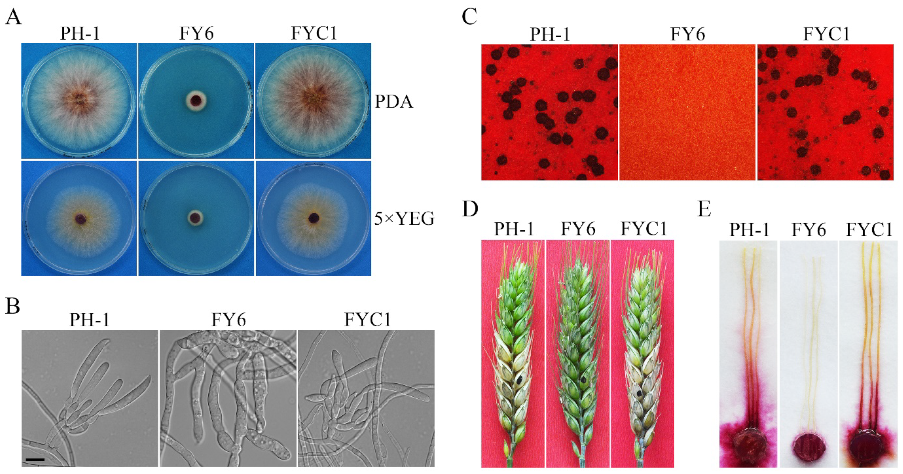
2.1. FgYCK1 Is Important for Vegetative Growth, Sexual/Asexual Development, and Pathogenesis

The FgYCK1 (FGRRES_10066) gene encodes a 453-aa protein that contains a typical protein kinase domain (12-288 aa) and a C-terminal di-cysteine motif (Figure S1). It shares 69%, 70%, and 58% identity with yeast Yck1, Yck2, and Yck3, respectively. When grown on PDA, the Fgyck1 deletion mutant (Table 1) formed compact colonies with limited aerial hyphae (Figure 1A) and grew at approximately 1.8 mm/day (Table 2), but the growth rate of the wild-type PH-1 was 11.3 mm/day. Microscopic examinations showed that the Fgyck1 mutant was blocked in the production of phialides and conidia in CMC cultures, while the PH-1 produced abundant conidia (123.8 × 10⁴ conidia/mL) (Figure 1B; Table 2). On mating plates, the Fgyck1 mutant failed to produce perithecia (Figure 1C). In infection assays with flowering wheat heads, the average disease index of Fgyck1 mutant was 0, which is much lower than that (8.8) of PH-1, indicating that the Fgyck1 mutant is non-pathogenic (Table 2; Figure 1D). The same result was also obtained in infection assays with corn silks (Figure 1E). When the full-length FgYCK1 allele was re-introduced into the Fgyck1 mutant, the resulting Fgyck1/FgYCK1 transformant (Table 1) was similar to the wild type in vegetative growth, conidiation, sexual reproduction, and pathogenesis (Figure 1; Table 2). Therefore, deletion of FgYCK1 is directly responsible for all the defects observed in the Fgyck1 mutant.

2.2. Spontaneous Suppressors of Fgyck1 Are Partially Recovered in Growth Rate and Conidiation

The Fgyck1 mutant was unstable and often produced fast-growing sectors in cultures older than 7 days (Figure 2A). A total of 25 spontaneous suppressors that grew over 18% faster than the original mutant were collected. These suppressor strains varied in growth rate and colony morphology as shown in Figure 2B with seven representative suppressor strains. Although they grew faster than the original Fgyck1 mutant, all the suppressor strains still grew slower than the wild-type strain PH-1 (Table S1; Figure 2B). When assayed for conidiation in CMC, 15 of the 25 suppressors produced conidia but at a significantly reduced level in comparison with the wild type (Table S1). Furthermore, conidia produced by these suppressors were shorter and had fewer septa than those of PH-1 (Table S1; Figure S2). Like the Fgyck1 mutant, all the 25 suppressor strains were nonpathogenic in infection assays with wheat heads (Figure 2C; Table S1) and corn silks (Figure 2D). They also were sterile and failed to produce perithecia on mating plates (Figure 2E). These results indicate that none of these suppressor strains were fully rescued in the defects of the Fgyck1 mutant although its defects in vegetative growth and conidiation were partially suppressed.

2.3. Identification of Mutations in Spontaneous Suppressor Strains of Fgyck1

In F. graminearum, spontaneous suppressor mutations can be efficiently identified by whole-genome sequencing [42,43]. The seven representative suppressor strains (S8, S12, S21, S22, S23, S24, and S25) and original Fgyck1 mutant were sequenced over 100× coverage by Illumina Hi-seq. In comparison with the genome sequence of the Fgyck1 mutant, a total of 11 mutations were identified in eight predicted genes (Table 3). All these mutations were verified by PCR amplification and sequencing analysis of the corresponding genes. Two of these seven suppressor strains, S12 and S23, had mutations in FGRRES_07218 that are homologous to yeast BNI4 (named FgBNI4 in this study). Three suppressor strains, S21, S24, and S25, had the D80N mutation in FGRRES_03646 that is homologous to the TNA1 high-affinity nicotinic acid permease gene in S. cerevisiae (named FgTNA1). For the other six genes, only a single suppressor mutation was identified in one of these seven suppressor strains (Table 3), including the nonsense, deletion, and missense mutations in genes orthologous to yeast IES4, MIG1, and MSS4, respectively.
Because spontaneous mutations in FgBNI4 and FgTNA1 were identified in more than one suppressor strain sequenced, we amplified and sequenced these two genes from the remaining 18 suppressor strains. Additional four suppressors were found to have mutations in the open reading frame (ORF) of FgBNI4. To our surprise, no additional suppressor strains had mutation in FgTNA1 (Table S2). The six suppressor strains with mutations in FgBNI4 differed from each other in genetic mutations (Table 3; Figure 3A). In suppressor S3, a nonsense mutation occurred at R490 (R490*), resulting in the truncation of 490-741 aa of the FgBni4 protein. Suppressor S12 had the G889−1T to AT mutation at the splicing site of the first intron, resulting in the retention of the 78-bp intron and a frame shift. Suppressors S10, S15, and S23 also had frame-shift mutations that were caused by deletion of CT2051–2052, AC1617–1618, and CT2134–2135, respectively. Interestingly, unlike the other five suppressors, S7 had one missense mutation that resulted in the R699 to C change in the FgBni4 protein.
Sequence alignment showed that the C-terminal region of FgBni4 (682–741 aa) is well conserved among its orthologs from other fungi (Figure S3) and named CCT (for conserved C-terminal tail) in this study. The R699C missense mutation in suppressor S7 occurred at a well-conserved R residue in this CCT region. Whereas the nonsense mutation at R490 and frameshift mutations caused by intron retention or deletion of AC1617–1618 resulted in the truncation of more than one third of FgBni4 protein, the CCT region was only partially truncated by ΔCT2051–2052 and ΔCT2134–2135 mutations in suppressors S10 and S23.
Figure 3. Suppressive mutations in FgBNI4 and their effects on the Fgyck1 mutant. (A) Schematic drawing of FgBni4 and positions of suppressor mutations. Sequence alignments of the conserved C-terminal region (black box) of FgBni4 with its orthologs from Fusarium oxysporum (Fo), Magnaporthe oryzae (Mo), Neurospora crassa (Nc), Botrytis cinerea (Bc), Aspergillus nidulans (An), Ustilago maydis (Um), Candida albicans (Ca), and Saccharomyces cerevisiae (Sc). The star (∗) indicates stop codon mutation. (B) Three-day-old PDA cultures of Fgyck1 (FY6), Fgyck1 FgBNI4R699C (RCY14), Fgyck1 FgBNI4ΔCT (LFY5), and Fgyck1 Fgbni4 (DK41) mutants. (C) Conidia of indicated strains harvested from 5-day-old CMC cultures. Bar, 10 μm.
Ijms 23 09106 g003

2.4. The R699C Mutation in FgBNI4 Is Verified for Its Suppressive Effect on Fgyck1

Because the Fgyck1 mutant was unstable, to verify the suppressive effect of the R699C mutation we had to first generate the FgBNI4R699C mutant. The R699C mutation was introduced into FgBNI4 by overlapping PCR with primers carrying the C2095GC to T2095GC mutation (Table S4). The resulting PCR product was then used to generate the FgBNI4R699C gene replacement construct (Figure S4A) with the hph hygromycin phosphotransferase as the selectable marker. After transformation of PH-1, transformants resistant to hygromycin were screened by PCR for the replacement of endogenous FgBNI4 with FgBNI4R699C (Figure S4A). The resulting FgBNI4R699C mutants were further confirmed by PCR and sequencing analysis.
We then transformed the FgYCK1 gene replacement construct carrying the neo marker into the FgBNI4R699C mutant. Transformants resistant to both hygromycin and geneticin were screened for deletion of FgYCK1 to identify the Fgyck1 FgBNI4R699C mutants (Figure S5B). Like suppressor S7, the Fgyck1 FgBNI4R699C mutant grew faster than the Fgyck1 mutants but slower than PH-1 (Figure 3B). These produced conidia, although conidiation was significantly reduced compared to the wild type (Table 2). Furthermore, conidia formed by the Fgyck1 FgBNI4R699C mutants were shorter and had fewer septa than the wild-type conidia (Figure 3C). In infection assays with wheat heads, the Fgyck1 FgBNI4R699C mutants were nonpathogenic (Figure S6A). On selfing mating plates, the Fgyck1 FgBNI4R699C mutants were sterile and failed to form perithecia (Figure S6B). These results confirm that the R699C mutation in FgBNI4 partially suppresses the defects of Fgyck1 in vegetative growth and conidiation but has no effect on its defects in pathogenesis (infectious growth) and sexual reproduction.

2.5. Deletion of Entire FgBNI4 and Truncation of Its CCT Region Have the Same Suppressive Effect on the Fgyck1 Mutant

Among all the suppressor strains with frameshift mutations, deletion of CT2134–2135 in S23 resulted in the shortest truncation of the CCT region of FgBni4 (truncation of 29 aa). To verify its suppressive effect on Fgyck1, we first generated the FgBNI4ΔCT gene replacement construct (Figure S4B) and transformed it into PH-1. The FgBNI4ΔCT mutant (Table 1) was verified for the deletion of CT2134–2135 by sequencing analysis (Figure S4B) and transformed with the FgYCK1 gene replacement construct (Figure S5B). The resulting Fgyck1 FgBNI4ΔCT double mutant (Table 1) was partially rescued in the defects of the Fgyck1 mutant in growth (Table 2; Figure 3B) and produced few conidia with morphological defects (Figure 3C). However, like suppressor S23, the Fgyck1 FgBNI4ΔCT mutant was nonpathogenic and sterile in selfing (Figure S6), indicating that truncation of the CCT region of FgBNI4 also partially rescued the defects of Fgyck1 mutant in growth and conidiation but had no effect on plant infection and perithecium development. Interestingly, all suppressor strains with mutations in FgBNI4 (S3, S7, S10, S12, S15 and S23) were partially rescued in conidiation and had the same defect in conidium morphology (Table S1; Figure S2).
To further characterize the relationship between FgYck1 and FgBni4, we also generated the Fgbni4 deletion mutant and transformed it with the FgYCK1 gene replacement construct (Figure S5). The resulting Fgbni4 Fgyck1 double mutant (Table 1) had similar phenotypes with the Fgyck1 FgBNI4ΔCT and Fgyck1 FgBNI4R699C mutants in growth, conidiation, sexual reproduction, and pathogenesis (Figure 3B,C; Figure S6). These results indicate that truncation of the C-terminal 29 aa residues had the same suppressive effects as deletion of the entire FgBNI4 on the Fgyck1 mutant. Therefore, the CCT region of FgBNI4 must be essential for its function. Other nonsense and frameshift mutations that occurred upstream from CT2134–2135 likely had the same effects as deletion of FgBNI4.

2.6. FgBNI4 Is Involved in Cell Wall Integrity and Hyphal Growth

The Fgbni4 mutant was slightly reduced in vegetative growth (Figure 4A; Table S3) but normal in asexual and sexual reproduction (Figure S7; Table S3). In infection assays with flowering wheat heads, the Fgbni4 mutant caused typical FHB symptoms in the inoculated wheat kernels and was able to spread to nearby spikelets (Figure S7C). Nevertheless, its disease index was slightly lower than that of PH-1 (Table S3). These results indicate that FgBNI4 is dispensable for asexual and sexual reproduction but plays a role in vegetative growth and plant infection.
In S. cerevisiae and C. albicans, Bni4 is involved in chitin deposition in cell wall [29,34]. To determine whether the Fgbni4 mutant is defective in cell wall integrity, we first assayed the effects of cell wall disturbing compounds Calcofluor white (CFW) and Congo Red (CR) on hyphal growth. In the presence of 200 μg/mL CFW or 300 μg/mL CR, the Fgbni4 mutant was more significantly reduced in growth rate than PH-1 (Figure 4A). We also assayed the effects of CFW and CR on conidium germination. After incubation in regular YEPD for 6 h, conidia of PH-1 and Fgbni4 mutant had no obvious defects in germination, although germ tubes of Fgbni4 mutant were shorter than those of PH-1 (Figure 4B). In the presence of 15 μg/mL CR, which had no obvious effect on PH-1, germination was not inhibited but germ tubes were much shorter in the Fgbni4 mutant. Swollen conidium and hyphal compartments, as well as empty conidium compartments were often observed in the mutant but not in PH-1 (Figure 4B). The addition of 20 μg/mL CFW to YEPD had similar but more severe effects on germ tube growth and cell morphology in the Fgbni4 mutant (Figure 4B,C). When the full-length FgBNI4 gene was transformed into the Fgbni4 mutant, the resulting transformants were normal in vegetative growth, pathogenicity, and responses to cell wall stressors (Figure 4). Therefore, FgBNI4 is important for cell wall integrity, which may be directly related to its roles in vegetative growth and plant infection in F. graminearum.
Figure 4. The involvement of FgBni4 in response to cell wall stresses in F. graminearum. (A) Colonies of wild-type strain (PH-1), Fgbni4 mutant (FB5), and Fgbni4/FgBNI4 complemented transformant (FBC9) grew on PDA plates supplemented without (CK) or with 200 μg/mL CFW or 300 μg/mL CR for 3 days. (B) Conidia of indicated strains were incubated in YEPD without (CK) or with 20 μg/mL CFW or 15 μg/mL CR for 6 h before examination. White arrows point to the swollen or ruptured cells of Fgbni4 mutant. Bar, 20 μm. (C) Hyphae length of indicated strains without (CK) or with CFW or CR treatments. The length of hyphae was measured by using Image J software.
Ijms 23 09106 g004

2.7. The Fgyck1 Mutant Is Defective in Maintaining Polarized Growth

To determine whether deletion of FgYCK1 affects polarized growth, we examined hyphal tips after incubation for 36 h on PDA. Whereas all the wild-type hyphae examined had normal hyphal tips, approximately 70% of the hyphae had swollen tips in the Fgyck1 mutant (Figure 5A). For some of these swollen hyphal tips, one or multiple new hyphae emerged and grew (Figure 5A), indicating the re-establishment of polarized growth. Therefore, FgYck1 likely plays an important role in maintaining polarized growth at hyphal tips in F. graminearum.
In filamentous fungi, defects or disturbances in actin assembly often lead to swollen hyphal tips [44,45]. To verify the role of FgYck1 in polarized growth, we transformed the F-actin marker LifeAct-GFP into PH-1 and the Fgyck1 mutant. In the wild type, LifeAct-GFP signals were mainly observed in the subapical collar region of hyphae and enriched at the core of Spitzenkörper (Spk) bodies at hyphal tips (Figure 5B). However, in the Fgyck1 mutant, LifeAct-GFP signals were observed in the peripherals of swollen hyphal tips and no strong LifeAct-GFP signals at the Spk core were observed when more than 50 swollen hyphal tips were examined (Figure 5B). We also examined more than 50 hyphae of the Fgyck1 mutant with relatively normal hyphal tips and enhanced LifeAct-GFP signals at the Spk core were not observed (Figure 5B). These data indicate that the deletion of the FgYCK1 gene affects the localization and enrichment of F-actin to the Spk1 core.
The polarisome complex mediates F-actin polarization and directs Spk vesicles to the hyphal apex [46]. FgSpa2 is the key component of the polarisome that is required for establishment and maintenance of polarized growth in F. graminearum and other fungi [47,48,49]. Therefore, we also transformed FgSPA2-GFP into PH-1 and the Fgyck1 mutant. As expected, FgSpa2-GFP showed a polarisome-like localization at all hyphal tips in the wild type (Figure 5C). However, when we examined more than 50 swollen hyphal tips of the Fgyck1 mutant, no FgSpa2-GFP signals were observed (Figure 5C). Although FgSpa2-GFP signals were present at the tip of normal hyphae of the Fgyck1 mutant, its polarisome-like accumulation was significantly impaired (Figure 5C). These results indicate that the loss of FgYck1 may affect the organization of the Spk body and polarisomes that are necessary to maintain polarized growth at the tip.

2.8. The Defect of Fgyck1 in Polarized Growth Is Alleviated by Deletion of FgBNI4

When hyphal tips were examined, the Fgyck1 Fgbni4 mutant still formed a few apical swollen bodies (Figure 6A). However, in comparison with the Fgyck1 mutant, the percentage of swollen hyphal tips was significantly reduced in the double mutant. Whereas about 70% of the hyphae had swollen tips in the Fgyck1 mutant, over 63% of hyphal tips were normal in the Fgyck1 Fgbni4 mutant (Figure 6B). When stained with CFW, irregular hyphal width and CFW staining were often observed in the Fgyck1 mutant but rare in the Fgyck1 Fgbni4 mutant (Figure 6C). Therefore, deletion of FgBNI4 significantly alleviates the polarized growth defect of the Fgyck1 mutant.
The irregular CFW staining in Fgyck1 indicates the alteration of cell wall composition (Figure 6C), which may be associated with the CWI signaling pathway. In F. graminearum, the Mgv1 MAPK regulates CWI pathway and the mgv1 deletion mutant also has severe growth defects [50]. Tip swelling observed in the Fgyck1 mutant may be related to defects in CWI signaling. To determine whether deletion of FgYCK1 affects the activation of the Mgv1 MAPK, we assayed its phosphorylation with a commercially available anti-TpEY phosphorylation-specific antibody. On Western blots with total proteins isolated from hyphae collected from 18 h YEPD cultures, the phosphorylation level of Mgv1 was reduced in the Fgyck1 mutant in comparison with the wild type (Figure 6D,E). However, the expression level of Mgv1 detected with an anti-Mgv1 antibody generated in this study was not affected by deletion of FgYCK1. In the Fgyck1 Fgbni4 double mutant, Mgv1 phosphorylation was recovered to the wild-type level (Figure 6D,E), indicating that deletion of FgBNI4 rescues the defect of Fgyck1 in Mgv1 activation.

2.9. Deletion of FgBNI4 Alleviates the Defects of Fgyck1 in FgRho1 Internalization and Vacuolar Fusion

Rho1 is a small GTPase that plays essential roles in polarized growth, actin organization, and cell wall integrity in fungi [51,52]. The rho1 mutants of N. crassa and F. oxysporum have similar defects with the Fgyck1 mutant, including swollen tips and altered cell wall composition [37,53]. Therefore, we speculated that deletion of FgYCK1 may negatively impact FgRho1 localization and function. To test this hypothesis, we transformed the GFP-FgRHO1 fusion construct into PH-1 and the Fgyck1 and Fgyck1 Fgbni4 mutants and observed the FgRho1 localization in 12 h hyphae. In all hyphae of wild type, the GFP-FgRho1 predominantly localized to the plasma membrane and vacuolar membrane (Figure 7A). In contrast, stronger GFP-FgRho1 signals were accumulated inside the vacuoles in >75% hyphae of the Fgyck1 mutant (Figure 7B), indicating an enhanced internalization of FgRho1. However, the internalization of FgRho1 was observed in <10% hyphae of the Fgyck1 Fgbni4 double mutant (Figure 7C). Furthermore, the Fgyck1 mutant appeared to have more and smaller vacuoles than the wild type (Figure 7), which may be related to defect in vacuole fusion caused by the internalization of FgRho1. The abundance and size of vacuoles were similar between PH-1 and the Fgyck1 Fgbni4 double mutant (Figure 7). These results indicate that deletion of FgYCK1 negatively impacts the localization and function of FgRho1 in F. graminearum, which may contribute the pleiotropic defects of the Fgyck1 mutant due to the importance of Rho1 in hyphal tip growth and cell wall integrity [36,37]. Deletion of FgBNI4 alleviates the defects of Fgyck1 in FgRho1 internalization and vacuole fusion, which may contribute to the recoveries of Fgyck1 Fgbni4 mutant in polarized growth and cell wall integrity.

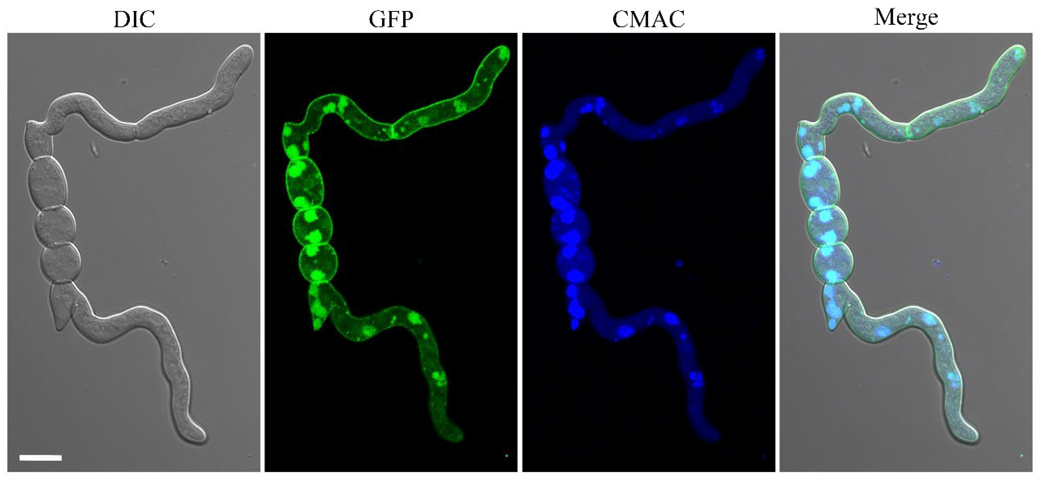
2.10. The FgYck1 Localizes to Plasma Membrane and Vacuolar Lumen

To confirm the subcellular localization of FgYck1, we generated the GFP-FgYCK1 under the control of the strong constitutive RP27 promoter [54] and expressed it in PH-1. In the 12 h hyphae of PH-1/GFP-FgYCK1 transformant, GFP-FgYck1 signals were detected mainly at the plasma membrane and vacuolar lumens that were stained with CMAC (7-amino-4-chloromethylcoumarin, Sigma-Aldrich, Eugene, OR, USA), and weaker GFP signals were also observed in the cytoplasm (Figure 8).

3. Discussion

Whereas S. cerevisiae has two paralogous casein kinase I genes YCK1 and YCK2 originated from recent whole-genome duplication, FgYCK1 is the only YCK1/2 ortholog in F. graminearum. Unlike the yeast yck1 yck2 double mutant that is inviable, the Fgyck1 deletion mutant was viable but had severe growth defects. However, the Fgyck1 mutant was blocked in conidiation. Even after incubation in CMC medium for over two weeks, no conidia were observed in mutant cultures. The production of conidia from phialides in F. graminearum is a similar blastospore formation process as the budding of yeast cells. Therefore, the YCK1 orthologs may have a conserved role in blastospore formation on phialides. In yeast, YCK1 and YCK2 are important for proper septin assembly and morphogenesis. In M. oryzae, the Moyck1 mutant is reduced in conidiation but still produces macroconidia [55], which are directly formed on conidiophores instead of phialides. However, microconidia are produced from phialides in M. oryzae [55] and the Moyck1 mutant may be blocked in the production of microconidia.
Like the Fgprp4, fng1, and a few other F. graminearum mutants with severe growth defects [42,56,57,58], the Fgyck1 mutant produced spontaneous suppressors with faster growth rates. By whole genome sequencing of seven selected suppressors, we identified mutations in seven predicted genes, including orthologs of yeast BNI4, TNA1, MIG1, and MSS4. Three of these had the same D80N mutation in FgTNA1, suggesting that this is a hot spot for spontaneous mutations. However, no additional mutation in FgTNA1 was identified in the remaining 18 suppressor strains by amplification and sequencing of its coding region. Interestingly, all three of the suppressors with the D80N mutation in FgTNA1 also had mutations in other genes. Therefore, we did not pursue further characterization of FgTNA1 in this study. One missense suppressor mutation was identified in the ortholog of A. nidulans CREA and S. cerevisiae MIG1. The Mig1/CreA protein is a major regulator of carbon catabolite repression in yeast and filamentous fungi [59,60]. Because Yck1 and Yck2 are involved in glucose sensing and signaling in yeast [61], it is likely that FgYck1 also is involved in the utilization of different carbon sources in F. graminearum, possibly via FgCreA.
FgBNI4 was selected for further characterization because it is the only other gene with mutations identified in more than one suppressor strain by whole-genome sequencing [32,33,34,35,36,37]. In F. graminearum, the Fgbni4 deletion mutant was normal in conidiation and sexual reproduction but was reduced in growth rate and virulence. Interestingly, five out of the six suppressor strains with mutations in FgBNI4 had nonsense or frameshift mutations that resulted in the truncation of its C-terminal region. The amino acid sequences of the FgBni4 orthologs are not well conserved among different fungi except the C-terminal 60 residues (named CCT region). The only missense mutation in FgBNI4 that occurred in this region in suppressor S7 is at R699, a well-conserved residue among its orthologs. In addition, the frameshift mutation in suppressor S23 (ΔCT2134–2135, L712fs) resulted in the truncation of only the last 29 residues. Furthermore, both the R699C mutation and deletion of CT2134–2135 had the same suppressive effects with deletion of the entire FgBNI4 gene on Fgyck1 mutant in vegetative growth and conidiation. Therefore, this CCT region must be essential for the function of FgBNI4. Because deletion of FgBNI4 and all the suppressor mutations in FgBNI4 failed to suppress the defects of Fgyck1 in sexual reproduction and plant proteins, we conclude that the genetic relationship between FgBNI4 and FgYCK1 is stage-specific during vegetative growth and asexual reproduction.
In S. cerevisiae, YCK1 and YCK2 have not been reported to physically or genetically interact with BNI4. In F. graminearum, mutations in the CCT region of FgBNI4 partially suppressed the growth defect of the Fgyck1 mutant. This genetic relationship between FgYCK1 and FgBNI4 may be related to their direct interaction and FgBni4 may be a target of FgYck1 kinase. Because Bni4 is a protein that mainly functions to interact with other proteins for their proper localization in yeast, it is possible that phosphorylation of FgBni4 by Fgyck1 is necessary for its interaction with some FgBni4-interacting proteins in F. graminearum. However, because Bni4 is a subunit of the Bni4-Glc7 phosphatase I complex, it is also possible that suppressive effects of mutations in FgBNI4 on Fgyck1 are related to the antagonistic effects of phosphorylation by FgYck1 and dephosphorylation by Glc7 on their common targets. Disruption of FgBNI4 will reduce or eliminate the dephosphorylation of FgYck1-phosphorylated proteins by the Bni4-Glc7 phosphatase I. Although Bni4 is not known to be phosphorylated by Yck1 or Yck2 in yeast, it is phosphorylated in a cell cycle-dependent manner by unknown kinases [62]. Yeast Bni4 is also phosphorylated by Slt2 and Kss1 MAP kinases that are orthologous to Mgv1 and Gpmk1, respectively [63]. In F. graminearum, it is possible that Mgv1 and Gpmk1 MAP kinases are also involved the phosphorylation of FgBni4. The mgv1 mutant had severe growth defects although the Gpmk1 mutant was only slightly reduced in growth. The Gpmk1 mgv1 double has not been reported but the Gpmk1 mgv1 Fghog1 triple mutant had more severe defects than the mgv1 mutant [64].
The majority of the vegetative hyphae of the Fgyck1 mutant had swollen tips [7,8]. However, conidium germination was not blocked and swollen tips of the Fgyck1 mutant often re-established polarized growth and produced one or more new hyphae. Therefore, although FgYCK1 is important for polarized growth, it is not essential for establishing polarized tip growth. Swollen hyphal tips observed in the Fgyck1 mutant had only dispersed, peripheral LifeAct-GFP signals, which is similar to the effects of inhibiting actin polymerization. The central actin core of Spk observed in hyphal tips of PH-1 was not observed in these swollen hyphal tips of the Fgyck1 mutant. Even in Fgyck1 hyphae with relatively morphologically normal tips, the enrichment of F-actin at the center of Spk1 was not observed. Consistent with these observations, the apical localization of polarisome scaffold protein FgSpa2 was not observed at swollen hyphal tips in the Fgyck1 mutant. Polarisome is a complex of proteins that nucleates actin cables for polarized cell growth in the budding yeast and filamentous fungi [65]. It is possible that FgYck1 regulates polarized growth by phosphorylating certain components of the polarisome. Deletion of FgBNI4 may negatively impact the interaction of Glc7 phosphatase I with the polarisome components that are phosphorylated by FgYck1 and their dephosphorylation, leading to a faster growth rate in the Fgyck1 Fgbni4 double mutant. In S. cerevisiae, some polarisome-related genes, including SPA2 and BNI1, have been found to interact with BNI4 in a synthetic genetic array analysis [33].
In S. cerevisiae, mutations in BNI4 affect the localization of the chitin synthase III and chitin synthesis at the bud neck [31]. It is possible that deletion of FgYCK1 also affects the expression or localization of chitin synthases in F. graminearum. Similar to the Fgyck1 mutant, the Gzchs5 and Gzchs7 mutants produce swollen hyphal tips in F. graminearum [66,67]. In C. albicans, the yck2 mutant is defective in cell wall integrity but has increased compensatory chitin deposition in the cell wall by up-regulating chitin synthase genes [26]. We noticed that the Fgyck1 mutant had a stronger CFW staining of the cell wall than the wild type and CFW staining became normal in the Fgyck1 Fgbni4 double mutant. Therefore, it is possible that deletion of FgYCK1 also results in a thickening of the cell wall by up-regulating GzChs5, GzChs6, and other enzymes involved in cell wall synthesis. Consistent with this hypothesis, we found that the Fgyck1 mutant was reduced in the phosphorylation of Mgv1 CWI MAP kinase [50] and deletion of FgBNI4 rescued its Mgv1 phosphorylation to the wild-type level.
In F. graminearum, deletion of FgYCK1 enhanced the internalization of FgRho1 into vacuoles, which was suppressed by deletion of FgBNI4. The vacuole fusion defect of the Fgyck1 mutant was also partially rescued in the Fgyck1 Fgbni4 double mutant. Therefore, it is likely that the defects of the Fgyck1 mutant and suppressive effects of mutations in FgBNI4 are related to their functional relationships with FgRho1 in F. graminearum. In yeast, Yck1 and Yck2 regulate the localization and activity of Mss4 for the synthesis of PI4,5P2 that is required for clathrin-mediated endocytosis [25,68] and involved in actin cytoskeleton organization and cell wall integrity through Rho1 signaling [25,69]. Interestingly, suppressor S12 had the N353T mutation in FgMSS4, which may partially suppress the defect of Fgyck1 mutant in the localization or activity of FgMss4. FgYck1 may regulate the internalization of FgRho1 by controlling PI4,5P2 synthesis via phosphorylation of FgMss4 in F. graminearum. Therefore, it is important to characterize the functional relationship among the well-conserved FgYck1 kinase, FgRho1, and FgBni4 in hyphal growth, PI4,5P2-dependent endocytosis, and cell wall integrity in future studies.
In summary, our study showed that the Fgyck1 mutant displayed pleiotropic defects in vegetative growth, conidiation, sexual development and pathogenicity. Deletion of FgYCK1 also affected polarized growth and cell wall integrity. Through characterizing natural suppressor mutants, we found a novel genetic interaction between FgYck1 and FgBni4 during vegetative growth and conidiation. Additionally, the loss of FgBNI4 can partially restore the phosphorylation of Mgv1 and correct the localization of FgRho1 in Fgyck1 mutant. Further characterization of FgYck1 and FgBni4 on PI4,5P2 synthesis and endocytosis on the plasma membrane will be helpful in defining and clarifying the relationship between FgYck1 and FgBni4 in F. graminearum.

4. Materials and Methods

Fungal strains and culture conditions: The wild-type strain PH-1 and its mutants listed in Table 1 were routinely cultured on PDA (20% potato, 2% glucose, and 1.5% agar) at 25 °C and preserved in 20% glycerol at −80 °C. Conidiation was examined with liquid CMC (1.5% carboxymethylcellulose, 0.1% NH4NO3, 0.0% MgSO4·7H2O, 0.1% yeast extract) cultures [70]. Mating on carrot agar plates and examination for perithecium formation and ascospore ejection were assayed as described [71]. Protoplast preparation and polyethylene glycol (PEG)-mediated transformation were performed as described [50,72]. For transformant selection, hygromycin B (CalBiochem, La Jolla, CA, USA), geneticin (Sigma, St. Louis, MO, USA), and zeocin (Invitrogen, Carlsbad, CA, USA) were added to the final concentration of 300, 400 and 700 μg/mL, respectively, to both the bottom and top agar. Fungal genomic DNA was extracted from mycelia harvested from 24 YEPD cultures by the cetyltrimethylammonium bromide (CTAB) protocol as described [73].
Infection assays: Conidia of PH-1 and mutant strains were harvested from 5-day-old CMC cultures and resuspended to a concentration of 2 × 10⁵ conidia/mL in sterile distilled water. For wheat head infection assays, the fifth floret from the bottom of each head of cultivar Xiaoyan 22 was inoculated with 10 μL of conidial suspension or culture blocks [50,74]. Inoculated wheat heads were covered with a plastic bag to keep humidity for 48 h. Infected wheat plants were examined at 14 days post-inoculation (dpi) to estimate the disease index [50]. Infection assays with corn silks were carried out as described [75].
Isolation of spontaneous suppressors and identification of suppressor mutations: Sectors with faster growth rate were isolated as spontaneous suppressors from the edge of Fgyck1 colonies formed on PDA plates as described [42,56,57]. To identify mutations in seven representative suppressor strains, genomic DNA samples were sequenced with the Illumina HiSeq-PE150 at Novogene Bioinformatics Institute (Beijing, China) to 50×coverage with pair-end libraries [76,77]. The resulting sequence reads were mapped onto the genome sequences of PH-1 and original Fgyck1 mutant as a reference using Bowtie 2 [78]. Mutations were identified by SAMtools with the default parameters [79]. Annotation of the mutation sites was performed with the Variant Effect Predictor (VEP) program [80]. To determine whether the remaining 18 suppressors of Fgyck1 have mutations in FgBNI4 and FgTNA1, PCR products amplified with primer pairs FgBNI4/TF-FgBNI4/TR and FgTNA1/TF-FgTNA1/TR (Table S4) were sequenced at Sango Biotech (Shanghai, China).
Generation of the Fgbni4 and Fgbni4 Fgyck1 mutants: To generate the Fgbni4 mutant, a 1.2-kb upstream and a 1.1-kb downstream flanking fragments of the FgBNI4 gene were amplified with primer pairs B1F-B2R and B3F-B4R (Table S4), respectively, and fused to the hph fragments amplified with primer pairs HT/F-HY/R and YG/F-HT/R from pCB1003 by overlapping PCR [81,82]. The resulting PCR products were co-transformed into protoplasts of PH-1. Hygromycin-resistant transformants were screened for FgBNI4 deletion by PCR with primer pairs H850-H852, B5F-B6R, B7F-H855R, and H866F-B8R. To generate the Fgyck1 Fgbni4 double mutant, the FgYCK1 replacement construct was generated with neomycin resistant cassette (neo) and transformed into the Fgbni4 mutant. Transformants resistant to both hygromycin and geneticin were screened by PCR with primer pairs G850-G852, Y5F-Y6R, Y7F-G855R, and G866F-Y8R (Table S4) for the deletion of FgYCK1. Optimal conditions for the PCR reactions were as follows: 5 min denaturation at 95 °C followed by 35 cycles of 95 °C for 1 min, 57 °C for 1 min, and 72 °C for 1 min. The reaction was completed with a 10-min extension at 72 °C. All PCR reactions were performed in a C1000 Touch Thermal Cycler (Bio-Rad, Munich, Germany).
Generation of the FgBNI4R699C Fgyck1 and FgBNI4ΔCT Fgyck1 mutants: A modified split marker approach [81] was used to generate the transformants with in situ mutations. A DNA fragment containing the last 1000-bp of FgBNI4 ORF and 400-bp terminator sequence was amplified by overlapping PCR [83] to introduce the R699C mutation with primer pairs P1F-P1R and P2F-P2R. The 800-bp DNA sequence downstream from the terminator sequence of FgBNI4 was also amplified. These two fragments were ligated to the hph fragments amplified with primer pairs HT/F-HY/R and YG/F-HT/R by overlapping PCR and transformed into protoplasts of PH-1. The resulting FgBNI4R699C mutants were confirmed by sequencing analyses. The FgYCK1 replacement construct (neo) was then transformed into the FgBNI4R699C mutant to generate the FgBNI4R699C Fgyck1 mutants. The same approach was used to generate the FgBNI4ΔCT and FgBNI4ΔCT Fgyck1 mutants. All the primers are listed in the Table S4.
Complementation assays with the Fgyck1 and Fgbni4 mutants: For complementation of the Fgyck1 mutant, the full-length FgYCK1 gene, including the 2.3-kb promotor and 1-kb terminator regions, was amplified and cloned into vector pFL7 (carrying geneticin resistance marker) by the gap repair approach [84]. The resulting construct was confirmed by sequencing analysis and transformed into the Fgyck1 mutant. For complementation of the Fgbni4 mutant, the entire FgBNI4 gene, with its promotor and terminator regions, was amplified and cloned into the GFP-tagged vector pKNTG [85] that carries the neo selectable marker by using the NovoRec® plus One step PCR Cloning kit (Novoprotein, Shanghai, China). The resulting construct was transformed into the Fgbni4 mutant. Both Fgyck1/FgYCK1 and Fgbni4/FgBNI4 transformants were confirmed by PCR analysis and assayed for phenotype changes.
Assays for defects in responses to cell wall stresses: The final concentration of 300 μg/mL Congo red (CR) and 200 μg/mL Calcofluor white (CFW) were added to PDA to assay vegetative growth at 25 °C as described [86,87]. Colony morphology was examined and photographed after incubation for 3, 5, or 7 days. To assay conidial germination, 20 μg/mL CFW or 15 μg/mL CR was added to freshly harvested conidia resuspended to 106 conidia/mL in YEPD and incubated for 6 h at 25 °C [88]. All experiments were repeated independently at least three times.
Generation of the GFP-FgRHO1, GFP-FgYCK1, and FgSPA2-GFP transformants: All GFP fusion constructs were generated using the NovoRec® plus One step PCR Cloning kit (Novoprotein, Shanghai, China). To generate the GFP-FgRHO1 construct, the entire FgRHO1 gene, including its terminator region, was amplified with primers GFP-RHO1/F and GFP-RHO1/R and fused with GFP in vector pKNTG-RP27, which was generated by inserting the constitutive RP27 promoter into pKNTG. The same approach was performed to generate the GFP-FgYCK1 construct. To generate the FgSPA2-GFP construct, the entire coding region of FgSPA2, including its native promoter, was amplified with primers FgSPA2/F and FgSPA2/R and cloned into the KpnI/HindIII-digested pKNTG. All resulting GFP fusion constructs were confirmed by sequencing analysis and transformed into protoplasts of PH-1 and the Fgyck1 mutant.
Western blot analysis: Total proteins were isolated from hyphae harvested from 18 h YEPD cultures [88]. The Bradford method was performed to confirm the protein concentration [89] and 10 µg of protein were loaded per lane for each samples. Proteins separated on 10% SDS-PAGE gel were transferred to nitrocellulose membranes [90]. Phosphorylation of Mgv1 was detected with the PhophoPlus p44/42 MAP kinase antibody kit (Cell Signaling Technology, Danvers, MA, USA) as described [90]. To detect the expression level of Mgv1, an anti-Mgv1 antibody was generated at the ABclonal Biotechnology (Wuhan, China) by injecting rabbits with a polypeptide of Mgv1 (333–349 aa). On Western blots, a 46-kD Mgv1 band was detected in PH-1 but not in the mgv1 mutant [50] with 1:5000 dilution of the resulting anti-Mgv1 antibody, indicating that it is suitable for Mgv1 detection. The ClarityTM western ECL substrate kit (Bio-Rad, Hercules, CA, USA) and ChemiDocTM XRS+ imaging system (Bio-Rad, Hercules, CA, USA) were used for Western blot analysis. The Image LabTM software was used to analyze the quantitative changes of the Mgv1 phosphorylation level. Each experiment was repeated at least three times.
Vacuole and cell wall staining: Hyphae harvested from 12 h YEPD cultures were stained with 10 μM CMAC (7-amino-4-chloromethylcoumarin, Sigma-Aldrich) as described [91] to visualize the vacuolar lumen. For cell wall staining, hyphae were stained with 10 μg/mL CFW (Sigma-Aldrich, St. Louis, MO, USA) for 5 min as described [92]. Samples were examined for CMAC and CFW staining signals with an Olympus BX53 epifluorescence microscope (Olympus, Tokyo, Japan) or Zeiss LSM880 confocal microscope (Carl Zeiss, Jena, Germany).

Supplementary Materials

The following supporting information can be downloaded at: https://www.mdpi.com/article/10.3390/ijms23169106/s1.

Author Contributions

Conceptualization, R.H.; Data curation, J.Z. and R.H.; Formal analysis, R.H.; Investigation, J.Z., D.H. and Q.L.; Methodology, D.H. and Q.L.; Software, D.H.; Visualization, J.Z.; Writing—original draft, J.Z.; Writing—review & editing, J.-R.X. and G.W. All authors have read and agreed to the published version of the manuscript.

Funding

This work was supported by grants from the Natural Science Foundation of Shannxi (No. 2020JM-157) and Chinese University Scientific Fund (No. 2452019217) to G.W., National Natural Science Foundation of China (No. 31801688) to R.H., and USWBSI and NSF to J.X.

Institutional Review Board Statement

Not applicable.

Informed Consent Statement

Not applicable.

Data Availability Statement

The data that support the findings of this study are available in the Supplementary Material of this article.

Acknowledgments

We thank Cong Jiang, Xue Zhang, Qinhu Wang, and Huiquan Liu for their fruitful discussions. We also thank Hua Zhao (State Key Laboratory of Crop Stress Biology for Arid Areas, Northwest A&F University, Yangling, China) for assistance with confocal microscopy.

Conflicts of Interest

The authors declare no conflict of interest.

References

  1. Bai, G.; Shaner, G. Management and resistance in wheat and barley to fusarium head blight. Annu. Rev. Phytopathol. 2004, 42, 135–161. [Google Scholar] [CrossRef] [PubMed]
  2. Goswami, R.S.; Kistler, H.C. Heading for disaster: Fusarium graminearum on cereal crops. Mol. Plant Pathol. 2004, 5, 515–525. [Google Scholar] [CrossRef] [PubMed]
  3. Trail, F. For blighted waves of grain: Fusarium graminearum in the postgenomics era. Plant Physiol. 2009, 149, 103–110. [Google Scholar] [CrossRef] [PubMed]
  4. Trail, F.; Gaffoor, I.; Vogel, S. Ejection mechanics and trajectory of the ascospores of Gibberella zeae (anamorph Fuarium graminearum). Fungal Genet. Biol. 2005, 42, 528–533. [Google Scholar] [CrossRef]
  5. Boenisch, M.J.; Broz, K.L.; Purvine, S.O.; Chrisler, W.B.; Nicora, C.D.; Connolly, L.R.; Freitag, M.; Baker, S.E.; Kistler, H.C. Structural reorganization of the fungal endoplasmic reticulum upon induction of mycotoxin biosynthesis. Sci. Rep. 2017, 7, 44296. [Google Scholar] [CrossRef]
  6. Lozowicka, B.; Iwaniuk, P.; Konecki, R.; Kaczynski, P.; Kuldybayev, N.; Dutbayev, Y. Impact of diversified chemical and biostimulator protection on yield, health status, mycotoxin level, and economic profitability in spring wheat (Triticum aestivum L.) cultivation. Agronomy 2022, 12, 258. [Google Scholar] [CrossRef]
  7. Wang, C.; Zhang, S.; Hou, R.; Zhao, Z.; Zheng, Q.; Xu, Q.; Zheng, D.; Wang, G.; Liu, H.; Gao, X.; et al. Functional analysis of the kinome of the wheat scab fungus Fusarium graminearum. PLoS Pathog. 2011, 7, e1002460. [Google Scholar] [CrossRef]
  8. Li, B.; Dong, X.; Li, X.; Chen, H.; Zhang, H.; Zheng, X.; Zhang, Z. A subunit of the HOPS endocytic tethering complex, FgVps41, is important for fungal development and plant infection in Fusarium graminearum. Environ. Microbiol. 2018, 20, 1436–1451. [Google Scholar] [CrossRef]
  9. Tuazon, P.T.; Traugh, J.A. Casein kinase I and II-multipotential serine protein kinases: Structure, function, and regulation. Adv. Second. Messenger Phosphoprot. Res. 1991, 23, 123–164. [Google Scholar]
  10. Guerra, B.; Issinger, O.G. Protein kinase CK2 and its role in cellular proliferation, development and pathology. Electrophoresis 1999, 20, 391–408. [Google Scholar] [CrossRef]
  11. Cheong, J.K.; Virshup, D.M. Casein kinase 1: Complexity in the family. Int. J. Biochem. Cell Biol. 2011, 43, 465–469. [Google Scholar] [CrossRef]
  12. Knippschild, U.; Gocht, A.; Wolff, S.; Huber, N.; Löhler, J.; Stöter, M. The casein kinase 1 family: Participation in multiple cellular processes in eukaryotes. Cell Signal 2005, 17, 675–689. [Google Scholar] [CrossRef]
  13. Sun, B.; Chen, L.; Cao, W.; Roth, A.F.; Davis, N.G. The yeast casein kinase Yck3p is palmitoylated, then sorted to the vacuolar membrane with AP-3-dependent recognition of a YXXϕ adaptin sorting signal. Mol. Biol. Cell 2004, 15, 1397–1406. [Google Scholar] [CrossRef]
  14. Peng, Y.; Grassart, A.; Lu, R.; Wong, C.C.; Yates III, J.; Barnes, G.; Drubin, D.G. Casein kinase 1 promotes initiation of clathrin-mediated endocytosis. Dev. Cell 2015, 32, 231–240. [Google Scholar] [CrossRef]
  15. Lord, C.; Bhandari, D.; Menon, S.; Ghassemian, M.; Nycz, D.; Hay, J.; Ghosh, P.; Ferro-Novick, S. Sequential interactions with Sec23 control the direction of vesicle traffic. Nature 2011, 473, 181–186. [Google Scholar] [CrossRef]
  16. Hoekstra, M.F.; Liskay, R.M.; Ou, A.C.; DeMaggio, A.J.; Burbee, D.G.; Heffron, F. HRR25, a putative protein kinase from budding yeast: Association with repair of damaged DNA. Science 1991, 253, 1031–1034. [Google Scholar] [CrossRef]
  17. Tanaka, C.; Tan, L.J.; Mochida, K.; Kirisako, H.; Koizumi, M.; Asai, E.; Sakoh-Nakatogawa, M.; Ohsumi, Y.; Nakatogawa, H. Hrr25 triggers selective autophagy–related pathways by phosphorylating receptor proteins. J. Cell Biol. 2014, 207, 91–105. [Google Scholar] [CrossRef]
  18. Wang, J.; Davis, S.; Menon, S.; Zhang, J.; Ding, J.; Cervantes, S.; Miller, E.; Jiang, Y.; Ferro-Novick, S. Ypt1/Rab1 regulates Hrr25/CK1δ kinase activity in ER–Golgi traffic and macroautophagy. J. Cell Biol. 2015, 210, 273–285. [Google Scholar] [CrossRef] [PubMed]
  19. De Assis, L.J.; Ulas, M.; Ries, L.N.A.; El Ramli, N.A.M.; Sarikaya-Bayram, O.; Braus, G.H.; Bayram, O.; Goldman, G.H. Regulation of Aspergillus nidulans CreA-mediated catabolite repression by the F-box proteins Fbx23 and Fbx47. Mbio 2018, 9, e00840-18. [Google Scholar] [CrossRef]
  20. Apostolaki, A.; Harispe, L.; Calcagno-Pizarelli, A.M.; Vangelatos, I.; Sophianopoulou, V.; Arst, H.N., Jr.; Peñalva, M.A.; Amillis, S.; Scazzocchio, C. Aspergillus nidulans CkiA is an essential casein kinase I required for delivery of amino acid transporters to the plasma membrane. Mol. Microbiol. 2012, 84, 530–549. [Google Scholar] [CrossRef]
  21. De Souza, C.P.; Hashmi, S.B.; Osmani, A.H.; Andrews, P.; Ringelberg, C.S.; Dunlap, J.C.; Osmani, S.A. Functional analysis of the Aspergillus nidulans kinome. PLoS ONE 2013, 8, e58008. [Google Scholar] [CrossRef] [PubMed]
  22. Shi, H.B.; Chen, N.; Zhu, X.M.; Su, Z.Z.; Wang, J.Y.; Lu, J.P.; Liu, X.H.; Lin, F.C. The casein kinase MoYck1 regulates development, autophagy, and virulence in the rice blast fungus. Virulence 2019, 10, 719–733. [Google Scholar] [CrossRef] [PubMed]
  23. Robinson, L.C.; Bradley, C.; Bryan, J.D.; Jerome, A.; Kweon, Y.; Panek, H.R. The Yck2 yeast casein kinase 1 isoform shows cell cycle-specific localization to sites of polarized growth and is required for proper septin organization. Mol. Biol. Cell 1999, 10, 1077–1092. [Google Scholar] [CrossRef]
  24. Roth, A.F.; Papanayotou, I.; Davis, N.G. The yeast kinase Yck2 has a tripartite palmitoylation signal. Mol. Biol. Cell 2011, 22, 2702–2715. [Google Scholar] [CrossRef]
  25. Audhya, A.; Emr, S.D. Regulation of PI4,5P2 synthesis by nuclear-cytoplasmic shuttling of the Mss4 lipid kinase. EMBO J. 2003, 22, 4223–4236. [Google Scholar] [CrossRef]
  26. Jung, S.I.; Rodriguez, N.; Irrizary, J.; Liboro, K.; Bogarin, T.; Macias, M.; Eivers, E.; Porter, E.; Filler, S.G.; Park, H. Yeast casein kinase 2 governs morphology, biofilm formation, cell wall integrity, and host cell damage of Candida albicans. PLoS ONE 2017, 12, e0187721. [Google Scholar]
  27. Görl, M.; Merrow, M.; Huttner, B.; Johnson, J.; Roenneberg, T.; Brunner, M. A PEST-like element in FREQUENCY determines the length of the circadian period in Neurospora crassa. EMBO J. 2001, 20, 7074–7084. [Google Scholar] [CrossRef]
  28. Liu, X.; Chen, A.; Caicedo-Casso, A.; Cui, G.; Du, M.; He, Q.; Lim, S.; Kim, H.J.; Hong, C.I.; Liu, Y. FRQ-CK1 interaction determines the period of circadian rhythms in Neurospora. Nat. Commun. 2019, 10, 4352. [Google Scholar] [CrossRef]
  29. DeMarini, D.J.; Adams, A.E.; Fares, H.; De Virgilio, C.; Valle, G.; Chuang, J.S.; Pringle, J.R. A septin-based hierarchy of proteins required for localized deposition of chitin in the Saccharomyces cerevisiae cell wall. J. Cell Biol. 1997, 139, 75–93. [Google Scholar] [CrossRef]
  30. Larson, J.R.; Bharucha, J.P.; Ceaser, S.; Salamon, J.; Richardson, C.J.; Rivera, S.M.; Tatchell, K. Protein phosphatase type 1 directs chitin synthesis at the bud neck in Saccharomyces cerevisiae. Mol. Biol. Cell 2008, 19, 3040–3051. [Google Scholar] [CrossRef]
  31. Kozubowski, L.; Panek, H.; Rosenthal, A.; Bloecher, A.; DeMarini, D.J.; Tatchell, K. A Bni4-Glc7 phosphatase complex that recruits chitin synthase to the site of bud emergence. Mol. Biol. Cell 2003, 14, 26–39. [Google Scholar] [CrossRef]
  32. Bloecher, A.; Tatchell, K. Dynamic localization of protein phosphatase type 1 in the mitotic cell cycle of Saccharomyces cerevisiae. J. Cell Biol. 2000, 149, 125–140. [Google Scholar] [CrossRef]
  33. Lesage, G.; Shapiro, J.; Specht, C.A.; Sdicu, A.M.; Ménard, P.; Hussein, S.; Tong, A.H.; Boone, C.; Bussey, H. An interactional network of genes involved in chitin synthesis in Saccharomyces cerevisiae. BMC Genet. 2005, 6, 8. [Google Scholar] [CrossRef]
  34. Rowbottom, L.; Munro, C.A.; Gow, N.A. Candida albicans mutants in the BNI4 gene have reduced cell-wall chitin and alterations in morphogenesis. Microbiology 2004, 150, 3243–3252. [Google Scholar] [CrossRef]
  35. Levin, D.E. Cell wall integrity signaling in Saccharomyces cerevisiae. Microbiol. Mol. Biol. Rev. 2005, 69, 262–291. [Google Scholar] [CrossRef]
  36. Levin, D.E. Regulation of cell wall biogenesis in Saccharomyces cerevisiae: The cell wall integrity signaling pathway. Genetics 2011, 189, 1145–1175. [Google Scholar] [CrossRef]
  37. Richthammer, C.; Enseleit, M.; Sanchez-Leon, E.; März, S.; Heilig, Y.; Riquelme, M.; Seiler, S. RHO1 and RHO2 share partially overlapping functions in the regulation of cell wall integrity and hyphal polarity in Neurospora crassa. Mol. Microbiol. 2012, 85, 716–733. [Google Scholar] [CrossRef]
  38. Eitzen, G.; Thorngren, N.; Wickner, W. Rho1p and Cdc42p act after Ypt7p to regulate vacuole docking. EMBO J. 2001, 20, 5650–5656. [Google Scholar] [CrossRef]
  39. Logan, M.R.; Jones, L.; Eitzen, G. Cdc42p and Rho1p are sequentially activated and mechanistically linked to vacuole membrane fusion. Biochem. Biophys. Res. Commun. 2010, 394, 64–69. [Google Scholar] [CrossRef]
  40. Cuomo, C.A.; Gueldener, U.; Xu, J.R.; Trail, F.; Turgeon, B.G.; Di Pietro, A.; Walton, J.D.; Ma, L.J.; Baker, S.E.; Rep, M.; et al. The Fusarium graminearum genome reveals a link between localized polymorphism and pathogen specialization. Science 2007, 317, 1400–1402. [Google Scholar] [CrossRef]
  41. Liang, J.; Fu, X.; Hao, C.; Bian, Z.; Liu, H.; Xu, J.R.; Wang, G. FgBUD14 is important for ascosporogenesis and involves both stage-specific alternative splicing and RNA editing during sexual reproduction. Environ. Microbiol. 2021, 23, 5052–5068. [Google Scholar] [CrossRef] [PubMed]
  42. Jiang, H.; Xia, A.; Ye, M.; Ren, J.; Li, D.; Liu, H.; Wang, Q.; Lu, P.; Wu, C.; Xu, J.R.; et al. Opposing functions of Fng1 and the Rpd3 HDAC complex in H4 acetylation in Fusarium graminearum. PLoS Genet. 2020, 16, e1009185. [Google Scholar] [CrossRef] [PubMed]
  43. Li, Y.; Zhang, X.; Hu, S.; Liu, H.; Xu, J.R. PKA activity is essential for relieving the suppression of hyphal growth and appressorium formation by MoSfl1 in Magnaporthe oryzae. PLoS Genet. 2017, 13, e1006954. [Google Scholar] [CrossRef]
  44. Wendland, J.; Philippsen, P. Determination of cell polarity in germinated spores and hyphal tips of the filamentous ascomycete Ashbya gossypii requires a rhoGAP homolog. J. Cell Sci. 2000, 113, 1611–1621. [Google Scholar] [CrossRef]
  45. Brent Heath, I.; Bonham, M.; Akram, A.; Gupta, G.D. The interrelationships of actin and hyphal tip growth in the ascomycete Geotrichum candidum. Fungal Genet. Biol. 2003, 38, 85–97. [Google Scholar] [CrossRef]
  46. Steinberg, G. Hyphal growth: A tale of motors, lipids, and the Spitzenkorper. Eukaryot. Cell 2007, 6, 351–360. [Google Scholar] [CrossRef]
  47. Meyer, V.; Arentshorst, M.; van den Hondel, C.A.; Ram, A.F. The polarisome component SpaA localises to hyphal tips of Aspergillus niger and is important for polar growth. Fungal Genet. Biol. 2008, 45, 152–164. [Google Scholar] [CrossRef]
  48. Li, C.; Yang, J.; Zhou, W.; Chen, X.L.; Huang, J.G.; Cheng, Z.H.; Zhao, W.S.; Zhang, Y.; Peng, Y.L. A spindle pole antigen gene MoSPA2 is important for polar cell growth of vegetative hyphae and conidia, but is dispensable for pathogenicity in Magnaporthe oryzae. Curr. Genet. 2014, 60, 255–263. [Google Scholar] [CrossRef]
  49. Zheng, H.; Li, L.; Yu, Z.; Yuan, Y.; Zheng, Q.; Xie, Q.; Li, G.; Abubakar, Y.S.; Zhou, J.; Wang, Z.; et al. FgSpa2 recruits FgMsb3, a Rab8 GAP, to the polarisome to regulate polarized trafficking, growth and pathogenicity in Fusarium graminearum. New Phytol. 2021, 229, 1665–1683. [Google Scholar] [CrossRef]
  50. Hou, Z.; Xue, C.; Peng, Y.; Katan, T.; Kistler, H.C.; Xu, J.R. A mitogen-activated protein kinase gene (MGV1) in Fusarium graminearum is required for female fertility, heterokaryon formation, and plant infection. Mol. Plant Microbe Interact. 2002, 15, 1119–1127. [Google Scholar] [CrossRef]
  51. Arkowitz, R.A.; Bassilana, M. Regulation of hyphal morphogenesis by Ras and Rho small GTPases. Fungal Biol. Rev. 2015, 29, 7–19. [Google Scholar] [CrossRef]
  52. Heinisch, J.J.; Rodicio, R. Protein kinase C in fungi-more than just cell wall integrity. FEMS Microbiol. Rev. 2018, 42, fux051. [Google Scholar] [CrossRef] [PubMed]
  53. Martínez-Rocha, A.L.; Roncero, M.I.; López-Ramirez, A.; Mariné, M.; Guarro, J.; Martínez-Cadena, G.; Di Pietro, A. Rho1 has distinct functions in morphogenesis, cell wall biosynthesis and virulence of Fusarium oxysporum. Cell Microbiol. 2008, 10, 1339–1351. [Google Scholar] [CrossRef] [PubMed]
  54. Bourett, T.M.; Sweigard, J.A.; Czymmek, K.J.; Carroll, A.; Howard, R.J. Reef coral fluorescent proteins for visualizing fungal pathogens. Fungal Genet. Biol. 2002, 37, 211–220. [Google Scholar] [CrossRef]
  55. Zhang, H.; Wu, Z.; Wang, C.; Li, Y.; Xu, J.R. Germination and infectivity of microconidia in the rice blast fungus Magnaporthe oryzae. Nat. Commun. 2014, 5, 4518. [Google Scholar] [CrossRef]
  56. Gao, X.; Jin, Q.; Jiang, C.; Li, Y.; Li, C.; Liu, H.; Kang, Z.; Xu, J.R. FgPrp4 kinase is important for spliceosome B-complex activation and splicing efficiency in Fusarium graminearum. PLoS Genet. 2016, 12, e1005973. [Google Scholar] [CrossRef]
  57. Li, C.; Zhang, Y.; Wang, H.; Chen, L.; Zhang, J.; Sun, M.; Xu, J.R.; Wang, C. The PKR regulatory subunit of protein kinase A (PKA) is involved in the regulation of growth, sexual and asexual development, and pathogenesis in Fusarium graminearum. Mol. Plant Pathol. 2018, 19, 909–921. [Google Scholar] [CrossRef]
  58. Chen, Z.; Zehraoui, E.; Atanasoff-Kardjalieff, A.K.; Strauss, J.; Studt, L.; Ponts, N. Effect of H2A.Z deletion is rescued by compensatory mutations in Fusarium graminearum. PLoS Genet. 2020, 16, e1009125. [Google Scholar] [CrossRef]
  59. Kelly, J.M. 13 The regulation of carbon metabolism in filamentous fungi. In Biochemistry and Molecular Biology; Springer International Publishing: Cham, Switzerland, 2016; pp. 321–340. [Google Scholar]
  60. Adnan, M.; Zheng, W.; Islam, W.; Arif, M.; Abubakar, Y.S.; Wang, Z.; Lu, G. Carbon catabolite repression in filamentous fungi. Int. J. Mol. Sci. 2017, 19, 10048. [Google Scholar] [CrossRef]
  61. Snowdon, C.; Johnston, M. A novel role for yeast casein kinases in glucose sensing and signaling. Mol. Biol. Cell 2016, 27, 3369–3375. [Google Scholar] [CrossRef]
  62. Zou, J.; Friesen, H.; Larson, J.; Huang, D.; Cox, M.; Tatchell, K.; Andrews, B. Regulation of cell polarity through phosphorylation of Bni4 by Pho85 G1 cyclin-dependent kinases in Saccharomyces cerevisiae. Mol. Biol. Cell 2009, 20, 3239–3250. [Google Scholar] [CrossRef]
  63. Pérez, J.; Arcones, I.; Gómez, A.; Casquero, V.; Roncero, C. Phosphorylation of Bni4 by MAP kinases contributes to septum assembly during yeast cytokinesis. FEMS Yeast Res. 2016, 16, fow060. [Google Scholar] [CrossRef]
  64. Ren, J.; Zhang, Y.; Wang, Y.; Li, C.; Bian, Z.; Zhang, X.; Liu, H.; Xu, J.R.; Jiang, C. Deletion of all three MAP kinase genes results in severe defects in stress responses and pathogenesis in Fusarium graminearum. Stress Biol. 2022, 2, 6. [Google Scholar] [CrossRef]
  65. Xie, Y.; Sun, J.; Han, X.; Turšić-Wunder, A.; Toh, J.D.; Hong, W.; Gao, Y.G.; Miao, Y. Polarisome scaffolder Spa2-mediated macromolecular condensation of Aip5 for actin polymerization. Nat. Commun. 2019, 10, 5078. [Google Scholar] [CrossRef]
  66. Kim, J.E.; Lee, H.J.; Lee, J.; Kim, K.W.; Yun, S.H.; Shim, W.B.; Lee, Y.W. Gibberella zeae chitin synthase genes, GzCHS5 and GzCHS7, are required for hyphal growth, perithecia formation, and pathogenicity. Curr. Genet. 2009, 55, 449–459. [Google Scholar] [CrossRef]
  67. Fernandes, C.; Gow, N.A.; Gonçalves, T. The importance of subclasses of chitin synthase enzymes with myosin-like domains for the fitness of fungi. Fungal Biol. Rev. 2016, 30, 1–14. [Google Scholar] [CrossRef]
  68. Yamamoto, W.; Wada, S.; Nagano, M.; Aoshima, K.; Siekhaus, D.E.; Toshima, J.Y.; Toshima, J. Distinct roles for plasma membrane PtdIns(4)P and PtdIns(4,5)P2 during receptor-mediated endocytosis in yeast. J. Cell Sci. 2018, 131, jcs207696. [Google Scholar] [CrossRef]
  69. Audhya, A.; Loewith, R.; Parsons, A.B.; Gao, L.; Tabuchi, M.; Zhou, H.; Boone, C.; Hall, M.N.; Emr, S.D. Genome-wide lethality screen identifies new PI4,5P2 effectors that regulate the actin cytoskeleton. EMBO J. 2004, 23, 3747–3757. [Google Scholar] [CrossRef]
  70. Zhou, X.; Heyer, C.; Choi, Y.E.; Mehrabi, R.; Xu, J.R. The CID1 cyclin C-like gene is important for plant infection in Fusarium graminearum. Fungal Genet. Biol. 2010, 47, 143–151. [Google Scholar] [CrossRef]
  71. Cavinder, B.; Sikhakolli, U.; Fellows, K.M.; Trail, F. Sexual development and ascospore discharge in Fusarium graminearum. J. Vis. Exp. 2012, 61, e3895. [Google Scholar] [CrossRef]
  72. Xu, H.; Ye, M.; Xia, A.; Jiang, H.; Huang, P.; Liu, H.; Hou, R.; Wang, Q.; Li, D.; Xu, J.R.; et al. The Fng3 ING protein regulates H3 acetylation and H4 deacetylation by interacting with two distinct histone modifying complexes. New Phytol. 2022. [Google Scholar] [CrossRef] [PubMed]
  73. Qian, H.; Du, J.; Chi, M.; Sun, X.; Liang, W.; Huang, J.; Li, B. The Y137H mutation in the cytochrome P450 FgCYP51B protein confers reduced sensitivity to tebuconazole in Fusarium graminearum. Pest Manag. Sci. 2018, 74, 1472–1477. [Google Scholar] [CrossRef] [PubMed]
  74. Ding, S.; Mehrabi, R.; Koten, C.; Kang, Z.; Wei, Y.; Seong, K.; Kistler, H.C.; Xu, J.R. Transducin beta-like gene FTL1 is essential for pathogenesis in Fusarium graminearum. Eukaryot. Cell 2009, 8, 867–876. [Google Scholar] [CrossRef] [PubMed]
  75. Seong, K.; Hou, Z.; Tracy, M.; Kistler, H.C.; Xu, J.R. Random insertional mutagenesis identifies genes associated with virulence in the wheat scab fungus Fusarium graminearum. Phytopathology 2005, 95, 744–750. [Google Scholar] [CrossRef]
  76. Sun, M.; Zhang, Y.; Wang, Q.; Wu, C.; Jiang, C.; Xu, J.R. The tri-snRNP specific protein FgSnu66 is functionally related to FgPrp4 kinase in Fusarium graminearum. Mol. Microbiol. 2018, 109, 494–508. [Google Scholar] [CrossRef]
  77. Jiang, C.; Cao, S.; Wang, Z.; Xu, H.; Liang, J.; Liu, H.; Wang, G.; Ding, M.; Wang, Q.; Gong, C.; et al. An expanded subfamily of G-protein-coupled receptor genes in Fusarium graminearum required for wheat infection. Nat. Microbiol. 2019, 4, 1582–1591. [Google Scholar] [CrossRef]
  78. Langmead, B.; Salzberg, S.L. Fast gapped-read alignment with Bowtie 2. Nat. Methods 2012, 9, 357–359. [Google Scholar] [CrossRef]
  79. Li, H.; Handsaker, B.; Wysoker, A.; Fennell, T.; Ruan, J.; Homer, N.; Marth, G.; Abecasis, G.; Durbin, R. The sequence alignment/map format and SAMtools. Bioinformatics 2009, 25, 2078–2079. [Google Scholar] [CrossRef]
  80. McLaren, W.; Pritchard, B.; Rios, D.; Chen, Y.; Flicek, P.; Cunningham, F. Deriving the consequences of genomic variants with the Ensembl API and SNP Effect Predictor. Bioinformatics 2010, 26, 2069–2070. [Google Scholar] [CrossRef]
  81. Catlett, N.L.; Lee, B.N.; Yoder, O.C.; Turgeon, B.G. Split-marker recombination for efficient targeted deletion of fungal genes. Fungal Genet. Newsl. 2003, 50, 9–11. [Google Scholar] [CrossRef]
  82. Heckman, K.L.; Pease, L.R. Gene splicing and mutagenesis by PCR-driven overlap extension. Nat. Protoc. 2007, 2, 924–932. [Google Scholar] [CrossRef]
  83. Urban, A.; Neukirchen, S.; Jaeger, K.E. A rapid and efficient method for site-directed mutagenesis using one-step overlap extension PCR. Nucleic Acids Res. 1997, 25, 2227–2228. [Google Scholar] [CrossRef]
  84. Zhou, X.; Li, G.; Xu, J.R. Efficient approaches for generating GFP fusion and epitope-tagging constructs in filamentous fungi. Methods Mol. Biol. 2011, 722, 199–212. [Google Scholar]
  85. Zheng, W.; Zhao, X.; Xie, Q.; Huang, Q.; Zhang, C.; Zhai, H.; Xu, L.; Lu, G.; Shim, W.B.; Wang, Z. A conserved homeobox transcription factor Htf1 is required for phialide development and conidiogenesis in Fusarium species. PLoS ONE 2012, 7, e45432. [Google Scholar] [CrossRef]
  86. Zheng, D.; Zhang, S.; Zhou, X.; Wang, C.; Xiang, P.; Zheng, Q.; Xu, J.R. The FgHOG1 pathway regulates hyphal growth, stress responses, and plant infection in Fusarium graminearum. PLoS ONE 2012, 7, e49495. [Google Scholar] [CrossRef]
  87. Wang, G.; Sun, P.; Gong, Z.; Gu, L.; Lou, Y.; Fang, W.; Zhang, L.; Su, L.; Yang, T.; Wang, B.; et al. Srk1 kinase, a SR protein-specific kinase, is important for sexual reproduction, plant infection and pre-mRNA processing in Fusarium graminearum. Environ. Microbiol. 2018, 20, 3261–3277. [Google Scholar] [CrossRef]
  88. Ren, J.; Li, C.; Gao, C.; Xu, J.R.; Jiang, C.; Wang, G. Deletion of FgHOG1 is suppressive to the mgv1 mutant by stimulating Gpmk1 activation and avoiding intracellular turgor elevation in Fusarium graminearum. Front. Microbiol. 2019, 10, 1073. [Google Scholar] [CrossRef]
  89. Kielkopf, C.L.; Bauer, W.; Urbatsch, I.L. Bradford assay for determining protein concentration. Cold Spring Harb. Protoc. 2020, 2020, pdb-prot102269. [Google Scholar] [CrossRef]
  90. Zhang, X.; Liu, W.; Li, Y.; Li, G.; Xu, J.R. Expression of HopAI interferes with MAP kinase signalling in Magnaporthe oryzae. Environ. Microbiol. 2017, 19, 4190–4204. [Google Scholar] [CrossRef]
  91. Zhang, X.; Wang, G.; Yang, C.; Huang, J.; Chen, X.; Zhou, J.; Li, G.; Norvienyeku, J.; Wang, Z. A HOPS protein, MoVps41, is crucially important for vacuolar morphogenesis, vegetative growth, reproduction and virulence in Magnaporthe oryzae. Front. Plant Sci. 2017, 8, 1091. [Google Scholar] [CrossRef]
  92. Li, C.; Melesse, M.; Zhang, S.; Hao, C.; Wang, C.; Zhang, H.; Hall, M.C.; Xu, J.R. FgCDC14 regulates cytokinesis, morphogenesis, and pathogenesis in Fusarium graminearum. Mol. Microbiol. 2015, 98, 770–786. [Google Scholar] [CrossRef]
Figure 1. Defects of the Fgyck1 mutant in vegetative growth, sexual/asexual reproduction and pathogenicity. (A) Three-day-old PDA and 5×YEG cultures of the wild-type PH-1, Fgyck1 mutant (FY6) and Fgyck1/FgYCK1 complemented transformant (FYC1). (B) Five-day-old CMC cultures of the same set of strains were examined for phialides and conidia. No conidia or phialides were observed in the Fgyck1 mutant. Bar, 10 μm. (C) Mating cultures were examined at 7 days post-fertilization (dpf) for perithecium formation. (D) Flowering wheat heads inoculated with the indicated strains were examined for head blight symptoms at 14 days post-inoculation (dpi). Black dots mark the inoculated spikelets. (E) Corn silks inoculated with culture blocks were photographed at 5 dpi.
Ijms 23 09106 g001
Figure 2. Spontaneous suppressors of the Fgyck1 mutant. (A) Sectors with faster growth rate and different pigmentation (marked with arrows) were observed in PDA cultures of the Fgyck1 mutant FY6 older than one week. (B) Five-day-old PDA cultures of the Fgyck1 mutant and seven representative suppressors with different growth rate and pigmentation. (C) Flowering wheat heads inoculated marked strains were photographed at 14 dpi. Black dots indicate the inoculated spikelets. (D) Corn silks inoculated with culture blocks of the same set of strains were photographed at 5 dpi. (E) Mating cultures were examined for perithecium formation at 7 dpf. All suppressors were non-pathogenic and failed to produce perithecia.
Ijms 23 09106 g002
Figure 5. The Fgyck1 mutant is defective in maintaining polarized hyphal growth. (A) Hyphae of PH-1 and Fgyck1 mutant (FY6) from 36-h-old PDA cultures. Swollen hyphal tips were observed in the Fgyck1 mutant and some of them produced one or multiple new tips or hyphae. Bar, 20 μm. (B) Hyphal tips of transformants of PH-1 and Fgyck1 mutant FY6 expressing the LifeAct-GFP construct were examined by confocal microscopy. The enrichment of LifeAct-GFP at the core of Spk is not observed in normal or swollen hyphal tips of the Fgyck1 mutant. Bar, 5 μm. (C) Hyphal tips of transformants of PH-1 and FY6 expressing the FgSpa2-GFP construct were examined by confocal microscopy. Localization of FgSpa2-GFP to the polarisome was absent in swollen tips and reduced in normal tips in the Fgyck1 mutant. Bar, 5 μm.
Ijms 23 09106 g005
Figure 6. Deletion of FgBNI4 partially suppresses the defects of Fgyck1 mutant in polarized growth and cell wall integrity. (A) Hyphae of 36-h-old PDA cultures of the wild type (PH-1) and the Fgyck1 (FY6) and Fgyck1 Fgbni4 (DK41) mutants were examined for swollen tips (marked with asterisks). Bar, 50 μm. (B) The percentage of hyphae with swollen tips in PH-1, FY6, and DK41. (C) Hyphae of the marked strains collected from 12 h YEPD cultures were stained with CFW and observed by confocal microscopy. Bar, 5 μm. (D) Total proteins isolated from 18 h vegetative hyphae were used for Western blot analyses with the anti-TpEY and anti-Mgv1 antibodies. (E) The relative phosphorylation level of Mgv1 in the Fgyck1 (FY6) and Fgyck1 Fgbni4 (DK41) mutants in comparison with PH-1 (arbitrarily set to 1). The intensities of bands detected on Western blots were analyzed with the Image LabTM Software 3.0 (Bio-Rad Laboratories, Hercules, CA, USA). For each strain, the phosphorylation level of Mgv1 was estimated by comparing the intensities of the band detected with the anti-TpEY antibody with the band detected with the anti-Mgv1 antibody.
Ijms 23 09106 g006
Figure 7. Localization of GFP-FgRho1 in the wild type, Fgyck1, and Fgyck1 Fgbni4 strains. (A) Hyphae of GFP-FgRHO1 transformant of the wild-type strain PH-1. (B) Representative germ tubes of Fgyck1 mutant expressing GFP-FgRHO1. The lower panels are close-up views of the swollen tip as marked. (C) Hyphae of Fgyck1 Fgbni4/GFP-FgRHO1 transformant. The right panels are close-up views of the framed sections on the left. For all the strains, hyphae were harvested from 12 h YEPD cultures and examined by confocal microscopy. The CMAC dye was used to stain the vacuolar lumen. Bar, 5 μm.
Ijms 23 09106 g007
Figure 8. Subcellular localization of GFP-FgYck1. Hyphae of PH-1/GFP-FgYCK1 transformant from 12-h-old YEPD cultures were examined by confocal microscopy. The GFP signals mainly localized to plasma membrane and vacuolar lumen. The CMAC dye was used to stain the vacuolar lumen. Bar, 10 μm.
Ijms 23 09106 g008
Table 1. Strains of Fusarium graminearum used in this study.
StrainBrief DescriptionReference
PH-1Wild type[40]
FY6Fgyck1 deletion mutant of PH-1[7]
PHLA1Transformant of PH-1 expressing LifeAct-GFP[41]
FYC1Complementary strain of Fgyck1 mutant FY6This study
RGY3, RGY5GFP-FgYCK1 transformant of PH-1This study
S1-S25Spontaneous suppressors of FY6This study
FB5Fgbni4 deletion mutant of PH-1This study
FB18Fgbni4 deletion mutant of PH-1This study
FBC9complementary strain of Fgbni4 mutant FB5This study
RC11FgBNI4R699C transformant of PH-1This study
LF41FgBNI4ΔCT transformant of PH-1This study
RCY14Fgyck1 FgBNI4R699C mutantThis study
LFY5Fgyck1 FgBNI4ΔCT mutantThis study
DK21, DK41Fgyck1 Fgbni4 double mutantsThis study
FYLA1, FYLA3LifeAct-GFP transformants of FY6This study
PHSP26FgSPA2-GFP transformant of PH-1This study
FYSP21, FYSP22FgSPA2-GFP transformants of FY6This study
PHR12, PHR19Transformants of PH-1 expressing GFP-FgRHO1This study
FYR2GFP-FgRHO1 transformant of FY6This study
DKR1, DKR2GFP-FgRHO1 transformants of DK21This study
Table 2. Vegetative growth, conidiation, and virulence of F. graminearum strains.
StrainGrowth Rate (mm/Day) aConidiation (×104 Conidia/mL) bDisease Index c
PH-1 (WT)11.3 ± 0.5 A*123.8 ± 7.7 A8.8 ± 1.6 A
FY6 (Fgyck1)1.8 ± 0.0 C00
FYC1 (Fgyck1/FgYCK1)11.2 ± 0.2 A129.1 ± 8.7 A8.8 ± 1.5 A
RCY14 (Fgyck1 FgBNI4R699C)3.0 ± 0.1 B45.2 ± 9.6 B0
LFY5 (Fgyck1 FgBNI4ΔCT)3.0 ± 0.1 B46.4 ± 8.0 B0
DK21 (Fgyck1 Fgbni4-21)3.2 ± 0.0 B45.8 ± 5.8 B0
DK41 (Fgyck1 Fgbni4-41)3.1 ± 0.0 B46.6 ± 5.8 B0
a Average radial growth per day on PDA plates. b Conidiation in 5-day-old CMC cultures. c Diseased spikelets per wheat head at 14 dpi. * Mean and standard deviation were calculated with data from three independent measurements. Different letters (A–C) indicate significant differences by Duncan’s multiple rage test (p = 0.05).
Table 3. Mutations identified in suppressor strains of Fgyck1 mutant by whole genome sequencing.
SuppressorPredicted GeneYeast HomologNucleotide ChangeAmino Acid Changes
S8FGRRES_10104IES4C568AA to TAAQ190 *
S12FGRRES_07218BNI4G889−1T to AT (intron 1)S297 fs IR
FGRRES_17518MSS4A1058 to CN353T
S21FGRRES_03646TNA1G238AT to AATD80N
FGRRES_17597TOK1G160GT to CGTG54R
S22FGRRES_06258noneΔG1473A1474N492 fs
S23FGRRES_07218BNI4ΔC2134T2135L712 fs
S24FGRRES_03646TNA1G238AT to AATD80N
FGRRES_09715MIG1ΔCTC920–922DP307
S25FGRRES_03646TNA1G238AT to AATD80N
FGRRES_03242noneΔCAT1113–1115DI371
* stop codon; fs, frame shift; IR, intron retention; Δ, deletion.
Publisher’s Note: MDPI stays neutral with regard to jurisdictional claims in published maps and institutional affiliations.

