Article

Unique Structural Fold of LonBA Protease from Bacillus subtilis, a Member of a Newly Identified Subfamily of Lon Proteases

1
Center for Structural Biology, National Cancer Institute, Frederick, MD 21702, USA
2
Basic Science Program, Frederick National Laboratory for Cancer Research, Frederick, MD 21702, USA
3
Shemyakin-Ovchinnikov Institute of Bioorganic Chemistry, Russian Academy of Sciences, Moscow 117997, Russia
4
Institute of Molecular and Industrial Biotechnology, Lodz University of Technology, 90-573 Lodz, Poland
*
Authors to whom correspondence should be addressed.
Int. J. Mol. Sci. 2022, 23(19), 11425; https://doi.org/10.3390/ijms231911425
Submission received: 19 August 2022 / Revised: 21 September 2022 / Accepted: 22 September 2022 / Published: 28 September 2022
(This article belongs to the Collection Feature Paper Collection in Biochemistry)

Abstract

:
ATP-dependent Lon proteases are key participants in the quality control system that supports the homeostasis of the cellular proteome. Based on their unique structural and biochemical properties, Lon proteases have been assigned in the MEROPS database to three subfamilies (A, B, and C). All Lons are single-chain, multidomain proteins containing an ATPase and protease domains, with different additional elements present in each subfamily. LonA and LonC proteases are soluble cytoplasmic enzymes, whereas LonBs are membrane-bound. Based on an analysis of the available sequences of Lon proteases, we identified a number of enzymes currently assigned to the LonB subfamily that, although presumably membrane-bound, include structural features more similar to their counterparts in the LonA subfamily. This observation was confirmed by the crystal structure of the proteolytic domain of the enzyme previously assigned as Bacillus subtilis LonB, combined with the modeled structure of its ATPase domain. Several structural features present in both domains differ from their counterparts in either LonA or LonB subfamilies. We thus postulate that this enzyme is the founding member of a newly identified LonBA subfamily, so far found only in the gene sequences of firmicutes.

1. Introduction

ATP-dependent Lon proteases are key components of the protein quality control (PQC) system. PQC plays a leading role in the maintenance of the cellular proteome in organisms belonging to all domains of life, both under normal and stress conditions. PQC system includes families of molecular chaperones and highly selective proteolytic enzymes [1,2,3,4,5]. Chaperones ensure the correct folding and refolding of cellular proteins and the formation of protein ensembles, and also prevent protein aggregation [6,7,8,9]. PQC proteases cleave damaged and abnormal proteins and control the level of regulatory proteins at all stages of the cell cycle [10,11,12,13]. Most chaperones, as well as all proteases of PQC, belong to heat shock proteins (Hsp) [14,15].
PQC proteases are bifunctional, oligomeric enzymes that degrade protein substrates utilizing a processive mechanism (without the release of large intermediates) [16,17,18,19]. Their proteolytic function is coupled to ATP hydrolysis. The protease components are represented by peptide hydrolases of four different classes, while the ATPase components are chaperones of the Hsp100 family belonging to a single superfamily of AAA+ proteins (ATPases Associated with various cellular Activities) [20,21,22,23]. The ATPase and protease components can be either individual subunits, forming heterooligomeric AAA+ proteases, or separate domains in a single polypeptide chain of homooligomeric enzymes. All AAA+ proteases form barrel-shaped complexes constructed from hexameric rings of AAA+ chaperones with a central axial pore, and coaxially docked hexameric or heptameric rings of peptidases forming a degradation chamber with internal active centers. The ATPase component recognizes a protein target to be degraded, remodels it by unfolding, and translocates into the proteolytic chamber for hydrolysis.
Homooligomeric Lon was the first identified ATP-dependent protease, detected during the screening of intracellular peptidases in Escherichia coli [24]. Named EcLon, this enzyme performs more than 50% of selective proteolysis in E. coli cells [25]. For many years, EcLon has served as the principal model used to study general characteristics of ATP-dependent proteolysis.
The lon genes from genomes of Gram-positive bacteria Bacillus subtilis and Brevibacillus brevis, analogous to the E. coli lon gene, were isolated and characterized already in the 1990s [26,27]. Sequences of the enzymes encoded by these genes (BsLon and BbLon) turned out to be highly similar to the EcLon: more than 54% of the residues are strictly conserved between EcLon and BsLon, or between EcLon and BbLon, and the overall similarity between these enzymes is as high as 84%. Primary structures of more than 100 Lon proteases from various organisms became available by the year 2000. Their comparative analysis showed that Lon proteases are non-classical serine peptidases, with catalytic centers formed by serine–lysine dyads [28,29,30].
Two types of peptidase centers (A and B) were noticed in the general pool of Lon proteases, differing in the environment of the catalytically active serine and lysine residues [29,30,31]. The identified differences were accompanied by variations in the architecture of the corresponding ATPase components of Lon proteases, demonstrated earlier [32]. Thus, the Lon family was subdivided into two subfamilies, LonA and LonB.
Definitions of the domain organization of Lon proteases used here follow the recently presented scheme [33] and are shown in Figure 1. A polypeptide chain of all Lon proteases contains an ATPase (A) domain followed by a protease (P) domain, and in some cases also additional domains. The A domain always includes an AAA+ module consisting of an α/β and α subdomains, and in some Lons, also additional subdomains (Figure 1).
Members of the LonA subfamily function in the cytoplasm of bacteria and in the mitochondria and chloroplasts of eukaryotes. In addition to the A and P domains, they also include an extended N-terminal region (Figure 1), which contains a fragment of a sequence with a coiled-coil (CC) conformation. The LonB subfamily predominantly consists of membrane-bound Lon proteases of archaea, with the membrane-anchoring (MA) domain embedded in the AAA+ module (Figure 1) [30,31]. Crystal structures of the P domains of EcLonA [34] and of Archaeoglobus fulgidus LonB (AfLonB) [35] revealed their unique fold, highly conserved in both subfamilies [30]. However, although in each subfamily the catalytic Ser and Lys amino acids in the P domains are separated by exactly 42 residues, an important distinction is the presence of a Pro residue before the catalytic Ser in LonA enzymes, whereas the corresponding residue in LonB enzymes is invariably Asp.
Based on unique features of the P domains of Lons observed in the crystal structures, Lon proteases were allocated to the SJ clan in the MEROPS peptide hydrolase database (https://www.ebi.ac.uk/merops/, accessed on 20 May 2022) [36]. Initially, the Lon family was divided into seven identification groups (ID). Bacterial and eukaryotic enzymes of ID groups 001-004 and 006 (75 members) had exclusively A-type active centers, and enzymes from archaea (ID group 005, 14 members) had B-type centers (Figure 1). Some Lon proteases with catalytic centers of both types were combined into a non-characterized ID group 00X (12 members) [30]. The names “LonA peptidase” and “LonB peptidase” were assigned to members of the ID groups 001 and 005, respectively.
Subsequently, another group of enzymes, whose protease and ATPase components resembled their counterparts in LonB proteases, was discovered in thermophilic bacteria [37]. These enzymes also selectively degrade unfolded protein substrates but utilize an ATP-independent mechanism, since they have lost their ATPase activity due to the replacement of some essential residues of ATPase centers. In the MEROPS database, these enzymes are represented as the third Lon subfamily, consisting so far of only eight members, and named LonC (ID S16.007) (Figure 1) [38].
The entire family of Lon proteases currently includes more than 21,000 members, divided into 25 ID groups. However, proteins of many groups either do not contain ATPase components or are non-peptidase homologues of Lon enzymes. True Lons are represented by enzymes of the main groups S16.(001-011) and a few others (for example, groups S16.A(02, 03, 06, 14), with the number of members ranging from 1 to 6). Nevertheless, no new variants of the active centers were identified, with the enzymes having protease centers of either the A or the B type in each ID group.
Recently, the ID group of archaeal LonBs (S16.005) significantly expanded due to the inclusion of some enzymes from bacteria representing Bacilli and Clostridia classes of the phylum Firmicutes [38]. These enzymes bear structural features of both LonA and LonB proteases, and their protease active centers belong to the A type (Figure 1), providing an exception to the current MEROPS classification of Lon proteases. Therefore, they can be assumed to represent a separate “hybrid” LonBA subfamily in the Lon family. We describe here the isolation, initial characterization, and a crystallographic and modeling study of the first representative of the proposed new subfamily—LonBA protease from B. subtilis.

2. Results and Discussion

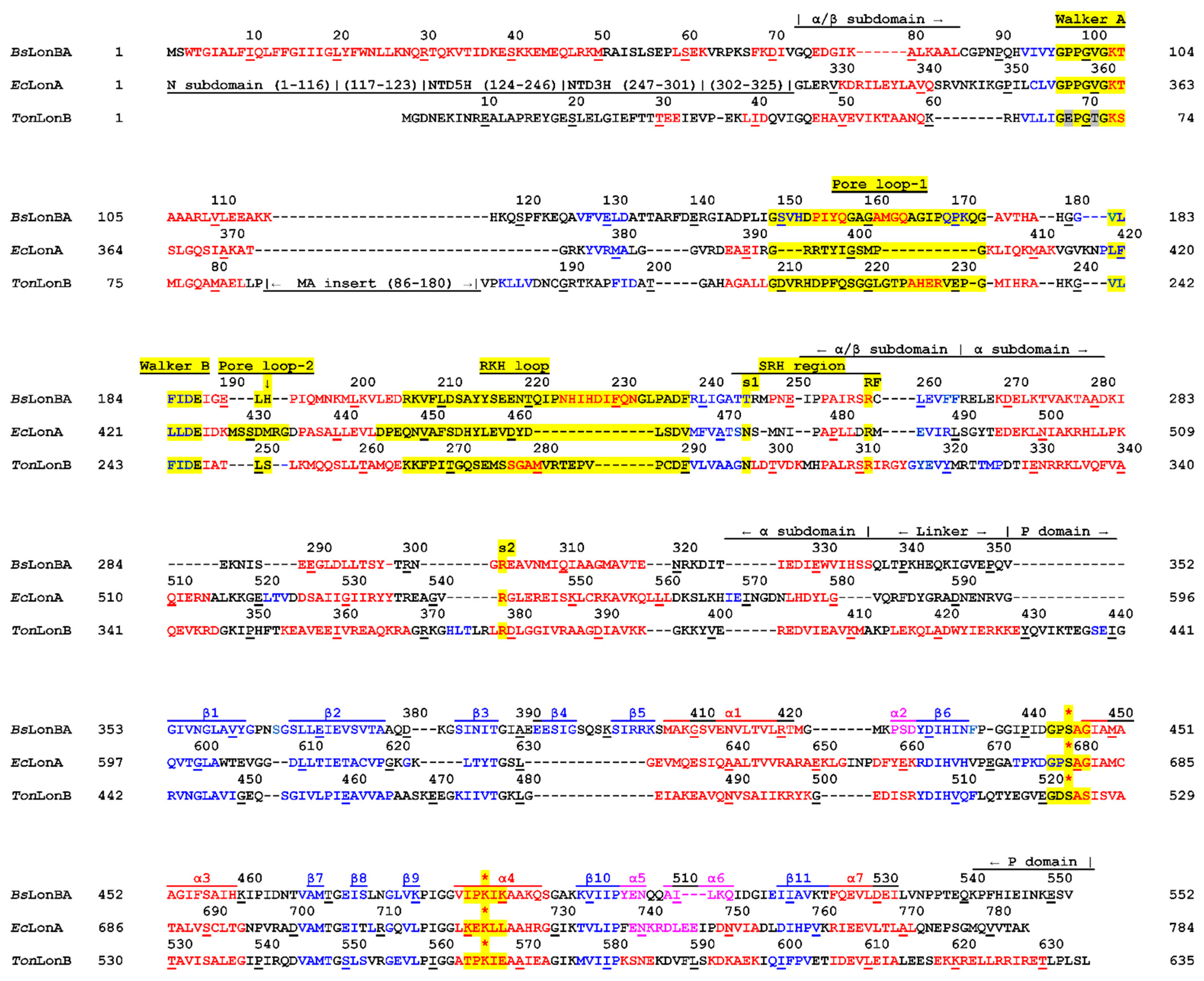
2.1. Analysis of the Primary Structures of Lon Proteases Belonging to ID Group S16.005

The ID group S16.005 (LonB peptidases) in the MEROPS database currently includes 145 members. Out of these, only 19 enzymes (13.1%) are LonB proteases of archaea, 40 (27.6%) are represented by enzymes from Bacilli, and 86 (59.3%) are from Clostridia. True archaeal proteases of this group are formed by ATPase and protease domains of B-type, and their AAA+ modules include an inserted MA domain with a transmembrane fragment (Figure 1) [30,39]. The amino acid sequences of bacterial enzymes of this ID group lack both the extended N-terminal region characteristic of LonAs, as well as the large, inserted MA domain inherent in LonBs. Nevertheless, their ATPase domains demonstrate a number of similarities with the ATPase domains found in both subfamilies of Lons, but their protease domains are more similar to A-type than to B-type Lons, which implies a “hybrid” domain organization of this new group of enzymes (Figure 1).
Despite the fact that the presence of a Lon protease in B. subtilis, which contains neither an N-terminal region as in LonA, nor a membrane anchor as in LonB, has been previously noted [39,40], this observation has not been followed up. There is no information in the literature on the isolation and study of Lon proteases from Clostridia that are assigned to ID groups S16.001 and S16.005. For these reasons, we selected B. subtilis as a source for purification and study of a model bacterial “hybrid” Lon protease (BsLonBA) of the S16.005 group.
The primary structures of LonA and LonBA proteases from eight bacillary sources were compared with the structure of the model enzyme, EcLonA (Figure S1). LonBAs were found to contain two functional domains (AAA+ module and P domain) connected by a 13- or 15-residue-long linker, as well as an N-terminal fragment consisting of 72 or 73 residues. Similarity levels within each fragment of the LonBA sequences were evaluated. It was found that the most conserved is the α/β subdomain of the AAA+ module (Figure 1), with a total sequence similarity of 90.0% (with 69.4% of strictly conserved residues), whereas the least conserved was the α subdomain (53.3% similarity and 27.8% identity). The N-terminal extension and the P domain show similar conservation: 75.3% similarity and 26.4% identity (for the N fragment), 71.1% similarity, and 38.5% identity (for the P domain).
According to the prediction (http://distillf.ucd.ie/porterpaleale/, accessed on 20 May 2022), the N-extension of the BsLonBA is predominantly α-helical (Figure S1). On that basis, we compared this fragment with helical subdomains NTD5H and NTD3H of LonA proteases. It was found that the LonBA N-fragments do not resemble either the NTD3H subdomain (Figure S1) or the NTD5H subdomain (not shown). There is also no sequence similarity between the N-terminal fragments of LonBAs and the corresponding fragments of LonB and LonC proteases (Figure S2).
While comparing the AAA+ modules of LonA and LonBA proteases, a distinction in size of the N-terminal α-helix, as well as differences in the distances between the enzyme consensus fragments, may be detected (Figure S1). In particular, in LonA proteases, the conserved Asp residue of the Walker B motif is separated from the Lys of the Walker A by 60 residues, whereas in LonBA proteases, this distance increases to 82 residues due to insertions and deletions that are localized before, after, and/or within Pore loop-1 (Figure S1). Other insertions or deletions are found in the loops connecting conserved secondary structure elements, and they are discussed in more structural terms below. On the contrary, the α subdomains of LonBA proteases are smaller than in LonAs, due to deletions of some residues in the regions of the first and third α helices (Figure S1).
Although the overall similarity between the P domains of LonAs and LonBAs is high and the distances between their catalytic residues (Ser 445 and Lys488 in BsLonBA; Ser679 and Lys722 in EcLonA, see Figure S1) are identical, the presence of an inserted fragment of 14 amino acids, which is embedded in a region located before the first α helices of the P domains of LonAs (E632-A647 in EcLonA), is significant (Figure S1). It can be noted that the P domains of bacillary LonAs and LonBAs do not contain any cysteine residues and therefore cannot form an internal disulfide bond, which is characteristic for EcLonA [34]. However, as will be shown below, the presence of the disulfide in the latter enzyme does not affect the conformation of its region compared to its counterpart in BsLonBA.
Thus, a comparison of the amino acid sequence of LonBA with the sequences of LonA, LonB, and LonC proteases suggests noticeable differences between these Lon subfamilies. We used the BsLonBA enzyme as a model representative of that proposed novel subfamily. The enzyme was cloned, expressed, and its basic enzymatic properties were studied. Additionally, the P domain of BsLonBA was crystallized and its structure was compared to structures of P domains of other members of the Lon family.

2.2. Preparation, Limited Proteolysis, and Preliminary Characterization of the Recombinant BsLonBA

Full-length BsLonBA was cloned and expressed in E. coli as a C-terminal His6-tagged protein (BsLonBA-H6), which appeared to be soluble only in the presence of detergents (CHAPS or DDM). To identify the domain organization of the enzyme, we used limited proteolysis by chymotrypsin, which previously allowed us to successfully determine the domain boundaries in EcLonA and study its nucleotide-dependent conformational changes [41]. Three main cleavage sites that led to the formation of stable fragments were identified (Figure S1). However, these sites (F238/R239, Y298/T299, and W331/V332) turned out to be of limited usefulness for fixing domain boundaries, since they are located in the α/β subdomain before its consensus sensor-1 (s1) residue, as well as in the central part and in the C-terminal fragment of the α subdomain, respectively. In addition, unlike EcLonA, limited proteolysis of BsLonBA was not affected by either ATP or Mg2+ ions. Cleavage of BsLonBA within the AAA+ module may indicate its partial disorder, presumably due to the insertions and deletions noted above. On the other hand, the absence of limited proteolysis sites within the region corresponding to the EcLonA P domain (residues 353–552 in BsLonBA) demonstrates its structural stability.
In terms of enzymatic properties, the newly isolated BsLonBA-H6 protease is radically different from the “classical” EcLonA. The latter enzyme, which also bore a C-terminal hexahistidine tag, was shown to hydrolyze ATP effectively, and, in the presence of a protein substrate, the ATPase activity increased significantly [42,43]. However, BsLonBA-H6 does not show the ability to hydrolyze ATP either in the absence or in the presence of a substrate protein.
The proteolytic activity of both Lon proteases was monitored through hydrolysis of β-casein. In the presence of the ATP-Mg2+ EcLonA-H6 degrades a protein substrate utilizing the processive mechanism, but in the absence of a nucleotide, the enzyme exhibits weak non-processive activity (Figure S3A). In both cases, bortezomib, an organoboronic serine protease inhibitor, completely suppresses the proteolytic function of EcLonA-H6. The BsLonBA-H6 protease does not cleave β-casein in the time interval in which the proteolytic activity of EcLonA-H6 was tested. However, when the reaction time is increased to 24 h or more, products of non-processive hydrolysis of the protein substrate are detected, both in the presence of ATP-Mg2+ and in the absence of effectors (Figure S3B). In addition, hydrolysis of the target protein is accompanied by noticeable autolytic degradation of the enzyme (Figure S3B). Thus, under the conditions utilized in this study, BsLonBA-H6 exhibits very low non-processive proteolytic activity, and at the same time bortezomib practically does not affect the function of the enzyme (Figure S3B).
The results presented above show that BsLonBA-H6 is significantly inferior to EcLonA-H6-protease in both the ATPase and proteolytic activities. However, peptidase activities of these enzymes are quite comparable. It was found that basic peptidase activities (the hydrolysis efficiency of PepTBE in the absence of a nucleotide) of BsLonBA-H6 and EcLonA-H6 differ by no more than 5–6 times (Figure S4, lines 1 and 3). Bortezomib exhibited an inhibitory effect on the peptidase activity of both enzymes (Figure S4, lines 2 and 4), but with different efficacy: the IC50 values differ by more than 20 times (0.05 μM for EcLonA-H6 and 1.15 μM for BsLonBA-H6). The presence of the ATP-Mg2+ complex leads to the activation of the peptidase activity of EcLonA-H6, but does not affect peptide hydrolysis by BsLonBA-H6, which is consistent with the absence of its ATPase activity.
Differences in the ATPase and proteolytic activities of BsLonBA and EcLonA may indicate that the formation of a functionally active conformation of the AAA+ module of the former may require either optimization of the enzyme isolation method or a search for a putative adaptor protein, not currently identified. Important effects of adapter proteins were previously shown, for example, for ClpCP [44,45]. A search for conditions that promote the formation of the functionally correct active structure of BsLonBA-H6-protease remains a future direction in the study of enzymes of this new Lon subfamily.

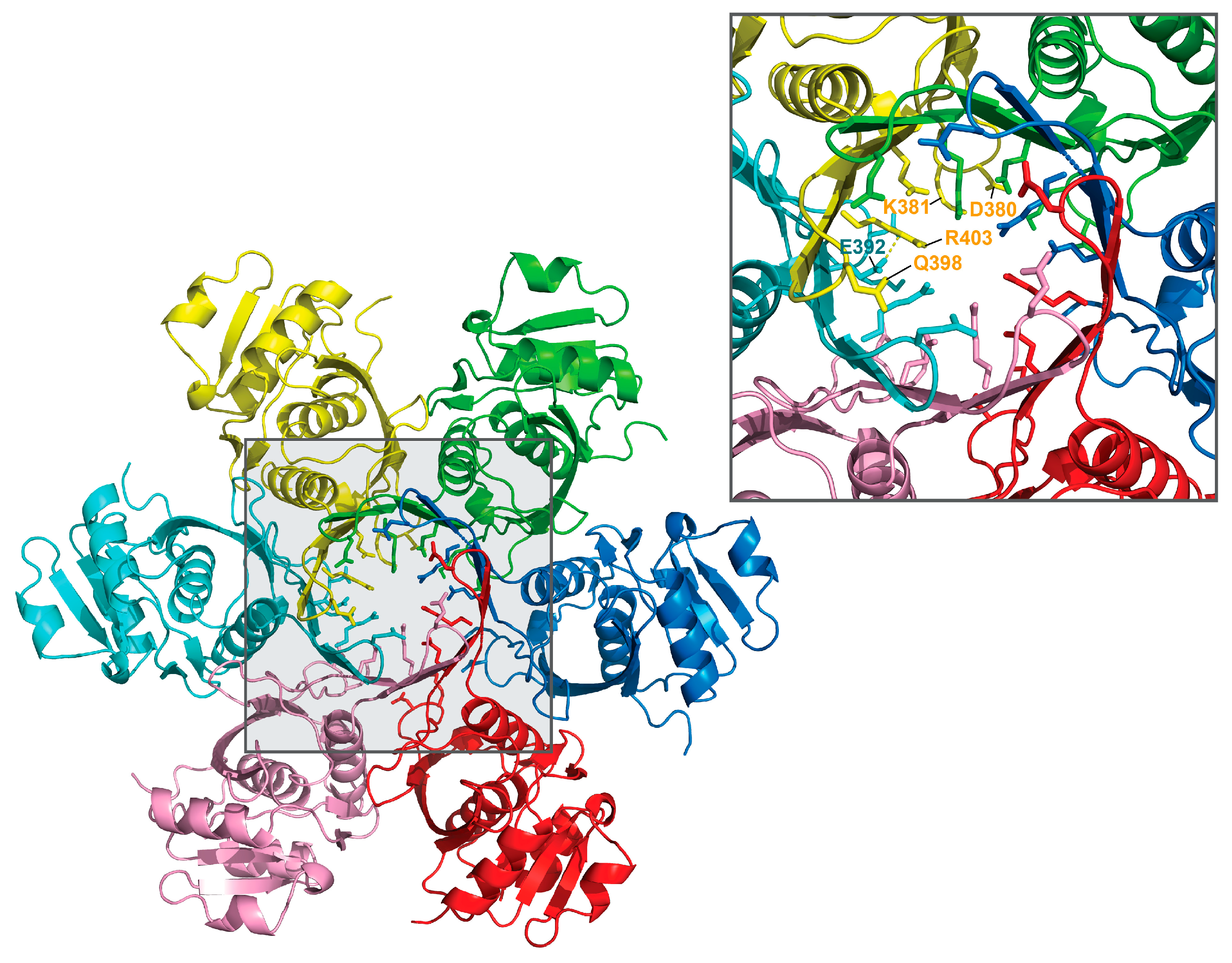
2.3. Crystal Structure of the P Domain of BsLonBA

The crystal structure of the P domain of BsLonBA (residues 347–552) was determined at the resolution of 1.9 Å. Crystals belong to the hexagonal space group P63 with two molecules (designated A and B) in the asymmetric unit, arranged around the crystallographic 3-fold axis. This arrangement results in the creation of an expected planar hexamer (Figure 2). Such a hexamer is similar to the hexameric P domains of EcLonA and AfLonB, to the P domains in the almost full-length hexameric proteases LonB from Thermococcus onnurineus (TonLonB) and LonC from Meiothermus taiwanensis (MtaLonC), as well as in cryoEM structures of full length human mitochondrial LonA (hMtLonA) (as recently reviewed [33]). It must be stressed, however, that the protein used for crystallization behaved as a dimer when run on a sizing column before crystallization experiments were set up. The fact that an isolated P domain of BsLonBA emulates other Lons, recreating hexamers in the crystal lattice, leads to an assumption that the structure of BsLonBA P domain mimics functional, hexameric organization of this protein. Similarly to other Lons, in the BsLonBA oligomer six P domains have a planar arrangement with the proteolytic chamber in the center. However, one structural element strikingly differentiates BsLonBA from other known Lons. The insertion of 14 residues (392–405) in the P domain of BsLonBA (Figure S1) creates a 12-stranded β-barrel in the core of the hexameric assembly formed by β-hairpin inserts of the neighboring subunits (Figure 2; a detailed description below).
The overall fold of the P domain of BsLonBA resembles the equivalent domains of enzymes belonging to A, B, and C subfamilies of Lon proteases. In BsLonBA it consists of a single polypeptide chain of ~200 amino acids, which forms 11 β-strands and seven helices (Figure 3 and Figure S2). The molecule can be divided into two subdomains with a linking fragment comprising residues 460–467. The core of the first subdomain is built of a 4-stranded β-sheet and three helices. A mixed β-sheet consisting of an N-terminal β-hairpin (β1–β2), β6, and β3 forms the back wall. The protruding part of the subdomain is formed by another β-hairpin (β4–β5), which participates in the formation of the central β-barrel of the hexamer. It precedes in the sequence the longest helix α1, which runs on the front side of the mixed β-sheet. Helix α1 and a short helical turn α2 (directly preceding β6 in the sequence) participate in the inter-protomer interactions with the back wall of the neighboring unit of the hexamer. Helix α1 also interacts with helix α3, which runs in the central part of the P domain and leads to the second subdomain. This subdomain includes a central three-stranded parallel β-sheet (β7–β10–β11) flanked by helix α7 from the bottom side and by helices α4 and α6 from the top side. A short helix α5 directly follows strand β10. The subdomain has also an extended loop with two very short β-strands (β8–β9), which are placed above the N-terminal side of the α3 and protrude towards α1, participating in interactions with the neighboring protomer.
The unique β4–β5 inserted hairpin in BsLonBA (392–405) is created by two five-residue-long strands composed mainly of polar residues and a four-residue-long connecting loop. Six inserts arranged around the three-fold symmetry axis complete a 12-stranded antiparallel β-barrel, where each insert forms nine hydrogen bonds with the neighboring subunit. In the central core of this barrel, two layers of charged or hydrophilic residues from each protomer form a planar arrangement with their side chains in an extended conformation and oriented towards the center of the barrel (Figure 4, top and middle ovals shaded gray). The top layer is formed by the side chains of Gln398. The width of the barrel opening within this layer is ~11 Å (Figure 4 and Figure 5). The second layer is formed by the side chains of conserved Glu392 and Arg403, that are poised towards the interior of the barrel, creating a second lid with a ~8 Å wide opening. An ion pair between the Nε atom of Arg403 side chain and Oε1 atom of Glu392 from two neighboring protomers seems to support the extended conformations of their side chains and their orientations towards the center of the barrel (Figure 5). The third layer is formed at the bottom of the central chamber of the P domain hexamer by the side chains of Asp380 and Lys381 from the same protomer, which are also oriented towards the center with the distance between their charged groups ~3.4 Å (Figure 4 and Figure 5). Here, the chamber widens to ~16.5 Å.

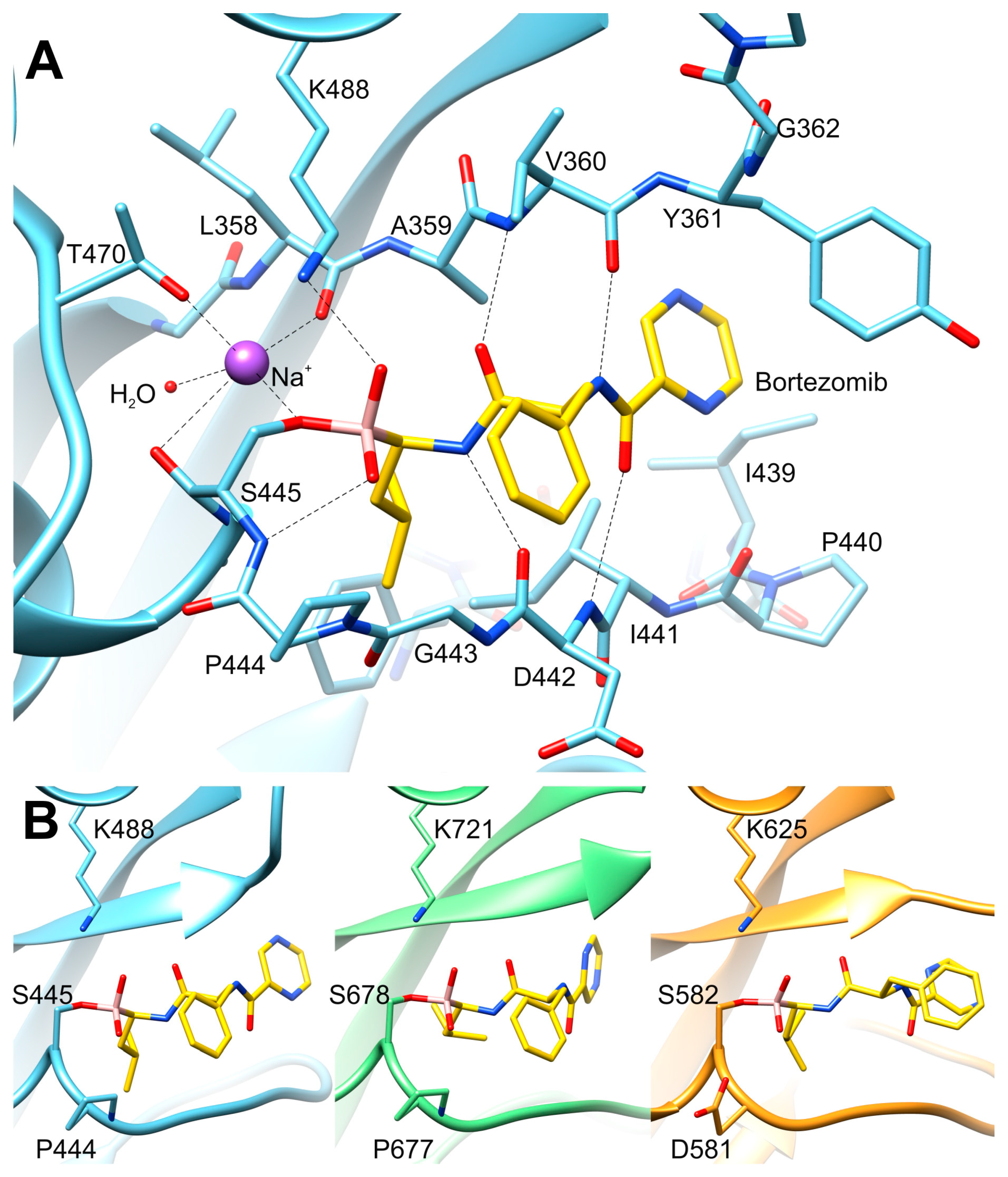
2.4. The Mode of Binding of Bortezomib to the P Domain of LonBA and Other Lon Proteases

We have captured the P domain in an inhibited state, in the complex with bortezomib, a covalently bound inhibitor. Unambiguous electron density maps (Figure 3) allowed us to determine the conformation of the inhibitor within the substrate-binding site. This site is created by a β-hairpin (β1–β2), a long loop connecting β6 and α3, and the protruding loop made of β8–β9. The catalytic Ser445 is located in the sequence just before α3, whereas Lys488, the other member of the catalytic dyad, is located within helix α4 (Figure 3). Such an architecture that includes several flexible structural elements allows accommodatinf a variety of conformations. Bortezomib is covalently bound to the Oγ of Ser445 via the boronate moiety. The inhibitor structurally mimics a peptide substrate with leucine and phenylalanine side chains, as well as pyrazinoic acid moiety. Within the active site, bortezomib creates β-sheet-like interactions with residues of β1 (A359, V360, Y361) on one side, and residues of the loop β6-α3 (I441, D442) on the other side (Figure 6A). Oxygen atoms of the boronate moiety make additional hydrogen bonds with the side-chain amine of Lys488 and the main chain nitrogen of Ser445. Analysis of the vicinity of the active site identified a putative sodium cation coordinated by carbonyl oxygens of Ser445 and Leu358, Oγ atoms of Thr470 and Ser445, and a water molecule (Figure 6A). The location and conformation of the inhibitor are similar to its counterparts in other Lon proteases (see below) (Figure 6B).
High-resolution crystal structures that include bortezomib-inhibited protease domains are available for hMtLonA (PDB ID 6x27), LonA from M. taiwanensis (MtaLonA) (4ypm), and MtaLonC (4fwd) [33]. There are currently no published data available for any LonB complexed with any ligand, including bortezomib. Among the available structures of different protease domains of LonB, AfLonB (exemplified by 1z0w) has an active site area that is significantly different from what is expected for an enzyme with no ligand bound, and LonB from Methanocaldococcus jannaschii (MjLonB, 1xhk) is in a self-inhibited conformation, in which Asp547, separated by two residues from the catalytic Ser550, occupies a part of the substrate-binding site. Although TonLonB (3k1j) has both the active site serine and lysine replaced by alanines, its substrate-binding site is not occupied. This enables the active site to adopt an open conformation for accepting a ligand, allowing direct comparison with BsLonBA inhibited by bortezomib.
The P domain of hMtLonA (6x27) can be superimposed on its BsLonBA counterpart with an rmsd of 1.34 Å for 155 Cα atoms, whereas the superposition of MtaLonA (4ypm) yields an rmsd of 1.54 Å for 146 Cα atoms, despite the amino acid identity compared to BsLonBA being larger (41%) for the latter enzyme than 35% for the former. The overall similarity of the P domains of BsLonBA and TonLonB (3k1j) is very high (1.34 Å for 145 Cα atoms, 43.5% identity). Although the identity of 24.3% is lower compared to MtaLonC (4fwd), the fold of these proteins is still very similar, with an rmsd of 1.53 Å for 156 Cα atoms.
The boronate group of bortezomib is covalently bound to the active site serine and the conformation of its main chain is virtually identical in all compared structures (Figure 6B). The extended inhibitor mimics a β strand and makes a number of hydrogen bonds with the main chain peptides of the two adjacent chains. The side moieties of bortezomib occupy the S1–S3 subsites of the substrate-binding crevice. In the structure of the hMtLonA complex (6x27), the P2 phenyl group of the inhibitor has two conformations, whereas its conformation in MtaLonC (4fwd) corresponds to the alternate conformation in 6x27 (not shown). This subsite is located on the surface of the enzyme and it is rather open to solvent.
Each of the four structures of Lon proteases complexed with bortezomib (listed above) contains two fully conserved solvent molecules in the immediate vicinity of the active site serine side chain. The distance between the Oγ atom of the catalytic serine and the first one of these solvent molecules varies in these structures between 2.62 and 2.88 Å, but this solvent molecule was annotated as water in hMtLonA (6x27) and MtaLonC (4fwd), as Mg2+ ion in MtaLonA (4ypm), and now as Na+ in BsLonBA. This molecule is within a hydrogen bond distance to three main chain carbonyl atoms, as well as to another conserved solvent molecule, invariably identified as water. The presence of five potential ligands of these solvent molecules, as well as the geometry of their interactions, makes it unlikely that they could be waters, since a water molecule could not be a hydrogen bond donor to three oxygens acting as acceptors. The distances between this molecule and its neighbors are significantly longer than the coordination distances expected for a Mg2+ ion, and the geometry of interactions is also not octahedral. Only the postulated Na+ ion could be reconciled with the observed geometry, since that ion is quite frequently found to be coordinated to five oxygen atoms [46]. This interpretation makes the previous proposal that a magnesium ion might be directly responsible for the transition between the inactive and active conformation of the active site of Lon rather unlikely.
A defining feature of the P domains of LonA proteases is the identity of the residues preceding the catalytic serine (Ser679 in EcLonA), which is always a proline, whereas the corresponding residue is an aspartic acid in LonBs and LonCs. Nevertheless, the main-chain torsion angles are virtually identical in the presence of bound inhibitors regardless of the nature of this residue (Figure 6B), making it unlikely that its identity plays a major role in establishing the catalytic properties of the protease domains of different subfamilies of Lon proteases.

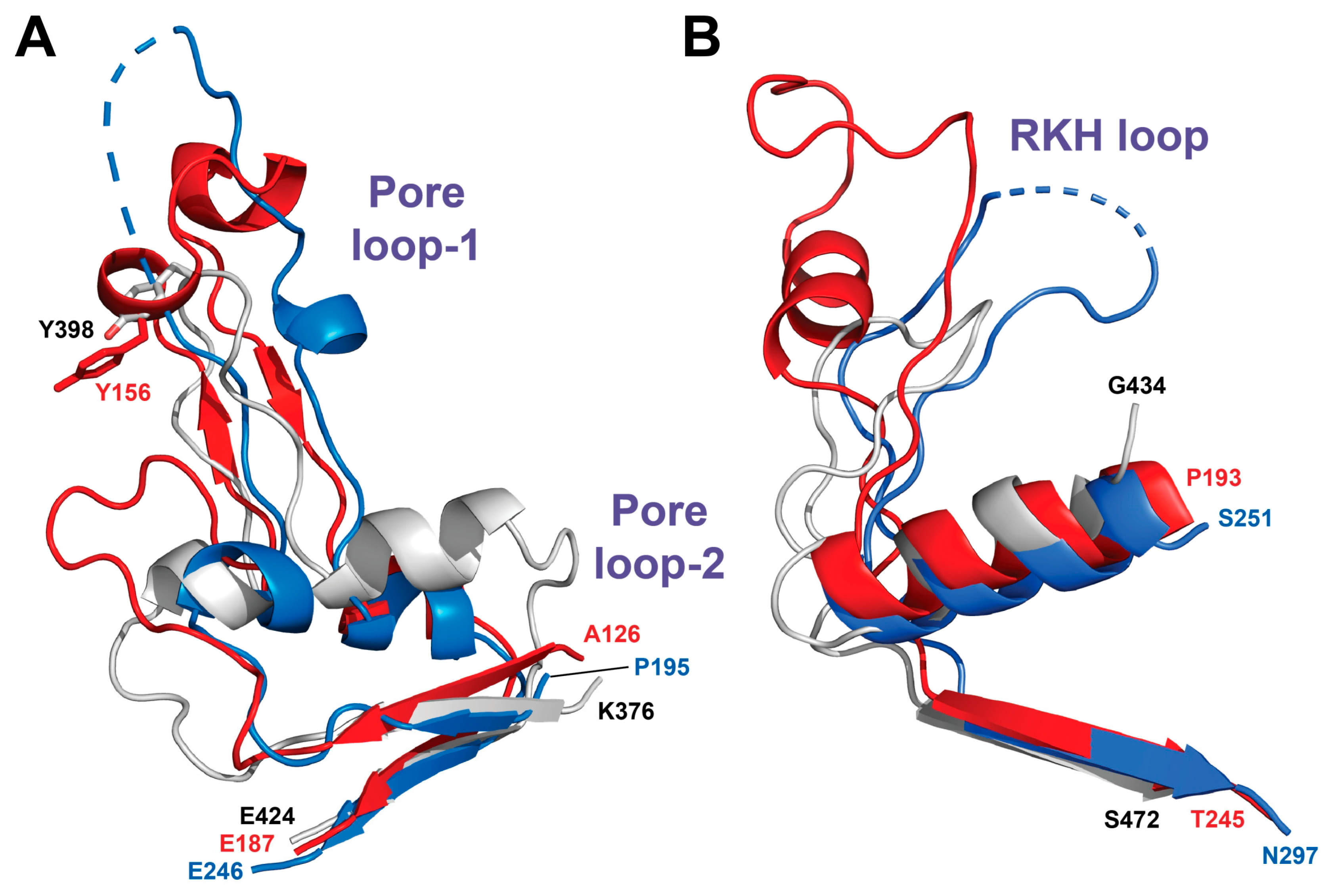
2.5. Comparison of the BsLonBA P Domain with Its Counterparts in Other Lon Proteases

In addition to the structure of the P domain of BsLonBA described here, six structures of its LonA counterparts, three of LonB, and one of LonC are currently available [33]. All of them start with long β-hairpins comprising strands β1 and β2 (Figure 3). Strand β1 participates in the formation of the ligand-binding site with residues interacting directly or via solvent with the bound inhibitor (Figure 6). The conformation and position of the tip of the β1–β2 loop vary in different Lons according to the functional state of the individual P domains, as well as the presence or absence of a bound ligand (Figure 7 and Figure 8).
The loop connecting strands β2 and β3 is one-residue longer in BsLonBA than in EcLonA, and two-residue shorter than in TonLonB (Figure 9). hMtLonA (6x27) has the longest insert in this loop compared to bacterial LonAs, as well as LonBs (~10 residues), which forms a flexible loop disordered in the structure. In BsLonBA, residues Asp380 and Lys381 of this loop in each protomer form the planar arrangement of their side chains in an extended conformation at the bottom of the central chamber of the P domain hexamer, as described above (Figure 4 and Figure 5). Interestingly, EcLonA and MjLonB (1xhk) also have topologically equivalent corresponding residues within this loop (Lys621 in EcLonA and Lys487 in MjLonB).
Four β strands (β1, β2, and β3, as well as β6, positioned between β2 and β3 (Figure 3)), form in BsLonBA a β-sheet that is also conserved in all Lon subfamilies. Unique among all subfamilies of Lon, only MtaLonC includes a helical twist after β3. In turn, BsLonBA is the sole exception that includes a protruding β-hairpin (β4–β5) that forms a β-barrel in the central part of the proteolytic chamber of the hexamer (Figure 2 and Figure 3). No other Lon with known structure has a similar insert that would correspond to β4–β5 of BsLonBA (Figure S2). The orientation of the longest helix α1 is slightly different in other known Lons than it is in BsLonBA. Additionally, the loop that follows, connecting helix α1 with strand β6, is the most variable region in different Lons (Figure 7). This loop can be longer, as in MjLonB, with no helical twist, or it may form two small helices, as in MtaLonA or BsLonBA. However, a common feature of this loop is that it creates a helical twist just before β6 (BsLonBA, LonA protease from Yersinia pestis (YpLonA), MtaLonA, hMtLonA, TonLonB, AfLonB) (shown for BsLonBA in Figure 3). The long flexible loop connecting strand β6 with helix α3 in BsLonBA carries the catalytic Ser445 (Figure 3). After that, the protruding region consisting of β7, as well as of the loop that comprises strands β8 and β9 and leads to helix α4 is structurally very similar in all Lons, with helix α4 carrying the catalytic Lys488 (Figure 3). The only exception is again MtaLonC, where α4 is longer and ends with an additional inserted loop created by seven residues (Figure S2). The region that follows (β10-α5) is rather similar in various Lons, although BsLonBA is characterized by a break in a shorter helix α5, which is followed by α6 and then β11. LonAs and LonC are characterized by a longer helix α5 and an insertion that follows it. This insertion forms a helix and a loop that leads to β11. In LonBs, there is no additional helix after α5. AfLonB has a long loop, whereas this loop is significantly shorter in MjLonB. After β11, the common feature of all Lons is the presence of α7 and some of them end with another helix close to the C terminus (TonLonB, AfLonB, and MtaLonC).
Comparison of the structure of P domain of BsLonBA with the structures of the corresponding domains in LonAs and LonBs, determined in either single-domain or multi-domain constructs, reveals a similar mechanism of transition of the state of the active site from an active conformation to an inactive one. Two elements of the active site area embracing the ligand binding area, which include on one side a flexible loop (residues 438–446 in BsLonBA, element 1) and on the other side a β1–β2 loop (residues 358–368, element 2, Figure 7), would change their conformation and mutual arrangement, yielding a particular functional state of the P domain (Figure 8). Thus, in the structure of BsLonBA, the active site of the P domain adopts an open conformation, with bortezomib bound in the center and flanked by both mobile elements listed above. In both the crystal structure of the P domain of EcLonA and cryo-EM structure of the full-length enzyme, their uninhibited active sites are found in an inactive conformation, although the mechanism to achieve this state was different. In the crystal structure of an isolated P domain (1rre), the fragment comprising element 2 moved to occupy the binding site area (Figure 8, shown in pink), whereas in the cryo-EM structure of the full-length enzyme (6u5z) a fragment comprising element 1 moved to occupy the same space, leaving the position of element 2 unchanged (Figure 8, shown in green). In the structure of hMtLonA (7krz, Figure 8, shown in cyan), the inhibitor, bortezomib, is bound in the active site, and the flanking elements remain in an open conformation, leaving the active site area undisturbed.
Although in the crystal structure of an unliganded, isolated P domain of MjLonB (1xhk, Figure 8, shown in magenta), the inactive state is supported by the shift of element 1 to occupy the active site area, in the crystal structure of the almost full-length TonLonB (3kij, Figure 8, shown in blue), the active site area assumes an open conformation even in the absence of a ligand.
While the mechanism of transition from the active to inactive state of the P domain that is assisted by the movement and conformational changes in two mobile elements of the active site seems to be consistent for several LonAs and LonBs, one example completely stands out. Significant differences were observed in the active site of an isolated P domain of AfLonB (1z0w, Figure 8, shown in yellow), where the part of the chain around the catalytic Ser509 was flipped away from the helix carrying the other catalytic residue, Lys552. As a result, the catalytic dyad no longer exists, and a fragment comprising element 1 moves to occupy the substrate binding site, establishing an inactive state of the enzyme. We can speculate that such a conformational change could be facilitated by the substitution of the conserved proline residue before the catalytic Ser in LonAs by the aspartate in LonBs, making such a dramatic flip of the main chain around this residue possible.

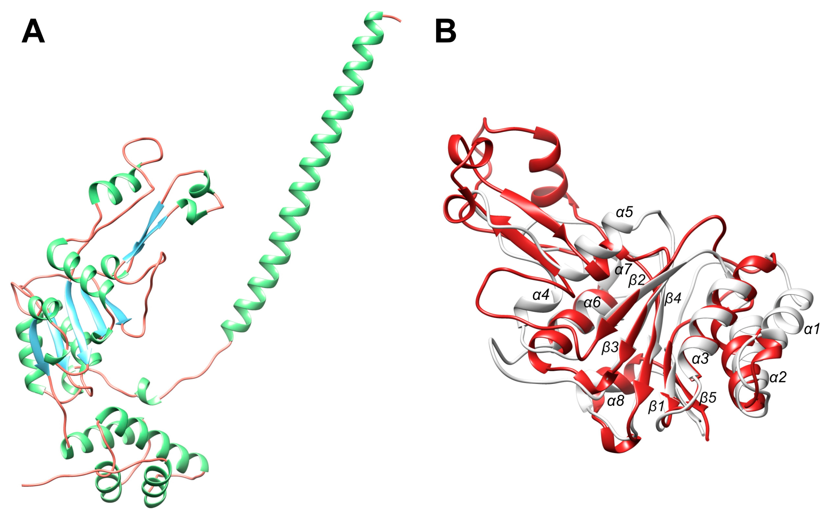
2.6. Model Building of the ATPase Domain of BsLonBA

Although we have not determined the structure of the AAA+ module of BsLonBA experimentally, a putative model was obtained with the prediction program AlphaFold2 [47] (Figure 10A). It was our expectation that some features that specifically differentiate LonBAs from other members of this family might be important in deciding whether BsLonBA should be considered as a representative of a new subfamily of Lons. The original division of the Lon family into the subfamilies LonA and LonB was based, at least in part, on the differences in the location of the enzymes in the cells. Whereas LonA is a soluble cytoplasmic or mitochondrial enzyme, LonB was found to be membrane-associated [30,48,49]. The fragment of LonB that carries the membrane-insertion sequence is located in the α/β subdomain between the Walker motifs A and B and consists of over 100 amino acids, some of which assume the helical conformation necessary for insertion into cell membranes. There is no corresponding insertion in the sequence of BsLonBA, although that enzyme may also be described as being membrane-associated according to programs that search for transmembrane fragments (including SMART, TMHMM, HMMTOP, TMDAS and others; see Materials and Methods). In this case, however, the membrane-insertion fragment is most likely located at the N terminus of the sequence. The model calculated by AlphaFold2 included a very long α helix (consisting of 52 amino acids) at the N terminus (Figure 10A), with the first ~20 residues being hydrophobic and thus capable of being inserted into a membrane.

2.7. Comparison of the Functionally Important Elements in the AAA+ Module of BsLonBA with Their Counterparts in LonA and LonB

An overall comparison between the structures of the AAA+ modules of BsLonBA and the corresponding fragment of EcLonA (6n2i) yields an rmsd of 2.7 Å for 195 Cα pairs, with sequence identity 18.5% and 19 gaps. An analogous comparison with the AAA+ module of TonLonB (4zpx) yields an rmsd of 2.7 Å for 214 Cα pairs, with sequence identity 23.8% and 15 gaps.
The AAA+ module of Lon proteases belongs to the HCLR clade, the name of which is derived from the four families that also include HslU/ClpX, ClpABC-CTD, and RuvB [50]. It differs from the simplest module, such as that found in the clamp loader clade [48] (exemplified by the structure with PDB ID 1njg [51]), by the presence of a β loop insertion that emerges from a single helix in the clamp loader, breaking the corresponding helix in Lon into two small helices: α4 and α5 (Figure 10B). From here on, a new numbering of the secondary structure elements for the A domain is used (Figure 10B). A set of conserved consensus fragments of the AAA+ module includes Walker motifs A (residues 356–363) and B (419–424), as well as residues forming sensor-1 (Asn473), sensor-2 (Arg542), and the arginine finger (Arg484), in EcLonA numbering. The corresponding residue numbers for TonLonB and BsLonBA are presented in Figure 9.
The central pore of a hexamer of Lon is lined with axial loops, required for protein unfolding and translocation. Three different pore loops, called Pore loop-1, Pore loop-2 and RKH, are found in almost all AAA+ unfoldases. They interact with polypeptide substrates and pull them into the pore in a concerted way, with nucleotide-dependent loop movements unfolding those substrates that otherwise cannot enter the pore. Pore loop-1 is involved in polypeptide translocation and consists of 11 residues in EcLonA (6n2i; [52]), 24 residues in TonLonB (4zpx; [53]), and 25 residues in the predicted structure of BsLonBA (Figure 9). According to the model this insert in BsLonBA resembles more closely the one that is present in LonB than in LonA (Figure 11A). Although the estimate of the size of this insertion is fairly accurate, the predicted helical turns at the tip of the loop may not be necessarily modeled properly in BsLonBA due to the lack of a proper template, since that part of the loop is disordered in TonLonB.
While in enzymes belonging to all Lon subfamilies the insertion of Pore loop-1 breaks a single helical fragment into two small helices (α4 and α5; but in the model of LonBA α4 is only a helical turn, Figure 10B), the axis of the second small helix that follows the insertion remains the same as in the preceding one in each enzyme (Figure 11A). However, the orientation of the composite helix axis in LonBA is closer to its counterpart in LonB and slightly deviates from the orientation of the corresponding element in both bacterial and mitochondrial LonAs (Figure 11A).
Pore loop-1 is located between the Walker A and Walker B motives. As was noted before, there is a large difference in the number of residues that separate Walker A from Walker B motif in LonBAs and LonAs (82 residues between the conserved Lys103 in the Walker A and the conserved Asp186 in the Walker B motif of BsLonBA, as compared to 60 residues in LonAs). The insertions responsible for this difference are localized in BsLonBA in two areas. One area includes two loops that connect strand β2 with the preceding helix α3 (residues 103–116) and with the following helix α4 (residues 388–391 and 204–208 in EcLonA and TonLonB, respectively), which is, as described above, only a helical turn in the modeled structure of BsLonBA (residues 145–148). The other insertions are found within Pore loop-1 (residues 149–173, 393–404 and 209–232 in BsLonBA, EcLonA and TonLonB, respectively) (Figure 9). On the other hand, there is a five-residue deletion in BsLonBA in the loop connecting helix α5 (residues 174–178) and strand β3 (residues 182–187) comprising residues of Walker B motif, as compared to the corresponding fragment of EcLonA (residues 411–418). This fragment of BsLonBA is more similar to the corresponding one in TonLonB (Figure 9).
Pore loop-2 in BsLonBA is very short, similar to its counterpart in TonLonB. It contains only two residues 191–192, unlike the corresponding Pore loop-2 in EcLonA that comprises eight residues (428–434) (Figure 9, and Figure 11A). In EcLonA, this loop is more extended into the central pore, enabling interactions with the substrate.
The RKH loop is the longest in BsLonBA (residues 206–238), its length closer to its counterpart in TonLonB (residues 265–290) than to the shorter loop in EcLonA (residues 445–466) (Figure 9, and Figure 11B). The location and surrounding of the sensor-1 and sensor-2 residues, as well as the arginine finger, are highly conserved in all three enzymes (Figure 12).
On the other hand, the loop comprising the SRH region with sensor-1 residue and the Walker A motif in BsLonBA are more similar to their counterparts in EcLonA. In TonLonB these two fragments bear small differences. The fragment comprising the SRH region with the s1 residue is longer than in both BsLonBA and EcLonA. Additionally, the conserved Pro residue in the Walker A motif (Pro98 in BsLonBA and Pro357 in EcLonA) is substituted by Glu68 in TonLonB. As a result, the longer SRH loop in the latter adopts a different conformation that permits the NH group of Leu298 (the next residue after s1) to interact with the side chain of Glu68 (Figure 13).

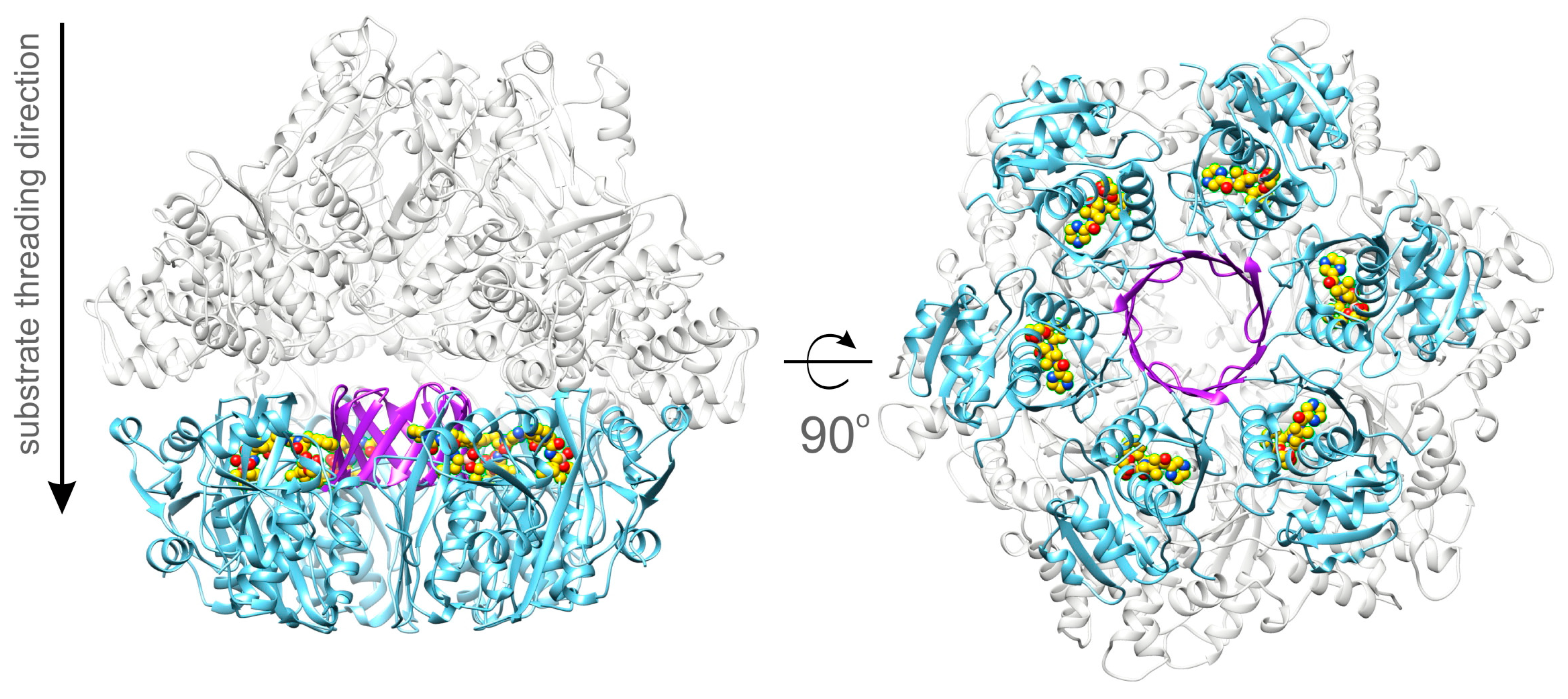
2.8. A Model of the Full-Length BsLonBA

A putative model of the almost full-length BsLonBA was obtained by superposition of the hexamer of the experimentally determined structures of its P domains on the hexamer of the full-length crystal structure of TonLonB (3k1j), followed by individual superposition of each AAA+ module (generated with AlphaFold2, with residues 1–56 truncated) on each AAA+ module of 3k1j. The resulting model is shown in Figure 14. However, since the crystal structure of TonLonB is quasi-symmetric in the absence of a bound substrate, it should be stressed that the resulting model of BsLonBA should be considered tentative, as structures of substrate-free, full-length members of the LonA subfamily are helical rather than planar. Nevertheless, this modeling exercise indicates that the core part of BsLonBA could resemble the structures of other members of the Lon family, although this still needs experimental verification. At this time, there are no data indicating whether the N-terminal fragment of BsLonBA is indeed folded into a helical conformation and whether it serves as a membrane anchor.
This model of the full-length BsLonBA suggests that a protein substrate threaded through the axial channel (A-pore) of the AAA+ module likely interacts with the external side of the unique barrel in the P domain hexamer formed by the β4–β5 hairpins (Figure 4). In addition, threading of the substrate through the barrel seems rather unlikely, because the substrate would miss the proteolytic active site. Moreover, the polar interior of the barrel (Figure 5) would act repulsively against bulky hydrophobic side chains of the processed substrate. In the generated model of the full-length BsLonBA, there is a ~20 Å gap between the bottom part of the A-pore and the inserted barrel. This provides enough space for the threaded substrate to be translocated to one of the proteolytic sites where it could be processed.

3. Materials and Methods

3.1. Analysis of the Amino acid Sequences of Lon Proteases

The amino acid sequences of the chaperones and proteases analyzed in this study were obtained from the UniProt Knowledgebase (http://www.uniprot.org/, accessed on 20 May 2022) and the MEROPS database (https://www.ebi.ac.uk/merops/, accessed on 20 May 2022).
For secondary structure prediction the following programs were used: Porter 5.0 (http://distilldeep.ucd.ie/porter/, accessed on 20 May 2022), PSSpred (https://zhanggroup.org/PSSpred/, accessed on 20 May 2022), Jped4 (http://www.compbio.dundee.ac.uk/jpred4/index.html, accessed on 20 May 2022) and RaptorX (http://raptorx.uchicago.edu/StructurePropertyPred/predict/, accessed on 20 May 2022).
The search for transmembrane fragments in protein sequences was carried out using programs SMART (http://smart.embl-heidelberg.de/smart/set_mode.cgi?NORMAL=1, accessed on 20 May 2022), TMHMM (https://services.healthtech.dtu.dk/service.php?TMHMM-2.0, accessed on 20 May 2022), HMMTOP (http://www.enzim.hu/hmmtop/html/submit.html, accessed on 20 May 2022) and MPEx (https://blanco.biomol.uci.edu/mpex/, accessed on 20 May 2022).
ExPASy server (www.expasy.org), PSIPRED (http://bioinf.cs.ucl.ac.uk/psipred, accessed on 20 May 2022), and CoCoPRED (http://www.csbio.sjtu.edu.cn/bioinf/CoCoPRED/, accessed on 20 May 2022) were used to identify the coiled-coil regions in proteins and for the subsequent topology analysis. The alignments of the protease sequences were performed using the Clustal Omega program (https://www.ebi.ac.uk/Tools/msa/clustalo/, accessed on 20 May 2022).

3.2. Molecular Cloning, Purification, Characterization, and Limited Proteolysis of the Recombinant BsLonBA

The gene encoding BsLonBA was amplified from the genome of B.s subtilis strain 168 using a pair of primers: the forward primer (5′-CGTACTACCTGCACCTCATGAGTTGGACAGGGATCGC-3′) and the reverse one (5′- GTCCTACCTGCATCCTCGAGAACGGATTCTTTATTGATTTCGATATG-3′). Both primers contained a BspMI site (underlined). After digestion with BspMI, the product was purified and ligated into the NcoI/XhoI sites of the isopropyl β-D-thiogalactoside (IPTG)-inducible E. coli expression vector pET28a(+) (Novagen, USA). The target form of BsLonBA protease contained an octapeptide (LEHHHHHH) at its C terminus (BsLonBA-H6). The construct was verified by DNA sequencing. E. coli strain BL21(DE3) (Novagen, USA) was transformed with the recombinant plasmid. Cells containing expression plasmid were grown to OD600 ~ 0.5 at 37 °C in 2YT broth containing 50 µg/mL kanamycin. Production of BsLonBA-H6 was induced with 0.1 mM IPTG for 4 h at 20–25 °C. The protein was purified to homogeneity from cleared cell lysate by sequential chromatography on Ni-Sepharose, Heparin-Sepharose, and Sephacryl S-300 columns (GE Healthcare, Sweden). It should be emphasized that due to the low solubility of BsLonBA-H6 protease, all stages of its isolation were carried out in the presence of 8 mM CHAPS detergent. The final product was judged to be >95% pure by SDS-PAGE. The protein concentration was quantified with Bradford reagent (Bio-Rad, Hercules, CA, USA).
A study of the enzymatic properties of BsLonBA-H6 was carried out in comparison with the corresponding properties of the EcLonA-H6 as described in [54]. The ATPase activity of Lon proteases was tested from the time-dependent accumulation of inorganic phosphate, a product of the ATP hydrolysis [55]. The proteolytic activity was followed electrophoretically by observing the hydrolysis of β-casein as a substrate [56]. The efficiency of the peptidase centers of Lons was estimated by hydrolysis of a low-molecular-weight substrate, the thiobenzyl ester of the N-substituted tripeptide (Suc-Phe-Leu-Phe-SBzl, PepTBE). The accumulation of the reaction product, benzyl thiolate, was recorded by its interaction with DTDP, leading to the formation of thiopyridone, the level of which was determined spectrophotometrically [57]. Due to the high propensity of BsLonBA-H6 to precipitation, the basic conditions for the determination of all types of enzymatic activity were: 50 mM Tris pH 8.3, 200 mM NaCl buffer containing CHAPS, and the temperature of 30 °C (See the Supplement).
Limited proteolysis of BsLonBA-H6 with chymotrypsin (Millipore-Sigma, Burlington, MA, USA) was performed in the absence or presence of ATP-Mg2+, following the previously described procedures [41]. The common solution consisted of 50 mM Tris pH 8.5, 200 mM NaCl and 8 mM CHAPS, with 0.8–1 mg/mL BsLonBA-H6 and 2 µg/mL chymotrypsin. In another set of experiments, these solutions were supplemented with 5 mM ATP and 20 mM MgCl2. All measurements were performed at 25 °C for 1.5 h and were monitored using SDS-PAGE. The boundaries of the chymotryptic fragments of BsLonBA-H6 were determined by N-terminal sequencing.

3.3. Cloning, Protein Expression, and Purification of the Protease Domain of BsLonBA

The P domain of BsLonBA, residues Gly347-Val552, was amplified from the full-length gene using the polymerase chain reaction (PCR) with the forward primer 5′-GGGGACAAGTTTGTACAAAAAAGCAGGCTCGGAGAACCTGTACTTCCAGGGTGTTGAACCACAGGTTGGCATTG-3′ and reverse primer 5′-GGGGACCACTTTGTACAAGAAAGCTGGGTTATTAAACGGATTCTTTATTGATTTCGATATGAAACGG-3′. The PCR amplicon was recombined into the Gateway® cloning vector pDONR221 (Thermo Fisher Scientific, USA) and the nucleotide sequence was confirmed by DNA sequencing. The open reading frame encoding the BsLonBA P domain, now with a recognition site for tobacco etch virus (TEV) protease (ENLYFQ/G) fused in-frame to its N terminus, was moved by recombination into the destination vector pDEST-527 (Protein Expression Laboratory, Frederick National Laboratory for Cancer Research, Frederick, Maryland, USA) to produce pJT519. This plasmid directs the expression of the BsLonBA P domain (G347-V552) with an N-terminal hexahistidine tag and an intervening TEV protease recognition site. The fusion protein was expressed in the E. coli strain BL21(DE3) (Invitrogen Thermo Fisher Scientific, Waltham, MA, USA). Cells containing expression plasmid were grown to mid-log phase (OD600~0.5) at 37 °C in LB broth containing 100 µg/mL ampicillin and 0.2% glucose. Overproduction of fusion protein was induced with 1 mM IPTG for 4 h at 30 °C. The cells were pelleted by centrifugation and stored at −80 °C.
All purification steps were performed at 4–8 °C. E. coli cell paste expressing the P domain was suspended in ice-cold 50 mM Tris pH 7.5, 200 mM NaCl, 25 mM imidazole buffer (buffer A) containing cOmplete® EDTA-free protease inhibitor cocktail tablets (Millipore-Sigma, Burlington, MA, USA). The cells were lysed with an APV-1000 homogenizer (SPX Corporation, Charlotte, NC, USA) at 9500–10,000 psi, and centrifuged at 30,000× g for 30 min. The supernatant was filtered through a 0.45 µm polyethersulfone membrane and applied to a 5 mL HisTrap FF column (Cytiva, Marlborough, MA, USA) equilibrated in buffer A. The column was washed to baseline with buffer A and then eluted with a linear gradient of imidazole to 500 mM. Fractions containing recombinant protein were pooled, concentrated using an Ultracel® 10 kDa ultrafiltration disc (Millipore-Sigma, Burlington, MA, USA), diluted with 50 mM Tris pH 7.5, 200 mM NaCl buffer to reduce the imidazole concentration to ~25 mM, and digested overnight with His7-tagged TEV protease [58]. The digest was applied to a 10 mL HisTrap FF column (2 × 5 mL columns in series) equilibrated in buffer A and the BsLonBA P domain emerged in the column effluent. The effluent was incubated overnight at 4 °C with 10 mM dithiothreitol, concentrated as above, and applied to a HiPrep 26/60 Sephacryl S-200 HR column (Cytiva, USA) equilibrated in 25 mM Tris pH 7.5, 200 mM NaCl, 2 mM TCEP buffer. The peak fractions containing recombinant protein were pooled and concentrated to 9–10 mg/mL (estimated from the absorbance at 280 nm using a molar extinction coefficient of 4470 M−1 cm−1 derived using the ExPASy ProtParam tool [59]). Aliquots were flash-frozen in liquid nitrogen and stored at −80 °C. The final product was judged to be >95% pure by SDS-PAGE. The molecular weight was confirmed by electrospray ionization mass spectroscopy.

3.4. Crystallographic Procedures

Samples of the P domain of BsLonBA (residues 347–552) were mixed with the inhibitor, bortezomib (Millipore-Sigma, Burlington, MA, USA), at the molar ratio 1:6 in a buffer consisting of 20 mM Tris pH 7.5, 200 mM NaCl, and 2 mM TCEP. Subsequently, 10 mM MgCl2 was added to the sample which was then concentrated to 46.4 mg/mL. Crystals were grown by the hanging drop method, by mixing 1 µL of sample with an equivalent amount of mother liquor consisting of 35% PEG400 at pH 7.8. Hexagonal plate crystals appeared in three days and fully grew to the size of 0.2 × 0.2 × 0.05 mm in 2 weeks. We were unable to grow crystals in the absence of bortezomib.
Diffraction data for the crystals with bound inhibitor were collected at the SER-CAT beamline ID-22 of the Advanced Photon Source, Argonne National Laboratory. The crystal used for data collection was flash cooled in liquid nitrogen without any additional cryoprotectant. Data were indexed and integrated with XDS [60] and scaled with Aimless [61] (Table 1). The structure was solved by molecular replacement using the structure of the P domain of EcLonA [34] (PDB ID 1rre) as a starting model. Subsequently, the structure was rebuilt in Phenix AutoBuild [62] and refined in Coot [63] and Refmac5 [64]. TLS parameters [65,66] were applied at a later stage of structure refinement. The quality of refined structures was controlled by factors Rwork and Rfree [67] and by geometric parameters. Procheck [68] and MolProbity [69] were used for the evaluation of the final model. Residues 352–547 could be modeled in the electron density maps for chain A, whereas we were able to trace only residues 348–535 in chain B. The difference, especially in the C-terminal part, is caused by the contacts with neighboring subunits in the crystal lattice. The final refinement statistics are presented in Table 1.

3.5. Modeling Procedures

The amino acid sequences of the ATPase (A) domain and the protease (P) domain of BsLonBA were submitted individually to the AlphaFold2 [70] ColabFold server (https://colab.research.google.com/github/sokrypton/ColabFold/blob/main/AphaFold2-.ipynb, accessed on 5 April 2022) and the program was run with default parameters. The highest-ranked model of each domain was used for comparisons with the experimental structure of the P domain and for the construction of a model of the hexamer. A comparison of the AlphaFold2 model of the P domain with the subsequently determined crystal structure resulted in an rmsd of 1.3 Å, indicating high accuracy of the prediction and, by extrapolation, suggesting that the prediction of the structure of the A domain might also be trustworthy.
A putative model of a hexamer of BsLonBA was obtained by superposition of the hexamer of the P domains created by application of crystallographic symmetry to the experimental coordinates on the hexamer of TonLonB, also obtained by applying the crystal symmetry operators to the coordinate set 3k1j [71]. A model of the A domain obtained with AlphaFold2 was superimposed on each A domain of the target hexamer in turn, after truncating residues 1–56 that were modeled as a single helix. The N-terminal part of the predicted structure includes a helix that might be inserted into the membrane and thus anchor this protease.
Figure 2, Figure 3, Figure 4, Figure 5 and Figure 6 were created with the program Chimera [72], whereas Figure 7, Figure 8 and Figure 10, Figure 11, Figure 12, Figure 13 and Figure 14 were drawn with PyMol [73]. Coordinates were aligned with the programs Align [74] and/or PyMol [73].

4. Conclusions

A Proposal to Assign LonBA as a Separate Lon Subfamily

Based on the analysis of the primary through quaternary structure of BsLonBA presented here, we propose that members of this subfamily of firmicute Lon proteases should be transferred from their current assignment in databases such as MEROPS, in which they share their designation with archaeal LonB enzymes. Although LonBAs are presumably membrane-bound, the insertion domain is located at their N terminus, thus differentiating them from LonB enzymes. Additionally, as discussed in detail above, some functionally important elements in the ATPase domain bear more resemblance to LonA, and the others to LonB. The catalytic centers of the protease domains resemble LonAs more than LonBs. An insertion that creates a unique 12-stranded β barrel in the protease domain of LonBA is not found in any other Lons. These features differentiate LonBAs from the already established A and B families of Lons. More complete characterization of the LonBA subfamily will require further studies of the biochemical properties and the biological role of these enzymes, as well as obtaining structural data for the full-length LonBAs.

Supplementary Materials

The supporting information can be downloaded at: https://www.mdpi.com/article/10.3390/ijms231911425/s1.

Author Contributions

A.G.: investigation; writing—final manuscript (equal). M.L.: investigation. A.G.A.: investigation. A.M.K.: investigation. G.T.L.: investigation. B.S.: investigation, writing—final manuscript (equal). S.C.: investigation. J.E.T.: investigation. I.V.S.: resources, investigation. A.W.: resources, supervision, writing—final manuscript (equal). T.V.R.: conceptualization, resources, supervision, writing—final manuscript (equal). All authors have read and agreed to the published version of the manuscript.

Funding

This research was funded in part by the Intramural Research Program of the NIH, National Cancer Institute, Center for Cancer Research, in part by the Russian Science Foundation (project no. 21-74-20154), and in part with Federal funds from the National Cancer Institute, National Institutes of Health, Department of Health and Human Services, under Contract No. 75N91019D00024. This contract number represents work performed within the scope of work of the severable FFRDC Bridge contract. The content of this publication does not necessarily reflect the views or policies of the Department of Health and Human Services, nor does mention of trade names, commercial products, or organizations imply endorsement by the U.S. Government.

Data Availability Statement

Coordinates and structure factors have been deposited in the Protein Data Bank with the accession number 8dvh.

Acknowledgments

We thank the Biophysics Resource in the Center for Structural Biology, NCI at Frederick for use of the LC-ESMS instrument. We acknowledge the use of beamline 22-ID of the Southeast Regional Collaborative Access Team (SER-CAT), located at the Advanced Photon Source, Argonne National Laboratory. Use of the Advanced Photon Source was supported by the U.S. Department of Energy, Office of Science, Office of Basic Energy Sciences, under Contract No. W-31-109-Eng-38.

Conflicts of Interest

The authors declare no competing financial interests.

Abbreviations

AAA+, ATPases Associated with diverse cellular Activities; ATP, adenosine triphosphate; PepTBE, Succinyl-Phe-Leu-Phe-SBzl; SDS–PAGE, sodium dodecyl sulfate–polyacrylamide gel electrophoresis; TCEP–Tris(2-carboxyethyl)phosphine; cryo-EM, cryo-electron microscopy; MA, membrane anchoring; MTS, mitochondial targeting sequence; NTD, N-terminal domain; PDB, Protein Data Bank; PQC, protein quality control; EcLonA, Escherichia coli LonA; BsLonA, Bacillus subtilis LonA; MtaLonA, Meiothermus taiwanensis LonA; hMtLonA, Human mitochondrial LonA; TtLonA, Thermus thermophilus LonA; AfLonB, Archaeoglobus fulgidus LonB; TonLonB, Thermococcus onnurineus LonB; MjLonB, Methanocaldococcus jannaschii LonB; MtaLonC, M. taiwanensis LonC.

References

  1. Gottesman, S.; Wickner, S.; Maurizi, M.R. Protein quality control: Triage by chaperones and proteases. Genes Dev. 1997, 11, 815–823. [Google Scholar] [CrossRef] [PubMed]
  2. Flanagan, J.; Bewley, M. Protein Quality Control in Bacterial Cells: Integrated Networks of Chaperones and ATP-Dependent Proteases. Gen. Eng. 2002, 24, 17–47. [Google Scholar] [CrossRef]
  3. Buchberger, A.; Bukau, B.; Sommer, T. Protein quality control in the cytosol and the endoplasmic reticulum: Brothers in arms. Mol. Cell 2010, 40, 238–252. [Google Scholar] [CrossRef] [PubMed]
  4. Kovacs, D.; Szabo, B.; Pancsa, R.; Tompa, P. Intrinsically disordered proteins undergo and assist folding transitions in the proteome. Arch. Biochem. Biophys. 2013, 531, 80–89. [Google Scholar] [CrossRef]
  5. Balchin, D.; Hayer-Hartl, M.; Hartl, F.U. In vivo aspects of protein folding and quality control. Science 2016, 353, aac4354. [Google Scholar] [CrossRef]
  6. Bukau, B.; Weissman, J.; Horwich, A. Molecular chaperones and protein quality control. Cell 2006, 125, 443–451. [Google Scholar] [CrossRef]
  7. Doyle, S.M.; Wickner, S. Hsp104 and ClpB: Protein disaggregating machines. Trends Biochem. Sci. 2009, 34, 40–48. [Google Scholar] [CrossRef]
  8. Clare, D.K.; Saibil, H.R. ATP-driven molecular chaperone machines. Biopolymers 2013, 99, 846–859. [Google Scholar] [CrossRef]
  9. Mattoo, R.U.; Goloubinoff, P. Molecular chaperones are nanomachines that catalytically unfold misfolded and alternatively folded proteins. Cell. Mol. Life Sci. 2014, 71, 3311–3325. [Google Scholar] [CrossRef]
  10. Sauer, R.T.; Bolon, D.N.; Burton, B.M.; Burton, R.E.; Flynn, J.M.; Grant, R.A.; Hersch, G.L.; Joshi, S.A.; Kenniston, J.A.; Levchenko, I.; et al. Sculpting the proteome with AAA+ proteases and disassembly machines. Cell 2004, 119, 9–18. [Google Scholar] [CrossRef] [Green Version]
  11. Striebel, F.; Kress, W.; Weber-Ban, E. Controlled destruction: AAA+ ATPases in protein degradation from bacteria to eukaryotes. Curr. Opin. Struct. Biol. 2009, 19, 209–217. [Google Scholar] [CrossRef]
  12. Sauer, R.T.; Baker, T.A. AAA+ proteases: ATP-fueled machines of protein destruction. Annu. Rev. Biochem. 2011, 80, 587–612. [Google Scholar] [CrossRef]
  13. Mahmoud, S.A.; Chien, P. Regulated Proteolysis in Bacteria. Annu. Rev. Biochem. 2018, 87, 677–696. [Google Scholar] [CrossRef]
  14. Richter, K.; Haslbeck, M.; Buchner, J. The heat shock response: Life on the verge of death. Mol. Cell 2010, 40, 253–266. [Google Scholar] [CrossRef]
  15. Jee, H. Size dependent classification of heat shock proteins: A mini-review. J. Exerc. Rehabil. 2016, 12, 255–259. [Google Scholar] [CrossRef]
  16. Nishii, W.; Suzuki, T.; Nakada, M.; Kim, Y.T.; Muramatsu, T.; Takahashi, K. Cleavage mechanism of ATP-dependent Lon protease toward ribosomal S2 protein. FEBS Lett. 2005, 579, 6846–6850. [Google Scholar] [CrossRef]
  17. Ondrovicova, G.; Liu, T.; Singh, K.; Tian, B.; Li, H.; Gakh, O.; Perecko, D.; Janata, J.; Granot, Z.; Orly, J.; et al. Cleavage site selection within a folded substrate by the ATP-dependent lon protease. J. Biol. Chem. 2005, 280, 25103–25110. [Google Scholar] [CrossRef]
  18. Mikita, N.; Cheng, I.; Fishovitz, J.; Huang, J.; Lee, I. Processive Degradation of Unstructured Protein by Escherichia coli Lon Occurs via the Slow, Sequential Delivery of Multiple Scissile Sites Followed by Rapid and Synchronized Peptide Bond Cleavage Events. Biochemistry 2013, 52, 5629–5644. [Google Scholar] [CrossRef]
  19. Li, S.; Hsieh, K.Y.; Kuo, C.I.; Su, S.C.; Huang, K.F.; Zhang, K.; Chang, C.I. Processive cleavage of substrate at individual proteolytic active sites of the Lon protease complex. Sci. Adv. 2021, 7, eabj7537. [Google Scholar] [CrossRef]
  20. Lupas, A.; Flanagan, J.M.; Tamura, T.; Baumeister, W. Self-compartmentalizing proteases. Trends Biochem. Sci. 1997, 22, 399–404. [Google Scholar] [CrossRef]
  21. Neuwald, A.F.; Aravind, L.; Spouge, J.L.; Koonin, E.V. AAA+: A class of chaperone-like ATPases associated with the assembly, operation, and disassembly of protein complexes. Genome Res. 1999, 9, 27–43. [Google Scholar] [CrossRef]
  22. Rotanova, T.V.; Melnikov, E.E. ATP-dependent proteases and proteolytic complexes involved into intracellular protein degradation. Biochem. (Mosc.) Suppl. Ser. B Biomed. Chem. 2008, 54, 512–530. [Google Scholar] [CrossRef]
  23. Bittner, L.M.; Arends, J.; Narberhaus, F. Mini review: ATP-dependent proteases in bacteria. Biopolymers 2016, 105, 505–517. [Google Scholar] [CrossRef]
  24. Swamy, K.H.; Goldberg, A.L. E. coli contains eight soluble proteolytic activities, one being ATP dependent. Nature 1981, 292, 652–654. [Google Scholar] [CrossRef]
  25. Gur, E.; Sauer, R.T. Recognition of misfolded proteins by Lon, a AAA(+) protease. Genes Dev. 2008, 22, 2267–2277. [Google Scholar] [CrossRef] [PubMed]
  26. Ito, K.; Udaka, S.; Yamagata, H. Cloning, characterization, and inactivation of the Bacillus brevis lon gene. J. Bacteriol. 1992, 174, 2281–2287. [Google Scholar] [CrossRef] [PubMed]
  27. Riethdorf, S.; Völker, U.; Gerth, U.; Winkler, A.; Engelmann, S.; Hecker, M. Cloning, nucleotide sequence, and expression of the Bacillus subtilis lon gene. J. Bacteriol. 1994, 176, 6518–6527. [Google Scholar] [CrossRef] [PubMed]
  28. Amerik, A.Y.; Antonov, V.K.; Gorbalenya, A.E.; Kotova, S.A.; Rotanova, T.V.; Shimbarevich, E.V. Site-directed mutagenesis of La protease. A catalytically active serine residue. FEBS Lett. 1991, 287, 211–214. [Google Scholar] [CrossRef]
  29. Rotanova, T.V.; Melnikov, E.E.; Tsirulnikov, K.B. Catalytic dyad Ser-Lys at the active site of Escherichia coli ATP-dependent Lon-proteinase. Bioorg. Khimiia 2003, 29, 97–99. [Google Scholar]
  30. Rotanova, T.V.; Melnikov, E.E.; Khalatova, A.G.; Makhovskaya, O.V.; Botos, I.; Wlodawer, A.; Gustchina, A. Classification of ATP-dependent proteases Lon and comparison of the active sites of their proteolytic domains. Eur. J. Biochem. 2004, 271, 4865–4871. [Google Scholar] [CrossRef]
  31. Rotanova, T.V.; Botos, I.; Melnikov, E.E.; Rasulova, F.; Gustchina, A.; Maurizi, M.R.; Wlodawer, A. Slicing a protease: Structural features of the ATP-dependent Lon proteases gleaned from investigations of isolated domains. Protein Sci. 2006, 15, 1815–1828. [Google Scholar] [CrossRef] [Green Version]
  32. Lupas, A.N.; Martin, J. AAA proteins. Curr. Opin. Struct. Biol. 2002, 12, 746–753. [Google Scholar] [CrossRef]
  33. Wlodawer, A.; Sekula, B.; Gustchina, A.; Rotanova, T.V. Structure and the mode of activity of Lon proteases from diverse organisms. J. Mol. Biol. 2022, 434, 167504. [Google Scholar] [CrossRef]
  34. Botos, I.; Melnikov, E.E.; Cherry, S.; Tropea, J.E.; Khalatova, A.G.; Rasulova, F.; Dauter, Z.; Maurizi, M.R.; Rotanova, T.V.; Wlodawer, A.; et al. The catalytic domain of Escherichia coli Lon protease has a unique fold and a Ser-Lys dyad in the active site. J. Biol. Chem. 2004, 279, 8140–8148. [Google Scholar] [CrossRef]
  35. Botos, I.; Melnikov, E.E.; Cherry, S.; Kozlov, S.; Makhovskaya, O.V.; Tropea, J.E.; Gustchina, A.; Rotanova, T.V.; Wlodawer, A. Atomic-resolution crystal structure of the proteolytic domain of Archaeoglobus fulgidus Lon reveals the conformational variability in the active sites of Lon proteases. J. Mol. Biol. 2005, 351, 144–157. [Google Scholar] [CrossRef]
  36. Rawlings, N.D.; Barrett, A.J. MEROPS: The peptidase database. Nucleic Acids Res. 2000, 28, 323–325. [Google Scholar] [CrossRef]
  37. Liao, J.H.; Kuo, C.I.; Huang, Y.Y.; Lin, Y.C.; Lin, Y.C.; Yang, C.Y.; Wu, W.L.; Chang, W.H.; Liaw, Y.C.; Lin, L.H.; et al. A Lon-Like Protease with No ATP-Powered Unfolding Activity. PLoS ONE 2012, 7, e40226. [Google Scholar]
  38. Rawlings, N.D.; Barrett, A.J.; Thomas, P.D.; Huang, X.; Bateman, A.; Finn, R.D. The MEROPS database of proteolytic enzymes, their substrates and inhibitors in 2017 and a comparison with peptidases in the PANTHER database. Nucleic Acids Res. 2018, 46, D624–D632. [Google Scholar] [CrossRef]
  39. Besche, H.; Zwickl, P. The Thermoplasma acidophilum Lon protease has a Ser-Lys dyad active site. Eur. J. Biochem. 2004, 271, 4361–4365. [Google Scholar] [CrossRef]
  40. Ward, D.E.; Shockley, K.R.; Chang, L.S.; Levy, R.D.; Michel, J.K.; Conners, S.B.; Kelly, R.M. Proteolysis in hyperthermophilic microorganisms. Archaea 2002, 1, 63–74. [Google Scholar] [CrossRef]
  41. Melnikov, E.E.; Andrianova, A.G.; Morozkin, A.D.; Stepnov, A.A.; Makhovskaya, O.V.; Botos, I.; Gustchina, A.; Wlodawer, A.; Rotanova, T.V. Limited proteolysis of E. coli ATP-dependent protease Lon—A unified view of the subunit architecture and characterization of isolated enzyme fragments. Acta Biochim. Pol. 2008, 55, 281–296. [Google Scholar] [CrossRef]
  42. Chung, C.H.; Goldberg, A.L. DNA stimulates ATP-dependent proteolysis and protein-dependent ATPase activity of protease La from Escherichia coli. Proc. Natl. Acad. Sci. USA 1982, 79, 795–799. [Google Scholar] [CrossRef] [Green Version]
  43. Andrianova, A.G.; Kudzhaev, A.M.; Serova, O.V.; Dergousova, N.I.; Rotanova, T.V. Role of alpha-Helical Domains in Functioning of ATP-Dependent Lon Protease of Escherichia coli. Russ. J. Bioorg. Chem. 2014, 40, 620–627. [Google Scholar] [CrossRef] [PubMed]
  44. Kirstein, J.; Schlothauer, T.; Dougan, D.A.; Lilie, H.; Tischendorf, G.; Mogk, A.; Bukau, B.; Turgay, K. Adaptor protein controlled oligomerization activates the AAA+ protein ClpC. EMBO J. 2006, 25, 1481–1491. [Google Scholar] [CrossRef]
  45. Wang, F.; Mei, Z.; Qi, Y.; Yan, C.; Hu, Q.; Wang, J.; Shi, Y. Structure and mechanism of the hexameric MecA-ClpC molecular machine. Nature 2011, 471, 331–335. [Google Scholar] [CrossRef] [PubMed]
  46. Zheng, H.; Cooper, D.R.; Porebski, P.J.; Shabalin, I.G.; Handling, K.B.; Minor, W. CheckMyMetal: A macromolecular metal-binding validation tool. Acta Crystallogr. 2017, D73, 223–233. [Google Scholar] [CrossRef]
  47. Jumper, J.; Evans, R.; Pritzel, A.; Green, T.; Figurnov, M.; Ronneberger, O.; Tunyasuvunakool, K.; Bates, R.; Žídek, A.; Potapenko, A.; et al. Highly accurate protein structure prediction with AlphaFold. Nature 2021, 596, 583–589. [Google Scholar] [CrossRef]
  48. Iyer, L.M.; Leipe, D.D.; Koonin, E.V.; Aravind, L. Evolutionary history and higher order classification of AAA+ ATPases. J. Struct. Biol. 2004, 146, 11–31. [Google Scholar] [CrossRef] [PubMed]
  49. Simmons, L.A.; Grossman, A.D.; Walker, G.C. Clp and Lon proteases occupy distinct subcellular positions in Bacillus subtilis. J. Bacteriol. 2008, 190, 6758–6768. [Google Scholar] [CrossRef] [PubMed]
  50. Miller, J.M.; Enemark, E.J. Fundamental Characteristics of AAA+ Protein Family Structure and Function. Archaea 2016, 2016, 9294307. [Google Scholar] [CrossRef] [PubMed]
  51. Podobnik, M.; Weitze, T.F.; O’Donnell, M.; Kuriyan, J. Nucleotide-induced conformational changes in an isolated Escherichia coli DNA polymerase III clamp loader subunit. Structure 2003, 11, 253–263. [Google Scholar] [CrossRef]
  52. Rotanova, T.V.; Andrianova, A.G.; Kudzhaev, A.M.; Li, M.; Botos, I.; Wlodawer, A.; Gustchina, A. New insights into structural and functional relationships between LonA proteases and ClpB chaperones. FEBS Open Bio 2019, 9, 1536–1551. [Google Scholar] [CrossRef] [Green Version]
  53. An, Y.J.; Na, J.H.; Kim, M.I.; Cha, S.S. Structural basis for the ATP-independent proteolytic activity of LonB proteases and reclassification of their AAA+ modules. J. Microbiol. 2015, 53, 711–717. [Google Scholar] [CrossRef]
  54. Kudzhaev, A.; Dubovtseva, E.; Serova, O.; Andrianova, A.; Rotanova, T. Effect of the Deletion of the (173–280) Fragment of the Inserted α-Helical Domain on the Functional Properties of ATP-Dependent Lon Protease from E. coli. Russ. J. Bioorg. Chem. 2018, 44, 518–527. [Google Scholar] [CrossRef]
  55. Bencini, D.A.; Wild, J.R.; O’Donovan, G.A. Linear one-step assay for the determination of orthophosphate. Anal. Biochem. 1983, 132, 254–258. [Google Scholar] [CrossRef]
  56. Laemmli, U.K. Cleavage of structural proteins during the assembly of the head of bacteriophage T4. Nature 1970, 227, 680–685. [Google Scholar] [CrossRef]
  57. Castillo, M.J.; Nakajima, K.; Zimmerman, M.; Powers, J.C. Sensitive substrates for human leukocyte and porcine pancreatic elastase: A study of the merits of various chromophoric and fluorogenic leaving groups in assays for serine proteases. Anal. Biochem. 1979, 99, 53–64. [Google Scholar] [CrossRef]
  58. Kapust, R.B.; Tözsér, J.; Fox, J.D.; Anderson, D.E.; Cherry, S.; Copeland, T.D.; Waugh, D.S. Tobacco etch virus protease: Mechanism of autolysis and rational design of stable mutants with wild-type catalytic proficiency. Protein Eng. 2001, 14, 993–1000. [Google Scholar] [CrossRef]
  59. Gasteiger, E.; Gattiker, A.; Hoogland, C.; Ivanyi, I.; Appel, R.D.; Bairoch, A. ExPASy: The proteomics server for in-depth protein knowledge and analysis. Nucleic Acids Res. 2003, 31, 3784–3788. [Google Scholar] [CrossRef]
  60. Kabsch, W. Integration, scaling, space-group assignment and post-refinement. Acta Crystallogr. 2010, D66, 133–144. [Google Scholar] [CrossRef]
  61. Evans, P.R.; Murshudov, G.N. How good are my data and what is the resolution? Acta Crystallogr. 2013, D69, 1204–1214. [Google Scholar] [CrossRef]
  62. Terwilliger, T.C.; Grosse-Kunstleve, R.W.; Afonine, P.V.; Moriarty, N.W.; Adams, P.D.; Read, R.J.; Zwart, P.H.; Hung, L.W. Iterative-build OMIT maps: Map improvement by iterative model building and refinement without model bias. Acta Crystallogr. 2008, D64, 515–524. [Google Scholar] [CrossRef]
  63. Emsley, P.; Lohkamp, B.; Scott, W.G.; Cowtan, K. Features and development of Coot. Acta Crystallogr. 2010, D66, 486–501. [Google Scholar] [CrossRef]
  64. Murshudov, G.N.; Skubak, P.; Lebedev, A.A.; Pannu, N.S.; Steiner, R.A.; Nicholls, R.A.; Winn, M.D.; Long, F.; Vagin, A.A. REFMAC5 for the refinement of macromolecular crystal structures. Acta Crystallogr. 2011, D67, 355–367. [Google Scholar] [CrossRef] [PubMed]
  65. Winn, M.D.; Isupov, M.N.; Murshudov, G.N. Use of TLS parameters to model anisotropic displacements in macromolecular refinement. Acta Crystallogr. 2001, D57, 122–133. [Google Scholar] [CrossRef]
  66. Winn, M.D.; Murshudov, G.N.; Papiz, M.Z. Macromolecular TLS refinement in REFMAC at moderate resolutions. Methods Enzymol. 2003, 374, 300–321. [Google Scholar] [CrossRef] [PubMed]
  67. Brünger, A.T. The free R value: A novel statistical quantity for assessing the accuracy of crystal structures. Nature 1992, 355, 472–474. [Google Scholar] [CrossRef]
  68. Laskowski, R.A.; MacArthur, M.W.; Moss, D.S.; Thornton, J.M. PROCHECK: Program to check the stereochemical quality of protein structures. J. Appl. Cryst 1993, 26, 283–291. [Google Scholar] [CrossRef]
  69. Chen, V.B.; Arendall, W.B., III; Headd, J.J.; Keedy, D.A.; Immormino, R.M.; Kapral, G.J.; Murray, L.W.; Richardson, J.S.; Richardson, D.C. MolProbity: All-atom structure validation for macromolecular crystallography. Acta Crystallogr. 2010, D66, 12–21. [Google Scholar] [CrossRef] [PubMed]
  70. Mirdita, M.; Schütze, K.; Moriwaki, Y.; Heo, L.; Ovchinnikov, S.; Steinegger, M. ColabFold—Making protein folding accessible to all. Nat. Methods 2022, 19, 679–682. [Google Scholar] [CrossRef] [PubMed]
  71. Cha, S.S.; An, Y.J.; Lee, C.R.; Lee, H.S.; Kim, Y.G.; Kim, S.J.; Kwon, K.K.; De Donatis, G.M.; Lee, J.H.; Maurizi, M.R.; et al. Crystal structure of Lon protease: Molecular architecture of gated entry to a sequestered degradation chamber. EMBO J. 2010, 29, 3520–3530. [Google Scholar] [CrossRef]
  72. Pettersen, E.F.; Goddard, T.D.; Huang, C.C.; Couch, G.S.; Greenblatt, D.M.; Meng, E.C.; Ferrin, T.E. UCSF Chimera--a visualization system for exploratory research and analysis. J. Comput. Chem. 2004, 25, 1605–1612. [Google Scholar] [CrossRef] [Green Version]
  73. DeLano, W.L. The PyMOL Molecular Graphics System; DeLano Scientific: San Carlos, CA, USA, 2002. [Google Scholar]
  74. Cohen, G.E. ALIGN: A program to superimpose protein coordinates, accounting for insertions and deletions. J. Appl. Crystallogr. 1997, 30, 1160–1161. [Google Scholar] [CrossRef]
Figure 1. Schematic block diagram of the different subfamilies of Lon proteases. Corresponding functional domains that are more similar to each other are identified by the same color (light blue and blue colors of the α/β subdomains characterize A and B types of AAA+ modules, respectively, whereas red and turquoise colors characterize the proteolytic (P) domains of A- and B-type). Consensus fragments of the AAA+ modules and P domains crucial to the enzymatic activity are shown together with selected distances between them. Additional sequences in α/β subdomains in LonB and LonC (Membrane Anchoring Subdomain, MA, and Helical Hairpin Extension, HHE) are shown in blue and green, respectively. NB, NBA and NC are the N-terminal extensions in proteases of B, BA, and C subfamilies. The N-terminal region of LonAs is formed by three subdomains, with NTD5H and NTD3H forming the so-called helical insertion subdomain, HI(CC) [33].
Ijms 23 11425 g001
Figure 2. Crystal packing of the protease domain of BsLonBA. Unit cell of BsLonBA crystals with six chains of the protease domain of BsLonBA organized around the crystallographic threefold symmetry axis depicted as a black triangle. The right panel shows the close-up view of the 12-stranded β-barrel created by hairpins β4–β5 of the six protease domains labeled A–F.
Ijms 23 11425 g002
Figure 3. Structural details of the protease domain of BsLonBA. Cartoon representation of the protease domain of BsLonBA inhibited by bortezomib. Structure is color-coded by the secondary structure elements: helices (green), β-strands (light blue), coil regions (salmon); the catalytic dyad residues (S445 and K488) are marked with blue; right panel shows bortezomib bound to the catalytic S445 with displayed Fo-Fc omit map contoured at 3σ level (green mesh).
Ijms 23 11425 g003
Figure 4. Hexamer of the protease (P) domain of BsLonBA (side view). Six protomers of the protease domain forming a hexamer of BsLonBA are shown in different colors. Three layers of charged or hydrophilic residues from each protomer form a planar arrangement (marked with shaded ovals) with their side chains (shown in sticks) oriented into the central chamber of a hexamer.
Ijms 23 11425 g004
Figure 5. Hexamer of the protease domain of BsLonBA (top view). Top view of the central chamber of the P domain with protruding side chains of the residues in an extended conformation (shown in sticks).
Ijms 23 11425 g005
Figure 6. Proteolytic active site of Lon proteases inhibited by bortezomib. (A) Structural details and major interactions of bortezomib and sodium ion bound to the active site of BsLonBA; (B) Side-by-side comparison of bortezomib inhibiting the active sites of various Lon proteases: the structures of BsLonBA, MtaLonA (4ypm), and hMtLonA (6x27) are shown from left to right.
Ijms 23 11425 g006
Figure 7. Mobile areas of the P domains of Lon proteases. Overall cartoon structure of the BsLonBA P domain (pale green) is used to highlight the mobile areas in various Lons. The first mobile area is a flexible surface loop (residues 422–429 in BsLonBA) with different lengths and conformations in all subfamilies of Lons from different sources (color coded). The other two mobile areas involve a fragment containing the catalytic Ser residue (residues 438–446 in BsLonBA, Element 1 shown in red) and an adjacent β-loop comprising residues 358–368 (Element 2 shown in red). The inhibitor, bortezomib, is shown in sticks.
Ijms 23 11425 g007
Figure 8. Transition from the active to the inactive state of the P domain. This transition is assisted by the movement and conformational changes in two mobile elements (1 and 2) in the active site. These superimposed fragments are shown in BsLonBA (red); LonAs: EcLonA (1rre and 6u5z, pink and green, respectively) and hMtLonA (7krz, cyan); LonBs: MjLonB (1xhk, magenta), TonLonB (3kij, blue), and AfLonB (1z0w, yellow). The inhibitor, bortezomib, bound to BsLonBA is shown in sticks, the P domain of BsLonBA is shown in a cartoon representation in pale green.
Ijms 23 11425 g008
Figure 9. Structure-based sequence alignment of BsLonBA, EcLonA, and TonLonB. Domain organization of Lon enzymes is shown. Red indicates amino acids that form α helices, magenta–310 helices, blue–β strands, and black color indicates amino acids that are not included in the regular secondary structure elements. Consensus sequence elements are highlighted in yellow: Walker motifs A and B, Pore loop-1, Pore loop-2, RKH loop, SRH region, the residues sensor-1 (s1), sensor-2 (s2), and arginine finger (RF), as well as fragments containing the catalytic serine (S*) and lysine (K*) residues.
Ijms 23 11425 g009
Figure 10. Topology of the A domain of BsLonBA. (A) Model of the A domain of BsLonBA based on structure prediction with AlphaFold2. Colors: α-Helices green, β-strands blue, irregular structure red. (B) Superposition of the core of the model of the A domain of BsLonBA shown in panel A (red) and the AAA+ module of the EcLonA (6u5z, white).
Ijms 23 11425 g010
Figure 11. Superposition of three different Pore loops in various Lon subfamilies. Pore loop-1 (A) and RKH loop (B) are longer in both BsLonBA (red) and TonLonB (blue) than the corresponding loops in EcLonA (white), while Pore loop-2 (A) is longer in the latter. Dashed lines indicate fragments that are not modeled in the experimental structures due to the lack of electron density.
Ijms 23 11425 g011
Figure 12. Sensor-1 and sensor-2 residues. Superposition of the ATPase domains of BsLonBA (red), EcLonA (white), and TonLonB (blue) reveals the structurally equivalent placement of the s1 and s2 residues in all three enzymes.
Ijms 23 11425 g012
Figure 13. Loop comprising the SRH region (shown as a ribbon) with the s1 residue and the Walker A motif (both shown in sticks and marked). Disposition of the two fragments comprising the SRH region and the Walker A motif in BsLonBA (red), EcLonA (white), and TonLonB (blue) is compared.
Ijms 23 11425 g013
Figure 14. Predicted model of the BsLonBA hexamer. The model represents the experimental hexameric crystallographic assembly of the P domains (light blue) and an approximate, possible organization of the A domains (gray). A model of the A domain was generated in AlphaFold; coordinates of the P domain assembly and six copies of the predicted A domain were superposed onto the structure of TonLonB (3k1j). The central 12-stranded β-barrel created by β4–β5 hairpins of the six protease domains is depicted in violet.
Ijms 23 11425 g014
Table 1. Data collection and refinement statistics.
Data CollectionLonBA
Space groupP63
Unit cell parameters
a, b, c (Å)
α, β, γ (˚)

89.9, 89.9, 83.95
90, 120, 90
Temperature (K)100
Wavelength (Å)1.000
Resolution (Å)84–1.9 (1.94–1.90)
Rmerge (%)4.4 (82.7)
Completeness (%)99.9 (99.0)
Mean I/σ(I)30.1 (1.8)
Total reflections294,057 (10,969)
Unique reflections30,425 (1942)
Redundancy9.7
CC1/21.000 (0.797)
B factor from Wilson plot (Å2)39.6
No. of molecules in a.u.2
Refinement statistics
Resolution (Å)30–1.9 (1.95–1.90)
Working set: no. of reflections28,500
Rfactor (%)19.4 (31.6)
Test set: no. of reflections951
Rfree (%)23.7 (34.2)
Protein atoms2831
Solvent atoms159
Ligand atoms58
Geometry statistics
R.m.s.d. bond distance (Å)0.01
R.m.s.d. bond angle (°)1.65
Isotropic average B-factor (Å2)47.0
Ramachandran plot (%)
Favored regions97
Allowed regions3
Disallowed regions0
PDB ID8dvh
Publisher’s Note: MDPI stays neutral with regard to jurisdictional claims in published maps and institutional affiliations.

