Article

Polysaccharides of Salsola passerina: Extraction, Structural Characterization and Antioxidant Activity

1
Institute of Physiology of Federal Research Centre “Komi Science Centre of the Urals Branch of the Russian Academy of Sciences”, 167982 Syktyvkar, Russia
2
School of Arts and Sciences, National University of Mongolia, Ulaanbaatar 14201, Mongolia
3
N.D. Zelinsky Institute of Organic Chemistry, Russian Academy of Sciences, 119991 Moscow, Russia
*
Author to whom correspondence should be addressed.
Int. J. Mol. Sci. 2022, 23(21), 13175; https://doi.org/10.3390/ijms232113175
Submission received: 3 October 2022 / Revised: 20 October 2022 / Accepted: 25 October 2022 / Published: 29 October 2022
(This article belongs to the Collection Feature Paper Collection in Biochemistry)

Abstract

:
The above-ground part of the Salsola passerine was found to contain ~13% (w/w) of polysaccharides extractable with water and aqueous solutions of ammonium oxalate and sodium carbonate. The fractions extracted with aqueous sodium carbonate solutions had the highest yield. The polysaccharides of majority fractions are characterized by similar monosaccharide composition; namely, galacturonic acid and arabinose residues are the principal components of their carbohydrate chains. The present study focused on the determination of antioxidant activity of the extracted polysaccharide fractions and elucidation of the structure of polysaccharides using nuclear magnetic resonance (NMR) spectroscopy. Homogalacturonan (HG), consisting of 1,4-linked residues of α-D-galactopyranosyluronic acid (GalpA), rhamnogalacturonan-I (RG-I), which contains a diglycosyl repeating unit with a strictly alternating sequence of 1,4-linked D-GalpA and 1,2-linked L-rhamnopyranose (Rhap) residues in the backbone, and arabinan, were identified as the structural units of the obtained polysaccharides. HMBC spectra showed that arabinan consisted of alternating regions formed by 3,5-substituted and 1,5-linked arabinofuranose residues, but there was no alternation of these residues in the arabinan structure. Polysaccharide fractions scavenged the 1,1-diphenyl-2-picrylhydrazyl (DPPH) radical at 0.2–1.8 mg/mL. The correlation analysis showed that the DPPH scavenging activity of polysaccharide fractions was associated with the content of phenolic compounds (PCs).

1. Introduction

Plants of the Amaranthaceae family are associated with noxious garden weeds and ruderal plants. Perennial or annual herbaceous flowering plants of various species of the Chenopodium genus, known as the goosefoots, grow almost everywhere in the world and are among the most common cosmopolitan weeds. However, this family also contains valuable, useful plants. The genus of halophyte plant Salsola L. is one of the largest in the family Amaranthaceae. Plants of this genus are characterized by rapid regeneration, the ability to grow large biomass, resistance to high environmental temperature, tolerance to soil salinity and to extended drought conditions. Therefore, the role of plants of this genus is great in saline, arid regions of various countries with developed distant pastures. Over 150 species of the genus Salsola L., including annual semi-dwarf and dwarf shrubs and woody trees, are distributed in arid and semi-arid regions of the Middle East, Asia, Europe and Africa [1].
Extracts and decoctions of plants of this genus are used in world folk medicine to treat bacterial and viral, cardiovascular, skin diseases, coughs and flu, and in cosmetics [2]. Previously, several biologically active compounds were isolated from different types of Salsola: flavonoids, phenolic acids, saponins, triterpenes, lignans, sterols, fatty acids, alcohols, alkaloids, coumarins, as well as nitrogenous cyanogenic, isoprenoid and sulphur-containing compounds [3,4,5,6,7]. Most of these studies focus on phenolic compounds (PCs), which attract a lot of attention because of the great antioxidant activity.
Pectic polysaccharides are a family of complex polysaccharides present in all plant primary cell walls [8]. The irregular block sugar chains and various macromolecular segments of the linear and ramified regions characterize the complicated structure of pectic polysaccharides. The final model of the primary structure of the pectin macromolecule and the model of its biosynthesis have not been developed to date. The studies spanning the last 100 years have made it possible to establish the structure but not interlinking of the main domains of pectins. Homogalacturonan (HG) forms linear regions and the backbone of the substituted galacturonans (rhamnogalacturonan II, xylogalacturonan and apiogalacturonan). Rhamnogalacturonan (RG) forms the backbone of the RG-I, in which the arabinans, the galactans and/or the arabinogalactans form the side chains [9].
Pectin is resistant in the human stomach and small intestine and has to be fermented in the large bowel by colonic bacteria. Therefore, pectin belongs to dietary fibers and possesses good prebiotic properties [10]. Pectin is highly valued as a functional food ingredient because of hypolipidemic, hypoglycemic, satiating, antibacterial and antitumor effects [11]. In particular, pectic polysaccharides from various sources show antioxidant activity [12,13,14,15,16]. Various authors suggest that galacturonic acid (GalA) scavenges free radicals [17,18]; therefore, the HG domain mediates antioxidant activity [18,19]. Moreover, the antioxidant activity of pectins may also be associated with RG-I [20]. Additionally, the side chains of pectin may be feruloylated in certain cases, which might explain its considerable antioxidant potential [21].
Based on the wide distribution and ability to produce a large biomass, it is of interest to isolate pectin polysaccharides from plants of the genus Salsola L., since pectins make up the bulk of the plant cell and are its biologically active compounds. In this study, we assume pectins can be biologically active substances of saltwort Salsola passerine, which remains unexplored both in terms of polysaccharide composition and low-weight molecular phytochemicals and polyphenols. S. passerina, like other plant species of the genus Salsola, grows effectively in salt soil areas and can gain high biomass in semi-arid desert conditions, providing food for camels, sheep, goats, cattle. S. passerina is one of the most common species in Mongolia.
The present study aims to determine the structure and antioxidant activity of polysaccharides isolated from the semi-shrubs of Salsola gemmascens ssp. passerina (Bunge) Botschantz (main name—Salsola passerina Bunge) and to identify the component responsible for antioxidant activity.

2. Results and Discussion

2.1. Isolation and Characterization of Polysaccharides

Five extractants—cold water, hot water, water acidified to pH 2.0, 0.7% aqueous solutions of ammonium oxalate and 0.5% aqueous solutions of sodium carbonate—were successively used to extract polysaccharides from S. passerine (Figure 1). We performed the extraction with each extractant out until there were no sugars in the corresponding extract. As a result, eleven polysaccharide fractions were obtained. Fractions extracted with the sodium carbonate solution had the highest yield and those extracted with cold water—the lowest. Polysaccharides isolated with cold water containing a significant amount of Man, GalA and Ara residues were the principal components of polysaccharides extracted by other extractants.
HW1, AC, OK1, SO1 were fractionated using a DEAE-cellulose (OH) column. As a result, three polysaccharide fractions were obtained from each fraction by elution with 0.01, 0.1 and 0.2 M NaCl. The polysaccharides of the obtained fractions had a similar monosaccharide composition. The GalA and Ara residues were the principal components of their carbohydrate chains. The exception was the HW1-1 fraction whose polysaccharides, similar to the polysaccharides of fractions extracted by cold water, were characterized by a significant content of Man and Glc residues (Table 1). This indicates that the extraction with cold water was incomplete, and a small part of the polysaccharides was extracted with hot water in the next step.
All parent fractions included protein components. Fractions extracted with sodium carbonate included the largest amount of protein (up to 39%). The largest part of the proteins was not connected to polysaccharides because it was removed during separation on DEAE-cellulose. However, a small part of the co-eluted protein seemed to be connected to polysaccharides.

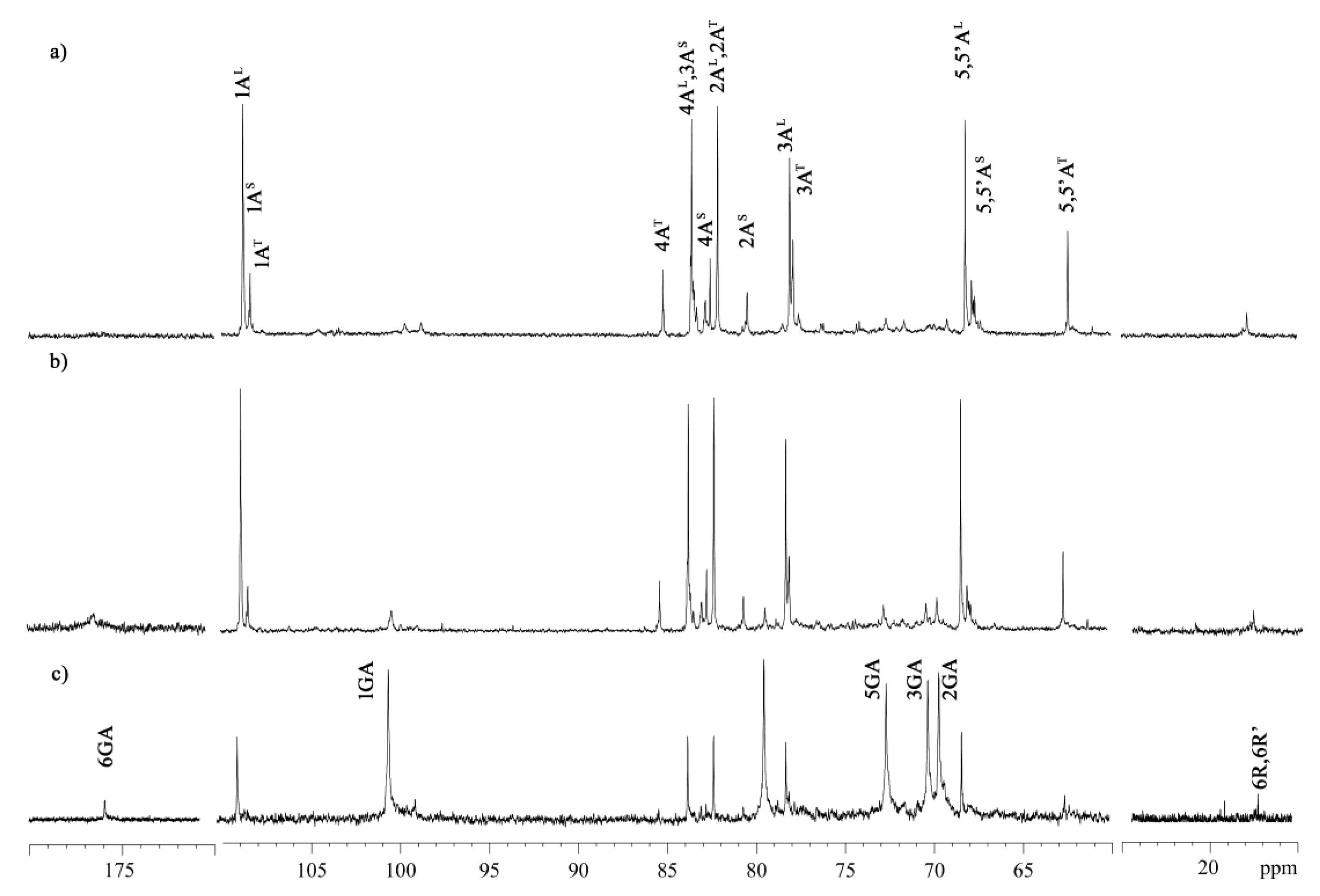
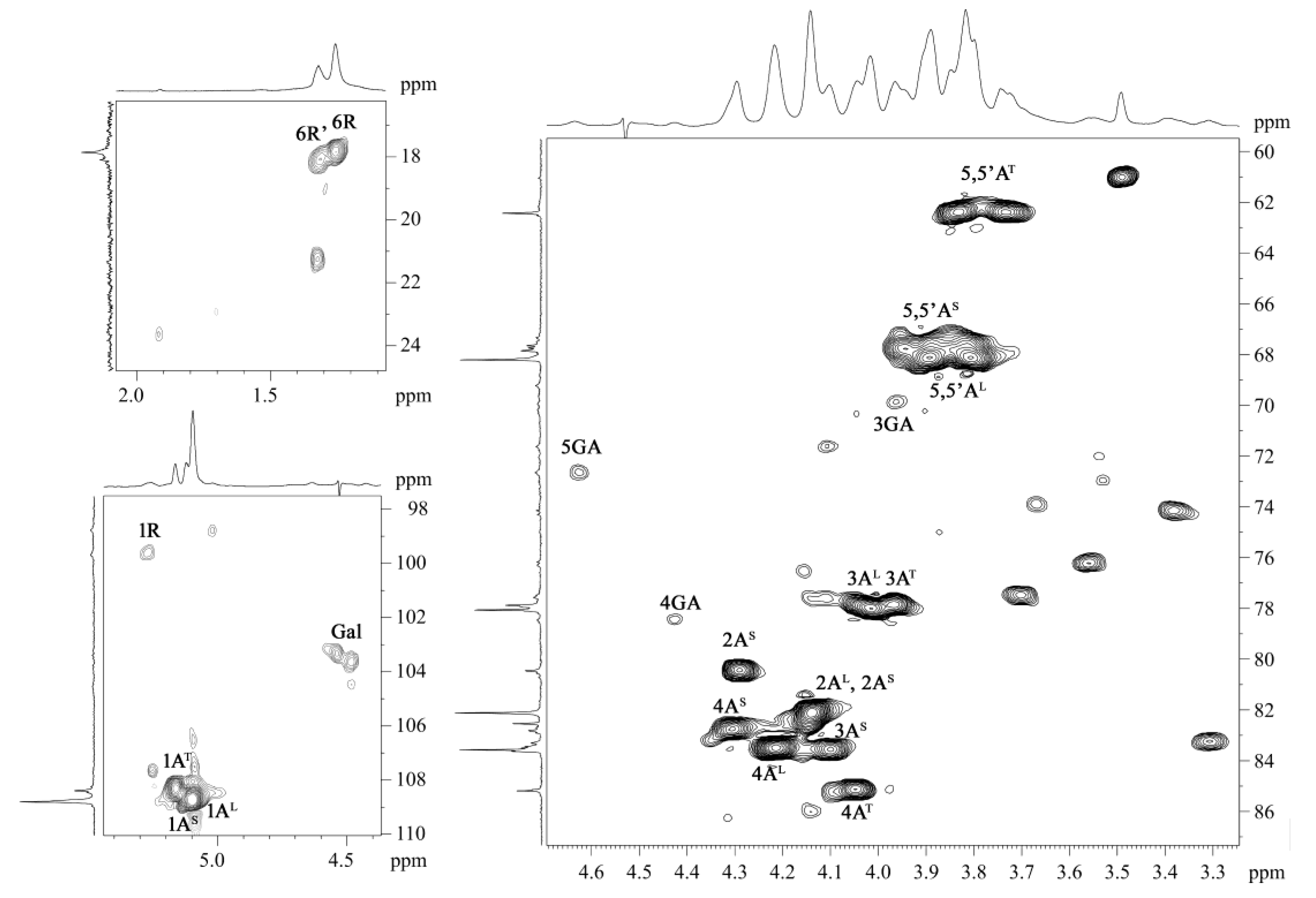
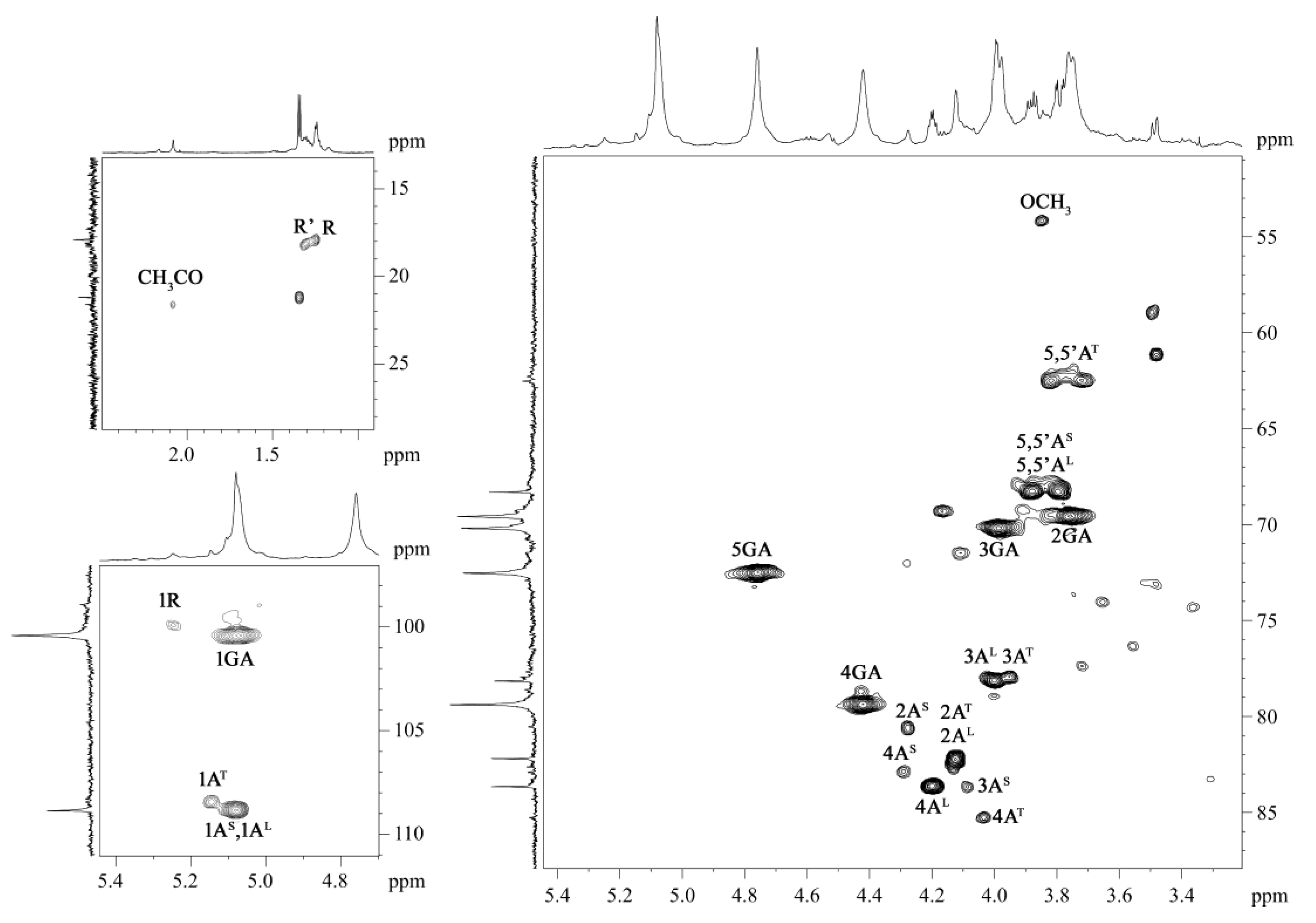
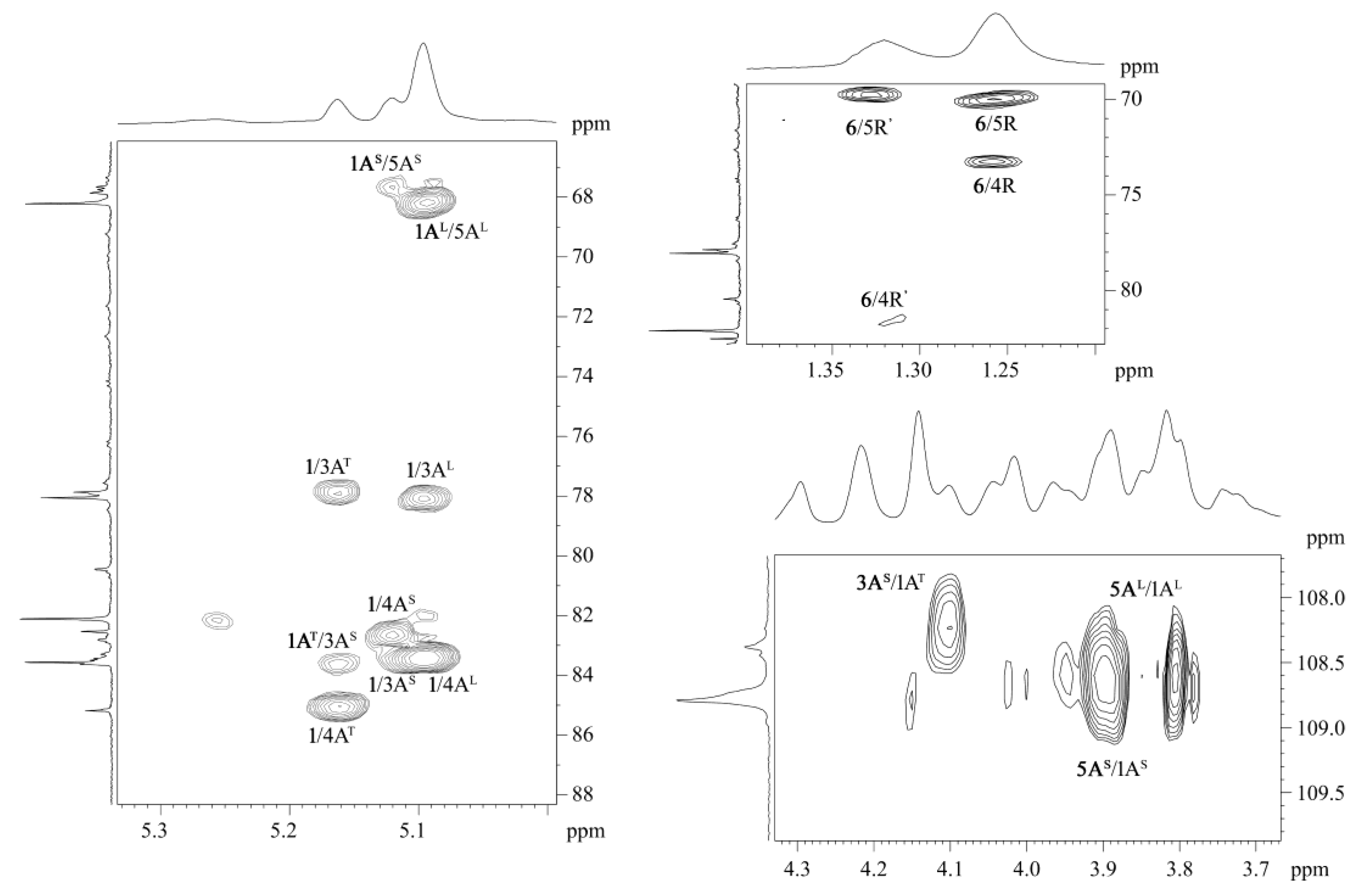
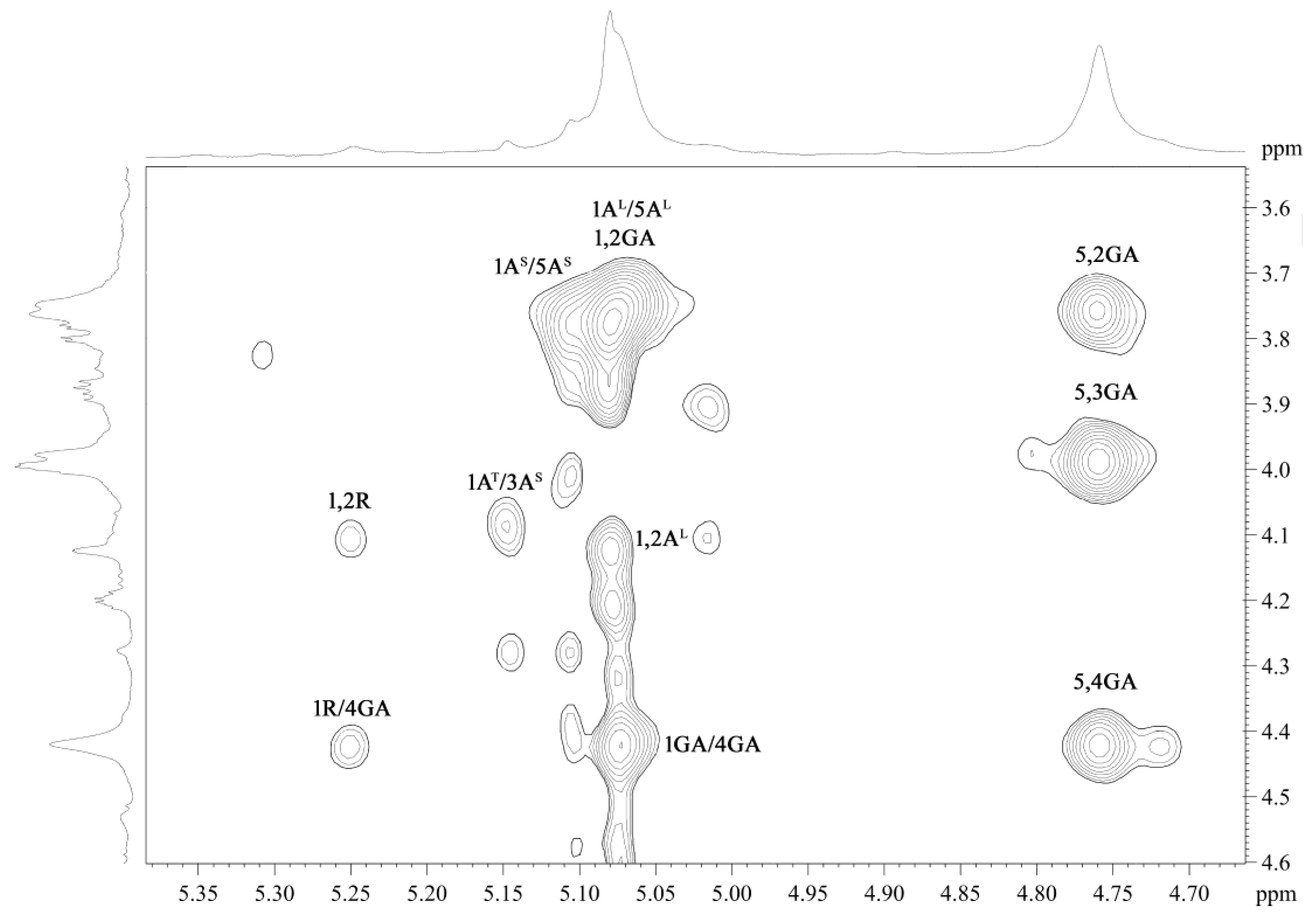
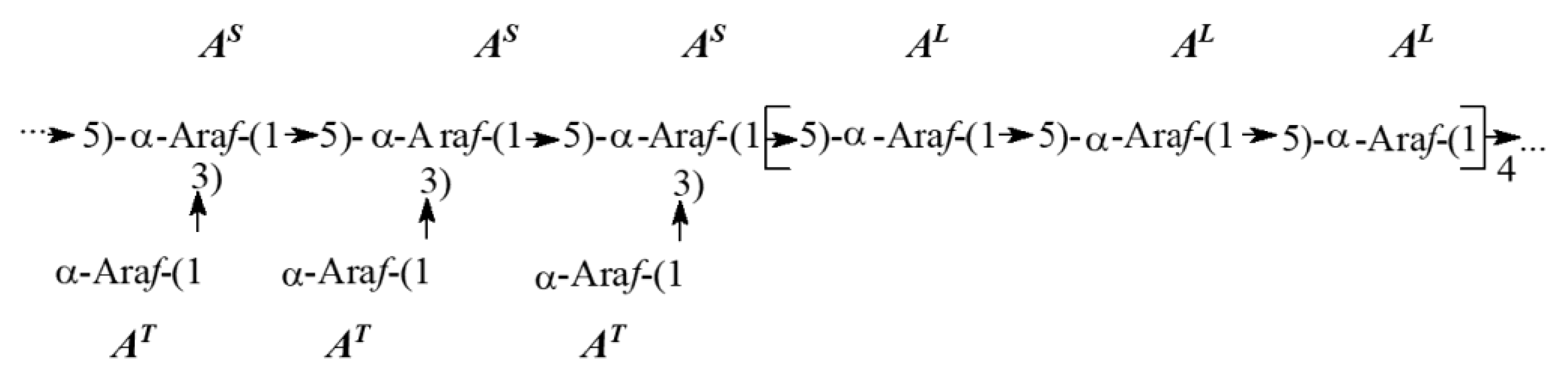
2.2. NMR Spectroscopic Study

Information about the structure of the main polysaccharides from S. passerines was obtained by a combined analysis of the NMR spectra of SO1-1, SO1-2 and SO1-3. The NMR spectra of the three samples were similar (Figure 2, Figure 3, Figure 4 and Figure 5). The 13C NMR spectra of the samples (Figure 2) were assigned using 1H, 13C heteronuclear single quantum coherence spectroscopy (HSQC) spectra. Analysis of the 1H, 13C HSQC spectra (Figure 3, Figure 4 and Figure 5, Table 2) revealed substitutions in the monosaccharide residues based on the comparison of their 13C chemical shifts with those of the parent pyranoses and furanoses [22] and considering the glycosylation effects in the 13C NMR spectra of the carbohydrates [23,24], as well as data from our previous NMR studies of pectins [25]. The correlated spectroscopy (COSY), total correlation spectroscopy (TOCSY), rotating frame Overhauser effect spectroscopy (ROESY) and heteronuclear multiple bond correlation (HMBC) spectra revealed residues of α-D-galactopiranoside uronic acid (GA in Table 2), α-L-rhamnopyranose (R) and α-arabinofuranose (A) in all three samples. Conclusions regarding monosaccharide composition, ring size and anomeric configuration were drawn based on the comparison of visible coupling constants and chemical shifts of the sugar residues and corresponding pyranoses [26,27] and furanoses [28,29].
The occurrence of 1,2-linked (label R) and 2,4-substituted (label R′) α-L-rhamnose residues in polysaccharides was confirmed by cross peaks at 1.25/17.9 ppm and 1.31/18.1 ppm in the 1H, 13C HSQC spectra (Figure 3, Figure 4 and Figure 5) and HMBC spectra (Figure 6 and Figure S2). The ROESY spectrum of SO1-3 (Figure 7) included an inter-residue correlation peak of the anomeric proton of Rha residues and H-4 of GalA residues at δH/H 5.26/4.44 ppm, confirming the RG-I regions in polysaccharides.
Three intense signals at δH 5.16, 5.12 and 5.09 ppm belonging to terminal nonreduction arabinose residues (label AT), 3,5-substituted arabinose residues (label AS) and 1,5-linked arabinose residues (label AL), respectively, were found in the anomeric region of 1H NMR spectra of SO1-1 and SO1-2 (Figure S1). In the anomeric region, the 1H NMR spectrum of SO1-3 signals of 3,5-Ara and 1,5-Ara overlaps the intense signal belonging to the 1,4-linked D-galacturonic acid residues (label GA) at δH 5.08 ppm.
The resonance of C-6 at δC 176.0 ppm indicated the predominance of non-methyl-esterified α-1,4-linked D-GalA residues in the structure of polysaccharides SO1-3 (Figure 2c), but the signal of low intensity at δH/C 3.86/54.4 ppm confirmed that some GalA residues were methyl esterified.
The following inter-residue correlations H-1(glycosylating GA)/H-4(glycosylated GA) at δH/H 5.08/4.44 ppm in the ROESY spectrum of SO1-3 (Figure 6) and H-4(glycosylated GA)/C-1(glycosylating GA) at δH/C 4.44/100.3 ppm in the HMBC spectrum of SO1-3 (Figure S2) indicated on the galacturonan (HG) in the studied polysaccharides.
The correlation peak at δH/C 2.09/21.56 ppm in the HSQC spectrum of SO1-3 (Figure 5) confirmed the O-acetylated residues in the structure of polysaccharides from S. passerina. No clear evidence was obtained for the attachment of the O-acetyl group to specific residues, since the intensity of their signals was low. Rha and GalA residues may be acetyl esterified [30]. The signals of O-acetyl groups are present only in the spectrum of sample SO1-3, which included polysaccharides with a high content of GalA, which may indirectly indicate the O-acetylation of GalA residues.
The resonance of C-6 at δC 176.0 ppm indicated the predominance of non-methyl-esterified α-1,4-linked D-GalA residues in the structure of polysaccharides (Figure 2).
The sequence of Ara residues in the repeating units was determined using the H/C correlations in the HMBC spectra and the H/H correlations in the ROESY spectra.
The inter-residue correlation peaks—H-1(glycosylating AS)/C-5(glycosylated AS) and H-1(glycosylating AL)/C-5(glycosylated AL) in HMBC spectra (Figure 7 and Figure S2) and H-1(AT)/H-3(AS) in ROESY spectra (Figure 6, Figures S3 and S4)—showed the side chains formed by single arabinose residues.
The average length of branches in the arabinan side chains, derived from the relative amounts of terminal and branched arabinose residues, was equal to one, confirming that the branches in the arabinan side chains comprised a single arabinose residue.
The ratio of AT, AS and AL was approximately 1:1:4 in the 1H NMR spectrum of SO1-1 and indicated that the lengths of the linear regions were four times the lengths of the branched regions.
The ratio of Ara residues in the spectra of SO1-2 and SO1-3 was not determined because of the overlap of the signal of the anomeric proton AL with the signal of the anomeric proton of GA in the 1H NMR spectrum (Figure S1).
Clear inter-residue correlation peaks in the HMBC spectra (Figure 7 and Figure S2) at δH/C 5.09/68.3 and 5.12/67.9 ppm for H-1(glycosylating AL)/C-5(glycosylated AL) and H-1(glycosylating AS)/C-5(glycosylated AS) mainly indicated that 3,5-Ara substituted 3,5-Ara, and 1,5-Ara substituted 1,5-Ara.
A possible structure of the repeating unit of the arabinan chain of polysaccharides from S. passerines is proposed below (Scheme 1), where the lengths of the structural regions are arbitrary.
In addition, the following low-intensity peaks were found in the anomeric region of the 1H, 13C HSQC spectra: at δC/H 98.78/5.26 ppm belonging to Rha residues in the RG-I regions, at δC/H 103.40/4.54, 103.80/4.49, 104.68/4.48 ppm belonging to Gal residues, respectively (Figure 3 and Figure 4).
The occurrence of 1,2-linked (label R) and 2,4-substituted (label R’) α-L-rhamnose residues in polysaccharides was confirmed by the cross peaks at 1.25/17.9 ppm and 1.31/18.1 ppm in the 1H, 13C HSQC spectra (Figure 3, Figure 4 and Figure 5) and the HMBC spectra (Figure 7 and Figure S2). The ROESY spectrum of SO1-3 (Figure 6) included an inter-residue correlation peak of the anomeric proton of Rha residues, and H-4 of the GalA residues at δH/H 5.26/4.44 ppm confirmed the RG-I regions in polysaccharides.
Thus, three structural domains were identified in the polysaccharides isolated from S. passerine: arabinan, HG and RG-I. Considering the intensity of signals in the NMR spectrum, SO1-1 is dominated by the arabinan units, while SO1-3 is dominated by the galacturonan units. In the present study, no links between them were established. Nonetheless, it is possible that they represent domains of a complex pectin macromolecule.
Arabinans have been found in the cell wall of several plants and are believed to form RG-I side chains [31]. However, most of the evidence is based on co-extraction and/or co-elution of RG-I and arabinans [32,33,34]. Only a few studies found that the L-Ara residues are covalently attached to rhamnose residues at the O-4 position of the RG-I backbone [35,36].
1,5-linked residues of α-L-arabinofuranose form both the backbone and the side chains of most of the arabinans studied [37]. Backbone residues are usually substituted at O-2 and/or O-3 and/or at both positions, with O-3 substitutions predominating [32]. However, several arabinans with a high percentage of substitution at the O-2 position have also been found [38,39]. Other structures of arabinans have also been described. For example, in arabinans, both the furanose and pyranose forms of arabinose were found [40]. Terminal β-arabinofuranose residues may glycosylate 1,5-linked α-arabinofuranose residues of the backbone at position O-5 [41]. Various degrees of branching have been found, including single, linear and branched oligomeric and polymeric chains, with different linkage types. The almost linear 1,5-arabinan associated with protein was isolated from red wine [42]. The arabinans in pectins often have single substituted side chains [32,43]. Arabinans from soybean [44], apple [45], the inner bark of Norway spruce [46] were found to have a highly branched structure. Arabinan-rich pectins, which constituted 50% of the total pectic polysaccharides, have been obtained from pea Pisum sativum L. [47].
The roles of arabinans in plant cell walls remain unclear. It was established that arabinans can be substituted by terminal phenolic esters, particularly feruloyl or coumaroyl esters. Ferulic acid groups may be ester linked to O-2 of the arabinose residues [48,49]. Feruloyl esters may determine guard cell wall flexibility by providing the cross-links between arabinans and other wall polymers; this testifies a unique role for arabinans in determining the physical and functional properties of guard cell walls [50].

2.3. DPPH Radical-Scavenging Activity

Polysaccharide fractions from S. passerine scavenged the DPPH radical at concentrations of 0.2–1.8 mg/mL. The half-maximal DPPH inhibitory concentration (IC50) of them is given in Table 3. CW2, CW3, SO1 and SO2 demonstrated the highest activity, which exceeded 2.51–2.96 times that of commercial apple pectin (AP) activity. CW2, CW3, SO1 and SO2 scavenged 67, 69, 55 and 67% of DPPH radicals at a concentration of 1 mg/mL. Other fractions were less effective and scavenged only 31–48% of DPPH radicals at a concentration of 1 mg/mL.
The DPPH radical scavenging assay is widely used to evaluate the antioxidant property of plant polysaccharides. The activity of CW2, CW3, SO1 and SO2 seems to be comparable to that of polysaccharides from cantaloupe rinds [51], hawthorn wine pomace [52], fruit bodies of Tremella fuciformis [53] and apple pomace [54]. It should be noted that some other polysaccharides demonstrated the same level of DPPH scavenging activity at lower concentrations. These include pectins from Chaenomeles sinensis fruits [55], Epilobium angustifolium L. [56], Thymus quinquecostatus Celak. leaves [57], Gardenia jasminoides J. Ellis flowers [58] and Ziziphus jujuba cv. Muzao [59].
The DPPH radical scavenging activity of the fractions obtained by DEAE-cellulose elution was compared with the activity of the parent fraction SO1 (Figure 8). The polysaccharides SO1-1, SO1-2 and SO1-3 obtained were less effective (p < 0.05) than the parent fraction SO1, exhibiting IC50 equal to 3.64 ± 0.18, 5.70 ± 0.92 and 5.70 ± 1.30 mg/mL, respectively.
On the basis of the yield and content of PCs in SO1-1, SO1-2, SO1-3 and SO1, most of the PCs providing antioxidant activity were removed by anion exchange chromatography, assuming that they were not bound to the polysaccharide chains. The sum contents of PCs in polysaccharides SO1-1, SO1-2, SO1-3 included about 18% from the content of PCs in the parent fraction SO1. It was detected that three fractions obtained on DEAE-cellulose provided only 24% of the DPPH radical scavenging activity of SO1, although they represented about 70% of the parent pectin (Table 1). This suggests that the antioxidant activity of SO1 was mainly provided by the associated PCs but not by polysaccharides.
The relationship between the chemical characteristics of polysaccharides and DPPH scavenging ability was further investigated using correlation analysis. The total content of sugars and the (Ara + Gal)/Xyl ratio correlated negatively, whereas the content of PCs, Gal and Man, as well as PDI correlated positively with DPPH scavenging activity (Table 4).
We tested the five regression models, subsequently removing the less significant factors (according to the p-value). The linear regression, including the contents of PCs and Man as independent variables, resulted in the best model for prediction (adj. R2 = 0.82, p = 0.000) (Table 5). The content of PCs was the only factor contributing significantly to DPPH scavenging activity (p = 0.000, β = 0.79).
Thus, the correlation analysis showed that the DPPH scavenging activity of the sample from S. passerine is associated with the content of PCs. This is consistent with the results of Ref [54], whose authors evaluated the activity of apple pectins, and our previous study on fireweed pectins [56]. It is known that PCs may bind covalently to the side chains of RG I through the Ara and Gal residues and may be involved in the cross-linking of macromolecules [60].
It was shown that the removal of PCs from polysaccharides reduces the antioxidant activity but does not completely abolish it [60]. Several authors suggest that the antioxidant activity of pectins may be due to the hydroxyl and carboxyl groups of GalA residues [52,61]. Previously, we showed that the antioxidant activity of fireweed pectins is partly related to the xylogalacturonan chains [56]. However, in the present study, we failed to find the polysaccharide chains responsible for the DPPH radical scavenging activity of Salsola pectins. The small sample size (n = 14), which determines the statistical power of multiple regression [62], may be the reason we failed to identify the polysaccharide chains that contribute to the antioxidant activity of Salsola pectins.

3. Materials and Methods

3.1. Materials

Biological material: plant material, consisting of yellow-green annual branches with spherical dwarf leaves, was collected in August 2019 from the semi-shrubs of Salsola gemmascens ssp. passerina (Bunge) Botschantz. = Caroxylon passerinum (Bunge) Akhani et E.H. Roalson (main name—Salsola passerina Bunge) growing in Mandal-ovoo soum, Ömnö-Govi province, Mongolia. They were identified by Prof. B.Oyuntsetseg (School of Arts and Sciences, National University of Mongolia). The plant material was washed with distilled water and dried with filter paper.
The chemicals used are described in the Supplementary (Appendix A).

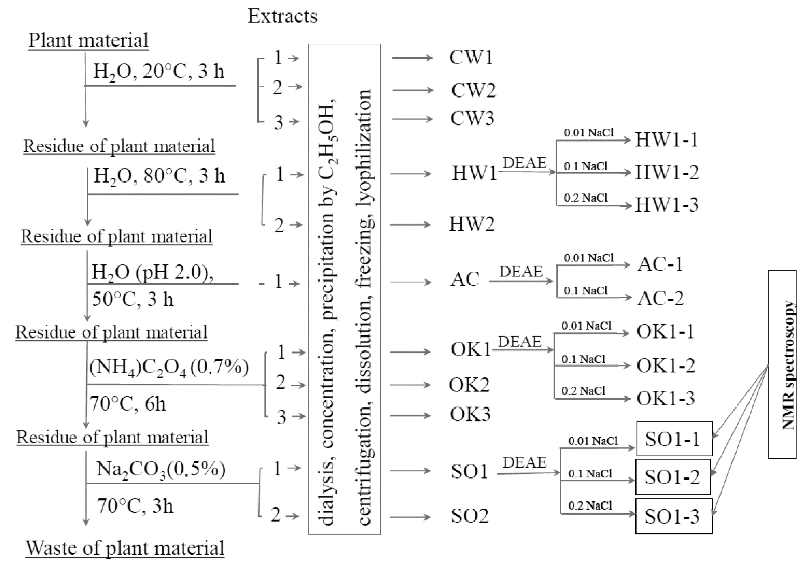
3.2. Isolation of Polysaccharides of S. passerina

Polysaccharides from the plant material were sequentially extracted, as described below; the extraction scheme is shown in Figure 1. At each stage, an exhaustive extraction of polysaccharides was carried out until the absence of reaction of the extract to the carbohydrate; the extraction mixtures were mixed in a mechanical stirrer.
Freshly picked plant material (234 g) was milled in a blender, distilled water (1 L) was added, and the resulting mixture was stirred in a mechanical mixer at 20 °C for 3 h. The mixture was centrifuged, and the residue of the plant material was treated again; the treatment was repeated three times. In the next stage, polysaccharides from the residues of plant materials were extracted with hot water at 80 °C for 3 h. The extraction was repeated twice (each time, the volume of added water was 1 L). Finally, the five aqueous extracts (three obtained with cold water (CW1, CW2, CW3) and two with hot water (HW1, HW2)) were obtained. Next, polysaccharides were extracted with acidified water (pH 2.0, 1 L) at 50 °C for 3 h. As a result, one extract (AC) was obtained. Next, polysaccharides were extracted with aqueous solutions of ammonium oxalate (0.7% w/v) at 70 °C for 6 h. The extraction was repeated three times (the first volume of salt solution added was 2 L; the second and third volumes were 1 L). Finally, the three extracts (OK1, OK2, OK3) were obtained. Next, polysaccharides were extracted with aqueous solutions of Na2CO3 (0.5% w/v) containing NaBH4 at 70 °C for 3 h. The extraction was repeated twice (the first volume of soda solution added was 3 L, the second—2 L). The two extracts (SO1, SO2) were obtained.
The carbohydrate content of each extract was detected using a phenol-sulfuric acid assay [63].
All extracts were dialyzed against distilled water for 48 h at 10 °C. Extracts SO1, SO2 were previously acidified with a diluted solution of acetic acid to pH 5.6. The dialyzed extracts were concentrated on a Heidolph 4002 rotary evaporator (Germany) under reduced pressure at 40 °C.
Polysaccharides were precipitated from the extracts with a four-fold volume of 95% ethanol, centrifuged, washed twice with 95% ethanol, dissolved in distilled water, frozen and lyophilized. The yields of the polysaccharide fractions obtained are expressed in % (w/w) of mass of dry plant material and are presented in Table 1.

3.3. Ion Exchange Chromatography of Polysaccharide Fractions

The major polysaccharide fractions HW1, AC, OK1, SO1 were separated on a DEAE-cellulose (OH-) column (2.5 cm × 40 cm). Each polysaccharide fraction (100 mg) was dissolved in 5 mL of 0.01 M NaCl, and the solution was applied to the column. The column was stepwise eluted with 0.01, 0.1, 0.2, 0.3, 0.5 and 1.0 M NaCl solution (400 mL of each eluent) at a flow rate of 0.9 mL/min. The fractions were collected at 12 min intervals using a low-pressure system Pharmacia Biotech (Sweden) with a FRAC-100 fraction collector, P-50 pump. The carbohydrate content in each tube was determined by the phenol–sulfuric acid method [63]. When separating each of the HW1, OK1, SO1, three major polysaccharide fractions were obtained (eluted with 0.01, 0.1 and 0.2 M NaCl). When separating AC, the fraction eluted 0.2 M NaCl was obtained as minor. In addition, minor fractions were obtained from all fractions by elution with 0.3, 0.5 and 1.0 M NaCl.
The separation procedure was repeated twice for HW1, OK1 and four times for AC, SO1. Data on the monosaccharide composition and the yield of the fractions are presented in Table 1 as a mean of these experiments.

3.4. General Analytical Methods

The content of uronic acids was determined as described earlier [64,65]. The quantitative determination of protein was calculated using the Bradford method [66]. The quantitative determination of phenolics was performed with the Folin–Ciocalteu reagent using gallic acid as a standard [67]. The content of neutral monosaccharides was determined by gas–liquid chromatography (GLC), as described earlier in detail [68]. The sugar concentration was determined at 490 nm using the phenol–sulfuric acid assay [63].
The relative molar mass distributions (RMM) (including Mn, Mw and PDI) of the polysaccharide samples were determined by size exclusion chromatography with high-performance liquid chromatography (HPSEC); the procedure was described in detail earlier [69].

3.5. Nuclear Magnetic Resonance Spectroscopy

All homo- and heteronuclear NMR experiments of the samples were carried out on a Bruker Avance 600 spectrometer (Germany) at a probe temperature of 303, 313 and 318 K, which provided a minimum overlap of the signal of deuterated water with the polymer signals. The procedures for preparing the polysaccharide samples and the conditions of the NMR experiments were described earlier [69].

3.6. Antioxidant Activity

The DPPH solution (0.2 mM, in ethanol) was added to the pectin solution (0.4–3.6 mg/mL water) in equal proportions (v/v) and mixed. After incubation at 25 °C for 1 h, the absorbance of the sample was measured at 517 nm. The scavenging activity of the pectins was measured at four different concentrations, and the half-maximal inhibitory concentration (IC50, mg/mL) values were calculated based on a polynomial regression curve [70].

3.7. Statistical Analysis

The significance of the difference among the means in determining the antioxidant activity was estimated with one-way analysis of variance (ANOVA) and Fisher’s least significant difference (LSD) post hoc test at p < 0.05. The relationship between the chemical characteristics and activity of polysaccharide fractions was evaluated by the calculation of the Pearson correlation coefficients and multiple linear regression analysis. All calculations were performed using the statistical package Statistica 10.0 (StatSoft, Inc., USA). The data were expressed as the means ± s.d. of three independent experiments.

4. Conclusions

Polysaccharide fractions isolated from S. passerine with water and aqueous solutions of ammonium oxalate and sodium carbonate were characterized by a similar composition, including polysaccharides, protein and PCs. HG, RG-I and arabinan with regions formed by 3,5-substituted and by 1,5-linked arabinose residues were identified as the principal units of the polysaccharides obtained. Polysaccharide fractions of S. passerine demonstrated a moderate antioxidant potential. Fractions isolated with cold water and sodium carbonate scavenged the DPPH radical in vitro to a much greater extent than commercial apple pectin. The correlation analysis of the composition and activity of polysaccharide fractions obtained by anionic exchange chromatography revealed that the antioxidant capacity of polysaccharides of S. passerine is mainly due to the associated PCs.

Supplementary Materials

The following supporting information can be downloaded at: https://www.mdpi.com/article/10.3390/ijms232113175/s1.

Author Contributions

Study design, conceptualization, polysaccharide isolation, V.G.; writing—review and editing, project administration, S.P.; measurement of antioxidant activity, V.S.; separation of polysaccharide fractions on an anion exchange resin, V.K.; collecting plant material and its primary treatment by organic solvents, F.V.; collecting plant material and its primary treatment by organic solvents, S.N.; experiments and analysis of NMR data, A.S.S. and A.S.D. All authors have read and agreed to the published version of the manuscript.

Funding

This study was supported by the Russian Science Foundation (grant number 21-73-20005).

Institutional Review Board Statement

Not applicable.

Informed Consent Statement

Not applicable.

Data Availability Statement

The data that support the findings of this study are available from the corresponding author upon reasonable request.

Acknowledgments

NMR spectra were registered on the equipment of the Zelinsky Institute of Organic Chemistry Shared Research Facility.

Conflicts of Interest

The authors declare no conflict of interest.

Appendix A

Chemical reagents: ethyl alcohol, ethanol, C2H5OH (96%, JSC Kirov Pharmaceutical Factory, Kirov, Russia); methyl alcohol, methanol, CH3OH (99.9%, Reakhim, Saint Petersburg, Russia); sodium hydroxide, NaOH (98%, Fluka, Buchs, Germany); sodium chloride, NaCl (99%, Sigma-Aldrich, St. Louis, MO, USA); chloroform, CHCl3 (>99.9%, Ekos-1, Staraya Kupavna, Russia); ammonium hydroxide solution, NH4OH (≥25 NH3 in H2O, >99.9%, Ekos-1, Staraya Kupavna, Russia); trifluoroacetic acid, CF3COOH (99%, Acros organics, Jersey City, NJ, USA); pyridine, C5H5N (99%, Ekos-1, Staraya Kupavna, Russia); acetic acid, CH3COOH (99.9%, Chimreactive, Moscow, Russia); sodium borohydride, NaBH4 (>98.5%, Sigma-Aldrich, USA); D2O (99.9 atom % D, Sigma-Aldrich, St. Louis, MO, USA); toluene, C6H5-CH3 (99%, Ekos-1, Staraya Kupavna, Russia); sulfuric acid, H2SO4 (>99.9%, Vekton, Penfield, NY, Russia); phenol, C6H5OH (99%, Reakhim, Saint Petersburg, Russia); 3,5-dimethylphenol, (CH3)2C6H3OH (≥99%, Sigma-Aldrich, USA); 1,4-α-D-polygalacturonase (Sigma, exo- and endo-activity 690 units/g, St. Louis, MO, USA); 1,1-diphenyl-2-picrylhydrazyl radical (Sigma-Aldrich, St. Louis, MO, USA); the Folin and Ciocalteu’s phenol reagent (Sigma-Aldrich, St. Louis, MO, USA).
For the ion exchange chromatography, we used DEAE-cellulose (Sigma-Aldrich, St. Louis, MO, USA).
The bovine serum albumin (≥96%, Sigma-Aldrich, USA); myo-inositol (≥99%, Sigma-Aldrich, USA), l-(+)-Rhap, l-(+)-Araf, d-(+)-Galp, D-(+)-Manp, d-(+)-xylose and d-(+)-Glcp (≥99%, Sigma-Aldrich, USA); d-(+)-GalpA monohydrate (≥97%, Sigma-Aldrich, USA); gallic acid (MP Biomedicals, USA) were used as standards.

References

  1. Mosyakin, S.L. A taxonomic synopsis of the genus Salsola (Chenopodiaceae) in North America. Ann. Mo. Bot. Gard. 1996, 83, 387–395. [Google Scholar] [CrossRef]
  2. Akhani, H.; Trimborn, P.; Ziegler, H. Photosynthetic Pathways In Chenopodiaceae from Africa, Asia and Europe with Their Ecological, Phytogeographical and Taxonomical Importance. Plant Syst. Evol. 1997, 206, 187–221. [Google Scholar] [CrossRef]
  3. Murshid, S.S.A.; Atoum, D.; Abou-Hussein, D.R.; Abdallah, H.M.; Hareeri, R.H.; Almukadi, H.; Edrada-Ebel, R. Genus Salsola: Chemistry, Biological Activities and Future Prospective—A Review. Plants 2022, 11, 714. [Google Scholar] [CrossRef] [PubMed]
  4. Tundis, R.; Menichini, F.; Conforti, F.; Loizzo, M.R.; Bonesi, M.; Statti, G.; Menichini, F. A Potential Role of Alkaloid Extracts from Salsola Species (Chenopodiaceae) in the Treatment of Alzheimer’s Disease. J. Enzym. Inhib. Med. Chem. 2009, 24, 818–824. [Google Scholar] [CrossRef] [PubMed] [Green Version]
  5. Tundis, R.; Loizzo, M.R.; Statti, G.A.; Menichini, F. Inhibitory Effects on the Digestive Enzyme Alpha-Amylase of Three Salsola Species (Chenopodiaceae) in Vitro. Pharmazie 2007, 62, 473–475. [Google Scholar]
  6. Hamed, A.I.; Masullo, M.; Sheded, M.G.; Mahalel, U.A.; Tawfik, M.M.; Perrone, A.; Piacente, S. Triterpene Saponins from Salsola Imbricata. Phytochem. Lett. 2011, 4, 353–356. [Google Scholar] [CrossRef]
  7. Beyaoui, A.; Chaari, A.; Ghouila, H.; Ali Hamza, M.; Ben Jannet, H. New Antioxidant Bibenzyl Derivative and Isoflavonoid from the Tunisian Salsola tetrandra Folsk. Nat. Prod. Res. 2012, 26, 235–242. [Google Scholar] [CrossRef]
  8. Ridley, B.L.; O’Neill, M.A.; Mohnen, D. Pectins: Structure, biosynthesis, and oligogalacturonide-related signaling. Phytochemistry 2001, 57, 929–967. [Google Scholar] [CrossRef]
  9. Voragen, A.G.J.; Coenen, G.-J.; Verhoef, R.P.; Schols, H.A. Pectin, a versatile polysaccharide present in plant cell walls. Struct. Chem. 2009, 20, 263–275. [Google Scholar] [CrossRef] [Green Version]
  10. Naqash, F.; Masoodi, F.A.; Rather, S.A.; Wani, S.M.; Gani, A. Emerging concepts in the nutraceutical and functional properties of pectin—A review. Carbohydr. Polym. 2017, 168, 227–239. [Google Scholar] [CrossRef]
  11. Minzanova, S.T.; Mironov, V.F.; Arkhipova, D.M.; Khabibullina, A.V.; Mironova, L.G.; Zakirova, Y.M.; Milyukov, V.A. Biological activity and pharmacological application of pectic polysaccharides: A review. Polymers 2018, 10, 1407. [Google Scholar] [CrossRef] [PubMed]
  12. Tan, M.; Chang, S.; Liu, J.; Li, H.; Xu, P.; Wang, P.; Wang, X.; Zhao, M.; Zhao, B.; Wang, L.; et al. Physicochemical Properties, Antioxidant and Antidiabetic Activities of Polysaccharides from Quinoa (Chenopodium quinoa Willd.) Seeds. Molecules 2020, 25, 3840. [Google Scholar] [CrossRef]
  13. Wu, D.-T.; Liu, W.; Han, Q.-H.; Wang, P.; Xiang, X.-R.; Ding, Y.; Zhao, L.; Zhang, Q.; Li, S.-Q.; Qin, W. Extraction Optimization, Structural Characterization, and Antioxidant Activities of Polysaccharides from Cassia Seed (Cassia obtusifolia). Molecules 2019, 24, 2817. [Google Scholar] [CrossRef] [PubMed] [Green Version]
  14. Klosterhoff, R.R.; Bark, J.M.; Glänzel, N.M.; Iacomini, M.; Martinez, G.R.; Winnischofer, S.M.B.; Cordeiro, L.M.C. Structure and intracellular antioxidant activity of pectic polysaccharide from acerola (Malpighia emarginata). Int. J. Biol. Macromol. 2018, 106, 473–480. [Google Scholar] [CrossRef] [PubMed]
  15. Patova, O.; Smirnov, V.; Golovchenko, V.; Vityazev, F.; Shashkov, A.; Popov, S. Structural, rheological and antioxidant properties of pectins from Equisetum arvense L. and Equisetum sylvaticum L. Carbohydr. Polym. 2019, 209, 239–249. [Google Scholar] [CrossRef]
  16. Zeng, F.; Chen, W.; He, P.; Zhan, Q.; Wang, Q.; Wu, H.; Zhang, M. Structural characterization of polysaccharides with potential antioxidant and immunomodulatory activities from Chinese water chestnut peels. Carbohydr. Polym. 2020, 246, 116551. [Google Scholar] [CrossRef]
  17. Ma, X.; Yu, J.; Jing, J.; Zhao, Q.; Ren, L.; Hu, Z. Optimization of Sunflower Head Pectin Extraction by Ammonium Oxalate and the Effect of Drying Conditions on Properties. Sci. Rep. 2021, 11, 10616. [Google Scholar] [CrossRef]
  18. Ning, X.; Liu, Y.; Jia, M.; Wang, Q.; Sun, Z.; Ji, L.; Mayo, K.H.; Zhou, Y.; Sun, L. Pectic Polysaccharides from Radix Sophorae tonkinensis Exhibit Significant Antioxidant Effects. Carbohydr. Polym. 2021, 262, 117925. [Google Scholar] [CrossRef]
  19. Zhang, T.; Shuai, M.; Ma, P.; Huang, J.; Sun, C.; Yao, X.; Chen, Z.; Min, X.; Yan, S. Purification, chemical analysis and antioxidative activity of polysaccharides from pH-modified citrus pectin after dialyzation. LWT 2020, 128, 109513. [Google Scholar] [CrossRef]
  20. Wang, W.; Ma, X.; Jiang, P.; Hu, L.; Zhi, Z.; Chen, J.; Ding, T.; Ye, X.; Liu, D. Characterization of pectin from grapefruit peel: A comparison of ultrasound-assisted and conventional heating extractions. Food Hydrocoll. 2016, 61, 730–739. [Google Scholar] [CrossRef]
  21. Wang, J.; Hu, S.; Nie, S.; Yu, Q.; Xie, M. Reviews on Mechanisms of In Vitro Antioxidant Activity of Polysaccharides. Oxid. Med. Cell Longev. 2016, 2016, 5692852. [Google Scholar] [CrossRef] [PubMed]
  22. Bock, K.; Pedersen, C. Carbon-13 Nuclear Magnetic Resonance Spectroscopy of Monosaccharides. In Advances in Carbohydrate Chemistry and Biochemistry; Academic Press: Cambridge, MA, USA, 1983; pp. 27–66. [Google Scholar] [CrossRef]
  23. Shashkov, A.S.; Lipkind, G.M.; Knirel, Y.A.; Kochetkov, N.K. Stereochemical Factors Determining the Effects of Glycosylation on The13C Chemical Shifts in Carbohydrates. Magn. Reson. Chem. 1988, 26, 735–747. [Google Scholar] [CrossRef]
  24. Lipkind, G.M.; Shashkov, A.S.; Knirel, Y.A.; Vinogradov, E.V.; Kochetkov, N.K. A Computer-Assisted Structural Analysis of Regular Polysaccharides on the Basis of 13C-n.m.r. Data. Carbohydr. Res. 1988, 175, 59–75. [Google Scholar] [CrossRef]
  25. Golovchenko, V.V.; Khramova, D.S.; Shashkov, A.S.; Otgonbayar, D.; Chimidsogzol, A.; Ovodov, Y.S. Structural Characterisation of the Polysaccharides from Endemic Mongolian Desert Plants and Their Effect on the Intestinal Absorption of Ovalbumin. Carbohydr. Res. 2012, 356, 265–272. [Google Scholar] [CrossRef] [PubMed]
  26. Altona, C.; Haasnoot, C.A.G. Prediction Of Anti And Gauche Vicinal Proton-Proton Coupling Constants in Carbohydrates: A Simple Additivity Rule for Pyranose Rings. Org. Magn. Reson. 1980, 13, 417–429. [Google Scholar] [CrossRef]
  27. Jansson, P.-E.; Kenne, L.; Widmalm, G. Computer-Assisted Structural Analysis of Polysaccharides with an Extended Version of Casper Using 1H- and 13C-n.m.r. Data. Carbohydr. Res. 1989, 188, 169–191. [Google Scholar] [CrossRef]
  28. Cyr, N.; Perlin, A.S. The Conformations of Furanosides. A 13C Nuclear Magnetic Resonance Study. Can. J. Chem. 1979, 57, 2504–2511. [Google Scholar] [CrossRef] [Green Version]
  29. Angyal, S.J. Hudson’s Rules of Isorotation as Applied to Furanosides, and the Conformations of Methyl Aldofuranosides. Carbohydr. Res. 1979, 77, 37–50. [Google Scholar] [CrossRef]
  30. Sengkhamparn, N.; Bakx, E.J.; Verhoef, R.; Schols, H.A.; Sajjaanantakul, T.; Voragen, A.G.J. Okra Pectin Contains an Unusual Substitution of Its Rhamnosyl Residues with Acetyl and Alpha-Linked Galactosyl Groups. Carbohydr. Res. 2009, 344, 1842–1851. [Google Scholar] [CrossRef]
  31. Albersheim, P.; Darvill, A.G.; O’Neill, M.A.; Schols, H.A.; Voragen, A.G.J. An Hypothesis: The Same Six Polysaccharides Are Components of the Primary Cell Walls of All Higher Plants. In Progress in Biotechnology; Elsevier: Amsterdam, The Netherlands, 1996; pp. 47–55. [Google Scholar] [CrossRef]
  32. Ding, H.H.; Cui, S.W.; Goff, H.D.; Chen, J.; Wang, Q.; Han, N.F. Arabinan-Rich Rhamnogalacturonan-I from Flaxseed Kernel Cell Wall. Food Hydrocoll. 2015, 47, 158–167. [Google Scholar] [CrossRef]
  33. Navarro, D.A.; Cerezo, A.S.; Stortz, C.A. NMR Spectroscopy and Chemical Studies of an Arabinan-Rich System from the Endosperm of the Seed of Gleditsia triacanthos. Carbohydr. Res. 2002, 337, 255–263. [Google Scholar] [CrossRef]
  34. Shakhmatov, E.G.; Toukach, P.V.; Makarova, E.N. Structural Studies of the Pectic Polysaccharide from Fruits of Punica granatum. Carbohydr. Polym. 2020, 235, 115978. [Google Scholar] [CrossRef] [PubMed]
  35. McNeil, M.; Darvill, A.G.; Albersheim, P. Structure of Plant Cell Walls: XII. Identification of Seven Differently Linked Glycosyl Residues Attached to O-4 of the 2,4-Linked L-Rhamnosyl Residues of Rhamnogalacturonan I. Plant Physiol. 1982, 70, 1586–1591. [Google Scholar] [CrossRef] [PubMed]
  36. Lau, J.M.; McNeil, M.; Darvill, A.G.; Albersheim, P. Treatment of Rhamnogalacturonan I with Lithium in Ethylenediamine. Carbohydr. Res. 1987, 168, 245–274. [Google Scholar] [CrossRef]
  37. Voragen, A.; Pilnik, W.; Thibault, J.-F.; Axelos, M.A.V.; Renard, C.M.G.C. Pectins. In Food Polysaccharides and Their Applications; Stephen, A.M., Ed.; Marcel Decker: New York, NY, USA, 1995; p. 654. [Google Scholar]
  38. Eriksson, I.; Andersson, R.; Westerlund, E.; Andersson, R.; Aman, P. Structural Features of an Arabinan Fragment Isolated from the Water-Soluble Fraction of Dehulled Rapeseed. Carbohydr. Res. 1996, 281, 161–172. [Google Scholar] [CrossRef]
  39. Habibi, Y.; Mahrouz, M.; Vignon, M.R. Arabinan-Rich Polysaccharides Isolated and Characterized from the Endosperm of the Seed of Opuntia Ficus-Indica Prickly Pear Fruits. Carbohydr. Polym. 2005, 60, 319–329. [Google Scholar] [CrossRef]
  40. Swamy, N.R.; Salimath, P.V. Arabinans from Cajanus Cajan Cotyledon. Phytochemistry 1991, 30, 263–265. [Google Scholar] [CrossRef]
  41. Cardoso, S.M.; Silva, A.M.S.; Coimbra, M.A. Structural Characterisation of the Olive Pomace Pectic Polysaccharide Arabinan Side Chains. Carbohydr. Res. 2002, 337, 917–924. [Google Scholar] [CrossRef]
  42. Belleville, M.P.; Williams, P.; Brillouet, J.M. A Linear Arabinan from a Red Wine. Phytochemistry 1993, 33, 227–229. [Google Scholar] [CrossRef]
  43. Westphal, Y.; Kühnel, S.; de Waard, P.; Hinz, S.W.A.; Schols, H.A.; Voragen, A.G.J.; Gruppen, H. Branched Arabino-Oligosaccharides Isolated from Sugar Beet Arabinan. Carbohydr. Res. 2010, 345, 1180–1189. [Google Scholar] [CrossRef]
  44. Aspinall, G.O.; Cottrell, I.W. Polysaccharides of Soybeans. VI. Neutral Polysaccharides from Cotyledon Meal. Can. J. Chem. 1971, 49, 1019–1022. [Google Scholar] [CrossRef] [Green Version]
  45. Aspinall, G.; Fanous, H. Structural Investigations on the Non-Starchy Polysaccharides of Apples. Carbohydr. Polym. 1984, 4, 193–214. [Google Scholar] [CrossRef]
  46. Le Normand, M.; Mélida, H.; Holmbom, B.; Michaelsen, T.E.; Inngjerdingen, M.; Bulone, V.; Paulsen, B.S.; Ek, M. Hot-Water Extracts from the Inner Bark of Norway Spruce with Immunomodulating Activities. Carbohydr. Polym. 2014, 101, 699–704. [Google Scholar] [CrossRef] [PubMed]
  47. Noguchi, M.; Hasegawa, Y.; Suzuki, S.; Nakazawa, M.; Ueda, M.; Sakamoto, T. Determination of Chemical Structure of Pea Pectin by Using Pectinolytic Enzymes. Carbohydr. Polym. 2020, 231, 115738. [Google Scholar] [CrossRef]
  48. Ishii, T.; Tobita, T. Structural Characterization of Feruloyl Oligosaccharides from Spinach-Leaf Cell Walls. Carbohydr. Res. 1993, 248, 179–190. [Google Scholar] [CrossRef]
  49. Saulnier, L.; Thibault, J.-F. Ferulic Acid and Diferulic Acids as Components of Sugar-Beet Pectins and Maize Bran Heteroxylans. J. Sci. Food Agric. 1999, 79, 396–402. [Google Scholar] [CrossRef]
  50. Jones, L.; Milne, J.L.; Ashford, D.; McQueen-Mason, S.J. Cell Wall Arabinan Is Essential for Guard Cell Function. Proc. Natl. Acad. Sci. USA 2003, 100, 11783–11788. [Google Scholar] [CrossRef] [Green Version]
  51. Kazemi, M.; Amiri Samani, S.; Ezzati, S.; Khodaiyan, F.; Hosseini, S.S.; Jafari, M. High-quality Pectin from Cantaloupe Waste: Eco-friendly Extraction Process, Optimization, Characterization and Bioactivity Measurements. J. Sci. Food Agric. 2021, 101, 6552–6562. [Google Scholar] [CrossRef]
  52. Sun, D.; Chen, X.; Zhu, C. Physicochemical Properties and Antioxidant Activity of Pectin from Hawthorn Wine Pomace: A Comparison of Different Extraction Methods. Int. J. Biol. Macromol. 2020, 158, 1239–1247. [Google Scholar] [CrossRef]
  53. Li, M.; Ma, F.; Li, R.; Ren, G.; Yan, D.; Zhang, H.; Zhu, X.; Wu, R.; Wu, J. Degradation of Tremella fuciformis Polysaccharide by a Combined Ultrasound and Hydrogen Peroxide Treatment: Process Parameters, Structural Characteristics, and Antioxidant Activities. Int. J. Biol. Macromol. 2020, 160, 979–990. [Google Scholar] [CrossRef]
  54. Wikiera, A.; Grabacka, M.; Byczyński, Ł.; Stodolak, B.; Mika, M. Enzymatically Extracted Apple Pectin Possesses Antioxidant and Antitumor Activity. Molecules 2021, 26, 1434. [Google Scholar] [CrossRef] [PubMed]
  55. Liu, H.-M.; Wei, Y.-N.; Yan, Y.-Y.; Wu, M.; Qin, G.-Y.; Wang, X.-D. Extraction and Characterization of Pectic Polysaccharides from Chaenomeles sinensis Fruit by Hot Compressed Water. BioResources 2019, 15, 854–868. [Google Scholar] [CrossRef]
  56. Popov, S.; Smirnov, V.; Kvashninova, E.; Khlopin, V.; Vityazev, F.; Golovchenko, V. Isolation, Chemical Characterization and Antioxidant Activity of Pectic Polysaccharides of Fireweed (Epilobium angustifolium L.). Molecules 2021, 26, 7290. [Google Scholar] [CrossRef] [PubMed]
  57. Li, X.; Wang, X.; Dong, Y.; Song, R.; Wei, J.; Yu, A.; Fan, Q.; Yao, J.; Shan, D.; Lv, F.; et al. Preparation, Structural Analysis, Antioxidant and Digestive Enzymes Inhibitory Activities of Polysaccharides from Thymus quinquecostatus Celak. Leaves. Ind. Crop. Prod. 2022, 175, 114288. [Google Scholar] [CrossRef]
  58. Chen, Q.; Xue, G.; Ni, Q.; Wang, Y.; Gao, Q.; Zhang, Y.; Xu, G. Physicochemical and Rheological Characterization of Pectin-rich Polysaccharides from Gardenia jasminoides J. Ellis Flower. Food Sci. Nutr. 2020, 8, 3335–3345. [Google Scholar] [CrossRef]
  59. He, Z.; Zhu, Y.; Bao, X.; Zhang, L.; Li, N.; Jiang, G.; Peng, Q. Optimization of Alkali Extraction and Properties of Polysaccharides from Ziziphus jujuba cv. Residue. Molecules 2019, 24, 2221. [Google Scholar] [CrossRef] [Green Version]
  60. Ghosh, D.; Karmakar, P. Insight into Anti-Oxidative Carbohydrate Polymers from Medicinal Plants: Structure-Activity Relationships, Mechanism of Actions and Interactions with Bovine Serum Albumin. Int. J. Biol. Macromol. 2021, 166, 1022–1034. [Google Scholar] [CrossRef]
  61. Guo, R.; Zhang, J.; Liu, X.; Li, X.; Sun, X.; Kou, Y.; Li, D.; Liu, Y.; Zhang, H.; Wu, Y. Pectic Polysaccharides from Biluochun Tea: A Comparative Study in Macromolecular Characteristics, Fine Structures and Radical Scavenging Activities in Vitro. Int. J. Biol. Macromol. 2022, 195, 598–608. [Google Scholar] [CrossRef]
  62. Hair, J.F. Pearson-Multivariate Data Analysis, 7/E-Joseph F. Hair, Jr, William C. Black, Barry J. Babin and Rolph E. Anderson. Pearson New Int. Ed. 2014, 30, 816. [Google Scholar]
  63. DuBois, M.; Gilles, K.A.; Hamilton, J.K.; Rebers, P.A.; Smith, F. Colorimetric Method for Determination of Sugars and Related Substances. Anal. Chem. 1956, 28, 350–356. [Google Scholar] [CrossRef]
  64. Scott, R.W. Colorimetric Determination of Hexuronic Acids in Plant Materials. Anal. Chem. 1979, 51, 936–941. [Google Scholar] [CrossRef]
  65. Usov, A.I.; Bilan, M.I.; Klochkova, N.G. Polysaccharides of Algae. 48. Polysaccharide Composition of Several Calcareous Red Algae: Isolation of Alginate from Corallina pilulifera P. et R. (Rhodophyta, Corallinaceae). Bot. Mar. 1995, 38, 43–51. [Google Scholar] [CrossRef]
  66. Bradford, M. A Rapid and Sensitive Method for the Quantitation of Microgram Quantities of Protein Utilizing the Principle of Protein-Dye Binding. Anal. Biochem. 1976, 72, 248–254. [Google Scholar] [CrossRef]
  67. Ruviaro, A.R.; Barbosa, P.D.P.M.; Martins, I.M.; de Ávila, A.R.A.; Nakajima, V.M.; Dos Prazeres, A.R.; Macedo, J.A.; Macedo, G.A. Flavanones biotransformation of citrus by-products improves antioxidant and ACE inhibitory activities in vitro. Food Biosci. 2020, 38, 100787. [Google Scholar] [CrossRef]
  68. Golovchenko, V.V.; Naranmandakh, S.; Ganbaatar, J.; Prilepskii, A.Y.; Burygin, G.L.; Chizhov, A.O.; Shashkov, A.S. Structural Investigation and Comparative Cytotoxic Activity of Water-Soluble Polysaccharides from Fruit Bodies of the Medicinal Fungus Quinine Conk. Phytochemistry 2020, 175, 112313. [Google Scholar] [CrossRef] [PubMed]
  69. Golovchenko, V.V.; Khlopin, V.A.; Patova, O.A.; Feltsinger, L.S.; Bilan, M.I.; Dmitrenok, A.S.; Shashkov, A.S. Pectin from Leaves of Birch (Betula pendula Roth.): Results of NMR Experiments and Hypothesis of the RG-I Structure. Carbohydr. Polym. 2022, 284, 119186. [Google Scholar] [CrossRef] [PubMed]
  70. Shogren, R.L.; Biswas, A. Preparation of Starch–Sodium Lignosulfonate Graft Copolymers via Laccase Catalysis and Characterization of Antioxidant Activity. Carbohydr. Polym. 2013, 91, 581–585. [Google Scholar] [CrossRef] [PubMed]
Figure 1. Scheme of isolation of polysaccharides from the S. passerine.
Ijms 23 13175 g001
Figure 2. 13C spectra of the SO1-1 (a), SO1-2 (b) and SO1-3 (c).
Ijms 23 13175 g002
Figure 3. Parts of 1H, 13C HSQC spectrum of the SO1-1. The corresponding parts of the 1H and 13C NMR spectra are shown along the X and Y axes, respectively. Arabic numerals refer to the carbon atoms in the residues, as designated in Table 2.
Ijms 23 13175 g003
Figure 4. Parts of 1H, 13C HSQC spectrum of the SO1-2. The corresponding parts of the 1H and 13C NMR spectra are shown along the X and Y axes, respectively. Arabic numerals refer to the carbon atoms in the residues, as designated in Table 2.
Ijms 23 13175 g004
Figure 5. Parts of 1H, 13C HSQC spectrum of the SO1-3. The corresponding parts of the 1H and 13C NMR spectra are shown along the X and Y axes, respectively. Arabic numerals refer to the carbon atoms in the residues, as designated in Table 2.
Ijms 23 13175 g005
Figure 6. Part of 1H, 1H ROESY spectrum of the SO1-1. The corresponding part of the 1H NMR spectrum is shown along the axes. Slashes refer to inter-residue correlation peaks, as designated in Table 2.
Ijms 23 13175 g006
Figure 7. Part of 1H, 13C HMBC spectrum of the SO1-1. The corresponding parts of the 1H and 13C NMR spectra are shown along the X and Y axes, respectively. Arabic numerals before slash refer to the protons, and those after slash refer to carbon atoms in the corresponding residues.
Ijms 23 13175 g007
Scheme 1. A possible structure of the repeating unit of the arabinan from S. passerines.
Ijms 23 13175 sch001
Figure 8. The DPPH radical scavenging activity of SO1 and of its components obtained by separation on DEAE-cellulose column. Data are presented as the mean ± SD of three independent experiments.
Ijms 23 13175 g008
Table 1. Chemical characteristics of pectic polysaccharides from the S. passerine.
FractionsYield, %Content, %Mw,
kg/mol
Mn,
kg/mol
PDI
GalA cRha cAra cXyl cMan cGlc cGal cTS dProtein dPCs d
CW10.40 a17.464.3313.7215.4522.4513.8212.7747.442.30.843.5915.622.79
CW20.14 a31.185.818.3910.4324.937.2312.0346.706.91.063.0912.744.95
CW30.09 a26.146.9216.138.1225.354.6712.6738.333.71.052.6814.043.75
HW10.88 a47.404.7911.869.0211.777.517.6564.023.70.796.0727.013.56
HW20.67 a50.443.6231.593.264.122.634.3570.271.90.771.4416.024.46
HW1-16.93 b10.120.5923.121.0345.4114.075.6545.000nd34.4714.752.34
HW1-231.30 b66.462.0914.718.780.830.576.5656.350nd56.6820.962.70
HW1-317.40 b79.074.604.915.680.420.764.5676.460.17nd124.3050.742.45
AC1.82 a47.613.9931.876.912.181.925.5252.052.00.455.3113.564.08
AC-117.22 b40.026.9014.7510.903.123.8120.5059.250nd---
AC-250.92 b58.468.2810.5012.831.511.646.7746.920nd80.0634.552.32
OK11.04 a56.994.6523.006.512.032.114.7176.011.80.687.5222.383.91
OK20.30 a59.434.5225.622.051.933.053.4164.172.90.6107.1725.884.14
OK30.45 a51.785.2027.805.092.752.974.4175.142.90.6124.2627.954.45
OK1-19.44 b32.5110.2747.130.001.431.946.7343.462.25nd293.3640.027.33
OK1-225.97 b29.006.0055.241.410.181.466.7181.050nd75.1121.653.47
OK1-329.98 b77.923.6411.721.530.000.734.4785.830nd92.2844.982.05
SO17.32 a55.486.8419.424.310.001.8312.1257.8415.61.2259.1958.094.46
SO20.12 a52.517.4624.721.930.003.3910.0062.9118.21.1239.2930.437.86
SO1-133.78 b21.418.0960.161.700.030.997.6388.5110.900.3444.41319.551.39
SO1-227.97 b33.724.1847.403.940.291.658.8277.840.020.393.3033.352.77
SO1-37.88 b60.716.6519.545.630.230.756.5068.315.080.481.9340.372.03
a—air-dried; b—in relation to the sample applied to the column; c—data were calculated as molar %; d—data were calculated as mass %; TS—total sugar content; PCs—phenolic compounds; nd—not determined; number (Mn) and weight (Mw) average relative molar masses and polydispersity indices (PDI) were measured using pullulan standards; the data are presented in the table as a mean of three experiments.
Table 2. Chemical shifts of the signals in the 1H and 13C NMR spectra of the SO1-1, SO1-2 and SO1-3 (323 K, D2O, TSP, δH 0.0, δC −1.6).
Residue13C NMR Chemical Shifts (δC) and 1H (δH, Italic), ppm
C-1
H-1
C-2
H-2
C-3
H-3
C-4
H-4
C-5
H-5,5′
C-6
H-6
→4)-α-D-GalpA-(1→
GA
100.3
5.08
69.6
3.77
70.2
4.00
79.3
4.44
72.6
4.75
176.1
→2)-α-Rhap-(1→4
R
99.5
5.26
77.6
4.12
68.8
3.89
74.1
3.40
69.6
3.82
17.90
1.25
→2,4)-α-Rhap-(1→4
R′
99.5
5.26
77.2
4.12
71.4
4.10
81.8
3.70
70.9
3.90
18.1
1.31
α-L-Araf-(1→3
AT
108.5
5.16
82.8
4.14
77.9
3.97
85.2
4.05
62.5
3.83; 3.73
→5)-α-L-Araf-(1→
AL
108.5
5.09
82.2
4.14
78.2
4.01
83.7
4.21
68.3
3.89; 3.81
→3,5)-α-L-Araf-(1→
AS
108.8
5.12
80.5
4.29
83.8
4.10
82.9
4.30
67.9
3.94; 3.84
TSP—trimethylsilylpropanoic acid.
Table 3. DPPH scavenging effect of polysaccharide fractions from S. passerine.
PectinIC50 (mg/mL)
CW11.14 ± 0.02 cd
CW20.69 ± 0.07 bc
CW30.66 ± 0.07 b
HW11.47 ± 0.22 d
HW21.58 ± 0.24 d
AC12.20 ± 0.41 e
OK11.63 ± 0.26 d
OK21.84 ± 0.03 de
OK31.64 ± 0.11 d
SO10.78 ± 0.06 bc
SO20.68 ± 0.08 b
AP1.96 ± 0.36 de
Trolox0.006 ± 0.001 a
Data are presented as the mean ± SD of three independent experiments. Different capital letters (a–e) show the significant differences (p < 0.05, LSD test).
Table 4. The Pearson correlation coefficients between the DPPH scavenging activity and the chemical characteristics of polysaccharide fractions from S. passerine (n = 14).
Second VariableRpSecond VariableRp
PCs−0.890.000Protein−0.510.062
Gal−0.590.026Glc−0.460.098
PDI−0.580.029Rha−0.340.236
Man−0.550.040Xyl/GalA−0.300.290
(Ara + Gal)/Rha0.580.028Xyl−0.270.342
Total sugar0.670.009Rha/GalA−0.280.335
Mw−0.110.713
Mn0.120.671
Ara/Xyl0.220.455
GalA0.230.430
GalA/NM0.250.384
GalA-Rha0.260.377
2Rha + Ara + Gal0.300.295
Ara0.520.059
Table 5. Multiple regression analysis of the DPPH scavenging activity.
VariableβStandard Error of βParameter EstimateStandard Errorp-Value
DependentIndependent
DPPH scavenging activityPCs−0.790.13−2.020.320.000
Man−0.260.13−0.020.010.062
Regression results: R2 = 0.847, adjusted R2 = 0.819, F2.11 = 30.438, p < 0.000, Standard estimate error = 0.31.
Publisher’s Note: MDPI stays neutral with regard to jurisdictional claims in published maps and institutional affiliations.

