Review

Amyloidogenesis: What Do We Know So Far?

by
Zeina Alraawi
1,†,
Nayan Banerjee
2,
Srujana Mohanty
3 and
Thallapuranam Krishnaswamy Suresh Kumar
1,*
1
Department of Chemistry and Biochemistry, Fulbright College of Art and Science, University of Arkansas, Fayetteville, AR 72701, USA
2
School of Chemical Sciences, Indian Association for the Cultivation of Science, 2A & 2B Raja S. C. Mullick Road, Jadavpur, Kolkata 700032, India
3
Department of Chemical Sciences, Indian Institute of Science Education and Research, Kolkata 741246, India
*
Author to whom correspondence should be addressed.
Cell and molecular Biology program.
Int. J. Mol. Sci. 2022, 23(22), 13970; https://doi.org/10.3390/ijms232213970
Submission received: 20 September 2022 / Revised: 1 November 2022 / Accepted: 8 November 2022 / Published: 12 November 2022
(This article belongs to the Special Issue Protein Folding and Misfolding — Structure and Functions 2.0)

Abstract

:
The study of protein aggregation, and amyloidosis in particular, has gained considerable interest in recent times. Several neurodegenerative diseases, such as Alzheimer’s (AD) and Parkinson’s (PD) show a characteristic buildup of proteinaceous aggregates in several organs, especially the brain. Despite the enormous upsurge in research articles in this arena, it would not be incorrect to say that we still lack a crystal-clear idea surrounding these notorious aggregates. In this review, we attempt to present a holistic picture on protein aggregation and amyloids in particular. Using a chronological order of discoveries, we present the case of amyloids right from the onset of their discovery, various biophysical techniques, including analysis of the structure, the mechanisms and kinetics of the formation of amyloids. We have discussed important questions on whether aggregation and amyloidosis are restricted to a subset of specific proteins or more broadly influenced by the biophysiochemical and cellular environment. The therapeutic strategies and the significant failure rate of drugs in clinical trials pertaining to these neurodegenerative diseases have been also discussed at length. At a time when the COVID-19 pandemic has hit the globe hard, the review also discusses the plausibility of the far-reaching consequences posed by the virus, such as triggering early onset of amyloidosis. Finally, the application(s) of amyloids as useful biomaterials has also been discussed briefly in this review.

1. Introduction

It is impressive how nature accurately implements molecular self-assembly through ordered growth of nanoscale building blocks with high efficiency in order to fabricate macromolecular architectures. Peptides folding onto proteins, nucleotide chains twisting around to form the DNA duplex and cells proliferating into tissues, to list a few, are some of the most astounding biological self-assembly processes nature has to offer [1,2,3,4,5].
During molecular self-assembly, molecules associate under equilibrium conditions to form structurally organized frameworks, stabilized via various intermolecular interactions, such as electrostatic interactions, van der Waals forces, stacking interactions, hydrogen bonds, and hydrophobic effects. All of these, in addition to the covalent and other intramolecular interactions, produce a well-ordered supramolecular structure. The topology of monomeric units play an important role in the architecture, design, composition, and characteristics of the self-assemblies produced [6,7]. Molecular self-assembly lies at an interdisciplinary interface of protein science, biochemistry, molecular engineering, polymer science, and materials science [8]. Molecular self-assembly of biomolecules, such as peptides and proteins, has emerged as a topic of crucial interest in recent times because of its relevance in biotechnology and health sciences. Protein structures gain their functionality through complex molecular self-organization processes that lead to protein folding and are affected by nature of solvent, osmolarity, pH, temperature, etc. This makes protein folding highly sensitive to its biophysiochemical environment, often leading to protein misfoldings which are at the root of various neurodegenerative disorders [9,10,11,12]. Moreover, the interaction of a ligand with its receptor has been related to self-assembly. The morphology of molecular assemblies produced by enzyme instructed assembly of small molecules is crucially dependent on ligand–receptor interactions. For example, it has been indicated that the ligand–receptor interaction changes the morphology of molecular assemblies produced by enzyme instructed assembly of small molecules to short nanofibers. In contrast, the absence of ligand–receptor interaction directs enzymatic dephosphorylation of precursor to generate the hydrogelator that self-assembles to form long nanofibers, thus preventing the nanofiber formation [13].
Aromatic amino acids, such as tryptophan, phenylalanine, and tyrosine, have reportedly been shown to have a tendency towards forming fibril-like self-assemblies [14,15]. Understanding the interactions that induce and stabilize the molecular self-assembly process have become a target in structural biochemistry for reducing the risk of fibrillation of various proteins and peptides associated with different diseases and for the safe storage of various proteinaceous concoctions of clinical relevance.

2. Single Amino Acid Self-Assemblies

Single amino acid self-assemblies have garnered a great deal of attention recently because of their role in metabolic disorders [16]. Single amino acids, such as phenylalanine, tyrosine, tryptophan, cysteine, and methionine have been found to cause disorders, such as phenylketonuria (PKU), tyrosinemia, hypertryptophanemia, cystinuria and hypermethioninemia, respectively [16,17,18]. Phenylalanine self-assemblies show similar properties to that of amyloid assemblies as they have characteristic birefringence, and Thioflavin T (ThT) fluorescence and electron diffraction patterns [19,20,21]. Besides being ordered like amyloid fibrils, they exhibit strong cytotoxic activities as well. Phenylalanine aggregates were observed in PKU, thus strongly indicating similar etiology of PKU with amyloid-related diseases [22,23,24]. Aromatic amino acids, such as phenylalanine, tyrosine, and tryptophan, are characterized by their high hydrophobic nature and their tendency to form β-sheet structures [19,20]. Their aromaticity has been proposed to promote amyloid fibrillation because of π-stacking that can energetically drive the self-assembly process forward and provide specific directionality and orientation through stacking [18,23]. In addition to their high hydrophobicity, low chain flexibility and β-sheet propensity, amyloid residues also have a planar steric profile that can be easily fit into ‘steric zipper’ structures in laminated β-sheets [25]. Although aromatic interactions are not necessary for amyloid formation, their presence has exhibited acceleration in the amyloid formation process [17,26]. However, not only aromatic amino acids, but also non-aromatic amino acids, such as cysteine and methionine, have been shown to self-assemble, forming very long fiber-like aggregates [27]. Inhibitors containing aromatic groups have also been synthesized. These mechanisms indicate that either assembly is blocked, or solubility of the amyloid species increases due to the introduction of an inhibitor based on the underlying principle that amyloid formation can be inhibited using modified molecular recognition elements [28]. Resveratrol is one such compound that has exhibited neuroprotective activities, such as promoting clearance of Aβ aggregations and reduction of plaques. It has been reported that resveratrol inhibits amyloid formation by IAPP and amyloid-β peptide [29].

3. The History of Amyloid Formation

In 1639, Nicholas Fontanus observed an abscess in the liver and white stones in the spleen in the autopsy of a man. This might have been the first case of sago spleen amyloidosis, a form of amyloid deposition in the spleen and the discovery of amyloids [30,31].
Amyloid comes from the Latin word amylym which means starch. The term amyloid was coined by Matthias Schleiden in 1838. It was during his iodine sulfuric acid test on plants that he referred to amyloids as starch-like [30]. In medical literature, the term amyloids was used by Virchow, describing corpora amylacea found in the liver of a patient during the autopsy, as starch-like deposits since they responded to the iodine sulfuric acid test just like starch [30]. However, in 1859, Kekule and Friedrich used protein staining dyes to show that these deposits are in fact proteinaceous in nature. Further studies concluded that amyloids are protein deposits of insoluble fibrils that cause amyloidosis [32]. Prions, another set of macromolecules of contemporary interest, are closely related to amyloids. The earliest case of prion disease in humans is suspectedly reported to be Kuru, a brain disease found in some cannibalistic tribes of New Guinea. However, the very first time that the infectious nature of prion disease was observed was during the accidental transmission of scrapie in sheep in 1937. The protein-only hypothesis or the prion hypothesis was proposed by Stanley Prusiner in 1982, for which he later received the Nobel Prize in Medicine in 1997. Prions are infectious agents in the form of misfolded proteins, which propagate by inducing protein misfolding. Prions have been reported to cause several proteins to aggregate into tightly packed beta-sheets, called an amyloid fold, relating them to amyloids [33,34]. One of the most common neurodegenerative diseases of recent times, Alzheimer’s disease, was first observed by Alois Alzheimer in 1907. While James Parkinson had first medically described another neurodegenerative dysfunction, Parkinson’s disease, in 1817, it was only in 1997 that it was found that alpha-synuclein protein misfolding causes Parkinson’s disease [35,36]. A protein derived from the twisted β-plated sheet fibrils found in cerebrovascular amyloidosis associated with Alzheimer’s was first isolated by Glenner and Wong in 1984 [30]. In 1875 three scientists, Cornil, Heschl, and Jürgens, independently established the importance of methyl violet stain to detect amyloids [16]. Congo red (CR), an aniline dye, was first developed by German chemist Paul Böttiger [37]. Eventually, Congo red (CR) became the most predominantly used dye to detect amyloids. When stained with Congo red (CR), amyloids give an apple-green birefringence under polarized light caused by the increased optical anisotropy after binding with the dye [38,39]. However, the evidence of the CR dye showing non-specific binding and also binding with non-amyloid substances suggests using it with discretion to determine the presence of amyloids. Many other staining methods have eventually been developed, such as staining with Thioflavin T (ThT), Congo Corinth, Benzopurpurin 4B, and Acid Fuchsin to detect the presence of amyloids. However, in current times, with the advent of spectroscopic prowess, several biophysical techniques have become the primary method for the detection of amyloids (Figure 1) [37].

4. The Structural Features of Amyloid Fibrils

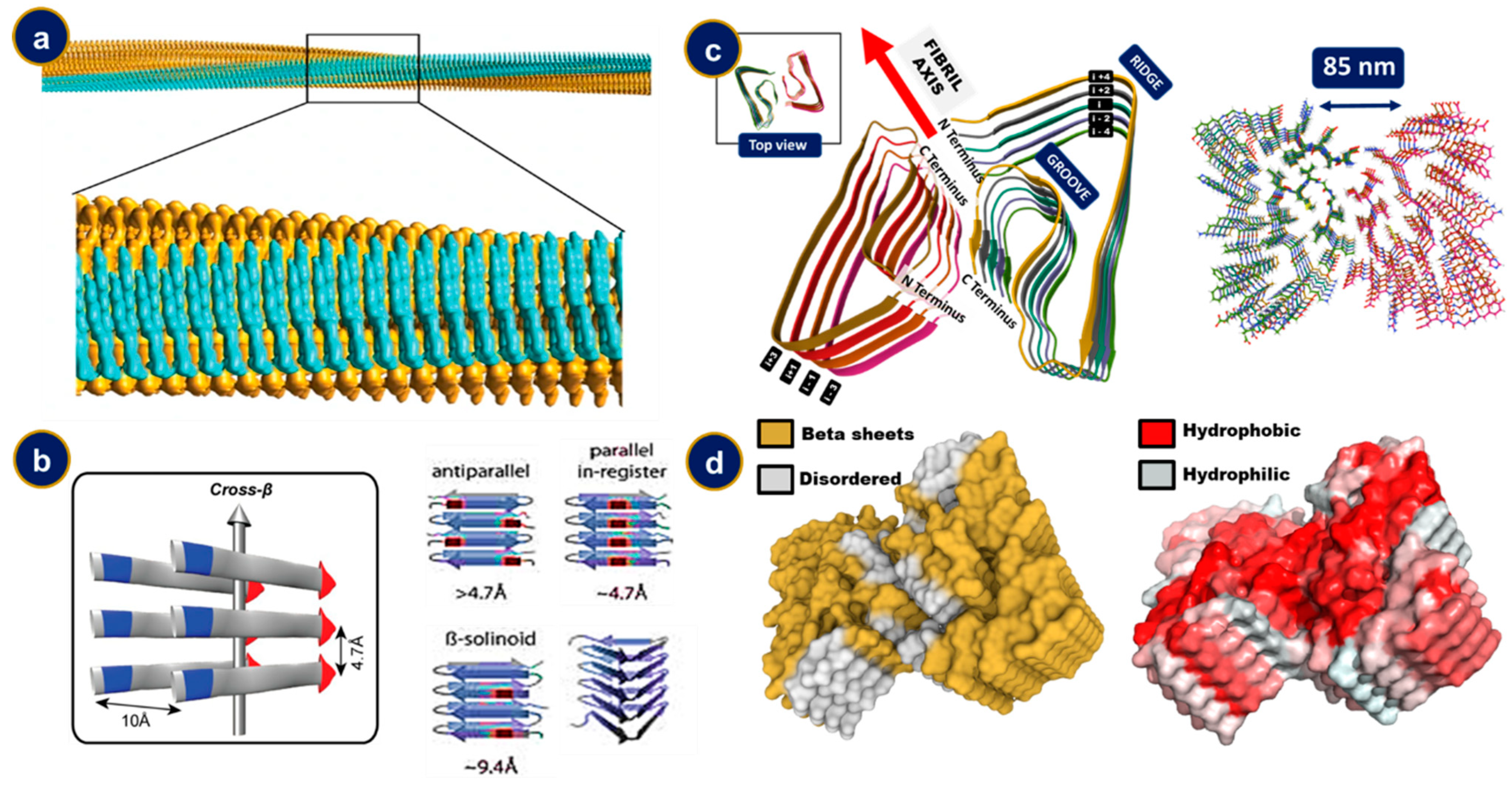
Amyloid depositions are found in various morphologies of which the meshed, fibril bundles and the star-shaped supercluster are the most common. While the fibrillar bundles are compact unidirectional deposits, the mesh morphology lacks a definite orientation and the star-shaped cluster consists of supercoils radiating in multiple directions [40,41]. Individual strands exist in a fibrillar, unbranched, thread-like state, generally 7–13 nm in diameter and a few micrometers in length [42]. The fibrils, in turn, comprise of 2–8 protofilaments which are 2–7 nm in diameter. These protofilaments may either associate in a twisted way with one another or laterally as flat ribbons of thickness 2–7 nm and about 30 nm wide [43]. The protofilaments possess an extended beta-sheet secondary structure with the individual beta-sheets being perpendicular to the longitudinal fiber axis, a structure referred to as the cross beta [44,45,46] (Figure 2). The etymology of the rather colloquial term now lies in the characteristic diffraction data of these fibrils where two distinct diffraction line sets at 0.47 nm and 1.0 nm correspond to the longitudinal inter-strand transverse stacking distances [47]. However, it is worthwhile to note that only a small percentage of the polypeptide stretch forms these beta associations while the remaining portions assemble as loops, tails, and/or intrinsically disordered structures [48,49,50]. The side chains in the residues forming the beta sheet assemblies interdigitate to form a hydrophobic water devoid region referred to as the dry region and constitute the steric zippers (pairs of self-complementary β-sheets) [51]. This water extrusion phenomenon upon amyloid formation drives the entropic gain for facilitating the aggregation.
The constituting β sheets may further fold upon themselves, leading to a great deal of polymorphism in amyloid fibrils. Both electrostatic and Van der Waals interactions, along with the contiguous array of hydrogen bonded residues, have an essential effect in attributing structural stability to these fibrils. Recent computational efforts further shed light on the importance of these non-bonding interactions in imparting mechanical properties, such as elasticity and thermodynamic stability, to these self-assemblies [54,55]. Often, two β-sheets come face to face, separated only by a loop–hinge region forming a structure termed as β sandwich [56]. An iterative pattern of the same kind leads to the formation of a supersecondary structural motif called superpleated β-sheet, where the individual β-sheets align parallel to each other. β-sheets can be in-register, when residues on one strand align with the same residue on the other strand, or out of register, when residues on one strand do not align with the same residues on the other strand [57,58]. Another intriguing motif prevalent in the amyloid family is that of β solenoids. In line with the etymology, it consists of an array of a polypeptide chain looping back on itself to form another layer. Clearly, this structure cannot be in-register. However, if the adjacent beta strands show a great deal of sequence similarity, it is termed pseudo-in-register. The β-solenoid, in turn, also encompasses a wide variety of structures classified on how the β strands coil around the central imaginary axis. The most notable ones prevalent in amyloid fibrils include β-helix, where the individual strands associate in a helical fashion with either two or three faces, and β-roll, consisting of two β-strands looping in a β-sandwiched fashion to form another identical layer [59]. Various computational as well as experimental efforts toward mapping the electrostatic surface potentials have bettered the understanding of formation, and shed light on the heterogeneity of these deposits [60].

5. Characterization of Amyloid Fibril Formation

Protein misfolding, aggregation and fibril formation have emerged as important areas in neurodegenerative diseases and recombinant biopharmaceutical products. Accordingly, different classical and updated biophysical techniques have been utilized to investigate the aggregation of proteins in vitro and in vivo. For example, the secondary structure of amyloidogenic proteins can be investigated using circular dichroism spectroscopy (CD), fibrils formation can be studied using molecular probes, and the morphology of the aggregation can be investigated via transmission electron microscopy (TEM) [61,62,63].
Secondary structures play a vital role in the aggregation process, and hence, knowledge about the secondary structure and perturbations to it is of paramount importance. For instance, CD analysis shows that unordered and α-helical structures are the main content of the monomers and oligomers of Aβ deposits [64,65,66]. CD spectroscopy has been employed in investigating the effect of transthyretin (TTR) on the structure of β-amyloid that causes cellular toxicity [67]. The structural characterization of Aβ42 amorphous aggregates using CD spectroscopy revealed that the addition of TTR-S (sequence YTIAALLSPYSYSRRRRR) to preformed fibrils caused a significant change in the secondary structure of Aβ42 from β-sheet to helical secondary structures that leads to protection against cytotoxicity (Figure 3a). Analysis of the tertiary structure of proteins by monitoring the changes in intrinsic fluorescence or the intensity of fluorescence of a specific dye, such as Thioflavin T (ThT), which can bind to β-sheet aggregates and increase fluorescence intensity significantly as it binds to more aggregates, has been used widely used in detection of the fibrils formation [68,69]. In a study that examined the structural changes of newt acidic fibroblast growth factor (nFGF-1) in the presence of the 2,2,2-trifluoroethanol (TFE), the fluorescence spectra of newt acidic fibroblast factor (nFGF-1) were investigated, and no change in the spectrum of the protein was observed at lower concentrations of TFE through mentoring the intensity of fluorescence at wavelength 350 to 308 nm. The fluorescence spectra of nFGF-1 exhibits an emission maximum at around 308 nm when the protein is folded because the fluorescence of the tryptophan residues are quenched by imidazole and pyrrole groups in the environment of the indole ring of the tryptophan residues [70]. However, at higher concentration of TFE (8 to 15% (v/v)), the ratio of the 350 to 308 nm fluorescence significantly increases, indicating significant change in the tertiary structure of the protein. The most significant effect of TFE on the structure of nFGF-1 was found at 15% (v/v) TFE (Figure 3b) [71]. Extrinsic fluorescence dependent on staining dye is also employed in fibrils formation analysis [72]. Thioflavin T (ThT) is a small molecule dye, whose derivatives are widely used to detect the aggregation of amyloidogenic proteins. ThT exhibits fluorescence emission maximum at around 485 nm wavelength due to its binding to the amyloid. It has been indicated that ThT binds to prefibrillar aggregates that contain the β-sheet groove binding site. According to the study, the binding site of prefibrillar aggregates of the β-sheet structure has lower (ThT) fluorescence intensity than fibrils. However, the (ThT) fluorescence of the prefibrillar species could not be detected in the study [68,73,74]. Besides extrinsic fluorescence, another staining method has been used in the amyloid analysis employing Congo red (CR). It has indicated that CR assisted in detecting amyloid fibrils in the form of deposits in the brain tissue and in vitro. During the investigation of the possible formation of amyloid structures in algal adhesive, the green–gold birefringence was observed after staining with Congo red [72,75,76] (Figure 3c). CR binds to the β-sheet structure found in amyloid fibrils and detects a significant change in the maximum absorbance from 490 to 540 nm and green birefringence with blended polarized light. 1-anilinonaphthalene 8-sulfonate (ANS), and 4,4′-bis-1-anilinonaphthalene 8-sulfonate (Bis-ANS), are fluorescent dyes incorporated in protein characterization since the 1950s, and they are applicable in the detection of protein aggregation [62,74]. The knowledge about the exposed hydrophobic patches in protein can be achieved via ANS binding assay when the fluorescence intensity is elevated, and the fluorescence maximum has blue shift due to the exposure of the hydrophobic parts on the surface of proteins [77].
Another stain that is frequently used in the investigation of amyloid fibrils is Congo red (CR). The dye binds to the β-sheet structure found in amyloid fibrils and detects a significant change in the highest absorbance from 490 to 540 nm and green birefringence with blended polarized light [78,79]. It has been demonstrated that CR detects amyloid fibrils in brain tissue [80]. Furthermore, in algal adhesive, the green–gold birefringence was observed after staining with CR indicating amyloid formation (Figure 3c).
X-ray diffraction has been used as a powerful and sensitive technique providing information about the structure and assembly of amyloid fibrils. For instance, it has been observed that the aggregation of newt protein acidic fibroblast factor (nFGF-1) induced by 2,2,2, trifluoroethanol (TFE) exhibits reflection mainly at 4.7 Å on the meridian and identical equatorial reflection at 10.3 Å, which indicates that the fibrils induced by TFE possess a cross β structure (Figure 3d) [81].
Fourier transform infrared spectroscopy (FTIR) is a nondestructive technique which has been widely used in the analysis of protein and peptide structure. Amide I and amide II are the two major bands investigated via FTIR. Amide I is primarily associated with the stretching vibration of C=O, while amide II is related to the bending of N-H and stretching of C-N vibrations of the peptide structure [82]. The frequency of the amide I band is mainly related to secondary structures of the protein and is based on various hydrogen-bonding interactions in the α-helix, β-sheet, β-turn, and unordered structures. The amide I vibrational frequencies of α-helices and β-sheets are about 1655 and 1630 cm−1, respectively, while the vibrational frequency of the same for the aggregated protein is about 1620–1625 cm−1 [83,84,85]. Native transthyretin (TTR) shows an absorption spectrum that has a wide amide I band with a maximum at 1630 cm−1, while (TTR) fibrils display a spectrum with an amide I band with a maximum at 1615 cm−1 and an additional low-intensity peak at 1684 cm−1 [86] (Figure 3e). Furthermore, FTIR can be employed to investigate protein misfolding and aggregation in cells and tissues and picture the in situ secondary structure of the amyloid plaques in brain tissue of AD patients using synchrotron Fourier transform infrared micro spectroscopy (FTIRM) [85,87].
Morphology analysis has emerged as an essential technique in investigating amyloid formation. Among the highest resolution microscopic techniques, transmission electron microscopy (TEM) and atomic force microscopy (AFM) have most widely been used for observing the morphology and structure of macromolecular assemblies involved in the pathogenesis of protein-misfolding diseases [88]. In the TEM technique, a cathode ray emitter is utilized to provide and expedite a high-voltage electron beam focused via electrostatic and electromagnetic lenses. While the electrons move through a thin and electron-transparent sample, an image is produced from the electrons transmitted over the sample. Then, the image is magnified and focused via a detached lens, which can then be displayed on an imaging screen [88]. Several pieces of information related to the structure of the sample can be inferred from the image. Then, the image is amplified via electromagnetic lenses and measured by a fluorescent screen, photographic plate, or a charge-coupled apparatus. Usually, macromolecular minerals are set onto a fine, metallic grid support covered by a thin, electron-transparent carbon film. Negative staining, rotary shadowing, and cryo-EM are helpful to negatively stain biological macromolecules to increase the comparison and decrease the image noise. TEM pictures provide visualization of the biological macromolecules in vitro and in vivo. For example, fibrils purified from the heart of a 51-year-old woman with light chain (AL) amyloidosis were visible through TEM under negative staining conditions [89] (Figure 3f). Using atomic force microscopy (AFM), a dynamic technique indeed, different kinds of surfaces can be visualized, encompassing, but not limited to, polymers, ceramics, composites, glass, and biological samples. The technique has been indicated as an important one in amyloid analysis, and it has been widely used to describe the morphology, assembly progress, and mechanical properties of amyloids [90,91]. There exist mainly two major techniques to use AFM. In the first technique, the needle tip is constantly attached to the sample and is referred to as the contact mode. In contrast, in the tapping mode, a rigid girder oscillates and can be close to the specimen, while another component of the oscillation expands into the abhorrent reign. Therefore, it occasionally connects the edge and the surface. In a study of self-aggregation of β-amyloid fibrils utilizing tapping mode atomic force microscopy, after one week of incubation, it was found that the fibrils of Aβ1–40 had high surface density and extended length (5.1 μm) (Figure 3g) [92].
Solid state nuclear magnetic resonance spectroscopy (ss NMR) has become one of the best choices for amyloid structure determination among different resonance techniques, and it is suitable for solid or solid-like samples for various reasons. Amyloid fibrils produce high quality results with this technique, because they have a recognized molecular structure, can be prepared with selective isotopic labeling in multi-milligram amounts, and in high concentrations, which yields a good signal to noise ratio as a result [93]. In a study describing a molecular sample for Aβ1–40 fibrils via solid state NMR and other experimental results, the N-terminus region of Aβ1–40 showed deranged fibrils for the entire structural arrangement, starting beyond Y10 (Figure 3h) [94].
Mass spectrometry, a well-known analytical technique, can detect the mass/charge value of the protein fragments. In addition, the technique has exhibited a vital role in detecting and characterizing proteins aggregation and fibrils formation. For instance, Aβ purified by two-dimensional reverse-phase high pressure liquid chromatography (HPLC) was investigated via amino acid sequencing and mass spectrometry after treatment with a lysylendopeptidase. The study revealed a vast peak that distributed asymmetrically around a mass of 4356.7, indicating that Aβ protein was not homogeneous [95] (Figure 3i). Analysis of the self-assembly of insulin via mass spectrometry is another example where the self-assembly and aggregation of insulin molecules were analyzed using nanoflow electrospray mass spectrometry [96].
Figure 3. Common biophysical techniques used in the characterization of amyloid fibrils: (a): CD spectra of Aβ 42 fibrils in presence and absence of TTR (figure adopted from [67]; (b): the intrinsic fluorescence of nFGF-1 in various concentrations of TFE (the figure was adopted from reference [71]; (c): staining with red Congo of plant (Prasiola linearis adhesive) (the figure adopted from [76]); (d): the X-ray fiber diffraction of the n-FGF1 fibrils induced by TFE (the figure was adopted from reference [77]); (e): FTIR for native (1) and (2) fibrils TTR (the figure was adopted from [97]; (f): TEM of fibrils extracted from heart tissue samples (the figure was adopted from reference [89]); (g): AFM of Aβ 1–40: aggregation of Aβ-1–40 (the figure was adopted from [92]; (h): the linewidths of 13C NMR for CO, Cα, and Cβ location in Aβ1–40 fibrils (the figure was adopted from [94]); (i): analysis of cerebral cortex Aβ by mass spectrometry. The broad peak reveals the non-homogenous shape of the protein (figure was adopted from reference [95]).
Ijms 23 13970 g003

6. Amyloid Precursor Protein and Its Involvement in Amyloid Formation

Amyloid precursor protein (APP) is a type 1 single-pass transmembrane glycoprotein, and one of its products of metabolism is the Aβ peptide [98,99]. The sequential proteolytic cleavages in APP lead to the generation of β-amyloid peptides (Aβ) [99]. APP is comprised of three domains: APP extracellular domain, APP transmembrane domain, and APP intracellular domain, and has three most common isoforms: APP695, APP751 and APP770 [100]. The entire structure of the APP is yet to be ascertained. However, the crystal structures of the E1, E2 subdomains, and the ACID domain are known [101,102]. The N-terminus, extracellular domain 1 of APP (E1 region) includes a growth factor-like domain (GFLD) and a copper-binding domain (CuBD) [103,104]. The extracellular domain 2 (E2 region), which is a linked linker, contains the RERMS sequence and the central APP domain [103]. The intracellular region of APP, called the APP intracellular C-terminus domain (AICD), is known for two proposed functions, as a transcriptional regulator and in regulation of its trafficking and endocytosis with the YENPTY motif [105,106].

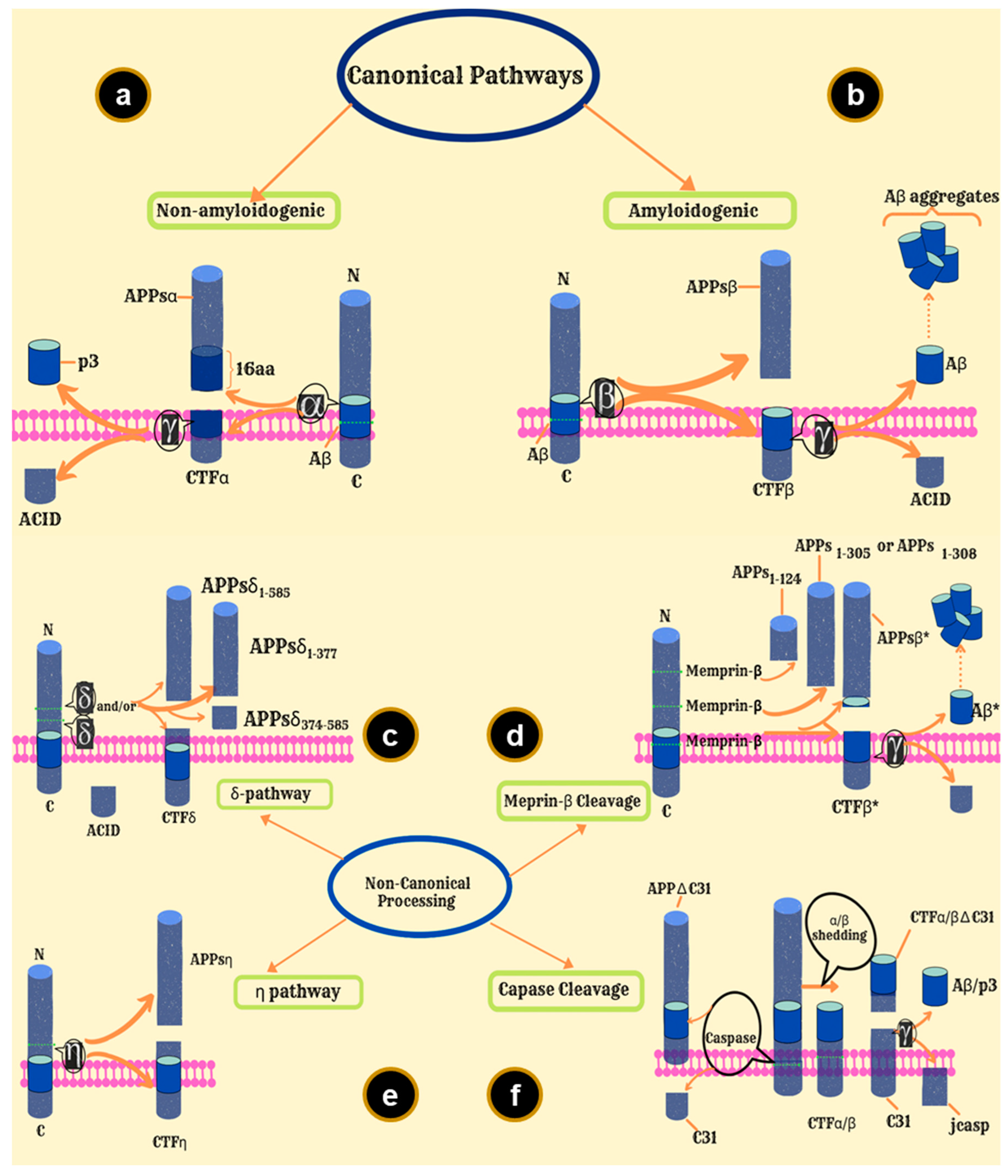
7. APP Processing Pathways

Pathogenic mutations in APP lead to a surge in Aβ peptide production and/or change in the ratio of certain Aβ peptides, and thus, have been linked to the early onset of AD [101,105,106]. APP processing occurs via the action of secretases. This leads to two processing pathways: amyloidogenic and non-amyloidogenic [107,108]. In the amyloidogenic pathway, APP is cleaved by β-secretase (BACE1 and BACE2) and releases soluble secreted APP (APPsβ), and membrane-tethered-β-carboxyl-terminal fragments (CTFβ) [109,110,111]. The γ-secretase then cleaves CTFβ with an endoproteolytic cut at the ε-site liberating ACID, and an exoproteolytic cut at the N-terminus generating Aβ [101,112,113]. On the other hand, in the non-amyloidogenic pathway, APP is cleaved by α-secretase to release APPsα and membrane-tethered α-carboxyl-terminal fragments (CTFα); γ-secretase then cleaves the latter to generate ACID and p3 [99,107,114]. Curiously, until 1992 it was believed that the cleavage of the CTF-β by γ-secretase required some membrane perturbation since the proteolytic cut had to occur at the transmembrane site. Therefore, it was postulated that following the β-secretase cleavage, a cleavage could occur at COOH terminus of the Aβ region, which would be facilitated by γ-secretase [115].
The amyloidogenic and non-amyloidogenic pathways are canonical pathways. There exist several non-canonical APP processing pathways [116]. In the η-secretase pathway, APP is cleaved within the extracellular domain releasing sAPP-η and C-terminal fragments (CTF-η) [117]. CTF-η is then processed either by α-secretase releasing the Aη-α peptide (leaving a CTF-α fragment) or by β-secretase releasing the Aη-β peptide (leaving a CTF-β), to the extracellular medium [118]. Meprin-β can cleave all APP isoforms at multiple sites on the N-terminal ectodomain, one of which releases APPsβ*, and the C-terminal ectodomain liberates CTFβ* [119,120]. CTFβ* is one residue shorter than CTFβ [101]. CTFβ is then processed by γ-secretase liberating Aβ* and AICD [121]. Aβ* is amino-terminally truncated Aβ2−x [101]. The APP C-terminus can also be cleaved by Asp664 to release [122]. In the caspase APP processing pathway, a small peptide, Jcasp, is generated by the cleavage of CTFs by caspase and γ-secretase [123,124]. In the δ-secretase pathway, three soluble APPsδ fragments and C-terminal fragment-δ (CTFδ) are produced upon cleavage by δ-secretase, which are then processed by β-secretase and γ-secretase, liberating Aβ and AICD [101,125] (Figure 4).

8. The Aβ Peptide

The exoproteolytic cut at the N-terminus (Aspartyl) of the membrane tethered 99 residues long CTF β by γ secretase is not precise [121,126]. Consequently, the resulting Aβ peptide shows heterogeneity in residue length and C-terminus polymorphism. The C-terminus of the peptide plays a pivotal role in the alpha helix to beta sheet conformational change during aggregation processes. Amidst the pool of various fragment lengths of these Aβ peptides, ranging from 37 to 51 residues, the 40-residue one is most abundant, followed by the 42 residues [3,19,20], the peptides denoted as Aβ40 and Aβ42, respectively [42,127,128]. These are formed from the larger Aβ fragments by endocytic processing [129,130]. The Aβ42 variant has been found to be more hydrophobic and has a higher amyloidogenic propensity than that of the Aβ40 counterpart [131,132,133]. The longer Aβ42 isoform of the peptide is formed in the Endoplasmic Reticulum (ER), while the shorter Aβ40 is formed in the trans-Golgi network [134,135].
Neprilysin (NEP), Endothelin Converting Enzymes (ECE 1&2), Matrix Metalloproteases (MMP 2) and Insulin Degrading Enzyme (IDE) are the principal metalloproteases responsible for the catabolism of Aβ peptides [136,137,138,139]. Aβ peptides also undergo lysosome mediated degradation, involving enzymes such as cathepsin B [140]. Plasmin, a serine protease, has also been found to have a degrading effect on both Aβ monomers and fibrils [141]. AD is often associated with mutations and/or underproduction of these enzymes, leading to decreased metabolism of Aβ peptides. Soluble Aβ fragments can cross the blood brain barrier to fall into the mainstream blood circulation [142]. This exchange is mediated by Low Density Lipoprotein (LDLP) receptor related proteins (LRPs) on the brain side and the receptor for advanced glycation end products (RAGE) on the luminal side [143,144]. Modifications in these vascular mechanisms might additionally contribute to increases in concentrations of Aβ fragments in the brain in AD related pathologies.
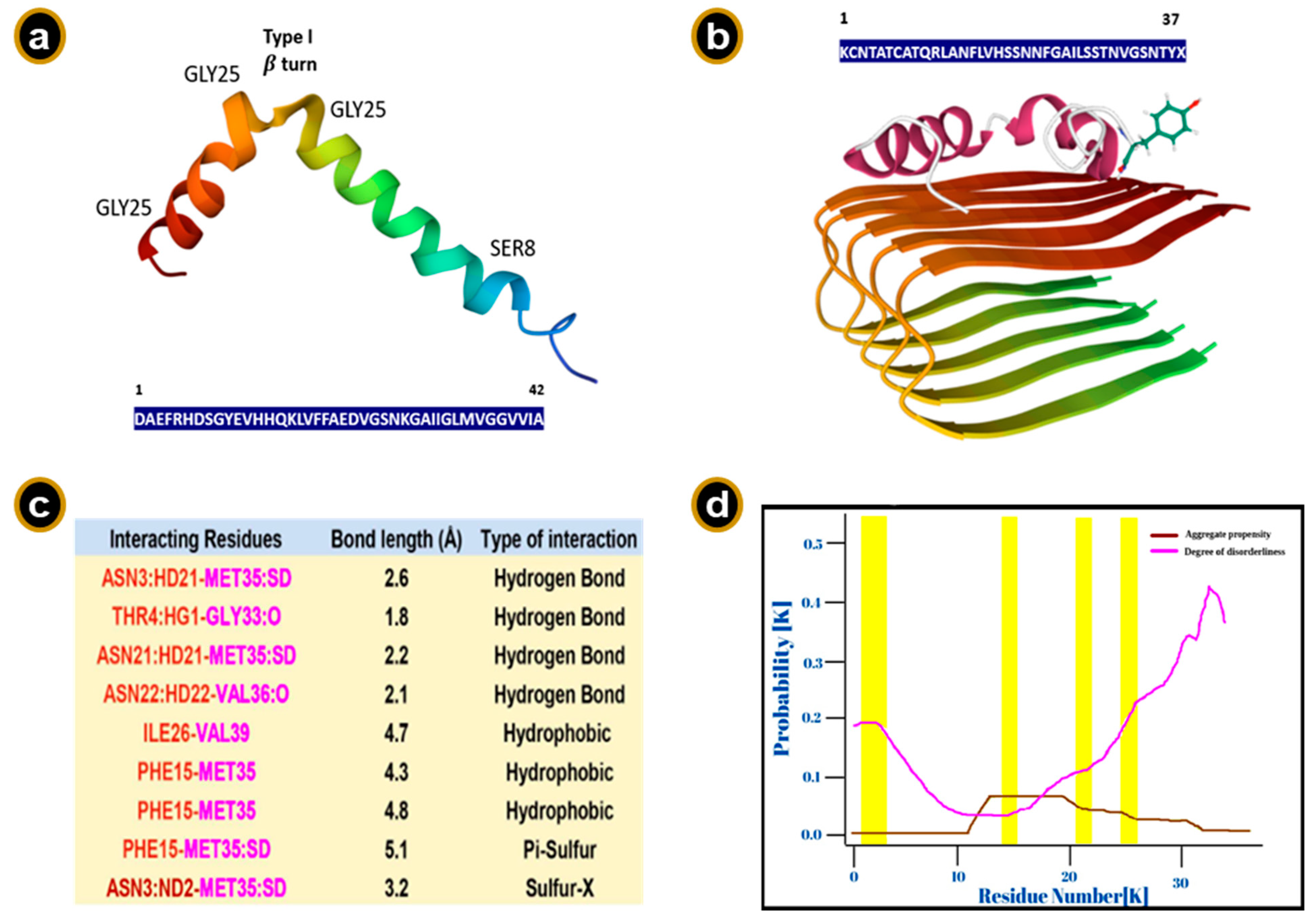
The structure of the Aβ peptide is dynamic in solution and depends on various factors, such as polarity, etc. The different conformations have been shown to exhibit varied propensities towards the amyloid formation. Moreover, different conformations of the monomer may favor different aggregation pathways, leading to varied kinetics in solution [145]. Aβ peptide is mainly α helical in organic solvents, whereas it assumes predominantly a β sheet structure in aqueous media, albeit subject to pH, concentration and incubation times [146,147]. In aqueous sodium dodecyl sulfate (SDS) media, the Aβ40 peptide between residues 1–14 is unstructured, the residues being polar and likely been solvated by water. The Aβ40 peptide has been shown to possess an α helical conformation at the C-terminus between residues 15–36 with a kink or hinge at residues 25–27 and a bend at residue 12 in membrane mimicking solutions. His-13 and Lys-16 have their sidechains lying close on the same side of the helix and are in close proximity. Moreover, 3D NMR studies report two helical regions, Aβ (8–25) and Aβ (28–38), being connected by a type I β turn (Figure 5a). The C-terminus of the Aβ42 isoform is more structured and has reduced flexibility due to a β hairpin structure formed by residues 31–34 and 38–41, probably attributing to its greater amyloidogenic propensity over the Aβ40 isoform. Recent studies show that the Aβ peptides can populate a multitude of discrete conformational clusters [148]. Furthermore, recent computational and experimental efforts reveal that the association of N-terminal hexapeptide A2T and A2V with human Aβ42 shows significantly lower amyloidogenic propensity by altering the conformational landscape of human Aβ42 [149,150].
Protein docking studies between Aβ peptides and human amylin reveal that the major interactions are in the intrinsically disordered stretches of the proteins, suggesting that amyloid aggregation could be driven by the disordered regions of these peptides (Figure 5b–d). Furthermore, these feasible interactions also hint at explaining the co-occurrence of AD, PD, and type-II diabetes [152]. Roughly, a quarter of the Aβ peptide surface is hydrophobic [153]. α helix to β sheet transition of the peptide is thought to involve deprotonation of side chains of the acidic residues Asp7, Glu11, Glu 22 and Asp23 at a pH greater than 4 and the subsequent protonation of the basic residues His6, His 13 and His 14. This event causes a disruption of the α helical structure and above a critical concentration leads to the formation of an oligomeric β sheet structure and fibrillization. Salt bridge electrostatic interactions between His-Asp/Glu residues and the hydrophobic Van der Waals interactions in Leu17-Ala21 residues play a major role in stabilizing the antiparallel arrangement of β sheet assembly defined by residues 13–23. We shall delve into an in-depth discussion about the self-assembly process of amyloid fibrils in the subsequent section(s) [25].

9. Sequence Requirements

Interestingly, even shorter fragments of the Aβ peptides are also capable of forming amyloid fibrils. Norstedt et al. proposed that the shortest fragment capable of amyloid formation is the decapeptide Aβ 14–23 [154]. Any mutations and/or substitutions in this region of the Aβ peptide have been shown to be generally associated with loss of fibril forming ability, indicating that this sequence forms the core of the Aβ fibrils. Almost a year later Tycko et al. showed that the hepta residue Aβ16–22: N-acetyl-Lys-Leu-Val-Phe-Phe-Ala-Glu-NH2, formed full length and highly ordered amyloid fibrils in vitro [155]. Substitutions for positions that stabilize the α-helical conformation (for instance, substituting Val18 by Ala) have been shown to reduce the amyloidogenic propensity of the peptides [156]. It is indeed intriguing that even single point substitutions can act as a veto against fibrillation. For instance, any substitution for Lys16 or Phe19/20 by Ala has been shown to block fibril formation in vitro [157,158,159,160]. Certain mutations indicate that induction of aromatic amino acids increase the amyloidogenic propensities, whereas some studies claim that mutations which replace aromatic side chains promote aggregation of the Aβ peptide, negating the role of π stacking in amyloid self-assembly [22,161]. Concurrently, there exist key regions where substitutions can enhance amyloidogenic propensities [162,163]. These substitutions enhance the hydrophobic interactions in the hydrophobic core by decreasing the electrostatic potential in the 17–21 region [164]. The residues 1–9 of Aβ N-terminus are exposed on the fibrillar surface and play a role in additionally stabilizing the fibrils via interfibrillar interactions. However, Aβ peptides with 1–9 excised region have been shown to form fibrils, indicating that residues 1–9 are not necessary for fibril formation [165,166].

10. Conservation of the Sequence

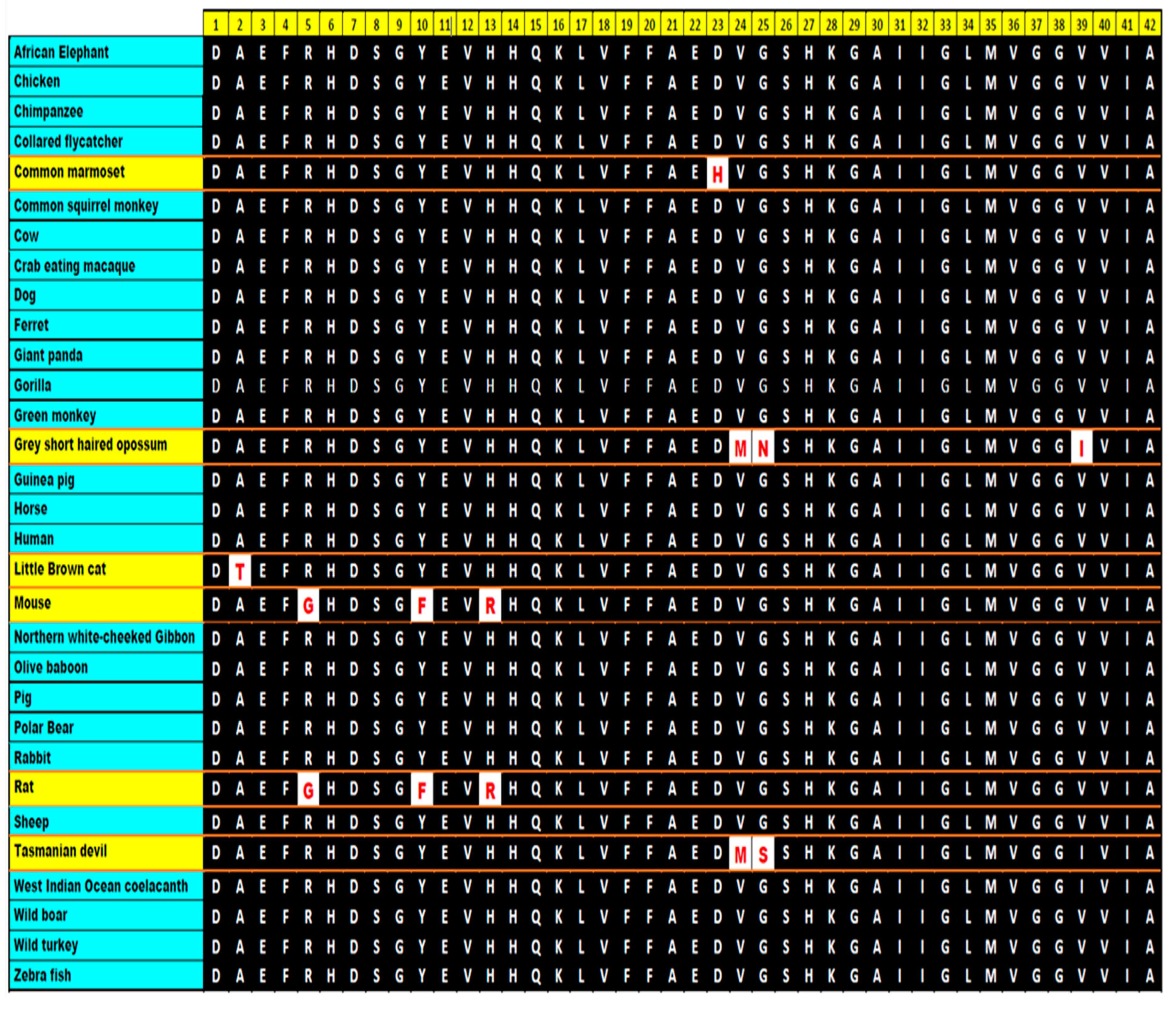
The Aβ peptide is not exclusive to humans. It occurs in a large variety of organisms, however, pathologies related to AD-type diseases are almost exclusively observed in humans alone [167]. The Aβ peptide sequence is highly conserved with only some point mutations in some species (Figure 6). Apart from chimpanzees, almost all non-human primates lack the typical neurofibrillary tangle pathology—a hallmark of AD, even though the NHPs have almost similar APP sequence and biochemistry in addition to Aβ sequence similarity [168]. Interestingly, there has been evidence that the soluble Aβ content in chimpanzees might be higher than those in clinical AD cases [169]. Canines, although genetically farther from humans than non-human primates (NHPs), exhibit substantial amounts of Aβ42 triggered amyloid depositions related to cognitive dysregulations. Moreover, the amyloid buildup starts much faster in canines compared to NHPs. However, the pathologies exhibit little or no neuronal loss. They also lack neuritic plaques or neurofibrillary tangle pathology, another point of contrast between AD in humans and age-related amyloid pathology in canines [170,171,172] (Figure 6).
Now, does it mean that amyloid formation is driven by a specific primary sequence or is it more generally an inherent property of polypeptides under a given biochemical environment? In this context, an interesting observation was reported by Guijarro et al., wherein they found PI3-SH3 small globular domain with a simple native fold formed amyloid-like aggregates at acidic pH, whereas at neutral pH no amyloid formation was observed [173,174,175,176].
A year later, Chiti et al. showed that acylphosphatase with very little propensity to adopt β sheet structure even in the partially folded state, could be directed to form amyloid-like aggregates in vitro. It clearly hinted that, probably, the driving criterion was not entirely the high propensity primary sequence but rather the correct denaturing conditions favoring the hydrophobic clumping forming aggregates [177]. Subsequently, it was also shown that homomeric polypeptides, i.e., polypeptides containing residues of same amino acids, especially those of aromatic amino acids, such as polylysine and polythreonine, could form amyloid-like fibrils [178]. More recently, it has also been shown that single amino acids, such as phenylalanine, can form amyloid assemblies, thus strengthening the hypothesis [179].

11. Protein Aggregation and Its Mechanisms

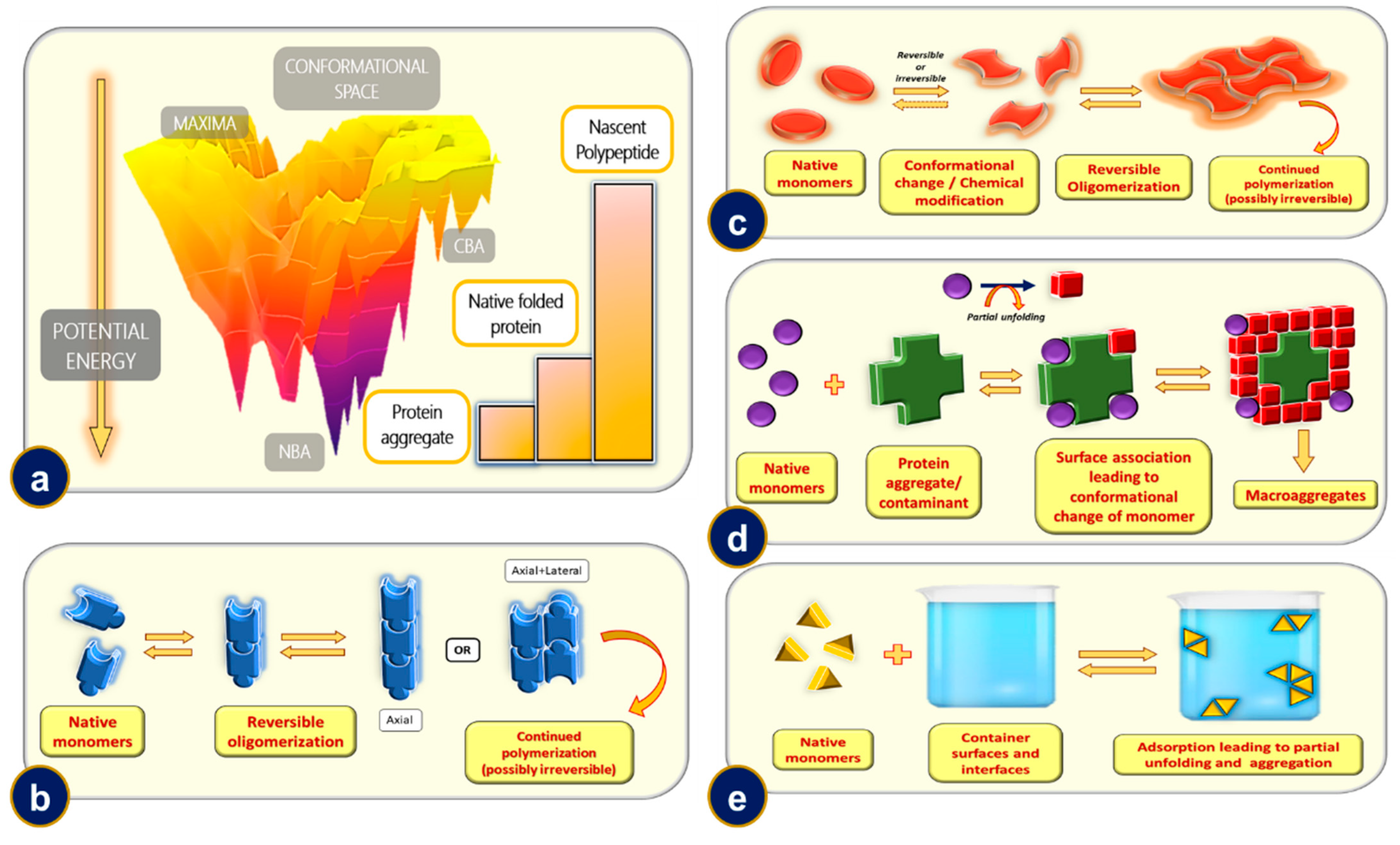
Proteins are the true “chameleons” when it comes to structural diversity. The same protein exists in markedly different structural forms depending on the chemical, physical and biological environments. It is a well-imbibed fact that the proteins keep folding up on themselves to gain stability, giving rise to various levels of structures: primary, secondary, tertiary, and quaternary. Protein folding, which is ubiquitous in nature, is a rather complex problem when it comes to predicting the final state starting from the primary structure, colloquially termed as the protein folding problem [180]. A rather appealing and comprehensive way of understanding the relative stabilities of the sibling structures of the same protein is by analyzing the potential energy surface diagram of the same, more well known as the folding funnel diagram [181]. The folding funnel is a multidimensional potential energy landscape which correlates various structures and/or levels of protein organization with their relative stabilities. The nascent or the unfolded protein chain (primary structure) occupies an intermediate energy level. The local minimas, also known as competing basins of attractions (CBAs), are generally populated by the folding intermediates and the misfolded protein structures. The global minima(s) or the native basins of attraction (NBAs) are populated by the most stable protein structures, which include the native folded state and aggregates of proteins. The topology of the energy landscape reveals yet another interesting aspect of the protein folding process. The folding funnel can either be a smooth one or a rugged one. The rugged funnel is generally an indication that molecular chaperones assist in the folding of the protein under consideration [182] (Figure 7a).
A smooth funnel scenario arises for small globular proteins where the native folded state is determined by the primary sequence alone. The most well-known example in this context is probably the case of ribonuclease folding, enunciated by the 1972 Nobel Laureate in Chemistry—Christian B. Anfinsen—in the principle now known as the Anfinsen’s dogma. Anfinsen, in his studies of ribonuclease folding, had postulated that a primary protein sequence could be used to predict the final folded structure of a protein uniquely, given that the native state minima in the potential energy surface (PES) is unchallenged by any other comparative structure of the protein, and the path leading from unfolded to folded state is kinetically and thermodynamically accessible without having to go through complex conformational requirements [183,184]. It is interesting to note that amyloids—our current topic of interest—are an exception to this dogma as they represent an additional minimum other than that of the native state of the protein in the folding funnel. Protein structure is highly dynamic, and temperature-induced conformational fluctuations lead to a sparse population of partial/misfolded states in the Boltzmannian ensemble [185]. Some of these structures have aggregation-prone residues exposed on the surface, and these structures contribute largely to initiate the aggregation cascade. It is however not necessary that these aggregation prone structures should always originate from the native state and can trace back their origin to pre-native form(s) of the protein. Protein–protein interactions are of utmost importance to probe aggregation mechanisms. While electrostatic interactions between protein monomers/oligomers have been shown to promote aggregation, the role of hydrophobic interactions in the same remains debatable. While one school of thought advocates that since introduction of hydrophobic amino acids at aggregation hotspots increases the aggregation propensity, hydrophobic interactions must be the major driving force behind protein aggregation; another school of thought argues that the hydrophobic patches occupy too small a region on the exposed protein surface to be able to mediate hydrophobic aggregation [186,187,188].
Protein aggregation is primarily entropy driven. Superficially it might seem that the process is entropically disfavored as aggregates should have lower entropy than free monomers/oligomers; however, this decrease is compensated more than enough by the entropy increase due to water molecules being extruded from aggregating regions of the interacting proteins. The overall entropy change for the process thus becomes positive [189]. While several mechanisms have been proposed for an overwhelmingly large statistical sample of proteins, almost all of them have a common skeletal framework. The initiation of protein aggregation takes place by formation and/or introduction of a nucleating center, which would drive forward the aggregation. We have tried to elucidate some simple aggregation models which can describe a fairly large number of protein aggregation pathways.

11.1. Mechanism-1: Reversible Oligomerization of Native Monomers

For proteins that have aggregation-prone residues exposed on the surface, the monomers may possess an intrinsic nature to oligomerize albeit reversibly until the formation of a critical nucleus size. It is interesting to note that in most cases the kinetic data suggest that koff (rate corresponding to monomer detachment from forming oligomer) is greater than kon (rate corresponding to the addition of a monomer to the forming oligomer) until the formation of a critical nucleus, after which the rate correlation reverses, thus favoring aggregation [189]. The formation of the critical nucleus might be explained based on stochastic fluctuations in the system. The forming nucleus may even catalyze the formation of another potential nucleus on its elongating surface, a process termed secondary nucleation. The larger oligomers over time thus have a much lower tendency to dissociate into native monomers, and the oligomerization eventually becomes irreversible. Depolymerization/oligomerization can also be inhibited in cases where the protein monomers associate chemically (for, e.g., via disulfide bridges). Insulin and Interleukin-1 receptor antagonist (rhIL-1RA) proteins are probably the best examples that fit this mechanistic model [190,191] (Figure 7b).

11.2. Mechanism-2: Aggregation of Conformationally/Chemically Modified Monomers

As discussed earlier, proteins populate an ensemble of structures given approximately by the Boltzmannian distribution. At physiological temperatures, although there might be negligible populations of mis/partially folded states with respect to the native form, with increasing temperature their population increases. These conformationally distinct monomers can then follow a mechanism as outlined in Mechanism-1 (reversible oligomerization). This initial change in the native monomer might even be brought about by chemicals (not necessarily denaturants). For example, proteolysis of a short stretch of polypeptide or glycosylation, changes brought about by pH might alter the aggregation propensity of the protein molecules [192,193,194]. The conformational and/or chemical changes in the native monomer might even be brought about by physical cues, such as pressure, etc. [195]. After the formation of a critical nucleus, it is possible that even the native monomers are recruited into the aggregating polymer. IFN-γ and G-CSF are reportedly thought to undergo aggregation via this mechanism [196,197,198] (Figure 7c).

11.3. Mechanism-3: Microaggregate and/or Contaminant Induced Aggregation

Nucleation as discussed is a critical step in aggregation. Expectedly, introduction of a nucleus of suitable size expedites the aggregation process. To draw an analogy, it is equivalent to the introduction of a seed in crystal solution to initiate and expedite crystallization. When the nucleus introduced, as an oligomer of the aggregating protein, it is termed as a homonucleating process, whereas when aggregation is induced by some external particle (aggregate of a different protein or rough micro beads), it is termed as heteronucleation. The introduction of a nucleus of a proper critical size (or larger) may immediately lead to an aggregation cascade while any nuclei of a size smaller than the critical size would be slower at inducing aggregation until it grows to reach the critical size [199,200,201,202] (Figure 7d).

11.4. Mechanism-4: Phase Interface or Rough Surface Induced Aggregation

At phase interface or on rough surfaces, the native monomers may undergo adsorption induced conformational change and/or partial unfolding. This event may trigger the formation of a critical nucleus at the surface/phase interface, mediating towards aggregation via the above-mentioned mechanism(s) [203,204,205]. In such cases, surfactants may reduce the aggregation by minimizing exposed surface area of the native monomers or that of the nucleus, thereby arresting oligomerization to a large extent. It should be noted that this bifurcation of mechanisms is purely artificial, and a given protein may aggregate through one or more of the mechanisms we highlighted [206,207] (Figure 7e).

12. Kinetics and Mechanisms of Amyloid Formation

Amyloids differ from amorphous protein deposits in the fact that they possess a highly ordered fibrillar morphology. It is worthwhile to discuss the kinetics alongside the various mechanisms that contribute to amyloid formation. For the purpose of enhanced readability of the present article, we have tried to keep the mathematical treatment to the bare minimum without compromising the vigor of the kinetic revelations. One of the first attempts to model kinetics of filamentous protein aggregation can be traced back to the 1960s, wherein Ossawa et al. modelled the polymerization of G-Actin to form filamentous F-Actin [208]. They showed that the dynamic length of an actin filament depended on the concentration of the free monomers as well as concentration of the existing fibrils along with distinct rate constants of polymerization and depolymerization [209].
This set the ground for the development of several primary NDP (nucleation dependent polymerization) models. There exist various flavors to these NDP models, however the basic essence revolves around formation of a nucleus of a critical size in a thermodynamically unfavored (but not stochastically impossible) process after which addition of monomers to the growing fibril becomes thermodynamically favored [210].
It would almost be blasphemous not to be mentioning Finke and Watzky’s Ockham Razor model [211]. Originally intended for modelling kinetics of metal nanoclusters, this model gained a surprising feat in describing amyloid kinetics at a macroscopic level [212]. This model can schematically be represented with two equations: M → M’ (rate constant k) and M + M’ → 2M’ (rate constant k’), where M represents free monomer in solution and M’ represents monomer unit in a forming polymer. The main drawback of the Finke–Watzky and other closely related models, such as the three-step nucleated conformational conversion (NCC) aggregation model [213], and the Gibson–Murphy two step aggregation model, is that they describe the kinetics in an averaged out way with the fine microscopic details remaining largely uncharted [214,215].
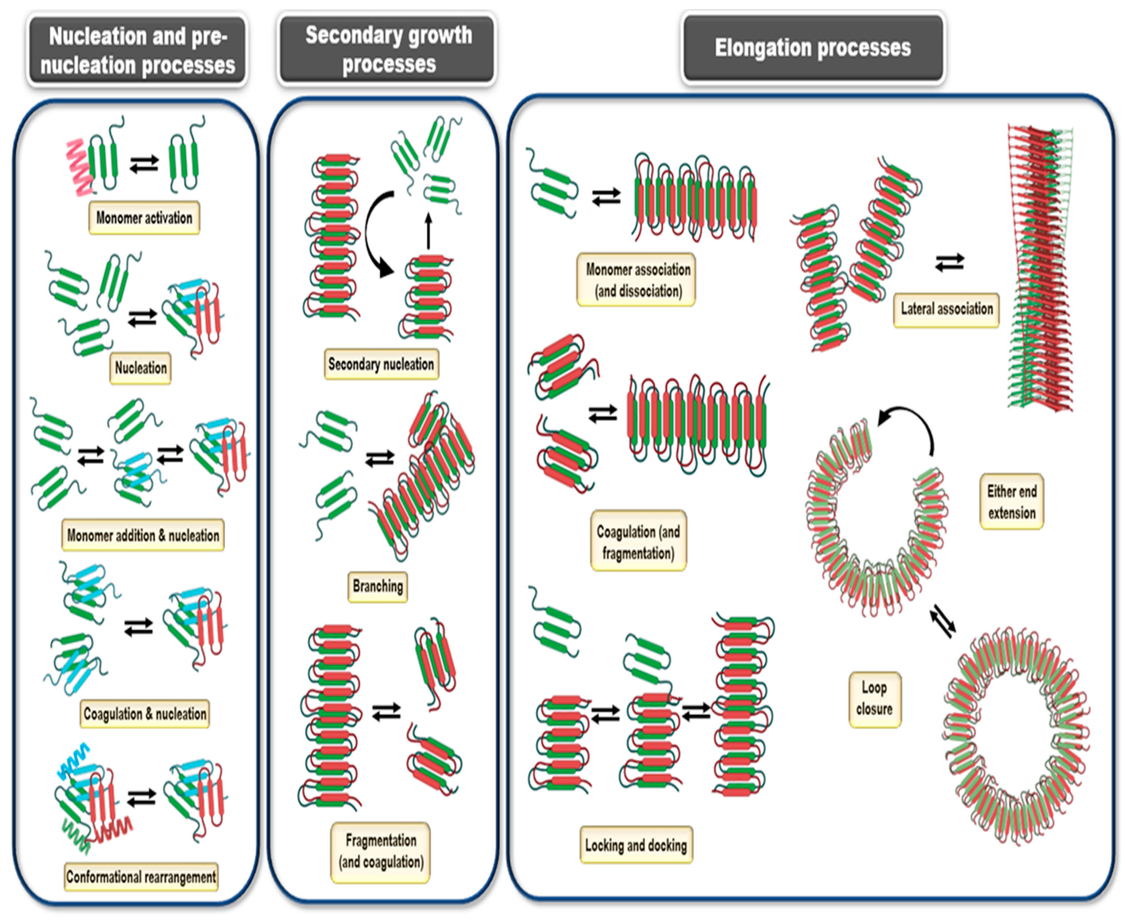
The models discussed so far were biased on the fact that nucleation was considered a single time event, treated as a discrete step in the mechanism with elongation necessarily following the nucleation event. However, the forming fibrils can also initiate nucleation on their surfaces in a process termed as secondary nucleation [216,217,218,219]. There even occur heterogenous or cross-seeding events, which account for the heterogeneity observed in neurofibrillary tangles, and closely related amyloid peptide fragments in the aggregates [220,221]. This further sparks risks of formation of hybrid aggregates affecting species, earlier immune to such aggregations, in an inter-species jump [151]. Moreover, the depolymerization event was being considered as the liberation of a single monomer unit from the extending fibril. However, it is plausible that a chunk of the formed fibril dissociates from the parent fibril in a fragmentation process [222]. In a reverse process, the fibril elongation in turn may be aided by coagulation of the fibril chunks. Coagulation is an important elongation process, perhaps not in the initial stages of fibril formation but in the later stages when the free monomer concentration is appreciably depleted. Coagulation may also assist in formation of the seeding nuclei at various stages [222,223]. As discussed earlier in the protein aggregation mechanisms, free monomers in solution and the monomers incorporated into the fibril may have a stark conformational difference. As such, the monomers may first associate with the elongating fibril, forming an intermediate species before being incorporated into the fibril in the required conformation, in a process called locking and docking [224]. In the initial days of amyloid chemistry, it was believed that the fibrillar topology was necessarily unbranched and was backed up by EM and AFM studies of the same. However, the subsequent development in modeling prowess yielded solutions which showed that there should in fact be no major energy barrier for lateral association [225]. Secondary nucleation on a preformed fibril may lead to lateral association, however the lateral growth may fragment up from the parent fibril to form a separate fibril growth altogether. In recent times, small fibrillar lateral growths have been observed in Aβ fibrils [226]. Smaller aggregates found in close vicinity of the large fibrillar masses further strengthen the lateral association hypothesis [21]. It is important to note that not all the aggregating intermediates lead to ordered fibril formation. The on pathway describes the obligate processes that lead to fibril assembly and off-pathways describe those aggregation intermediates which directly cannot lead to fibril formation [227]. These off-pathway aggregates are reportedly shown to have enhanced toxic effects in vivo [228]. The off-pathway aggregation also contributes to fibril assembly, albeit indirectly. Some recent studies, however, discourage the practice of classifying oligomers unstructured precursors to the amyloid fibrils on a binary basis, either on or off-pathway, and stress on a broader and more encompassing definition.
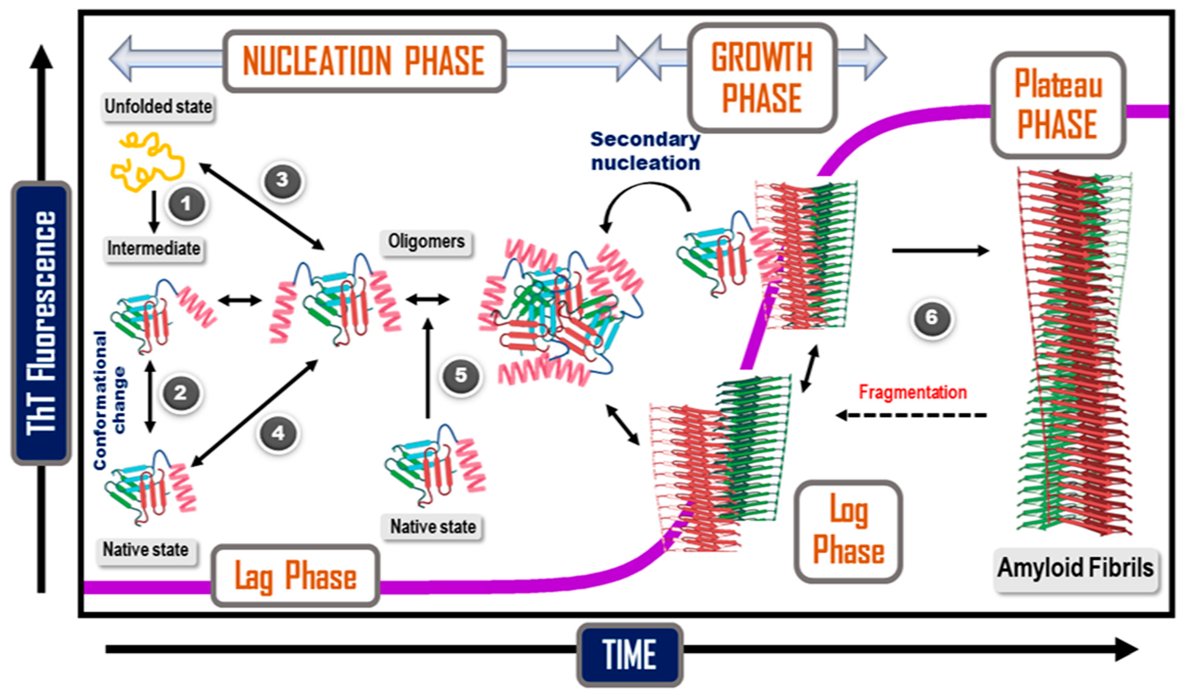
The morphology of amyloid fibrils in nature can be even more bizarre. Apart from the fibrillar species we discussed so far, closed circular loop fibrils are also found along with the regular fibrillar aggregates in protein systems, such as HSA, FtsZ (from E. coli), α-synuclein, β-lactoglobulin, αS2-casein, crystallin proteins, and apoC-II [229,230,231,232,233,234,235,236] (Figure 8). These closed-loop fragments negatively regulate fibrillation. The incompletely formed fibrils undergo an end-to-end circular association, thus forming a thermodynamic sink for elongating fibrils [237]. The kinetic models for amyloid self-assembly were developed mainly for fitting into empirical curves rather than for prediction purposes. As we mentioned earlier, almost all of these kinetic models bear the same essence: a sigmoidal growth curve described with a lag phase, an exponential growth phase, and a plateau phase (Figure 9). The different flavors added cumulatively closely fit to the empirical data and deconvoluted the underlying microscopic processes. For instance, by considering secondary nucleation and associated secondary processes, including appropriate terms into the rate law, a closer and more realistic fit to the kinetic data can be obtained. The integrated rate law utilizing such an approach was postulated by Cohen et al. [238,239,240].
M ( t ) M ( ) = 1 ( B + + C + B + + C + e κ t B + C + e κ t B + + C + ) k 2 κ k ˜ e k t
For a description of the variables and parameters used please refer to Table 1.
The mathematical details of the treatment and the various techniques, such as numerical integration, along with the analytical treatments have been reported and reviewed elsewhere [241,242]. Indeed, it has been found for Aβ42 that in the absence or low concentration of fibrils, fibril elongation takes place majorly through primary nucleation processes. However, once a critical fibril concentration is achieved, secondary nucleation takes over as the dominant elongating phenomenon [216,243]. The self-assembly is further promoted by a positive feedback loop existing between the monomeric and fibrillar forms [240,244].
The native monomer similarly can either undergo partial unfolding and/or associate with the forming nucleus [181,183,184]. When the concentration of amyloid fibrils increases significantly, the secondary processes take over as the dominant phenomenon [185].
Analytical approaches to explain the kinetic curve reveal an interesting underlying fact. The lag phase and the exponential phases might not necessarily be corresponding to the nucleation and elongation events. The phases might rather correspond to a combination of different molecular level events. For instance, fibril elongation by primary processes might correspond to the slower lag phase, while secondary nucleation and fragmentation to form additional nuclei might be the major contributors in the rapid exponential growth phase [243].

13. The Clinical Effects of Amyloid Formation

Amyloidosis refers to various disorders that result from a protein misfolding in which soluble proteins aggregate and convert to insoluble amyloid fibrils [242]. As a result, functional and structural organ damage can be caused by these amyloid deposits [47,245,246].
The extracellular deposition of amyloid fibrils in tissues are involved in different human diseases. Examples include Alzheimer’s disease, Parkinson’s disease, dialysis-related amyloidosis, type-II diabetes, etc. (Table 2) [246]. In amyloidosis, amyloid fibrils build up in tissues, leading to various symptoms, such as reduction in weight, loss in energy, spleen augmentation, tongue augmentation, weakness, bleeding, swelling of legs, numbness, etc. [247,248]. Amyloid depositions can sometimes be limited to one organ or site of the body, known as localized amyloidosis. In contrast, the fibril deposition found in various organs and tissues in the body is termed as systemic. An example of localized amyloidosis is found in Alzheimer’s disease and diabetes mellitus [248]. Localized amyloid can be found in skin, eyelid, conjunctiva, breast, larynx, bronchial tree, lung, and genitourinary tract. In general, low counts of clonal plasma cells can be detected in the biopsy sample [249]. In systemic amyloidosis, symptoms appear as a result of progressive disease in organs and tissues, such as in light chain (AL) amyloidosis and transthyretin amyloidosis (ATTR) [250].
Alzheimer’s disease (AD) is considered a complicated neurodegenerative disorder. The main histopathological characteristics of AD are plaques and neurofibrillary tangles that Alois Alzheimer previously described [251]. It has been believed that the building up of amyloid β-peptide (Aβ) in neural tissue is one of the main causes of AD pathogenesis. After the accumulation of (Aβ), the production of neurofibrillary tangles consisting of tau protein results as a consequence of imbalance between the production and clearance of Aβ species [246]. There are various indications about the amyloid hypothesis that regard amyloids as the toxic cause of neural/synaptic damage and dementia. For example, the existence of amyloid in neuritic plaques in AD, the genetics of inherited AD, the contribution of mutations of amyloid precursor protein (APP), Alzheimer-like changes that are found in middle-aged patients with Down syndrome, the molecular biology of Aβ production from APP, the neurotoxicity of amyloid in tissue culture, and detection on transgenic mouse models of AD with human mutant genes, are all indicators of the amyloid hypothesis of AD. It has been demonstrated that AD, known by plaques Aβ peptide, and neurofibrillary tangles (NFTs), composed of intracellular filaments aggregation of hyperphosphorylated tau protein [252]. According to the amyloid cascade hypothesis, the deposition of Aβ induces the pathology of tau protein via induction of the intraneuronal production of NFTs which consist of hyperphosphorylated tau protein [253,254]. Therefore, there has been enormous research demonstrating the association between Aβ and AD. Moreover, there are abundant therapeutic approaches being developed to reduce the production and accumulation of Aβ in the brain that might lead to preventing AD [255,256]. On the other hand, it has been indicated that there is no correlation between the number of amyloid deposits in the brain and the degree of cognitive impairment that the patient has. For example, there are humans without symptoms of AD, but they have Aβ deposits [257].
Neuritic plaques consisting of the high level of insoluble Aβ in the brain parenchyma is a pathology often associated with AD. In addition, the neurofibrillary tangles in various conditions might have the same significant morphological characteristics, but could have a unique structure of tau isoforms [258]. There is no agreement over the stage classification of AD between clinicians, neuropsychologists, and pathologists. Research efforts on disease mechanisms are largely hindered by the lack of an in-life diagnostic test [166]. It has been demonstrated that immunization of AD patients with full-length Aβ42 (AN-1792) removed amyloid plaques from the brain. However, it could not stop the neurodegenerative progression [259,260]. The mechanism that causes progress in neurodegeneration is not clear. Excess buildup of misfolded hyperphosphorylated tau causing more neuron damage might be the closest hypothesis to explain it. An additional reasonable rationale might be that a specific construction or constructions of Aβ cause the death of several neurons, a typical of the condition. There is an indication that both the solubility and the amount of Aβ in various pools could be highly relevant in AD. Imaging technologies, utilizing amyloid imaging agents depending on the chemical structure of histologic dyes, can be used to investigate amyloid pathology during disease progression in AD patients [256]. Various unsuccessful clinical attempts of anti-Aβ drugs have been made so far. In contrast, in September 2016, a study revealed that an anti-amyloid antibody diminished Aβ levels in the brain and reduced the rate of failure of cognitive action in people with medium or preclinical Alzheimer’s condition [261]. However, not all researchers are persuaded that Aβ leads to Alzheimer’s and are introducing a diversity of other reasonable inhibitors for the harmful array of signaling cascades that eventually drowns brain cells in AD [16]. The US FDA approved aducanumab, a human IgG1 (the most abundant immunoglobulins in the human serum) anti-Aβ monoclonal antibody selective against Aβ aggregates as the most important disease-modeling cure for Alzheimer’s [262]. Lecanemb was raised as an anti-Aβ protofibril antibody. The drug successfully reduces the aggregated Aβ in the brain. Therefore, it is approved by FDA [263].
Amyloid precursor protein (APP) and Aβ species, which control neurodegeneration, develop via the activation of the protein kinase signaling cascade in AD. This highlights the influential role of protein kinase that should be utilized in the treatment of AD patients [264]. Recently, an interesting study was conducted with mice carrying human Aβ only in their hepatic cells. The study indicated that the protein was migrated through blood via lipoproteins containing a high amount of triglyceride, like it is in humans, and finally diffused into the brain. The mice displayed considerable neurodegeneration and brain atrophy, which was related to neurovascular inflammation and dysfunction of cerebral capillaries, both generally noticed in AD. This indicates that there is a possibility of induction of neurodegeneration by peripherally derived Aβ species, suggesting that Aβ produced in the liver might be an important factor in human AD [265]. Based on the amyloid cascade hypothesis, Aβ is produced by the sequential cleavage of the transmembrane protein amyloid precursor protein (APP) via α,β and γ secretases. The cleavage of APP at the plasma membrane is controlled by α-secretase, while β and γ secretases govern the production of Aβ via amyloidogenic pathway [246,263]. Therefore, manipulating the β and γ secretases cloud reduces the production of Aβ and prevent its accumulation from depositing into soluble oligomers and fibrils that initiate oxidative injury, microglial and astrocytic activity besides prevention kinase/phosphatase activity that cause neuronal death [266]. Furthermore, elevation and aggregation of Aβ are correlated with the chronic response of the innate immune system by activating microglia via various immunological receptors [267]. Despite the various trials of anti-Aβ therapies, there is evidence that pathogens trigger soluble Aβ to aggregate into insoluble deposits to prevent the leaking of the pathogen or stop it [268]. For example, recently, it has been indicated that there is a correlation between Aβ and protection and repair of the central nervous system due to the antimicrobial and antiviral function of Aβ oligomer and consecutive fibril formation [269].
On the other hand, some researchers believe that tau protein is more relevant in dementia, and consequently suggest targeting tau protein may be a more meaningful approach than focusing on the development of anti-amyloid therapy in protecting corporeal action in Alzheimer’s disease. However, it should be noted that improvement the effectiveness of the therapy with tau protein is not an easy task. There are various antibodies for the protein, and two vaccines are currently under clinical investigation. As a treatment for AD, tau protein is still in its nascent stage [270]. In AD, amyloid deposits accumulate to produce plaques in the extracellular space, and tau proteins occur in the form of neurofibrillary tangles present in the intracellular spaces that are important hallmarks in the final post-mortem of clinical AD cases [271,272]. It is believed that Alzheimer’s disease typically begins at an age of 15–20 years in people with a family history of AD and 20–30 years in sporadic people before indication of any clinical symptoms [273,274]. Unfortunately, when the clinical phenotype is identified, significant neuronal and synaptic degeneration and massive neuroinflammatory changes have already occurred, ushering the need for early detection of AD [270].

14. Prion

Prion diseases are infectious neurodegenerative diseases that are almost always fatal [33]. Prion diseases are characterized by the accumulation of the protease-resistant prion protein in the central nervous system, which sometimes forms amyloid-like plaques [275]. Although the exact mechanism is not known, it is believed that the conversion of the cell-surface glycoprotein (PrPC) to conformationally altered isoform (PrPSc) when the infectious materials of PrPSc interact with the host PrPC causes the replication of prion [33,276]. The conformational change of the human prion protein into a self-replicating amyloid-like form is associated with a class of fatal diseases, transmissible spongiform encephalopathies [276,277]. When caused by spontaneous prion misfolding, they lead to sporadic Creutzfeldt–Jakob disease, while mutations in the prion protein gene cause familial Creutzfeldt–Jakob disease, Gerstmann–Sträussler Scheinker syndrome, and fatal familial insomnia [276,278]. In this context, both prion and Aβ and prion protein are related to neurodegenerative disorders, such as Creutzfeldt–Jakob and Alzheimer’s, respectively. However, prions are related to neurodegenerative diseases that occur in humans and animals [279]. It was considered that prions are unique because they are infectious. However, it has been indicated that that Aβ proliferates in a manner that prions do [280].

15. Future Perspectives

Proteins assemble and convert to insoluble aggregates that cannot be degraded and amyloid fibrils, related to various diseases, are produced. Some of the frequent amyloid diseases are Alzheimer’s disease, diabetes type-II, and mad cow disease [47].
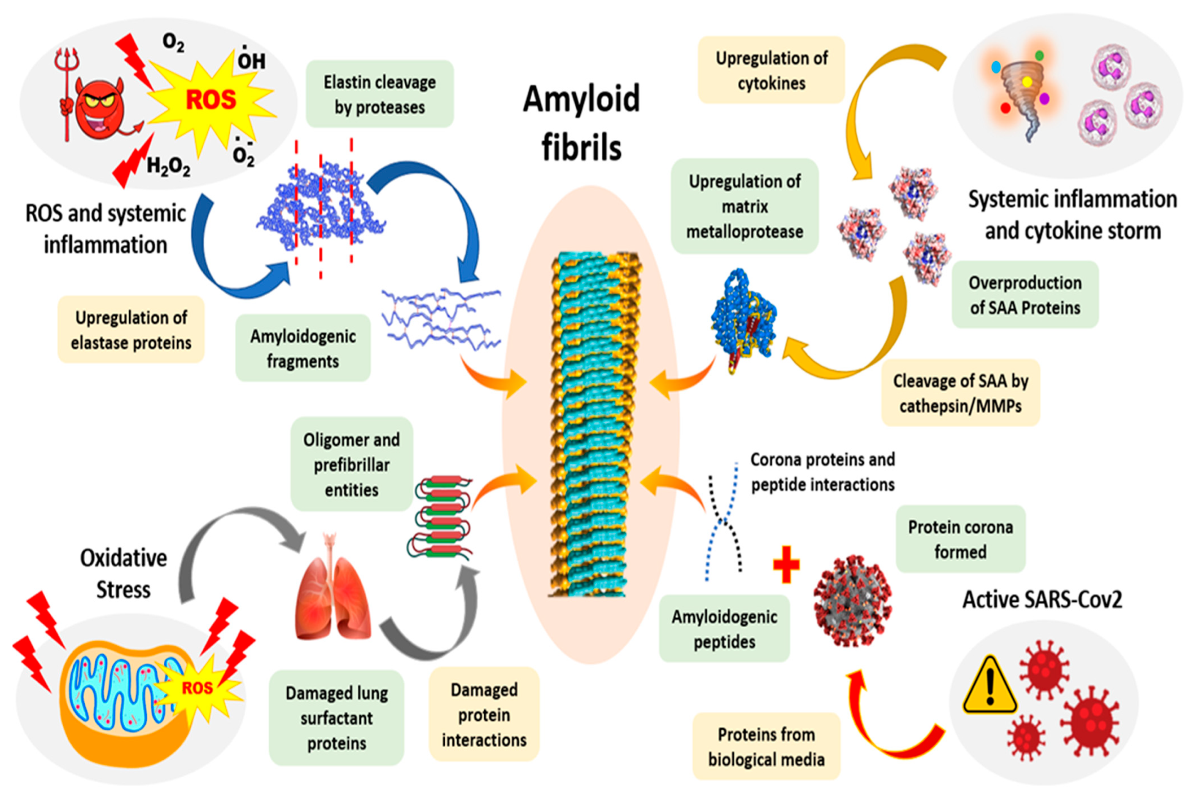
Sever acute respiratory syndrome corona virus 2 (SARS-CoV-2), the causative of the coronavirus-19 (COVID-19 ) pandemic, is a virus that needs no introduction in contemporary times at least. Intriguingly, there have been recent hypotheses and evidence suggesting a possible link between COVID-19 and amyloidosis [281] (Figure 10). The first observation of amyloidosis in such cases of acute respiratory inflictions dates back to 2002 wherein the patients showed a buildup of elastin amyloids. Such respiratory syndromes and viral infections are almost ubiquitously involved with cytokine storm, which upregulates metalloprotease expressions [282,283,284]. These in turn cleave the serum amyloid A proteins, generating a pool of potential amyloidogenic peptides [285]. Furthermore, these cases are also possibly responsible for the upregulation of elastase enzymes, which free elastin fibers from their intrinsic mesh and coerce them into amyloidogenic aggregations [286]. AA amyloidosis is hypothesized to be responsible for renal, lung and multiple organ failures in COVID-19 [287]. ARDS cases are associated with extreme conditions of oxidative stress. Oxidative stress can both directly and indirectly lead to amyloidosis. The leakage of reactive oxygen species from mitochondria is possibly enhanced, leading to more frequent misfolding in proteins, possibly driving amyloidosis [287]. Another hypothesis in place suspects that these viral infections might bring about biochemical changes at cellular levels, favoring the release of amyloidogenic peptides and eventually leading to amyloidosis [288,289].
Heme-Aβ has been shown to be involved in the release of partially reduced oxygen species (PROS) during the TCA cycle in mitochondria [289,290]. These PROS are mainly responsible for the degradation of neurotransmitters and have been shown to be associated with Alzheimer’s disease. The correlation of increased oxidative stress with SARS-CoV-2 infection further sparks possibilities of early AD onset in COVID-19 affected patients.
More recently, synthetic amyloids have also gained clout as important functional next-generation biomaterials, finding uses in drug delivery, food industry, vaccines, water purification, tissue engineering, and as biosensors [291,292,293,294,295,296].
Recently, Biogen and Eisai’s drug Aducanumab, a monoclonal antibody against β-amyloid was approved by the FDA. ADUHELM™ is the first-ever and the only drug as of yet that addresses the apparent pathology of Alzheimer’s disease [297]. The root cause of AD remains debated with β-amyloid aggregation in the AD brain, being mostly completed in the early clinical stages, while tau protein continues to accumulate throughout the course of the disease [166,298]. Accordingly, amyloids have emerged as topic of interest in physical, chemical, biological, and medical research. Advancement in biophysical techniques, such as solid-state nuclear magnetic resonance spectroscopy (ssNMR), X-ray and electron diffraction cryo-electron microscopy (cryo-EM), and mass spectrometry, which assist in analysis of amyloids, aim to provide a better understanding of the molecular mechanisms underlying the amyloid cascade. It is an exhilarating time to live in indeed, to see what the future holds for the cure of AD and amyloid research.

Author Contributions

Conceptualization, Z.A., N.B. and S.M.; writing—original draft preparation, Z.A., N.B. and S.M.; writing—review and editing, Z.A., N.B., S.M., and T.K.S.K.; visualization, Z.A., N.B. and S.M.; supervision, T.K.S.K.; project administration, T.K.S.K.; funding acquisition, T.K.S.K. All authors have read and agreed to the published version of the manuscript.

Funding

The work was supported by the Department of Energy (grant number DE-FG02-01ER15161) and the National Institutes of Health/National Cancer Institute (NIH/NCI) (1 RO1 CA 172631) and the NIH through the COBRE (P30 GM103450) to (T.K.S.K.)

Institutional Review Board Statement

Not applicable.

Informed Consent Statement

Not applicable.

Conflicts of Interest

The authors declare no conflict of interest.

Abbreviations

AD: Alzheimer’s; PKU: Phenylketonuria; ThT: Thioflavin T; CR: Congo Red; APP: Amyloid Precursor Protein; CuBD; Copper-Binding Domain; GFLD: Growth Factor-Like Domain; AICD: APP intracellular C-terminus domain; CTF: C-terminal fragment; IAPP: Islet amyloid polypeptide; BACE: Beta-site APP Cleaving Enzyme; CD: circular dichroism spectroscopy; TEM: transmission electron micriecopy;FGF-1: newt acidic fibroblast growth factor; TRR: transthyretin; TFE: 2,2,2-trifluoroethanol;ANS:1-anilinonaphthalene8-sulfonate; BIS-ANS:4,4′-bis-1-anilinonaphthalene 8-sulfonate; FTIR: Fourier Transform Infrared; AFM: atomic force microscopy; ssNMR: sloid state nuclear magnetic resonance spectroscopy; HPLC: high pressure liquid chromatography; ATTR: Transthyretin amyloidosis; CTF: Carboxy terminal fragment; NEP: Neprilysin; ECE: Endothelin Converting Enzyme; MMP: Matrix Metalloprotease; IDE: Insulin Degrading Enzyme; LDLP: Low Density Lipoprotein; LRPs: Low Density Lipoprotein receptor related proteins; RAGE: Receptor for Advanced Glycation End products; SDS: Sodium Dodecyl Sulfate; NHPs: Non-Human Primates; CBAs: Competing Basins of Attractions; NBAs: Native Basins of Attraction; PES: Potential Energy Surface; IL: Interleukin; ILRA: Interleukin Receptor antagonist; IFN: Interferon; G-CSF: Granulocyte Colony Stimulating Factor; NDP: Nucleation Dependent Polymerization; NCC: Nucleated Conformational Conversion; SARS-CoV-2: Severe Acute Respiratory Syndrome Coronavirus 2; COVID-19: Coronavirus Disease 2019.

References

  1. Gates, B.D.; Xu, Q.; Stewart, M.; Ryan, D.; Willson, C.G.; Whitesides, G.M. New approaches to nanofabrication: Molding, printing, and other techniques. Chem. Rev. 2005, 105, 1171–1196. [Google Scholar] [CrossRef] [PubMed]
  2. Love, J.C.; Estroff, L.A.; Kriebel, J.K.; Nuzzo, R.G.; Whitesides, G.M. Self-assembled monolayers of thiolates on metals as a form of nanotechnology. Chem. Rev. 2005, 105, 1103–1170. [Google Scholar] [CrossRef] [PubMed]
  3. Otero, R.; Gallego, J.M.; de Parga, A.L.V.; Martin, N.; Miranda, R. Molecular Self-Assembly at Solid Surfaces. Adv. Mater. 2011, 23, 5148–5176. [Google Scholar] [CrossRef]
  4. Zhou, Y.; Huang, W.; Liu, J.; Zhu, X.; Yan, D. Self-assembly of hyperbranched polymers and its biomedical applications. Adv. Mater. 2010, 22, 4567–4590. [Google Scholar] [CrossRef] [PubMed]
  5. Chen, C.-L.; Bromley, K.M.; Moradian-Oldak, J.; DeYoreo, J.J. In situ AFM study of amelogenin assembly and disassembly dynamics on charged surfaces provides insights on matrix protein self-assembly. J. Am. Chem. Soc. 2011, 133, 17406–17413. [Google Scholar] [CrossRef] [Green Version]
  6. Agheli, H.; Malmström, J.; Larsson, E.M.; Textor, M.; Sutherland, D.S. Large area protein nanopatterning for biological applications. Nano Lett. 2006, 6, 1165–1171. [Google Scholar] [CrossRef]
  7. Whitesides, G.M.; Mathias, J.P.; Seto, C.T. Molecular self-assembly and nanochemistry: A chemical strategy for the synthesis of nanostructures. Science 1991, 254, 1312–1319. [Google Scholar] [CrossRef]
  8. Reches, M.; Gazit, E. Molecular self-assembly of peptide nanostructures: Mechanism of association and potential uses. Curr. Nanosci. 2006, 2, 105–111. [Google Scholar] [CrossRef] [Green Version]
  9. Kumaraswamy, P.; Sethuraman, S.; Krishnan, U.M. Hierarchical self-assembly of Tjernberg peptide at nanoscale. Soft Matter 2013, 9, 2684–2694. [Google Scholar] [CrossRef]
  10. Lesk, A.M.; Rose, G.D. Folding units in globular proteins. Proc. Natl. Acad. Sci. USA 1981, 78, 4304–4308. [Google Scholar] [CrossRef]
  11. Weber, J.K.; Pande, V.S. Protein folding is mechanistically robust. Biophys. J. 2012, 102, 859–867. [Google Scholar] [CrossRef] [PubMed] [Green Version]
  12. Yadav, S.; Sharma, A.K.; Kumar, P. Nanoscale self-assembly for therapeutic delivery. Front. Bioeng. Biotechnol. 2020, 8, 127. [Google Scholar] [CrossRef] [PubMed] [Green Version]
  13. Haburcak, R.; Shi, J.; Du, X.; Yuan, D.; Xu, B. Ligand–receptor interaction modulates the energy landscape of enzyme-instructed self-assembly of small molecules. J. Am. Chem. Soc. 2016, 138, 15397–15404. [Google Scholar] [CrossRef] [PubMed]
  14. Kar, K.; Ibrar, S.; Nanda, V.; Getz, T.M.; Kunapuli, S.P.; Brodsky, B. Aromatic interactions promote self-association of collagen triple-helical peptides to higher-order structures. Biochemistry 2009, 48, 7959–7968. [Google Scholar] [CrossRef] [PubMed] [Green Version]
  15. Shaham-Niv, S.; Rehak, P.; Vuković, L.; Adler-Abramovich, L.; Král, P.; Gazit, E. Formation of apoptosis-inducing amyloid fibrils by tryptophan. Isr. J. Chem. 2017, 57, 729–737. [Google Scholar] [CrossRef]
  16. Koshti, B.; Ramesh, S.; Kshtriya, V.; Walia, S.; Bhatia, D.; Gour, N. Amyloid like aggregates formed by the self-assembly of proline and Hydroxyproline. Biol. Med. Chem. 2021. [Google Scholar] [CrossRef]
  17. Koshti, B.; Kshtriya, V.; Singh, R.; Walia, S.; Bhatia, D.; Joshi, K.B.; Gour, N. Unusual Aggregates Formed by the Self-Assembly of Proline, Hydroxyproline, and Lysine. ACS Chem. Neurosci. 2021, 12, 3237–3249. [Google Scholar] [CrossRef]
  18. Parthasarathy, A.; Cross, P.J.; Dobson, R.C.J.; Adams, L.E.; Savka, M.A.; Hudson, A.O. A three-ring circus: Metabolism of the three proteogenic aromatic amino acids and their role in the health of plants and animals. Front. Mol. Biosci. 2018, 5, 29. [Google Scholar] [CrossRef]
  19. Makwana, K.M.; Mahalakshmi, R. Implications of aromatic–aromatic interactions: From protein structures to peptide models. Protein Sci. 2015, 24, 1920–1933. [Google Scholar] [CrossRef] [Green Version]
  20. Uversky, V.N. The alphabet of intrinsic disorder: II. Various roles of glutamic acid in ordered and intrinsically disordered proteins. Intrinsically Disord. Proteins 2013, 1, e24684. [Google Scholar] [CrossRef]
  21. Wei, G.; Su, Z.; Reynolds, N.P.; Arosio, P.; Hamley, I.W.; Gazit, E.; Mezzenga, R. Self-assembling peptide and protein amyloids: From structure to tailored function in nanotechnology. Chem. Soc. Rev. 2017, 46, 4661–4708. [Google Scholar] [CrossRef] [PubMed]
  22. Armstrong, A.H.; Chen, J.; McKoy, A.F.; Hecht, M.H. Mutations that replace aromatic side chains promote aggregation of the Alzheimer’s Aβ peptide. Biochemistry 2011, 50, 4058–4067. [Google Scholar] [CrossRef] [PubMed] [Green Version]
  23. Biancalana, M.; Makabe, K.; Koide, A.; Koide, S. Aromatic cross-strand ladders control the structure and stability of β-rich peptide self-assembly mimics. J. Mol. Biol. 2008, 383, 205–213. [Google Scholar] [CrossRef] [PubMed] [Green Version]
  24. Shaham-Niv, S.; Adler-Abramovich, L.; Schnaider, L.; Gazit, E. Extension of the generic amyloid hypothesis to nonproteinaceous metabolite assemblies. Sci. Adv. 2015, 1, e1500137. [Google Scholar] [CrossRef] [PubMed] [Green Version]
  25. Zagorski, M.G.; Barrow, C.J. NMR studies of amyloid. beta.-peptides: Proton assignments, secondary structure, and mechanism of an. alpha.-helix. fwdarw.. beta.-sheet conversion for a homologous, 28-residue, N-terminal fragment. Biochemistry 1992, 31, 5621–5631. [Google Scholar] [CrossRef] [PubMed]
  26. Tu, L.-H.; Raleigh, D.P. Role of aromatic interactions in amyloid formation by islet amyloid polypeptide. Biochemistry 2013, 52, 333–342. [Google Scholar] [CrossRef] [PubMed]
  27. Gour, N.; Koshti, B.; Shah, D.; Kshatriya, V.S.; Agrawal-Rajput, R.; Pandey, M. Single amino acid based self-assemblies of Cysteine and Methionine. ACS Chem. Neurosci. 2018, 10. [Google Scholar] [CrossRef]
  28. Adler-Abramovich, L.; Vaks, L.; Carny, O.; Trudler, D.; Magno, A.; Caflisch, A.; Frenkel, D.; Gazit, E. Phenylalanine assembly into toxic fibrils suggests amyloid etiology in phenylketonuria. Nat. Chem. Biol. 2012, 8, 701–706. [Google Scholar] [CrossRef]
  29. Liu, G.; Gaines, J.C.; Robbins, K.J.; Lazo, N.D. Kinetic profile of amyloid formation in the presence of an aromatic inhibitor by nuclear magnetic resonance. ACS Med. Chem. Lett. 2012, 3, 856–859. [Google Scholar] [CrossRef] [Green Version]
  30. Tanskanen, M. “Amyloid”—Historical Aspects. In Amyloidosis; IntechOpen: London, UK, 2013; p. 1. [Google Scholar]
  31. Kyle, R.A. Amyloidosis: A convoluted story. Br. J. Haematol. 2001, 114, 529–538. [Google Scholar] [CrossRef]
  32. Nishitsuji, K.; Saito, H.; Uchimura, K. Enzymatic remodeling of heparan sulfate: A therapeutic strategy for systemic and localized amyloidoses? Neural Regen. Res. 2016, 11, 408. [Google Scholar] [CrossRef] [PubMed]
  33. Soto, C. Prion hypothesis: The end of the controversy? Trends Biochem. Sci. 2011, 36, 151–158. [Google Scholar] [CrossRef] [PubMed] [Green Version]
  34. Zabel, M.D.; Reid, C. A brief history of prions. Pathog. Dis. 2015, 73, ftv087. [Google Scholar] [CrossRef] [PubMed] [Green Version]
  35. Goetz, C.G. The history of Parkinson’s disease: Early clinical descriptions and neurological therapies. Cold Spring Harb. Perspect. Med. 2011, 1, a008862. [Google Scholar] [CrossRef] [Green Version]
  36. Stefanis, L. α-Synuclein in Parkinson’s disease. Cold Spring Harb. Perspect. Med. 2012, 2, a009399. [Google Scholar] [CrossRef] [Green Version]
  37. Yakupova, E.I.; Bobyleva, L.G.; Vikhlyantsev, I.M.; Bobylev, A.G. Congo Red and amyloids: History and relationship. Biosci. Rep. 2019, 39, BSR20181415. [Google Scholar] [CrossRef] [Green Version]
  38. Howie, A.J.; Owen-Casey, M.P. Discrepancies between descriptions and illustrations of colours in Congo red-stained amyloid, and explanation of discrepant colours. Amyloid 2010, 17, 109–117. [Google Scholar] [CrossRef]
  39. Linke, R.P. Congo red staining of amyloid: Improvements and practical guide for a more precise diagnosis of amyloid and the different amyloidoses. In Protein Misfolding, Aggregation, and Conformational Diseases; Springer: Berlin/Heidelberg, Germany, 2006; pp. 239–276. [Google Scholar]
  40. Toyama, B.H.; Weissman, J.S. Amyloid Structure: Conformational Diversity and Consequences. Annu. Rev. Biochem. 2011, 80, 557–585. [Google Scholar] [CrossRef] [Green Version]
  41. Sunde, M.; Serpell, L.C.; Bartlam, M.; Fraser, P.E.; Pepys, M.B.; Blake, C.C.F. Common core structure of amyloid fibrils by synchrotron X-ray diffraction. J. Mol. Biol. 1997, 273, 729–739. [Google Scholar] [CrossRef] [Green Version]
  42. Chen, G.F.; Xu, T.H.; Yan, Y.; Zhou, Y.R.; Jiang, Y.; Melcher, K.; Xu, H.E. Amyloid beta: Structure, biology and structure-based therapeutic development. Acta Pharmacol. Sin. 2017, 38, 1205–1235. [Google Scholar] [CrossRef]
  43. Kollmer, M.; Meinhardt, K.; Haupt, C.; Liberta, F.; Wulff, M.; Linder, J.; Handl, L.; Heinrich, L.; Loos, C.; Schmidt, M.; et al. Electron tomography reveals the fibril structure and lipid interactions in amyloid deposits. Proc. Natl. Acad. Sci. USA 2016, 113, 5604–5609. [Google Scholar] [CrossRef] [PubMed] [Green Version]
  44. Iadanza, M.G.; Jackson, M.P.; Hewitt, E.W.; Ranson, N.A.; Radford, S.E. A new era for understanding amyloid structures and disease. Nat. Rev. Mol. Cell Biol. 2018, 19, 755–773. [Google Scholar] [CrossRef] [PubMed]
  45. Nelson, R.; Sawaya, M.R.; Balbirnie, M.; Madsen, A.Ø.; Riekel, C.; Grothe, R.; Eisenberg, D. Structure of the cross-β spine of amyloid-like fibrils. Nature 2005, 435, 773–778. [Google Scholar] [CrossRef] [PubMed] [Green Version]
  46. Gorkovskiy, A.; Thurber, K.R.; Tycko, R.; Wickner, R.B. Locating folds of the in-register parallel β-sheet of the Sup35p prion domain infectious amyloid. Proc. Natl. Acad. Sci. USA 2014, 111, E4615–E4622. [Google Scholar] [CrossRef] [PubMed] [Green Version]
  47. Chiti, F.; Dobson, C.M. Protein Misfolding, Amyloid Formation, and Human Disease: A Summary of Progress Over the Last Decade. Annu. Rev. Biochem. 2017, 86, 27–68. [Google Scholar] [CrossRef] [PubMed]
  48. Jahn, T.R.; Makin, O.S.; Morris, K.L.; Marshall, K.E.; Tian, P.; Sikorski, P.; Serpell, L.C. The common architecture of cross-β amyloid. J. Mol. Biol. 2010, 395, 717–727. [Google Scholar] [CrossRef] [PubMed]
  49. DeForte, S.; Uversky, V.N. Order, disorder, and everything in between. Molecules 2016, 21, 1090. [Google Scholar] [CrossRef] [Green Version]
  50. Van Der Lee, R.; Buljan, M.; Lang, B.; Weatheritt, R.J.; Daughdrill, G.W.; Dunker, A.K.; Fuxreiter, M.; Gough, J.; Gsponer, J.; Jones, D.T. Classification of intrinsically disordered regions and proteins. Chem. Rev. 2014, 114, 6589–6631. [Google Scholar] [CrossRef]
  51. Xu, S.H. Cross-beta-Sheet Structure in Amyloid Fiber Formation. J. Phys. Chem. B 2009, 113, 12447–12455. [Google Scholar] [CrossRef]
  52. Schmidt, M.; Wiese, S.; Adak, V.; Engler, J.; Agarwal, S.; Fritz, G.; Westermark, P.; Zacharias, M.; Fändrich, M. Cryo-EM structure of a transthyretin-derived amyloid fibril from a patient with hereditary ATTR amyloidosis. Nat. Commun. 2019, 10, 5008. [Google Scholar] [CrossRef]
  53. Smith, J.E.; Mowles, A.K.; Mehta, A.K.; Lynn, D.G. Looked at life from both sides now. Life 2014, 4, 887–902. [Google Scholar] [CrossRef] [PubMed] [Green Version]
  54. Yoon, G.; Lee, M.; Kim, J.I.; Na, S.; Eom, K. Role of sequence and structural polymorphism on the mechanical properties of amyloid fibrils. PLoS ONE 2014, 9, e88502. [Google Scholar] [CrossRef] [PubMed]
  55. Makin, O.S.; Atkins, E.; Sikorski, P.; Johansson, J.; Serpell, L.C. Molecular basis for amyloid fibril formation and stability. Proc. Natl. Acad. Sci. USA 2005, 102, 315–320. [Google Scholar] [CrossRef] [PubMed] [Green Version]
  56. Biancalana, M.; Makabe, K.; Koide, S. Minimalist design of water-soluble cross-β architecture. Proc. Natl. Acad. Sci. USA 2010, 107, 3469–3474. [Google Scholar] [CrossRef] [PubMed] [Green Version]
  57. Del Pozo-Yauner, L.; Becerril, B.; Ochoa-Leyva, A.; Rodríguez-Ambriz, S.L.; Carrión, J.I.P.; Zavala-Padilla, G.; Sánchez-López, R.; Velasco, D.A.F. The structural determinants of the immunoglobulin light chain amyloid aggregation. In Physical Biology of Proteins and Peptides; Springer: Berlin/Heidelberg, Germany, 2015; pp. 1–28. [Google Scholar]
  58. Maity, B.K.; Das, A.K.; Dey, S.; Moorthi, U.K.; Kaur, A.; Dey, A.; Surendran, D.; Pandit, R.; Kallianpur, M.; Chandra, B.; et al. Ordered and Disordered Segments of Amyloid-β Drive Sequential Steps of the Toxic Pathway. ACS Chem. Neurosci. 2019, 10, 2498–2509. [Google Scholar] [CrossRef]
  59. Schmidt, A.; Annamalai, K.; Schmidt, M.; Grigorieff, N.; Fändrich, M. Cryo-EM reveals the steric zipper structure of a light chain-derived amyloid fibril. Proc. Natl. Acad. Sci. USA 2016, 113, 6200–6205. [Google Scholar] [CrossRef] [Green Version]
  60. Lee, G.; Lee, W.; Lee, H.; Woo Lee, S.; Sung Yoon, D.; Eom, K.; Kwon, T. Mapping the surface charge distribution of amyloid fibril. Appl. Phys. Lett. 2012, 101, 043703. [Google Scholar] [CrossRef]
  61. Bhak, G.; Lee, J.-H.; Hahn, J.-S.; Paik, S.R. Granular assembly of α-synuclein leading to the accelerated amyloid fibril formation with shear stress. PLoS ONE 2009, 4, e4177. [Google Scholar] [CrossRef] [Green Version]
  62. Lee, J.-H.; Bhak, G.; Lee, S.-G.; Paik, S.R. Instantaneous amyloid fibril formation of α-synuclein from the oligomeric granular structures in the presence of hexane. Biophys. J. 2008, 95, L16–L18. [Google Scholar] [CrossRef] [Green Version]
  63. Ha, C.; Park, C.B. Template-directed self-assembly and growth of insulin amyloid fibrils. Biotechnol. Bioeng. 2005, 90, 848–855. [Google Scholar] [CrossRef]
  64. Wu, J.W.; Breydo, L.; Isas, J.M.; Lee, J.; Kuznetsov, Y.G.; Langen, R.; Glabe, C. Fibrillar oligomers nucleate the oligomerization of monomeric amyloid β but do not seed fibril formation. J. Biol. Chem. 2010, 285, 6071–6079. [Google Scholar] [CrossRef] [PubMed] [Green Version]
  65. Kayed, R.; Pensalfini, A.; Margol, L.; Sokolov, Y.; Sarsoza, F.; Head, E.; Hall, J.; Glabe, C. Annular protofibrils are a structurally and functionally distinct type of amyloid oligomer. J. Biol. Chem. 2009, 284, 4230–4237. [Google Scholar] [CrossRef] [PubMed] [Green Version]
  66. Morgado, I.; Wieligmann, K.; Bereza, M.; Rönicke, R.; Meinhardt, K.; Annamalai, K.; Baumann, M.; Wacker, J.; Hortschansky, P.; Malešević, M. Molecular basis of β-amyloid oligomer recognition with a conformational antibody fragment. Proc. Natl. Acad. Sci. USA 2012, 109, 12503–12508. [Google Scholar] [CrossRef] [PubMed] [Green Version]
  67. Cao, Q.; Anderson, D.H.; Liang, W.Y.; Chou, J.; Saelices, L. The inhibition of cellular toxicity of amyloid-β by dissociated transthyretin. J. Biol. Chem. 2020, 295, 14015–14024. [Google Scholar] [CrossRef] [PubMed]
  68. Biancalana, M.; Koide, S. Molecular mechanism of Thioflavin-T binding to amyloid fibrils. Biochim. Et Biophys. Acta-Proteins Proteom. 2010, 1804, 1405–1412. [Google Scholar] [CrossRef] [Green Version]
  69. Levine Iii, H. Thioflavine T interaction with synthetic Alzheimer’s disease β-amyloid peptides: Detection of amyloid aggregation in solution. Protein Sci. 1993, 2, 404–410. [Google Scholar] [CrossRef] [PubMed]
  70. Arunkumar, A.I.; Kumar, T.K.S.; Kathir, K.M.; Srisailam, S.; Wang, H.M.; Leena, P.S.T.; Chi, Y.H.; Chen, H.C.; Wu, C.H.; Wu, R.T. Oligomerization of acidic fibroblast growth factor is not a prerequisite for its cell proliferation activity. Protein Sci. 2002, 11, 1050–1061. [Google Scholar] [CrossRef] [PubMed] [Green Version]
  71. Srisailam, S.; Kumar, T.K.S.; Rajalingam, D.; Kathir, K.M.; Sheu, H.-S.; Jan, F.-J.; Chao, P.-C.; Yu, C. Amyloid-like Fibril Formation in an All β-Barrel Protein PARTIALLY STRUCTURED INTERMEDIATE STATE (S) IS A PRECURSOR FOR FIBRIL FORMATION. J. Biol. Chem. 2003, 278, 17701–17709. [Google Scholar] [CrossRef] [Green Version]
  72. Maezawa, I.; Hong, H.S.; Liu, R.; Wu, C.Y.; Cheng, R.H.; Kung, M.P.; Kung, H.F.; Lam, K.S.; Oddo, S.; LaFerla, F.M. Congo red and thioflavin-T analogs detect Aβ oligomers. J. Neurochem. 2008, 104, 457–468. [Google Scholar] [CrossRef]
  73. Reinke, A.A.; Gestwicki, J.E. Insight into amyloid structure using chemical probes. Chem. Biol. Drug Des. 2011, 77, 399–411. [Google Scholar] [CrossRef]
  74. Lindgren, M.; Hammarström, P. Amyloid oligomers: Spectroscopic characterization of amyloidogenic protein states. FEBS J. 2010, 277, 1380–1388. [Google Scholar] [CrossRef] [PubMed] [Green Version]
  75. Nilsson, M.R. Techniques to study amyloid fibril formation in vitro. Methods 2004, 34, 151–160. [Google Scholar] [CrossRef] [PubMed]
  76. Mostaert, A.S.; Higgins, M.J.; Fukuma, T.; Rindi, F.; Jarvis, S.P. Nanoscale mechanical characterisation of amyloid fibrils discovered in a natural adhesive. J. Biol. Phys. 2006, 32, 393–401. [Google Scholar] [CrossRef] [Green Version]
  77. Hawe, A.; Sutter, M.; Jiskoot, W. Extrinsic fluorescent dyes as tools for protein characterization. Pharm. Res. 2008, 25, 1487–1499. [Google Scholar] [CrossRef] [PubMed] [Green Version]
  78. Howie, A.J.; Brewer, D.B. Optical properties of amyloid stained by Congo red: History and mechanisms. Micron 2009, 40, 285–301. [Google Scholar] [CrossRef] [PubMed]
  79. Şen, S.; Başdemir, G. Diagnosis of renal amyloidosis using Congo red fluorescence. Pathol. Int. 2003, 53, 534–538. [Google Scholar] [CrossRef]
  80. Setti, S.E.; Raymick, J.; Hanig, J.; Sarkar, S. In vivo demonstration of Congo Red labeled amyloid plaques via perfusion in the Alzheimer disease rat model. J. Neurosci. Methods 2021, 353, 109082. [Google Scholar] [CrossRef]
  81. Hamley, I.W.; Castelletto, V.; Moulton, C.; Myatt, D.; Siligardi, G.; Oliveira, C.L.P.; Pedersen, J.S.; Abutbul, I.; Danino, D. Self-assembly of a modified amyloid peptide fragment: pH-responsiveness and nematic phase formation. Macromol. Biosci. 2010, 10, 40–48. [Google Scholar] [CrossRef]
  82. Haris, P.I.; Severcan, F. FTIR spectroscopic characterization of protein structure in aqueous and non-aqueous media. J. Mol. Catal. B Enzym. 1999, 7, 207–221. [Google Scholar] [CrossRef]
  83. Byler, D.M.; Susi, H. Examination of the secondary structure of proteins by deconvolved FTIR spectra. Biopolym. Orig. Res. Biomol. 1986, 25, 469–487. [Google Scholar] [CrossRef]
  84. Goormaghtigh, E.; Ruysschaert, J.-M.; Raussens, V. Evaluation of the information content in infrared spectra for protein secondary structure determination. Biophys. J. 2006, 90, 2946–2957. [Google Scholar] [CrossRef] [PubMed] [Green Version]
  85. Miller, L.M.; Wang, Q.; Telivala, T.P.; Smith, R.J.; Lanzirotti, A.; Miklossy, J. Synchrotron-based infrared and X-ray imaging shows focalized accumulation of Cu and Zn co-localized with β-amyloid deposits in Alzheimer’s disease. J. Struct. Biol. 2006, 155, 30–37. [Google Scholar] [CrossRef] [PubMed]
  86. Choo, L.-P.i.; Wetzel, D.L.; Halliday, W.C.; Jackson, M.; LeVine, S.M.; Mantsch, H.H. In situ characterization of beta-amyloid in Alzheimer’s diseased tissue by synchrotron Fourier transform infrared microspectroscopy. Biophys. J. 1996, 71, 1672–1679. [Google Scholar] [CrossRef] [Green Version]
  87. Rak, M.; Del Bigio, M.R.; Mai, S.; Westaway, D.; Gough, K. Dense-core and diffuse Aβ plaques in TgCRND8 mice studied with synchrotron FTIR microspectroscopy. Biopolym. Orig. Res. Biomol. 2007, 87, 207–217. [Google Scholar] [CrossRef]
  88. Li, H.; Rahimi, F.; Sinha, S.; Maiti, P.; Bitan, G.; Murakami, K. Amyloids and protein aggregation–analytical methods. Encycl. Anal. Chem. 2009, 1–32. [Google Scholar] [CrossRef]
  89. Annamalai, K.; Gührs, K.H.; Koehler, R.; Schmidt, M.; Michel, H.; Loos, C.; Gaffney, P.M.; Sigurdson, C.J.; Hegenbart, U.; Schönland, S. Polymorphism of amyloid fibrils in vivo. Angew. Chem. Int. Ed. 2016, 55, 4822–4825. [Google Scholar] [CrossRef] [Green Version]
  90. Linke, W.A.; Grützner, A. Pulling single molecules of titin by AFM—Recent advances and physiological implications. Pflügers Arch.-Eur. J. Physiol. 2008, 456, 101–115. [Google Scholar] [CrossRef]
  91. Li, Q.; Song, J.; Saura-Múzquiz, M.; Besenbacher, F.; Christensen, M.; Dong, M. Magnetic properties of strontium hexaferrite nanostructures measured with magnetic force microscopy. Sci. Rep. 2016, 6, 25985. [Google Scholar] [CrossRef] [Green Version]
  92. Serem, W.K.; Bett, C.K.; Ngunjiri, J.N.; Garno, J.C. Studies of the growth, evolution, and self-aggregation of β-amyloid fibrils using tapping-mode atomic force microscopy. Microsc. Res. Tech. 2011, 74, 699–708. [Google Scholar] [CrossRef]
  93. Tycko, R. Characterization of amyloid structures at the molecular level by solid state nuclear magnetic resonance spectroscopy. Methods Enzymol. 2006, 413, 103–122. [Google Scholar]
  94. Petkova, A.T.; Ishii, Y.; Balbach, J.J.; Antzutkin, O.N.; Leapman, R.D.; Delaglio, F.; Tycko, R. A structural model for Alzheimer’s β-amyloid fibrils based on experimental constraints from solid state NMR. Proc. Natl. Acad. Sci. USA 2002, 99, 16742–16747. [Google Scholar] [CrossRef] [PubMed]
  95. Mori, H.; Takio, K.; Ogawara, M.; Selkoe, D.J. Mass spectrometry of purified amyloid beta protein in Alzheimer’s disease. J. Biol. Chem. 1992, 267, 17082–17086. [Google Scholar] [CrossRef]
  96. Nettleton, E.J.; Tito, P.; Sunde, M.; Bouchard, M.; Dobson, C.M.; Robinson, C.V. Characterization of the oligomeric states of insulin in self-assembly and amyloid fibril formation by mass spectrometry. Biophys. J. 2000, 79, 1053–1065. [Google Scholar] [CrossRef] [Green Version]
  97. Zandomeneghi, G.; Krebs, M.R.H.; McCammon, M.G.; Fändrich, M. FTIR reveals structural differences between native β-sheet proteins and amyloid fibrils. Protein Sci. 2004, 13, 3314–3321. [Google Scholar] [CrossRef] [PubMed] [Green Version]
  98. Lorenzo, A.; Yuan, M.; Zhang, Z.; Paganetti, P.A.; Sturchler-Pierrat, C.; Staufenbiel, M.; Mautino, J.; Vigo, F.S.; Sommer, B.; Yankner, B.A. Amyloid β interacts with the amyloid precursor protein: A potential toxic mechanism in Alzheimer’s disease. Nat. Neurosci. 2000, 3, 460–464. [Google Scholar] [CrossRef]
  99. Zheng, H.; Koo, E.H. Biology and pathophysiology of the amyloid precursor protein. Mol. Neurodegener. 2011, 6, 27. [Google Scholar] [CrossRef] [Green Version]
  100. Nguyen, K.V. The human β-amyloid precursor protein: Biomolecular and epigenetic aspects. Biomol. Concepts 2015, 6, 11–32. [Google Scholar] [CrossRef] [Green Version]
  101. Müller, U.C.; Deller, T.; Korte, M. Not just amyloid: Physiological functions of the amyloid precursor protein family. Nat. Rev. Neurosci. 2017, 18, 281–298. [Google Scholar] [CrossRef]
  102. Radzimanowski, J.; Simon, B.; Sattler, M.; Beyreuther, K.; Sinning, I.; Wild, K. Structure of the intracellular domain of the amyloid precursor protein in complex with Fe65-PTB2. EMBO Rep. 2008, 9, 1134–1140. [Google Scholar] [CrossRef] [Green Version]
  103. Lee, H.N.; Jeong, M.S.; Jang, S.B. Molecular Characteristics of Amyloid Precursor Protein (APP) and Its Effects in Cancer. Int. J. Mol. Sci. 2021, 22, 4999. [Google Scholar] [CrossRef]
  104. Baumkötter, F.; Schmidt, N.; Vargas, C.; Schilling, S.; Weber, R.; Wagner, K.; Fiedler, S.; Klug, W.; Radzimanowski, J.; Nickolaus, S. Amyloid precursor protein dimerization and synaptogenic function depend on copper binding to the growth factor-like domain. J. Neurosci. 2014, 34, 11159–11172. [Google Scholar] [CrossRef] [PubMed]
  105. Bekris, L.M.; Yu, C.-E.; Bird, T.D.; Tsuang, D. The Genetics of Alzheimer’s Disease and Parkinson’s Disease. Neurochem. Mech. Dis. 2011, 695–755. [Google Scholar] [CrossRef]
  106. Julia, T.C.W.; Goate, A.M. Genetics of β-amyloid precursor protein in Alzheimer’s disease. Cold Spring Harb. Perspect. Med. 2017, 7, a024539. [Google Scholar]
  107. Chow, V.W.; Mattson, M.P.; Wong, P.C.; Gleichmann, M. An overview of APP processing enzymes and products. Neuromol. Med. 2010, 12, 1–12. [Google Scholar] [CrossRef] [Green Version]
  108. Zhang, X.; Song, W. The role of APP and BACE1 trafficking in APP processing and amyloid-β generation. Alzheimer Res. Ther. 2013, 5, 46. [Google Scholar] [CrossRef]
  109. Hussain, I.; Powell, D.J.; Howlett, D.R.; Chapman, G.A.; Gilmour, L.; Murdock, P.R.; Tew, D.G.; Meek, T.D.; Chapman, C.; Schneider, K. ASP1 (BACE2) cleaves the amyloid precursor protein at the β-secretase site. Mol. Cell. Neurosci. 2000, 16, 609–619. [Google Scholar] [CrossRef]
  110. Farzan, M.; Schnitzler, C.E.; Vasilieva, N.; Leung, D.; Choe, H. BACE2, a β-secretase homolog, cleaves at the β site and within the amyloid-β region of the amyloid-β precursor protein. Proc. Natl. Acad. Sci. USA 2000, 97, 9712–9717. [Google Scholar] [CrossRef] [Green Version]
  111. Cole, S.L.; Vassar, R. The Alzheimer’s disease β-secretase enzyme, BACE1. Mol. Neurodegener. 2007, 2, 22. [Google Scholar] [CrossRef] [Green Version]
  112. Xu, X. γ-Secretase catalyzes sequential cleavages of the AβPP transmembrane domain. J. Alzheimer Dis. 2009, 16, 211–224. [Google Scholar] [CrossRef] [Green Version]
  113. Steiner, H.; Fukumori, A.; Tagami, S.; Okochi, M. Making the final cut: Pathogenic amyloid-β peptide generation by γ-secretase. Cell Stress 2018, 2, 292. [Google Scholar] [CrossRef] [Green Version]
  114. De Strooper, B.; Vassar, R.; Golde, T. The secretases: Enzymes with therapeutic potential in Alzheimer disease. Nat. Rev. Neurol. 2010, 6, 99–107. [Google Scholar] [CrossRef] [PubMed] [Green Version]
  115. Selkoe, D.J. Alzheimer’s disease: Genes, proteins, and therapy. Physiol. Rev. 2001, 81, 741–766. [Google Scholar] [CrossRef] [PubMed]
  116. Coronel, R.; Palmer, C.; Bernabeu-Zornoza, A.; Monteagudo, M.; Rosca, A.; Zambrano, A.; Liste, I. Physiological effects of amyloid precursor protein and its derivatives on neural stem cell biology and signaling pathways involved. Neural Regen. Res. 2019, 14, 1661. [Google Scholar] [PubMed]
  117. Willem, M.; Tahirovic, S.; Busche, M.A.; Ovsepian, S.V.; Chafai, M.; Kootar, S.; Hornburg, D.; Evans, L.D.B.; Moore, S.; Daria, A. η-Secretase processing of APP inhibits neuronal activity in the hippocampus. Nature 2015, 526, 443–447. [Google Scholar] [CrossRef]
  118. Reinhard, C.; Hébert, S.S.; De Strooper, B. The amyloid-β precursor protein: Integrating structure with biological function. EMBO J. 2005, 24, 3996–4006. [Google Scholar] [CrossRef] [Green Version]
  119. Becker-Pauly, C.; Pietrzik, C.U. The metalloprotease meprin β is an alternative β-secretase of APP. Front. Mol. Neurosci. 2017, 9, 159. [Google Scholar] [CrossRef] [Green Version]
  120. Jefferson, T.; Čaušević, M.; Auf dem Keller, U.; Schilling, O.; Isbert, S.; Geyer, R.; Maier, W.; Tschickardt, S.; Jumpertz, T.; Weggen, S. Metalloprotease meprin β generates nontoxic N-terminal amyloid precursor protein fragments in vivo. J. Biol. Chem. 2011, 286, 27741–27750. [Google Scholar] [CrossRef] [Green Version]
  121. Fernandez, M.A.; Biette, K.M.; Dolios, G.; Seth, D.; Wang, R.; Wolfe, M.S. Transmembrane substrate determinants for γ-secretase processing of APP CTFβ. Biochemistry 2016, 55, 5675–5688. [Google Scholar] [CrossRef] [Green Version]
  122. Banwait, S.; Galvan, V.; Zhang, J.; Gorostiza, O.F.; Ataie, M.; Huang, W.; Crippen, D.; Koo, E.H.; Bredesen, D.E. C-terminal cleavage of the amyloid-β protein precursor at Asp664: A switch associated with Alzheimer’s disease. J. Alzheimer Dis. 2008, 13, 1–16. [Google Scholar] [CrossRef]
  123. Zhang, H.; Ma, Q.; Zhang, Y.W.; Xu, H. Proteolytic processing of Alzheimer’s β-amyloid precursor protein. J. Neurochem. REVIEW 2012, 120, 9–21. [Google Scholar] [CrossRef] [Green Version]
  124. Nhan, H.S.; Chiang, K.; Koo, E.H. The multifaceted nature of amyloid precursor protein and its proteolytic fragments: Friends and foes. Acta Neuropathol. 2015, 129, 1–19. [Google Scholar] [CrossRef] [PubMed] [Green Version]
  125. Eggert, S.; Thomas, C.; Kins, S.; Hermey, G. Trafficking in Alzheimer’s disease: Modulation of APP transport and processing by the transmembrane proteins LRP1, SorLA, SorCS1c, Sortilin, and Calsyntenin. Mol. Neurobiol. 2018, 55, 5809–5829. [Google Scholar] [CrossRef]
  126. Haskins, J.W.; Nguyen, D.X.; Stern, D.F. Neuregulin 1-activated ERBB4 interacts with YAP to induce Hippo pathway target genes and promote cell migration. Sci. Signal. 2014, 7, ra116. [Google Scholar] [CrossRef] [Green Version]
  127. Seubert, P.; Vigo-Pelfrey, C.; Esch, F.; Lee, M.; Dovey, H.; Davis, D.; Sinha, S.; Schiossmacher, M.; Whaley, J.; Swindlehurst, C. Isolation and quantification of soluble Alzheimer’s β-peptide from biological fluids. Nature 1992, 359, 325–327. [Google Scholar] [CrossRef] [PubMed]
  128. Gu, L.; Guo, Z. Alzheimer’s Aβ42 and Aβ40 peptides form interlaced amyloid fibrils. J. Neurochem. 2013, 126, 305–311. [Google Scholar] [CrossRef] [PubMed] [Green Version]
  129. Olsson, F.; Schmidt, S.; Althoff, V.; Munter, L.M.; Jin, S.; Rosqvist, S.; Lendahl, U.; Multhaup, G.; Lundkvist, J. Characterization of intermediate steps in amyloid beta (Aβ) production under near-native conditions. J Biol Chem 2014, 289, 1540–1550. [Google Scholar] [CrossRef] [Green Version]
  130. Takami, M.; Nagashima, Y.; Sano, Y.; Ishihara, S.; Morishima-Kawashima, M.; Funamoto, S.; Ihara, Y. γ-Secretase: Successive tripeptide and tetrapeptide release from the transmembrane domain of β-carboxyl terminal fragment. J. Neurosci. 2009, 29, 13042–13052. [Google Scholar] [CrossRef] [Green Version]
  131. De, S.; Wirthensohn, D.C.; Flagmeier, P.; Hughes, C.; Aprile, F.A.; Ruggeri, F.S.; Whiten, D.R.; Emin, D.; Xia, Z.; Varela, J.A. Different soluble aggregates of Aβ42 can give rise to cellular toxicity through different mechanisms. Nat. Commun. 2019, 10, 1541. [Google Scholar] [CrossRef] [PubMed] [Green Version]
  132. Yan, Y.; Wang, C. Aβ42 is more rigid than Aβ40 at the C terminus: Implications for Aβ aggregation and toxicity. J. Mol. Biol. 2006, 364, 853–862. [Google Scholar] [CrossRef]
  133. Kim, W.; Hecht, M.H. Sequence determinants of enhanced amyloidogenicity of Alzheimer Aβ42 peptide relative to Aβ40. J. Biol. Chem. 2005, 280, 35069–35076. [Google Scholar] [CrossRef] [Green Version]
  134. Cook, D.G.; Forman, M.S.; Sung, J.C.; Leight, S.; Kolson, D.L.; Iwatsubo, T.; Lee, V.M.Y.; Doms, R.W. Alzheimer’s Aβ(1–42) is generated in the endoplasmic reticulum/intermediate compartment of NT2N cells. Nat. Med. 1997, 3, 1021–1023. [Google Scholar] [CrossRef] [PubMed]
  135. Greenfield, J.P.; Tsai, J.; Gouras, G.K.; Hai, B.; Thinakaran, G.; Checler, F.; Sisodia, S.S.; Greengard, P.; Xu, H. Endoplasmic reticulum and trans-Golgi network generate distinct populations of Alzheimer β-amyloid peptides. Proc. Natl. Acad. Sci. USA 1999, 96, 742–747. [Google Scholar] [CrossRef] [PubMed]
  136. Marr, R.A.; Guan, H.; Rockenstein, E.; Kindy, M.; Gage, F.H.; Verma, I.; Masliah, E.; Hersh, L.B. Neprilysin regulates amyloid β peptide levels. J. Mol. Neurosci. 2004, 22, 5–11. [Google Scholar] [CrossRef]
  137. Eckman, E.A.; Reed, D.K.; Eckman, C.B. Degradation of the Alzheimer’s amyloid β peptide by endothelin-converting enzyme. J. Biol. Chem. 2001, 276, 24540–24548. [Google Scholar] [CrossRef] [Green Version]
  138. Qiu, W.Q.; Walsh, D.M.; Ye, Z.; Vekrellis, K.; Zhang, J.; Podlisny, M.B.; Rosner, M.R.; Safavi, A.; Hersh, L.B.; Selkoe, D.J. Insulin-degrading enzyme regulates extracellular levels of amyloid β-protein by degradation. J. Biol. Chem. 1998, 273, 32730–32738. [Google Scholar] [CrossRef] [Green Version]
  139. Liao, M.-C.; Van Nostrand, W.E. Degradation of soluble and fibrillar amyloid β-protein by matrix metalloproteinase (MT1-MMP) in vitro. Biochemistry 2010, 49, 1127–1136. [Google Scholar] [CrossRef] [Green Version]
  140. Oberstein, T.J.; Utz, J.; Spitzer, P.; Klafki, H.W.; Wiltfang, J.; Lewczuk, P.; Kornhuber, J.; Maler, J.M. The role of Cathepsin B in the degradation of Aβ and in the production of Aβ peptides starting with Ala2 in cultured astrocytes. Front. Mol. Neurosci. 2021, 13, 615740. [Google Scholar] [CrossRef]
  141. Tucker, H.M.; Kihiko, M.; Caldwell, J.N.; Wright, S.; Kawarabayashi, T.; Price, D.; Walker, D.; Scheff, S.; McGillis, J.P.; Rydel, R.E. The plasmin system is induced by and degrades amyloid-β aggregates. J. Neurosci. 2000, 20, 3937–3946. [Google Scholar] [CrossRef] [Green Version]
  142. Deane, R.; Bell, R.D.; Sagare, A.; Zlokovic, B.V. Clearance of amyloid-β peptide across the blood-brain barrier: Implication for therapies in Alzheimer’s disease. CNS Neurol. Disord.-Drug Targets (Former. Curr. Drug Targets-CNS Neurol. Disord.) 2009, 8, 16–30. [Google Scholar] [CrossRef]
  143. Deane, R.; Du Yan, S.; Submamaryan, R.K.; LaRue, B.; Jovanovic, S.; Hogg, E.; Welch, D.; Manness, L.; Lin, C.; Yu, J. RAGE mediates amyloid-β peptide transport across the blood-brain barrier and accumulation in brain. Nat. Med. 2003, 9, 907–913. [Google Scholar] [CrossRef]
  144. Yamada, K.; Hashimoto, T.; Yabuki, C.; Nagae, Y.; Tachikawa, M.; Strickland, D.K.; Liu, Q.; Bu, G.; Basak, J.M.; Holtzman, D.M. The low density lipoprotein receptor-related protein 1 mediates uptake of amyloid β peptides in an in vitro model of the blood-brain barrier cells. J. Biol. Chem. 2008, 283, 34554–34562. [Google Scholar] [CrossRef] [PubMed] [Green Version]
  145. Levine, Z.A.; Larini, L.; LaPointe, N.E.; Feinstein, S.C.; Shea, J.-E. Regulation and aggregation of intrinsically disordered peptides. Proc. Natl. Acad. Sci. USA 2015, 112, 2758–2763. [Google Scholar] [CrossRef]
  146. Wei, G.; Shea, J.-E. Effects of solvent on the structure of the Alzheimer amyloid-β (25–35) peptide. Biophys. J. 2006, 91, 1638–1647. [Google Scholar] [CrossRef] [PubMed] [Green Version]
  147. Takano, K. Amyloid β conformation in aqueous environment. Curr. Alzheimer Res. 2008, 5, 540–547. [Google Scholar] [CrossRef] [PubMed]
  148. Sgourakis, N.G.; Merced-Serrano, M.; Boutsidis, C.; Drineas, P.; Du, Z.; Wang, C.; Garcia, A.E. Atomic-level characterization of the ensemble of the Aβ(1-42) monomer in water using unbiased molecular dynamics simulations and spectral algorithms. J. Mol. Biol. 2011, 405, 570–583. [Google Scholar] [CrossRef] [PubMed] [Green Version]
  149. Chakraborty, S.; Das, P. Emergence of alternative structures in amyloid beta 1-42 monomeric landscape by N-terminal hexapeptide amyloid inhibitors. Sci. Rep. 2017, 7, 9941. [Google Scholar] [CrossRef] [Green Version]
  150. Lin, T.-W.; Chang, C.-F.; Chang, Y.-J.; Liao, Y.-H.; Yu, H.-M.; Chen, Y.-R. Alzheimer’s amyloid-β A2T variant and its N-terminal peptides inhibit amyloid-β fibrillization and rescue the induced cytotoxicity. PLoS ONE 2017, 12, e0174561. [Google Scholar] [CrossRef] [Green Version]
  151. Chaudhuri, P.; Prajapati, K.P.; Anand, B.G.; Dubey, K.; Kar, K. Amyloid cross-seeding raises new dimensions to understanding of amyloidogenesis mechanism. Ageing Res. Rev. 2019, 56, 100937. [Google Scholar] [CrossRef]
  152. Moreno-Gonzalez, I.; Edwards Iii, G.; Salvadores, N.; Shahnawaz, M.; Diaz-Espinoza, R.; Soto, C. Molecular interaction between type 2 diabetes and Alzheimer’s disease through cross-seeding of protein misfolding. Mol Psychiatry 2017, 22, 1327–1334. [Google Scholar] [CrossRef] [Green Version]
  153. Thacker, D.; Sanagavarapu, K.; Frohm, B.; Meisl, G.; Knowles, T.P.J.; Linse, S. The role of fibril structure and surface hydrophobicity in secondary nucleation of amyloid fibrils. Proc. Natl. Acad. Sci. USA 2020, 117, 25272–25283. [Google Scholar] [CrossRef]
  154. Tjernberg, L.O.; Callaway, D.J.E.; Tjernberg, A.; Hahne, S.; Lilliehöök, C.; Terenius, L.; Thyberg, J.; Nordstedt, C. A Molecular Model of Alzheimer Amyloid β-Peptide Fibril Formation*. J. Biol. Chem. 1999, 274, 12619–12625. [Google Scholar] [CrossRef] [PubMed] [Green Version]
  155. Balbach, J.J.; Ishii, Y.; Antzutkin, O.N.; Leapman, R.D.; Rizzo, N.W.; Dyda, F.; Reed, J.; Tycko, R. Amyloid fibril formation by Aβ16-22, a seven-residue fragment of the Alzheimer’s β-amyloid peptide, and structural characterization by solid state NMR. Biochemistry 2000, 39, 13748–13759. [Google Scholar] [CrossRef] [PubMed]
  156. Fritzsch, J.; Korn, A.; Surendran, D.; Krueger, M.; Scheidt, H.A.; Mote, K.R.; Madhu, P.K.; Maiti, S.; Huster, D. Probing the Influence of Single-Site Mutations in the Central Cross-β Region of Amyloid β (1–40) Peptides. Biomolecules 2021, 11, 1848. [Google Scholar] [CrossRef] [PubMed]
  157. Sinha, S.; Lopes, D.H.J.; Bitan, G. A key role for lysine residues in amyloid β-protein folding, assembly, and toxicity. ACS Chem. Neurosci. 2012, 3, 473–481. [Google Scholar] [CrossRef] [PubMed] [Green Version]
  158. Bernstein, S.L.; Wyttenbach, T.; Baumketner, A.; Shea, J.-E.; Bitan, G.; Teplow, D.B.; Bowers, M.T. Amyloid β-protein: Monomer structure and early aggregation states of Aβ42 and its Pro19 alloform. J. Am. Chem. Soc. 2005, 127, 2075–2084. [Google Scholar] [CrossRef]
  159. Xie, L.; Luo, Y.; Wei, G. Aβ(16–22) Peptides Can Assemble into Ordered β-Barrels and Bilayer β-Sheets, while Substitution of Phenylalanine 19 by Tryptophan Increases the Population of Disordered Aggregates. J. Phys. Chem. B 2013, 117, 10149–10160. [Google Scholar] [CrossRef]
  160. Tjernberg, L.O.; Näslund, J.; Lindqvist, F.; Johansson, J.; Karlström, A.R.; Thyberg, J.; Terenius, L.; Nordstedt, C. Arrest of-Amyloid Fibril Formation by a Pentapeptide Ligand (∗). J. Biol. Chem. 1996, 271, 8545–8548. [Google Scholar] [CrossRef] [Green Version]
  161. Gazit, E. A possible role for π-stacking in the self-assembly of amyloid fibrils. FASEB J. 2002, 16, 77–83. [Google Scholar] [CrossRef]
  162. Davis, J.; Van Nostrand, W.E. Enhanced pathologic properties of Dutch-type mutant amyloid beta-protein. Proc. Natl. Acad. Sci. USA 1996, 93, 2996–3000. [Google Scholar] [CrossRef] [Green Version]
  163. Choi, B.; Kim, N.H.; Jin, G.Y.; Kim, Y.S.; Kim, Y.H.; Eom, K. Sequence-dependent aggregation-prone conformations of islet amyloid polypeptide. Phys. Chem. Chem. Phys. 2021, 23, 22532–22542. [Google Scholar] [CrossRef]
  164. Baumketner, A.; Krone, M.G.; Shea, J.-E. Role of the familial Dutch mutation E22Q in the folding and aggregation of the 15–28 fragment of the Alzheimer amyloid-β protein. Proc. Natl. Acad. Sci. USA 2008, 105, 6027–6032. [Google Scholar] [CrossRef] [PubMed] [Green Version]
  165. Paravastu, A.K.; Petkova, A.T.; Tycko, R. Polymorphic fibril formation by residues 10–40 of the Alzheimer’s β-amyloid peptide. Biophys. J. 2006, 90, 4618–4629. [Google Scholar] [CrossRef] [PubMed]
  166. Ulamec, S.M.; Brockwell, D.J.; Radford, S.E. Looking beyond the core: The role of flanking regions in the aggregation of amyloidogenic peptides and proteins. Front. Neurosci. 2020, 14, 611285. [Google Scholar] [CrossRef] [PubMed]
  167. Murphy, M.P.; LeVine, H., 3rd. Alzheimer’s disease and the amyloid-beta peptide. J. Alzheimer Dis. 2010, 19, 311–323. [Google Scholar] [CrossRef] [Green Version]
  168. Walker, L.C.; Kitt, C.A.; Schwam, E.; Buckwald, B.; Garcia, F.; Sepinwall, J.; Price, D.L. Senile plaques in aged squirrel monkeys. Neurobiol. Aging 1987, 8, 291–296. [Google Scholar] [CrossRef]
  169. Rosen, R.F.; Farberg, A.S.; Gearing, M.; Dooyema, J.; Long, P.M.; Anderson, D.C.; Davis-Turak, J.; Coppola, G.; Geschwind, D.H.; Paré, J.F.; et al. Tauopathy with paired helical filaments in an aged chimpanzee. J. Comp. Neurol. 2008, 509, 259–270. [Google Scholar] [CrossRef] [Green Version]
  170. Satou, T.; Cummings, B.J.; Head, E.; Nielson, K.A.; Hahn, F.F.; Milgram, N.W.; Velazquez, P.; Cribbs, D.H.; Tenner, A.J.; Cotman, C.W. The progression of β-amyloid deposition in the frontal cortex of the aged canine. Brain Res. 1997, 774, 35–43. [Google Scholar] [CrossRef] [Green Version]
  171. Cummings, B.J.; Head, E.; Afagh, A.J.; Milgram, N.W.; Cotman, C.W. β-amyloid accumulation correlates with cognitive dysfunction in the aged canine. Neurobiol. Learn. Mem. 1996, 66, 11–23. [Google Scholar] [CrossRef] [Green Version]
  172. Cummings, B.J.; Satou, T.; Head, E.; Milgram, N.W.; Cole, G.M.; Savage, M.J.; Podlisny, M.B.; Selkoe, D.J.; Siman, R.; Greenberg, B.D. Diffuse plaques contain C-terminal Aβ42 and not Aβ40: Evidence from cats and dogs. Neurobiol. Aging 1996, 17, 653–659. [Google Scholar] [CrossRef]
  173. Batra-Safferling, R.; Granzin, J.; Mödder, S.; Hoffmann, S.; Willbold, D. Structural studies of the phosphatidylinositol 3-kinase (PI3K) SH3 domain in complex with a peptide ligand: Role of the anchor residue in ligand binding. Biol. Chem. 2010, 391, 33–42. [Google Scholar] [CrossRef]
  174. Koyama, S.; Yu, H.; Dalgarno, D.C.; Shin, T.B.; Zydowsky, L.D.; Schreiber, S.L. Structure of the Pl3K SH3 domain and analysis of the SH3 family. Cell 1993, 72, 945–952. [Google Scholar] [CrossRef]
  175. Röder, C.; Vettore, N.; Mangels, L.N.; Gremer, L.; Ravelli, R.B.G.; Willbold, D.; Hoyer, W.; Buell, A.K.; Schröder, G.F. Atomic structure of PI3-kinase SH3 amyloid fibrils by cryo-electron microscopy. Nat. Commun. 2019, 10, 3754. [Google Scholar] [CrossRef] [PubMed]
  176. Orte, A.; Birkett, N.R.; Clarke, R.W.; Devlin, G.L.; Dobson, C.M.; Klenerman, D. Direct Characterization of Amyloidogenic Oligomers by Single-Molecule Fluorescence. Proc. Natl. Acad. Sci. USA 2008, 105, 14424–14429. [Google Scholar] [CrossRef] [PubMed] [Green Version]
  177. Chiti, F.; Webster, P.; Taddei, N.; Clark, A.; Stefani, M.; Ramponi, G.; Dobson, C.M. Designing conditions for in vitro formation of amyloid protofilaments and fibrils. Proc. Natl. Acad. Sci. USA 1999, 96, 3590–3594. [Google Scholar] [CrossRef] [PubMed] [Green Version]
  178. LeBrasseur, N. Polyamino acids take amyloid form. J. Cell Biol. 2002, 159, 538–539. [Google Scholar] [CrossRef] [Green Version]
  179. Tomar, D.; Chaudhary, S.; Jena, K.C. Self-assembly of L-phenylalanine amino acid: Electrostatic induced hindrance of fibril formation. RSC Adv. 2019, 9, 12596–12605. [Google Scholar] [CrossRef] [Green Version]
  180. Dill, K.A.; Ozkan, S.B.; Shell, M.S.; Weikl, T.R. The protein folding problem. Annu. Rev. Biophys. 2008, 37, 289. [Google Scholar] [CrossRef]
  181. Karplus, M. Behind the folding funnel diagram. Nat. Chem. Biol. 2011, 7, 401–404. [Google Scholar] [CrossRef]
  182. Balchin, D.; Hayer-Hartl, M.; Hartl, F.U. Recent advances in understanding catalysis of protein folding by molecular chaperones. FEBS Lett. 2020, 594, 2770–2781. [Google Scholar] [CrossRef]
  183. Anfinsen, C.B. Principles that govern the protein folding chains. Science 1973, 181, 223–230. [Google Scholar] [CrossRef] [Green Version]
  184. Sivertsson, E.M.; Itzhaki, L.S. When ribosomes pick the structure. Nat. Chem. 2014, 6, 378–379. [Google Scholar] [CrossRef] [PubMed]
  185. Shortle, D. Propensities, probabilities, and the Boltzmann hypothesis. Protein Sci. 2003, 12, 1298–1302. [Google Scholar] [CrossRef] [PubMed]
  186. Buell, A.K.; Hung, P.; Salvatella, X.; Welland, M.E.; Dobson, C.M.; Knowles, T.P.J. Electrostatic effects in filamentous protein aggregation. Biophys. J. 2013, 104, 1116–1126. [Google Scholar] [CrossRef] [PubMed] [Green Version]
  187. Beerten, J.; Schymkowitz, J.; Rousseau, F. Aggregation prone regions and gatekeeping residues in protein sequences. Curr. Top. Med. Chem. 2012, 12, 2470–2478. [Google Scholar] [CrossRef]
  188. March, D.; Bianco, V.; Franzese, G. Protein unfolding and aggregation near a hydrophobic interface. Polymers 2021, 13, 156. [Google Scholar] [CrossRef]
  189. Forsdyke, D.R. Entropy-driven protein self-aggregation as the basis for self/not-self discrimination in the crowded cytosol. J. Biol. Syst. 1995, 3, 273–287. [Google Scholar] [CrossRef]
  190. Pekar, A.H.; Frank, B.H. Conformation of proinsulin. Comparison of insulin and proinsulin self-association at neutral pH. Biochemistry 1972, 11, 4013–4016. [Google Scholar] [CrossRef]
  191. Alford, J.R.; Kendrick, B.S.; Carpenter, J.F.; Randolph, T.W. High concentration formulations of recombinant human interleukin-1 receptor antagonist: II. Aggregation kinetics. J. Pharm. Sci. 2008, 97, 3005–3021. [Google Scholar] [CrossRef]
  192. Levine, P.M.; Galesic, A.; Balana, A.T.; Mahul-Mellier, A.-L.; Navarro, M.X.; De Leon, C.A.; Lashuel, H.A.; Pratt, M.R. α-Synuclein O-GlcNAcylation alters aggregation and toxicity, revealing certain residues as potential inhibitors of Parkinson’s disease. Proc. Natl. Acad. Sci. USA 2019, 116, 1511–1519. [Google Scholar] [CrossRef] [Green Version]
  193. Gao, Y.-Z.; Xu, H.-H.; Ju, T.-T.; Zhao, X.-H. The effect of limited proteolysis by different proteases on the formation of whey protein fibrils. J. Dairy Sci. 2013, 96, 7383–7392. [Google Scholar] [CrossRef] [Green Version]
  194. An, D.; Li, L. The effect of limited proteolysis by trypsin on the formation of soy protein isolate nanofibrils. J. Chem. 2020, 2020, 8185037. [Google Scholar] [CrossRef]
  195. Ledward, D.A. Effects of pressure on protein structure. Int. J. High Press. Res. 2000, 19, 1–10. [Google Scholar] [CrossRef]
  196. Kendrick, B.S.; Carpenter, J.F.; Cleland, J.L.; Randolph, T.W. A transient expansion of the native state precedes aggregation of recombinant human interferon-γ. Proc. Natl. Acad. Sci. USA 1998, 95, 14142–14146. [Google Scholar] [CrossRef] [Green Version]
  197. Krishnan, S.; Chi, E.Y.; Webb, J.N.; Chang, B.S.; Shan, D.; Goldenberg, M.; Manning, M.C.; Randolph, T.W.; Carpenter, J.F. Aggregation of granulocyte colony stimulating factor under physiological conditions: Characterization and thermodynamic inhibition. Biochemistry 2002, 41, 6422–6431. [Google Scholar] [CrossRef] [PubMed]
  198. Raso, S.W.; Abel, J.; Barnes, J.M.; Maloney, K.M.; Pipes, G.; Treuheit, M.J.; King, J.; Brems, D.N. Aggregation of granulocyte-colony stimulating factor in vitro involves a conformationally altered monomeric state. Protein Sci. 2005, 14, 2246–2257. [Google Scholar] [CrossRef]
  199. Linse, S.; Cabaleiro-Lago, C.; Xue, W.-F.; Lynch, I.; Lindman, S.; Thulin, E.; Radford, S.E.; Dawson, K.A. Nucleation of protein fibrillation by nanoparticles. Proc. Natl. Acad. Sci. USA 2007, 104, 8691–8696. [Google Scholar] [CrossRef] [PubMed] [Green Version]
  200. Chi, E.Y.; Weickmann, J.; Carpenter, J.F.; Manning, M.C.; Randolph, T.W. Heterogeneous nucleation-controlled particulate formation of recombinant human platelet-activating factor acetylhydrolase in pharmaceutical formulation. J. Pharm. Sci. 2005, 94, 256–274. [Google Scholar] [CrossRef] [PubMed]
  201. Cabaleiro-Lago, C.; Szczepankiewicz, O.; Linse, S. The effect of nanoparticles on amyloid aggregation depends on the protein stability and intrinsic aggregation rate. Langmuir 2012, 28, 1852–1857. [Google Scholar] [CrossRef]
  202. Srivastava, A.K.; Pittman, J.M.; Zerweck, J.; Venkata, B.S.; Moore, P.C.; Sachleben, J.R.; Meredith, S.C. β-Amyloid aggregation and heterogeneous nucleation. Protein Sci. 2019, 28, 1567–1581. [Google Scholar] [CrossRef]
  203. Sethuraman, A.; Belfort, G. Protein structural perturbation and aggregation on homogeneous surfaces. Biophys. J. 2005, 88, 1322–1333. [Google Scholar] [CrossRef] [Green Version]
  204. Co, N.T.; Li, M.S. Effect of Surface Roughness on Aggregation of Polypeptide Chains: A Monte Carlo Study. Biomolecules 2021, 11, 596. [Google Scholar] [CrossRef] [PubMed]
  205. Kopp, M.R.G.; Wolf Pérez, A.-M.; Zucca, M.V.; Capasso Palmiero, U.; Friedrichsen, B.; Lorenzen, N.; Arosio, P. An accelerated surface-mediated stress assay of antibody instability for developability studies. MAbs 2020, 12, 1815995. [Google Scholar] [CrossRef] [PubMed]
  206. Vargo, K.B.; Stahl, P.; Hwang, B.; Hwang, E.; Giordano, D.; Randolph, P.; Celentano, C.; Hepler, R.; Amin, K. Surfactant impact on interfacial protein aggregation and utilization of surface tension to predict surfactant requirements for biological formulations. Mol. Pharm. 2020, 18, 148–157. [Google Scholar] [CrossRef] [PubMed]
  207. Hansted, J.G.; Wejse, P.L.; Bertelsen, H.; Otzen, D.E. Effect of protein–surfactant interactions on aggregation of β-lactoglobulin. Biochim. Et Biophys. Acta (BBA)-Proteins Proteom. 2011, 1814, 713–723. [Google Scholar] [CrossRef] [PubMed]
  208. Oosawa, F.; Asakura, S.; Hotta, K.; Imai, N.; Ooi, T. G-F transformation of actin as a fibrous condensation. J. Polym. Sci. 1959, 37, 323–336. [Google Scholar] [CrossRef]
  209. Oosawa, F.; Kasai, M. A theory of linear and helical aggregations of macromolecules. J. Mol. Biol. 1962, 4, 10–21. [Google Scholar] [CrossRef]
  210. Auer, S.; Dobson, C.M.; Vendruscolo, M. Characterization of the nucleation barriers for protein aggregation and amyloid formation. HFSP J. 2007, 1, 137–146. [Google Scholar] [CrossRef] [PubMed] [Green Version]
  211. Morris, A.M.; Watzky, M.A.; Agar, J.N.; Finke, R.G. Fitting neurological protein aggregation kinetic data via a 2-step, Minimal/“Ockham’s Razor” Model: The Finke− Watzky mechanism of nucleation followed by autocatalytic surface growth. Biochemistry 2008, 47, 2413–2427. [Google Scholar] [CrossRef]
  212. Watzky, M.A.; Finke, R.G. Transition metal nanocluster formation kinetic and mechanistic studies. A new mechanism when hydrogen is the reductant: Slow, continuous nucleation and fast autocatalytic surface growth. J. Am. Chem. Soc. 1997, 119, 10382–10400. [Google Scholar] [CrossRef]
  213. Serio, T.R.; Cashikar, A.G.; Kowal, A.S.; Sawicki, G.J.; Moslehi, J.J.; Serpell, L.; Arnsdorf, M.F.; Lindquist, S.L. Nucleated conformational conversion and the replication of conformational information by a prion determinant. Science 2000, 289, 1317–1321. [Google Scholar] [CrossRef] [Green Version]
  214. Kayed, R.; Head, E.; Thompson, J.L.; McIntire, T.M.; Milton, S.C.; Cotman, C.W.; Glabe, C.G. Common structure of soluble amyloid oligomers implies common mechanism of pathogenesis. Science 2003, 300, 486–489. [Google Scholar] [CrossRef] [PubMed] [Green Version]
  215. Gibson, T.J.; Murphy, R.M. Inhibition of insulin fibrillogenesis with targeted peptides. Protein Sci. 2006, 15, 1133–1141. [Google Scholar] [CrossRef] [PubMed]
  216. Knowles, T.P.J.; Waudby, C.A.; Devlin, G.L.; Cohen, S.I.A.; Aguzzi, A.; Vendruscolo, M.; Terentjev, E.M.; Welland, M.E.; Dobson, C.M. An Analytical Solution to the Kinetics of Breakable Filament Assembly. Science 2009, 326, 1533–1537. [Google Scholar] [CrossRef] [PubMed] [Green Version]
  217. Padrick, S.B.; Miranker, A.D. Islet amyloid: Phase partitioning and secondary nucleation are central to the mechanism of fibrillogenesis. Biochemistry 2002, 41, 4694–4703. [Google Scholar] [CrossRef]
  218. Ramachandran, G.; Udgaonkar, J.B. Evidence for the existence of a secondary pathway for fibril growth during the aggregation of tau. J. Mol. Biol. 2012, 421, 296–314. [Google Scholar] [CrossRef] [PubMed]
  219. Cohen, S.I.A.; Linse, S.; Luheshi, L.M.; Hellstrand, E.; White, D.A.; Rajah, L.; Otzen, D.E.; Vendruscolo, M.; Dobson, C.M.; Knowles, T.P.J. Proliferation of amyloid-β42 aggregates occurs through a secondary nucleation mechanism. Proc. Natl. Acad. Sci. USA 2013, 110, 9758–9763. [Google Scholar] [CrossRef] [Green Version]
  220. Jucker, M.; Walker, L.C. Pathogenic protein seeding in Alzheimer disease and other neurodegenerative disorders. Ann. Neurol. 2011, 70, 532–540. [Google Scholar] [CrossRef] [Green Version]
  221. Tripathi, T.; Khan, H. Direct interaction between the β-amyloid core and tau facilitates cross-seeding: A novel target for therapeutic intervention. Biochemistry 2020, 59, 341–342. [Google Scholar] [CrossRef] [Green Version]
  222. Xue, W.-F.; Hellewell, A.L.; Hewitt, E.W.; Radford, S.E. Fibril fragmentation in amyloid assembly and cytotoxicity: When size matters. Prion 2010, 4, 20–25. [Google Scholar] [CrossRef] [Green Version]
  223. Modler, A.J.; Gast, K.; Lutsch, G.; Damaschun, G. Assembly of amyloid protofibrils via critical oligomers—A novel pathway of amyloid formation. J. Mol. Biol. 2003, 325, 135–148. [Google Scholar] [CrossRef]
  224. Schor, M.; Mey, A.S.J.S.; Noe, F.; MacPhee, C.E. Shedding light on the Dock–Lock mechanism in amyloid fibril growth using Markov State Models. J. Phys. Chem. Lett. 2015, 6, 1076–1081. [Google Scholar] [CrossRef]
  225. Chiricotto, M.; Melchionna, S.; Derreumaux, P.; Sterpone, F. Multiscale aggregation of the amyloid Aβ16–22 peptide: From disordered coagulation and lateral branching to Amorphous prefibrils. J. Phys. Chem. Lett. 2019, 10, 1594–1599. [Google Scholar] [CrossRef] [PubMed]
  226. Andersen, C.B.; Yagi, H.; Manno, M.; Martorana, V.; Ban, T.; Christiansen, G.; Otzen, D.E.; Goto, Y.; Rischel, C. Branching in amyloid fibril growth. Biophys. J. 2009, 96, 1529–1536. [Google Scholar] [CrossRef] [PubMed] [Green Version]
  227. Dear, A.J.; Meisl, G.; Šarić, A.; Michaels, T.C.T.; Kjaergaard, M.; Linse, S.; Knowles, T.P.J. Identification of on-and off-pathway oligomers in amyloid fibril formation. Chem. Sci. 2020, 11, 6236–6247. [Google Scholar] [CrossRef] [PubMed]
  228. Lee, M.-C.; Yu, W.-C.; Shih, Y.-H.; Chen, C.-Y.; Guo, Z.-H.; Huang, S.-J.; Chan, J.C.C.; Chen, Y.-R. Zinc ion rapidly induces toxic, off-pathway amyloid-β oligomers distinct from amyloid-β derived diffusible ligands in Alzheimer’s disease. Sci. Rep. 2018, 8, 4772. [Google Scholar] [CrossRef] [PubMed]
  229. Lashuel, H.A.; LaBrenz, S.R.; Woo, L.; Serpell, L.C.; Kelly, J.W. Protofilaments, filaments, ribbons, and fibrils from peptidomimetic self-assembly: Implications for amyloid fibril formation and materials science. J. Am. Chem. Soc. 2000, 122, 5262–5277. [Google Scholar] [CrossRef] [PubMed] [Green Version]
  230. González, P.A.; Carreño, L.J.; Coombs, D.; Mora, J.E.; Palmieri, E.; Goldstein, B.; Nathenson, S.G.; Kalergis, A.M. T cell receptor binding kinetics required for T cell activation depend on the density of cognate ligand on the antigen-presenting cell. Proc. Natl. Acad. Sci. USA 2005, 102, 4824–4829. [Google Scholar] [CrossRef] [Green Version]
  231. Conway, K.A.; Lee, S.-J.; Rochet, J.-C.; Ding, T.T.; Williamson, R.E.; Lansbury, P.T. Acceleration of oligomerization, not fibrillization, is a shared property of both α-synuclein mutations linked to early-onset Parkinson’s disease: Implications for pathogenesis and therapy. Proc. Natl. Acad. Sci. USA 2000, 97, 571–576. [Google Scholar] [CrossRef] [Green Version]
  232. Jordens, S.; Adamcik, J.; Amar-Yuli, I.; Mezzenga, R. Disassembly and reassembly of amyloid fibrils in Water—Ethanol mixtures. Biomacromolecules 2011, 12, 187–193. [Google Scholar] [CrossRef]
  233. Thorn, D.C.; Meehan, S.; Sunde, M.; Rekas, A.; Gras, S.L.; MacPhee, C.E.; Dobson, C.M.; Wilson, M.R.; Carver, J.A. Amyloid fibril formation by bovine milk κ-casein and its inhibition by the molecular chaperones αS-and β-casein. Biochemistry 2005, 44, 17027–17036. [Google Scholar] [CrossRef]
  234. Meehan, S.; Knowles, T.P.; Baldwin, A.J.; Smith, J.F.; Squires, A.M.; Clements, P.; Treweek, T.M.; Ecroyd, H.W.; Tartaglia, G.; Vendruscolo, M. Characterisation of amyloid fibril formation by small heat-shock proteins, human alphaA-, alphaB-and R120G alphaB-crystallins. J. Mol. Biol. 2007, 372, 470–484. [Google Scholar] [CrossRef] [PubMed]
  235. Ecroyd, H.; Carver, J.A. Crystallin proteins and amyloid fibrils. Cell. Mol. Life Sci. 2009, 66, 62–81. [Google Scholar] [CrossRef] [PubMed]
  236. Hatters, D.M.; MacPhee, C.E.; Lawrence, L.J.; Sawyer, W.H.; Howlett, G.J. Human apolipoprotein C-II forms twisted amyloid ribbons and closed loops. Biochemistry 2000, 39, 8276–8283. [Google Scholar] [CrossRef] [PubMed]
  237. Yang, S.; Griffin, M.D.W.; Binger, K.J.; Schuck, P.; Howlett, G.J. An equilibrium model for linear and closed-loop amyloid fibril formation. J. Mol. Biol. 2012, 421, 364–377. [Google Scholar] [CrossRef] [Green Version]
  238. Cohen, S.I.A.; Vendruscolo, M.; Welland, M.E.; Dobson, C.M.; Terentjev, E.M.; Knowles, T.P.J. Nucleated polymerization with secondary pathways. I. Time evolution of the principal moments. J. Chem. Phys. 2011, 135, 08B615. [Google Scholar] [CrossRef] [Green Version]
  239. Cohen, S.I.A.; Vendruscolo, M.; Dobson, C.M.; Knowles, T.P.J. Nucleated polymerization with secondary pathways. II. Determination of self-consistent solutions to growth processes described by non-linear master equations. J. Chem. Phys. 2011, 135, 08B611. [Google Scholar] [CrossRef] [Green Version]
  240. Scheidt, T.; Łapińska, U.; Kumita, J.R.; Whiten, D.R.; Klenerman, D.; Wilson, M.R.; Cohen, S.I.A.; Linse, S.; Vendruscolo, M.; Dobson, C.M.; et al. Secondary nucleation and elongation occur at different sites on Alzheimer’s amyloid-β aggregates. Sci. Adv. 2019, 5, eaau3112. [Google Scholar] [CrossRef] [Green Version]
  241. Gillam, J.E.; MacPhee, C.E. Modelling amyloid fibril formation kinetics: Mechanisms of nucleation and growth. J. Phys. Condens. Matter 2013, 25, 373101. [Google Scholar] [CrossRef]
  242. Karamanos, T.K.; Kalverda, A.P.; Thompson, G.S.; Radford, S.E. Mechanisms of amyloid formation revealed by solution NMR. Prog. Nucl. Magn. Reson. Spectrosc. 2015, 88, 86–104. [Google Scholar] [CrossRef] [Green Version]
  243. Arosio, P.; Knowles, T.P.; Linse, S. On the lag phase in amyloid fibril formation. Phys. Chem. Chem. Phys. 2015, 17, 7606–7618. [Google Scholar] [CrossRef] [Green Version]
  244. Chen, S.; Chen, S.T.; Sun, Y.; Xu, Z.; Wang, Y.; Yao, S.Y.; Yao, W.B.; Gao, X.D. Fibroblast growth factor 21 ameliorates neurodegeneration in rat and cellular models of Alzheimer’s disease. Redox Biol. 2019, 22, 101133. [Google Scholar] [CrossRef] [PubMed]
  245. Xu, S.H. Aggregation drives “misfolding” in protein amyloid fiber formation. Amyloid-J. Protein Fold. Disord. 2007, 14, 119–131. [Google Scholar] [CrossRef] [PubMed]
  246. Hardy, J.; Selkoe, D.J. The amyloid hypothesis of Alzheimer’s disease: Progress and problems on the road to therapeutics. Science 2002, 297, 353–356. [Google Scholar] [CrossRef] [PubMed] [Green Version]
  247. Palladini, G.; Sachchithanantham, S.; Milani, P.; Gillmore, J.; Foli, A.; Lachmann, H.; Basset, M.; Hawkins, P.; Merlini, G.; Wechalekar, A.D. A European collaborative study of cyclophosphamide, bortezomib, and dexamethasone in upfront treatment of systemic AL amyloidosis. Blood J. Am. Soc. Hematol. 2015, 126, 612–615. [Google Scholar] [CrossRef] [Green Version]
  248. Hazenberg, B.P.C. Amyloidosis: A clinical overview. Rheum. Dis. Clin. 2013, 39, 323–345. [Google Scholar] [CrossRef] [Green Version]
  249. Westermark, P. Localized AL amyloidosis: A suicidal neoplasm? Upsala J. Med. Sci. 2012, 117, 244–250. [Google Scholar] [CrossRef]
  250. Rh, F.; Rl, C.; Skinner, M. The systemic amyloidoses. N. Engl. J. Med. 1997, 337, 898–909. [Google Scholar]
  251. Nordberg, A. Amyloid plaque imaging in vivo: Current achievement and future prospects. Eur. J. Nucl. Med. Mol. Imaging 2008, 35, 46–50. [Google Scholar] [CrossRef] [Green Version]
  252. Lewis, J.; Dickson, D.W.; Lin, W.-L.; Chisholm, L.; Corral, A.; Jones, G.; Yen, S.-H.; Sahara, N.; Skipper, L.; Yager, D. Enhanced neurofibrillary degeneration in transgenic mice expressing mutant tau and APP. Science 2001, 293, 1487–1491. [Google Scholar] [CrossRef] [Green Version]
  253. Heneka, M.T.; Carson, M.J.; El Khoury, J.; Landreth, G.E.; Brosseron, F.; Feinstein, D.L.; Jacobs, A.H.; Wyss-Coray, T.; Vitorica, J.; Ransohoff, R.M. Neuroinflammation in Alzheimer’s disease. Lancet Neurol. 2015, 14, 388–405. [Google Scholar] [CrossRef] [Green Version]
  254. Hardy, J.A.; Higgins, G.A. Alzheimer’s disease: The amyloid cascade hypothesis. Science 1992, 256, 184–185. [Google Scholar] [CrossRef] [PubMed]
  255. Drachman, D.A. The amyloid hypothesis, time to move on: Amyloid is the downstream result, not cause, of Alzheimer’s disease. Alzheimer Dement. 2014, 10, 372–380. [Google Scholar] [CrossRef]
  256. Serpell, L.C. Alzheimer’s amyloid fibrils: Structure and assembly. Biochim. Et Biophys. Acta (BBA)—Mol. Basis Dis. 2000, 1502, 16–30. [Google Scholar] [CrossRef]
  257. Kuner, P.; Bohrmann, B.; Tjernberg, L.O.; Näslund, J.; Huber, G.; Celenk, S.; Grüninger-Leitch, F.; Richards, J.G.; Jakob-Rœtne, R.; Kemp, J.A. Controlling polymerization of β-amyloid and prion-derived peptides with synthetic small molecule ligands. J. Biol. Chem. 2000, 275, 1673–1678. [Google Scholar] [CrossRef] [PubMed] [Green Version]
  258. Lee, V.M.Y.; Goedert, M.; Trojanowski, J.Q. Neurodegenerative tauopathies. Annu. Rev. Neurosci. 2001, 24, 1121–1159. [Google Scholar] [CrossRef] [PubMed]
  259. Holmes, C.; Boche, D.; Wilkinson, D.; Yadegarfar, G.; Hopkins, V.; Bayer, A.; Jones, R.W.; Bullock, R.; Love, S.; Neal, J.W. Long-term effects of Aβ42 immunisation in Alzheimer’s disease: Follow-up of a randomised, placebo-controlled phase I trial. Lancet 2008, 372, 216–223. [Google Scholar] [CrossRef] [Green Version]
  260. Holtzman, D.M. Moving towards a vaccine. Nature 2008, 454, 419–420. [Google Scholar] [CrossRef]
  261. Alzheimer’s-Association. Beta-Amyloid and the Amyloid Hypothesis; Alzheimer’s-Association: Chicago, IL, USA, 2017. [Google Scholar]
  262. Tampi, R.R.; Forester, B.P.; Agronin, M. Aducanumab: Evidence from clinical trial data and controversies. Drugs Context 2021, 10. [Google Scholar] [CrossRef]
  263. Swanson, C.J.; Zhang, Y.; Dhadda, S.; Wang, J.; Kaplow, J.; Lai, R.Y.K.; Lannfelt, L.; Bradley, H.; Rabe, M.; Koyama, A. A randomized, double-blind, phase 2b proof-of-concept clinical trial in early Alzheimer’s disease with lecanemab, an anti-Aβ protofibril antibody. Alzheimer Res. Ther. 2021, 13, 80. [Google Scholar] [CrossRef]
  264. Engin, A.B.; Engin, A. Alzheimer’s Disease and Protein Kinases. In Protein Kinase-Mediated Decisions Between Life and Death; Springer: Berlin/Heidelberg, Germany, 2021; pp. 285–321. [Google Scholar]
  265. Kauwe, G.; Tracy, T.E. Amyloid beta emerges from below the neck to disable the brain. PLoS Biol. 2021, 19, e3001388. [Google Scholar] [CrossRef]
  266. Schwartz, M.F.; Buxbaum, L.J.; Montgomery, M.W.; Fitzpatrick-DeSalme, E.; Hart, T.; Ferraro, M.; Lee, S.S.; Coslett, H.B. Naturalistic action production following right hemisphere stroke. Neuropsychologia 1998, 37, 51–66. [Google Scholar] [CrossRef]
  267. Weggen, S.; Eriksen, J.L.; Das, P.; Sagi, S.A.; Wang, R.; Pietrzik, C.U.; Findlay, K.A.; Smith, T.E.; Murphy, M.P.; Bulter, T. A subset of NSAIDs lower amyloidogenic Aβ42 independently of cyclooxygenase activity. Nature 2001, 414, 212–216. [Google Scholar] [CrossRef] [PubMed] [Green Version]
  268. Brothers, H.M.; Gosztyla, M.L.; Robinson, S.R. The physiological roles of amyloid-β peptide hint at new ways to treat Alzheimer’s disease. Front. Aging Neurosci. 2018, 10, 118. [Google Scholar] [CrossRef] [Green Version]
  269. Urbanc, B. Cross-Linked Amyloid β-Protein Oligomers: A Missing Link in Alzheimer’s Disease Pathology? J. Phys. Chem. B 2021, 125, 1307–1316. [Google Scholar] [CrossRef] [PubMed]
  270. Pluta, R.; Ułamek-Kozioł, M. Tau protein-targeted therapies in Alzheimer’s disease: Current state and future perspectives. Exon Publ. 2020, 69–82. [Google Scholar] [CrossRef]
  271. Ashe, K.H.; Aguzzi, A. Prions, prionoids and pathogenic proteins in Alzheimer disease. Prion 2013, 7, 55–59. [Google Scholar] [CrossRef] [Green Version]
  272. Colin, M.; Dujardin, S.; Schraen-Maschke, S.; Meno-Tetang, G.; Duyckaerts, C.; Courade, J.-P.; Buee, L. From the prion-like propagation hypothesis to therapeutic strategies of anti-tau immunotherapy. Acta Neuropathol. 2020, 139, 3–25. [Google Scholar] [CrossRef] [Green Version]
  273. Rafii, M.S. Targeting tau protein in Alzheimer’s disease. Lancet 2016, 388, 2842–2844. [Google Scholar] [CrossRef]
  274. Sochocka, M.; Donskow-Łysoniewska, K.; Diniz, B.S.; Kurpas, D.; Brzozowska, E.; Leszek, J. The gut microbiome alterations and inflammation-driven pathogenesis of Alzheimer’s disease—A critical review. Mol. Neurobiol. 2019, 56, 1841–1851. [Google Scholar] [CrossRef] [Green Version]
  275. Uversky, V.N.; Fink, A.L. Pathways to amyloid fibril formation: Partially folded intermediates in the fibrillation of natively unfolded proteins. Amyloid Proteins Beta Sheet Conform. Dis. 2005, 246–273. [Google Scholar] [CrossRef]
  276. Westergard, L.; Christensen, H.M.; Harris, D.A. The cellular prion protein (PrPC): Its physiological function and role in disease. Biochim. Et Biophys. Acta (BBA)-Mol. Basis Dis. 2007, 1772, 629–644. [Google Scholar] [CrossRef] [PubMed] [Green Version]
  277. Giles, K.; Woerman, A.L.; Berry, D.B.; Prusiner, S.B. Bioassays and inactivation of prions. Cold Spring Harb. Perspect. Biol. 2017, 9, a023499. [Google Scholar] [CrossRef] [PubMed]
  278. Scheckel, C.; Aguzzi, A. Prions, prionoids and protein misfolding disorders. Nat. Rev. Genet. 2018, 19, 405–418. [Google Scholar] [CrossRef] [PubMed]
  279. Aguzzi, A.; Haass, C. Games played by rogue proteins in prion disorders and Alzheimer’s disease. Science 2003, 302, 814–818. [Google Scholar] [CrossRef]
  280. Meyer-Luehmann, M.; Coomaraswamy, J.; Bolmont, T.; Kaeser, S.; Schaefer, C.; Kilger, E.; Neuenschwander, A.; Abramowski, D.; Frey, P.; Jaton, A.L. Exogenous induction of cerebral ß-amyloidogenesis is governed by agent and host. Science 2006, 313, 1781–1784. [Google Scholar] [CrossRef]
  281. Sinha, N.; Thakur, A.K. Likelihood of amyloid formation in COVID-19-induced ARDS. Trends Microbiol 2021, 29, 967–969. [Google Scholar] [CrossRef]
  282. Song, P.; Li, W.; Xie, J.; Hou, Y.; You, C. Cytokine storm induced by SARS-CoV-2. Clin. Chim. Acta 2020, 509, 280–287. [Google Scholar] [CrossRef]
  283. Fajgenbaum, D.C.; June, C.H. Cytokine storm. N. Engl. J. Med. 2020, 383, 2255–2273. [Google Scholar] [CrossRef]
  284. Földvári-Nagy, L.; Schnabel, T.; Dörnyei, G.; Korcsmáros, T.; Lenti, K. On the role of bacterial metalloproteases in COVID-19 associated cytokine storm. Cell Commun. Signal. 2021, 19, 7. [Google Scholar] [CrossRef]
  285. Wang, W.; Khatua, P.; Hansmann, U.H.E. Cleavage, downregulation, and aggregation of serum amyloid A. J. Phys. Chem. B 2020, 124, 1009–1019. [Google Scholar] [CrossRef]
  286. Skinner, M.; Stone, P.; Shirahama, T.; Connors, L.H.; Calore, J.; Cohen, A.S. The association of an elastase with amyloid fibrils. Proc. Soc. Exp. Biol. Med. 1986, 181, 211–214. [Google Scholar] [CrossRef] [PubMed]
  287. Cheignon, C.; Tomas, M.; Bonnefont-Rousselot, D.; Faller, P.; Hureau, C.; Collin, F. Oxidative stress and the amyloid beta peptide in Alzheimer’s disease. Redox Biol. 2018, 14, 450–464. [Google Scholar] [CrossRef] [PubMed]
  288. Sait, A.; Angeli, C.; Doig, A.J.; Day, P.J.R. Viral involvement in Alzheimer’s disease. ACS Chem. Neurosci. 2021, 12, 1049–1060. [Google Scholar] [CrossRef] [PubMed]
  289. Miwata, H.; Yamada, T.; Okada, M.; Kudo, T.; Kimura, H.; Morishima, T. Serum amyloid A protein in acute viral infections. Arch. Dis. Child. 1993, 68, 210–214. [Google Scholar] [CrossRef] [Green Version]
  290. Roy, M.; Pal, I.; Nath, A.K.; Dey, S.G. Peroxidase activity of heme bound amyloid β peptides associated with Alzheimer’s disease. Chem. Commun. 2020, 56, 4505–4518. [Google Scholar] [CrossRef]
  291. Maji, S.K.; Schubert, D.; Rivier, C.; Lee, S.; Rivier, J.E.; Riek, R. Amyloid as a depot for the formulation of long-acting drugs. PLoS Biol. 2008, 6, e17. [Google Scholar] [CrossRef]
  292. Pal, S.; Goswami, S.; Das, D. Cross β amyloid assemblies as complex catalytic machinery. Chem. Commun. 2021, 57, 7597–7609. [Google Scholar] [CrossRef]
  293. Kapil, N.; Singh, A.; Singh, M.; Das, D. Efficient MoS2 Exfoliation by Cross-β-Amyloid Nanotubes for Multistimuli-Responsive and Biodegradable Aqueous Dispersions. Angew. Chem. Int. Ed. 2016, 55, 7772–7776. [Google Scholar] [CrossRef]
  294. Das, S.; Jacob, R.S.; Patel, K.; Singh, N.; Maji, S.K. Amyloid fibrils: Versatile biomaterials for cell adhesion and tissue engineering applications. Biomacromolecules 2018, 19, 1826–1839. [Google Scholar] [CrossRef]
  295. Peydayesh, M.; Mezzenga, R. Protein nanofibrils for next generation sustainable water purification. Nat. Commun. 2021, 12, 3248. [Google Scholar] [CrossRef]
  296. Zeng, H.; Zhai, L.; Qiao, T.; Yu, Y.; Zhang, J.; Li, D. Efficient removal of As (V) from aqueous media by magnetic nanoparticles prepared with Iron-containing water treatment residuals. Sci. Rep. 2020, 10, 9335. [Google Scholar] [CrossRef] [PubMed]
  297. Cummings, J.; Aisen, P.; Apostolova, L.G.; Atri, A.; Salloway, S.; Weiner, M. Aducanumab: Appropriate use recommendations. J. Prev. Alzheimer Dis. 2021, 8, 398–410. [Google Scholar] [CrossRef] [PubMed]
  298. Das, B.C.; Pradhan, S.; Ojha, D.P.; Das, A.; Hosmane, N.S.; Das, S. The role of tau protein in diseases. Ann. Adv. Chem. 2018, 2, 001–016. [Google Scholar]
Figure 1. Chronology of events in the history of amyloid research.
Ijms 23 13970 g001
Figure 2. The structural features of amyloid fibrils: (a) 3D map of ATTR amyloid fibril constructed from cryo-EM image. Cyan depicts the N-terminus residue density and ochre depicts the C-terminus residue density (figure reproduced from [52]); (b) typical cross-beta-sheet motifs found in amyloids (the figure reproduced from [53]); (c) top and side views of the cartoon depiction of an amyloid fibril fragment showing the non-planar sub-units in a staggered conformation. The top view depicts a dry interface devoid of water formed due to interdigitation of the side chains of residues on opposite chains; (d) secondary structure and hydrophobicity mappings on the amyloid beta fibrils. The hydrophobic interactions in the core (red) bear testimony to its exceptional thermodynamic stability.
Ijms 23 13970 g002
Figure 4. The canonical amyloid precursor protein (APP) processing pathways: (a) processing by α-secretase along the non-amyloidogenic pathway occurs in the amyloid-β (Aβ) region, liberates APPsα (α-secretase-generated APP ectodomain fragment) and generates p3; (b) processing along the amyloidogenic pathway generates Aβ (through β-secretase and γ-secretase cleavage) and liberates APPsβ. The schematic shows the non-canonical APP processing; (c) cleavage by δ-secretase produces three soluble APPsδ fragments and C-terminal fragment-δ (CTFδ), which are further processed by β-secretase and γ-secretase; (d) cleavage by meprin-β at three sites gives rise to three soluble fragments (top right panel). APPsβ* contains one additional residue compared with APPsβ; (e) cleavage by η-secretase gives rise to soluble APPsη and CTFη, which is further processed by α-secretase or β-secretase to generate Aη-α or Aη-β; (f) caspases cleave within the intracellular domain to yield C31 and after subsequent γ-secretase cleavage.
Ijms 23 13970 g004
Figure 5. (a) Structure and sequence of human Aβ peptide; (b) amylin binds with Aβ peptide through its various interacting residues, resulting in a strong association (binding affinity energy = −8.83 kcal/mol; (c) possible interacting residues of human amylin-red (PDB ID: 2L86) and β sheet structure of Aβ1–42-magenta (PDB ID:2BEG); (d) computational analysis of disorder (red curve) and aggregation promoting(magenta) regions on human amylin-red (PDB ID: 2L86) upon binding with interacting residues (yellow) of Aβ1–42 (PDB ID:2BEG). (Figure b–d were reproduced from [151]).
Ijms 23 13970 g005
Figure 6. Primary sequence of the Aβ peptide in different organisms arranged in lexicographical order. The sequence is highly conserved except for rare point amino acid mutations in some organisms.
Ijms 23 13970 g006
Figure 7. Aggregation mechanisms: (a) the potential energy surface for a protein folding process. The competing basins of attractions are populated by the folding intermediates which have higher energy than the native folded protein. The native basin of attraction is a low-lying critical point, populated by the functionally folded native protein. The aggregated being thermodynamically stabler lie even more downhill, populating the lowest points of the PES; (b) various mechanisms for modeling protein aggregation: reversible oligomerization of the native monomers leading to irreversible macroaggregates; (c) conformational change of native monomer followed by b1 mechanism; (d) microaggregate/contaminant induced surface aggregation leading to irreversible polymerization and aggregation; (e) aggregation induced at phase interface or on rough surfaces.
Ijms 23 13970 g007
Figure 8. Self-assembly of amyloid fibrils is triggered and facilitated by a number of preceding processes, which include prenucleation and nucleation, secondary growth, and elongation processes, occurring both chronologically and simultaneously to yield the thermodynamically stable fibrils.
Ijms 23 13970 g008
Figure 9. Sigmoidal kinetic curve for amyloid formation probed by using ThT fluorescence. Thioflavin T bound to polymeric amyloid fibrils and increase in fluorescence intensity signaled increased formation of amyloid fibrils. The nascent unfolded polypeptide may either drop down into a native folding cascade via partially folded intermediates (1) and/or may directly be recruited in nucleus formation (3). The native monomer similarly can either undergo partial unfolding (2) and/or associate with the forming nucleus (4,5). When concentration of amyloid fibrils increases significantly, the secondary processes (6) take over as the dominant.
Ijms 23 13970 g009
Figure 10. SARS-CoV-2 induced acute respiratory syndromes lead to upregulation of ROS and inflammation that can potentially lead to amyloidosis.
Ijms 23 13970 g010
Table 1. Variable description for Amyloid Kinetics Rate Law by Cohen et al. [227,228,229].
VariableDescription
M ( t ) Total fibril mass at a time t
M ( ) Final fibril mass
k n Rate constant for primary nucleation
k + Rate constant for monomer addition and elongation
k o f f Rate constant for depolymerization
k Rate constant for fragmentation
k 2 Rate constant for secondary nucleation
m ( 0 ) Initial concentration of soluble monomers in solution
n c Rate order with respect to monomer (primary pathway)
n 2 Rate order with respect to monomer (secondary pathway)
λ 2 k + k n m ( 0 ) n c
κ 2 k + k 2 m ( 0 ) n 2 + 1
B ± k ± k ˜ 2 κ
C ± ± λ 2 2 κ 2
k 2 κ 2 n 2 ( n 2 + 1 ) + 2 λ 2 n c
k ˜ k 2 4 C + C κ 2
Table 2. Various amyloid related diseases.
S.No.DiseaseProtein/PeptideStructure
1.AD (Alzheimer’s Disease)Aβ (β-Amyloid peptide)Natively unfolded
2.HCHWA (Dutch Hereditary Cerebral Hemorrhage With Amyloidosis)Aβ (β-Amyloid peptide)Natively unfolded
3.(CAA) Cerebral Amyloid AngiopathyAβ (β-Amyloid peptide)Natively unfolded
4.Familial British dementiaCystine Cross-linked Amyloid BriNatively unfolded
5.(HD) Huntington DiseaseHuntingtinUnfolded exon 1 which forms fibrils
6.SBMA
(Spinal and Bulbar
DNA-binding
Muscular Atrophy)
Androgen receptor proteinLigand-binding and domains α-helical;
N-terminal—natively unfolded
7.NIID (Neuronal Intranuclear Inclusion Disease)Ataxin-1Natively unfolded
8.SCA (Spinocerebellar Ataxia)Ataxin-1Natively unfolded
9.DRPLA (hereditary Dentatorubral Pallidoluysian Atrophy)Atrophin-1Probably natively unfolded
10.Type II Diabetes MellitusAmylinNatively unfolded
11.MCT (Medullary Carcinoma of Thyroid)CalcitoninNatively unfolded
12.PD (Parkinson’s disease)α-SynucleinNatively unfolded
13.DLBD (Diffuse Lewy Bodies Disease)α-SynucleinNatively unfolded
14.LBVAD (Lewy Bodies Variant of Alzheimer’s Disease)α-SynucleinNatively unfolded
15.DLB (Dementia with Lewy Bodies)α-SynucleinNatively unfolded
16.MSA (Multiple System Atrophy)α-SynucleinNatively unfolded
17.Pick’s DiseaseTau proteinNatively unfolded
18.PSP (Progressive Supranuclear Palsy)Tau proteinNatively unfolded
19.CJD (Creutzfeldt–Jakob Disease)Prion proteinN-terminal fragment—
natively unfolded;
C-terminal domain:
α-helical
20.GSS (Gerstmann–Straussler Schneiker Syndrome)Prion proteinN-terminal fragment—
natively unfolded;
C-terminal domain:
α-helical
21.FII (Fatal Familial Insomnia)Prion proteinN-terminal fragment—
natively unfolded;
C-terminal domain:
α-helical
22.KuruPrion proteinN-terminal fragment—
natively unfolded;
C-terminal domain:
α-helical
23.BSE (Bovine Spongiform)Prion proteinN-terminal fragment—
natively unfolded;
C-terminal domain:
α-helical
24.ScrapiePrion proteinN-terminal fragment—
natively unfolded;
C-terminal domain:
α-helical
25.Spongiform EncephalopathyPrion proteinN-terminal fragment—
natively unfolded;
C-terminal domain:
α-helical
26.Amyotrophic lateral sclerosisSuperoxide dismutase 1β-sheet and Ig-like
27.Familial Amyloidotic PolyneuropathyTransthyretin mutantsβ-sheet
28.Amyloid light chain (AL)
amyloidosis
Immunoglobulin (Ig) light chainsβ-sheet and Ig-like
29.Senile systemic amyloidosisWild-type transthyretinβ-sheet
30.Haemodialysis-related amyloidosisβ2-microglobulinβ-sheet and Ig-like
31.Lysozyme amyloidosisLysozyme mutantsα-helical and β-sheet
32.Apolipoprotein A1 amyloidosisApo A-1 fragmentsIntrinsically disordered
33.Injection-localized amyloidosisInsulinα-helical and insulin-like
34.Progressive supranuclear palsyTau proteinIntrinsically disordered
35.Argyrophilic grain diseaseTau proteinIntrinsically disordered
36.Tangle predominant dementiaTau proteinIntrinsically disordered
37.Chronic traumatic encephalopathyTau proteinIntrinsically disordered
38.GangliogliomaTau proteinIntrinsically disordered
39.MeningioangiomatosisTau proteinIntrinsically disordered
40.Subacute sclerosing panencephalitisTau proteinIntrinsically disordered
41.Lead encephalopathyTau proteinIntrinsically disordered
42.Tuberous sclerosisTau proteinIntrinsically disordered
43.Hallervorden–Spatz diseaseTau proteinIntrinsically disordered
44.LipofuscinosisTau proteinIntrinsically disordered
45.Familial Danish dementiaTau proteinIntrinsically disordered
46.Heavy-chain amyloidosisFragments of immunoglobulin heavy chainAll-β, Ig-like
47.AA amyloidosisFull or N-terminal fragments of serum amyloid A protein (SAA)All-α, SA-like four-helix bundle
48.Hereditary visceral amyloidosisβ2-microglobulinAll-β, Ig-like
49.ApoAII amyloidosis (mainly renal)C-term extended apolipoprotein A-IIUnknown
50.ApoAIV amyloidosis (many organs)N-term fragments of apolipoprotein A-IV
(ApoAIV)
Unknown
51.ApoCII amyloidosisApolipoprotein C-IIAll-α, unknown fold
52.Fibrinogen amyloidosisFragments of fibrinogenα-chainUnknown
53.Atrial amyloidosisAtrial natriuretic factor(ANF)Intrinsically disordered
54.Pituitary prolactinomaN-term fragments of prolactin (PRL)Unknown
55.Aortic medial amyloidosisMedinIntrinsically disordered
56.Gelatinous drop-like corneal dystrophyLactotransferrinα + β, periplasmic binding protein-like II
57.Calcifying epithelial odontogenic tumorsOdontogenic Ameloblast Associated protein (ODAM)Unknown
58.Pulmonary alveolar proteinosisPulmonary surfactant-associated protein C (SP-C)All α-transmembrane helical fragment
59.Renal amyloidosisLeukocyte cell-derived chemotaxin-2 (LECT-2)Unknown
60.Lattice corneal dystrophy, type 1C-term fragments of kerato-epithelinUnknown
61.Lattice corneal dystrophy, type 3AC-term fragments of kerato-epithelinUnknown
62.Lattice corneal dystrophy, Avellino typeC-term fragments of kerato-epithelinUnknown
63.Seminal vesicle amyloidosisSemenogelin-1 (SGI)Unknown
64.Prostate cancerAll-α, EF hand-likeProteins S100A8/A9
65.Injection-localized amyloidosisEnfuvirtideUnknown
66.Frontotemporal dementia with ParkinsonismTau proteinNatively unfolded
67.Icelandic hereditary cerebral amyloid angiopathyMutant of cystatin CA + β, cystatin-like
68.Spinocerebellar ataxia 17TATA box-binding protein with polyQ expansionA + β, TBP-like
69.Hereditary cerebral haemorrhage with amyloidosisMutants of amyloid βNatively unfolded
70.Cataractγ-CrystallinsAll-β, γ-crystallin-like
71.Pulmonary alveolar proteinosisLung surfactant protein CUnknown
72.Inclusion-body myositisAmyloid β peptideNatively unfolded
73.Cutaneous lichen amyloidosisKeratinsUnknown
74.Secondary systemic amyloidosisSerum amyloid AUnknown
75.Familial Amyloid AApoA1Polyneuropathy IIUnknown
76.Hereditary nonneuropathic systemic amyloidosisALysUnknown
Publisher’s Note: MDPI stays neutral with regard to jurisdictional claims in published maps and institutional affiliations.