Article

Peripheral Ion Channel Genes Screening in Painful Small Fiber Neuropathy

by
Milena Ślęczkowska
1,2,†,
Rowida Almomani
2,3,†,
Margherita Marchi
4,
Erika Salvi
4,
Bianca T A de Greef
2,
Maurice Sopacua
2,
Janneke G J Hoeijmakers
2,
Patrick Lindsey
1,
Stephen G Waxman
5,6,
Giuseppe Lauria
4,
Catharina G Faber
2,*,
Hubert J M Smeets
1,2 and
Monique M Gerrits
7
1
Department of Toxicogenomics, Maastricht University, 6229 ER Maastricht, The Netherlands
2
Department of Neurology, School of Mental Health and Neuroscience, Maastricht University Medical Centre+, 6229 ER Maastricht, The Netherlands
3
Department of Medical Laboratory Sciences, Jordan University of Science and Technology, Irbid 22110, Jordan
4
Neuroalgology Unit, IRCCS Foundation “Carlo Besta” Neurological Institute, 20133 Milan, Italy
5
Department of Neurology, Yale University School of Medicine, New Haven, CT 06510, USA
6
Center for Neuroscience and Regeneration Research, Yale University School of Medicine, New Haven, CT 06510, USA
7
Department of Clinical Genetics, Maastricht University Medical Centre+, 6229 HX Maastricht, The Netherlands
*
Author to whom correspondence should be addressed.
These authors contributed equally to this work.
Int. J. Mol. Sci. 2022, 23(22), 14095; https://doi.org/10.3390/ijms232214095
Submission received: 3 October 2022 / Revised: 9 November 2022 / Accepted: 10 November 2022 / Published: 15 November 2022
(This article belongs to the Collection Feature Papers in Molecular Genetics and Genomics)

Abstract

:
Neuropathic pain is a characteristic feature of small fiber neuropathy (SFN), which in 18% of the cases is caused by genetic variants in voltage-gated sodium ion channels. In this study, we assessed the role of fifteen other ion channels in neuropathic pain. Patients with SFN (n = 414) were analyzed for ANO1, ANO3, HCN1, KCNA2, KCNA4, KCNK18, KCNN1, KCNQ3, KCNQ5, KCNS1, TRPA1, TRPM8, TRPV1, TRPV3 and TRPV4 variants by single-molecule molecular inversion probes–next-generation sequencing. These patients did not have genetic variants in SCN3A, SCN7A-SCN11A and SCN1B-SCN4B. In twenty patients (20/414, 4.8%), a potentially pathogenic heterozygous variant was identified in an ion-channel gene (ICG). Variants were present in seven genes, for two patients (0.5%) in ANO3, one (0.2%) in KCNK18, two (0.5%) in KCNQ3, seven (1.7%) in TRPA1, three (0.7%) in TRPM8, three (0.7%) in TRPV1 and two (0.5%) in TRPV3. Variants in the TRP genes were the most frequent (n = 15, 3.6%), partly in patients with high mean maximal pain scores VAS = 9.65 ± 0.7 (n = 4). Patients with ICG variants reported more severe pain compared to patients without such variants (VAS = 9.36 ± 0.72 vs. VAS = 7.47 ± 2.37). This cohort study identified ICG variants in neuropathic pain in SFN, complementing previous findings of ICG variants in diabetic neuropathy. These data show that ICG variants are central in neuropathic pain of different etiologies and provides promising gene candidates for future research.

1. Introduction

Neuropathic pain (NeuP) is defined as a pain condition usually caused by progressive nerve disease [1]. NeuP symptoms are often described as a shooting or burning pain accompanied by allodynia, hyperalgesia, sensory dysfunction and autonomic complaints [2]. Chronic pain is common in peripheral neuropathy, including diabetic neuropathy (DN) and small fiber neuropathy (SFN), where Aδ-fibres and C-fibres are affected [3,4]. Patients suffering from neuropathic pain report major negative impact on quality of life [5]. Unfortunately, the currently available treatment has a moderate effect and often does not bring the expected pain relief [2,6]. Several conditions such as diabetes mellitus, autoimmune disorders, viral infections, inflammatory disorders and chemotherapy have been linked to NeuP, but the pathophysiology is largely unresolved [2]. An increasing number of reports highlight a role for genetic factors involved in pain development [1,7,8]; still, in more than 80% of the cases, a possible genetic factor is unknown [9].
In the last two decades, alterations of voltage-gated sodium ion channels (VGSCs) have been reported to be caused by genetic mutations in the underlying genes [10,11,12,13]. VGSCs are transmembrane polypeptides responsible for the generation and conduction of action potentials in excitable cells [14]. Gain-of-function (GOF) variants of SCN9A, SCN10A and SCN11A have been reported in several pain-related diseases, including SFN [10,11,12,14,15], adding up to 12% in patients with pure SFN [3]. Screening of all VGSCs genes, including SCN3A, SCN7A-SCN11A and SCN1B-SCN4B, increased the number of patients with NeuP with an identified (potential) underlying cause to 18.1% [9].
In the literature, other ion channels’ genes (ICGs) have also been reported in pain modulation, mainly transient receptor potential (TRP) cation channels [16], potassium voltage-gated (Kv) channels [17], hyperpolarization-activated and cyclic nucleotide-gated channels (HCN) [18] and Ca2+-activated Cl- channels, also known as anoctamins (ANO) [19]. TRP channels function as thermal, chemical and mechanical sensors [16]. Kv are a group of potassium channels involved in the modulation of sensory neuron excitability and pain processing [17]. HCN channels exhibit wide expression in peripheral nerves, and their impaired functioning has been linked to neuropathic pain [18]. The most studied member of the ANO family, ANO1, has been found to interact with TRPV1, leading to increased pain in sensory neurons [20]. Moreover, ANO3 modulates nociception in the dorsal root ganglion (DRG) via enhancement of the sodium-activated potassium channel Slack activity [21].
In a previous study, we investigated the role of variants in 15 ICG in different patient cohorts with painful and painless DN. GOF and LOF variants were present in both groups, suggesting that ICG variants contribute to NeuP [22]. Therefore, in this study, we extended the analysis of these ICG to a different patient cohort of neuropathic pain, SFN. We focused specifically on patients with SFN without a defined underling genetic cause, as analysis of the SCN3A, SCN7A-SCN11A and SCN1B-SCN4B genes was negative. We performed single-molecule molecular inversion probes–next-generation sequencing (smMIPs-NGS) to analyze exons and exon–intron junctions and classified the identified variants.

2. Results

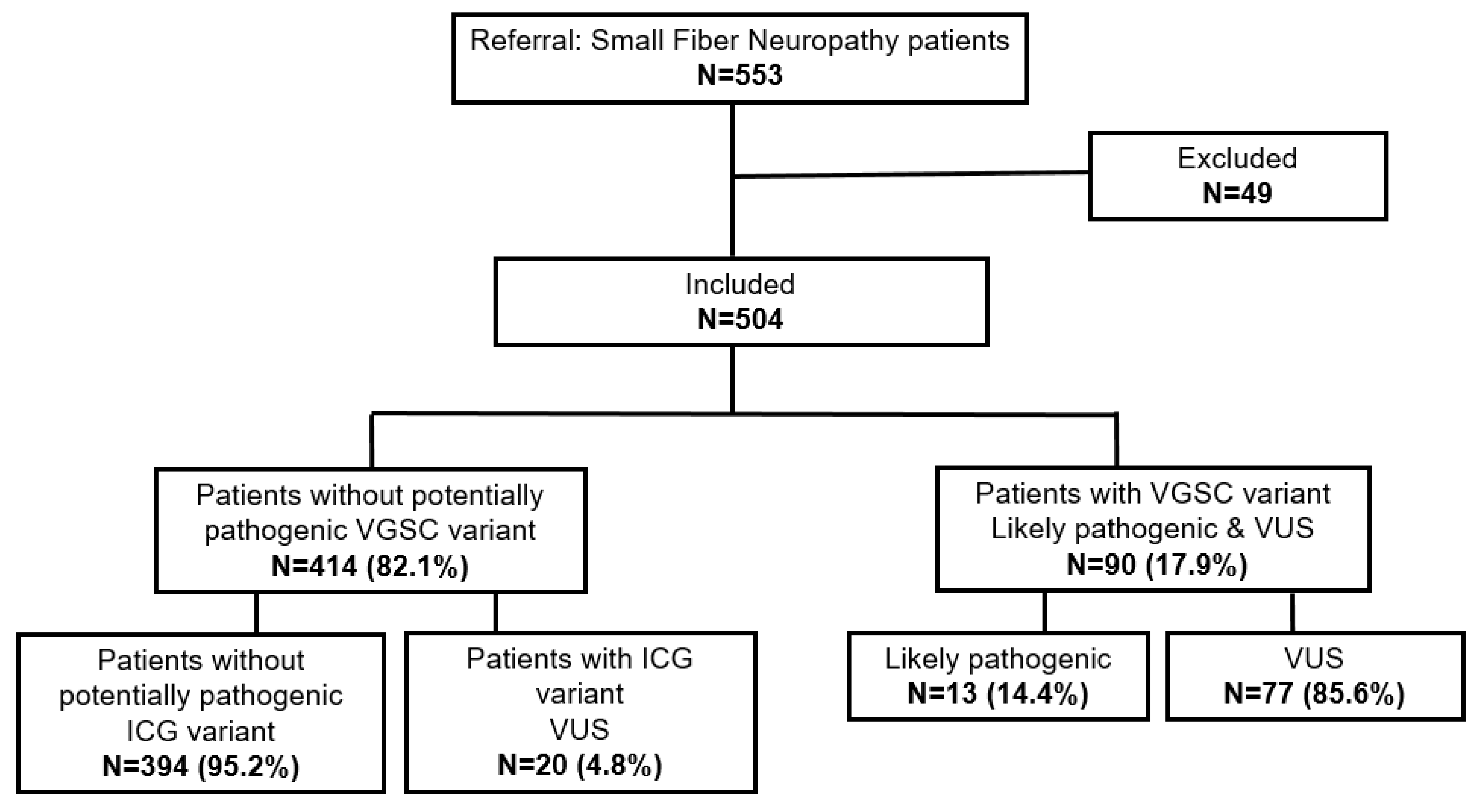
2.1. smMIPs-NGS of Patients with SFN

The performance of smMIPs-NGS, capture efficiency and coverage of targeted exons and exon-flanking sequences (±20 bps) for a 15-ICG panel were assessed as described previously [22]. The average coverage of these regions (>30x/bp) was 93%. The targeted sequences of ANO3 (2946 bp), KCNA2 (1500 bp), KCNA4 (1962 bp), KCNK18 (1155 bp), KCNQ3 (2619 bp), KCNQ5 (2856 bp), TRPA1 (3360 bp), TRPM8 (3315 bp), TRPV1 (2520 bp) and TRPV4 (2616 bp) had an average coverage >90%, and for five targeted gene areas (ANO1 (2961 bp), HCN1 (2673 bp), KCNN1 (1632 bp), KCNS1 (1581 bp), TRPV3 (2376 bp)) the average coverage was at least 84%. Four exons (exon 1, exon 12 and exon 20 of ANO1, and exon 3 of TRPV1) had poor coverage (<20x/bp) or were completely missing. The calculated capture efficiency of each probe and coverage of on-target regions was reproducible per region, per run and between samples. A total of 49 out of 553 samples were excluded due to low quality of DNA, insufficient number of reads or incomplete clinical information (Figure 1).

2.2. Genetic Variants Identified in Ion Channel Genes

Among 414 patients negative for SCN3A, SCN7A-SCN11A and SCN1B-SCN4B screening, 19 different potentially pathogenic variants were identified in ICGs. All detected variants were classified as VUS and localized in seven ICG genes: ANO3, KCNK18, KCNQ3, TRPA1, TRPM8, TRPV1 and TRPV3. In total, two missense variants were detected in ANO3, one frameshift variant leading to a shorter protein in KCNK18, two missense in KCNQ3, four missense and two nonsense variants in TRPA1, three missense in TRPM8, three missense in TRPV1, one splice variant in the donor site of intron 9 and one missense in TRPV3 (Table 1). All of them were heterozygous. The detected nonsense/frameshift variants were predicted either to cause a shortening of the protein length due to a premature stop codon or, most likely, cause nonsense-mediated decay (NMD) of the mRNA, while the splice site variant in the donor site of intron 9 (TRPV3 c.1242+1G>A) was predicted to alter the protein sequence via alternative splicing.
Although the best predictable functional effect will be lack of the protein due to NMD or truncated, nonfunctional polypeptides (KCNK18 c.1107del, TRPA1 c.1177C>T and c.1954C>T), the substitution in conserved functional areas of the protein may affect proper functioning. Three variants (TRPV1 c.1348A>G and c.1735C>T and TRPV3 c.2006T>C) were found in transmembrane domains. One variant (ANO3 c.2656A>T) was detected in a linker between transmembrane domains, three variants were located in ankyrin repeats (ANK) (TRPA1 c.932C>A and c.980A>G and TRPV1 c.914T>G), three in the N-terminus (TRPM8 c.665A>G and c.1102C>T), four in the C-terminus (ANO3 c.3100G>C, KCNQ3 c.1885G>A and c.1706A>G, TRPA1 c.3136A>G and TRPM8 c.2945C>T) and one in the inositol-phosphate binding site of TRPA1 (c.3136A>G).
Each ICG variant was detected in one patient, with the exception of TRPA1 c.3136A>G, which was present in two siblings affected by SFN (Table 1). Most of the reported ICG variants were novel (n = 16/19, 84%). The TRPA1 c.1954C>T variant has been seen before in a German painful-diabetic-peripheral-neuropathy patient [22]. The majority of the identified (n = 14/19, 73.7%) variants were located in TRP genes. No potential causing variant was identified in eight genes from our gene panel: ANO1, HCN1, KCNA2, KCNA4, KCNQ5, KCNN1, KCNS1 and TRPV4. None of the patients with an identified VGSC variant had an additional variant in one of the other ICGs (Figure 1).

2.3. Patients with SFN with an ICG Variant Compared to Patients without ICG/VGSC Variant

We did not observe statistically significant differences between patients with a potentially pathogenic ICG variant compared to patients without such a variant (no ICG/VGSC variant) in relation to mean age of onset of neuropathy (52.2 ± 13.3 vs. 54.3 ± 13.9 years old, p = 0.786), neuropathy duration (n = 12, 4.4 ± 5.0 years vs. n = 277, 8.1 ± 9.3, p = 0.183), positive family history (n = 5, 38.5% vs. n = 50, 22.2%, p = 0.088), abnormal TTT (n = 9, 100% vs. n = 280, 91.8%, p = 1) and abnormal skin biopsy (n = 14, 77.8% vs. n = 129, 42%, p = 0.0056) (Table 2). Detailed clinical characteristics of patients with SFN and an ICG variant are available in the Supplementary Materials.
During the clinical assessment, multiple questionnaires, including the SFN Symptom Inventory Questionnaire (SFN-SIQ) [23] and the Visual Analogue Scale, were given to the patients (Table 3). All analyzed patients that completed the questionnaire reported severe pain; however, individuals carrying a potentially pathogenic ICG variant (n = 7) had higher maximal pain scores VAS = 9.36 ± 0.72 compared to those in the group without such an ICG/VGSC variant (n = 204, VAS = 7.47 ± 2.37 (Table 3)). Each individual with ICG reported maximal pain above the mean maximal pain calculated for patients without an ICG/VGSC variant. The patient with an ANO3 variant reported maximal pain (VAS = 9.7), the patient with KCNQ3 c.1885G>A variant reported VAS = 8.7, and the patient with KCNQ3 c.1706A>G reported VAS = 8.5. Interestingly, three out of four TRPA1-positive patients evaluated maximal pain as VAS = 10, which is the highest possible value in the scale. Only one patient carrying the TRPA1 variant, specifically c.2065A>G, reported VAS = 8.6. Therefore, the maximal pain scores in the group of patients having variants in the same gene were higher than those in the subgroup without an ICG/VGSC variant (Table 3).
Several features, including SFN-specific symptoms like a changed sweating pattern, diarrhea, constipation, micturition problems, dry eyes, dry mouth, orthostatic dizziness, palpitations, hot flashes, sensitive leg skin, burning feet, sheet intolerance legs and restless legs were evaluated for the SFN patients (Table 4 and Table 5). All patients with an ICG variant for which these data were available (n = 6) reported often or always burning feet and the restless legs symptom. Next to that, the most commonly mentioned complaint among patients with ICG variants was sheet intolerance on legs, which presented often or always (n = 5/6, 83.3%) and sometimes (n = 1/6, 16.7%) (Table 6). However, burning feet, sheet intolerance and restless legs were also the most commonly mentioned features in patients without an ICG/VGSC variant. Burning feet presented always and often (n = 213, 79.1%), restless legs presented always and often (n = 156, 57.4%) and sheet intolerance presented always and often (n = 126, 46.3% (Table 7)). Statistical significance was not reached for any of investigated features.

3. Discussion

3.1. Summary of ICG Screening in Patients with Small Fiber Neuropathy

In 414 patients with no (potentially) pathogenic variants in the SCN3A, SCN7A-SCN11A and SCN1B-SCN4B genes, twenty patients (4.8%) had one potentially pathogenic heterozygous variant in ICG. The detected variants were located in seven genes and were present in two patients (0.5%) in ANO3, one (0.2%) in KCNK18, two (0.5%) in KCNQ3, seven (1.7%) in TRPA1, 3 (0.7%) in TRPM8, three (0.7%) in TRPV1 and two (0.5%) in TRPV3. Most of the detected variants were novel; however, three variants have been reported before. The ANO3 c.3100G>C variant is present in the Human Gene Mutation Database (HGMD) and described as likely disease-causing with questionable pathogenicity in primary torsion dystonia [24]. Two KCNQ3 variants have been reported in ClinVar; c.1885G>A was reported two times, once as a VUS in benign familial neonatal seizures and as once as likely benign without linking to a specific phenotype, while the c.1706A>G variant was reported as a VUS, and the phenotype was not provided. Our patients did not have complaints typical for dystonia and familial neonatal seizures.
In our study population, each ICG variant was detected only once, with exception of the TRPA1 c.3136A>G variant present in a brother and sister with SFN, which was considered as one independent finding. None of the identified ICG variants have been reported before in neuropathic pain, except for the TRPA1 c.1954C>T variant, which we published recently, present in a 73-year-old male diagnosed with painful DN [22].
The frequency of potentially pathogenic ICG variants in our population (4.8%) was slightly lower than the frequency in patients with painful DN (5.4%) [22]. The most frequent variants in the SFN cohort were located in TRP genes, in total in fifteen patients (3.6%), which is consistent with data obtained for painful DN, where seven individuals (3.3%) had identified VUS TRP variants, three (1.4%) in TRPA1, three (1.4%) in TRPM8, one (0.5%) in TRPV4. Different heterozygous missense VUS were also present in three (1.4%) painful DN patients in ANO3 and two different KCNK18 variants leading to a premature stop codon in two (0.9%) individuals with painful DN [22]. Therefore, our data suggest that although the same genes may be involved, different variants underlay SFN and painful DN. No potentially pathogenic variants were detected in eight genes: ANO1, HCN1, KCNA2, KCNA4, KCNQ5, KCNN1, KCNS1 and TRPV4. Five of them, KCNA2, KCNA4, KCNQ5, KCNN1 and KCNS1, were also negative for screening in a diabetic neuropathy population [22]. Interestingly in an SFN cohort, variants have been detected in three genes: KCNQ3, TRPV1 and TPRV3, which did not appear in painful DN [22]. Therefore, although it seems that ICG variants can play a role in DN and SFN, it cannot be concluded due to the limited cohort sizes that specific ICG or ICG genes are responsible for either clinical manifestation. The list of genes, however, is a promising candidate for future research.

3.2. VUS Meaning in Context of Protein Function

All detected variants were classified as VUS, which reflects the limitations of bioinformatics analysis of novel variants in absence of functional data or segregation in the family [25,26]. In this study, we only relied on in silico prediction of pathogenicity, therefore, it is difficult to draw a definite conclusion about variant causality, which demands further validation. However, it is likely that a number of these VUS will change to (possibly) pathogenic, as they clearly have a strong impact on the protein. The nonsense/frameshift/splice site variants cause altered protein length or protein absence due to nonsense-mediated decay (NMD). Moreover, substitutions are present in conserved, functionally relevant regions of the protein, most likely affecting protein function, like channel opening and heat sensitivity (variants in the transmembrane domain) [27], channel gating and desensitization (variants in the linker between transmembrane domains) [28], protein–protein interactions (variation in ANK) [29,30], protein stability and folding (variants in N- and C-terminus) [31] and binding sites, such as the inositholphospathe binding site in the case of TRPA1 c.3136A>G, leading to channel inactivation [32].

3.3. ICG Variants in Relation to Patients’ Clinical Manifestations

3.3.1. Anoctamin 3

Anoctamin 3 is a member of the Ca2+-activated chloride channel family involved in pain processing and thermoregulation [33]. ANO3 indirectly inhibits pain signaling via enhancing sodium-activated potassium (SLACK) channels in DRG [21]. Since ANO3 knockout results in a decreased pain threshold in tested rats, GOF variants are expected in pain-related phenotypes [21]. Consistent with that, two heterozygous missense variants with predicted GOF were detected in individuals affected with painful DN [22]. In our study cohort of SFN patients, two heterozygous missense variants (ANO3 c.2656A>T and c.3100G>C) have been identified in two unrelated subjects. Both of them reported burning pain, allodynia and sheet intolerance. TTT was abnormal in both cases; moreover, a female patient identified cold temperature as a pain-provoking factor. Altogether, it may link the patients’ clinical manifestation with disrupted thermoregulation.

3.3.2. Potassium Channels

Potassium channels are important in regulation of nociceptor excitability in sensory neurons [34]. A heterozygous deletion of one nt in KCNK18 c.1107del leading to a frameshift and a shortened C-terminus has been identified in an SFN patient. Although missense and frameshift mutations with a premature stop codon in KCNK18 are linked mainly to migraine, some studies indicate that downregulation of KCNK18 contributes to neuropathic pain [35,36]. Two patients from our cohort harbored two different heterozygous missense variants in KCNQ3 c.1885G>A and c.1706A>G. Interestingly, both of them had a similar phenotype with numb sensation and burning pain in the feet. In both cases, pain complaints were getting worse at rest. Additionally, both patients had disturbed warm and cold sensation in their feet. However, one patient reported that cold increased and warmth reduced pain symptoms, while the other patient did not indicate a temperature influence on pain. GOF mutations in KCNQ3 are recognized as contributors to pain resilience [37], therefore, a loss-of-function (LOF) effect of detected variants would be expected in our patients with SFN and neuropathic pain.

3.3.3. TRP Channels

Variants located in the TRP genes constitute the largest group of variants identified in our study cohort. TRP channels constitute a large, divergent gene family with a well-documented role in mediating nociceptive behaviors in response to thermal, chemical and osmotic stimuli [38]. In total, six variants were detected in TRPA1 (two nonsense and four missense), three missense variants in TRPM8, three missense in TRPV1 and two variants in TRPV3 (one missense and one splice variant). TRPA1 channel activity has been functionally linked to pain hypersensitivity, cold hyperalgesia and mechanical nociception in inflammatory and neuropathic pain models [39,40,41]. The GOF mutation N855S in TRPA1 has been associated with familial episodic pain syndrome. The TRPA1 R919* variant is segregating in a family with the cram-fasciculation syndrome [42,43]. Interestingly, the R919* GOF effect was suggested based on clinical improvement with carbamazepine; however, it was not demonstrated whether the truncated TRPA1 protein was expressed at all [43]. In our study, one patient heterozygous for the missense VUS c.980A>G had complaints of numb sensation and painful cramps in the fingers, lower legs and feet, worsening after exercise. Several VUS leading to premature stop codons or nonsense-mediated RNA-decay variants have been reported in painful DN [22], and with the presence of two nonsense variants in our patient cohort (c.1177C>T and c.1954C>T in painful DN), a LOF mechanism would in our opinion be more likely. The clinical manifestation in the patients with TRPA1 VUS was diverse in our cohort; however, the most common complaints were numb sensation, tingling and burning pain. Four out of seven patients reported pain increase after exercise, and all the patients with performed TTT had abnormal TTT values.
Three different heterozygous VUS have been detected in patients with disturbed thermal sensation (abnormal TTT or QST) in TRPM8. Additionally, the patient with c.665A>G VUS reported increased pain due to warm temperature. As TRPM8 is a modulator of cold sensation and a potential target for neuropathic pain treatment [38], those variants remain interesting candidates for functional validation and determine a GOF and LOF effect.
TRPV1 and TRPV3 are heat-activated proteins involved in thermosensation and pain perception [38]. In addition to that, TRPV3 activation plays a role in enhanced itch sensation [44]. In our study, two patents had a TRPV3 variant (c.20006T>C combined with itch episodes and a splice-site variant c.1242+1G>A r.spl?), and three patients were heterozygous for a missense variant in TRPV1 (c.914T>G or c.1348A>G or c.1735C>T). Two of them reported increased pain complaints during rest, which might interfere with night rest. Interestingly, no pain change was reported in response of warm/cold temperature. The literature indicates that upregulation of TRPV1 increases amplification of pain signals [38]. Consistently with that, the GOF Q85R mutation of TRPV1 has been linked to neuropathic pain [45]. On the other hand, TRPV1 missense variants localized in transmembrane domains have been reported in diabetic neuropathy patients without pain [22], which highlights the need of functional variant validation before drawing a definite conclusion.

3.4. Patients with an ICG Variant Had More Severe Clinical Manifestations and Higher Pain Scores

Due to the small number of patients with an ICG variant, it is difficult to draw a definite conclusion whether these patients had more severe clinical manifestations in relation to basic clinical features or SFN-SIQ when compared to those of patients without an ICG/VGSC variant. It seems that patients with an ICG variant tended to have more frequent abnormal skin biopsies and a shorter duration of neuropathy; however, statistical significance was not reached for any of these features. There is evidence implicating enhanced Na+ influx produced by gain-of-function Nav1.7 mutations as a trigger for calcium-importing reverse Na+/Ca2+ exchange that can contribute to axonal degeneration [46]; further work will be needed to determine whether ionic imbalances due to any of the variants found in this study are injurious to axons. Nevertheless, patients with an ICG variant reported higher maximal pain VAS = 9.36 ± 0.72 than patients without an ICG/VGSC variant VAS = 7.47 ± 2.37 did. All of the patients with an ICG variant had severe mean maximal pain VAS ≥ 8.5, always higher than the mean calculated for patients without an ICG/VGSC variant. Interestingly, patients with TRPA1 reported very severe pain (mean max pain VAS = 9.65 ± 0.7), and three of them gave VAS = 10, which is the highest possible score. These data are consistent with a previous study showing that mean maximal pain was higher for painful DN patients with an ICG variant vs. painful DN without an ICG variant [22].

4. Material and Methods

4.1. Study Population

From June 2014 to September 2016, 553 patients diagnosed with SFN were recruited at Maastricht University Medical Center+ (MUMC+), Maastricht, The Netherlands and at Fondazione I.R.C.C.S. Istituto Neurologico Carlo Besta (FINCB), Milan, Italy for this study. MUMC+ is a tertiary referral center for SFN in the Netherlands, and FINCB is a national referral center for several neurological conditions, including SFN. Medical history and clinical data were collected and recorded as described previously [12]. The diagnosis of SFN was confirmed based on clinical symptoms of SFN combined with reduced intraepidermal nerve fiber density (IEND) in skin biopsy without signs of large fiber involvement and/or abnormal temperature threshold testing (TTT) and/or nerve conduction study (NCS) [47]. The assessment of neuropathic pain was performed using the visual analog scale (VAS), where the scores are recorded by marking a value on a 10 cm line in which 0 represents “no pain” and 10 is “the worst pain that you can imagine”, and the 11-point Numerical Rating Scale (PI-NRS) ranging from 0 to 10, where 0 means “no pain” and 10 refers to “the worst pain imaginable” [48,49]. Only patients with diagnosed SFN and neuropathic pain with VAS > 3 were included. In this study, we focused on patients negative for SCN3A, SCN7A-SCN11A and SCN1B-SCN4B variant screening. Most of the included patients (n = 348, 69%) were Dutch, while 156 (31%) patients were from Italy. The mean age of recruited SFN patients was 54.1 years (SD 14.2 years), with the mean age of complaints onset of 42.2 years (SD 13.6 years). More females than males were present in the study population. There were 59.5% (n = 300) of females and 40.5% (n = 204) of males. In both populations, females were over-represented, with 59.8% (n = 208) females in the Dutch cohort and 59% (n = 92) females in the Italian cohort, respectively. Twenty-three percent of the patients (n = 65) reported positive cases of neuropathy in the family, while negative family history was registered for 77% (n = 217). The data for familial cases of neuropathy was incomplete for 222 patients. More than one-third of the recruited patients had an abnormal skin biopsy (n = 120, 36.5%).

4.2. DNA Extraction and Storage

Genomic DNA was from blood samples using a NucleoSpin8 Blood Isolation kit (Macherey-Nagel, Düren, Germany) or a QIAamp DNA Blood Maxi Kit, Puregene® Blood Core Kit (Qiagen, Hilden, Germany). DNA extraction was performed according to the manufacturers’ instructions and stored in −20 °C.

4.3. smMIPs-NGS of Peripheral Ion Channels and Data Analysis

The smMIP-NGS of exonic regions with ±20 bps exon-flanking sequences of the 15 ion channel genes (Table 8) was performed according to a previously established protocol [50]. The detailed description of the technique, smMIPs characteristics, number of probes per targeted coding region per gene and smMIPs-NGS pipeline used for data analysis have been described before [22].

4.4. Variant Classification

Exonic and ±20 bp exon-flanking intron variants with >20x coverage and an alternative variant call of >30% were included for the analysis. The SNPs with high frequency, defined as dbSNP > 5%, ExAC > 5% and >1% in our Dutch in-house database of 12,244 exomes, were excluded from the analysis. All variants were examined individually, including BAM file visualization and variant interpretation performed in Alamut Visual software (Interactive Biosoftware, Rouen, France). Variants were classified according to the practice guidelines of the Association for Clinical Genetic Science (ACGS) [51].

4.5. Statistical Analysis

The analysis of continuous variables was performed using the independent Student’s t-test. Categorical variables were analyzed using the chi-square test or Fisher’s exact test in the case of small counts <5. Statistical significance of <0.05 was applied.

5. Conclusions

In a well-characterized cohort of patients with SFN, we identified twenty patients (4.8%) carrying a potentially pathogenic variant in ICG. Patients with an ICG variant might have more severe pain than patients without a (potentially) pathogenic ICG/VGSC variant. This complements previous results of the analysis of the same ICG in a large cohort of painful and painless DN. Moreover, it extends the list of potential pain-related genes, reporting variants in KCNQ3, TRPV1 and TPRV3 that were not present in painful DN. Apparently, ICGs are fundamental for both SFN and DN, although the numbers are too small to unambiguously identify gene-or variant-specific manifestations. Although all of the variants were classified as VUS and the consequences of the variation cannot be certainly predicted in silico, they are still promising risk factors for neuropathic pain as well as promising gene candidates for future research.

Supplementary Materials

The following supporting information can be downloaded at: https://www.mdpi.com/article/10.3390/ijms232214095/s1.

Author Contributions

M.Ś.: conceptualization; data curation; formal analysis; investigation; writing—original draft; writing—review and editing. R.A.: conceptualization; data curation; formal analysis; supervision; validation; writing—review and editing. M.M.: investigation; methodology; software; writing—review and editing. E.S.: investigation; methodology; software; writing—review and editing. B.T.A.d.G.: data curation; investigation; writing—review and editing. M.S.: data curation; investigation; writing—review and editing. J.G.J.H.: conceptualization; data curation; investigation; writing—review and editing. P.L.: formal analysis; methodology; software; writing—review and editing. S.G.W.: conceptualization; funding acquisition; resources; writing—review and editing. G.L.: conceptualization; funding acquisition; resources; writing—review and editing. C.G.F.: conceptualization; funding acquisition; resources; supervision; writing—review and editing. H.J.M.S.: conceptualization; supervision; writing—review and editing. M.M.G.: conceptualization; data curation; formal analysis; investigation; methodology; supervision; writing—review and editing. All authors have read and agreed to the published version of the manuscript.

Funding

This work was supported by funding from the Molecule-to-Man Pain Network, a European Commission Multi-Center Collaborative Projects through the European Union’s Horizon 2020 research and innovation program under grant agreement No. 721841 and the European Union seventh framework program for the PROPANE study under grant agreement No. 602273. The funding source had no involvement in study design, data collection, analysis and interpretation of data.

Institutional Review Board Statement

All subjects gave their informed consent for inclusion before they participated in the study. The study was conducted in accordance with the Declaration of Helsinki, and the protocol was approved by the Local Medical Ethical Committee; Comitato Etico Regione Lombardia Sezione Fondazione IRCCS Instituto Neurologico “Carlo Besta” (n. 56, 07 November 2018) and METC azM/UM (METC 17-4-035.1/ivb, 11 May 2017). Patient information was anonymized prior to data analysis.

Informed Consent Statement

Informed consent was obtained from all subjects involved in the study.

Conflicts of Interest

Professor Stephen Waxman received SGW report grants from the European Union’s Horizon 2020 research and innovation program Marie Sklodowska-Curie grant for PAIN-Net, Molecule-to-Man Pain Network (grant no. 721841) and European Union 7th Framework Programme (grant no. 602273) for the PROPANE study, was supported in part by the Rehabilitation Research and Development Service and Biomedical Laboratory Research Service, Department of Veterans Affairs, has served as a consultant to ChromoCell Corp. and Sangamo Therapeutics, and has served on the Scientific Advisory Board of OliPass Corporation, MedTronics and Navega Therapeutics, outside the submitted work.

References

  1. Calvo, M.; Davies, A.J.; Hébert, H.L.; Weir, G.A.; Chesler, E.J.; Finnerup, N.B.; Levitt, R.C.; Smith, B.H.; Neely, G.G.; Costigan, M.; et al. The Genetics of Neuropathic Pain from Model Organisms to Clinical Application. Neuron 2019, 104, 637–653. [Google Scholar] [CrossRef] [PubMed]
  2. Cavalli, E.; Mammana, S.; Nicoletti, F.; Bramanti, P.; Mazzon, E. The neuropathic pain: An overview of the current treatment and future therapeutic approaches. Int. J. Immunopathol. Pharmacol. 2019, 33, 2058738419838383. [Google Scholar] [CrossRef] [Green Version]
  3. Eijkenboom, I.; Sopacua, M.; Hoeijmakers, J.G.J.; de Greef, B.T.A.; Lindsey, P.; Almomani, R.; Marchi, M.; Vanoevelen, J.; Smeets, H.J.M.; Waxman, S.G.; et al. Yield of peripheral sodium channels gene screening in pure small fibre neuropathy. J. Neurol. Neurosurg. Psychiatry 2019, 90, 342–352. [Google Scholar] [CrossRef]
  4. Colloca, L.; Ludman, T.; Bouhassira, D.; Baron, R.; Dickenson, A.H.; Yarnitsky, D.; Freeman, R.; Truini, A.; Attal, N.; Finnerup, N.B.; et al. Neuropathic pain. Nat. Rev. Dis. Prim. 2017, 3, 17002. [Google Scholar] [CrossRef] [PubMed] [Green Version]
  5. McCarberg, B.H.; Billington, R. Consequences of neuropathic pain: Quality-of-life issues and associated costs. Am. J. Manag. Care 2006, 12, S263–S268. [Google Scholar] [PubMed]
  6. Spallone, V.; Greco, C. Painful and painless diabetic neuropathy: One disease or two? Curr. Diabetes Rep. 2013, 13, 533–549. [Google Scholar] [CrossRef]
  7. Nguyen, P.T.; Yarov-Yarovoy, V. Towards Structure-Guided Development of Pain Therapeutics Targeting Voltage-Gated Sodium Channels. Front. Pharmacol. 2022, 13, 842032. [Google Scholar] [CrossRef]
  8. Mogil, J.S. Sources of Individual Differences in Pain. Annu. Rev. Neurosci. 2021, 44, 1–25. [Google Scholar] [CrossRef]
  9. Almomani, R.; Sopacua, M.; Marchi, M.; Ślęczkowska, M.; Lindsey, P.; Greef, B.T.d.; Hoeijmakers, J.G.J.; Salvi, E.; Merkies, I.S.J.; Fadavi, H.; et al. Genetic profiling of sodium channels in painful and painless diabetic and idiopathic small fiber neuropathy. 2022; in preparation. [Google Scholar]
  10. Huang, J.; Han, C.; Estacion, M.; Vasylyev, D.; Hoeijmakers, J.G.; Gerrits, M.M.; Tyrrell, L.; Lauria, G.; Faber, C.G.; Dib-Hajj, S.D.; et al. Gain-of-function mutations in sodium channel Na(v)1.9 in painful neuropathy. Brain J. Neurol. 2014, 137, 1627–1642. [Google Scholar] [CrossRef] [Green Version]
  11. Faber, C.G.; Lauria, G.; Merkies, I.S.; Cheng, X.; Han, C.; Ahn, H.S.; Persson, A.K.; Hoeijmakers, J.G.; Gerrits, M.M.; Pierro, T.; et al. Gain-of-function Nav1.8 mutations in painful neuropathy. Proc. Natl. Acad. Sci. USA 2012, 109, 19444–19449. [Google Scholar] [CrossRef] [PubMed]
  12. Faber, C.G.; Hoeijmakers, J.G.; Ahn, H.S.; Cheng, X.; Han, C.; Choi, J.S.; Estacion, M.; Lauria, G.; Vanhoutte, E.K.; Gerrits, M.M.; et al. Gain of function Naν1.7 mutations in idiopathic small fiber neuropathy. Ann. Neurol. 2012, 71, 26–39. [Google Scholar] [CrossRef] [PubMed]
  13. Alsaloum, M.; Estacion, M.; Almomani, R.; Gerrits, M.M.; Bönhof, G.J.; Ziegler, D.; Malik, R.; Ferdousi, M.; Lauria, G.; Merkies, I.S.; et al. A gain-of-function sodium channel β2-subunit mutation in painful diabetic neuropathy. Mol. Pain 2019, 15, 1744806919849802. [Google Scholar] [CrossRef] [PubMed] [Green Version]
  14. Hoeijmakers, J.G.; Faber, C.G.; Merkies, I.S.; Waxman, S.G. Painful peripheral neuropathy and sodium channel mutations. Neurosci. Lett. 2015, 596, 51–59. [Google Scholar] [CrossRef]
  15. Dib-Hajj, S.D.; Rush, A.M.; Cummins, T.R.; Hisama, F.M.; Novella, S.; Tyrrell, L.; Marshall, L.; Waxman, S.G. Gain-of-function mutation in Nav1.7 in familial erythromelalgia induces bursting of sensory neurons. Brain J. Neurol. 2005, 128, 1847–1854. [Google Scholar] [CrossRef]
  16. Basso, L.; Altier, C. Transient Receptor Potential Channels in neuropathic pain. Curr. Opin. Pharmacol. 2017, 32, 9–15. [Google Scholar] [CrossRef]
  17. Busserolles, J.; Tsantoulas, C.; Eschalier, A.; López García, J.A. Potassium channels in neuropathic pain: Advances, challenges, and emerging ideas. Pain 2016, 157 (Suppl. S1), S7–S14. [Google Scholar] [CrossRef]
  18. Rivolta, I.; Binda, A.; Masi, A.; DiFrancesco, J.C. Cardiac and neuronal HCN channelopathies. Pflug. Arch. Eur. J. Physiol. 2020, 472, 931–951. [Google Scholar] [CrossRef]
  19. Cho, H.; Yang, Y.D.; Lee, J.; Lee, B.; Kim, T.; Jang, Y.; Back, S.K.; Na, H.S.; Harfe, B.D.; Wang, F.; et al. The calcium-activated chloride channel anoctamin 1 acts as a heat sensor in nociceptive neurons. Nat. Neurosci. 2012, 15, 1015–1021. [Google Scholar] [CrossRef]
  20. Takayama, Y.; Uta, D.; Furue, H.; Tominaga, M. Pain-enhancing mechanism through interaction between TRPV1 and anoctamin 1 in sensory neurons. Proc. Natl. Acad. Sci. USA 2015, 112, 5213–5218. [Google Scholar] [CrossRef] [Green Version]
  21. Huang, F.; Wang, X.; Ostertag, E.M.; Nuwal, T.; Huang, B.; Jan, Y.N.; Basbaum, A.I.; Jan, L.Y. TMEM16C facilitates Na+-activated K+ currents in rat sensory neurons and regulates pain processing. Nat. Neurosci. 2013, 16, 1284–1290. [Google Scholar] [CrossRef] [PubMed]
  22. Ślęczkowska, M.; Almomani, R.; Marchi, M.; de Greef, B.T.A.; Sopacua, M.; Hoeijmakers, J.G.J.; Lindsey, P.; Salvi, E.; Bönhof, G.J.; Ziegler, D.; et al. Peripheral Ion Channel Gene Screening in Painful- and Painless-Diabetic Neuropathy. Int. J. Mol. Sci. 2022, 23, 7190. [Google Scholar] [CrossRef] [PubMed]
  23. Brouwer, B.A.; Bakkers, M.; Hoeijmakers, J.G.; Faber, C.G.; Merkies, I.S. Improving assessment in small fiber neuropathy. J. Peripher. Nerv. Syst. 2015, 20, 333–340. [Google Scholar] [CrossRef] [PubMed]
  24. Zech, M.; Gross, N.; Jochim, A.; Castrop, F.; Kaffe, M.; Dresel, C.; Lichtner, P.; Peters, A.; Gieger, C.; Meitinger, T.; et al. Rare sequence variants in ANO3 and GNAL in a primary torsion dystonia series and controls. Mov. Disord. Off. J. Mov. Disord. Soc. 2014, 29, 143–147. [Google Scholar] [CrossRef] [PubMed]
  25. Perrone, F.; Cacace, R.; van der Zee, J.; Van Broeckhoven, C. Emerging genetic complexity and rare genetic variants in neurodegenerative brain diseases. Genome Med. 2021, 13, 59. [Google Scholar] [CrossRef] [PubMed]
  26. Federici, G.; Soddu, S. Variants of uncertain significance in the era of high-throughput genome sequencing: A lesson from breast and ovary cancers. J. Exp. Clin. Cancer Res. 2020, 39, 46. [Google Scholar] [CrossRef] [Green Version]
  27. Grandl, J.; Kim, S.E.; Uzzell, V.; Bursulaya, B.; Petrus, M.; Bandell, M.; Patapoutian, A. Temperature-induced opening of TRPV1 ion channel is stabilized by the pore domain. Nat. Neurosci. 2010, 13, 708–714. [Google Scholar] [CrossRef] [Green Version]
  28. Papke, D.; Grosman, C. The role of intracellular linkers in gating and desensitization of human pentameric ligand-gated ion channels. J. Neurosci. 2014, 34, 7238–7252. [Google Scholar] [CrossRef] [Green Version]
  29. Gaudet, R. A primer on ankyrin repeat function in TRP channels and beyond. Mol. Biosyst. 2008, 4, 372–379. [Google Scholar] [CrossRef] [Green Version]
  30. Mosavi, L.K.; Cammett, T.J.; Desrosiers, D.C.; Peng, Z.Y. The ankyrin repeat as molecular architecture for protein recognition. Protein Sci. Publ. Protein Soc. 2004, 13, 1435–1448. [Google Scholar] [CrossRef] [Green Version]
  31. Krishna, M.M.; Englander, S.W. The N-terminal to C-terminal motif in protein folding and function. Proc. Natl. Acad. Sci. USA 2005, 102, 1053–1058. [Google Scholar] [CrossRef] [PubMed]
  32. Brewster, M.S.J.; Gaudet, R. How the TRPA1 receptor transmits painful stimuli: Inner workings revealed by electron cryomicroscopy. Bioessays 2015, 37, 1184–1192. [Google Scholar] [CrossRef] [PubMed] [Green Version]
  33. Wang, T.F.A.; Chen, C.; Huang, F.; Feng, S.J.; Tien, J.S.; Braz, J.M.; Basbaum, A.I.; Jan, Y.N.; Jan, L.Y. TMEM16C is involved in thermoregulation and protects rodent pups from febrile seizures. Proc. Natl. Acad. Sci. USA 2021, 118, e2023342118. [Google Scholar] [CrossRef] [PubMed]
  34. Tsantoulas, C. Emerging potassium channel targets for the treatment of pain. Curr. Opin. Support Palliat. Care 2015, 9, 147–154. [Google Scholar] [CrossRef] [PubMed]
  35. Zhou, J.; Yang, C.X.; Zhong, J.Y.; Wang, H.B. Intrathecal TRESK gene recombinant adenovirus attenuates spared nerve injury-induced neuropathic pain in rats. Neuroreport 2013, 24, 131–136. [Google Scholar] [CrossRef] [PubMed]
  36. Zhou, J.; Chen, H.T.; Yang, C.X.; Zhong, J.Y.; He, W.Y.; Xiong, Q.M. Reversal of TRESK Downregulation Alleviates Neuropathic Pain by Inhibiting Activation of Gliocytes in the Spinal Cord. Neurochem. Res. 2017, 42, 1288–1298. [Google Scholar] [CrossRef]
  37. Yuan, J.H.; Estacion, M.; Mis, M.A.; Tanaka, B.S.; Schulman, B.R.; Chen, L.; Liu, S.; Dib-Hajj, F.B.; Dib-Hajj, S.D.; Waxman, S.G. KCNQ variants and pain modulation: A missense variant in Kv7.3 contributes to pain resilience. Brain Commun. 2021, 3, fcab212. [Google Scholar] [CrossRef]
  38. Gonzalez-Ramirez, R.; Chen, Y.; Liedtke, W.B.; Morales-Lazaro, S.L. TRP Channels and Pain. In Neurobiology of TRP Channels; Emir, T.L.R., Ed.; CRC Press: Boca Raton, FL, USA, 2017; pp. 125–147. [Google Scholar] [CrossRef]
  39. Obata, K.; Katsura, H.; Mizushima, T.; Yamanaka, H.; Kobayashi, K.; Dai, Y.; Fukuoka, T.; Tokunaga, A.; Tominaga, M.; Noguchi, K. TRPA1 induced in sensory neurons contributes to cold hyperalgesia after inflammation and nerve injury. J. Clin. Investig. 2005, 115, 2393–2401. [Google Scholar] [CrossRef] [Green Version]
  40. Dai, Y.; Wang, S.; Tominaga, M.; Yamamoto, S.; Fukuoka, T.; Higashi, T.; Kobayashi, K.; Obata, K.; Yamanaka, H.; Noguchi, K. Sensitization of TRPA1 by PAR2 contributes to the sensation of inflammatory pain. J. Clin. Investig. 2007, 117, 1979–1987. [Google Scholar] [CrossRef] [Green Version]
  41. McNamara, C.R.; Mandel-Brehm, J.; Bautista, D.M.; Siemens, J.; Deranian, K.L.; Zhao, M.; Hayward, N.J.; Chong, J.A.; Julius, D.; Moran, M.M.; et al. TRPA1 mediates formalin-induced pain. Proc. Natl. Acad. Sci. USA 2007, 104, 13525–13530. [Google Scholar] [CrossRef]
  42. Kremeyer, B.; Lopera, F.; Cox, J.J.; Momin, A.; Rugiero, F.; Marsh, S.; Woods, C.G.; Jones, N.G.; Paterson, K.J.; Fricker, F.R.; et al. A gain-of-function mutation in TRPA1 causes familial episodic pain syndrome. Neuron 2010, 66, 671–680. [Google Scholar] [CrossRef] [PubMed] [Green Version]
  43. Nirenberg, M.J.; Chaouni, R.; Biller, T.M.; Gilbert, R.M.; Paisán-Ruiz, C. A novel TRPA1 variant is associated with carbamazepine-responsive cramp-fasciculation syndrome. Clin. Genet. 2018, 93, 164–168. [Google Scholar] [CrossRef] [PubMed]
  44. Cui, T.T.; Wang, G.X.; Wei, N.N.; Wang, K. A pivotal role for the activation of TRPV3 channel in itch sensations induced by the natural skin sensitizer carvacrol. Acta Pharmacol. Sin. 2018, 39, 331–335. [Google Scholar] [CrossRef] [PubMed] [Green Version]
  45. Yuan, J.H.; Schulman, B.R.; Effraim, P.R.; Sulayman, D.H.; Jacobs, D.S.; Waxman, S.G. Genomic analysis of 21 patients with corneal neuralgia after refractive surgery. Pain Rep. 2020, 5, e826. [Google Scholar] [CrossRef]
  46. Estacion, M.; Vohra, B.P.S.; Liu, S.; Hoeijmakers, J.; Faber, C.G.; Merkies, I.S.J.; Lauria, G.; Black, J.A.; Waxman, S.G. Ca2+ toxicity due to reverse Na+/Ca2+ exchange contributes to degeneration of neurites of DRG neurons induced by a neuropathy-associated Nav1.7 mutation. J. Neurophysiol. 2015, 114, 1554–1564. [Google Scholar] [CrossRef] [Green Version]
  47. De Greef, B.T.A.; Hoeijmakers, J.G.J.; Gorissen-Brouwers, C.M.L.; Geerts, M.; Faber, C.G.; Merkies, I.S.J. Associated conditions in small fiber neuropathy—A large cohort study and review of the literature. Eur. J. Neurol. 2018, 25, 348–355. [Google Scholar] [CrossRef] [Green Version]
  48. Delgado, D.A.; Lambert, B.S.; Boutris, N.; McCulloch, P.C.; Robbins, A.B.; Moreno, M.R.; Harris, J.D. Validation of Digital Visual Analog Scale Pain Scoring With a Traditional Paper-based Visual Analog Scale in Adults. J. Am. Acad. Orthop. Surgeons. Glob. Res. Rev. 2018, 2, e088. [Google Scholar] [CrossRef]
  49. Williamson, A.; Hoggart, B. Pain: A review of three commonly used pain rating scales. J. Clin. Nurs. 2005, 14, 798–804. [Google Scholar] [CrossRef]
  50. Almomani, R.; Marchi, M.; Sopacua, M.; Lindsey, P.; Salvi, E.; Koning, B.; Santoro, S.; Magri, S.; Smeets, H.J.M.; Martinelli Boneschi, F.; et al. Evaluation of molecular inversion probe versus TruSeq® custom methods for targeted next-generation sequencing. PLoS ONE 2020, 15, e0238467. [Google Scholar] [CrossRef]
  51. Richards, S.; Aziz, N.; Bale, S.; Bick, D.; Das, S.; Gastier-Foster, J.; Grody, W.W.; Hegde, M.; Lyon, E.; Spector, E.; et al. Standards and guidelines for the interpretation of sequence variants: A joint consensus recommendation of the American College of Medical Genetics and Genomics and the Association for Molecular Pathology. Genet. Med. Off. J. Am. Coll. Med. Genet. 2015, 17, 405–424. [Google Scholar] [CrossRef]
Figure 1. SFN patients analyzed using smMIP-NGS for (potentially) pathogenic ICG and VGSC variants. ICG, ion channel gene; VGSC, voltage-gated sodium ion channel; VUS, variant with uncertain clinical significance.
Ijms 23 14095 g001
Table 1. Potentially pathogenic variants of ion channel genes identified in patients with SFN (n = 504).
Genec.Position &p.PositionNumber of PatientsClassification According to Richards et.al 2015LocationMAF gnomAD (%)
ANO3c.2656A>Tp.(Ile886Phe)1VUSLinker between transmembrane domain VII and VIII0
c.3100G>Cp.(Gly1034Arg)1VUSC-terminus0.011
KCNK18c.1107delp.(Met370Cysfs*?)1VUSFrame shift starting at codon Met3700.0016
KCNQ3c.1885G>Ap.(Val629Ile)1VUSC-terminus0.052
c.1706A>Gp.(Asp569Gly)1VUSC-terminus0.0018
TRPA1c.932C>Ap.(Thr311Asn)1VUSAnkyrin repeat VIII-containing domain0.044
c.980A>Gp.(Tyr327Cys)1VUSAnkyrin repeat VIII-containing domain0
c.1177C>Tp.(Arg393*)1VUSStop codon in Ankyrin repeat X–containing domain0.018
c.1954C>Tp.(Arg652*)1VUSCytoplasmic domain between ANK repeats and transmembrane domain I0.015
c.2065A>Gp.(Met689Val)1VUSCytoplasmic domain between ANK repeats and transmembrane domain I0.0068
c.3136A>Gp.(Lys1046Glu)2 *VUSinositol-phosphate binding site in C-terminus0.0008
TRPM8c.665A>Gp.(Asn222Ser)1VUSN-terminus0.0004
c.1102C>Tp.(Arg368Trp)1VUSN-terminus0.0016
c.2945C>Tp.(Thr982Met)1VUSC-terminus0.005
TRPV1c.914T>Gp.(Phe305Cys)1VUSAnkyrin repeat V-containing domain0.00054
c.1348A>Gp.(Thr450Ala)1VUSTransmembrane domain I0
c.1735C>Tp.(Arg579Cys)1VUSTransmembrane domain V0.0017
TRPV3c.1242+1G>Ap.? ^1VUSDonor splice site of intron 90.0029
c.2006T>Cp.(Leu669Pro)1VUSTransmembrane domain VI0.0019
c. position, location cDNA; p. position, location in protein; MAF gnomAD, minor allele frequency; VUS, variant with uncertain clinical significance. & Variants detected were annotated according to the guidelines of the Human Genome Variation Society using reference sequence GRCh37 and transcript numbers, NM_001313726.1 (ANO3); NM_181840.1 (KCNK18), NM_004519.3 (KCNQ3), NM_007332.2 (TRPA1), NM_024080.4 (TRPM8), NM_080706.3 (TRPV1), NM_001258205.1 (TRPV3). All detected variants are heterozygous. * Variant detected two times in siblings, ^ changed protein length due to splicing event (loss of donor splice site of intron 9).
Table 2. Comparison of clinical features of patients with small fiber neuropathy and ICG variant vs. no ICG/VGSC variant.
 Patients with SFN and ICG Variant
N = 20
Patients with SFN without ICG/VGSC Variant
N = 394
Mean age at recruitment [years ± SD]52.2 (±13.3)54.2 (±13.9)
Females (n, %)11 (55.0)240 (±60.9)
Males (n, %)9 (45.0)154 (±39.1)
Mean age of onset neuropathy [years ± SD]46.2 (±12.2)47.1 (±13.4)
Duration of neuropathy [years ± SD]4.4 (±5.0)8.1 (±9.3)
Positive family history for neuropathy (n, %)5 (38.5)50 (22.2)
Negative family history for neuropathy (n, %)8 (61.5)175 (77.8)
Normal skin biopsy (n, %)4 (22.2)178 (58)
Abnormal skin biopsy (n, %)14 (77.8)129 (42)
Normal TTT (n, %)0 (0)25 (8.2)
Abnormal TTT (n, %)9 (100)280 (91.8)
ICG, ion channel gene; VGSC, voltage-gated sodium ion channel; SD, standard variation. Patients with incomplete data not included in the table. Differences between patients with SFN and ICG variant vs. patients with SFN without ICG and VGSC variant were not statistically significant.
Table 3. Mean pain scores in patients with small fiber neuropathy.
 Patient with ANO3 Variant
(n = 1)
Patients with KCNQ3 Variant
(n = 2)
Patient with TRPA1 Variant
(n = 4)
Patients with ICG Variant (n = 7)Patients without ICG Variant and without VGSC Variant (n = 204)
Maximal pain
[VAS] [±SD]
9.78.6 (±0.14)9.65 (±0.7)9.36 (±0.72)7.47 (±2.37)
ICG, ion channel gene; VGSC, voltage-gated sodium ion channel; SD, standard variation. Pain evaluated using the visual analog scale (VAS), where 0 represents “no pain” and 10 is “the worst pain that you can imagine”. Patients with incomplete data not included in the table.
Table 4. Clinical features of SFN patients carrying an ICG variant.
GeneVariantGenderOnset ComplaintsNCSTTTIENFDItchMuscle CrampsWarmth InfluenceCold InfluenceExercise InfluenceRest InfluenceTemperature SensationPain SensationAllodyniaSleep Pattern
ANO3(Gly1034Arg)M18na/nnNN--NNnYn
KCNQ3(Val629Ile)M49a/na/na/nNN-Y ↑Y ↑Y ↑n-Yn
KCNQ3(Asp569Gly)M46na/nnNY---Y ↑--Y-
TRPA1(Arg393*)M50a/na/na/nNYNNY ↑-n-Ya/n
TRPA1(Arg652*)F17na/nnNNY ↑Y ↑Y ↑NYa/n
TRPA1(Met689Val)F34na/na/nYNY ↑-Y ↑Y ↑-Ya/n
TRPA1(Tyr327Cys)M45n-a/nNY--------
TRPM8(Arg368Trp)M45na/na/nNN----n--
TRPV1(Arg579Cys)M43na/nnYYNN-Y ↑nYa/n
p. position, location in protein; M, male; F, female; NCS, nerve conduction study; n, normal; a/n, abnormal; -, data incomplete; TTT, thermal threshold testing; IENFD, intraepidermal nerve fiber densities; Y, yes; N, no; ↓, decreased; ↑, increased. Patients with incomplete data not included in the table.
Table 5. Clinical features of SFN patients without an ICG/VGSC variant.
FeatureNCS N = 308TTT
N = 305
IENFD
N = 307
Temperature Sensation
N = 204
Pain Sensation
N = 145
Sleep Pattern
N = 130
Normal297, 96.4%25, 8.2%178, 58%120, 58.8%73, 50.3%35, 26.9%
Abnormal11, 3.6%280, 91.8%129, 42%84, 41.2%
(1, 0.5% ↑, 83, 40.7% ↓)
72, 49.7%
(33, 22.8% ↑, 39, 26.9% ↓)
95, 73.1%
FeatureItch
N = 308
Muscle Cramps
N = 308
Warmth Influence
N = 172
Cold Influence
N = 169
Exercise Influence
N = 195
Rest Influence
N = 123
Allodynia
N = 211
yes25, 8.1%56, 18.2%84, 48.8%
(63, 36.6% complains ↑,
21, 12.2% complains ↓)
82, 48.5%
(54, 32% complains ↑,
28, 16.6% complains ↓)
161, 82.6%
(144,. 73.8% complains ↑,
17, 8.7% complains ↓)
54, 68.3%
(45, 36.6% complains ↑,
39, 31.7% complains ↓)
193, 91.5%
no283, 91.9%252, 81.8%88, 51.2%87, 51.5%34, 17.4%39, 31.7%18, 8.5%
NCS, nerve conduction study; n, normal; a/n, abnormal; -, not determined; TTT, thermal threshold testing; IENFD, intraepidermal nerve fiber densities; Y, yes; N, no; ↓, decreased; ↑, increased.
Table 6. Complaints reported in the Small Fiber Neuropathy Symptom Inventory Questionnaire (SFN-SIQ) by SFN patients carrying a variant in ICG.
GeneVariantSweating ChangeDiarrheaConstipationMicturition ProblemDry EyesDry MouthDizziness on StandingPalpitationsHot FlashesHypersensitivity of Leg’s SkinBurning FeetSheet IntoleranceRestless Leg
ANO3(Gly1034Arg)4113342-14444
KCNQ3(Val629Ile)2222121121323
KCNQ3(Asp569Gly)3223222223434
TRPA1(Arg393*)-233132124444
TRPA1(Arg652*)3232133223333
TRPA1(Met689Val)3332443334344
Number value 1–4 expresses frequency of complaints; 1, never; 2, sometimes; 3, often; 4, always; -, not determined.
Table 7. Patients with SFN without a (potentially) pathogenic ICG/VGSC variant reporting Small Fiber Neuropathy Symptom Inventory Questionnaire (SFN-SIQ) complaints.
Frequency of ComplaintSweating Change
N = 268
Diarrhea
N = 272
Constipation
N = 275
Micturition Problem N = 273Dry Eyes
N = 273
Dry Mouth
N = 273
Orthostatic
Dizziness
N = 271
Palpitations
N = 268
Hot Flashes
N = 270
Hypersensitivity of Leg’s Skin
N = 273
Burning Feet
n = 269
Sheet Intolerance
N = 272
Restless Leg
N = 272
never56, 20.9%114, 41.9%115, 41.8%91, 33.3%95, 34.8%54, 19.8%73, 26.9%105, 39.2%86, 31.9%52, 19.0%21, 7.8%70, 25.7%36, 13.2%
sometimes94, 35.1%110, 40.4%86, 31.3%75, 27.5%87, 31.9%97, 35.5%130, 48.0%108, 40.3%94, 34.8%75, 27.5%35, 13.0%76, 27.9%80, 29.4%
often85, 31.7%44, 16.2%55, 20.0%80, 29.3%65, 23.8%91, 33.3%59, 21.8%54, 20.1%86, 31.9%70, 25.6%101, 37.5%68, 25.0%91, 33.5%
always33, 12.3%4, 1.5%19, 6.9%27, 9.9%26, 9.5%31, 11.4%9, 3.3%1, 0.4%4, 1.5%76, 27.8%112, 41.6%58, 21.3%65, 23.9%
N, number of patients that completed the SFN-SIQ related question.
Table 8. Gene panel.
Ion Channel FamilyGeneOMIM NumberFull Gene Name
AnoctaminsANO1610108Anoctamin 1, calcium activated chloride channel
ANO3610110Anoctamin 3
Non-selective
cation channels
HCN1602780Hyperpolarization activated cyclic nucleotide-gated potassium channel 1
Potassium channelsKCNA2176262Potassium voltage-gated channel, shaker-related subfamily, member 2
KCNA4176266Potassium voltage-gated channel, shaker-related subfamily, member 4
KCNK18613655Potassium channel, subfamily K, member 18
KCNN1602982Potassium intermediate/small conductance calcium-activated channel, subfamily N, member 1
KCNQ3602232Potassium voltage-gated channel, KQT-like subfamily, member 3
KCNQ5607357Potassium voltage-gated channel, KQT-like subfamily, member 5
KCNS1602905Potassium voltage-gated channel, delayed-rectifier, subfamily S, member 1
Transient receptorsTRPA1604775Transient receptor potential cation channel, subfamily A, member 1
TRPM8606678Transient receptor potential cation channel, subfamily M, member 8
TRPV1602076Transient receptor potential cation channel, subfamily V, member 1
TRPV3607066Transient receptor potential cation channel, subfamily V, member 3
TRPV4605427Transient receptor potential cation channel, subfamily V, member 4
Publisher’s Note: MDPI stays neutral with regard to jurisdictional claims in published maps and institutional affiliations.

