Article

A Highly Unstable and Elusive Plasmid That Encodes the Type III Secretion System Is Necessary for Full Virulence in the Marine Fish Pathogen Photobacterium damselae subsp. piscicida

Departamento de Microbioloxía e Parasitoloxía, Instituto de Acuicultura, Universidade de Santiago de Compostela, 15782 Santiago de Compostela, Spain
*
Author to whom correspondence should be addressed.
These authors contributed equally to this work.
Current address: Department of Biology, College of Science, Al-Hussein Bin Talal University, Ma’an 71111, Jordan.
§
Current address: International Medical Research Center (iMReC), Aqaba 77110, Jordan.
Int. J. Mol. Sci. 2022, 23(9), 4729; https://doi.org/10.3390/ijms23094729
Submission received: 8 April 2022 / Revised: 21 April 2022 / Accepted: 22 April 2022 / Published: 25 April 2022
(This article belongs to the Collection Microbial Virulence Factors)

Abstract

:
The marine bacterium Photobacterium damselae subsp. piscicida (Pdp) causes photobacteriosis in fish and important financial losses in aquaculture, but knowledge of its virulence factors is still scarce. We here demonstrate that an unstable plasmid (pPHDPT3) that encodes a type III secretion system (T3SS) is highly prevalent in Pdp strains from different geographical origins and fish host species. We found that pPHDPT3 undergoes curing upon in vitro cultivation, and this instability constitutes a generalized feature of pPHDPT3-like plasmids in Pdp strains. pPHDPT3 markers were detected in tissues of naturally-infected moribund fish and in the Pdp colonies grown directly from the fish tissues but were undetectable in a fraction of the colonies produced upon the first passage of the primeval colonies on agar plates. Notably, cured strains exhibited a marked reduction in virulence for fish, demonstrating that pPHDPT3 is a major virulence factor of Pdp. The attempts to stabilize pPHDPT3 by insertion of antibiotic resistance markers by allelic exchange caused an even greater reduction in virulence. We hypothesize that the existence of a high pressure to shed pPHDPT3 plasmid in vitro caused the selection of clones with off-target mutations and gene rearrangements during the process of genetic modification. Collectively, these results show that pPHDPT3 constitutes a novel, hitherto unreported virulence factor of Pdp that shows a high instability in vitro and warn that the picture of Pdp virulence genes has been historically underestimated, since the loss of the T3SS and other plasmid-borne genes may have occurred systematically in laboratories for decades.

1. Introduction

Photobacterium damselae subsp. piscicida (hereafter dubbed Pdp), a bacterium of the family Vibrionaceae, was first isolated in 1963 from natural populations of white perch (Morone americana) and striped bass (Morone saxatilis) affected by an unknown bacterium in the Chesapeake Bay (Maryland, USA) [1]. Since then, Pdp has become one of the most devastating pathogens in marine aquaculture, causing a septicaemic disease termed pasteurellosis, pseudotuberculosis, or photobacteriosis, affecting fish species of high commercial value [2,3], but vaccination strategies are far from being satisfactorily effective [4]. Hence, it is necessary to gain a deeper understanding of the underlying mechanisms of Pdp pathogenicity. Infections by Pdp are characterized by a bacteremia with disseminated tissue necrosis and pronounced cytopathology [5]. The best characterized virulence factor of Pdp is AIP56, a systemically disseminated AB-type toxin that induces apoptosis of macrophages and neutrophils [6,7,8] and is secreted through the type II secretion system [9]. AIP56 is encoded within the small virulence plasmid pPHDP10 [6] that has a high prevalence and stability in Pdp populations [10]. In addition, many Pdp isolates harbor pPHDP70, a ~70 kb plasmid that encodes the functions for synthesis and utilization of siderophore piscibactin [11]. The curing of pPHDP70 under laboratory conditions caused a reduction of virulence in a turbot fish model, but the role of piscibactin as a virulence factor needs additional investigation. A recent study reported that pPHDP70-like plasmids were present in 91 of 103 Pdp isolates tested, demonstrating a high prevalence, but not ubiquity in collections of isolates of this pathogen [10].
Despite being a pathogen recognized since 1964 and with a great financial impact on marine aquaculture, sequencing of the Pdp genome was not accomplished until 2017, when the genomes of two Spanish, one Japanese and one American isolate were published in different studies [12,13,14]. Recently, the high quality genome sequences of two Australian Pdp isolates have been published [15]. These studies clearly showed that Pdp genomes contain numerous insertion sequence (IS) elements in a multicopy fashion, accounting for 12–17% of the total genome [12]. As a result of IS proliferation, the Pdp genomes exhibit a large number of pseudogenes and of gene deletions, as well as a dramatic loss of biological functions [12] in comparison with its sibling subspecies Photobacterium damselae subsp. damselae (hereafter dubbed Pdd), a generalist and opportunistic pathogen whose genome has not undergone the massive IS expansion of Pdp [16]. This process of gene decay in Pdp is considered to be associated with a progressive adaptation to a host-dependent lifestyle, and the acquisition of virulence plasmids by horizontal gene transfer is a major driving force in the speciation process that shaped the two subspecies of P. damselae [17].
Recently, the presence of genes of the type III secretion system (T3SS) in two strains, PP3 and SNW-8.1, was published for the first time in Pdp [18]. The type III secretion system (T3SS) is a needle-like macromolecular apparatus used by several Gram negative pathogenic bacteria to inject effector proteins into the target eukaryotic host cells [19,20]. The effectors manipulate host cell physiology and cause diverse cellular responses and cell damage [19,21]. T3SS genes have been reported in several species of fish pathogenic bacteria [22], and specifically in members of the family Vibrionaceae as Vibrio parahaemolyticus [23], Vibrio alginolyticus [24] and Vibrio harveyi [25], among others.
Unexpectedly, a recent study in our laboratory unveiled that the T3SS genes, far from being an exception of some isolates, are highly prevalent in Pdp strains from different hosts and geographical origins isolated between 1980 and 2015 [10]. Notably, the same study warned about the spontaneous loss of T3SS genes in Pdp strain PP3 during the attempts to genetically modify this strain, and the loss of T3SS gene markers vscJ, vopD and vopB occurred simultaneously with the loss of putative plasmid-borne genes traC and parAB, suggesting that the T3SS might be encoded within an unstable genetic element. However, the Illumina-based, draft genome sequence available at that time for Pdp PP3 was highly fragmented due to the presence of transposase genes in a multicopy fashion, which prevented deciphering the genome context of the T3SS genes [10].
In the present study, the sequencing of the Pdp PP3 genome using a PacBio approach has resolved the PP3 genome into two chromosomes and six putative plasmids and unveiled the location of the T3SS within a large plasmid that we dubbed pPHDPT3. This plasmid was highly unstable in culture conditions, its loss occurring as fast as upon a single subculture step on agar plates. In vitro loss of pPHDPT3 markers was demonstrated to constitute a generalized feature of Pdp strains isolated from diseased fish. The virulence for fish of spontaneously cured strains was notably reduced in comparison with the stocks of parental strains that contained plasmid-bearing cells. Collectively, this study provides evidence that pPHDPT3 plasmid constitutes a hitherto unreported virulence factor of Pdp that is prevalent in Pdp populations and is highly unstable in vitro, and warns that the picture of Pdp virulence genes has been historically biased and underestimated.

2. Results

2.1. General Genome Features of Pdp PP3

In a recent study, we found the presence of T3SS-related genes in the draft genome sequences of two Pdp strains PP3 and SNW-8.1 obtained by Illumina sequencing [18]. To investigate the genetic context of T3SS genes in the Pdp genome, we carried out the genome sequencing of these two Pdp strains using a PacBio approach. This resolved the PP3 genome into ten contigs predicted to represent chromosomes I and II and up to six putative plasmids (Table 1). Sequence analysis revealed that the genes for the T3SS are contained within a plasmid of 133,065 bp which was here dubbed pPHDPT3. The remaining contigs corresponded to a pPHDP10-like plasmid, a pPHDP70-like plasmid, a 286-Kb plasmid encoding tetracycline resistance determinants, and two medium-size putative plasmids, one of which was predicted to include three unassembled contigs (Table 1). Surprisingly, the PacBio-derived sequence of strain SNW-8.1 did not contain the sequences of the T3SS and other pPHDPT3 genes (data not shown), as a consequence of pPHDPT3 plasmid instability and curing (see below).
The Pdp PP3 chromosomes I and II exhibit sizes (3.1 and 1.1 Mb, respectively) similar to other Pdp genomes [12,15]. We identified 644 transposase genes in the PP3 genome, belonging to 18 distinct families. Of note, chromosome II was particularly abundant in the density of IS elements. The distribution of ISs within families and replicons in the PP3 genome is summarized in Table 2.
The pPHDP10 plasmid of PP3 (8115 bb) shared the same structure and similar sequence length with homologous plasmids in two recently sequenced Pdp strains from Australia while differing in size and in gene arrangement from the prototype pPHDP10 sequence (9631 bp), first reported in the European Pdp strain MT1415 (Supplementary Figure S1). This small plasmid that encodes AIP56 toxin, a major virulence factor of Pdp, has undergone insertions of IS elements in its structure, being frequent the successive events of nested integration of transposase genes. The observation that all the pPHDP10 versions maintain a similar backbone of conserved genes that have escaped IS-mediated inactivation, clearly suggests that these conserved genes fulfill important functions for plasmid maintenance and/or for bacterial fitness and virulence.
The pPHDP70 plasmid and its variants, encode the functions for the synthesis and utilization of siderophore piscibactin and has been proposed to constitute a virulence factor of Pdp for fish [11]. Initially considered to be a plasmid exclusive to Pdp strains from the European continent, it has been detected in strains from Asia [10] and from Australia [15]. The PP3 genome also contains a pPHDP70-like plasmid, whose size (81,347 bp) and gene structure are highly similar to the plasmids of Australian strains and larger than the pPHDP70 plasmid first reported and sequenced in strain DI21 isolated in Spain (68,686 bp) (Supplementary Figure S2).
IS elements are present in the structure of pPHDP70 plasmids (Supplementary Figure S2), but there are conserved modules that have escaped pseudogenization and IS insertion, indicative of their importance in bacterial fitness and in virulence. The hallmark of these conserved modules is the pathogenicity island that contains the piscibactin siderophore genes. Interestingly, the PP3 and the two Australian pPHDP70-like plasmids (which are larger in size than the DI21 version), contain some genes and pseudogenes related to conjugal transfer (as TraF), which are absent from the DI21 plasmid. These scattered, type IV-related, genes might constitute remnants of an ancient version of pPHDP70 that initially contained the elements to be self-conjugative. The process of gene decay, pseudogenization and gene deletion undergone by Pdp genomes in its adaptation to a host-dependent lifestyle might have eroded the conjugation machinery of the primeval pPHDP70.
PP3 contains a large plasmid of 286 Kb dubbed here as pPHDP286 (Table 1), that harbors two tetracycline resistance genes, tetB and tetM. No close homologs of this plasmid have been previously reported in Pdp, but similarity searches unveiled that it shares large sequence modules with a putative plasmid sequenced in a Pdd isolate (Wu-1) (GenBank Acc. No. CP018299) (Supplementary Figure S3). In accordance with the presence of tetB and tetM genes, we found that Pdp PP3 is resistant to oxytetracycline, an antibiotic widely used in aquaculture, whereas DI21, an extensively studied Pdp strain that does not contain these gene markers [12] tested sensitive to this antibiotic (Supplementary Figure S3). Last, two small putative plasmids of ~37 and 23 kb in the PP3 genome, are highly similar to plasmids previously reported in two Australian strains (AS-16-0540-1 and AS-16-0555-7) (Supplementary Figure S4). One of these plasmids is constituted by contig SRHT02000007.1, whereas the second plasmid is predicted to include the three unassembled contigs SRHT02000001.1, SRHT02000002.1 and SRHT02000003.1.

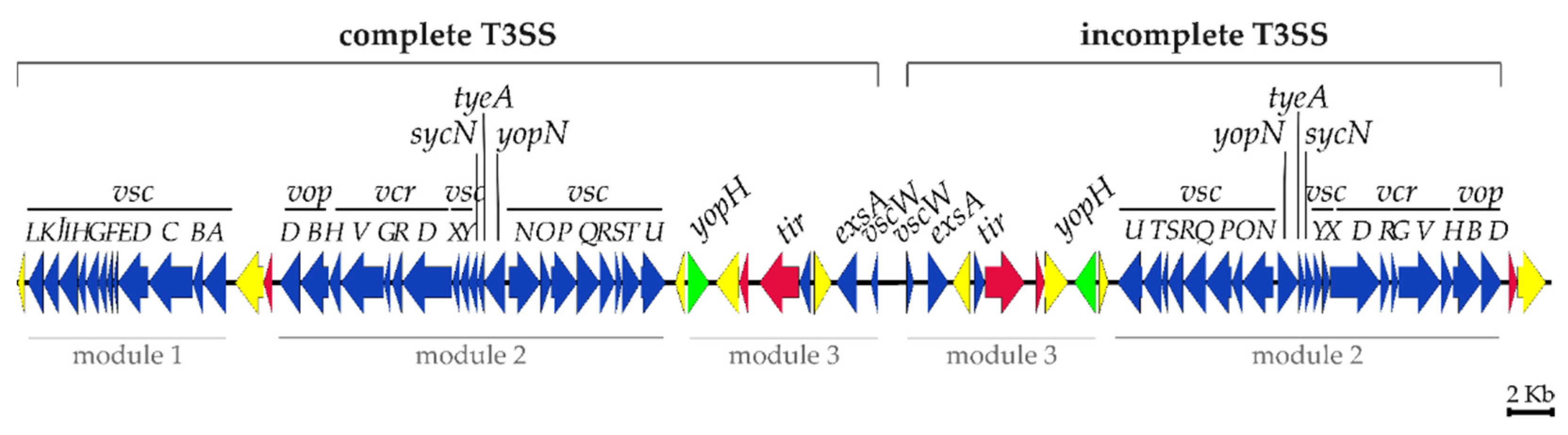
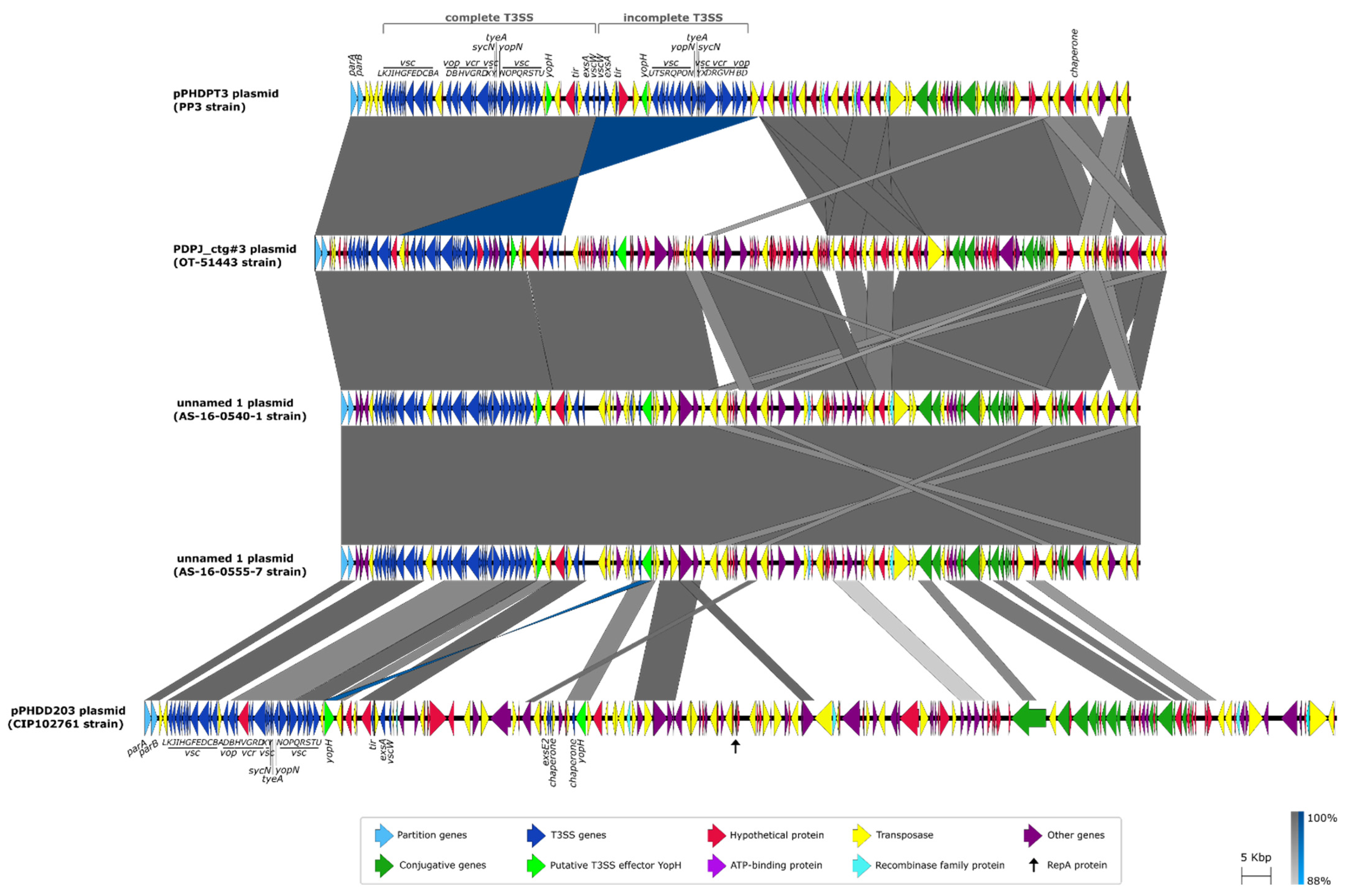
2.2. Sequence Analysis of pPHDPT3, a Highly Unstable Plasmid That Encodes the T3SS Genes in Pdp

As noted above, genome sequencing of PP3 has unveiled the plasmid location of the genes encoding the T3SS, a potential virulence factor of Pdp that has not been functionally characterized so far. The structure and gene organization of the pPHDPT3 sequence obtained with the PacBio approach is shown in Figure 1. pPHDPT3 has an average G+C content of 43%, similar to the host genome (40.9%). A total of 146 coding sequences (CDS) were predicted by the NCBI Prokaryotic Genome Annotation Pipeline, of which 28 were annotated as hypothetical proteins (Supplementary Table S1). Notably, 33 CDS correspond to IS that sum up 29.6 Kb, accounting for 22% of the pPHDPT3 sequence. Six different IS families are present in pPHDPT3, and most of them occur in multicopy (Figure 1; Table 2). IS elements are not uniformly distributed along the plasmid and concentrate in specific regions that separate putative functional modules. The pPHDPT3 IS1 copies have 99% sequence similarity with IS1 of the co-resident plasmids pPHDP10 and pPHDP70, suggesting a high capacity of expansion of this IS, that is present in very high copies in the two Pdp chromosomes, as reported in other Pdp genomes [12,15]. The pPHDPT3 partitioning system is represented by parAB genes.
Notably, sequence analysis did not reveal homology between pPHDPT3 and a known Rep protein, and the analysis of the pPHDPT3 sequence using the DoriC 10.0 database [26] failed to detect replication origin sequences. Last, no sequences were predicted with similarity to the oriV of the Marine RNA-based (MRB) plasmid family [27], suggesting that replication of pPHDPT3 may be governed by novel sequences.
A prominent feature of pPHDPT3 is the 62,153 bp region that accounts for half of the plasmid sequence and consists of two 100% identical, paralogous copies of T3SS genes organized in opposite orientations, in a mirror-like structure flanked by a transposase gene downstream from vopBD genes. The two divergently-oriented gene sets differ by the presence in one of the duplicated sets of a module containing vscABCDEFGHIJKL genes in a single copy (Figure 2).
The complete structure of the T3SS includes a basal body that crosses the inner and outer membranes of the bacterial cell, a cytoplasmic sorting platform that plays a role in effector selection and needle creation, and a needle that extends to the extracellular space and is capable of interacting with the host cell membrane [28]. The T3SS gene cluster of pPHDPT3 includes the structural genes of the secretion machinery (Supplementary Table S1) and is divided into three modules separated by transposase genes that do not disrupt any gene function, clearly suggesting the existence of a selective pressure to maintain the T3SS functional. The first module includes vscABCDEFGHIJKL genes which are all transcribed from the same strand and are flanked by two different transposase genes. A central, second module includes vscNOPQRSTUYX, yopN, tyeA, sycN, vcrDRGVH, and vopBD genes. A third module contains genes encoding a predicted effector protein YopH-like, a Tir type III secretion system chaperone, a putative transcriptional regulator ExsA, VscW type III secretion system pilotin, transposases and hypothetical proteins (Figure 2). This third module constitutes the symmetry axis point that separates the two paralogous copies of the T3SS genes. The incomplete T3SS gene cluster is 100% identical (at the nucleotide sequence level) to the complete T3SS cluster but lacks vscABCDEFGHIJKL genes.
pPHDPT3 exhibits conserved synteny with plasmids sequenced in the genomes of two Australian (AS-16-0540-1 and AS-16-0555-7) and one Japanese (OT-51443) Pdp strain (Figure 3). The similarity was also found with pPHDD203, a plasmid reported in the type strain (CIP102761) of the sibling subspecies Pdd, although homology is restricted to the T3SS gene cluster and to scattered plasmid regions (Figure 3). None of these homologous plasmids have been functionally characterized so far [13,15,29]. A comparison of the whole sequences of these five plasmids reveals conserved genetic architecture among them, as well as specific regions (Figure 3).
pPHDPT3 has a 15 kb module of 11 predicted genes traALEKBVC, trbI and traWDI of the conjugative transfer apparatus, and is divided into three submodules by IS elements. A comparison of homologous transfer regions in plasmids of the two subspecies shows that the tra region in the Pdp pPHDPT3-like plasmids is much shorter than in Pdd plasmids (Supplementary Figure S5). This comparison provides evidence of the loss of conjugation genes between traD and traW, represented by the absence of traGHFNU and trbBC genes in Pdp compared to Pdd. It is noticeable that the same gene deletions are found in the two Japanese and the two Australian Pdp strains. These findings point to the existence of a process of gene decay in pPHDPT3, a plasmid that was likely acquired by conjugation and has subsequently undergone gene deletions during the process of Pdp adaptation to a host-dependent lifestyle.
A feature unique to pPHDPT3 of the PP3 strain is the presence of two copies of T3SS genes. In contrast, the PP3 plasmid lacks a large sequence that is otherwise conserved in the other Pdp strains. Among other functions, this plasmid region absent in the PP3 plasmid includes a gene encoding an ArdC-like antirestriction protein (locus tag IC628_RS22200 in Pdp AS-16-0555-7), that is 89% identical to a protein encoded within the Pdp plasmid pPHDP60 (GenBank accession number AGE91731) [30]. Regardless of these sequence divergences among strains, the name of pPHDPT3 is here proposed to name all the Pdp plasmids encoding the T3SS that bear similarities among them. It is interesting to note that, in contrast to the four Pdp plasmids, the homologous plasmid in the Pdd type strain CIP102761 encodes a predicted RepA protein (locus tag VDA_RS23700) (Figure 3).
One of the hallmarks of the T3SS reported in other species of the family Vibrionaceae is the presence of genes encoding diverse effector proteins that differ considerably among species, and sequence similarity does not always predict accurately their biological roles in the target host cell [31]. Sequence analysis predicts that the Pdp PP3 pPHDPT3 sequence encodes a YopH-like putative tyrosine phosphatase protein, that shows similarity to T3SS-dependent effectors in Aeromonas spp., Yersinia spp. and other genera (Figure 4).

2.3. pPHDPT3 Is Highly Unstable and Undergoes Curing upon Cultivation in Laboratory

As indicated above, in this study we conducted a PacBio-based sequencing of the genomes of two Pdp strains in which the presence of T3SS genes was first reported, namely PP3 and SNW-8.1. Analysis of the PP3 sequence allowed us to close the sequence of pPHDPT3, the plasmid encoding the T3SS. However, surprisingly, the PacBio sequence files of SNW-8.1 did not contain any sequence related to pPHDPT3 (data not shown), even though the Illumina-based sequence of this same strain conducted in a previous study, unveiled the presence of T3SS gene clusters [18]. In light of the present study, this observation is explained by the random selection of an isolated, plasmid-cured colony of SNW-8.1 as the starting point for genomic DNA extraction for the PacBio sequencing.
We observed that Pdp strains PP3 and SNW-8.1 freshly streaked on a TSA-1 agar plate directly from the frozen stocks, tested positive for T3SS markers on the condition that PCR was conducted using the bacterial biomass of the confluent growth on the first streaks as the source of DNA template for PCR, where it is expected that a fraction of cells still bears the plasmid. However, isolated colonies will be originated from single cells that may or may not harbor the plasmid. Indeed, we found that the majority of the isolated colonies from the same plate tested negative for the T3SS gene markers, indicating pPHDPT3 plasmid loss in the majority of the isolated colonies (Figure 5). Similar results of pPHDPT3 curing in isolated colonies were found in two additional Pdp strains of our collection, RPM820.1 and SA-08-2352.
Specific PCR amplifications targeting the aip56 gene carried on pPHDP10 plasmid and frpA gene of pPHDP70 plasmid were conducted in parallel, in order to compare the stability of these two co-resident plasmids with that of pPHDPT3. All the colonies tested positive for pPHDP10 gene markers, corroborating that this plasmid is highly stable in culture conditions. Similarly, most, but not all colonies, tested positive for pPHDP70 markers, indicative of some degree of instability of this plasmid (Figure 5).
The percentage of colonies that lost pPHDPT3 plasmid proved to be similar when bacteria were cultivated at 25 and at 18 °C, suggesting that plasmid curing is not prevented by cultivation at low temperatures (data not shown).

2.4. The Instability of pPHDPT3 Is a Generalized Feature of Pdp Isolates: T3SS Genes Are Highly Prevalent in Pdp Isolates and Undergo Loss during Cultivation

Intrigued by this rapid loss of pPHDPT3 genes in Pdp isolates maintained in frozen stocks, we wondered whether this is a generalized feature of Pdp strains from different sources. In addition, we wanted to assess whether the Pdp strains recently isolated from disease outbreaks in fish farms in the European area are positive for pPHDPT3 markers and whether such markers are unstable upon cultivation. To assess this, we analyzed a collection of 51 Pdp strains stored at −80 °C in our laboratory. Twenty of them were isolated from diseased European sea bass individuals suffering photobacteriosis in the Atlantic and the Mediterranean basins between 2019 and 2021 and had undergone either one or a maximum of two isolation steps from the primeval TSA agar plates upon fish specimen analysis. The additional thirty-one Pdp strains were isolated from diverse sources, between 1980 and 2015. All these strains were streaked from the frozen stocks, and PCR was conducted with the cell biomass in the area of confluent growth. PCR results demonstrated a very high prevalence of the T3SS gene markers vscJ, yopN, vopD and vscQ in the cell biomass, as well as of the virulence plasmids pPHDP10 and pPHDP70 (Table 3). In recent strains (isolates from 2014 onwards), the prevalence of plasmid markers was 100%.
We then randomly selected 10 of these strains, representatives of different decades, and performed the same PCR analyses using 10 isolated colonies per strain. As a result, we found that a variable number of isolated colonies tested negative for the four pPHDPT3 plasmid markers simultaneously. Successive passages of those isolated colonies that had yielded positive amplification, caused the eventual loss of the pPHDPT3 markers in all the analyzed colonies, whereas 100% of colonies maintained pPHDP10, and ~80–90% of colonies maintained pPHDP70 (data not shown). These results provide strong evidence that T3SS genes are also encoded in a pPHDPT3-like unstable plasmid in the analyzed Pdp isolates, and warn that plasmid curing and, concomitantly, loss of T3SS genes, occur after very few in vitro passages.
We attempted to obtain the sequences of pPHDPT3-like plasmids from two recently isolated Pdp strains, using a PacBio approach, with the aim of ascertaining the genetic diversity among this group of elusive plasmids. Even though we selected two Pdp strains that had undergone a very reduced number of subculturing passages upon primeval isolation, and despite the final DNA extraction testing positive by PCR for T3SS gene markers, the sequencing results did not contain any sequence related to pPHDPT3, whereas the sequences of chromosome I and II, and of pPHDP10 and pPHDP70 plasmids were resolved with high accuracy (data not shown). These results demonstrate that the ratio of in vitro curing of pPHDPT3 is dramatically fast, and indicates that despite PCR detection of plasmid markers in the DNA sample, the amount of real pPHDPT3-plasmid DNA may not be sufficient for successful sequencing. The reasons for the apparently higher stability of the plasmid in the Pdp PP3 strain (stability that allowed for pPHDPT3 sequencing, albeit it also becomes easily cured upon subcultivation) in comparison with other Pdp strains are so far unknown.

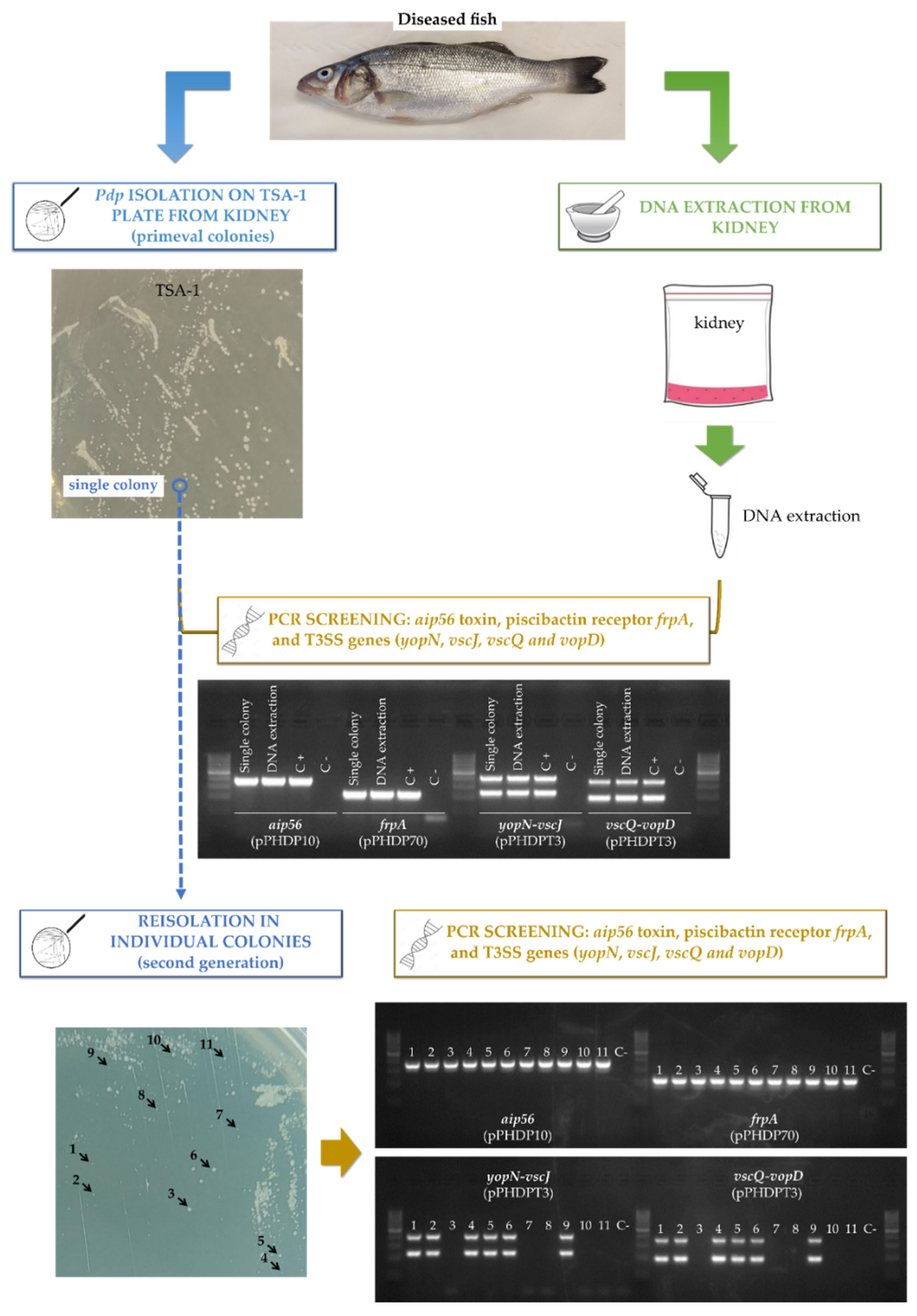
2.5. Tissues of Naturally-Infected Sea Bass Fish Suffering Photobacteriosis Test Positive for pPHDPT3 Genes, and These Become Lost following In Vitro Cultivation

Up to this point, the laboratory evidence pointing at instability and curing of pPHDPT3 in Pdp has been obtained using Pdp isolates maintained at −80 °C. In order to assess whether pPHDPT3 presence and further curing can be followed directly from the fish specimens, we analyzed naturally-infected, moribund European sea bass fish from fish farms suffering photobacteriosis outbreaks in field conditions. Immediately upon fish arrival at our laboratory, spleen and kidney samples were taken to purify DNA and to conduct PCR amplification of pPHDPT3 genes and, in parallel, the same specimens were cultivated on TSA-1 plates and incubated at 18 °C until presumptive Pdp colonies appeared. Positive amplification was confirmed for pPHDPT3 markers vscJ, yopN, vopD and vscQ, as well as for the stable gene marker aip56 (encoded within pPHDP10) (Figure 6). All the analyzed fish specimens tested positive by PCR for pPHDPT3 markers, and for all the samples we confirmed the growth of Pdp colonies after 4–5 days at 18 °C, and these colonies also tested positive for vscJ, yopN, vopD and vscQ. When these primeval isolated colonies were subjected to a further streak isolation step, ~50% of the new isolated colonies tested negative for pPHDPT3 markers, indicative of plasmid curing after the first passage on agar plates (Figure 6).

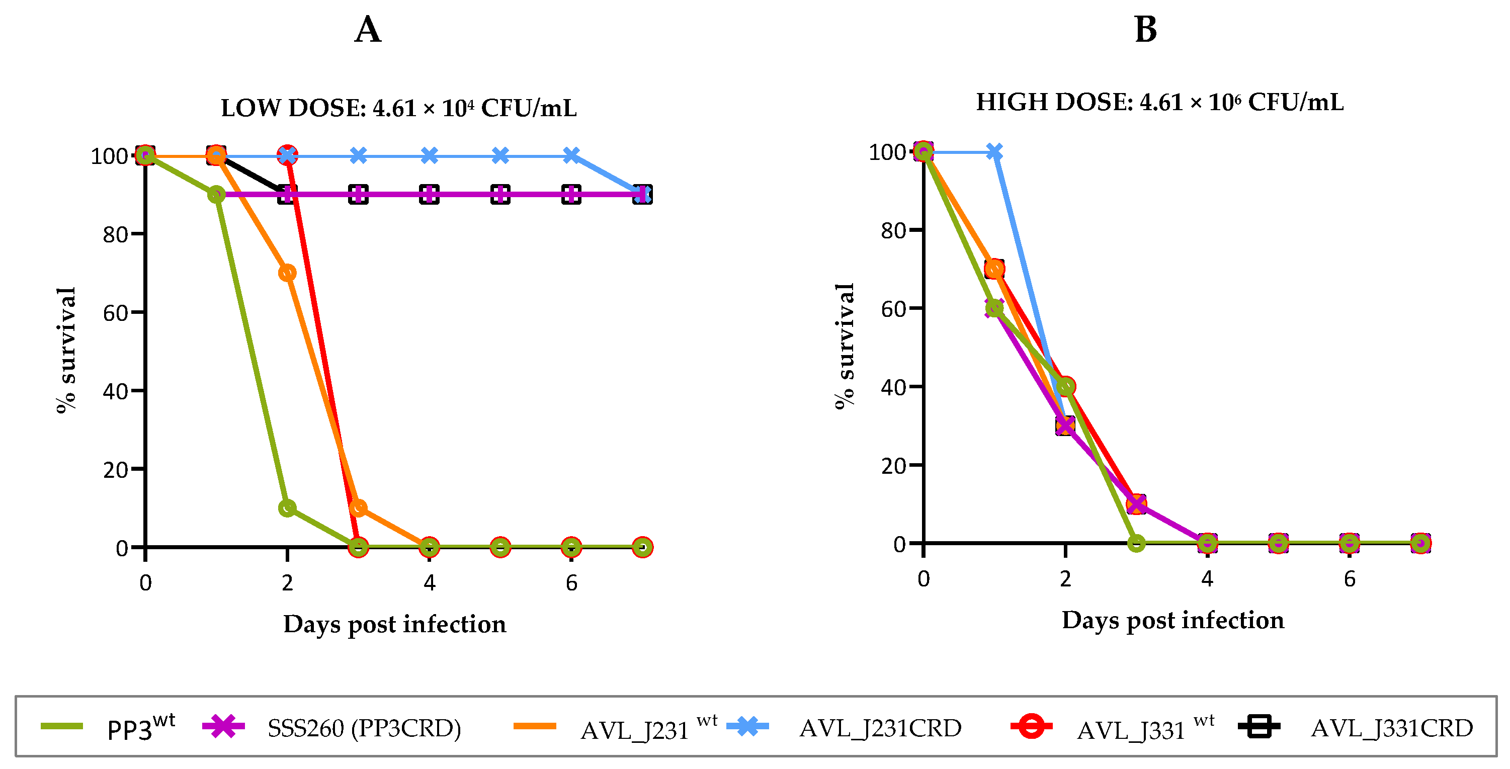
2.6. pPHDPT3 Is Essential for Maximal Virulence in Pdp: Virulence for Fish Is Markedly Reduced in Cured Strains

To assess the role of pPHDPT3 in virulence, we selected an isolated colony of Pdp PP3 that underwent spontaneous pPHDPT3 curing and that tested positive for the virulence plasmids pPHDP10 and pPHDP70 (cured strain dubbed SSS260). Bacterial suspensions of SSS260, and of the original parental strain PP3 (bulk cell mass testing positive by PCR for pPHDPT3), were inoculated into European sea bass fish at a sharp dose of ~4.6 × 10⁴ CFU/fish and mortalities were quantified for 7 d. Remarkably, the parental strain killed 100% of fish within the first 4 d post-inoculation, whereas the pPHDPT3-cured strain killed 10% of fish, thus demonstrating a strong impairment of virulence when the inoculated strain is cured of pPHDPT3 plasmid (Figure 7A).
Since we have demonstrated that pPHDPT3 is gradually lost after fish specimens’ cultivation on agar plates and subsequent passages, we wanted to assess if freshly isolated Pdp strains directly from diseased fish were more virulent than their isogenic pPHDPT3-cured derivatives. Hence, we inoculated in sea bass fish a dose of ~4.6 × 10⁴ CFU/fish of a Pdp strain (AVL_J231) freshly isolated from a moribund fish retrieved from a fish farm undergoing a photobacteriosis outbreak. Inoculation of this strain caused 100% of fish death within 4 d (Figure 7A). In parallel, a single colony of AVL_J231 was picked from the primeval agar plate and subcultured, and a colony that tested negative for pPHDPT3 markers was selected. This pPHDPT3-cured strain (AVL_J231CRD), which proved to test positive for the virulence plasmids pPHDP10 and pPHDP70, only caused death of one fish in the seventh day post-inoculation (Figure 7A). A similar result was obtained when another freshly isolated strain AVL_J331 and its pPHDPT3-cured derivative (AVL_J331CRD) were assayed (Figure 7A). These results altogether provide strong evidence that pPHDPT3 curing impairs virulence of Pdp for fish, and warns that plasmid curing may have been occurring for decades in laboratories and causing a gradual loss of virulence of stored Pdp strains when subcultured.
Despite the virulence attenuation in pPHDPT3-cured strains shown above, it is well documented that laboratory isolates of Pdp do cause fish death when used in experimental inoculations. We, therefore, hypothesized that pPHDPT3-cured strains would maintain a lower degree of virulence compared to plasmid-harboring strains. To gain further insight into this, we conducted fish virulence assays using a dose of ~4.6 × 10⁶ CFU/fish, i.e., 100-fold higher than the previously used dose, with all the strains tested above including their plasmidless derivatives. At this high dose, all the pPHDPT3-cured strains killed 100% of fish within the first 4 d post-inoculation (Figure 7B). These data are in agreement with all the previous studies reporting that Pdp strains grown in vitro, are indeed pathogenic for fish in experimental assays. These results shed light on the hypothesis that laboratory strains only cause fish death in experimental infections when high doses are inoculated, while pPHDPT3 plasmid-bearing strains cause fish death at much lower doses.

2.7. pPHDPT3 Instability Impedes the Accurate Genetic Manipulation of the Plasmid

In order to dissect the roles of specific pPHDPT3 plasmid genes in the virulence of Pdp for fish, we attempted to generate in-frame deletions of selected plasmid genes by allelic exchange. First, we generated constructs to delete the two alleles of vcrD gene, encoding one of the structural elements of the T3SS machinery (see methods). We succeeded in selecting a first recombination cross-over that integrated a suicide plasmid of the pGP704 family into pPHDPT3. This integration was possible by selecting KanR, provided by the resistance marker in the suicide plasmid. However, we were unable despite reiterated attempts, to obtain a second recombinant that would eliminate the suicide plasmid. Instead, 100% of the Pdp PP3 colonies grown under the conditions that would select for the second recombination cross-over (growth in presence of 15% sucrose without pressure for kanamycin resistance) all tested negative for pPHDPT3 markers (data not shown), indicative of plasmid curing.
We next designed a strategy to force pPHDPT3 stabilization, which included the allelic exchange of each of the two vcrD paralogous alleles (vcrD1 and vcrD2) by a chloramphenicol resistance gene CmR, to generate the two possible vcrD single mutants. Next, using the vcrD1 mutant as a starting point, we exchanged the vcrD2 allele with a kanamycin resistance gene KanR, in order to obtain a double mutant labeled with two different resistance genes. By maintaining the presence of the respective antibiotics during all the process of mutant construction, we would expect the allelic exchange to be successful and, at the same time, to stabilize pPHDPT3. As expected, antibiotic pressure eventually allowed the substitution of each vcrD allele with a resistance marker (Supplementary Figure S6). This allelic exchange procedure confirmed that indeed the pPHDPT3 plasmid of Pdp PP3 contains a duplicated set of T3SS genes. However, we observed that some plasmid gene markers were no longer detected by PCR after the process of mutant construction, indicating that the artificial pressure for maintaining the plasmid, eventually caused the selection of sequence rearrangements and gene loss in pPHDPT3 and, most likely, in the chromosomes (data not shown). In support of this hypothesis of off-target genome alterations caused during mutant construction, we found that the single and double vcrD mutants were nonvirulent for fish at the highest dose tested (~4.6 × 10⁶ CFU/fish) (data not shown), i.e., they were much more impaired in virulence than the cured strains. This high instability of the Pdp genome, with the presence of IS elements in high copy numbers, and the additional instability of the virulence plasmid pPHDPT3, should warn researchers about the difficulties of generating isogenic mutants for accurate virulence tests with this important pathogen.

3. Discussion

After more than 55 years since its first report as a fish pathogen [1], Pdp continues to be a major threat to the cultures of many species of fish of importance in marine aquaculture worldwide [32,33,34,35,36,37]. In the past two decades, research has focused on the identification and the study of virulence factors in Pdp, the most important so far being the apoptosis-inducing toxin AIP56 encoded within plasmid pPHDP10 [6] and the siderophore piscibactin system encoded within plasmid pPHDP70 [11]. Notably, it was not until 2019 that the presence of genes of the type III secretion system was reported in this pathogen [18]. Soon afterward, it was reported that genes of the T3SS are more the norm than the exception in Pdp isolates, and it was suggested that the T3SS might be encoded within an unstable plasmid [10].
A discovery that has been reinforced in recent years is the presence of multiple plasmids in Pdp genomes [13,14,15], and indeed the best studied virulence factors are plasmid encoded [6,11]. In the present study, we report that Pdp PP3 harbors several plasmids and three of them constitute proved virulence plasmids, pPHDP10, pPHDP70 and pPHDPT3. Despite this clear link between plasmids and virulence in Pdp, very little is known so far about the stability of plasmid replicons in Pdp cells. pPHDPT3 contains 33 IS of six different families but the T3SS genes are free of IS insertions, indicative of selective pressure for maintaining these gene sets intact. We have demonstrated that pPHDPT3 is highly unstable in laboratory culture conditions, and it becomes gradually lost following Pdp isolation on agar plates from fish tissues. The reasons for this high instability are so far unknown. Plasmids have evolved a variety of mechanisms that ensure their maintenance after cell division. Changes in the regulation or in the proficiency of these mechanisms caused by in vitro cultivation of bacteria in absence of selection from a fish host, and selective deletions of certain plasmid sequences might alter plasmid stability. Control of plasmid replication and copy number constitutes a major point for plasmid stability [38,39]. Additional strategies include mechanisms for actively partitioning plasmid copies during cell division [40,41], and post-segregational killing mechanisms, such as toxin-antitoxin systems, that selectively kill daughter cells lacking the plasmid [42,43]. In addition, it has to be taken into account that the growth rate can differ substantially between plasmid-free and plasmid-containing cells [44]. Hence, pPHDPT3 plasmid loss might be a consequence of an imperfect functioning of replication/maintenance/partition mechanisms at the molecular level, selective gene deletions occurring during the transition from in vivo to in vitro growth, and/or a consequence of the natural selection that favors the growth of cells that lack the plasmid. pPHDPT3 is a large plasmid that likely constitutes a metabolic burden for replicating cells in vitro, and thus plasmid loss may result in increased fitness outside the fish host. It is evident that Pdp cells must have mechanisms to maintain the plasmid in vivo, but not in vitro. We did not find sequences related to plasmid replication in pPHDPT3. Considering the high instability of this plasmid in vitro, and the high content of IS elements, it is not unreasonable to hypothesize that plasmid regions necessary for replication as well as for maintenance might undergo selective deletion and loss upon in vitro cultivation. This hypothesis, which will deserve an in-depth investigation, would explain why this plasmid is so unstable, and why replication-related sequences have not been so far identified in genomic analyses. Selective deletions of sequences necessary for replication and maintenance might also explain the differential stability of pPHDPT3-like plasmids in different Pdp strains. This differential stability is exemplified by the presence of enough plasmid DNA in PP3 stocks that enabled PacBio sequencing, in contrast to our inability to sequence homologous plasmids in other Pdp strains.
In this line of thought, virulence plasmids that undergo loss and/or sequence rearrangements upon in vitro cultivation have been reported in a variety of bacterial species. A plasmid that encodes nematicidal Cry toxins in Bacillus thuringiensis proved to be highly unstable in vitro, with loss occurring within a single growth cycle [45]. Of note, Aeromonas salmonicida, a major pathogen of fish in aquaculture, harbors a plasmid encoding the T3SS that is highly prone to undergo genetic rearrangements and T3SS gene loss when A. salmonicida is grown at 25 °C, but not when it is grown at 18 °C, and recombination between homologous IS elements has been found to be responsible for deletion and loss of discrete plasmid regions [46,47]. In this study we noticed that pPHDPT3 was unstable during cultivation under laboratory conditions at 25 °C and at 18 °C.
Our observations on plasmid loss in Pdp are strongly reminiscent of a similar case very well documented in Shigella species. Shigella spp. harbor pINV, a ca. 210 kb virulence plasmid containing a pathogenicity island (PAI) of ∼30 kb that encodes a T3SS essential for virulence [48,49]. It was found that plasmid instability differed between S. flexneri and S. sonnei. pINV is more stable in S. flexneri, a species that is mainly transmitted through the environment, and whose pINV variants contain a toxin-antitoxin (TA) system that stabilizes the plasmid [50]. S. flexneri can lose the PAI or the entire plasmid during in vitro growth [51]. In contrast, in S. sonnei, considered a host-adapted pathogen, pINV has undergone specific, discrete deletions of the genes encoding two TA systems, strongly impairing its stability when grown outside the host at environmental temperatures [52]. This process of TA gene deletions is likely associated with the recent emergence of S. sonnei as a host-adapted pathogen. Subsequent studies demonstrated that S. flexneri pINV undergoes loss of plasmid regions (including the T3SS) under laboratory conditions, and such deletions occurred through intra-molecular recombination events between ISs flanking T3SS genes [53]. Similarly, Pdp is considered to have diverged from its sibling subspecies, Pdd, by a process of adaptation to a host-dependent lifestyle, where the high number of IS elements have contributed to gene deletions and pseudogenization that shaped the Pdp genome [12]. The presence of large numbers of IS elements confers a high degree of genetic plasticity to pPHDPT3, with the potential to undergo structural rearrangements, fast sequence variations and gene deletions. In support of this idea, we were unable to obtain an isogenic deletion mutant of a pPHDPT3-borne T3SS gene, because the pressure for plasmid maintenance via integration of antibiotic resistance genes into the structure of pPHDPT3 eventually caused the selection of plasmid reorganization events.
The high instability of pPHDPT3-family plasmids, which are highly prevalent in isolates of this pathogen, warns about the fact that its proneness to be lost in cultivation has prevented researchers from detecting and studying these plasmids in Pdp isolates worldwide for decades. We demonstrate here that pPHDPT3 gene markers are virtually ubiquitous in all the Pdp strains being isolated nowadays from diseased European sea bass in the European area, both in the Atlantic and in the Mediterranean basins. The primeval colonies growing on agar plates upon fish sampling, all tested positive for pPHDPT3 markers. However, laboratory handling and culturing causes the loss of these unstable genetic elements, with special risk when isolated colonies are picked with the intention of establishing new bacterial stocks and starting cultures for research. This has undoubtedly caused an underestimation of the actual virulence gene content of the Pdp cells infecting fish in the field. In fact, although pPHDPT3 gene markers are highly prevalent in Pdp isolates from diverse geographical origins and isolation sources, the presence of such genes was not discovered and published until as recently as 2019 [18]. Another point that will deserve further investigation, is the differential stability of pPHDPT3 plasmid across Pdp strains. In this study, we were able to sequence pPHDPT3 from the genomic DNA of Pdp PP3, a strain isolated from Seriola quinqueradiata in Japan, that underwent an indeterminate number of passages before it reached our laboratory. Similarly, two recent studies have published Pdp genomes that contain a pPHDPT3-like plasmid [13,15] in strains from Japan and Australia. However, we failed to detect pPHDPT3 sequences after a PacBio sequencing of three additional Pdp strains. This failing is particularly informative for SNW-8.1, a strain from our laboratory that initially allowed the sequencing of pPHDPT3 pieces by an Illumina strategy [18] but failed to contain the plasmid sequence when a PacBio approach was accomplished later on, and only a single subculture step of the original strain differed between these two sequencing approaches.
The virulence data obtained in this study with plasmid-bearing and with plasmid-cured cultures, clearly demonstrate that plasmid curing causes a strong impairment in the degree of virulence of Pdp for fish, albeit pPHDPT3 curing does not completely abolish the pathogenicity of cured strains. These results are in agreement, on the one hand, with the widely reported observations that the Pdp strains routinely used for experimental infections do cause fish death and, on the other hand, with the observations that some freshly isolated Pdp strains exhibited an unexpectedly higher degree of virulence than usual [54,55]. We hypothesize that the unusually-highly virulent strains reported in some studies correspond to primeval stocks that still preserve the T3SS plasmid in a percentage of cells, whereas the majority of the laboratory strains that have lost the plasmid exhibit higher LD50 values. In support of this hypothesis, we were able to detect by PCR the presence of pPHDPT3 markers in a stock of Pdp strain PC554.2 that underwent some subculture steps from the stock originally reported as an unusually highly virulent isolate [55], but the genome sequencing of this strain in our present study demonstrated the absence of pPHDPT3 sequences (data not shown). The virulence reduction in Pdp stocks that have lost pPHDPT3 plasmid upon isolation and subculturing might explain why some studies have reported a 100-fold difference in LD50 among Pdp strains isolated in the same case study [56], a difference otherwise very difficult to explain in a pathogen that is highly clonal and serologically homogeneous [55,57,58].
Functions encoded within pPHDPT3 are candidates to play a role in virulence. The Pdp T3SS includes a YopH-like tyrosine phosphatase effector, whose homologs in other bacterial species play a major role in virulence, as reported in Aeromonas salmonicida [59] and in Yersinia spp. [60,61,62]. While the structural genes of T3SS are conserved among related species, effector proteins are highly diverse and some of them are specific for each pathogen. Moreover, fish pathogens are not an exception to this [22]. In this scenario, it is feasible that yet-unknown proteins with effector function and with other roles are encoded in pPHDPT3-like plasmids in different Pdp strains.
Recently isolated Pdp strains in the European Mediterranean and Atlantic basins test positive for pPHDPT3. Similarly, in a recent study, we reported that genes now known to be pPHDPT3-borne, are highly prevalent in Pdp isolates from diverse fish hosts and geographical locations, between 1980 and 2015 [10]. Considering that recently sequenced Pdp strains from Japan and Australia also harbor a pPHDPT3-like plasmid [13,15], it is reasonable to propose that pPHDPT3 is a virulence plasmid that is virtually ubiquitous in Pdp isolates currently circulating in the world. This high prevalence of pPHDPT3 plasmid and its high instability in vitro, clearly warn that Pdp cells in vivo surely exhibit virulence features that have passed unadvertised to microbiologists and fish pathologists since the beginning of Pdp-caused outbreaks in aquaculture in the 1960s. Acquisition of the large virulence plasmid pINV encoding a T3SS led to the emergence of Shigella spp. from Escherichia coli and enabled bacteria to invade epithelial cells [63]. Similarly, it is tempting to speculate that pPHDPT3 might allow Pdp to infect fish through ways and/or infective dynamics that have not been so far reproduced under laboratory conditions, considering that plasmid-cured strains have likely been used extensively in experimental infections in laboratories.

4. Materials and Methods

4.1. Bacteria, Plasmids, and Media

The bacteria and plasmids used in this study are listed in Table 4. Pdp strains were routinely grown on tryptic soy agar (TSA-1) or in tryptic soy broth (TSB-1) supplemented with an extra 0.5% NaCl (1% NaCl final concentration) at 25 °C or 18 °C. Escherichia coli strains were grown on Luria Bertani agar (LB) plates or Luria Bertani Broth at 37 °C. When necessary, antibiotics were used at the following concentrations: kanamycin (Kan) at 50 µg mL−1, ampicillin (Amp) at 50 µg mL−1, chloramphenicol (Cm) at 5 µg mL−1 or 20 µg mL−1. Antimicrobial susceptibility patterns to tetracycline (12 μg per disc) and oxytetracycline (30 μg per disc) were determined by disc diffusion tests on TSA-1 plates, using bacterial suspensions adjusted to an optical density at 600 nm (OD600) of 0.5 in saline solution (0.85% NaCl wt/vol). The diameter (in mm) of the inhibition zones around discs was measured and photographed after 24-h incubation at 25 °C.

4.2. DNA Sequencing, Annotation, and Comparative Genomics Analyses

High-purity genomic DNA of Pdp strains for genome sequencing was extracted using the G NOME DNA kit (MP Biomedicals) according to the manufacturer’s instructions, and the DNA was subjected to sequencing following the PacBio procedure (SNPsaurus, Eugene, OR, USA). The assembly by Flye 2.4.1 [68] was generated from 1,336,131,874 total bases, for 222 read depth over the genome. The read length N50/N90 was 10795/6571. Coding sequences (CDS) were predicted by the NCBI Prokaryotic Genome Annotation Pipeline [69]. Additional information of gene annotation and gene functions was obtained using the rapid annotations of subsystems technology (RAST) and the BLASTP database [70,71]. The CGView server database was used to obtain a circular graphical map representation of the pPHDPT3 plasmid genome [72]. Comparative analysis of nucleotide sequences was performed using BLASTN and BLASTP databases [71], and MAUVE and EasyFig programs [73,74]. The PacBio-derived sequence of the Pdp PP3 genome was deposited in GenBank database under Accession number SRHT00000000.

4.3. PCR Amplifications

Primers used in this study are listed in Table 5. These include primers for mutant construction, and for screening of gene presence. Markers related to pPHDPT3 plasmid backbone: parAB; genes of the T3SS: vcrD, vscJ, vopD, vscQ and yopN. PCR primers to test aip56 gene (pPHDP10 plasmid) and frpA genes (pPHDP70 plasmid) were used to screen for the presence of these two virulence plasmids. PCR amplifications were routinely performed using the NZYTaq II 2× Green Master kit following the manufacturer’s instructions; annealing temperatures were adjusted according to the corresponding primer pairs. Colony PCR was routinely conducted by picking an isolated colony and suspending it in 30 µL of sterile distilled water. Three µL of this suspension were directly added to the PCR tube to be used as a DNA template.

4.4. Phylogenetic Analysis

The phylogenetic tree of YopH-like putative effectors was constructed in MEGA-X [75]. Protein sequences were aligned using Clustal W [76]. The evolutionary histories were inferred using the Neighbor-Joining method [77]. The percentage of replicate trees in which the associated taxa clustered together in the bootstrap test (1000 replicates) is shown next to the branches. The evolutionary distances were computed using the Poisson Correction method [78], and are in the units of the number of amino acid substitutions per site.

4.5. Processing of Diseased Fish Tissues for PCR and Pdp Cultivation

European sea bass (Dicentrarchus labrax) fish individuals were obtained from farms located on the Atlantic and the Mediterranean coasts of Spain that were undergoing outbreaks of photobacteriosis caused by Pdp. Fish were transported to our laboratory under refrigerated conditions and immediately dissected upon arrival. Kidney and spleen samples were cultured on TSA-1 plates and incubated at 18 °C for 3–4 days until presumptive Pdp colonies appeared. Direct colony-PCR was conducted with isolated colonies using appropriate specific primers. In parallel, samples of the kidney and spleen were aseptically removed for total DNA extraction using Easy-DNATM (Invitrogen), following the manufacturer’s recommendations. Routinely, 200 ng of total DNA was used as a template for PCR reactions to detect the presence of Pdp gene markers in the tissues of naturally infected fish.

4.6. Selection of pPHDPT3-Cured Strains for Virulence Assays

Parental Pdp strains PP3, AVL_J231 and AVL_J331 were streaked on TSA-1 plates and incubated at 18 °C until isolated colonies were observed. Five isolated colonies per strain were re-streaked on fresh TSA-1 plates and incubated at 18 °C. When isolated colonies appeared, between 10–20 colonies per strain were subjected to PCR screening for pPHDPT3 markers (vscJ, vopD, yopN, vscQ) as well as for the pPHDP10 plasmid marker aip56, and the pPHDP70 marker frpA. Colonies that tested negative for all the pPHDPT3 markers but tested positive for aip56 and frpA were considered pPHDPT3-cured colonies and kept for further experiments. This process led to obtaining the cured versions of strains PP3 (dubbed SSS260), AVL_J231 (AVL_J231CRD) and AVL_J331 (AVL_J331CRD) (Table 4).

4.7. Virulence Assays

European sea bass fish (Dicentrarchus labrax) with an average weight of 5 g were selected to perform virulence tests using three parental Pdp strains and their respective pPHDPT3-cured derivatives, as well as three vcrD mutant strains (SSS256, SSS299 and SSS325). Fish were acclimated in 100 L aquaria at 24 °C for three days before the injection. For dose preparation, bacterial biomass at the confluent growth zone of the agar plate from 48 h cultures at 18 °C was suspended in saline solution (0.85% NaCl) and adjusted at a sharp OD600 = 0.3. Serial decimal dilutions were prepared in saline solution and bacterial counts were carefully calculated. Fish were inoculated intraperitoneally with 0.1 mL of bacterial suspensions in saline solution (0.85% NaCl) using either a low dose (~4.61 × 10⁴ CFU/fish) or a high dose (~4.61 × 10⁶ CFU/fish). A group of 10 fish was inoculated with saline solution as a control. For each strain and dose assayed, 10 fish were inoculated, and mortality was recorded over a period of 7 d after injection. Dead fish were aseptically dissected, bacteria were recovered from the kidney by culturing on TSA-1 agar plates, and colonies were screened by PCR using gene markers for pPHDPT3, pPHDP10 and pPHDP70 plasmids. The protocols of animal experimentation used in this study have been reviewed and approved by the Animal Ethics Committee of the Universidade de Santiago de Compostela, Spain (Ethical approval code 15004/14/003).

4.8. Allelic-Exchange Mutagenesis of vcrD Gene Alleles of the T3SS

The coding sequence of each of the two vcrD alleles (vcrD1 and vcrD2) was replaced with antibiotic resistance genes following an allelic exchange procedure using the suicide vector pNidKan, a pCVD442 derivative [66]. Two fragments of ~2 kb, upstream (fragment 1–2) and downstream (fragment 3–4) of each vcrD coding sequence were PCR-amplified. Fragment 1–2 was amplified using primers vcrD_XhoI_1 and vcrD_BamHI_2 while fragment 3–4 was amplified using primers vcrD_BamHI_3 and vcrD_NotI_4. After being digested with restriction enzymes, the two fragments were ligated so that the resulting DNA sequence generated an in-frame, 1697-bp deletion, of the vcrD coding sequence. In order to fix the unstable pPHDPT3 plasmid, the construction containing the deletion was cut with BamHI and an 882 bp chloramphenicol resistance gene (cat) amplified from the pKD3 plasmid [67] was inserted. In addition, another version for mutant construction was generated by cutting the aforementioned deletion construction with BamHI and inserting there an 1190 bp PCR-amplified kanamycin resistance gene (kan) from the pKD4 plasmid [67]. The cat-labelled construction for mutation of vcrD alleles was used to obtain single vcrD1 and vcrD2 mutants independently (SSS256 and SSS325 mutant strains, respectively). Besides, in order to obtain a double mutant, the cat-labelled vcrD1 mutant was used as the basis to construct a double mutant (SSS299) by using the kan-labelled construction that was used to delete vcrD2 gene. The mutant allele containing the cat gene was introduced into the suicide vector pNidKan and propagated into E. coli β3914. The mutant construction containing the kan gene was inserted into the suicide vector pNidKan and propagated into E. coli S17-1-λpir. E. coli donor strains were conjugated with Pdp recipient strains by mixing equal volumes of log-phase cultures of donor and recipient strains on TSA plates prepared with seawater. First recombinants for single vcrD1 mutant construction were selected on TSA-1 plates supplemented with chloramphenicol at 5 µg mL−1 (Cm5). For double mutant construction, the colonies resulting from the conjugation between E. coli S17-1-λpir and Pdp PP3 SSS256 ∆vcrD1-cat (single mutant) were selected on TSA-1 plates supplemented with Cm5 and kanamycin at 50 µg mL−1 (Kan50). To select for the second recombination of the single mutant construction containing the cat gene, TSA-1 plates supplemented with Cm5 and sucrose (15% [wt/vol]) were used. TSA-1 plates supplemented with Cm5 + Kan50 and sucrose (15% [wt/vol]) were used to select double mutant colonies containing the kan gene plus the cat gene.

5. Conclusions

This study provides evidence that pPHDPT3 plasmid constitutes a novel, hitherto unreported virulence factor of Pdp. The high in vitro instability of pPHDPT3-like plasmids warns that the picture of Pdp virulence genes has been historically biased and underestimated. The realization of the prevalence of plasmids encoding the T3SS and additional genes in Pdp will surely change our view of this pathogen from now on.

Supplementary Materials

The following supporting information can be downloaded at: https://www.mdpi.com/article/10.3390/ijms23094729/s1.

Author Contributions

Conceptualization, C.R.O. and A.V.; methodology, A.V. and C.R.O.; software, A.V.; formal analysis, S.A., A.V. and C.R.O.; investigation, S.A., A.V. and C.R.O.; resources, A.V. and C.R.O.; data curation, S.A., A.V. and C.R.O.; writing—original draft preparation, S.A. and C.R.O.; writing—review and editing, C.R.O. and A.V.; supervision, C.R.O. and A.V.; project administration, C.R.O.; funding acquisition, C.R.O. All authors have read and agreed to the published version of the manuscript.

Funding

This work was supported by the Agencia Estatal de Investigación (AEI) of Spain co-funded by the FEDER Programme from the European Union (Grant No. PID2019-110558RB-I00) to Carlos R. Osorio, and by Xunta de Galicia (Spain) [Grant No. ED431C 2018/18].

Institutional Review Board Statement

The protocols of animal experimentation used in this study have been reviewed and approved by the Animal Ethics Committee of the Universidade de Santiago de Compostela, Spain (Ethical approval code 15004/14/003).

Data Availability Statement

The PacBio-derived sequence of the Pdp PP3 genome was deposited in GenBank database under Accession number SRHT00000000.

Acknowledgments

We are highly grateful to the Instituto Galego de Formacion en Acuicultura (IGaFA) (Illa de Arousa, Galicia, Spain), for their valuable support in providing European sea bass fish for the virulence experiments.

Conflicts of Interest

The authors declare no conflict of interest. The funders had no role in the design of the study; in the collection, analyses, or interpretation of data; in the writing of the manuscript, or in the decision to publish the results.

References

  1. Snieszko, S.; Bullock, G.; Hollis, E.; Boone, J. Pasteurella sp. from an epizootic of white perch (Roccus americanus) in Chesapeake Bay tidewater areas. J. Bacteriol. 1964, 88, 1814–1815. [Google Scholar] [CrossRef] [PubMed] [Green Version]
  2. Romalde, J.L. Photobacterium damselae subsp. piscicida: An integrated view of a bacterial fish pathogen. Int. Microbiol. 2002, 5, 3–9. [Google Scholar] [CrossRef] [PubMed]
  3. Andreoni, F.; Magnani, M. Photobacteriosis: Prevention and diagnosis. J. Immunol. Res. 2014, 2014, 793817. [Google Scholar] [CrossRef] [PubMed] [Green Version]
  4. Håstein, T.; Gudding, R.; Evensen, O. Bacterial vaccines for fish—An update of the current situation worldwide. Dev. Biol. 2005, 121, 55–74. [Google Scholar]
  5. do Vale, A.; Marques, F.; Silva, M.T. Apoptosis of sea bass (Dicentrarchus labrax L.) neutrophils and macrophages induced by experimental infection with Photobacterium damselae subsp. piscicida. Fish Shellfish Immunol. 2003, 15, 129–144. [Google Scholar] [CrossRef]
  6. do Vale, A.; Silva, M.T.; dos Santos, N.M.; Nascimento, D.S.; Reis-Rodrigues, P.; Costa-Ramos, C.; Ellis, A.E.; Azevedo, J.E. AIP56, a novel plasmid-encoded virulence factor of Photobacterium damselae subsp. piscicida with apoptogenic activity against sea bass macrophages and neutrophils. Mol. Microbiol. 2005, 58, 1025–1038. [Google Scholar] [CrossRef]
  7. Silva, D.S.; Pereira, L.M.; Moreira, A.R.; Ferreira-da-Silva, F.; Brito, R.M.; Faria, T.Q.; Zornetta, I.; Montecucco, C.; Oliveira, P.; Azevedo, J.E.; et al. The apoptogenic toxin AIP56 is a metalloprotease A-B toxin that cleaves NF-κb P65. PLoS Pathog. 2013, 9, e1003128. [Google Scholar] [CrossRef] [Green Version]
  8. Pereira, L.M.; Pinto, R.D.; Silva, D.S.; Moreira, A.R.; Beitzinger, C.; Oliveira, P.; Sampaio, P.; Benz, R.; Azevedo, J.E.; dos Santos, N.M.; et al. Intracellular trafficking of AIP56, an NF-κB cleaving toxin from Photobacterium damselae subsp. piscicida. Infect. Immun. 2014, 82, 5270–5285. [Google Scholar] [CrossRef] [Green Version]
  9. do Vale, A.; Pereira, C.; Osorio, C.R.; dos Santos, N.M.S. The apoptogenic toxin AIP56 is secreted by the type II secretion system of Photobacterium damselae subsp. piscicida. Toxins 2017, 9, 368. [Google Scholar] [CrossRef] [Green Version]
  10. Abushattal, S.; Vences, A.; Osorio, C.R. A virulence gene typing scheme for Photobacterium damselae subsp. piscicida, the causative agent of fish photobacteriosis, reveals a high prevalence of plasmid-encoded virulence factors and of type III secretion system genes. Aquaculture 2020, 521, 735057. [Google Scholar] [CrossRef]
  11. Osorio, C.R.; Rivas, A.J.; Balado, M.; Fuentes-Monteverde, J.C.; Rodríguez, J.; Jiménez, C.; Lemos, M.L.; Waldor, M.K. A transmissible plasmid-borne pathogenicity island confers piscibactin biosynthesis in the fish pathogen Photobacterium damselae subsp. piscicida. Appl. Environ. Microbiol. 2015, 81, 5867–5879. [Google Scholar] [CrossRef] [PubMed] [Green Version]
  12. Balado, M.; Benzekri, H.; Labella, A.M.; Claros, M.G.; Manchado, M.; Borrego, J.J.; Osorio, C.R.; Lemos, M.L. Genomic analysis of the marine fish pathogen Photobacterium damselae subsp. piscicida: Insertion sequences proliferation is associated with chromosomal reorganisations and rampant gene decay. Infect. Genet. Evol. 2017, 54, 221–229. [Google Scholar] [CrossRef] [PubMed]
  13. Aoki, T.; Teru, Y.; Morimoto, N.; Kono, T.; Sakai, M.; Takano, T.; Hawke, J.P.; Fukuda, Y.; Takeyama, H.; Hikima, J.I. Complete genome sequence of Photobacterium damselae subsp. piscicida strain OT-51443 isolated from Yellowtail (Seriola quinqueradiata) in Japan. Genome Announc. 2017, 5, e00404-17. [Google Scholar] [CrossRef] [PubMed] [Green Version]
  14. Teru, Y.; Hikima, J.I.; Kono, T.; Sakai, M.; Takano, T.; Hawke, J.P.; Takeyama, H.; Aoki, T. Whole-genome sequence of Photobacterium damselae subsp. piscicida strain 91-197, isolated from hybrid striped bass (Morone sp.) in the United States. Genome Announc. 2017, 5, e00600-17. [Google Scholar] [CrossRef] [PubMed] [Green Version]
  15. Baseggio, L.; Silayeva, O.; Buller, N.; Landos, M.; Englestädter, J.; Barnes, A.C. Complete, closed and curated genome sequences of Photobacterium damselae subsp. piscicida isolates from Australia indicate mobilome-driven localized evolution and novel pathogenicity determinants. Microb. Genom. 2021, 7, 000562. [Google Scholar] [CrossRef]
  16. Osorio, C.R.; Vences, A.; Matanza, X.M.; Terceti, M.S. Photobacterium damselae subsp. damselae, a generalist pathogen with unique virulence factors and high genetic diversity. J. Bacteriol. 2018, 200, e00002-18. [Google Scholar] [CrossRef] [Green Version]
  17. Osorio, C.R. Photobacterium damselae: How horizontal gene transfer shaped two different pathogenic lifestyles in a marine bacterium. In Horizontal Gene Transfer; Villa, T.G., Viñas, M., Eds.; Springer Nature Switzerland AG: Cham, Switzerland, 2019; pp. 175–199. [Google Scholar]
  18. Abushattal, S.; Vences, A.; dos Santos, N.M.; do Vale, A.; Osorio, C.R. Draft genome sequences of Photobacterium damselae subsp. piscicida SNW-8.1 and PP3, two fish-isolated strains containing a type III secretion system. Microbiol. Res. Announc. 2019, 8, e00426-19. [Google Scholar] [CrossRef] [Green Version]
  19. Abrusci, P.; McDowell, M.A.; Lea, S.M.; Johnson, S. Building a secreting nanomachine: A structural overview of the T3SS. Curr. Opin. Struct. Biol. 2014, 25, 111–117. [Google Scholar] [CrossRef] [Green Version]
  20. Spreter, T.; Yip, C.K.; Sanowar, S.; André, I.; Kimbrough, T.G.; Vuckovic, M.; Pfuetzner, R.A.; Deng, W.; Angel, C.Y.; Finlay, B.B. A conserved structural motif mediates formation of the periplasmic rings in the type III secretion system. Nat. Struct. Mol. Biol. 2009, 16, 468–476. [Google Scholar] [CrossRef] [Green Version]
  21. Galán, J.E.; Wolf-Watz, H. Protein delivery into eukaryotic cells by type III secretion machines. Nature 2006, 444, 567–573. [Google Scholar] [CrossRef]
  22. Rahmatelahi, H.; El-Matbouli, M.; Menanteau-Ledouble, S. Delivering the pain: An overview of the type III secretion system with special consideration for aquatic pathogens. Vet. Res. 2021, 52, 146. [Google Scholar] [CrossRef] [PubMed]
  23. Makino, K.; Oshima, K.; Kurokawa, K.; Yokoyama, K.; Uda, T.; Tagomori, K.; Iijima, Y.; Najima, M.; Nakano, M.; Yamashita, A.; et al. Genome sequence of Vibrio parahaemolyticus: A pathogenic mechanism distinct from that of V. cholerae. Lancet 2003, 361, 743–749. [Google Scholar] [CrossRef]
  24. Zhao, Z.; Chen, C.; Hu, C.Q.; Ren, C.H.; Zhao, J.J.; Zhang, L.P.; Luo, P.; Wang, Q.B. The type III secretion system of Vibrio alginolyticus induces rapid apoptosis, cell rounding and osmotic lysis of fish cells. Microbiology 2010, 156, 2864–2872. [Google Scholar] [CrossRef] [PubMed] [Green Version]
  25. Henke, J.M.; Bassler, B.L. Quorum sensing regulates type III secretion in Vibrio harveyi and Vibrio parahaemolyticus. J. Bacteriol. 2004, 186, 3794–3805. [Google Scholar] [CrossRef] [Green Version]
  26. Luo, H.; Gao, F. DoriC 10.0: An updated database of replication origins in prokaryotic genomes including chromosomes and plasmids. Nucleic Acids Res. 2019, 47, D74–D77. [Google Scholar] [CrossRef] [Green Version]
  27. Le Roux, F.; Davis, B.M.; Waldor, M.K. Conserved small RNAs govern replication and incompatibility of a diverse new plasmid family from marine bacteria. Nucleic Acids Res. 2011, 39, 1004–1013. [Google Scholar] [CrossRef] [Green Version]
  28. Burkinshaw, B.J.; Strynadka, N.C. Assembly and structure of the T3SS. Biochim. Biophys. Acta 2014, 1843, 1649–1663. [Google Scholar] [CrossRef] [Green Version]
  29. Vences, A.; Rivas, A.J.; Lemos, M.L.; Husmann, M.; Osorio, C.R. Chromosome-encoded hemolysin, phospholipase, and collagenase in plasmidless isolates of Photobacterium damselae subsp. damselae contribute to virulence for fish. Appl. Environ. Microbiol. 2017, 83, e00401-17. [Google Scholar] [CrossRef] [Green Version]
  30. Balado, M.; Lemos, M.L.; Osorio, C.R. Genetic characterization of pPHDP60, a novel conjugative plasmid from the marine fish pathogen Photobacterium damselae subsp. piscicida. Plasmid 2013, 70, 154–159. [Google Scholar] [CrossRef]
  31. Osorio, C.R. T3SS effectors in Vibrios: Homology in sequence, diversity in biological functions? Virulence 2018, 9, 721–723. [Google Scholar] [CrossRef]
  32. Liu, P.C.; Lin, J.Y.; Lee, K.K. Virulence of Photobacterium damselae subsp. piscicida in cultured cobia Rachycentron canadum. J. Basic Microbiol. 2003, 43, 499–507. [Google Scholar] [CrossRef] [PubMed]
  33. Essam, H.M.; Abdellrazeq, G.S.; Tayel, S.I.; Torky, H.A.; Fadel, A.H. Pathogenesis of Photobacterium damselae subspecies infections in sea bass and sea bream. Microb. Pathog. 2016, 99, 41–50. [Google Scholar] [CrossRef] [PubMed]
  34. Costa, J.Z.; McCarthy, Ú.; Perez, O.; Ramos, E.; Rodriguez, M.; Monterroso, O.; Riera, R. Occurrence of Photobacterium damselae subsp. piscicida in sea-cage farmed meagre (Argyrosomus regius) in Tenerife, Canary Islands, Spain. Thalassas 2017, 33, 65–71. [Google Scholar] [CrossRef]
  35. Leu, M.; Wong, S.; Tay, Y.; Bandara, E.; Tsai, M. Infection of farmed sub-adult semicircle angelfish, Pomacanthus semicirculatus in Taiwan caused by Photobacterium damselae subsp. piscicida. Bull. Eur. Ass. Fish Pathol. 2019, 39, 137–144. [Google Scholar]
  36. Pham, T.H.; Cheng, T.C.; Wang, P.C.; Chen, S.C. Genotypic diversity, and molecular and pathogenic characterization of Photobacterium damselae subsp. piscicida isolated from different fish species in Taiwan. J. Fish Dis. 2020, 43, 757–774. [Google Scholar] [CrossRef]
  37. Shimada, M.T.; Yunis-Aguinaga, J.; Cueva-Quiroz, V.A.; Engracia, J.R.; Mourino, J.L.P.; Claudiano, G.S.; Moraes, F.R.; Moraes, J.R.E. Isolation and characterization of pathology in case of massive mortality by Photobacterium damselae subsp. piscicida in Rachycentron canadum. Biosci. J. 2020, 36, 1732–1741. [Google Scholar] [CrossRef]
  38. Chattoraj, D.K. Control of plasmid DNA replication by iterons: No longer paradoxical. Mol. Microbiol. 2000, 37, 467–476. [Google Scholar] [CrossRef]
  39. Brantl, S. Antisense RNAs in plasmids: Control of replication and maintenance. Plasmid 2002, 48, 165–173. [Google Scholar] [CrossRef]
  40. Gerdes, K.; Howard, M.; Szardenings, F. Pushing and pulling in prokaryotic DNA segregation. Cell 2010, 141, 927–942. [Google Scholar] [CrossRef] [Green Version]
  41. Salje, J.; Gayathri, P.; Löwe, J. The ParMRC system: Molecular mechanisms of plasmid segregation by actin-like filaments. Nat. Rev. Microbiol. 2010, 8, 683–692. [Google Scholar] [CrossRef]
  42. Hayes, F. Toxins-antitoxins: Plasmid maintenance, programmed cell death, and cell cycle arrest. Science 2003, 301, 1496–1499. [Google Scholar] [CrossRef] [PubMed]
  43. Schuster, C.F.; Bertram, R. Toxin-antitoxin systems are ubiquitous and versatile modulators of prokaryotic cell fate. FEMS Microbiol. Lett. 2013, 340, 73–85. [Google Scholar] [CrossRef] [PubMed]
  44. Lau, B.T.C.; Malkus, P.; Paulsson, J. New quantitative methods for measuring plasmid loss rates reveal unexpected stability. Plasmid 2013, 70, 353–361. [Google Scholar] [CrossRef] [PubMed] [Green Version]
  45. Sheppard, A.E.; Nakad, R.; Saebelfeld, M.; Masche, A.C.; Dierking, K.; Schulenburg, H. High instability of a nematicidal Cry toxin plasmid in Bacillus thuringiensis. J. Invertebr. Pathol. 2016, 133, 34–40. [Google Scholar] [CrossRef]
  46. Stuber, K.; Burr, S.E.; Braun, M.; Wahli, T.; Frey, J. Type III secretion genes in Aeromonas salmonicida subsp. salmonicida are located on a large thermolabile virulence plasmid. J. Clin. Microbiol. 2003, 41, 3854–3856. [Google Scholar] [CrossRef] [Green Version]
  47. Tanaka, K.H.; Vincent, A.T.; Emond-Rheault, J.-G.; Adamczuk, M.; Frenette, M.; Charette, S.J. Plasmid composition in Aeromonas salmonicida subsp. salmonicida 01-B526 unravels unsuspected type three secretion system loss patterns. BMC Genom. 2017, 18, 528. [Google Scholar] [CrossRef] [Green Version]
  48. Buchrieser, C.; Glaser, P.; Rusniok, C.; Nedjari, H.; D’Hauteville, H.; Kunst, F.; Sansonetti, P.; Parsot, C. The virulence plasmid pWR100 and the repertoire of proteins secreted by the type III secretion apparatus of Shigella flexneri. Mol. Microbiol. 2000, 38, 760–771. [Google Scholar] [CrossRef]
  49. Venkatesan, M.M.; Goldberg, M.B.; Rose, D.J.; Grotbeck, E.J.; Burland, V.; Blattner, F.R. Complete DNA sequence and analysis of the large virulence plasmid of Shigella flexneri. Infect. Immun. 2001, 69, 3271–3285. [Google Scholar] [CrossRef] [Green Version]
  50. Sayeed, S.; Reaves, L.; Radnedge, L.; Austin, S. The stability region of the large virulence plasmid of Shigella flexneri encodes an efficient postsegregational killing system. J. Bacteriol. 2000, 182, 2416–2421. [Google Scholar] [CrossRef] [Green Version]
  51. Sasakawa, C.; Kamata, K.; Sakai, T.; Murayama, S.Y.; Makino, S.; Yoshikawa, M. Molecular alteration of the 140-megadalton plasmid associated with loss of virulence and Congo red binding activity in Shigella flexneri. Infect. Immun. 1986, 51, 470–475. [Google Scholar] [CrossRef] [Green Version]
  52. McVicker, G.; Tang, C.M. Deletion of toxin-antitoxin systems in the evolution of Shigella sonnei as a host-adapted pathogen. Nat. Microbiol. 2016, 2, 16204. [Google Scholar] [CrossRef] [PubMed]
  53. Pilla, G.; McVicker, G.; Tang, C.M. Genetic plasticity of the Shigella virulence plasmid is mediated by intra-and inter-molecular events between insertion sequences. PLoS Genet. 2017, 13, e1007014. [Google Scholar] [CrossRef] [PubMed]
  54. Kawakami, H.; Suzukawa, K.; Yamashita, H.; Kohno, Y.; Kono, T.; Sakai, M. Mass mortality of wild largescale blackfish Girella punctata caused by Photobacterium damsela subsp. piscicida. Fish Pathol. 1999, 34, 87–88. [Google Scholar]
  55. Magariños, B.; Romalde, J.L.; López-Romalde, S.; Moriñigo, M.A.; Toranzo, A.E. Pathobiological characterisation of Photobacterium damselae subsp. piscicida isolated from cultured sole (Solea senegalensis). Eur. Ass. Fish Pathol. 2003, 23, 183–190. [Google Scholar]
  56. Zorrilla, I.; Chabrillón, M.; Arijo, S.; Díaz-Rosales, P.; Martínez-Manzanares, E.; Balebona, M.C.; Moriñigo, M.A. Bacteria recovered from diseased cultured gilthead sea bream (Sparus aurata L.) in southwestern Spain. Aquaculture 2003, 218, 11–20. [Google Scholar] [CrossRef]
  57. Magariños, B.; Romalde, J.L.; Bandín, I.; Fouz, B.; Toranzo, A.E. Phenotypic, antigenic and molecular characterization of Pasteurella piscicida strains isolated from fish. Appl. Environ. Microbiol. 1992, 58, 3316–3322. [Google Scholar] [CrossRef] [Green Version]
  58. Hawke, J.P.; Thune, R.L.; Cooper, R.K.; Judice, E.; Kelly-Smith, M. Molecular and phenotypic characterization of strains of Photobacterium damselae subsp. piscicida isolated from hybrid striped bass cultured in Louisiana, USA. J. Aquat. Animal Health 2003, 15, 189–201. [Google Scholar] [CrossRef]
  59. Dacanay, A.; Knickle, L.; Solanky, K.S.; Boyd, J.M.; Walter, J.A.; Brown, L.L.; Johnson, S.C.; Reith, M. Contribution of the type III secretion system (TTSS) to virulence of Aeromonas salmonicida subsp. salmonicida. Microbiology 2006, 152, 1847–1856. [Google Scholar] [CrossRef] [Green Version]
  60. Guan, K.L.; Dixon, J.E. Protein tyrosine phosphatase activity of an essential virulence determinant in Yersinia. Science 1990, 249, 553–556. [Google Scholar] [CrossRef]
  61. Bliska, J.B.; Guan, K.L.; Dixon, J.E.; Falkow, S. Tyrosine phosphate hydrolysis of host proteins by an essential Yersinia virulence determinant. Proc. Natl. Acad. Sci. USA 1991, 88, 1187–1191. [Google Scholar] [CrossRef] [Green Version]
  62. Shaban, L.; Nguyen, G.T.; Mecsas-Faxon, B.D.; Swanson, K.D.; Tan, S.; Mecsas, J. Yersinia pseudotuberculosis YopH targets SKAP2-dependent and independent signaling pathways to block neutrophil antimicrobial mechanisms during infection. PLoS Pathog. 2020, 16, e1008576. [Google Scholar] [CrossRef] [PubMed]
  63. Lan, R.; Reeves, P.R. Escherichia coli in disguise: Molecular origins of Shigella. Microbes Infect. 2002, 4, 1125–1132. [Google Scholar] [CrossRef]
  64. Herrero, M.; de Lorenzo, V.; Timmis, K.N. Transposon vectors containing non-antibiotic resistance selection markers for cloning and stable chromosomal insertion of foreign genes in gram-negative bacteria. J. Bacteriol. 1990, 172, 6557–6567. [Google Scholar] [CrossRef] [Green Version]
  65. Le Roux, F.; Binesse, J.; Saulnier, D.; Mazel, D. Construction of a Vibrio splendidus mutant lacking the metalloprotease gene vsm by use of a novel counterselectable suicide vector. Appl. Environ. Microbiol. 2007, 73, 777–784. [Google Scholar] [CrossRef] [PubMed] [Green Version]
  66. Mouriño, S.; Osorio, C.R.; Lemos, M.L. Characterization of heme uptake cluster genes in the fish pathogen Vibrio anguillarum. J. Bacteriol. 2004, 186, 6159–6167. [Google Scholar] [CrossRef] [PubMed] [Green Version]
  67. Datsenko, K.A.; Wanner, B.L. One-step inactivation of chromosomal genes in Escherichia coli K-12 using PCR products. Proc. Natl. Acad. Sci. USA 2000, 97, 6640–6645. [Google Scholar] [CrossRef] [Green Version]
  68. Kolmogorov, M.; Yuan, J.; Lin, Y.; Pevzner, P.A. Assembly of long, error-prone reads using repeat graphs. Nat. Biotechnol. 2019, 37, 540–546. [Google Scholar] [CrossRef]
  69. Tatusova, T.; DiCuccio, M.; Badretdin, A.; Chetvernin, V.; Nawrocki, E.P.; Zaslavsky, L.; Lomsadze, A.; Pruitt, K.D.; Borodovsky, M.; Ostell, J. NCBI Prokaryotic Genome Annotation Pipeline. Nucleic Acids Res. 2016, 44, 6614–6624. [Google Scholar] [CrossRef]
  70. Aziz, R.K.; Bartels, D.; Best, A.A.; DeJongh, M.; Disz, T.; Edwards, R.A.; Formsma, K.; Gerdes, S.; Glass, E.M.; Kubal, M.; et al. The RAST Server: Rapid annotations using subsystems technology. BMC Genom. 2008, 9, 75. [Google Scholar] [CrossRef] [Green Version]
  71. Altschul, S.F.; Madden, T.L.; Schäffer, A.A.; Zhang, J.; Zhang, Z.; Miller, W.; Lipman, D.J. Gapped BLAST and PSI-BLAST: A new generation of protein database search programs. Nucleic Acids Res. 1997, 25, 3389–3402. [Google Scholar] [CrossRef] [Green Version]
  72. Grant, J.R.; Stothard, P. The CGView Server: A comparative genomics tool for circular genomes. Nucleic Acids Res. 2008, 36, W181–W184. [Google Scholar] [CrossRef] [PubMed]
  73. Sullivan, M.J.; Petty, N.K.; Beatson, S.A. Easyfig: A genome comparison visualizer. Bioinformatics 2011, 27, 1009–1010. [Google Scholar] [CrossRef] [PubMed]
  74. Darling, A.E.; Mau, B.; Perna, N.T. ProgressiveMauve: Multiple genome alignment with gene gain, loss and rearrangement. PLoS ONE 2010, 5, e11147. [Google Scholar] [CrossRef] [PubMed] [Green Version]
  75. Kumar, S.; Stecher, G.; Li, M.; Knyaz, C.; Tamura, K. MEGA X: Molecular Evolutionary Genetics Analysis across Computing Platforms. Mol. Biol. Evol. 2018, 35, 1547–1549. [Google Scholar] [CrossRef] [PubMed]
  76. Thompson, J.D.; Higgins, D.G.; Gibson, T.J. CLUSTAL W: Improving the sensitivity of progressive multiple sequence alignment through sequence weighting, position-specific gap penalties and weight matrix choice. Nucleic Acids Res. 1994, 22, 4673–4680. [Google Scholar] [CrossRef] [PubMed] [Green Version]
  77. Saitou, N.; Nei, M. The neighbor-joining method: A new method for reconstructing phylogenetic trees. Mol. Biol. Evol. 1987, 4, 406–425. [Google Scholar] [CrossRef]
  78. Zuckerkandl, E.; Pauling, L. Evolutionary divergence and convergence in proteins. In Evolving Genes and Proteins; Bryson, V., Vogel, H.J., Eds.; Academic Press: New York, NY, USA, 1965; pp. 97–166. [Google Scholar]
Figure 1. Gene map of pPHDPT3 plasmid of Pdp strain PP3 (GenBank accession no. SRHT02000010.1). Arrows represent predicted open reading frames and the orientation of the arrows indicates the direction of transcription for each gene. Map was generated using GCView.
Ijms 23 04729 g001
Figure 2. Schematic representation of the complete (loci E4T26_023645- E4T26_023860) and the incomplete (loci E4T26_023865- E4T26_024010) T3SS gene clusters in pPHDPT3 plasmid of Pdp strain PP3. T3SS genes (blue arrows) are arranged in three modules separated by transposases (yellow arrows) and hypothetical proteins (red arrows). The gene for the putative effector YopH is highlighted in green.
Ijms 23 04729 g002
Figure 3. Comparative structural analysis of T3SS-harboring plasmids in four Pdp strains: PP3, OT-51443, AS-16-0540-1 and AS-16-0555-7, and in the Pdd type strain CIP102761. Grey-shaded and blue vertical blocks between sequences indicate the homologous regions, the color intensity denoting the identity levels (from 94 to 100%).
Ijms 23 04729 g003
Figure 4. Phylogenetic tree depicting the relationships between Pdp and Pdd YopH-like tyrosine phosphatases encoded within pPHDPT3-like plasmids, and homologous effector proteins in other bacterial species. The NCBI GenBank accession numbers of the sequences used in the alignment are indicated. Branch length denotes the number of substitutions per site. Bootstrap values are shown next to the branches. Phylogenetic tree was constructed using MEGA-X.
Ijms 23 04729 g004
Figure 5. Detection of pPHDPT3 curing in Pdp isolated colonies by direct colony-PCR, on agar plates incubated at 25 °C. A colony screening was conducted by PCR amplifications targeted to gene markers of plasmids pPHDP10 (aip56), pPHDP70 (frpA), and pPHDPT3 (parAB, vscJ, and vopD). Four different Pdp strains, PP3, SNW-8.1, RPM820.1 and SA-08-2352 were used. Purified genomic DNA of Pdp PP3 was used as positive control. Ten single colonies were randomly selected for each strain to test the stability of pPHDPT3 plasmid plus the cell biomass at the area of confluent growth for each strain. Lane numbers from one to ten denote single colonies of each strain and the biomass is labeled as B-letter. All the chosen colonies yielded a positive result for the aip56 gene demonstrating the high stability of pPHDP10 plasmid. Evidence of some degree of curing for plasmid pPHDP70 was also detected. The absence of amplification of parAB, vscJ, and vopD markers was indicative of pPHDPT3 curing, and demonstrates the high instability of pPHDPT3 plasmid. Molecular mass ruler of 100 bp is shown at the left and right ends of each gel.
Ijms 23 04729 g005
Figure 6. Direct evidence of pPHDPT3 curing upon isolation of Pdp colonies from naturally infected European sea bass. Naturally infected fish individuals suffering photobacteriosis were processed in parallel for direct DNA isolation from kidney and for direct plating on TSA-1. Primeval Pdp colonies were PCR-assayed for pPHDPT3 markers and selected colonies were re-streaked on TSA-1 to obtain “second generation” colonies. Plasmid markers were detected in DNA from infected kidney as well as in 100% primeval colonies. The PCR analysis of the second generation colonies yielded ~50% colonies negative for all pPHDPT3 markers, indicative of plasmid curing. Numbers 1 to 11 denote eleven “second generation” colonies tested individually. As a control, the markers for the more stable plasmids pPHDP10 and pPHDP70 were amplified in 100% of the second generation colonies.
Ijms 23 04729 g006
Figure 7. Survival (%) of sea bass fish after intraperitoneal injection of the P. damselae subsp. piscicida wild type strains PP3, AVL_J231 and AVL_J331 testing positive for pPHDPT3, and their respective cured versions (denoted with a CRD ending). A total of 10 fish were inoculated per strain at two different doses of 4.61 × 10⁴ CFU/fish (A), and 4.61 × 10⁶ CFU/fish (B). The respective control fish group (10 fish inoculated with 0.1 mL of sterile 0.85% NaCl solution) did not register any mortalities (data not shown).
Ijms 23 04729 g007
Table 1. General features of Pdp PP3 genome sequenced with the PacBio approach in this study.
PP3 Genome (PacBio)
GenBank Accession no.SRHT02000000
Genome size (bp)4,918,480
% GC40.90%
Genes (total)4868
CDSs (total)4687
ContigsAccession no.size (bp)
Chromosome ISRHT02000009.13,184,080
Chromosome IISRHT02000005.11,164,185
pPHDP10-likeSRHT02000004.18115
pPHDP70-likeSRHT02000008.181,347
pPHDPT3SRHT02000010.1133,065
pPHDP286SRHT02000006.1286,884
plasmid unnamed1SRHT02000003.125,333
plasmid unnamed1SRHT02000002.19318
plasmid unnamed1SRHT02000001.12958
plasmid unnamed2SRHT02000007.123,195
Table 2. Distribution of transposases in the major replicons of the Pdp PP3 genome sequenced in this study.
IS FamilyChromosome IChromosome IIpPHDP10pPHDP70pPHDPT3pPHDP286
IS1-like element ISPda1 family transposase1831620340
IS1 family transposase53290000
IS91 family transposase23403601
IS3 family transposase17240121
IS91-like element ISPda2 family transposase91403150
IS4 family transposase471000
IS4-like element ISVsa5 family transposase000008
Transposase zinc-binding domain-containing protein331300
IS30 family transposase100000
IS6 family transposase100060
Heteromeric transposase endonuclease subunit TnsA100000
ISNCY family transposase100001
IS5/IS1182 family transposase000040
Tn3 family transposase000020
Tn3-like element ISKox2 family transposase000001
IS110 family transposase000001
IS256 family transposase000001
Unclassified transposase000100
Total2962795173314
Table 3. Prevalence of virulence markers of plasmids pPHDP10, pPHDP70 and pPHDPT3 in 51 Pdp isolates from disease outbreaks between 1963 and 2021. (+, gene presence; −, gene absence).
StrainFishLocationYearpPHDP10pPHDP70pPHDPT3
aip56frpAvscJyopNvopDvscQ
ATCC17911Roccus americanusUSA1963+
MZS8001Seriola quinqueradiataJapan1980++++
EPOY 8803 IIEpinephelus akaaraJapan1988
P3333Seriola quinqueradiataJapan1990++++++
DI91Sparus aurataSpain1990++++++
B51Dicentrarchus labraxSpain1991++++++
ATLIT2Morone sp. Israel1993++
O69ESparus aurataGreece1994++++++
DS11Sparus aurataSpain1995+
619.1Sparus aurataPortugal1996++++++
LgB53/02Solea senegalensisSpain2001+
PC609.1Sparus aurataSpain2002++++++
TW515/02Solea senegalensisSpain2002++++++
PC813.1Sparus aurataSpain2003++++++
PC715.1Sparus aurataSpain2003++
ACC29.1Solea senegalensisPortugal2004++++++
ACC71.1Solea senegalensisPortugal2006++++++
ACC72.1Solea senegalensisPortugal2006++++++
RPM 820.1Solea senegalensisSpain2007++++++
AQV6.1Solea senegalensisPortugal2007++++++
357/08Argyrosomus regiusSpain2008++++++
SA-08-2352Mugil cephalusFrance2008++++++
AQV22.1Sparus aurataPortugal2008++++++
AQV28.1Dicentrarchus labraxPortugal2008++++++
AQP8.1Sparus aurataPortugal2009++++++
AQP19.1Dicentrarchus labraxPortugal2009++++++
SA082369TScophthalmus maximusFrance2009+
SNW8.1Salmo salarSpain2014++++++
SNW13.1Salmo salarSpain2014++++++
AQV67.1Dicentrarchus labraxPortugal2015++++++
AQV69.1Solea senegalensisPortugal2015++++++
Pdp-AVL1902Dicentrarchus labraxTurkey (Aegean Sea)2019++++++
Pdp-AVL2001Dicentrarchus labraxSpain (Mediterranean)2020++++++
Pdp-AVL2004Dicentrarchus labraxSpain (Mediterranean)2020++++++
Pdp-AVL2005Dicentrarchus labraxSpain (Mediterranean)2020++++++
Pdp-AVL2006Dicentrarchus labraxSpain (Atlantic)2020++++++
Pdp-AVL2011Dicentrarchus labraxSpain (Atlantic)2020++++++
Pdp-AVL2012Dicentrarchus labraxSpain (Atlantic)2020++++++
Pdp-AVL2017Dicentrarchus labraxTurkey (Aegean Sea)2020++++++
Pdp-AVL2101Dicentrarchus labraxSpain (Mediterranean)2021++++++
Pdp-AVL2106Dicentrarchus labraxSpain (Mediterranean)2021++++++
Pdp-AVL2107Dicentrarchus labraxSpain (Mediterranean)2021++++++
Pdp-AVL2109Dicentrarchus labraxSpain (Mediterranean)2021++++++
Pdp-AVL2111Dicentrarchus labraxSpain (Mediterranean)2021++++++
Pdp-AVL2112Dicentrarchus labraxSpain (Mediterranean)2021++++++
Pdp-AVL2113Dicentrarchus labraxSpain (Atlantic)2021++++++
Pdp-AVL2114Dicentrarchus labraxSpain (Atlantic)2021++++++
Pdp-AVL2115Dicentrarchus labraxSpain (Atlantic)2021++++++
Pdp-AVL2122Dicentrarchus labraxTurkey (Aegean Sea)2021++++++
Pdp-AVL2134Dicentrarchus labraxGreece (Aegean Sea)2021++++++
Pdp-AVL2135Dicentrarchus labraxGreece (Aegean Sea)2021++++++
Table 4. Bacterial strains and plasmids used in this study.
Strain or PlasmidDescriptionReference/Source
P. damselae subsp. piscicida
PP3Diseased Seriola quinqueradiata, Japan[18]
SNW 8.1Diseased Salmo salar, Spain[18]
DI21Diseased Sparus aurata, Spain[57]
RPM 820.1Diseased Solea senegalensis, Spain[10]
SA-08-2352Diseased Mugil cephalus, France[10]
AVL_J231Diseased Dicentrarchus labrax, SpainThis study
AVL_J231CRDAVL_J231 cured of pPHDPT3 This study
AVL_J331Diseased Dicentrarchus labrax, SpainThis study
AVL_J331CRDAVL_J331 cured of pPHDPT3This study
SSS256PP3 ∆vcrD1::catThis study
SSS325PP3 ∆vcrD2::catThis study
SSS299PP3 ∆vcrD1::catvcrD2::kanThis study
SSS260PP3 cured of pPHDPT3This study
Escherichia coli
DH5αCloning strainLaboratory stock
S17-1-λpirRP4-2 (Kan::Tn7,Tc::Mu-1) pro-82 λpir recA1 endA1 thiE1 hsdR17 creC510[64]
β3914RP4-2-Tc::Mu ∆dapA::(erm-pir) gyrA462 zei-298::Tn10 (KanR EmR TcR)[65]
Plasmids
pNidKanSuicide vector, pCVD442 derivative, KanR[66]
pKD3Template for cat gene amplification (CmR)[67]
pKD4Template for kan gene amplification (KanR)[67]
Table 5. Oligonucleotides used in this study.
OligonucleotidesSequence (5′-3′) aSize (bp)
Tetracycline efflux pump tetB
3′_int_tetBTGAAGTGGTTCGGTTGGTTA319
5′_int_tetBAATAGCACCCACACCGTTGC
Tetracycline-resistance ribosomal protection protein tetM
5′_int_tetMTTCAACAGCCGTTTGCAGCA247
3′_int_tetMTCTTTATAGTGGCGTACTGC
aip56 toxin gene (pPHDP10 plasmid)
aip56_FTCACGTTACAGGCTCTAGTG388
aip56_RGCATTCAACTGAACTGTCGG
frpA piscibactin receptor gene (pPHDP70 plasmid)
frpA_5′GTGGTGTCACTTACAGCGAT229
frpA_3′GAGACAGAAAACGTCACAGC
parAB partition genes (pPHDPT3 plasmid)
parA_PP3_FTCGTTCGTTTGGAGAATGGC582
parB_PP3_RGCCAATCGCAGGGTAGAACT
T3SS gene vscJ (pPHDPT3 plasmid)
vscJ_PP3_FGCGGAGACGAAATCAGATCG239
vscJ_PP3_RTCAGGCCGAACTTTACACCG
T3SS gene vopD (pPHDPT3 plasmid)
vopD_PP3_FGTAATACCTGCAAGCACACC322
vopD_PP3_RCAATCGGCGATCAAGCTAGA
T3SS gene yopN (pPHDPT3 plasmid)
yopN_PP3_FGCGCTCAACCACATCCTTGT431
yopN_PP3_RAAGCGCATGAGCTGGTTTCC
T3SS gene vscQ (pPHDPT3 plasmid)
vscQ_FGGCGTTGCTCAGTAGCCAAA363
vscQ_RGAGACCTGAAGCAGCACTTG
T3SS gene vopD (pPHDPT3 plasmid)
vopD_FGTAATACCTGCAAGCACACC
vopD_RTCACTATCGCTGCTTACACT185
vcrD delection
vcrD_Xhol_1GCCTCGAGGAGAAGAGCCATCAGTACCT2041
vcrD_BamHI_2GCGGATCCTTCTAGCGGTGTGGTGATGT
vcrD_BamHI_3GCGGATCCTTAGCGCTTGACCCTTCTGT2027
vcrD_NotI_4GCGCGGCCGCACTTGAACATCCGCTAAGCC
vcrD_int_FATGCTTGCCGTGATGCTACT1943
vcrD_int_RGACACCACCAGTACAGGTTT
Kanamycin resistance gene from pKD4 plasmid (kan)
kanR_pKD4_BamHI_5′GCGGATCCTAGAAAGCCAGTCCGCAGAA1190
kanR_pKD4_BamHI_3′GCGGATCCGAAGCCCAACCTTTCATAGA
Chloramphenicol resistance gene from pKD3 plasmid (cat)
cat_pKD3_BamHI_5′GCGGATCCTACCTGTGACGGAAGATCAC882
cat_pKD3_BamHI_3′GCGGATCCGGAACTTCATTTAAATGGCG
a Underlined letters indicate the target of restriction enzymes.
Publisher’s Note: MDPI stays neutral with regard to jurisdictional claims in published maps and institutional affiliations.

