Article

H2O2 Mediates VEGF- and Flow-Induced Dilations of Coronary Arterioles in Early Type 1 Diabetes: Role of Vascular Arginase and PI3K-Linked eNOS Uncoupling

1
Department of Medical Physiology, Cardiovascular Research Institute, School of Medicine, Texas A&M University Health Science Center, Bryan, TX 77807, USA
2
Department of Companion Animal Clinical Sciences, Faculty of Veterinary Medicine, Kasetsart University, Bangkok 10900, Thailand
*
Authors to whom correspondence should be addressed.
Int. J. Mol. Sci. 2023, 24(1), 489; https://doi.org/10.3390/ijms24010489
Submission received: 30 November 2022 / Revised: 17 December 2022 / Accepted: 24 December 2022 / Published: 28 December 2022

Abstract

:
In diabetes, the enzyme arginase is upregulated, which may compete with endothelial nitric oxide (NO) synthase (eNOS) for their common substrate L-arginine and compromise NO-mediated vasodilation. However, this eNOS uncoupling can lead to superoxide production and possibly vasodilator hydrogen peroxide (H2O2) formation to compensate for NO deficiency. This hypothesis was tested in coronary arterioles isolated from pigs with 2-week diabetes after streptozocin injection. The NO-mediated vasodilation induced by flow and VEGF was abolished by NOS inhibitor L-NAME and phosphoinositide 3-kinase (PI3K) inhibitor wortmannin but was not affected by arginase inhibitor Nω-hydroxy-nor-L-arginine (nor-NOHA) or H2O2 scavenger catalase in control pigs. With diabetes, this vasodilation was partially blunted, and the remaining vasodilation was abolished by catalase and wortmannin. Administration of L-arginine or nor-NOHA restored flow-induced vasodilation in an L-NAME sensitive manner. Diabetes did not alter vascular superoxide dismutase 1, catalase, and glutathione peroxidase mRNA levels. This study demonstrates that endothelium-dependent NO-mediated coronary arteriolar dilation is partially compromised in early type 1 diabetes by reducing eNOS substrate L-arginine via arginase activation. It appears that upregulated arginase contributes to endothelial NO deficiency in early diabetes, but production of H2O2 during PI3K-linked eNOS uncoupling likely compensates for and masks this disturbance.

1. Introduction

Diabetes mellitus is a common metabolic disease characterized by persistently high blood sugar levels resulting from a defect in insulin production (type 1) or a deficiency in insulin production/utilization (type 2). It is accompanied by a reduction in coronary perfusion and the dysregulation of coronary blood flow and vasomotor activity [1,2,3,4], especially with the presence of endothelial dysfunction in the microcirculation [3,5,6,7,8,9,10]. Shear stress, derived from the frictional force exerted on vascular endothelial cells by flowing blood, is essential for vascular homeostasis and a crucial determinant of vasomotor tone, vascular remodeling, and the development of thrombosis and atherosclerosis [11]. The ability of coronary microvessels to respond to increased shear stress, named flow-induced vasodilation [12], via the endothelial release of nitric oxide (NO) contributes to the regulation of coronary blood flow to match myocardial oxygen demand during metabolic activation [13,14,15]. The impairment of flow-induced dilation in the coronary vascular resistance arterioles has been recognized in humans [16,17] and animals [6,18] with diabetes. However, the cellular mechanism underlying the coronary vasomotor dysfunction evoked by diabetes remains incompletely understood. It has been shown that vascular endothelial growth factor (VEGF) exerts ligand-dependent vasodilation via VEGF receptor 2 (VEGFR2) activation and stimulation of NO production from endothelial NO synthase (eNOS) [19,20]. Various vascular complications have been linked to VEGF signaling pathways, including coronary artery disease, retinopathy, nephropathy, neuropathy, and peripheral vascular diseases in diabetic patients [21,22]. On the other hand, shear stress also activates VEGFR2 to exert ligand-independent vasodilation for the flow regulation [20,23]. VEGFR2 activation stimulates Akt phosphorylation and the subsequent regulation of eNOS activity plays a pivotal role in maintaining NO homeostasis [20,24].
Compromised endothelial function and reduced NO bioavailability are generally associated with diabetic vascular complications. The eNOS function can be altered by several diseases, including diabetes, to produce superoxide instead of NO [25,26]. This alteration is referred to as “eNOS uncoupling” [25,27,28,29]. Interestingly, it has been shown that hydrogen peroxide (H2O2) is formed from superoxide via superoxide dismutase and can modulate various endothelial functions in health and disease [30,31]. H2O2 is a potent vasodilator in coronary arterioles [32] and has been implicated as an endothelium-derived hyperpolarizing factor in many organs, including human and porcine coronary circulations [33]. However, it is unclear whether the reactive oxygen species generated by the uncoupled eNOS and the production of the vasodilator H2O2 can play a role in compensating for or modulating endothelium-dependent NO-mediated dilation of coronary arterioles. Although VEGFR2 can participate in diabetes-induced vascular complications, the impact of diabetes on vasomotor function mediated by VEGFR2 activation via ligand-dependent (i.e., VEGF) and -independent (i.e., shear stress) mechanisms has not been evaluated. Moreover, it has been implicated that upregulation of arginase, an L-arginine-consuming enzyme, can lead to eNOS uncoupling under vascular stress [34,35,36,37]. However, the role of arginase in eNOS uncoupling and H2O2 signaling in the participation of vasomotor dysregulation under early diabetic insult has not been investigated.
Because the development of microvascular complications is commonly associated with diabetes [38] and coronary artery disease is a major cause of death in diabetes [39,40], understanding the mechanism of coronary microvascular dysfunction elicited by early diabetes is important for designing effective therapy to treat coronary blood flow dysregulation and prevent organ damage. In the present study, we investigated the interaction between eNOS, arginase, NO, and H2O2 in modulating endothelium-dependent dilations of coronary arterioles in response to increased flow and VEGF in pigs 2 weeks after the induction of type 1 diabetes by streptozocin injection. We tested the hypothesis that the formation of vasodilator H2O2, due to eNOS uncoupling, compensates for the endothelial NO deficiency for coronary arteriolar dilation in the early stages of diabetes. The vasomotor activity was evaluated using an isolated and pressurized vessel approach to eliminate confounding influences from changes in hemodynamic and humoral factors that cannot be excluded under in vivo preparations.

2. Results

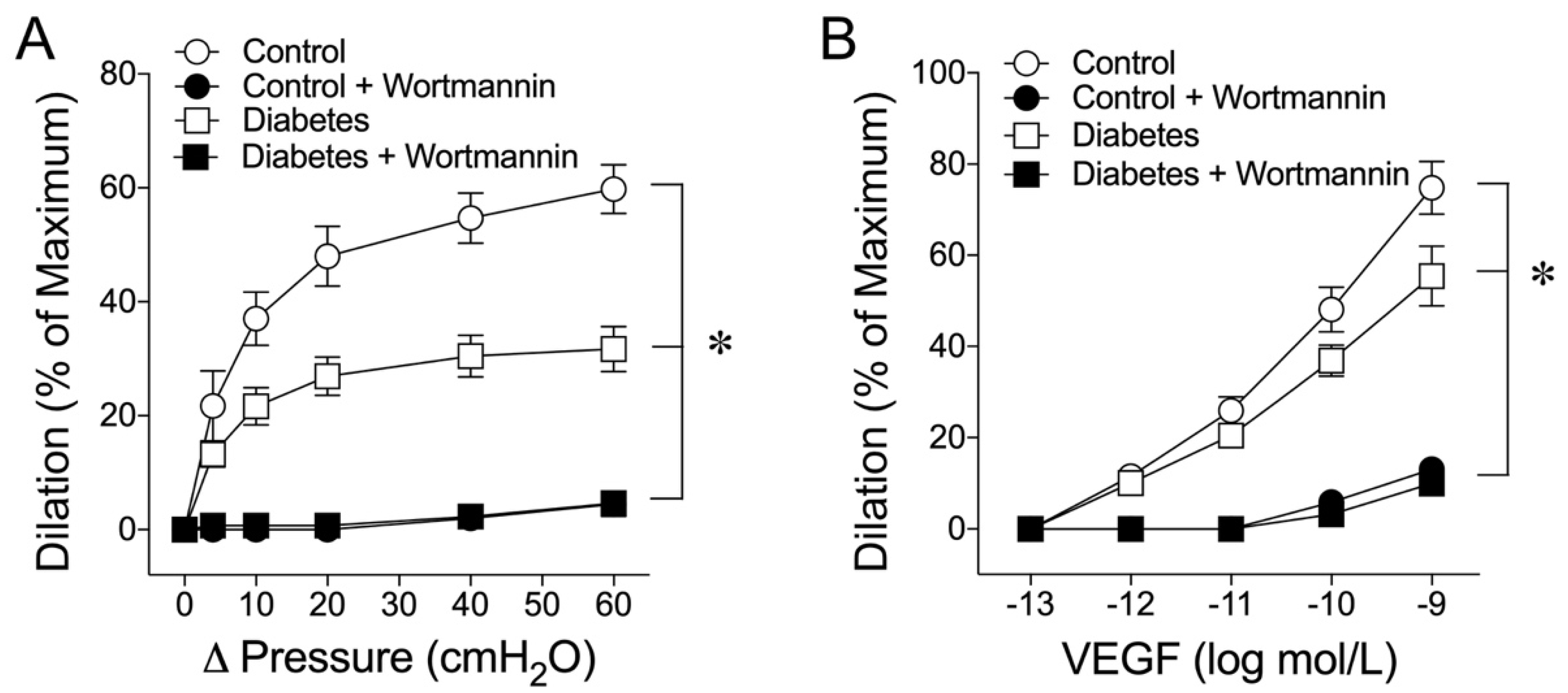
2.1. Early Diabetes Impairs Flow- and VEGF-Induced Vasodilations

In the present study, the average maximum internal diameters for the arterioles from the control and diabetic pigs were 84 ± 2 µm and 79 ± 3 µm, respectively. The vessels were pressurized to 60 cmH2O hydrostatic pressure without flow by two independent reservoirs [12]. The reason for selecting this luminal pressure was based on the in vivo coronary pressure measurements of about 40–50 mmHg in this size of coronary arterioles [41]. The pressurized control vessels developed spontaneous basal tone about 60% to 65% of the maximum diameter within 30–40 min under a 36–37 °C bath temperature. The vessels from the diabetic pigs developed a higher level of basal tone about 50% to 55% of the maximal diameter. The luminal flow of the vessel was initiated by moving the pressure reservoirs in an opposite direction to create a pressure gradient (Δ pressure) across the vessel longitudinally without changing the luminal pressure as described previously [12]. This dual-reservoir system eliminated the confounding effect of the pressure-dependent myogenic response during changes in the luminal flow [14]. The control vessels dilated to elevated flow in a manner dependent upon the magnitude of the pressure gradient, and administration of NOS inhibitor Nω-nitro-L-arginine methyl ester (L-NAME, 10 µmol/L) completely blocked this flow-induced vasodilation (Figure 1A). The vessels isolated from diabetic animals also exhibited significant vasodilation to the elevated flow, but the magnitude was compromised compared with that of the control animals (Figure 1A). Administration of L-NAME also completely abolished the flow-induced dilation in diabetic vessels (Figure 1A). Administration of VEGF caused concentration-dependent dilations of both the control and diabetic vessels in a manner sensitive to L-NAME. However, the diabetic vessels were less responsive to VEGF (Figure 1B).

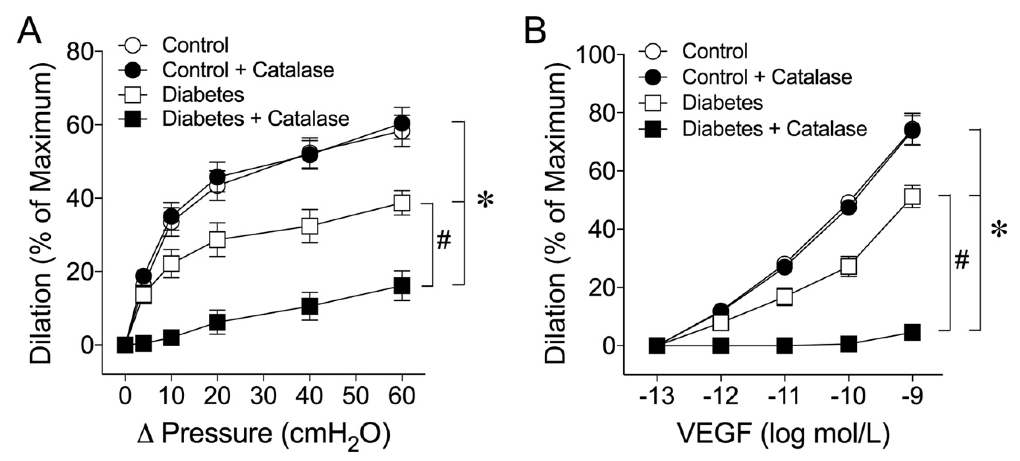
2.2. Role of H2O2 in Flow- and VEGF-Induced Coronary Arteriolar Dilations

To address whether H2O2 contributes to the flow- and VEGF-induced vasomotor responses, the vessels were treated with H2O2 scavenger catalase (1000 units/mL). Catalase did not affect the flow-induced dilation in the control vessels. However, in the vessels from the diabetic animals, the vasodilation in response to increased flow was compromised and the addition of catalase further blunted the vasodilation (Figure 2A). Similarly, VEGF-induced dilation was compromised in diabetic vessels. Administration of catalase did not affect VEGF-induced dilation in the control vessels but abolished the vasodilator response in the diabetic vessels (Figure 2B).

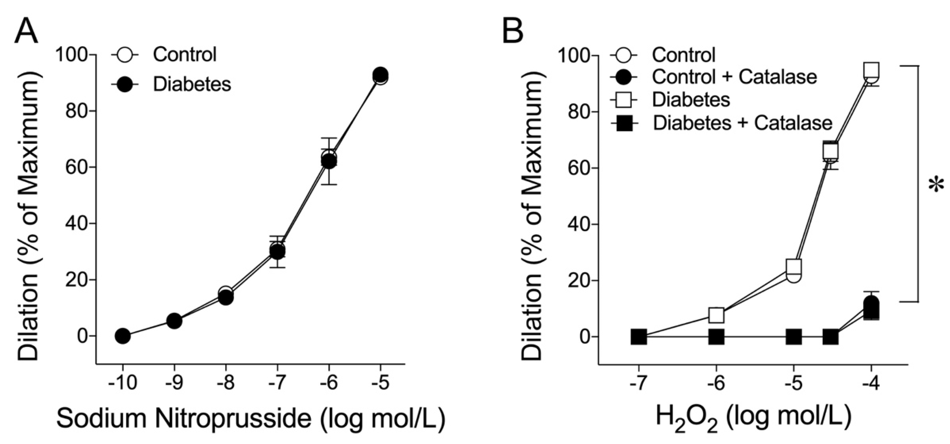
2.3. Coronary Arteriolar Dilations to NO Donor and H2O2

It is unclear whether early diabetes affects the response of the blood vessel to the NO donor or reactive oxygen species H2O2. To address this issue, the dilations of coronary arterioles to NO donor sodium nitroprusside and H2O2 were examined and compared in vessels isolated from the control and diabetic pigs. Administration of sodium nitroprusside caused similar concentration-dependent dilations of both control and diabetic vessels (Figure 3A). Coronary arterioles also dilated in response to H2O2 in a similar manner in both the control and diabetic vessels, and this vasodilation was abolished by catalase (1000 units/mL), except for the highest concentration of H2O2 (Figure 3B).

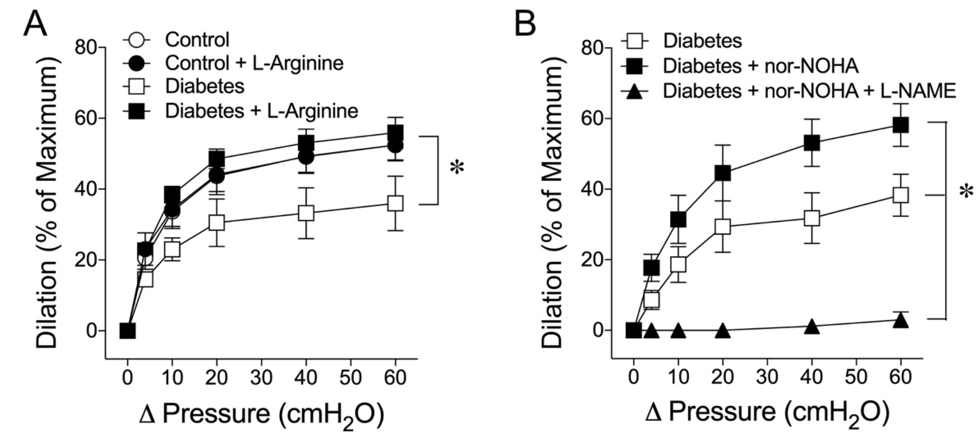
2.4. Vascular L-Arginine Availability and the Role of Arginase in Early Diabetes

The impacts of early diabetes on L-arginine availability and arginase activity for flow-induced dilation were investigated in coronary arterioles isolated from pigs with and without diabetes. Administration of L-arginine (3 mmol/L) to the control vessels did not affect flow-induced dilation. However, the vasodilation that was blunted by diabetes showed significant improvement by L-arginine, and the vasodilation magnitude was restored to the control level (Figure 4A). Furthermore, treating the diabetic vessels with arginase inhibitor Nω-hydroxy-nor-L-arginine (nor-NOHA; 0.1 mmol/L) restored flow-induced dilation, in a manner sensitive to L-NAME (Figure 4B).

2.5. Role of Phosphoinositide 3-Kinase (PI3K) in Flow- and VEGF-Induced Vasodilations

Activation of PI3K can lead to eNOS activation and NO production. However, it is unclear whether this signaling pathway contributes to the dilations of coronary arterioles elicited by flow and VEGF. It is also unclear whether this pathway is compromised in early diabetes. Therefore, the involvement of PI3K in these vasomotor responses was investigated and compared in coronary arterioles isolated from the control and diabetic pigs. Administration of PI3K inhibitor wortmannin (0.1 µmol/L) did not alter resting vascular tone but eliminated dilations in response to elevated flow (Figure 5A) and VEGF (Figure 5B) in both the control and diabetic vessels.

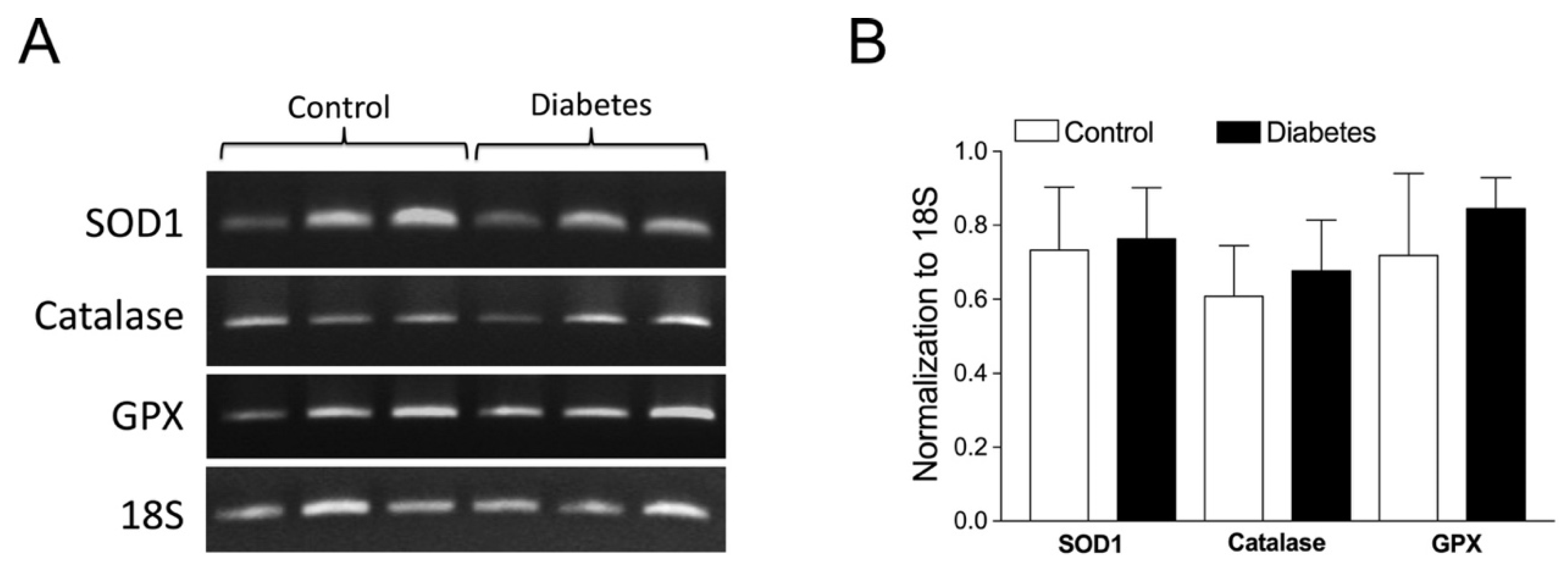
2.6. Role of Vascular Superoxide Dismutase 1 (SOD1), Catalase, and Glutathione Peroxidase (GPX) mRNA Expressions
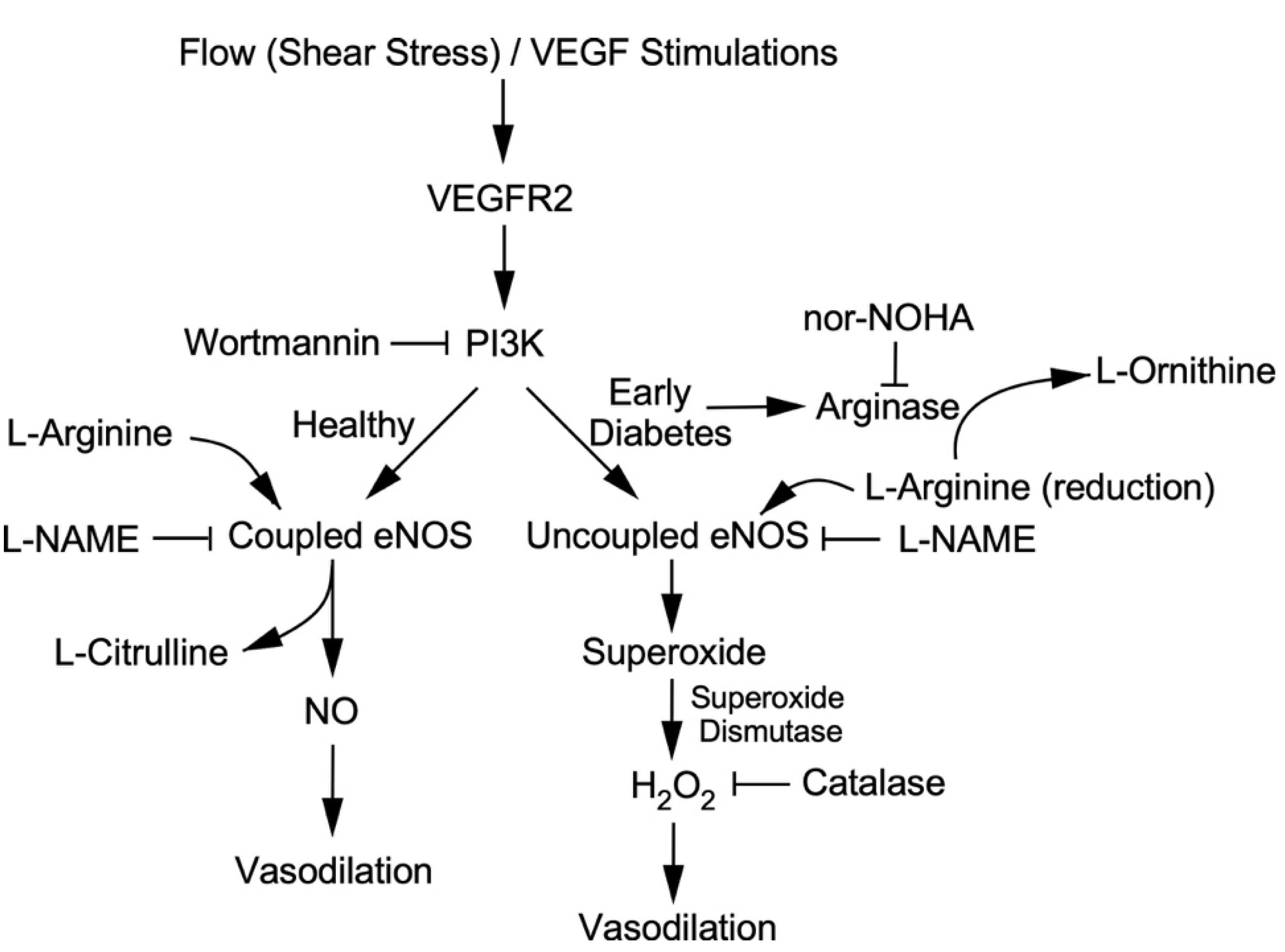
To assess whether vascular antioxidants are altered by early diabetes, the mRNA expressions of SOD1, catalase, and GPX were evaluated in coronary arterioles isolated from pigs with and without early diabetes (Figure 6A). The quantitative analysis of mRNA expression showed that the mRNA expressions of these antioxidant genes were comparable between the control and diabetic vessels (Figure 6B).

3. Discussion

The present study showed that the endothelium-dependent coronary arteriolar dilations to elevated flow and VEGF are impaired in the early stages (2 weeks of hyperglycemia) of type 1 diabetes in pigs, a large animal model resembling human cardiovascular physiology and pathophysiology. The compromised NO-mediated vasodilation can be restored by the supplementation of eNOS substrate L-arginine or by the inhibition of arginase activity. The released H2O2 during both flow and VEGF stimulations compensates for the NO deficiency and maintains, at least in part, the endothelial function. Early diabetes is likely to increase coronary arteriolar arginase activity leading to eNOS uncoupling and consequently promotes the formation of H2O2 to substitute for NO to mediate arteriolar dilation to increased flow and to VEGF.
We have previously shown that NO-dependent coronary arteriolar dilations in response to pharmacological stimulation by serotonin are impaired in pigs subjected to 2 weeks of type 1 diabetes [10]. However, it remains to be determined whether the physiological stimulation with the elevated flow in the coronary microcirculation is affected by early diabetes. This question is important because this vasomotor response is expected to play a critical role in regulating coronary blood flow during metabolic activation [12,13,42,43,44]. Our results showed that coronary arteriolar dilation to increased flow is partially compromised in the early stages of diabetes (Figure 1A). This finding agrees with the reduced flow-induced dilation in the brachial artery as one of the early changes in cardiovascular parameters in human patients with type 1 diabetes [45], although it is unclear whether this impairment extends to the microcirculation. In human patients with type 1 diabetes, the increased flow elicited by the endothelium-dependent vasodilator acetylcholine or by a physiological stimulation following a sudden release of arterial occlusion (i.e., reactive hyperemia) was compromised in the skin microcirculation [46]. Moreover, the coronary blood flow reserve, which is known to be directly related to the ability of coronary microvessels to dilate, was reportedly reduced in young patients with type 1 diabetes without microalbuminuria and autonomic neuropathy [47] or any clinical cardiovascular abnormalities [48]. Interestingly, the adenosine-induced increase in coronary blood flow was compromised in young men with uncomplicated type 1 diabetes [49]. These clinical studies may suggest the development of coronary arteriolar dysfunction related to NO deficiency because it has been shown that adenosine stimulates the endothelial release of NO to exert dilation in small coronary arterioles [50,51]. Because endothelial NO contributes significantly to the capacity of coronary flow reserve [52] and vasodilation elicited by shear stress (Figure 1), thus linking to metabolic activation [43], the negative impact of early diabetes on blood flow regulation in the coronary microcirculation is apparent.
Myocardial perfusion is optimally regulated to match the metabolic activity of the myocardium by adjusting microvascular resistance located at the size of arterioles less than 150 µm in diameter [53]. In the presence of NOS blockade, administration of catalase to the intact coronary circulation causes a substantial reduction in coronary arteriolar dilation in response to pacing-induced increases in myocardial oxygen consumption [54], indicating the important role of endogenous H2O2 in mediating coronary arteriolar dilation in vivo. However, the specific cellular source of H2O2 cannot be identified based on the in vivo experiments alone because of the complexity of coronary blood flow regulation and the disturbance of multiple confounding factors that cannot be separated in the intact animal preparations. Moreover, the mechanism of coupling H2O2 production to vasomotor regulation in coronary arterioles has not been established. Combining both in vivo and in vitro approaches, it was demonstrated that H2O2 released from the cardiomyocytes couples myocardial oxygen consumption to coronary blood flow via a feed-forward mechanism [55]. Interestingly, H2O2 predominantly acts on small coronary arterioles less than 100 µm in diameter [54], the size of vessels that is consistent with the current study. Furthermore, H2O2 has been shown to exert a cardioprotective role in coronary ischemia-reperfusion injury by compensating for impaired NO-mediated dilation in vivo [56]. However, the underlying mechanism is unclear and whether coronary arterioles are the source of H2O2 in the disease state has not been determined. In the present study, we found that the NO-mediated dilations in small coronary arterioles (~50 µm with basal tone) in response to flow and VEGF were compromised by about 40% in early diabetes. However, H2O2 appears to play a compensatory role in maintaining the remaining dilations of diseased vessels because catalase nearly abolished these responses only in vessels isolated from the diabetic animals (Figure 2). Because the isolated vessels were free from cardiomyocytes, this compensatory response appears to be derived from the blood vessel per se. It is worth noting that the vasodilation in response to the NO donor sodium nitroprusside was not altered by diabetes (Figure 3A), suggesting that the compromised vasodilation was not related to the alteration of smooth muscle NO signaling. In isolated coronary arterioles, the dilation to H2O2 was found to be mediated by the activation of endothelium-dependent signaling through the cycloxygenase-1-mediated release of prostaglandin E2 and the direct relaxation of smooth muscle through the opening of calcium-activated potassium channels [32]. These endothelium-dependent and -independent signaling pathways appear to be intact in early diabetes because vasodilation induced by H2O2 was not affected (Figure 3B).
The alteration in eNOS function might be the culprit in the present study because both flow- and VEGF-induced dilations were sensitive to eNOS blockade in both healthy and diabetic vessels (Figure 1). Because of the involvement of H2O2 in mediating dilations in diabetic vessels (Figure 2) in a manner sensitive to L-NAME (Figure 1), it is likely that the H2O2-mediated compensatory response originated from the eNOS signaling in the diseased vessel. This context was supported by the restoration of NO-mediated dilations by a high concentration of eNOS substrate L-arginine in the diabetic vessels (Figure 4A). The deficiency of L-arginine might have caused the uncoupling of eNOS for superoxide production and the subsequent formation of H2O2 [25,26,27,28,29] via superoxide dismutase in diabetic vessels. In addition, the increased activity of L-arginine-consuming enzyme arginase might have contributed to the eNOS uncoupling by reducing L-arginine bioavailability because the impaired vasodilation can be restored by the arginase inhibitor nor-NOHA (Figure 4B). Although arginase is constitutively expressed in the coronary vasculature [57], it can be upregulated and consequently contribute to endothelial NO deficiency and vascular dysfunction in various forms of vascular diseases and medical conditions such as aging [58], ischemia-reperfusion injury [34], systemic hypertension [36], hemorrhagic shock [59], and diabetes mellitus [37] by initiating or following oxidative stress and inflammation associated with L-arginine deficiency [60]. However, in the present study, arginase activity was not assessed, because of insufficient microvascular protein samples available for these experiments. This limitation may be addressed if a high-sensitivity assay kit is available in the future.
A previous report, using the same diabetic pig model as in the present study, showed that sequential activation of lectin-like oxidized low-density lipoprotein receptor-1, c-Jun N-terminal kinase, and arginase elicits superoxide-dependent oxidative stress with impaired endothelial NO-mediated coronary arteriolar dilation to serotonin [10]. However, the source of the oxidant is unclear and the administration of catalase in the previous study did not affect the diabetes-induced coronary endothelial dysfunction in response to serotonin [10]. Interestingly, in the same diabetic pig model, the impaired endothelium-dependent NO-mediated dilations of retinal arterioles to bradykinin and histamine are not affected by L-NAME [61]. However, treating diabetic retinal vessels with nor-NOHA, but not superoxide dismutase mimetic TEMPOL, preserves both histamine- and bradykinin-induced dilations in an L-NAME-sensitive manner. These studies suggest the crucial role of arginase activation in impairing endothelial NO function that can be associated with or dissociated from oxidative stress, depending upon the tissue (i.e., coronary vs. retinal arterioles) or the pharmacological agent. Moreover, those previous studies do not suggest a role of H2O2 in compensating for the NO deficiency, although the contribution of arginase activation to endothelial dysfunction is evident [10,61]. The reason for these inconsistent results on the H2O2 contribution is unclear, but they may be related to the different mechanisms of eNOS activation between pharmacological vs. mechanical (shear stress) stimulations. Serotonin, histamine, and bradykinin are known to activate eNOS through phosphorylation of the Ser-1179/1177 site [62,63] and activation of the enzyme through dephosphorylation at Thr-497 [64]. Interestingly, both shear stress and VEGF phosphorylate eNOS at Ser-635 with a slower activation of the enzyme compared to the phosphorylation of Ser-1179 [65]. This is consistent with our observations that both shear stress and VEGF took about 2–3 min to dilate the vessel in contrast to the vasodilation induced by serotonin, histamine, and bradykinin within 30 s of applying the agent [10,61]. It is worth noting that catalase did not affect resting vascular tone in either the control or diabetic vessels and the role of H2O2 was only observed under eNOS activations by shear stress and VEGF in the diabetic vessels (Figure 2). It is likely that the different sites of signaling in uncoupled eNOS might contribute to the generation of superoxide and the formation of H2O2 in response to shear stress and VEGF stimulations.
The context of the compensatory role of H2O2 generation from eNOS uncoupling can be supported by the blocking effect of PI3K inhibitor wortmannin on vasodilations to VEGF and shear stress (Figure 5). In cultured endothelial cells, PI3K is phosphorylated and activated by VEGFR2 following shear stress [66] or VEGF [67] stimulations. In the intact blood vessels, we found that VEGF and luminal flow/shear stress can activate VEGFR2 for vasodilation via ligand-dependent and ligand-independent mechanisms, respectively [20]. It appears that these two stimulation pathways converge to eNOS via PI3K signaling for NO release because inhibition of PI3K by wortmannin abolished both dilations in the control and diabetic vessels (Figure 5) similar to the blockade of eNOS by L-NAME (Figure 1). Our findings in the intact coronary arterioles are consistent with previous studies showing that wortmannin blocks NO production from cultured endothelial cells stimulated by shear stress [23] or VEGF [68]. Therefore, blocking upstream eNOS signaling by wortmannin is expected to eliminate the component of H2O2 in vasodilations to VEGF and shear stress in a similar fashion as that exerted by L-NAME (Figure 1) and catalase (Figure 2) if the uncoupled eNOS is the source of H2O2. Interestingly, in cultured pulmonary artery endothelial cells, H2O2 has been shown to activate eNOS for NO production, and overexpression of catalase can significantly attenuate the shear stress-induced increase in H2O2, eNOS activation, and NO generation [69]. However, under our experimental settings, the antioxidant gene expressions, including catalase, were unaltered in diabetic coronary arterioles (Figure 6). Moreover, endothelial NO does not participate in the dilation of porcine coronary arterioles to H2O2 [32]. Therefore, the increase in NO production by eNOS, due secondarily to H2O2 formation, is unlikely to be involved in the present study, at least in the early stages of diabetes/hyperglycemia.
In the present study, we conclude that mild oxidative stress, i.e., H2O2 formation during PI3K-linked eNOS uncoupling, corresponding to arginase activation and L-arginine deficiency, might be beneficial in terms of compensating for the NO deficiency to maintain the vasomotor function in the coronary microcirculation in early diabetes (Figure 7). However, this compensatory mechanism is not sufficient to fully restore the reduced endothelium-dependent NO-mediated function. This notion is consistent with the finding that the H2O2 released from the collateral microvessels improves the vasomotor function with exercise training in the ischemic heart [30]. On the contrary, prolonged hyperglycemia may exacerbate redox dysregulation in the vasculature, thereby providing positive feedback for developing adverse diabetic complications [70]. It is worth noting that the excessive production and prolonged exposure of microvessels to H2O2 can severely impair NO-mediated endothelial function by further reducing L-arginine availability through hydroxyl radical-dependent upregulation of arginase [35]. Therefore, redox signaling can be a double-edged sword in the microcirculation, which may help tissue survival during early disease stress by compensating for vasomotor regulation in one way and eliciting uncontrolled global oxidative stress and irreversible vascular damage for tissue injury in another way in the advanced stages of disease development. The early treatment of microvascular dysfunction by arginase inhibition might be a worthwhile strategy to consider for preventing the progression of microvascular complications toward severe tissue injury.

4. Materials and Methods

4.1. Porcine Diabetes Model

The approval of animal procedures was obtained from the Texas A&M University Health Science Center/Baylor Scott and White Institutional Animal Care and Use Committee (Temple, TX, USA). Male Yorkshire pigs, aged 8–12 weeks old and weighing 8–11 kg, were obtained from Real Farms (San Antonio, TX, USA). The cytotoxic glucose analog streptozocin (STZ; Zanosar®, 200 mg/kg in saline; Teva Parenteral Medicines, Irvine, CA, USA) was given intravenously via an ear vein (37 pigs) as described in detail in our previous studies to develop type 1 diabetes [71,72]. In the control group, saline was given intravenously instead of STZ (32 pigs). The pigs were nurtured for a period of 2 weeks. All animals were granted free access to water and fed with glucose syrup mixed with hog chow to prevent a sudden drop in blood sugar, especially during the first 24 h after STZ injection. The pigs were allowed free access to water and commercial pig chow afterwards. The general condition, body weight, and the level of blood glucose assessed with a Bayer Contour glucometer (Bayer HealthCare, Mishawaka, IN, USA) were recorded in all pigs.
The pigs that developed sustained hyperglycemia with a fasting blood glucose level between 250 and 600 mg/dL were included as a diabetic group in the present study. Pigs with sustained blood glucose above 600 mg/dL were treated with insulin (Humulin® 70/30, 2–8 units; Lilly; Indianapolis, IN, USA) to keep the glucose level between 250 and 600 mg/dL. Insulin was not given within 48 h before the terminal surgery. After the 2-week period, the pigs were sedated with Telazol (4–8 mg/kg intramuscularly; TW Medical Veterinary Supply, Austin, TX, USA), gas anesthetized with 2% to 5% isoflurane (Baxter Healthcare Co., Deerfield, IL, USA), heparinized with heparin (1000 U/kg, intravenously via marginal ear vein; Cardinal Health, Dublin, OH, USA), and intubated. The heart was excised and immediately kept on iced (5 °C) saline.

4.2. Isolation and Cannulation of Coronary Arterioles

Microscopic dissection of individual subepicardial arterioles (≈1 mm in length; ~20–60 µm in situ diameter) from the surrounding cardiac tissue was performed [41]. The coronary arterioles were then cannulated and ligated on each end with glass micropipettes containing a physiological saline solution (PSS)-albumin (1%; Thermo Fisher Scientific, Bridgewater, NJ, USA). The coronary arterioles were then pressurized to 60 cmH2O intraluminal pressure without flow by two independent pressure reservoir systems [12]. The inner diameter of coronary arterioles was continuously monitored using videomicroscopic techniques throughout the experiments, as described previously [12,41].

4.3. Study of Vasomotor Function

The cannulated coronary arterioles were bathed in physiological saline solution with albumin at 36 to 37 °C to allow the development of basal tone (stable within 60 min). To evaluate the effect of diabetes on vasomotor function, flow-induced vasodilation to a step-wise increase in pressure gradient (from 0 to 60 cmH2O) and concentration-dependent vasodilations to VEGF (1 pmol/L to 1 nmol/L) [20,73] and sodium nitroprusside (10 nmol/L to 10 µmol/L) [74] were recorded in vessels isolated from the 2-week diabetic and saline control pigs. To assess the involvement of uncoupled eNOS in the diabetes-induced effect, vasodilation was examined after treating the vessels with L-NAME (10 μmol/L) [50], catalase (1000 U/mL) [10], or L-arginine (3 mmol/L) [75] for 30 min. The role of vascular arginase in flow-induced dilation was assessed after treating the vessels with arginase inhibitor nor-NOHA (0.1 mmol/L; Cayman Chemical, Ann Arbor, MI, USA) [10,61]. The impact of PI3K on the VEGF- and flow-induced vasodilations was evaluated after incubation with PI3K inhibitor wortmannin (0.1 µmol/L) for 30 min [20].

4.4. RNA Isolation and Reverse Transcription Polymerase Chain Reaction (RT-PCR) Analysis of Antioxidant Transcripts

Total RNA was isolated from the coronary arterioles (3–4 vessels per sample pooled from one heart) of similar size to those used for functional studies, as we described previously [57]. Sets of primers specific for SOD1 (sense: 5′-CAGGTCCTCACTTCAATCC-3′, antisense: 5′-CCAAACGACTTCCASCAT-3′) (Accession no.NM_001190422.1), catalase (sense: 5′-CGAAGGCGAAGGTGTTTG-3′, antisense: 5′-AGTGTGCGATCCATATCC-3′) (Accession no. NM_214301.2), and glutathione peroxidase (GPX, sense: 5′-CACAACGGTGCGGGACTA-3′, antisense: 5′-CATTGCGACACACTGGAGAC-3′) (Accession no. NM_214201.1) genes were engineered (Sigma-Genosys, The Woodlands, TX, USA). Using 0.5 µg of total RNA per sample, RT-PCR was conducted, as we described previously [57]. The PCR reaction was optimized and run for 35 cycles for all genes. The level of expression of SOD1, catalase, and GPX transcripts was normalized to that of 18S transcripts (sense: 5′-GCGGCTTTGGTGACTCTA-3′, antisense: 5′-CTGCCTCCTTGGATGTG-3′) (Accession no. NR_002170.3).

4.5. Chemicals

All drugs were obtained from Sigma-Aldrich (St. Louis, MO, USA) except as specifically stated. Wortmannin was dissolved at 10 mmol/L in dimethyl sulfoxide, with subsequent concentrations diluted in PSS. All other drugs were dissolved in PSS. The final concentration of dimethyl sulfoxide in the vessel bath did not exceed 0.03% by volume. The 0.03% dimethyl sulfoxide had no significant effect on vessel viability, vasodilator responses, or maintenance of tone as previously reported [76].

4.6. Data Analysis

At the end of each experiment, the changes in the arteriolar diameter in response to the pressure gradient (flow-induced dilation) or vasoactive agonists were normalized to the maximum diameter changes in response to 0.1 mmol/L sodium nitroprusside in the presence of calcium-free PSS with EDTA (1 mmol/L) [77]. Diameter changes were expressed as % of maximum dilation. Data are reported as mean ± SEM and n value describes the number of vessels studied (one per pig per treatment group) in functional analysis or the number of pigs used in molecular studies. Student’s t-test or ANOVA followed by the Bonferroni multiple range test was used to determine the significance of experimental interventions, as appropriate. A value of p < 0.05 was considered significant.

Author Contributions

Conceptualization, N.T., T.W.H. and L.K.; conducting the experiments, N.T.; methodology, N.T., T.W.H. and L.K.; data analysis, N.T., L.K. and T.W.H.; writing—original draft preparation, N.T.; writing—review and editing, N.T., T.W.H. and L.K.; funding acquisition, L.K., T.W.H. and N.T. All authors have read and agreed to the published version of the manuscript.

Funding

This work was supported by the NIH R01EY018420 to T.W.H., and by the Office of the Ministry of Higher Education, Science, Research and Innovation and the Thailand Science Research and Innovation through the Kasetsart University Reinventing Program 2021 to N.T.

Institutional Review Board Statement

The experimental procedures and protocols were carried out under the guidance of the Animal Care and Use Committee at the Texas A&M University Health Science Center and Baylor Scott and White Health.

Informed Consent Statement

Not applicable.

Data Availability Statement

The datasets generated and/or analyzed for the current study are available from the corresponding authors upon reasonable request.

Acknowledgments

The authors gratefully acknowledge Wenjuan Xu and Xin Xu for their technical support and assistance in the collection of the heart samples.

Conflicts of Interest

The authors declare no conflict of interest. The funders had no role in the design of the study; in the collection, analyses, or interpretation of data; in the writing of the manuscript; or in the decision to publish the results.

References

  1. Momose, M.; Abletshauser, C.; Neverve, J.; Nekolla, S.G.; Schnell, O.; Standl, E.; Schwaiger, M.; Bengel, F.M. Dysregulation of coronary microvascular reactivity in asymptomatic patients with type 2 diabetes mellitus. Eur. J. Nucl. Med. Mol. Imaging 2002, 29, 1675–1679. [Google Scholar] [CrossRef] [PubMed]
  2. Tickerhoof, M.M.; Farrell, P.A.; Korzick, D.H. Alterations in rat coronary vasoreactivity and vascular protein kinase C isoforms in Type 1 diabetes. Am. J. Physiol. Heart Circ. Physiol. 2003, 285, H2694–H2703. [Google Scholar] [CrossRef] [PubMed] [Green Version]
  3. Prior, J.O.; Quinones, M.J.; Hernandez-Pampaloni, M.; Facta, A.D.; Schindler, T.H.; Sayre, J.W.; Hsueh, W.A.; Schelbert, H.R. Coronary circulatory dysfunction in insulin resistance, impaired glucose tolerance, and type 2 diabetes mellitus. Circulation 2005, 111, 2291–2298. [Google Scholar] [CrossRef] [PubMed] [Green Version]
  4. Murthy, V.L.; Naya, M.; Foster, C.R.; Gaber, M.; Hainer, J.; Klein, J.; Dorbala, S.; Blankstein, R.; Di Carli, M.F. Association between coronary vascular dysfunction and cardiac mortality in patients with and without diabetes mellitus. Circulation 2012, 126, 1858–1868. [Google Scholar] [CrossRef] [PubMed] [Green Version]
  5. Sabe, S.A.; Feng, J.; Sellke, F.W.; Abid, M.R. Mechanisms and clinical implications of endothelium-dependent vasomotor dysfunction in coronary microvasculature. Am. J. Physiol. Heart Circ. Physiol. 2022, 322, H819–H841. [Google Scholar] [CrossRef] [PubMed]
  6. Koller, A.; Balasko, M.; Bagi, Z. Endothelial regulation of coronary microcirculation in health and cardiometabolic diseases. Intern. Emerg. Med. 2013, 8 (Suppl. S1), 51–54. [Google Scholar] [CrossRef] [Green Version]
  7. Kibel, A.; Selthofer-Relatic, K.; Drenjancevic, I.; Bacun, T.; Bosnjak, I.; Kibel, D.; Gros, M. Coronary microvascular dysfunction in diabetes mellitus. J. Int. Med. Res. 2017, 45, 1901–1929. [Google Scholar] [CrossRef] [Green Version]
  8. Salvatore, T.; Galiero, R.; Caturano, A.; Vetrano, E.; Loffredo, G.; Rinaldi, L.; Catalini, C.; Gjeloshi, K.; Albanese, G.; Di Martino, A.; et al. Coronary microvascular dysfunction in diabetes mellitus: Pathogenetic mechanisms and potential therapeutic options. Biomedicines 2022, 10, 2274. [Google Scholar] [CrossRef]
  9. Boodhwani, M.; Sodha, N.R.; Mieno, S.; Xu, S.H.; Feng, J.; Ramlawi, B.; Clements, R.T.; Sellke, F.W. Functional, cellular, and molecular characterization of the angiogenic response to chronic myocardial ischemia in diabetes. Circulation 2007, 116, I-31–I-37. [Google Scholar] [CrossRef] [Green Version]
  10. Hein, T.W.; Xu, X.; Ren, Y.; Xu, W.; Tsai, S.H.; Thengchaisri, N.; Kuo, L. Requisite roles of LOX-1, JNK, and arginase in diabetes-induced endothelial vasodilator dysfunction of porcine coronary arterioles. J. Mol. Cell. Cardiol. 2019, 131, 82–90. [Google Scholar] [CrossRef]
  11. Baeyens, N.; Bandyopadhyay, C.; Coon, B.G.; Yun, S.; Schwartz, M.A. Endothelial fluid shear stress sensing in vascular health and disease. J. Clin. Investig. 2016, 126, 821–828. [Google Scholar] [CrossRef] [PubMed]
  12. Kuo, L.; Davis, M.J.; Chilian, W.M. Endothelium-dependent, flow-induced dilation of isolated coronary arterioles. Am. J. Physiol. 1990, 259, H1063–H1070. [Google Scholar] [CrossRef] [PubMed]
  13. Liao, J.C.; Kuo, L. Interaction between adenosine and flow-induced dilation in coronary microvascular network. Am. J. Physiol. 1997, 272, H1571–H1581. [Google Scholar] [CrossRef] [PubMed]
  14. Kuo, L.; Chilian, W.M.; Davis, M.J. Interaction of pressure- and flow-induced responses in porcine coronary resistance vessels. Am. J. Physiol. 1991, 261, H1706–H1715. [Google Scholar] [CrossRef] [PubMed]
  15. Goodwill, A.G.; Dick, G.M.; Kiel, A.M.; Tune, J.D. Regulation of coronary blood flow. Compr. Physiol. 2017, 7, 321–382. [Google Scholar] [CrossRef] [Green Version]
  16. Nguyen, M.T.; Pham, I.; Valensi, P.; Rousseau, H.; Vicaut, E.; Laguillier-Morizot, C.; Nitenberg, A.; Cosson, E. Flow-mediated-paradoxical vasoconstriction is independently associated with asymptomatic myocardial ischemia and coronary artery disease in type 2 diabetic patients. Cardiovasc. Diabetol. 2014, 13, 20. [Google Scholar] [CrossRef] [Green Version]
  17. Lockhart, C.J.; Agnew, C.E.; McCann, A.; Hamilton, P.K.; Quinn, C.E.; McCall, D.O.; Plumb, R.D.; McClenaghan, V.C.; McGivern, R.C.; Harbinson, M.T.; et al. Impaired flow-mediated dilatation response in uncomplicated Type 1 diabetes mellitus: Influence of shear stress and microvascular reactivity. Clin. Sci. 2011, 121, 129–139. [Google Scholar] [CrossRef] [Green Version]
  18. Zhang, Y.; Liu, J.; Luo, J.Y.; Tian, X.Y.; Cheang, W.S.; Xu, J.; Lau, C.W.; Wang, L.; Wong, W.T.; Wong, C.M.; et al. Upregulation of angiotensin (1-7)-mediated signaling preserves endothelial function through reducing oxidative stress in diabetes. Antioxid Redox Signal. 2015, 23, 880–892. [Google Scholar] [CrossRef] [Green Version]
  19. Fogarty, J.A.; Muller-Delp, J.M.; Delp, M.D.; Mattox, M.L.; Laughlin, M.H.; Parker, J.L. Exercise training enhances vasodilation responses to vascular endothelial growth factor in porcine coronary arterioles exposed to chronic coronary occlusion. Circulation 2004, 109, 664–670. [Google Scholar] [CrossRef] [Green Version]
  20. Hein, T.W.; Rosa, R.H., Jr.; Ren, Y.; Xu, W.; Kuo, L. VEGF receptor-2-linked PI3K/calpain/SIRT1 activation mediates retinal arteriolar dilations to VEGF and shear stress. Investig. Opthalmol. Vis. Sci. 2015, 56, 5381–5389. [Google Scholar] [CrossRef]
  21. Mercier, C.; Rousseau, M.; Geraldes, P. Growth factor deregulation and emerging role of phosphatases in diabetic peripheral artery disease. Front. Cardiovasc. Med. 2020, 7, 619612. [Google Scholar] [CrossRef] [PubMed]
  22. Caldwell, R.B.; Bartoli, M.; Behzadian, M.A.; El-Remessy, A.E.; Al-Shabrawey, M.; Platt, D.H.; Caldwell, R.W. Vascular endothelial growth factor and diabetic retinopathy: Pathophysiological mechanisms and treatment perspectives. Diabetes Metab. Res. Rev. 2003, 19, 442–455. [Google Scholar] [CrossRef] [PubMed]
  23. Jin, Z.G.; Ueba, H.; Tanimoto, T.; Lungu, A.O.; Frame, M.D.; Berk, B.C. Ligand-independent activation of vascular endothelial growth factor receptor 2 by fluid shear stress regulates activation of endothelial nitric oxide synthase. Circ. Res. 2003, 93, 354–363. [Google Scholar] [CrossRef] [PubMed] [Green Version]
  24. Blanes, M.G.; Oubaha, M.; Rautureau, Y.; Gratton, J.P. Phosphorylation of tyrosine 801 of vascular endothelial growth factor receptor-2 is necessary for Akt-dependent endothelial nitric-oxide synthase activation and nitric oxide release from endothelial cells. J. Biol. Chem. 2007, 282, 10660–10669. [Google Scholar] [CrossRef] [PubMed] [Green Version]
  25. Rochette, L.; Lorin, J.; Zeller, M.; Guilland, J.C.; Lorgis, L.; Cottin, Y.; Vergely, C. Nitric oxide synthase inhibition and oxidative stress in cardiovascular diseases: Possible therapeutic targets? Pharmacol. Ther. 2013, 140, 239–257. [Google Scholar] [CrossRef] [PubMed]
  26. Kietadisorn, R.; Juni, R.P.; Moens, A.L. Tackling endothelial dysfunction by modulating NOS uncoupling: New insights into its pathogenesis and therapeutic possibilities. Am. J. Physiol. Endocrinol. Metab. 2012, 302, E481–E495. [Google Scholar] [CrossRef] [Green Version]
  27. Meza, C.A.; La Favor, J.D.; Kim, D.H.; Hickner, R.C. Endothelial dysfunction: Is there a hyperglycemia-induced imbalance of NOX and NOS? Int. J. Mol. Sci. 2019, 20, 3775. [Google Scholar] [CrossRef] [Green Version]
  28. Münzel, T.; Daiber, A.; Ullrich, V.; Mülsch, A. Vascular consequences of endothelial nitric oxide synthase uncoupling for the activity and expression of the soluble guanylyl cyclase and the cGMP-dependent protein kinase. Arter. Thromb. Vasc. Biol. 2005, 25, 1551–1557. [Google Scholar] [CrossRef]
  29. van Etten, R.W.; de Koning, E.J.; Verhaar, M.C.; Gaillard, C.A.; Rabelink, T.J. Impaired NO-dependent vasodilation in patients with Type II (non-insulin-dependent) diabetes mellitus is restored by acute administration of folate. Diabetologia 2002, 45, 1004–1010. [Google Scholar] [CrossRef] [Green Version]
  30. Thengchaisri, N.; Shipley, R.; Ren, Y.; Parker, J.; Kuo, L. Exercise training restores coronary arteriolar dilation to NOS activation distal to coronary artery occlusion: Role of hydrogen peroxide. Arter. Thromb. Vasc. Biol. 2007, 27, 791–798. [Google Scholar] [CrossRef]
  31. Cai, H. Hydrogen peroxide regulation of endothelial function: Origins, mechanisms, and consequences. Cardiovasc. Res. 2005, 68, 26–36. [Google Scholar] [CrossRef] [PubMed] [Green Version]
  32. Thengchaisri, N.; Kuo, L. Hydrogen peroxide induces endothelium-dependent and -independent coronary arteriolar dilation: Role of cyclooxygenase and potassium channels. Am. J. Physiol. Heart Circ. Physiol. 2003, 285, H2255–H2263. [Google Scholar] [CrossRef] [PubMed] [Green Version]
  33. Shimokawa, H.; Morikawa, K. Hydrogen peroxide is an endothelium-derived hyperpolarizing factor in animals and humans. J. Mol. Cell. Cardiol. 2005, 39, 725–732. [Google Scholar] [CrossRef] [PubMed]
  34. Hein, T.W.; Zhang, C.; Wang, W.; Chang, C.I.; Thengchaisri, N.; Kuo, L. Ischemia-reperfusion selectively impairs nitric oxide-mediated dilation in coronary arterioles: Counteracting role of arginase. FASEB J. 2003, 17, 2328–2330. [Google Scholar] [CrossRef] [PubMed]
  35. Thengchaisri, N.; Hein, T.W.; Wang, W.; Xu, X.; Li, Z.; Fossum, T.W.; Kuo, L. Upregulation of arginase by H2O2 impairs endothelium-dependent nitric oxide-mediated dilation of coronary arterioles. Arter. Thromb. Vasc. Biol. 2006, 26, 2035–2042. [Google Scholar] [CrossRef] [Green Version]
  36. Zhang, C.; Hein, T.W.; Wang, W.; Miller, M.W.; Fossum, T.W.; McDonald, M.M.; Humphrey, J.D.; Kuo, L. Upregulation of vascular arginase in hypertension decreases nitric oxide-mediated dilation of coronary arterioles. Hypertension 2004, 44, 935–943. [Google Scholar] [CrossRef] [Green Version]
  37. Johnson, F.K.; Johnson, R.A.; Peyton, K.J.; Shebib, A.R.; Durante, W. Arginase promotes skeletal muscle arteriolar endothelial dysfunction in diabetic rats. Front. Immunol. 2013, 4, 119. [Google Scholar] [CrossRef] [Green Version]
  38. Tooke, J.E. Microvascular function in human diabetes. A physiological perspective. Diabetes 1995, 44, 721–726. [Google Scholar] [CrossRef]
  39. Kannel, W.B.; McGee, D.L. Diabetes and cardiovascular disease. The Framingham study. JAMA 1979, 241, 2035–2038. [Google Scholar] [CrossRef]
  40. Fuller, J.H. Mortality trends and causes of death in diabetic patients. Diabete Metab. 1993, 19, 96–99. [Google Scholar]
  41. Kuo, L.; Davis, M.J.; Chilian, W.M. Myogenic activity in isolated subepicardial and subendocardial coronary arterioles. Am. J. Physiol. Heart Circ. Physiol. 1988, 255, H1558–H1562. [Google Scholar] [CrossRef] [PubMed]
  42. Cornelissen, A.J.; Dankelman, J.; VanBavel, E.; Spaan, J.A. Balance between myogenic, flow-dependent, and metabolic flow control in coronary arterial tree: A model study. Am. J. Physiol. Heart Circ. Physiol. 2002, 282, H2224–H2237. [Google Scholar] [CrossRef] [PubMed]
  43. Quyyumi, A.A.; Dakak, N.; Andrews, N.P.; Gilligan, D.M.; Panza, J.A.; Cannon, R.O., 3rd. Contribution of nitric oxide to metabolic coronary vasodilation in the human heart. Circulation 1995, 92, 320–326. [Google Scholar] [CrossRef] [PubMed]
  44. Canty, J.M., Jr.; Iyer, V.S. Hydrogen peroxide and metabolic coronary flow regulation. J. Am. Coll. Cardiol. 2007, 50, 1279–1281. [Google Scholar] [CrossRef] [PubMed] [Green Version]
  45. Bradley, T.J.; Slorach, C.; Mahmud, F.H.; Dunger, D.B.; Deanfield, J.; Deda, L.; Elia, Y.; Har, R.L.; Hui, W.; Moineddin, R.; et al. Early changes in cardiovascular structure and function in adolescents with type 1 diabetes. Cardiovasc. Diabetol. 2016, 15, 31. [Google Scholar] [CrossRef] [Green Version]
  46. de, M.M.A.S.; Clemente, E.L.; de Lourdes Guimaraes Rodrigues, M.; Torres Valenca, D.C.; Gomes, M.B. Assessment of microvascular endothelial function in type 1 diabetes using laser speckle contrast imaging. J. Diabetes Complicat. 2017, 31, 753–757. [Google Scholar] [CrossRef]
  47. Pitkanen, O.P.; Nuutila, P.; Raitakari, O.T.; Ronnemaa, T.; Koskinen, P.J.; Iida, H.; Lehtimaki, T.J.; Laine, H.K.; Takala, T.; Viikari, J.S.; et al. Coronary flow reserve is reduced in young men with IDDM. Diabetes 1998, 47, 248–254. [Google Scholar] [CrossRef]
  48. Dankowski, R.; Wierzchowiecki, M.; Naskret, D.; Michalski, M.; Kandziora, M.; Biegalski, W.; Poprawski, K. Association between retinopathy, microalbuminuria and coronary perfusion in young patients with type 1 diabetes mellitus. Kardiol. Pol. 2008, 66, 262–268. [Google Scholar]
  49. Sundell, J.; Laine, H.; Nuutila, P.; Ronnemaa, T.; Luotolahti, M.; Raitakari, O.; Knuuti, J. The effects of insulin and short-term hyperglycaemia on myocardial blood flow in young men with uncomplicated type I diabetes. Diabetologia 2002, 45, 775–782. [Google Scholar] [CrossRef] [Green Version]
  50. Hein, T.W.; Belardinelli, L.; Kuo, L. Adenosine A2A receptors mediate coronary microvascular dilation to adenosine: Role of nitric oxide and ATP-sensitive potassium channels. J. Pharmacol. Exp. Ther. 1999, 291, 655–664. [Google Scholar]
  51. Hein, T.W.; Kuo, L. cAMP-independent dilation of coronary arterioles to adenosine: Role of nitric oxide, G proteins, and KATP channels. Circ. Res. 1999, 85, 634–642. [Google Scholar] [CrossRef] [PubMed] [Green Version]
  52. Nakamura, R.; Egashira, K.; Arimura, K.; Machida, Y.; Ide, T.; Tsutsui, H.; Shimokawa, H.; Takeshita, A. Increased inactivation of nitric oxide is involved in impaired coronary flow reserve in heart failure. Am. J. Physiol. Heart Circ. Physiol. 2001, 281, H2619–H2625. [Google Scholar] [CrossRef] [PubMed]
  53. Jones, C.J.; Kuo, L.; Davis, M.J.; Chilian, W.M. Distribution and control of coronary microvascular resistance. Adv. Exp. Med. Biol. 1993, 346, 181–188. [Google Scholar] [CrossRef] [PubMed]
  54. Yada, T.; Shimokawa, H.; Hiramatsu, O.; Shinozaki, Y.; Mori, H.; Goto, M.; Ogasawara, Y.; Kajiya, F. Important role of endogenous hydrogen peroxide in pacing-induced metabolic coronary vasodilation in dogs in vivo. J. Am. Coll. Cardiol. 2007, 50, 1272–1278. [Google Scholar] [CrossRef] [Green Version]
  55. Saitoh, S.; Zhang, C.; Tune, J.D.; Potter, B.; Kiyooka, T.; Rogers, P.A.; Knudson, J.D.; Dick, G.M.; Swafford, A.; Chilian, W.M. Hydrogen peroxide: A feed-forward dilator that couples myocardial metabolism to coronary blood flow. Arterioscler. Thromb. Vasc. Biol. 2006, 26, 2614–2621. [Google Scholar] [CrossRef] [PubMed] [Green Version]
  56. Yada, T.; Shimokawa, H.; Hiramatsu, O.; Haruna, Y.; Morita, Y.; Kashihara, N.; Shinozaki, Y.; Mori, H.; Goto, M.; Ogasawara, Y.; et al. Cardioprotective role of endogenous hydrogen peroxide during ischemia-reperfusion injury in canine coronary microcirculation in vivo. Am. J. Physiol. Heart Circ. Physiol. 2006, 291, H1138–H1146. [Google Scholar] [CrossRef] [PubMed] [Green Version]
  57. Zhang, C.; Hein, T.W.; Wang, W.; Chang, C.I.; Kuo, L. Constitutive expression of arginase in microvascular endothelial cells counteracts nitric oxide-mediated vasodilatory function. FASEB J. 2001, 15, 1264–1266. [Google Scholar] [CrossRef] [Green Version]
  58. Berkowitz, D.E.; White, R.; Li, D.; Minhas, K.M.; Cernetich, A.; Kim, S.; Burke, S.; Shoukas, A.A.; Nyhan, D.; Champion, H.C.; et al. Arginase reciprocally regulates nitric oxide synthase activity and contributes to endothelial dysfunction in aging blood vessels. Circulation 2003, 108, 2000–2006. [Google Scholar] [CrossRef] [Green Version]
  59. Johnson, R.A.; Durante, W.; Craig, T.; Peyton, K.J.; Myers, J.G.; Stewart, R.M.; Johnson, F.K. Vascular arginase contributes to arteriolar endothelial dysfunction in a rat model of hemorrhagic shock. J. Trauma 2010, 69, 384–391. [Google Scholar] [CrossRef]
  60. Kuo, L.; Hein, T.W. Vasomotor regulation of coronary microcirculation by oxidative stress: Role of arginase. Front. Immunol. 2013, 4, 237. [Google Scholar] [CrossRef] [Green Version]
  61. Hein, T.W.; Omae, T.; Xu, W.; Yoshida, A.; Kuo, L. Role of arginase in selective impairment of endothelium-dependent nitric oxide synthase-mediated dilation of retinal arterioles during early diabetes. Investig. Opthalmol. Vis. Sci. 2020, 61, 36. [Google Scholar] [CrossRef] [PubMed]
  62. Richardson, S.M.; Maleque, M.A.; Motley, E.D. 3-Morpholinosyndnonimine inhibits 5-hydroxytryptamine-induced phosphorylation of nitric oxide synthase in endothelial cells. Cell. Mol. Biol. 2003, 49, 1385–1389. [Google Scholar] [PubMed]
  63. Asada, M.; Ebihara, S.; Yamanda, S.; Niu, K.; Okazaki, T.; Sora, I.; Arai, H. Depletion of serotonin and selective inhibition of 2B receptor suppressed tumor angiogenesis by inhibiting endothelial nitric oxide synthase and extracellular signal-regulated kinase 1/2 phosphorylation. Neoplasia 2009, 11, 408–417. [Google Scholar] [CrossRef] [PubMed]
  64. Harris, M.B.; Ju, H.; Venema, V.J.; Liang, H.; Zou, R.; Michell, B.J.; Chen, Z.P.; Kemp, B.E.; Venema, R.C. Reciprocal phosphorylation and regulation of endothelial nitric-oxide synthase in response to bradykinin stimulation. J. Biol. Chem. 2001, 276, 16587–16591. [Google Scholar] [CrossRef] [Green Version]
  65. Boo, Y.C.; Hwang, J.; Sykes, M.; Michell, B.J.; Kemp, B.E.; Lum, H.; Jo, H. Shear stress stimulates phosphorylation of eNOS at Ser635 by a protein kinase A-dependent mechanism. Am. J. Physiol. Heart Circ. Physiol. 2002, 283, H1819–H1828. [Google Scholar] [CrossRef] [Green Version]
  66. Tzima, E.; Irani-Tehrani, M.; Kiosses, W.B.; Dejana, E.; Schultz, D.A.; Engelhardt, B.; Cao, G.; DeLisser, H.; Schwartz, M.A. A mechanosensory complex that mediates the endothelial cell response to fluid shear stress. Nature 2005, 437, 426–431. [Google Scholar] [CrossRef]
  67. Dayanir, V.; Meyer, R.D.; Lashkari, K.; Rahimi, N. Identification of tyrosine residues in vascular endothelial growth factor receptor-2/FLK-1 involved in activation of phosphatidylinositol 3-kinase and cell proliferation. J. Biol. Chem. 2001, 276, 17686–17692. [Google Scholar] [CrossRef] [Green Version]
  68. Youn, J.Y.; Wang, T.; Cai, H. An ezrin/calpain/PI3K/AMPK/eNOSs1179 signaling cascade mediating VEGF-dependent endothelial nitric oxide production. Circ. Res. 2009, 104, 50–59. [Google Scholar] [CrossRef] [Green Version]
  69. Kumar, S.; Sud, N.; Fonseca, F.V.; Hou, Y.; Black, S.M. Shear stress stimulates nitric oxide signaling in pulmonary arterial endothelial cells via a reduction in catalase activity: Role of protein kinase Cδ. Am. J. Physiol. Cell. Mol. Physiol. 2010, 298, L105–L116. [Google Scholar] [CrossRef] [Green Version]
  70. Cheng, X.; Siow, R.C.; Mann, G.E. Impaired redox signaling and antioxidant gene expression in endothelial cells in diabetes: A role for mitochondria and the nuclear factor-E2-related factor 2-Kelch-like ECH-associated protein 1 defense pathway. Antioxid Redox Signal. 2011, 14, 469–487. [Google Scholar] [CrossRef]
  71. Hein, T.W.; Potts, L.B.; Xu, W.; Yuen, J.Z.; Kuo, L. Temporal development of retinal arteriolar endothelial dysfunction in porcine type 1 diabetes. Investig. Opthalmol. Vis. Sci. 2012, 53, 7943–7949. [Google Scholar] [CrossRef] [PubMed]
  72. Chen, Y.L.; Xu, W.; Rosa, R.H., Jr.; Kuo, L.; Hein, T.W. Hyperglycemia enhances constriction of retinal venules via activation of the reverse-mode sodium-calcium exchanger. Diabetes 2019, 68, 1624–1634. [Google Scholar] [CrossRef] [PubMed]
  73. Shi, L.; Zhao, M.; Abbey, C.A.; Tsai, S.H.; Xie, W.; Pham, D.; Chapman, S.; Bayless, K.J.; Hein, T.W.; Rosa, R.H., Jr.; et al. Newly identified peptide, peptide Lv, promotes pathological angiogenesis. J. Am. Heart Assoc. 2019, 8, e013673. [Google Scholar] [CrossRef] [PubMed]
  74. Thengchaisri, N.; Hein, T.W.; Ren, Y.; Kuo, L. Activation of coronary arteriolar PKCβ2 impairs endothelial NO-mediated vasodilation: Role of JNK/Rho kinase signaling and xanthine oxidase activation. Int. J. Mol. Sci. 2021, 22, 9763. [Google Scholar] [CrossRef]
  75. Hein, T.W.; Kuo, L. LDLs impair vasomotor function of the coronary microcirculation: Role of superoxide anions. Circ. Res. 1998, 83, 404–414. [Google Scholar] [CrossRef] [Green Version]
  76. Thengchaisri, N.; Hein, T.W.; Ren, Y.; Kuo, L. Endothelin-1 impairs coronary arteriolar dilation: Role of p38 kinase-mediated superoxide production from NADPH oxidase. J. Mol. Cell. Cardiol. 2015, 86, 75–84. [Google Scholar] [CrossRef] [PubMed] [Green Version]
  77. Tsai, S.H.; Lu, G.; Xu, X.; Ren, Y.; Hein, T.W.; Kuo, L. Enhanced endothelin-1/Rho-kinase signalling and coronary microvascular dysfunction in hypertensive myocardial hypertrophy. Cardiovasc. Res. 2017, 113, 1329–1337. [Google Scholar] [CrossRef]
Figure 1. The role of NO in mediating flow- and VEGF-induced dilations was investigated in coronary arterioles isolated from control and diabetic pigs. (A): The elevated luminal flow, driven by the longitudinal pressure gradient (Δ pressure) across the vessel, caused vasodilation in normal pigs (n = 9). This vasodilation was compromised in the vessels isolated from diabetic pigs (n = 6). Administration of NOS inhibitor L-NAME (10 µmol/L) abolished the flow-induced vasodilation in both the control (n = 9) and diabetic pigs (n = 6). (B): Coronary arterioles from the control pigs (n = 7) dilated to VEGF in a concentration-dependent manner. VEGF-induced vasodilation was compromised by diabetes (n = 5). Administration of L-NAME abolished the vasodilation in both the control (n = 7) and diabetic (n = 5) vessels. The resting vascular tones were not altered before and after L-NAME in either control (63 ± 1% vs. 62 ± 1%) or diabetic (55 ± 3% vs. 58 ± 4%) vessels. * p < 0.05 vs. control, # p < 0.05 vs. diabetes.
Ijms 24 00489 g001
Figure 2. The role of H2O2 in mediating flow- and VEGF-induced dilations was investigated in coronary arterioles isolated from control and diabetic pigs. (A): The vasodilation induced by a stepwise increase in pressure gradient (Δ pressure), i.e., flow, in control pigs (n = 6) was not affected by H2O2 scavenger catalase (1000 units/mL; n = 6). The blunted flow-induced dilation in diabetic vessels (n = 8) was further inhibited by catalase (n = 8). (B): The vasodilation induced by VEGF was not affected by catalase in the control pigs (n = 5). In diabetic pigs (n = 6), the vasodilation to VEGF was blunted and the remaining dilation was abolished by catalase. The resting vascular tones were not altered before and after catalase in either the control (61 ± 2% vs. 62 ± 2%) or diabetic (60 ± 2% vs. 60 ± 2%) vessels. * p < 0.05 vs. control, # p < 0.05 vs. diabetes.
Ijms 24 00489 g002
Figure 3. Responses of coronary arterioles to NO donor sodium nitroprusside and H2O2. (A) Both the control (n = 5) and diabetic (n = 5) vessels dilated to sodium nitroprusside in a concentration-dependent manner without differences. (B) H2O2 produced similar dilations in the control (n = 5) and diabetic (n = 6) vessels and catalase (1000 units/mL) abolished the dilations in the control (n = 5) and diabetic (n = 6) vessels in the same manner. * p < 0.05, control/diabetes vs. catalase treatment.
Ijms 24 00489 g003
Figure 4. The impacts of L-arginine availability and arginase activity on flow-induced dilation were evaluated in coronary arterioles from pigs with and without diabetes. (A) Administration of L-arginine (3 mmol/L; n = 6) did not alter flow-induced dilation in the control vessels (n = 6) but the impaired vasodilation by diabetes (n = 5) was restored by L-arginine (n = 5). (B) The blunted flow-induced dilation in diabetic vessels (n = 6) was reversed by arginase inhibitor nor-NOHA (0.1 mmol/L; n = 6). Co-incubation with nor-NOHA and L-NAME abolished the flow-induced dilation of coronary arterioles in diabetic pigs (n = 4). The resting vascular tones were not altered before and after L-arginine in either control (59 ± 4% vs. 58 ± 3%) or diabetic (62 ± 3% vs. 63 ± 4%) vessels. nor-NOHA also did not alter the resting tone in diabetic vessels (62 ± 3% vs. 62 ± 4%). * p < 0.05 vs. diabetes.
Ijms 24 00489 g004
Figure 5. Effect of PI3K inhibition on flow- and VEGF-induced coronary arteriolar dilations. (A) The dilation of coronary arterioles to increased luminal flow (Control; n = 4) was attenuated by diabetes (n = 4) and administration of PI3K inhibitor wortmannin (0.1 µmol/L) abolished dilation of the control (n = 5) and diabetic (n = 5) vessels. (B) The VEGF-induced vasodilation (Control; n = 5) was abolished by wortmannin (n = 5) and the dilation of the diabetic vessels (n = 5) was also abolished by wortmannin (n = 5). The resting vascular tones were not altered before and after wortmannin in either control (64 ± 2% vs. 64 ± 2%) or diabetic (62 ± 3% vs. 65 ± 2%) vessels. * p < 0.05 vs. diabetes.
Ijms 24 00489 g005
Figure 6. Effect of early diabetes on the mRNA expressions of SOD1, catalase, and GPX in isolated coronary arterioles. (A) The expressions of antioxidant genes (SOD1, catalase, and GPX) were determined by RT-PCR and the expressions were normalized to the 18S transcripts. (B) The quantitative analysis showed no difference in the antioxidant gene expressions between the control (n = 3) and diabetic (n = 3) coronary arterioles.
Ijms 24 00489 g006
Figure 7. The diagram summarizes the pathways involved in the compensatory generation of H2O2 for NO deficiency in coronary arterioles due to arginase activation in the early stages of type 1 diabetes. L-Arginine is used by eNOS and arginase for the synthesis of NO and L-ornithine, respectively. In healthy coronary arterioles, shear stress and VEGF elicit endothelium-dependent NO-mediated vasodilation via activation of VEGR2/PI3K signaling. In the early stages of diabetes, arginase is activated and competes with eNOS for L-arginine. The reduction in L-arginine availability compromises the production of NO but causes PI3K-linked eNOS uncoupling and consequently promotes superoxide production and H2O2 formation via superoxide dismutase. The compromised NO-mediated vasodilation in early diabetes is compensated by H2O2. The inhibitors used for probing the signaling pathway are indicated.
Ijms 24 00489 g007
Disclaimer/Publisher’s Note: The statements, opinions and data contained in all publications are solely those of the individual author(s) and contributor(s) and not of MDPI and/or the editor(s). MDPI and/or the editor(s) disclaim responsibility for any injury to people or property resulting from any ideas, methods, instructions or products referred to in the content.

