

Will the Interactions of Some Platinum (II)-Based Drugs with B-Vitamins Reduce Their Therapeutic Effect in Cancer Patients? Comparison of Chemotherapeutic Agents such as Cisplatin, Carboplatin and Oxaliplatin—A Review

Abstract
:1. Introduction
Motivation of Study
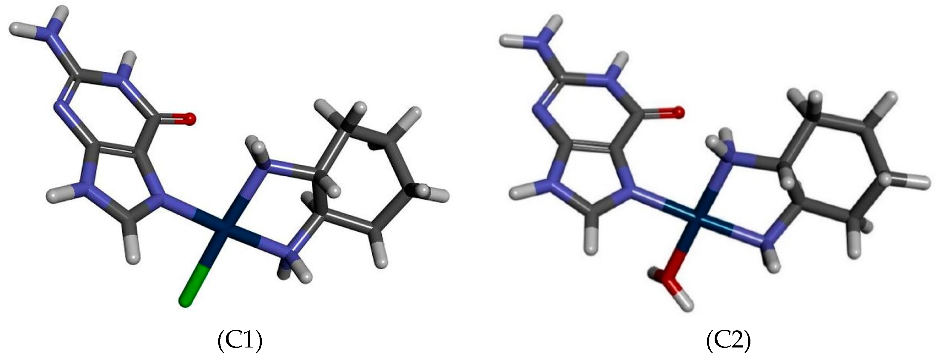
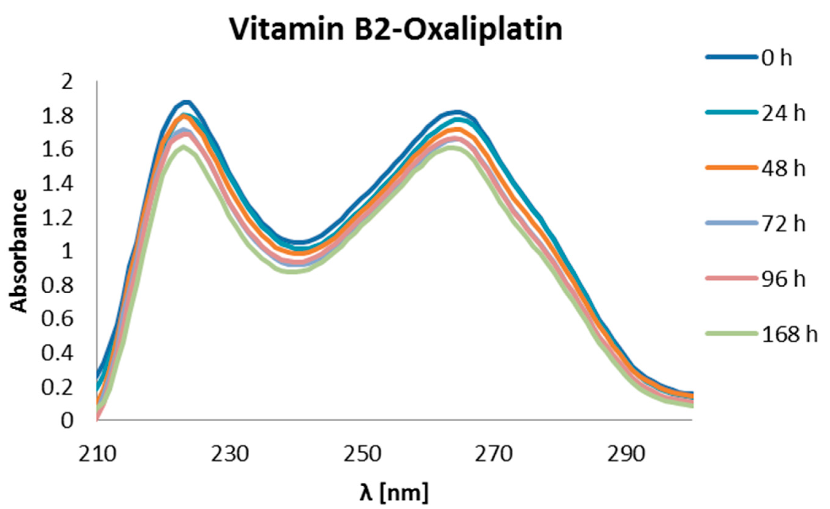
2. Results
3. Experimental Analysis
4. Conclusions
5. Future Directions
Author Contributions
Funding
Institutional Review Board Statement
Informed Consent Statement
Acknowledgments
Conflicts of Interest
References
- World Cancer Research Fund; American Institute for Cancer. Diet, Nutrition, Physical Activity and Breast Cancer; Continuous Update Project, Expert Report 2018; World Cancer Research Fund International: London, UK, 2018. [Google Scholar]
- Wiseman, L.R.; Adkins, J.C.; Plosker, G.L.; Goa, K.L. Oxaliplatin: A review of its use in the management of metastatic colorectal cancer. Drugs Aging 1999, 14, 459–475. [Google Scholar] [CrossRef] [PubMed]
- Rosenberg, B.; Vancamo, L.; Trosko, J.E.; Mansour, V.H. Platinum Compounds: A New Class of Potent Antitumour Agents. Nature 1969, 222, 385–386. [Google Scholar] [CrossRef] [PubMed]
- Rosenberg, B.; Van Camp, L.; Krigas, T. Inhibition of cell division in Escherichia coli by electrolysis products from a platinum electrode. Nature 1965, 205, 698–699. [Google Scholar] [CrossRef] [PubMed]
- Rixe, O.; Ortuzar, W.; Alvarez, M.; Parker, R.; Reed, E.; Paull, K.; Fojo, T. Oxaliplatin, tetraplatin, cisplatin, and carboplatin: Spectrum of activity in drug-resistant cell lines and in the cell lines of the national cancer institute’s anticancer drug screen panel. Biochem. Pharmacol. 1996, 52, 1855–1865. [Google Scholar] [CrossRef]
- Ho, G.Y.; Woodward, N.; Coward, J.I.G. Cisplatin versus carboplatin: Comparative review of therapeutic management in solid malignancies. Crit. Rev. Oncol. Hematol. 2016, 102, 37–46. [Google Scholar] [CrossRef] [Green Version]
- Frezza, M.; Hindo, S.; Chen, D.; Davenport, A.; Schmitt, S.; Tomco, D.; Dou, Q.P. Novel metals and metal complexes as platforms for cancer therapy. Curr. Pharm. Des. 2010, 16, 1813–1825. [Google Scholar] [CrossRef] [Green Version]
- Desoize, B.; Madoulet, C. Particular aspects of platinum compounds used at present in cancer treatment. Crit. Rev. Oncol. Hematol. 2002, 42, 317–325. [Google Scholar] [CrossRef]
- Fuertes, M.A.; Alonso, C.; Pérez, J.M. Biochemical modulation of cisplatin mechanisms of action: Enhancement of antitumor activity and circumvention of drug resistance. Chem. Rev. 2003, 103, 645–662. [Google Scholar] [CrossRef]
- Van Zyl, B.; Tang, D.; Bowden, N.A. Biomarkers of platinum resistance in ovarian cancer: What can we use to improve treatment. Endocr. Relat. Cancer 2018, 25, R303–R318. [Google Scholar] [CrossRef] [Green Version]
- Abotaleb, M.; Kubatka, P.; Caprnda, M.; Varghese, E.; Zolakova, B.; Zubor, P.; Opatrilova, R.; Kruzliak, P.; Stefanicka, P.; Büsselberg, D. Chemotherapeutic agents for the treatment of metastatic breast cancer: An update. Biomed. Pharmacother. 2018, 101, 458–477. [Google Scholar] [CrossRef]
- Rugo, H.S.; Olopade, O.I.; DeMichele, A.; Yau, C.; van ’t Veer, L.J.; Buxton, M.B.; Hogarth, M.; Hylton, N.M.; Paoloni, M.; Perlmutter, J.; et al. Adaptive Randomization of Veliparib–Carboplatin Treatment in Breast Cancer. N. Engl. J. Med. 2016, 375, 23–34. [Google Scholar] [CrossRef] [PubMed]
- Mikuła-Pietrasik, J.; Witucka, A.; Pakuła, M.; Uruski, P.; Begier-Krasińska, B.; Niklas, A.; Tykarski, A.; Książek, K. Comprehensive review on how platinum- and taxane-based chemotherapy of ovarian cancer affects biology of normal cells. Cell. Mol. Life Sci. 2019, 76, 681–697. [Google Scholar] [CrossRef] [PubMed] [Green Version]
- Gebremedhn, E.G.; Shortland, P.J.; Mahns, D.A. The incidence of acute oxaliplatin-induced neuropathy and its impact on treatment in the first cycle: A systematic review. BMC Cancer 2018, 18, 410. [Google Scholar] [CrossRef] [PubMed] [Green Version]
- Chaney, S.G.; Campbell, S.L.; Bassett, E.; Wu, Y. Recognition and processing of cisplatin- and oxaliplatin-DNA adducts. Crit. Rev. Oncol. Hematol. 2005, 53, 3–11. [Google Scholar] [CrossRef] [PubMed]
- Di Francesco, A.M.; Ruggiero, A.; Riccardi, R. Cellular and molecular aspects of drugs of the future: Oxaliplatin. Cell. Mol. Life Sci. 2002, 59, 1914–1927. [Google Scholar] [CrossRef]
- Martinez-Balibrea, E.; Martínez-Cardús, A.; Ginés, A.; Ruiz de Porras, V.; Moutinho, C.; Layos, L.; Manzano, J.L.; Bugés, C.; Bystrup, S.; Esteller, M.; et al. Tumor-Related Molecular Mechanisms of Oxaliplatin Resistance. Mol. Cancer Ther. 2015, 14, 1767–1776. [Google Scholar] [CrossRef] [Green Version]
- Martin, L.P.; Hamilton, T.C.; Schilder, R.J. Platinum resistance: The role of DNA repair pathways. Clin. Cancer Res. 2008, 14, 1291–1295. [Google Scholar] [CrossRef] [Green Version]
- Meyerhardt, J.A.; Mayer, R.J. Systemic therapy for colorectal cancer. N. Engl. J. Med. 2005, 352, 476–487. [Google Scholar] [CrossRef]
- Seetharam, R.N. Oxaliplatin: Preclinical perspectives on the mechanisms of action, response and resistance. Ecancermedicalscience 2009, 3, 153. [Google Scholar] [CrossRef]
- Sarmah, A.; Saha, S.; Bagaria, P.; Kinkar Roy, R. On the complementarity of comprehensive decomposition analysis of stabilization energy (CDASE) Scheme and supermolecular approach. Chem. Phys. 2012, 394, 29–35. [Google Scholar] [CrossRef]
- Sarmah, A.; Roy, R.K. Understanding the preferential binding interaction of aqua-cisplatins with nucleobase guanine over adenine: A density functional reactivity theory based approach. RSC Adv. 2013, 3, 2822. [Google Scholar] [CrossRef]
- Wang, D.; Lippard, S.J. Cellular processing of platinum anticancer drugs. Nat. Rev. Drug Discov. 2005, 4, 307–320. [Google Scholar] [CrossRef] [PubMed]
- Göschl, S.; Schreiber-Brynzak, E.; Pichler, V.; Cseh, K.; Heffeter, P.; Jungwirth, U.; Jakupec, M.A.; Berger, W.; Keppler, B.K. Comparative studies of oxaliplatin-based platinum(iv) complexes in different in vitro and in vivo tumor models. Metallomics 2017, 9, 309–322. [Google Scholar] [CrossRef] [PubMed] [Green Version]
- Mantri, Y.; Lippard, S.J.; Baik, M.-H. Bifunctional binding of cisplatin to DNA: Why does cisplatin form 1,2-intrastrand cross-links with ag but not with GA? J. Am. Chem. Soc. 2007, 129, 5023–5030. [Google Scholar] [CrossRef] [PubMed] [Green Version]
- Spiegel, K.; Rothlisberger, U.; Carloni, P. Cisplatin Binding to DNA Oligomers from Hybrid Car-Parrinello/Molecular Dynamics Simulations. J. Phys. Chem. B 2004, 108, 2699–2707. [Google Scholar] [CrossRef]
- Kelland, L.R.; Farrell, N.P. Platinum-Based Drugs in Cancer Therapy; Humana Press: Totowa, NJ, USA, 2000; Volume 7, ISBN 1-59259-012-8. [Google Scholar]
- Schewe, G. Platinum-Based Drugs in Cancer Therapy; Humana Press: Totowa, NJ, USA, 2010; ISBN 9781617370915. [Google Scholar]
- Riddell, I.A.; Lippard, S.J. Cisplatin and oxaliplatin: Our current understanding of their actions. Met. Ions Life Sci. 2018, 18, 1–42. [Google Scholar]
- Riddell, I.A.; Lippard, S.J.; Brabec, V.; Kasparkova, J.; Menon, V.; Farrell, N.P.; Taylor, K.M. Metallo-Drugs: Development and Action of Anticancer Agents; Walter de Gruyter GmbH & Co KG: Berlin, Germany, 2018; Volume 18. [Google Scholar]
- Dasari, S.; Tchounwou, P.B. Cisplatin in cancer therapy: Molecular mechanisms of action. Eur. J. Pharmacol. 2014, 740, 364–378. [Google Scholar] [CrossRef] [Green Version]
- de Sousa, G.F.; Wlodarczyk, S.R.; Monteiro, G. Carboplatin: Molecular mechanisms of action associated with chemoresistance. Braz. J. Pharm. Sci. 2014, 50, 693–702. [Google Scholar] [CrossRef]
- Alcindor, T.; Beauger, N. Oxaliplatin: A review in the era of molecularly targeted therapy. Curr. Oncol. 2011, 18, 18–25. [Google Scholar] [CrossRef] [Green Version]
- Woynarowski, J.M.; Faivre, S.; Herzig, M.C.S.; Arnett, B.; Chapman, W.G.; Trevino, A.V.; Raymond, E.; Chaney, S.G.; Vaisman, A.; Varchenko, M.; et al. Oxaliplatin-induced damage of cellular DNA. Mol. Pharmacol. 2000, 58, 920–927. [Google Scholar] [CrossRef]
- Pasetto, L.M.; D’Andrea, M.R.; Rossi, E.; Monfardini, S. Oxaliplatin-related neurotoxicity: How and why? Crit. Rev. Oncol. Hematol. 2006, 59, 159–168. [Google Scholar] [CrossRef] [PubMed]
- Hah, S.S.; Stivers, K.M.; De Vere White, R.W.; Henderson, P.T. Kinetics of carboplatin-DNA binding in genomic DNA and bladder cancer cells as determined by accelerator mass spectrometry. Chem. Res. Toxicol. 2006, 19, 622–626. [Google Scholar] [CrossRef] [PubMed] [Green Version]
- Brabec, V.; Kasparkova, J. Modifications of DNA by platinum complexes. Drug Resist. Updat. 2005, 8, 131–146. [Google Scholar] [CrossRef]
- Fuertes, M.A.; Castilla, J.; Alonso, C.; Pérez, J.M. Novel concepts in the development of platinum antitumor drugs. Curr. Med. Chem. Anticancer. Agents 2002, 2, 539–551. [Google Scholar] [CrossRef] [PubMed]
- Fischer, J.; Robin Ganellin, C. Analogue-Based Drug Discovery; John Wiley & Sons: Hoboken, NJ, USA, 2006; ISBN 3527312579. [Google Scholar]
- Hodgkinson, E.; Neville-Webbe, H.L.; Coleman, R.E. Magnesium depletion in patients receiving cisplatin-based chemotherapy. Clin. Oncol. R. Coll. Radiol. 2006, 18, 710–718. [Google Scholar] [CrossRef] [PubMed]
- Larson, C.A.; Blair, B.G.; Safaei, R.; Howell, S.B. The role of the mammalian copper transporter 1 in the cellular accumulation of platinum-based drugs. Mol. Pharmacol. 2009, 75, 324–330. [Google Scholar] [CrossRef] [Green Version]
- Ishida, S.; Lee, J.; Thiele, D.J.; Herskowitz, I. Uptake of the anticancer drug cisplatin mediated by the copper transporter Ctr1 in yeast and mammals. Proc. Natl. Acad. Sci. USA 2002, 99, 14298–14302. [Google Scholar] [CrossRef] [Green Version]
- Howell, S.B.; Safaei, R.; Larson, C.A.; Sailor, M.J. Copper transporters and the cellular pharmacology of the platinum-containing cancer drugs. Mol. Pharmacol. 2010, 77, 887–894. [Google Scholar] [CrossRef] [Green Version]
- Holzer, A.K.; Manorek, G.H.; Howell, S.B. Contribution of the major copper influx transporter CTR1 to the cellular accumulation of cisplatin, carboplatin, and oxaliplatin. Mol. Pharmacol. 2006, 70, 1390–1394. [Google Scholar] [CrossRef]
- Johnstone, T.C.; Suntharalingam, K.; Lippard, S.J. The next generation of platinum drugs: Targeted Pt (II) agents, nanoparticle delivery, and Pt (IV) prodrugs. Chem. Rev. 2016, 116, 3436–3486. [Google Scholar] [CrossRef] [Green Version]
- Han, C.H.; Khwaounjoo, P.; Hill, A.G.; Miskelly, G.M.; McKeage, M.J. Predicting effects on oxaliplatin clearance: In vitro, kinetic and clinical studies of calcium- and magnesium-mediated oxaliplatin degradation. Sci. Rep. 2017, 7, 4073. [Google Scholar] [CrossRef] [PubMed]
- Baik, M.-H.; Friesner, R.A.; Lippard, S.J. Theoretical Study of Cisplatin Binding to Purine Bases: Why Does Cisplatin Prefer Guanine over Adenine? J. Am. Chem. Soc. 2003, 125, 14082–14092. [Google Scholar] [CrossRef] [PubMed]
- Szefler, B.; Czeleń, P. Docking of cisplatin on fullerene derivatives and some cube rhombellane functionalized homeomorphs. Symmetry 2019, 11, 874. [Google Scholar] [CrossRef] [Green Version]
- Szefler, B.; Czeleń, P.; Szczepanik, A.; Cysewski, P. Does the affinity of cisplatin to B-vitamins impair the therapeutic effect in the case of patients with lung cancer-consuming carrot or beet juice? Anticancer Agents Med. Chem. 2019, 19, 1775–1783. [Google Scholar] [CrossRef]
- Szefler, B.; Czeleń, P.; Krawczyk, P. The Affinity of Carboplatin to B-Vitamins and Nucleobases. Int. J. Mol. Sci. 2021, 22, 3634. [Google Scholar] [CrossRef]
- Szefler, B.; Czeleń, P.; Kruszewski, S.; Siomek-Górecka, A.; Krawczyk, P. The assessment of physicochemical properties of Cisplatin complexes with purines and vitamins B group. J. Mol. Graph. Model. 2022, 113, 108144. [Google Scholar] [CrossRef]
- Szefler, B.; Czeleń, P.; Wojtkowiak, K.; Jezierska, A. Affinities to Oxaliplatin: Vitamins from B Group vs. Nucleobases. Int. J. Mol. Sci. 2022, 23, 10567. [Google Scholar] [CrossRef]
- Farrell, N.P. Preclinical perspectives on the use of platinum compounds in cancer chemotherapy. Semin. Oncol. 2004, 31, 1–9. [Google Scholar] [CrossRef]
- Shabalin, I.; Dauter, Z.; Jaskolski, M.; Minor, W.; Wlodawer, A. Crystallography and chemistry should always go together: A cautionary tale of protein complexes with cisplatin and carboplatin. Acta Crystallogr. Sect. D Biol. Crystallogr. 2015, 71, 1965–1979. [Google Scholar] [CrossRef] [Green Version]
- Ahmad, S. Platinum–DNA interactions and subsequent cellular processes controlling sensitivity to anticancer platinum complexes. Chem. Biodivers. 2010, 7, 543–566. [Google Scholar] [CrossRef]
- Stipanuk, M.H.; Caudill, M. Biochemical, Physiological, and Molecular Aspects of Human Nutrition; Saunders: St. Louis, MO, USA, 2013; ISBN 9781455746293. [Google Scholar]
- Stargrove, M.B.; Treasure, J.; McKee, D.L. Herb, Nutrient, and Drug Interactions: Clinical Implications and Therapeutic Strategies; Mosby Elsevier: Amsterdam, The Netherlands, 2008; ISBN 9780323029643. [Google Scholar]
- Polskie Towarzystwo Farmaceutyczne. Farmakopea Polska X; Urząd Rejestracji Produktów Leczniczych, Wyrobów Medycznych i Produktów Biobójczych: Warszawa, Poland, 2014; ISBN 978-83-63724-47-4. [Google Scholar]
- Ghosal, A.; Said, H.M. Mechanism and regulation of vitamin B2 (riboflavin) uptake by mouse and human pancreatic β-cells/islets: Physiological and molecular aspects. Am. J. Physiol. Liver Physiol. 2012, 303, G1052–G1058. [Google Scholar] [CrossRef] [PubMed]
- Kennedy, D. B Vitamins and the Brain: Mechanisms, Dose and Efficacy—A Review. Nutrients 2016, 8, 68. [Google Scholar] [CrossRef] [PubMed] [Green Version]
- Zielińska-Dawidziak, M.; Grajek, K.; Olejnik, A.; Czaczyk, K.; Grajek, W. Transport of high concentration of thiamin, riboflavin and pyridoxine across intestinal epithelial cells Caco-2. J. Nutr. Sci. Vitaminol. 2008, 54, 423–429. [Google Scholar] [CrossRef] [Green Version]
- White, E.; Patterson, R.E.; Kristal, A.R.; Thornquist, M.; King, I.; Shattuck, A.L.; Evans, I.; Satia-Abouta, J.; Littman, A.J.; Potter, J.D. VITamins and Lifestyle Cohort Study: Study Design and Characteristics of Supplement Users. Am. J. Epidemiol. 2004, 159, 83–93. [Google Scholar] [CrossRef] [PubMed] [Green Version]
- Winkler, C.; Wirleitner, B.; Schroecksnadel, K.; Schennach, H.; Fuchs, D. Beer down-regulates activated peripheral blood mononuclear cells in vitro. Int. Immunopharmacol. 2006, 6, 390–395. [Google Scholar] [CrossRef] [PubMed]


| ΔGr (kcal/mol) | Reaction | ||
|---|---|---|---|
| Cisplatin | Oxaliplatin | Carboplatin | |
| −18.90 | −13.87 | 149.53 | cis[Pt*/**/***]+/+/2+ + [B1(N7)]+ → cis[Pt/**/***-B1(N7)]2+/2+/3+ |
| −20.89 | −16.24 | 144.60 | cis[Pt*/**/***]+/+/2+ + [B1(N1)]+ → cis[Pt/**/***-B1(N1)]2+/2+/3+ |
| −3.75 | −1.46 | 233.04 | cis[Pt*/**/***]+/+/2+ + B2(N7) → cis[Pt/**/***-B2(N7)]+/+/2+ |
| −24.90 | −22.32 | −6.3 | cis[Pt*/**/***]+/+/2+ + B3(N7) → cis[Pt/**/***-B3(N7)]+/+/2+ |
| −15.61 | −17.06 | −12.96 | cis[Pt*/**/***]+/+/2+ + B6(N7) → cis[Pt/**/***-B6(N7)]+/+/2+ |
| −28.17 | −33.46 | −30.89 | cis[Pt*/**/***]+/+/2+ + GUA(N7)→cis[Pt/**/***-GUA(N7)]+/+/2+ |
| Number of Reaction | ΔGr | Reaction |
|---|---|---|
| 1 | −46.48 | [Pt*]+ + [B1(N7)]+ → [Pt*-B1(N7)]2+ |
| 2 | −48.91 | [Pt*]+ + [B1(N1)]+ → [Pt*-B1(N1)]2+ |
| 3 | −41.55 | [Pt*]+ + B2 → [Pt*-B2]+ |
| 4 | −39.37 | [Pt*]+ + B3 → [Pt*-B3]+ |
| 5 | −45.93 | [Pt*]+ + B6 → [Pt*-B6]+ |
| 6 | −46.10 | [Pt*]+ + GUA → [Pt*-GUA]+ |
| ΔGr (kcal/mol) | Reaction | ||
|---|---|---|---|
| Cisplatin | Oxaliplatin | Carboplatin | |
| −20.23 | −25.10 | 149.53 | cis[Pt*/**/***]2+/2+/+ + [B1(N7)]+ → cis[Pt/**-B1(N7)]3+/3+/2+ |
| −22.64 | −30.15 | 144.60 | cis[Pt*/**/***]2+/2+/+ + [B1(N1)]+ → cis[Pt/**-B1(N1)]3+/3+/2+ |
| −7.84 | −13.64 | 233.04 | cis[Pt*/**/***]2+/2+/+ + B2(N7) → cis[Pt/**-B2(N7)]2+/2+/+ |
| −27.28 | −35.53 | −6.3 | cis[Pt*/**/***]2+/2+/+ + B3(N7) → cis[Pt/**-B3(N7)]2+/2+/+ |
| −24.66 | −30.60 | −12.96 | cis[Pt*/**/***]2+/2+/+ + B6(N7) → cis[Pt/**-B6(N7)]2+/2+/+ |
| −37.58 | −50.03 | −30.89 | cis[Pt*/**/***]2+/2+/+ + GUA(N7)→cis[Pt/**-GUA(N7)]2+/2+/+ |
| Number of Reaction | ΔGr | Reaction |
|---|---|---|
| 1 | −46.45 | [Pt**]2+ + [B1(N7)]+ → [Pt**-B1(N7)]3+ |
| 2 | −50.58 | [Pt**]2+ + [B1(N1)]+ → [Pt**-B1(N1)]3+ |
| 3 | −42.96 | [Pt**]2+ + B2 → [Pt**-B2]2+ |
| 4 | −40.61 | [Pt**]2+ + B3 → [Pt**-B3]2+ |
| 5 | −47.11 | [Pt**]2+ + B6 → [Pt**-B6]2+ |
| 6 | −51.01 | [Pt**]2+ + GUA → [Pt**-GUA]2+ |
| BCP | PtClDACH-Gua | BCP | PtH2ODACH-Gua | ||||
|---|---|---|---|---|---|---|---|
| ρBCP(e×a0−3) | ∇2ρBCP(e×a0−5) | V (r) | ρBCP(e×a0−3) | ∇2ρBCP(e×a0−5) | V (r) | ||
| Cl-Pt | 1.02 × 10⁻¹ | 1.97 × 10⁻¹ | -1.27 × 10⁻¹ | H2O-Pt | 9.54 × 10⁻² | 4.69 × 10⁻¹ | −1.65 × 10⁻¹ |
| N-Pt | 1.25 × 10⁻¹ | 4.53 × 10⁻¹ | −2.06 × 10⁻¹ | N-Pt | 1.28 × 10⁻¹ | 4.31 × 10⁻¹ | −2.07 × 10⁻¹ |
| H2N-Pt | 1.25 × 10⁻¹ | 4.12 × 10⁻¹ | −1.96 × 10⁻¹ | H2N-Pt | 1.39 × 10⁻¹ | 3.77 × 10⁻¹ | −2.12 × 10⁻¹ |
| H2N-Pt | 1.26 × 10⁻¹ | 4.15 × 10⁻¹ | -1.99 × 10⁻¹ | H2N-Pt | 1.27 × 10⁻¹ | 4.04 × 10⁻¹ | −1.99 × 10⁻¹ |
| O…HN | 1.84 × 10⁻² | 7.27 × 10⁻² | −1.23 × 10⁻² | O…HN | 1.90 × 10⁻² | 7.36 × 10⁻² | −1.27 × 10⁻² |
Disclaimer/Publisher’s Note: The statements, opinions and data contained in all publications are solely those of the individual author(s) and contributor(s) and not of MDPI and/or the editor(s). MDPI and/or the editor(s) disclaim responsibility for any injury to people or property resulting from any ideas, methods, instructions or products referred to in the content. |
© 2023 by the authors. Licensee MDPI, Basel, Switzerland. This article is an open access article distributed under the terms and conditions of the Creative Commons Attribution (CC BY) license (https://creativecommons.org/licenses/by/4.0/).
Share and Cite
Szefler, B.; Czeleń, P. Will the Interactions of Some Platinum (II)-Based Drugs with B-Vitamins Reduce Their Therapeutic Effect in Cancer Patients? Comparison of Chemotherapeutic Agents such as Cisplatin, Carboplatin and Oxaliplatin—A Review. Int. J. Mol. Sci. 2023, 24, 1548. https://doi.org/10.3390/ijms24021548
Szefler B, Czeleń P. Will the Interactions of Some Platinum (II)-Based Drugs with B-Vitamins Reduce Their Therapeutic Effect in Cancer Patients? Comparison of Chemotherapeutic Agents such as Cisplatin, Carboplatin and Oxaliplatin—A Review. International Journal of Molecular Sciences. 2023; 24(2):1548. https://doi.org/10.3390/ijms24021548
Chicago/Turabian StyleSzefler, Beata, and Przemysław Czeleń. 2023. "Will the Interactions of Some Platinum (II)-Based Drugs with B-Vitamins Reduce Their Therapeutic Effect in Cancer Patients? Comparison of Chemotherapeutic Agents such as Cisplatin, Carboplatin and Oxaliplatin—A Review" International Journal of Molecular Sciences 24, no. 2: 1548. https://doi.org/10.3390/ijms24021548