Article

Improved Simulated-Daylight Photodynamic Therapy and Possible Mechanism of Ag-Modified TiO2 on Melanoma

Key Laboratory of Biomedical Information Engineering of Ministry of Education, School of Life Science and Technology, Institute of Biomedical Analytical Technology and Instrumentation, Xi’an Jiaotong University, Xi’an 710048, China
*
Author to whom correspondence should be addressed.
Int. J. Mol. Sci. 2023, 24(8), 7061; https://doi.org/10.3390/ijms24087061
Submission received: 8 March 2023 / Revised: 1 April 2023 / Accepted: 9 April 2023 / Published: 11 April 2023
(This article belongs to the Special Issue Photobiology and Medical Biomaterials Research)

Abstract

:
Simulated-daylight photodynamic therapy (SD-PDT) may be an efficacious strategy for treating melanoma because it can overcome the severe stinging pain, erythema, and edema experienced during conventional PDT. However, the poor daylight response of existing common photosensitizers leads to unsatisfactory anti-tumor therapeutic effects and limits the development of daylight PDT. Hence, in this study, we utilized Ag nanoparticles to adjust the daylight response of TiO2, acquire efficient photochemical activity, and then enhance the anti-tumor therapeutic effect of SD-PDT on melanoma. The synthesized Ag-doped TiO2 showed an optimal enhanced effect compared to Ag-core TiO2. Doping Ag into TiO2 produced a new shallow acceptor impurity level in the energy band structure, which expanded optical absorption in the range of 400–800 nm, and finally improved the photodamage effect of TiO2 under SD irradiation. Plasmonic near-field distributions were enhanced due to the high refractive index of TiO2 at the Ag-TiO2 interface, and then the amount of light captured by TiO2 was increased to induce the enhanced SD-PDT effect of Ag-core TiO2. Hence, Ag could effectively improve the photochemical activity and SD-PDT effect of TiO2 through the change in the energy band structure. Generally, Ag-doped TiO2 is a promising photosensitizer agent for treating melanoma via SD-PDT.

1. Introduction

Melanoma is a commonly occurring severe skin malignancy induced by melanocytes [1]. The incidence of melanoma is ever-increasing. It is traditionally considered to be metastatically invasive as it can invade and spread to neighboring tissues [2]. Additionally, it is resistant to chemotherapeutic drugs and radiation therapy [3,4]. Therefore, more effective therapeutic strategies for melanoma need to be developed [5,6]. Photodynamic therapy (PDT) can selectively destroy diseased cells or tissues as they are more sensitive to light irradiation [7]. During PDT, a photosensitizer in a singlet ground state undergoes visible or near-infrared irradiation, absorbs energy, and attains an excited triplet state through intersystem crossing [8]. The triplet state of the photosensitizer reacts with oxygen or the substrate through electron/hydrogen atom or energy transfer processes, producing reactive oxygen species, especially singlet oxygen, to damage biological components (e.g., amino acids, unsaturated lipids, and DNA bases) [9]. Because singlet oxygen can diffuse by only 10–20 nm during its lifetime of 0.01–0.04 µs, the intracellular damage targets of PDT are very close to the intracellular localization of the photosensitizer. Therefore, PDT might be a non-invasive, effective treatment strategy for melanoma cancer therapy [10].
PDT efficiency relies on three primary factors: the photosensitizer, light, and molecular oxygen, as per the PDT mechanism [11,12]. The frequently utilized light sources for PDT include coherent light sources (argon and argon-pumped lasers, solid-state lasers, metal vapor-pumped dye lasers, and optical parametric oscillator lasers) as well as non-coherent light sources (fluorescent lamps, halogen lamps, metal halide lamps, xenon arc lamps, and phosphor-coated sodium lamps) [13]. Specifically, coherent light sources can provide high-power output and are widely used in PDT [14]. However, they can cause severe pain, erythema, and edema during irradiation. These side effects are usually intolerable and can even make patients refuse treatment [15,16]. To solve this problem, daylight PDT or simulated-daylight PDT has attracted much attention. Although it cannot penetrate deep tissues, it is very safe, nearly painless, well-tolerated, and mostly nonsurgical [17,18,19,20,21]. Because skin disease is a superficial disease, it has no limitation of tissue depth. Using daylight PDT for skin disease is more effective than using traditional laser PDT [22]. Some studies have shown that although daylight PDT is effective for some skin diseases, it has no obvious therapeutic effect on melanoma [22,23]. This might be because the response of existing photosensitizers to daylight is very low in melanoma. Therefore, specific photosensitizers with a high response to daylight need to be developed to treat melanoma.
Titanium dioxide (TiO2) is a typical biocompatible semiconductor oxide metallic nanomaterial, which is used worldwide for different applications [24,25,26,27,28]. In 1985, Wake et al. used TiO2 to kill microbes through photochemical sterilization under metal halide lamp irradiation, which indicated that TiO2 could be used in the field of PDT [29,30,31,32]. TiO2 is photoactive in the presence of UV light, which provides a basis for its application in daylight PDT [24,33,34]. However, TiO2 is ineffective as a daylight photodynamic therapeutic agent for treating cancer as it responds poorly to sunlight. To shift the TiO2 absorption spectrum to the visible region to expand the daylight response range, several approaches have been proposed [35,36]. Among them, doping with the metal ions using transition metal or non-metal ions to change the optoelectronic features of TiO2 could significantly shift the optical response of TiO2. In addition, modifying with plasmonic metallic nanoparticles to combine the photocatalytic properties of TiO2 and the optical properties of plasmonic nanoparticles could extend the photocatalytic activity of TiO2 from UV light to visible or even to the NIR range of radiation. Among all metallic materials, silver (Ag) exhibits the most interesting physical properties and unique optical properties [37]. Hence, in this study, Ag-modified TiO2 nanomaterials with varying structures (Ag-doped TiO2 and Ag-core TiO2) were synthesized to improve the limited SD-PDT effect on melanoma by increasing the daylight response. The improvement in the photochemical activity and the therapeutic effect of PDT were compared and the possible mechanisms were theoretically studied. Generally, the described TiO2 modification method based on Ag significantly increased the photochemical properties of the metallic nanomaterials. The synthesized Ag-doped TiO2 was found to be a promising agent for treating melanoma using daylight PDT.

2. Results

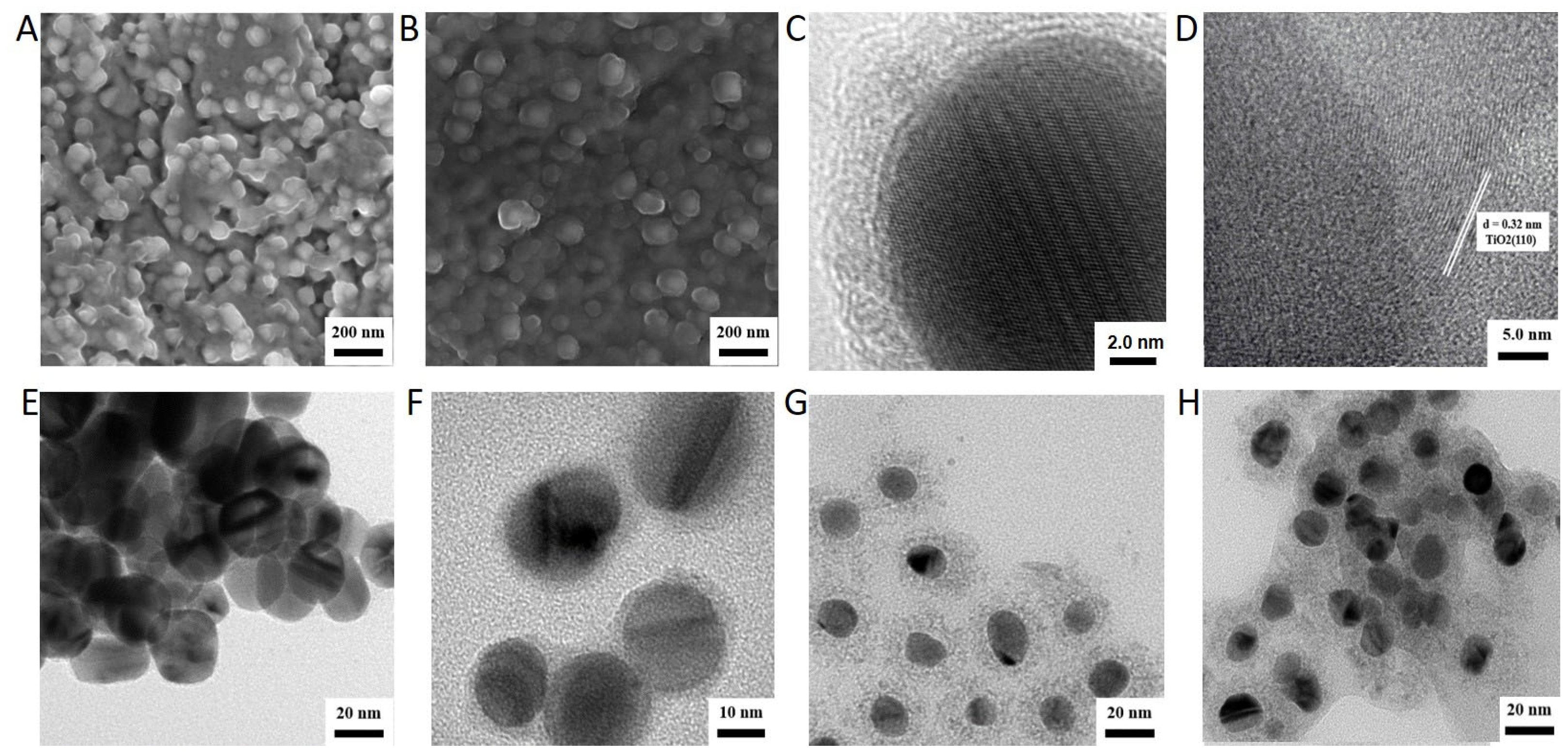
2.1. Synthesis and Characterization of Ag-Modified TiO2 with Different Structure

To increase the light response range of TiO2 and improve the simulated-daylight PDT effect of TiO2 on melanoma, Ag-modified TiO2 was synthesized. Based on the different optical properties of nanomaterials with different structures, Ag-doped TiO2 and Ag-core TiO2 were synthesized, respectively. The TEM results of the synthesized TiO2 were comparable to P25 (the commercialized TiO2 nanoparticles) purchased from Sigma, and Ag-doped TiO2 showed that the size of the sphere was 100 nm (Figure 1A,B). Nonetheless, the absorption spectrum of the Ag-doped TiO2 displayed a prominent redshift (Figure 2A), which may be attributed to alterations in the energy band structure. The XRD findings demonstrated that the lattice structure of TiO2 remained unaltered following Ag being doped into TiO2 (Figure 2B). In Ag-core TiO2, the addition of sodium bicarbonate to the reaction mixture led to the formation of a TiO2 shell enveloping Ag nanoparticles. The crystal lattice structure of TiO2 with a spacing of 0.32 nm appeared after calcination (Figure 1C,D). The TEM results revealed that the synthesized silver had a uniform particle size (Figure 1E). The thickness of the shell of TiO2 increased with the increase in the concentration of sodium bicarbonate to 1.5 mL. When the concentration of sodium bicarbonate was 0.9 mL and 1.3 mL, the thickness of the TiO2 shell was about 5 nm and 18.7 nm, respectively (Figure 1F,G). When the concentration of sodium bicarbonate was 1.5 mL, the shell of TiO2 agglomerated (Figure 1H). The results of the energy spectrum analysis from TEM-EDS also confirmed that silver was successfully coated by TiO2 (Ag 81.43%, Ti 7.36%, and O 11.21%) (Figure 2D). The results of the absorption spectrum showed that the Ag-doped TiO2 showed absorption in the range of 400–800 nm, which was similar to the absorption of the synthesized TiO2 and higher than the absorption of P25 (Figure 2A). The Ag-core TiO2 showed a prominent red shift and a decrease in the intensity of the absorption spectrum with an increase in the thickness of the TiO2 shell (Figure 2C).

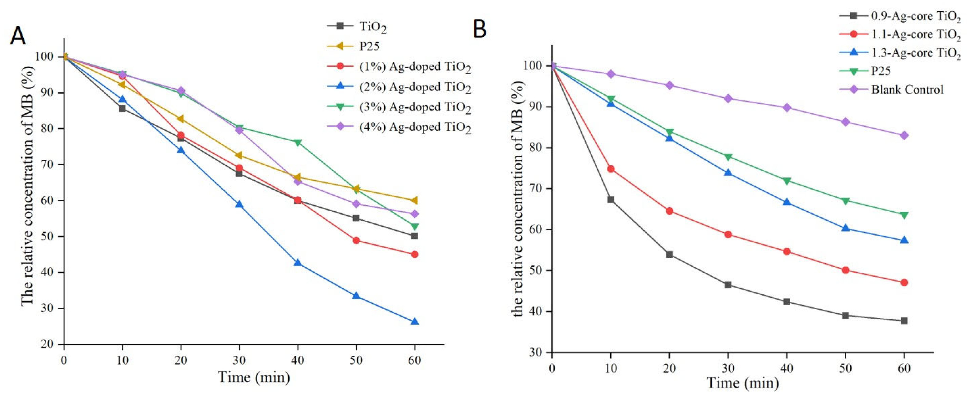
2.2. Comparative Analysis of the Photocatalytic Activity of Ag-Modified TiO2 with Different Structures

The photochemical activities of Ag-modified TiO2 were evaluated through the photocatalytic degradation of methylene blue. As shown in Figure 3A, methylene blue could be degraded under TiO2 induction after Ag was added to Ag-doped TiO2, but this degradation effect was not observed for free TiO2. Upon reaching a 2% Ag concentration, the degradation rate rose from 40% to 70%. As shown in Figure 3B, methylene blue was degraded by 62.3% and 36.3% under the induction of Ag-core TiO2 (5 nm thick) and P25, respectively. The catalytic efficiency of Ag-core TiO2 decreased as the thickness of the shell increased, and the Ag-core TiO2 (5 nm thick) of the shell had the highest catalytic efficiency. Hence, Ag-modified TiO2 significantly increased the photochemical activity of TiO2. Compared to Ag-core TiO2, Ag-doped TiO2 exhibited better photochemical activity.

2.3. Comparative Analysis of the Cytotoxicity Assay and Phototoxicity of Ag-Modified TiO2 with Different Structures

To evaluate the safety and photodamage effects of the synthesized Ag-doped TiO2 and Ag-core TiO2, we determined the viability of A375 cells (human melanoma cell line) without irradiation and with irradiation, by performing a CCK-8 assay. Cell viability induced by 50 µg/mL TiO2 in different reagents was higher than 90%, and no noticeable difference was found between them, which indicated that the synthesized reagents had negligible dark cytotoxicity in A375 cells. When the concentration of TiO2 decreased, the cell viability increased further (Figure 4A). Hence, the synthesized Ag-doped TiO2 and Ag-core TiO2 were safe for usage. As shown in Figure 4B, after irradiation by daylight, 1 µg/mL TiO2 in the different reagents could not inhibit A375 cells, and 50 µg/mL TiO2 in the different reagents could strongly inhibit them. Specifically, the simulated-daylight photodamage effect of Ag-doped TiO2 was higher than that of Ag-core TiO2. For example, cell viability decreased to 24.5% and 31.6% after induction by Ag-doped TiO2 and Ag-core TiO2, respectively. However, cell viability only decreased to 67.6% and 60.1% after induction by P25 and the synthesized free TiO2 at 50 µg/mL.
The photodamage effect increased with the increase in the irradiation dosage (Figure 3C). However, after irradiation with 40 J/cm2, the untreated cells could also be partially inhibited, and the cell survival rate of A375 was only about 71%. Based on the principle of little toxic effect on normal tissue occurring to the greatest extent during PDT, the irradiation dosage for the synthesized Ag-modified TiO2 could be controlled below 40 J/cm2. Overall, Ag could effectively improve the photodamage effect of TiO2 under simulated-daylight irradiation, and Ag-doped TiO2 had a more significant daylight PDT effect on melanoma.

2.4. Comparative Analysis of ROS Generation by Ag-Modified TiO2 with Different Structures

During PDT, generated ROS can lead to the damage of cellular components and then induce cell death. Hence, the ability to generate ROS determines the PDT effect. To determine the simulated-daylight PDT effect induced by Ag-modified TiO2, the ability to generate ROS was evaluated using a DCFH-DA probe—the most widely used probe for detecting intracellular H2O2 and oxidative stress. As per the fluorescence imaging findings, ROS were generated after induction by P25, the synthesized TiO2, and Ag-modified TiO2, and the induction was higher when Ag-modified TiO2 was used (Figure 5A). The fluorescence intensity of DCFH-DA increased obviously in Ag-modified TiO2 (Figure 5B). Compared with P25, Ag-core TiO2 and Ag-doped TiO2 resulted in significant increases, at average values of 1.75-fold and 1.95-fold, respectively. Hence, the ROS levels induced by Ag-doped TiO2 were higher than those induced by Ag-core TiO2. The inhibitory activity induced by all reagents containing TiO2 could be effectively weakened by using a quenching agent of ROS. After being treated with P25, the synthesized TiO2, Ag-core TiO2, and Ag-doped TiO2, as well as 20 mM histidine, cell activities increased from 67.6%, 60%, 31.6%, and 24.5% to 85.3%, 80.8%, 60.2%, and 55.6%, respectively. This revealed that the degree of inhibition induced by Ag-modified TiO2 was higher than that induced by the synthesized TiO2 or P25 (Figure 5C). Hence, our findings showed that Ag-modified TiO2, especially Ag-doped TiO2, efficiently improved simulated-daylight PDT by enhancing the ability to generate ROS.

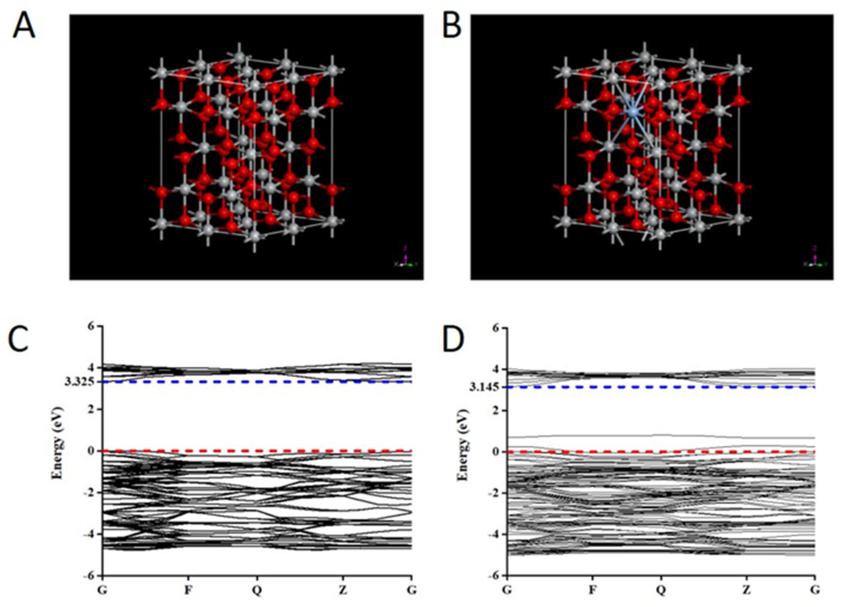
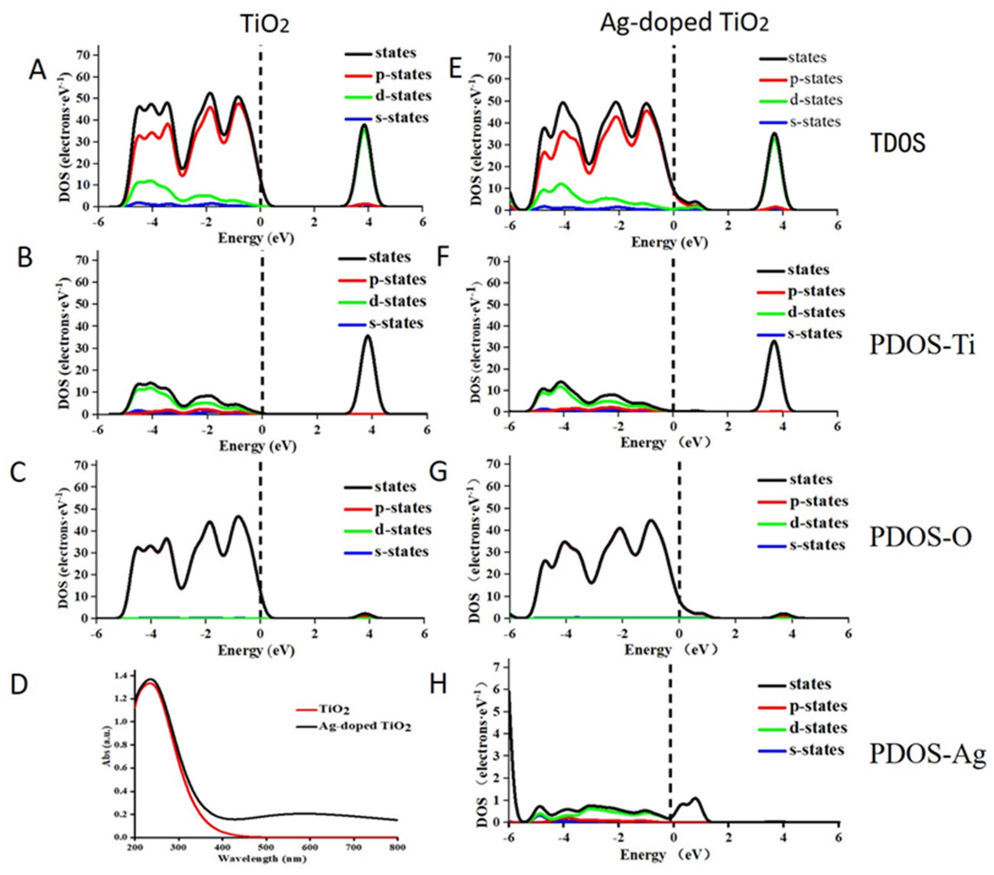
2.5. The Theoretical Mechanistic Analysis

The results of the experiment showed that modification with Ag could effectively enhance the photodamage effect of TiO2 under simulated-daylight irradiation. Specifically, Ag-doped TiO2 had a more significant daylight PDT effect on melanoma. This might be due to an increase in the response of TiO2 to daylight facilitated by Ag. To confirm whether this mechanism was used, changes in the optical absorption properties of TiO2 induced by doping with Ag were used, based on density functional theory using the CASTEP code. First, a supercell of TiO2 (containing 16 Ti atoms and 32 O atoms) and 2% Ag-doped TiO2 (containing 15 Ti atoms, 32 O atoms and 1 Ag atom) was constructed and used as the later calculation model (Figure 6A,B). Then, the changes in the band structure and the density of the states of pure TiO2 and Ag-doped TiO2 were calculated. The energy of the band gap of pure TiO2 was 3.325 eV, which then decreased to 3.14 eV when Ag was doped (Figure 6C,D). Additionally, compared to the band structure of pure TiO2, two new impurity energy levels were introduced, and one of them passed through the Fermi level, which indicated that it can act as an acceptor at the shallow impurity energy level as a bound state of a hole (Figure 6C,D). This shallow acceptor impurity level could enhance the separation of electron-hole pairs, produce free conduction holes, decrease the recombination of photo-generated electrons and holes, and decrease the energy required for the electrons to escape. The computed results of the total density of states and partial density of states of pure TiO2 and Ag-doped TiO2 showed that the 4d-orbital electrons of Ag atoms were used to introduce the new impurity energy levels and produce the new electronic states through strong mixing with the p-orbital states of oxygen atoms (Figure 7). Hence, these two impurity energy levels of Ag-doped TiO2 mainly occurred due to doping with Ag. Generally, the doped Ag atom has an influence on the TiO2 energy structure and induced bandgap narrowing. Ag is the main reason for the the increase in the daylight response of Ag-doped TiO2. Ag-doped TiO2 expanded optical absorption in the range of 400–800 nm and improved the photodamage effect of TiO2 under daylight irradiation.
First principles analysis can be used to analyze the properties of materials with a crystal structure but not of materials with a core–shell structure. To determine the cause of the improvement in the SD-PDT effect of TiO2 induced by Ag-core TiO2, discrete dipole approximation simulations were studied because these changes might be induced by the localized surface plasmon resonance enhancement effect. First, a series of Ag-core TiO2 complexes with a constant Ag core and TiO2 shells of different thicknesses (0 nm, 5 nm, 10 nm, 15 nm, and 20 nm) were calculated. As shown in Figure 8A, the results showed that the absorption spectrum has an obvious red shift and a significant decrease in the intensity with an increase in the thickness of the TiO2 shell. These results matched the absorption spectrum data we measured. Plasmonic near-field distribution showed a strong change in the Ag-core TiO2 (Figure 8B). The electric near-field intensities were considerably enhanced due to the high refractive index of TiO2 at the Ag- TiO2 interface. With the increase in the electric near-field intensity, the amount of light captured by TiO2 and the PDT effect increased. The relationship between the field enhancement effect and the thickness of the TiO2 shell results showed that the field enhancement effect gradually weakened and the ability of the TiO2 shell to capture light decreased with an increase in the thickness of the shell, which occurred probably because the shell affected the movement of photo-generated electrons and holes (Figure 8C). Hence, the Ag-core TiO2 with a 5 nm thick shell had the highest photocatalytic efficiency.

3. Discussion

Photodynamic therapy is an effective therapeutic strategy for skin disease in clinical therapy [38]. However, it is often accompanied by severe adverse effects during treatment. A large number of patients are unable to continue treatment due to these adverse effects. In 2008, daylight PDT was first introduced as a less painful, outdoors alternative to conventional PDT, with similar clinical effectiveness [39]. However, daylight PDT efficacy was often dependent on weather conditions. For example, in the U.K., daylight PDT is practical between the months of March or April and September or October, when the temperature is above 10 °C in the day (from 9:00 to 18:00) and the fluence rate reaches 130 W/m2 [40]. In addition, to avoid patient exposure to harmful wavelengths of ultraviolet radiation during daylight PDT, organic sunscreens should be used to prevent sun damage. To provide a controlled, daylight PDT environmental setting and remove the disadvantage of exposure to harmful ultraviolet radiation, SD-PDT has been investigated using an indoor daylight-simulating lamp. Wulf and co-workers reported that four different lamp candidates (18 W red-, 140 W red-, and 50 W white-light-emitting diode lamps and halogen lamps from 250 W slide projectors as well as 400 W overhead projectors for SD-PDT were able to photobleach a PPIX photosensitizer completely [39]. Calzavara-Pinto et.al. revealed that SD-PDT using a lamp with an output confined to the red waveband (630 ± 5 nm) and a polychromatic white LED lamp (400–700 nm) can represent a valid therapeutic method for Actinic cheilitis [41]. In our study, the SD-PDT effect can be obtained under the irradiation of a sunlight Xenon lamp with an emission spectral range of 380 nm to 700 nm. Hence, a sunlight Xenon lamp (380–700 nm) is also a useful lamp candidate for SD-PDT. However, in our study, we did not evaluate and compare the SD-PDT effect of Ag-modified TiO2 under other lamp sources. In further studies, more detailed comparative research may be needed to obtain a better SD-PDT anti-tumor therapeutic effect.
In this study, an investigation to improve the strategy to increase the simulating-daylight response of existing photosensitizers is the main purpose of the research that we want. TiO2 is a potent oxygen radical generator. However, it is limited in SD-PDT by the necessity to use ultraviolet irradiation with low tissue penetration and its harmful impact on the human body. To maximize the visible light absorption of TiO2, inorganic compounds were usually doped to the TiO2 during their preparation, because this process can narrow the bandgap in the TiO2 nanoparticle’s structure and decrease the necessary activation energy. Among these inorganic compounds, noble metals (such as gold (Au), silver (Ag), platinum (Pt), and palladium (Pd)) were used to dope TiO2, one after another [33]. All absorption ranges of TiO2 were shifted to longer wavelengths and enhanced photocatalytic activities under visible light were obtained to different degrees after doping. However, compared with the other noble metals used, Ag has been regarded as a better candidate due to its higher catalytic activity and ROS generation ability [41,42]. Hence, Ag-doped TiO2 may be suitable for daylight PDT or SD-PDT. Unfortunately, there are few study reports that shows that Ag-doped TiO2 is used in daylight PDT or SD-PDT. However, Alshamsan et.al. revealed that Ag-doped TiO2 has the potential to selectively kill cancer cells while sparing normal cells through ROS generation in HepG2 (human liver cancer cell line) [43]. It gave us a reason to conduct research and evaluate the SD-PDT effect of Ag-doped TiO2 on melanoma. In this study, the results showed that the limited photochemical activity and SD-PDT effect of TiO2 could be improved significantly through doping Ag to the TiO2. In addition, our results showed that the degree of the improvement photochemical activity was independent of the concentration introduction of Ag into TiO2. This may be caused by the synthesized Ag-doped TiO2 complex having different light responses with different concentrations of Ag under simulated-daylight irradiation. This change in light response was not entirely dependent on the doped Ag concentration. Lu and co-workers measured Ag-doped TiO2 photocatalysts with different concentrations of Ag (1–5%) in their previous study. They reported that 2% Ag-doped TiO2 had the highest photocatalytic activity under ultraviolet radiation and 5% Ag-doped TiO2 had the highest photocatalytic activity under solar light [40]. Hence, the introduction of the different concentrations of Ag into TiO2 may cause the different changes in light response. Generally, in our study, Ag-doped TiO2 with a certain concentration of Ag efficiently improved TiO2 photochemical activity compared with TiO2.
Besides the Ag-doped TiO2 complex, TiO2-coated Ag nanoparticles have found applications in many fields because they can combine the surface plasmon resonance properties of a Ag core and the photoactivity of the TiO2 shell [44]. Tunable optical properties can be obtained through a change in the ratio of the core radius and shell thickness. Hence, Ag-core TiO2 was also usually used to increase the optical absorption of TiO2 and extend its absorption region to that of visible light. As with Ag-doped TiO2, there are few study reports that show that Ag-core TiO2 is used in daylight PDT or SD-PDT, and the improvement in the photocatalysts’ effect was not compared directly between Ag-doped TiO2 and Ag-core TiO2 in any other study. To find a better improvement strategy to increase the simulating-daylight response of a TiO2 photosensitizer, the improvement in the photocatalytic activity and SD-PDT effect induced by Ag-doped TiO2 and Ag-coreTiO2 was compared. The results showed that the described TiO2 modification method based on Ag significantly increased the photochemical properties of TiO2 In addition, the synthesized Ag-doped TiO2 was found to be a promising agent for treating melanoma using daylight PDT, and doping Ag to TiO2 is the optimal enhanced strategy.
Several studies revealed that the introduction of Ag into TiO2 improves TiO2’s photochemical activity due to two mechanisms. (1) Ag can act as an electron acceptor to increase the separation efficiency of a photogenerated electron-hole pair because its Fermi level is below the conduction band of TiO2; (2) The generation of a local surface plasmon resonance effect extends the visible light absorption range and increases the photocatalytic efficiency of TiO2 [45]. Hence, in this study, first principles analysis was performed for Ag-doped TiO2 and the discrete dipole approximation for the Ag-core TiO2 was calculated. The results showed that a new shallow acceptor impurity level appeared in the energy band structure of Ag-doped TiO2, which decreased the recombination of photo-generated electrons and holes and the energy needed for the excitation of electrons. This expanded the light response range of TiO2 and made it more responsive to sunlight. A strong field enhancement effect was obtained at the interface between the TiO2 shell and the Ag core of Ag-core TiO2, which increased the amount of light captured by TiO2 and improved its photochemical activity. These are consistent with the previously described mechanism. These further confirm the reliability of our research on this improvement strategy to increase the simulating-daylight response of a TiO2 photosensitizer. Hence, Ag-doped TiO2 is a promising photosensitizer agent for treating melanoma with daylight PDT.

4. Material and Methods

4.1. Reagents and the Cell Lines

Silver nitrate (AgNO3), tetra butyl titanate, and P25 (TiO2 nanoparticles) were purchased from Sigma. TiCl3 was purchased from Aladdin. Absolute ethanol, sodium citrate, solidum borohydride (NaBH4), butyl alcohol, sodium bicarbonate (NaHCO3), and N-butanol were purchased from Tianjin Fuyu Chemical Co., Ltd. and Tianjin Tianli Chemical Reagent Co., Ltd. (Tianjin, China). A Cell Counting Kit (CCK-8) was purchased from Dojindo (Japan). DCFH-DA was purchased from Beyotime Company (Shanghai, China). Human melanoma cell line A375 was obtained from the Cell Bank of the Chinese Academy of Sciences (Shanghai, China) [46]. The A375 cells were cultured in DMEM medium (HyClone) supplemented with 10% fetal bovine serum (HyClone) and 1% penicillin/streptomycin in a humidified incubator at 37 °C with 5% CO2.

4.2. Synthesis and Modification of TiO2

Ag-doped TiO2: Nanosized TiO2 was synthesized using the sol-gel process. Briefly, 5 mL of glacial acetic acid and 20 mL of absolute ethanol were added to 3 mL of ddH2O and stirred at room temperature for 30 min. A total of 5 mL of tetra-butyl titanate and 10 mL of absolute ethanol were mixed and added to the above solution. After stirring for 30 min, the solution was dispersed ultrasonically for 20 min, left to stand for 24 h at room temperature, and then dried at 80 °C for 12 h. The obtained dried product was fully milled and then cauterized to 450 °C in a muffle for 2 h. Purified TiO2 was finally obtained. As with TiO2, 3 mL of 25 mg, 50 mg, 75 mg, or 100 mg AgNO3 and 5 mL of glacial acetic acid were added in turn to 20 mL of absolute ethanol and stirred at room temperature for 30 min and then added to 15 mL of a tetra-butyl titanate solution containing 10 mL of absolute ethanol. After stirring, dispersing, drying, milling, and cauterizing, the Ag-doped TiO2 with a mixing ratio of 1%, 2%, 3%, and 4% was obtained.
Ag-core TiO2: The Ag nanoparticles were produced utilizing the seed growth method. Initially, a 20 mL 1% sodium citrate solution diluted with 75 mL of ddH2O was stirred for 15 min at 70 °C. Next, 1.7 mL of 1% AgNO3 and 2 mL of 0.1% NaBH4 were added and stirred for 1 h at the same temperature. This process led to the creation of a 4 nm sized silver seed solution. Second, 2 mL of sodium citrate solution diluted with 80 mL of ddH2O was stirred for 15 min at 130 °C. Next, 10 mL of silver seed solution and 1.7 mL of AgNO3 were added in turn, stirred for 1 h at the same temperature, and then centrifuged repeatedly. The purified Ag nanoparticles were yielded. Next, 0.3 mL of TiCl3, the different concentrations of 0.2 M sodium bicarbonate (0.9 mL, 1.1 mL, 1.3 mL, and 1.5 mL) and the silver nanoparticle solution were added in turn to 8 mL of ddH2O, stirred for 30 min, and then washed with ddH2O and absolute ethanol, respectively. Furthermore, 10 mL of N-butanol was added. The mixing solution was heated in oil baths at 100 °C for 10 min and then dried, milled, and cauterized. Finally, Ag-core TiO2 with TiO2 shells of different thicknesses were synthesized.

4.3. Characterization of Ag-Doped TiO2 and Ag-Core TiO2

The morphologies of Ag-doped TiO2 and Ag-core TiO2 were observed using transmission electron microscopy (TEM; JEM-2100, JEOL, Tokyo, Japan). Absorption spectra of Ag-doped TiO2 and Ag-core TiO2 were recorded using an ultraviolet–visible spectrophotometer (V-550 UV/VIS, JASCO, Tokyo, Japan). X-ray diffraction was conducted using an X-ray diffractometer (XPert Powder, PANalytical B.V. Netherlands). An energy dispersive spectrometer was used to observe the distribution pattern of various elements (Ag, Ti, and O) using TEM-EDS (JEM-2100 Plus, JEOL Ltd., Japan), operating with an accelerating voltage of 200 kV. The photocatalytic capability of Ag-doped TiO2 (1%, 2%, 3%, and 4%) and Ag-core TiO2 (5 nm, 10 nm, 15 nm, and 20 nm) were evaluated through the photocatalytic degradation of methylene blue under a simulated sunlight Xenon lamp with an emission spectral range of 380 nm to 700 nm. The irradiation time was 10 min at 665 nm. The absorption intensity was recorded using an ultraviolet–visible spectrophotometer (V-550 UV/VIS, JASCO, Tokyo, Japan).

4.4. Cell Viability Analysis

The cytotoxicity assay and phototoxicity assay to evaluate the safety and simulated-daylight PDT effect on melanoma cells (A375 cell line) were measured using a CCK-8 assay (Cell Counting Kit-8—allows for sensitive colorimetric assays for the determination of cell viability in cell proliferation and cytotoxicity assays). Briefly, A375 cells (8000/well) were seeded to sterile 96-well flat-bottomed plates and incubated overnight in a humidified incubator at 37 °C with 5% CO2. Diluted Ag-doped TiO2 and Ag-core TiO2 with the different concentrations (1 μg/mL, 5 μg/mL, 10 μg/mL, 20 μg/mL, 50 μg/mL) were added to corresponding cells in the 96-well flat-bottomed plates. After incubation for 6 h, the medium containing reagent was replaced by fresh cell culture medium. For the cytotoxicity experiment, the plates were then incubated for 24 h in a humidified incubator. In the phototoxicity experiment, the cells were irradiated with a sunlight Xenon lamp at 30 J/cm2 for 15 min and then incubated for 12 h in a humidified incubator. To assess the influence of the irradiation dose on the phototoxicity effect, the cells were treated with 50 μg/mL of the Ag-doped TiO2 or Ag-core TiO2 previously mentioned. Then, they were irradiated with the daylight Xenon lamp at 10 J/cm2, 20 J/cm2, 30 J/cm2, 40 J/cm2, or 50 J/cm2 for 15 min before being incubated for 12 h in a humidified incubator. Finally, all the treated cells were measured for absorbance levels at 450 nm using a microplate reader (Infinite M200 Pro., Tecan, Switzerland). The absorbance levels of cells were calculated as
OD   of   treated   group - OD   of   blank   control   group OD   of   control   group - OD   of   blank   control   group × 100 %
OD is optical density.

4.5. Generation of ROS

ROS is an indirect factor to induce cell damage on PDT. Therefore, the ability of the generation of ROS was measured with a DCFH-DA probe using a Nikon eclipse Ti fluorescence microscope (Nikon, Japan). Briefly, 2.5 × 10³ A375 cells were seeded to sterile 3.5 mL flat-bottomed plates and incubated overnight in a humidified incubator at 37 °C with 5% CO2. Then, the cells were treated with diluted Ag-doped TiO2, Ag-core TiO2, the synthesized TiO2, and P25 for 6 h, washed with PBS twice, irradiated with the sunlight Xenon lamp at 40 J/cm2, incubated with 10 μmol/L DCFH-DA for 20 min at 37 °C in complete darkness, washed with PBS again, and then imaged using a FACScan system or detected using a fluorescence spectrophotometer under the excitation of 488 nm light. To quantify the ability of the generation of ROS, the fluorescence intensity of DCFH-DA was measured using a fluorescence spectrophotometer to detect the concentration of ROS in cells after being treated by Ag-doped TiO2, Ag-core TiO2, the synthesized TiO2, and P25 under a simulated-sunlight Xenon lamp irradiation. The treated cells under the same conditions as above were harvested, incubated with 10 μmol/L DCFH-DA for 10 min at 37 °C in complete darkness, and then centrifuged, washed with PBS, and measured using a fluorescence spectrophotometer under an excitation of 488 nm light. In order to reveal the role of ROS more directly in simulated-daylight PDT induced by Ag-modified TiO2, inhibition tests were measured using a quenching agent of ROS (histidine). After being treated with the different agents containing TiO2, the cells were treated with 20 mM histidine for 30 min, washed with PBS, and then irradiated with the sunlight Xenon lamp. The cell activity was detected using CCK-8 analysis, as before.

4.6. First-Principles Analysis for Ag-Doped TiO2

Based on crystallographic principles, the shape, electronic environment, and other parameters of the crystal cell will change when some atoms are substituted with allochthonous atoms in this cell. Therefore, the change in sunlight response induced by Ag-doped TiO2 is most likely because some Ti atoms are substituted with Ag atoms. The energy band structure, the total density of states and the partial density of states, and the optical absorption properties were determined via density functional theory using the CASTEP code [47].

4.7. Discrete Dipole Approximation for Ag-Core TiO2

It has been reported that the photocatalysis of TiO2 could be enhanced using metal particle doping, polymer nanocomposites, core–shell nanoparticles, and so on, based on the localized surface plasmon resonance enhancement effect [48]. These electric field enhancement factors can be quantified and analyzed using simulations based on discrete dipole approximation [49]. Hence, theoretical mechanistic analysis of the change in sunlight response induced by silver-core TiO2 in this study was employed using discrete dipole approximation simulations using the DDSCAT program.

5. Conclusions

To improve the limited effect of daylight PDT on melanoma due to the low daylight response of commonly used photosensitizers, Ag-mediated TiO2 nanomaterials with different structures were synthesized, and then the improvement in the photocatalytic activity and PDT effect of these nanomaterials were compared. As per the findings, Ag effectively and significantly increased the photochemical activity and the PDT effect of TiO2 under simulated-daylight irradiation. Ag-doped TiO2 exhibited superior photocatalytic activity and a greater daylight-PDT-induced anti-tumor effect compared to Ag-core TiO2. To determine the mechanism, first principles analysis was conducted utilizing Ag-doped TiO2, whereas the discrete dipole approximation for Ag-core TiO2 was calculated. The results showed that doping Ag into TiO2 led to the formation of a new shallow acceptor impurity level in the energy band structure, which then enhanced the separation of electron-hole pairs, produced free conduction holes, reduced the recombination of photo-generated electrons and holes, and decreased the energy required for electrons to escape. This increased optical absorption in the range of 400–800 nm, which improved the photodamage effect of TiO2 under simulated-daylight irradiation. The plasmonic near-field distribution increased due to the high refractive index of TiO2 at the Ag- TiO2 interface, which increased the amount of light captured by TiO2 and enhanced the induction of the daylight PDT effect. In addition, when the thickness of the shell increased, the shell affected the movement of photo-generated electrons and holes, which decreased the overall photochemical activity. Overall, Ag proved to be highly effective in enhancing the photochemical activity and PDT effect of TiO2 when exposed to simulated-daylight irradiation on melanoma. Thus, Ag-doped TiO2 exhibits great potential as a photosensitizer agent for treating melanoma with daylight PDT.

Author Contributions

Data curation, J.X.; Funding acquisition: C.Y.; Investigation, Y.Y.; Methodology: J.W.; Project administration, Z.Z.; Validation, S.W.; Writing—original draft, J.X.; Writing—review and editing, C.Y. All authors have read and agreed to the published version of the manuscript.

Funding

This research was funded by the National Natural Science Foundation of China under Grant Nos. 62175198, 61975160, U22A2092 and the Key Research and Development Program of Shaanxi Province under Grant No. 2022ZDLSF04-09.

Institutional Review Board Statement

Not applicable.

Informed Consent Statement

Not applicable.

Data Availability Statement

Data sharing not applicable.

Acknowledgments

The TEM study was undertaken at the International Center for Dielectric Research (ICDR), Xi’an Jiaotong University, Xi’an, China. The authors thank Chuansheng Ma for his help in using the TEM facility.

Conflicts of Interest

The authors declare no conflict of interest.

References

  1. Ahmed, B.; Qadir, M.I.; Ghafoor, S. Malignant Melanoma: Skin Cancer-Diagnosis, Prevention, and Treatment. Crit. Rev. Eukaryot. Gene Expr. 2020, 30, 291–297. [Google Scholar] [CrossRef] [PubMed]
  2. Aladowicz, E.; Ferro, L.; Vitali, G.C.; Venditti, E.; Fornasari, L.; Lanfrancone, L. Molecular networks in melanoma invasion and metastasis. Future Oncol. 2013, 9, 713–726. [Google Scholar] [CrossRef] [PubMed]
  3. La Porta, C.A. Mechanism of drug sensitivity and resistance in melanoma. Curr. Cancer Drug Targets 2009, 9, 391–397. [Google Scholar] [CrossRef] [PubMed]
  4. Kozar, I.; Margue, C.; Rothengatter, S.; Haan, C.; Kreis, S. Many ways to resistance: How melanoma cells evade targeted therapies. Biochim. Biophys. Acta Rev. Cancer 2019, 1871, 313–322. [Google Scholar] [CrossRef] [PubMed]
  5. Davis, L.E.; Shalin, S.C.; Tackett, A.J. Current state of melanoma diagnosis and treatment. Cancer Biol. Ther. 2019, 20, 1366–1379. [Google Scholar] [CrossRef] [PubMed] [Green Version]
  6. Jenkins, R.W.; Fisher, D.E. Treatment of Advanced Melanoma in 2020 and Beyond. J. Invest. Dermatol. 2021, 141, 23–31. [Google Scholar] [CrossRef]
  7. Zhang, Q.; Li, L. Photodynamic combinational therapy in cancer treatment. J. BUON 2018, 23, 561–567. [Google Scholar]
  8. Wang, Y.Y.; Liu, Y.C.; Sun, H.W.; Guo, D.S. Type I photodynamic therapy by organic-inorganic hybrid materials: From strategies to applications. Coordin. Chem. Rev. 2019, 395, 46–62. [Google Scholar] [CrossRef]
  9. Chen, D.; Xu, Q.; Wang, W.; Shao, J.; Huang, W.; Dong, X. Type I Photosensitizers Revitalizing Photodynamic Oncotherapy. Small 2021, 17, e2006742. [Google Scholar] [CrossRef]
  10. Juzeniene, A.; Moan, J. The history of PDT in Norway Part one: Identification of basic mechanisms of general PDT. Photodiagn. Photodyn. Ther. 2007, 4, 3–11. [Google Scholar] [CrossRef]
  11. Rkein, A.M.; Ozog, D.M. Photodynamic therapy. Dermatol. Clin. 2014, 32, 415–425, x. [Google Scholar] [CrossRef] [PubMed]
  12. Rodrigues, J.A.; Correia, J.H. Enhanced Photodynamic Therapy: A Review of Combined Energy Sources. Cells 2022, 11, 3995. [Google Scholar] [CrossRef] [PubMed]
  13. Algorri, J.F.; Ochoa, M.; Roldan-Varona, P.; Rodriguez-Cobo, L.; Lopez-Higuera, J.M. Light Technology for Efficient and Effective Photodynamic Therapy: A Critical Review. Cancers 2021, 13, 3484. [Google Scholar] [CrossRef] [PubMed]
  14. Kim, M.M.; Darafsheh, A. Light Sources and Dosimetry Techniques for Photodynamic Therapy. Photochem. Photobiol. 2020, 96, 280–294. [Google Scholar] [CrossRef] [Green Version]
  15. Kasche, A.; Luderschmidt, S.; Ring, J.; Hein, R. Photodynamic therapy induces less pain in patients treated with methyl aminolevulinate compared to aminolevulinic acid. J. Drugs Dermatol. 2006, 5, 353–356. [Google Scholar]
  16. Anand, S.; Yasinchak, A.; Govande, M.; Shakya, S.; Maytin, E.V. Painless versus conventional photodynamic therapy for treatment of actinic keratosis: Comparison of cell death and immune response in a murine model. Proc. SPIE Int. Soc. Opt. Eng. 2019, 10860, 61–70. [Google Scholar]
  17. Beiki, D.; Eggleston, I.M.; Pourzand, C. Daylight-PDT: Everything under the sun. Biochem. Soc. Trans. 2022, 50, 975–985. [Google Scholar] [CrossRef]
  18. Garcia-Gil, M.F.; Gracia-Cazana, T.; Cerro-Munoz, P.; Bernal-Masferrer, L.; Navarro-Bielsa, A.; Gilaberte, Y. Fully home-based methyl aminolevulinate daylight photodynamic therapy for actinic keratosis of the face or scalp: A real life open study. Dermatol. Ther. 2022, 35, e15879. [Google Scholar] [CrossRef]
  19. Karrer, S.; Szeimies, R.M.; Philipp-Dormston, W.G.; Gerber, P.A.; Prager, W.; Datz, E.; Zeman, F.; Muller, K.; Koller, M. Repetitive Daylight Photodynamic Therapy versus Cryosurgery for Prevention of Actinic Keratoses in Photodamaged Facial Skin: A Prospective, Randomized Controlled Multicentre Two-armed Study. Acta Derm. Venereol. 2021, 101, adv00355. [Google Scholar] [CrossRef]
  20. O’Mahoney, P.; Khazova, M.; Eadie, E.; Ibbotson, S. Measuring Daylight: A Review of Dosimetry in Daylight Photodynamic Therapy. Pharmaceuticals 2019, 12, 143. [Google Scholar] [CrossRef] [Green Version]
  21. Sjoholm, A.; Claeson, M.; Paoli, J. Measurements of illuminance in simulated daylight photodynamic therapy. Photodermatol. Photoimmunol. Photomed. 2022, 38, 564–570. [Google Scholar] [CrossRef] [PubMed]
  22. Galvao, L.E.; Goncalves, H.S.; Botelho, K.P.; Caldas, J.C. Daylight photodynamic therapy—Experience and safety in treatment of actinic keratoses of the face and scalp in low latitude and high brightness region. An. Bras. Dermatol. 2017, 92, 142–144. [Google Scholar] [CrossRef] [PubMed] [Green Version]
  23. Shi, L.; Liu, P.; Wu, J.; Ma, L.; Zheng, H.; Antosh, M.P.; Zhang, H.; Wang, B.; Chen, W.; Wang, X. The effectiveness and safety of X-PDT for cutaneous squamous cell carcinoma and melanoma. Nanomedicine 2019, 14, 2027–2043. [Google Scholar] [CrossRef] [PubMed]
  24. Ziental, D.; Czarczynska-Goslinska, B.; Mlynarczyk, D.T.; Glowacka-Sobotta, A.; Stanisz, B.; Goslinski, T.; Sobotta, L. Titanium Dioxide Nanoparticles: Prospects and Applications in Medicine. Nanomaterials 2020, 10, 387. [Google Scholar] [CrossRef] [Green Version]
  25. Jafari, S.; Mahyad, B.; Hashemzadeh, H.; Janfaza, S.; Gholikhani, T.; Tayebi, L. Biomedical Applications of TiO2 Nanostructures: Recent Advances. Int. J. Nanomed. 2020, 15, 3447–3470. [Google Scholar] [CrossRef]
  26. Kafshgari, M.H.; Goldmann, W.H. Insights into Theranostic Properties of Titanium Dioxide for Nanomedicine. Nano-Micro Lett. 2020, 12, 22. [Google Scholar] [CrossRef] [Green Version]
  27. Paul, S.; Rahman, M.A.; Bin Sharif, S.; Kim, J.H.; Siddiqui, S.E.T.; Hossain, M.A. TiO2 as an Anode of High-Performance Lithium-Ion Batteries: A Comprehensive Review towards Practical Application. Nanomaterials 2022, 12, 2034. [Google Scholar] [CrossRef]
  28. Zhang, W.; Tian, Y.; He, H.; Xu, L.; Li, W.; Zhao, D. Recent advances in the synthesis of hierarchically mesoporous TiO(2) materials for energy and environmental applications. Natl. Sci. Rev. 2020, 7, 1702–1725. [Google Scholar] [CrossRef] [Green Version]
  29. Matsunaga, T.; Tomoda, R.; Nakajima, T.; Wake, H. Photoelectrochemical Sterilization of Microbial-Cells by Semiconductor Powders. Fems. Microbiol. Lett. 1985, 29, 211–214. [Google Scholar] [CrossRef]
  30. Wang, P.; Zhang, L.W.; Zhang, Z.X.; Wang, S.J.; Yao, C.P. Influence of Parameters on Photodynamic Therapy of Au@TiO2-HMME Core-Shell Nanostructures. Nanomaterials 2022, 12, 1358. [Google Scholar] [CrossRef]
  31. Yang, C.C.; Tsai, M.H.; Li, K.Y.; Hou, C.H.; Lin, F.H. Carbon-Doped TiO(2) Activated by X-Ray Irradiation for the Generation of Reactive Oxygen Species to Enhance Photodynamic Therapy in Tumor Treatment. Int. J. Mol. Sci. 2019, 20, 2072. [Google Scholar] [CrossRef] [PubMed] [Green Version]
  32. He, Y.L.; Wang, S.J.; Zhang, L.W.; Xin, J.; Wang, J.; Yao, C.P.; Zhang, Z.X.; Yang, C.C. Sensitized TiO2 nanocomposites through HMME linkage for photodynamic effects. J. Biomed. Opt. 2016, 21, 128001. [Google Scholar] [CrossRef] [PubMed]
  33. Liao, C.; Li, Y.; Tjong, S.C. Visible-Light Active Titanium Dioxide Nanomaterials with Bactericidal Properties. Nanomaterials 2020, 10, 124. [Google Scholar] [CrossRef] [PubMed] [Green Version]
  34. Zhang, H.; Zhang, H.; Zhu, X.; Zhang, X.; Chen, Q.; Chen, J.; Hou, L.; Zhang, Z. Visible-light-sensitive titanium dioxide nanoplatform for tumor-responsive Fe2+ liberating and artemisinin delivery. Oncotarget 2017, 8, 58738–58753. [Google Scholar] [CrossRef] [Green Version]
  35. Zhang, P.; Yu, Y.; Wang, E.; Wang, J.; Yao, J.; Cao, Y. Structure of nitrogen and zirconium co-doped titania with enhanced visible-light photocatalytic activity. ACS Appl. Mater. Interfaces 2014, 6, 4622–4629. [Google Scholar] [CrossRef]
  36. Yu, Y.; Wen, W.; Qian, X.Y.; Liu, J.B.; Wu, J.M. UV and visible light photocatalytic activity of Au/TiO(2) nanoforests with Anatase/Rutile phase junctions and controlled Au locations. Sci. Rep. 2017, 7, 41253. [Google Scholar] [CrossRef] [Green Version]
  37. Sharma, R.K.; Yadav, S.; Dutta, S.; Kale, H.B.; Warkad, I.R.; Zboril, R.; Varma, R.S.; Gawande, M.B. Silver nanomaterials: Synthesis and (electro/photo) catalytic applications. Chem. Soc. Rev. 2021, 50, 11293–11380. [Google Scholar] [CrossRef]
  38. Sun, J.; Zhao, H.; Fu, L.; Cui, J.; Yang, Y. Global Trends and Research Progress of Photodynamic Therapy in Skin Cancer: A Bibliometric Analysis and Literature Review. Clin. Cosmet. Investig. Dermatol. 2023, 16, 479–498. [Google Scholar] [CrossRef]
  39. Lerche, C.M.; Heerfordt, I.M.; Heydenreich, J.; Wulf, H.C. Alternatives to Outdoor Daylight Illumination for Photodynamic Therapy--Use of Greenhouses and Artificial Light Sources. Int. J. Mol. Sci. 2016, 17, 309. [Google Scholar] [CrossRef] [Green Version]
  40. O’Mahoney, P.; Khazova, M.; Higlett, M.; Lister, T.; Ibbotson, S.; Eadie, E. Use of illuminance as a guide to effective light delivery during daylight photodynamic therapy in the U.K. Br. J. Dermatol. 2017, 176, 1607–1616. [Google Scholar] [CrossRef] [Green Version]
  41. Arisi, M.; Galli, B.; Pisani, E.G.; La Rosa, G.; Licata, G.; Rovaris, S.; Tomasi, C.; Rossi, M.; Venturini, M.; Spiazzi, L.; et al. Randomized Clinical Trial of Conventional versus Indoor Daylight Photodynamic Therapy for Treatment of Actinic Cheilitis. Dermatol. Ther. 2022, 12, 2049–2061. [Google Scholar] [CrossRef] [PubMed]
  42. Wu, L.; Pei, X.; Mei, M.; Li, Z.; Lu, S. Study on Photocatalytic Performance of Ag/TiO(2) Modified Cement Mortar. Materials 2022, 15, 4031. [Google Scholar] [CrossRef] [PubMed]
  43. Ahamed, M.; Khan, M.A.M.; Akhtar, M.J.; Alhadlaq, H.A.; Alshamsan, A. Ag-doping regulates the cytotoxicity of TiO(2) nanoparticles via oxidative stress in human cancer cells. Sci. Rep. 2017, 7, 17662. [Google Scholar] [CrossRef] [PubMed] [Green Version]
  44. Li, Q.; Zhang, Z. Bonding and Anti-bonding Modes of Plasmon Coupling Effects in TiO2-Ag Core-shell Dimers. Sci. Rep. 2016, 6, 19433. [Google Scholar] [CrossRef] [PubMed] [Green Version]
  45. Chakhtouna, H.; Benzeid, H.; Zari, N.; Qaiss, A.E.K.; Bouhfid, R. Recent progress on Ag/TiO(2) photocatalysts: Photocatalytic and bactericidal behaviors. Environ. Sci. Pollut. Res. Int. 2021, 28, 44638–44666. [Google Scholar] [CrossRef] [PubMed]
  46. Yu, B.; Wang, Y.; Yu, X.; Zhang, H.; Zhu, J.; Wang, C.; Chen, F.; Liu, C.; Wang, J.; Zhu, H. Cuprous oxide nanoparticle-inhibited melanoma progress by targeting melanoma stem cells. Int. J. Nanomed. 2017, 12, 2553–2567. [Google Scholar] [CrossRef] [PubMed] [Green Version]
  47. Elegbeleye, I.F.; Maluta, N.E.; Maphanga, R.R. Density Functional Theory Study of Optical and Electronic Properties of (TiO2) (n = 5,8,68) Clusters for Application in Solar Cells. Molecules 2021, 26, 955. [Google Scholar] [CrossRef] [PubMed]
  48. Lv, S.; Du, Y.; Wu, F.; Cai, Y.; Zhou, T. Review on LSPR assisted photocatalysis: Effects of physical fields and opportunities in multifield decoupling. Nanoscale Adv. 2022, 4, 2608–2631. [Google Scholar] [CrossRef]
  49. DePrince, A.E.; Hinde, R.J. Accurate Computation of Electric Field Enhancement Factors for Metallic Nanoparticles Using the Discrete Dipole Approximation. Nanoscale Res. Lett. 2010, 5, 592–596. [Google Scholar] [CrossRef] [Green Version]
Figure 1. Transmission electron microscopy image of Ag-modified TiO2. (A) Transmission electron microscopy image of TiO2; (B) Transmission electron microscopy image of Ag-doped TiO2; (C) Transmission electron microscopy image of Ag-core TiO2 with a high resolution before the calcination treatment; (D) The crystal lattice structure of TiO2 in Ag-core TiO2 observed using transmission electron microscopy image with a high resolution after the calcination treatment; (E) Transmission electron microscopy image of Ag; (F) Transmission electron microscopy image of Ag-core TiO2 with 5 nm thick TiO2 shell (sodium bicarbonate at 0.9 mL); (G) Transmission electron microscopy image of Ag-core TiO2 with 20 nm thick TiO2 shell (sodium bicarbonate at 1.3 mL); (H) Transmission electron microscopy image of Ag-core TiO2 with shell of TiO2 agglomerated (sodium bicarbonate at 1.5 mL).
Ijms 24 07061 g001
Figure 2. Properties of Ag-modified TiO2. (A) UV-vis absorption spectra of Ag-doped TiO2 compared with commercially available TiO2 (P25) and the synthesized TiO2; (B) XRD of Ag-doped TiO2 compared with P25 and the synthesized TiO2; (C) UV-vis absorption spectra of Ag-core TiO2 with the different thickness of the shell; (D) TME-EDS of Ag-core TiO2.
Ijms 24 07061 g002
Figure 3. The photocatalytic degradation of methylene blue (MB) induced by Ag-doped TiO2 (A) and Ag-core TiO2 (B).
Ijms 24 07061 g003
Figure 4. The cytotoxicity assay and phototoxicity of Ag-modified TiO2 through a CCK-8 assay; (A) Cell viability without irradiation—the cytotoxicity assay of Ag-modified TiO2 compared with P25 and the synthesized TiO2; (B) Cell viability after irradiation by daylight—the phototoxicity assay of Ag-modified TiO2 compared with P25 and the synthesized TiO2 with different concentrations of TiO2; (C) Cell viability after different irradiation dosage—the phototoxicity assay of Ag-modified TiO2 compared with P25 and the synthesized TiO2. *, p < 0.05, represents statistically significant difference between P25, the synthesized TiO2, Ag-core TiO2, the Ag-doped TiO2 group, and the control group.
Ijms 24 07061 g004
Figure 5. The ROS generation induced by Ag-core TiO2 and Ag-doped TiO2 and ROS inhibition using histidine compared with P25 and the synthesized TiO2; (A) Fluorescence imaging of ROS generation using DCFH-DA probe on A375 cells; (B) Fluorescence intensity assay of ROS generation for DCFH-DA probe on A375 cells. (C): ROS inhibition using 20 mM histidine for 30 min on A375 cells. The bar is 20 μm or SD. *, p < 0.05, represents statistically significant difference between the treated histidine group and the untreated histidine group in P25, the synthesized TiO2, Ag-core TiO2, Ag-doped TiO2, and control.
Ijms 24 07061 g005
Figure 6. The crystalline structures of TiO2 and Ag-doped TiO2 (A,B) and the band structure of TiO2 and Ag-doped TiO2 (C,D) obtained through density functional theory analysis. The abscissa axis is in the indicated Brillouin zone for tetragonal structure of TiO2. The dashed red lines (energy zero) represent the valence-band maximum. The blue lines represent the minimum band gap at the G point.
Ijms 24 07061 g006
Figure 7. The total density of states (DOS) and the partial density of states (PDOS) for the projected states of Ti, O, and Ag corresponding to TiO2 (AC) and Ag-doped TiO2 (EH), and the calculated optical absorption spectrum of TiO2 and Ag-TiO2 (D) obtained through density functional theory analysis.
Ijms 24 07061 g007
Figure 8. The simulated absorption spectrum (A) and the near-field enhancement distribution (B) and enhancement intensity (C) of Ag-core TiO2 with the increasing thickness of the TiO2 shell (from 5–20 nm).
Ijms 24 07061 g008
Disclaimer/Publisher’s Note: The statements, opinions and data contained in all publications are solely those of the individual author(s) and contributor(s) and not of MDPI and/or the editor(s). MDPI and/or the editor(s) disclaim responsibility for any injury to people or property resulting from any ideas, methods, instructions or products referred to in the content.

