Article

SkQ3 Exhibits the Most Pronounced Antioxidant Effect on Isolated Rat Liver Mitochondria and Yeast Cells

by
Anton G. Rogov
1,
Tatyana N. Goleva
1,
Dinara A. Aliverdieva
2 and
Renata A. Zvyagilskaya
3,*
1
National Research Center “Kurchatov Institute”, 123182 Moscow, Russia
2
Precaspian Institute of Biological Resources, Daghestan Federal Research Center of the Russian Academy of Sciences, 367000 Makhachkala, Russia
3
A.N. Bach Institute of Biochemistry, Research Center of Biotechnology of the Russian Academy of Sciences, 119071 Moscow, Russia
*
Author to whom correspondence should be addressed.
Int. J. Mol. Sci. 2024, 25(2), 1107; https://doi.org/10.3390/ijms25021107
Submission received: 27 November 2023 / Revised: 8 January 2024 / Accepted: 11 January 2024 / Published: 16 January 2024

Abstract

:
Oxidative stress is involved in a wide range of age-related diseases. A critical role has been proposed for mitochondrial oxidative stress in initiating or promoting these pathologies and the potential for mitochondria-targeted antioxidants to fight them, making their search and testing a very urgent task. In this study, the mitochondria-targeted antioxidants SkQ1, SkQ3 and MitoQ were examined as they affected isolated rat liver mitochondria and yeast cells, comparing SkQ3 with clinically tested SkQ1 and MitoQ. At low concentrations, all three substances stimulated the oxidation of respiratory substrates in state 4 respiration (no ADP addition); at higher concentrations, they inhibited the ADP-triggered state 3 respiration and the uncoupled state, depolarized the inner mitochondrial membrane, contributed to the opening of the mPTP (mitochondrial permeability transition pore), did not specifically affect ATP synthase, and had a pronounced antioxidant effect. SkQ3 was the most active antioxidant, not possessing, unlike SkQ1 or MitoQ, prooxidant activity with increasing concentrations. In yeast cells, all three substances reduced prooxidant-induced intracellular oxidative stress and cell death and prevented and reversed mitochondrial fragmentation, with SkQ3 being the most efficient. These data allow us to consider SkQ3 as a promising potential therapeutic agent to mitigate pathologies associated with oxidative stress.

1. Introduction

Mitochondrial diseases, a pool of clinically heterogeneous pathologies [1,2], are caused by a wide range of factors, including mutations in the mitochondrial [3] or nuclear genome [1], leading to mitochondrial dysfunction in cells and tissues. Mitochondrial dysfunction is associated with impairment of the oxidative phosphorylation system [4,5]; however, some pathologies may not affect mitochondrial energy functions and are caused by defects in cell signaling systems [6,7], mitophagy processes [8,9] or programmed cell death [10].
A common factor in mitochondrial dysfunctions of different etiologies is oxidative stress [11,12,13], characterized by an increase in reactive oxygen species (ROS), generally as a result of an imbalance between their mitochondrial production and the activity of cell antioxidant systems. Mitochondrial reactive oxygen species (mROS) are a decisive factor in the mechanism underlying muscle atrophy and weakness (e.g., sarcopenia) [14] and may play a role in the pathogenesis of multiple sclerosis [15], age-related chronic diseases [16], type II diabetes associated with obesity [17] and senescence in mtDNA-mutant mice [18]. In long-lived neurons, where functionally active mitochondria must be maintained throughout life, the role of mROS has been hypothesized [19] as being critical to normal function and pathology (Alzheimer’s, Parkinson’s, motoneuron and Huntington’s diseases); in cancer cells, ROS production contributes to the development of more aggressive phenotypes [20]. Under severe oxidative stress, ATP levels are reduced; Ca2+ cytosol levels increase; cellular components, including DNA, proteins and lipids are damaged [21]; and multicellular signaling pathways are destroyed, resulting in the escalation of various pathologies [22,23] and induction of a program of cell death [24], which in multicellular organisms, is often associated with a systemic inflammation response [10,25]. Thus, reducing the generation of mROS and oxidative stress in cells could be a promising therapeutic strategy for the treatment of mitochondrial diseases [26,27,28,29].
The most promising in this respect could be mitochondrial-directed antioxidants, which are transported predominantly in the mitochondria (the sites of mROS formation). They should contain an antioxidant connected by a linker to a cation, which predetermines the transport of the compound in mitochondria according to the membrane potential, generated on the cytoplasmic and inner mitochondrial membrane (they are negatively charged) [30,31] As a result, their concentrations in mitochondria would increase by several orders of magnitude in comparison with the initial concentrations. If lipophilic mitochondrial-targeted antioxidants have a high distribution coefficient in the membrane, their concentration in the lipid bilayer may increase by several orders of magnitude. Moreover, if lipophilic mitochondria-targeted antioxidants have as an antioxidant ubiquinone (Q), the natural component of the electron transfer chain of mitochondria, they can be reduced (regenerated) by components of the respiratory chain, which ensures their repetitive functions. The first mitochondria-targeted antioxidant, MitoQ 10-(4,5-dimethoxy-2-methyl-3,6-dioxo-1,4-cyclohexadien-1-yl)decyltriphenylphosphonium) containing the positively charged lipophilic cation triphenylphosphonium bonded by a C 10 aliphatic chain with ubiquinone, was synthesized by the Murphy’s group [32].
Academician V. P. Skulachev replaced ubiquinone in the molecule of MitoQ with the potentially more powerful natural antioxidant plastoquinone, normally functioning in the photosynthetic electron transport chain (in chloroplasts) under severe conditions of elevated oxygen concentrations and ROS production [33]. Within the framework of a project supervised by Skulachev at the A.N. Belozersky Research Institute of Physico-Chemical Biology, MSU, Moscow, Russia, a series of plastoquinone derivatives (referred to collectively as SkQs, where Sk means “penetrating cation”, “Skulachev ion” is a term put forward by D. Green [34] and Q is plastoquinone) were synthesized. SkQ1 (10-(4,5-dimethyl-3,6-dioxo-1,4-cyclohexadien-1-yl)decyltriphenylphosphonium) was the first one [33].
SkQ1 and other cationic derivatives of plastoquinone were found to easily penetrate through bilayer lipid membranes forming a diffusion potential of the expected value [31]. They are selectively accumulated in cell mitochondria, prevent the oxidation of mitochondrial cardiolipin upon oxidative stress [35], protect from ROS-induced apoptosis or necrosis and protein carbonylation in aging animals, lengthen the lifespan of a wide circle of eukaryotes (from fungi to mammals) [36] and retard the development of approximately 30 typical symptoms of senescence and senile diseases [31,35,36,37,38,39,40,41].
Within the pool of mitochondrial-targeted antioxidants of the SkQ family, SkQ3 is a conjugate of methylplastoquinone and triphenylphosphonium, 10-(2,4,5-trimethyl-3,6-dioxo-1,4-cyclohexadien-1-yl)decyltriphenylphosphonium [31].
SkQ3 increases the lifespan of Arabidopsis thaliana rosette and reduces the oxidative stress during PAMP (pathogen-associated molecular pattern)-dependent immune responses in Nicotiana tabacum stomata cells [42]. Like other SkQ family compounds [33], SkQ3 increases the state 4 respiration supported by succinate in mitochondria isolated from the rat heart [31] and aerobic yeast Dipodascus magnusii [43]. This uncoupling activity of SkQ3 can be associated with the joint action of cationic antioxidant and endogenous fatty acids as it is almost totally abolished by BSA [31]. In isolated rat heart mitochondria, SkQ3, at very low concentrations, exhibits antioxidant activity, which is replaced by a weak prooxidant activity with an increasing concentration [31].
Although SkQ3 possesses antioxidant properties and has all the advantages of SkQs, to date, there has not been a comprehensive study on its effects on mitochondria. There is also no complete understanding of how SkQ properties can be related to the structure of their quinone fragment.
The study was mainly aimed to compare the properties of the less investigated compound SkQ with the better-studied SkQ and MitoQ, which are already therapeutic. Additionally, we wanted to understand whether there is a correlation between the structure of the quinone fragment of the substances used and their antioxidant properties. As models, we used isolated tightly-coupled rat liver mitochondria (RLM) and the yeast D. magnusii cells, marked by respiratory metabolism closely resembling that of mammalian cells [44,45] and vigorous growth on a variety of simple, well defined and inexpensive media; giant cells of this yeast make them a unique object for the visualization of mitochondria in a cell [46].
It was found that despite minimal differences in the structures, SkQ3 differs fundamentally from SkQ1 and MitoQ, lacking prooxidant activity at high concentrations, a property, which is of great importance for the potential therapeutic use of the substance. In this regard, SkQ3 is similar to other members of the SkQ family, SkQT1 and SkQThy, but without their shortcomings. In summary, the data obtained allow us to consider SkQ3 as a promising potential therapeutic agent to mitigate pathologies associated with oxidative stress.

2. Results

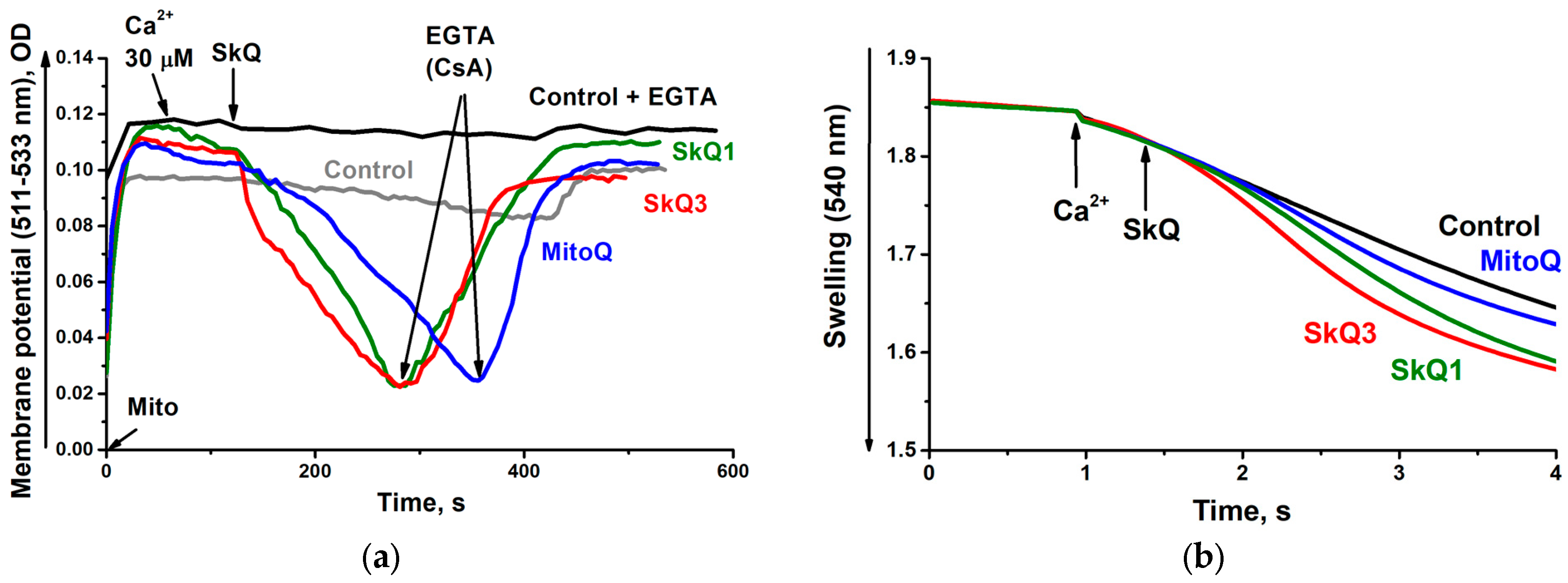
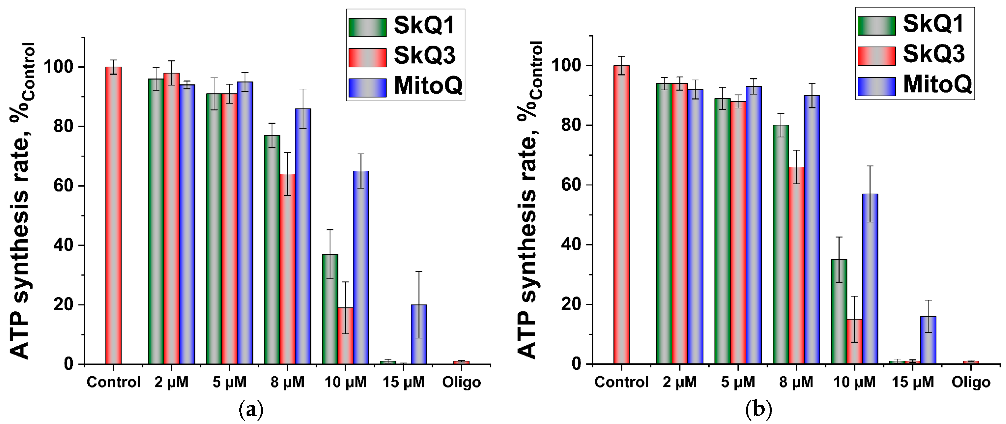
2.1. Effects of SkQ3, SkQ1 and MitoQ Supplementation on Isolated Rat Liver Mitochondria

In the first part of this study, we examined the mitochondria-targeted antioxidants SkQ3, SkQ1 and MitoQ and how they affect the functional parameters of the isolated tightly-coupled rat liver mitochondria (RLM), with a special focus on SkQ3, by comparing it with clinically tested SkQ1 and MitoQ [47,48,49].
Figure 1 shows SkQ1, SkQ3 and MitoQ structures.
Isolated RLM used in this study met all the conventional and some additional criteria of physiological integrity, as evidenced by their ability to maintain distinctive state 4-to-3 respiration transitions upon successive additions of ADP (Figure 2a,b); high respiratory control values, calculated based on Chance and Williams [50,51], upon the oxidation of both succinate and NAD-dependent substrates (Figure 2a,b); ADP/O ratios upon the oxidation of succinate and NAD-dependent substrates approaching 2 and 3, respectively, which was close to the theoretically expected maxima for these substrates; the stimulation of state 4 respiration by AMP instead of ADP, indicating the presence of adenylate kinase in the mitochondrial intermembrane space and hence the intactness of the outer mitochondrial membrane; long retention of the maximal ΔΨ value in the absence of oligomycin, which proves the absence of proton leakage.
At low concentrations, all three substances studied stimulated the oxidation of succinate (Figure 3a) and NAD-dependent substrates (glutamate and malate) (Figure 3b) in state 4 respiration, which is a formal indicator of their uncoupling effect, most probably due to discharging of the membrane potential as a result of their electrophoretic movement into mitochondria and/or the cycling of endogenous fatty acids. SkQ1 was the strongest uncoupler, while SkQ3 exhibited (showed) a moderate uncoupling effect.
At significantly higher concentrations, all three substances studied similarly inhibited state 3 respiration (in the presence of 0.9 mM ADP) (Figure 4a) and uncoupled respiration (in the presence of 2 μM carbonylcyanide m-chlorophenylhydrazone (CCCP) (Figure 4b) in mitochondria oxidizing succinate, thus suggesting the absence of the specific inhibition of ATP synthase or adenine nucleotide translocase. SkQ3 had a more pronounced inhibitory effect on state 3 or the uncoupled state of respiration, causing the same effect at lower concentrations.
All three substances studied depolarized the inner mitochondrial membrane (Figure 5a). SkQ3 reduced the membrane potential more effectively than SkQ1 or MitoQ. None of the substances caused the high-amplitude swelling of mitochondria (Figure 5b), indicating that there was no detectable action of SkQ3, SkQ1 or MitoQ on nonspecific mitochondrial pores or ion channels. Alamethicin (5 μM), a hydrophobic 1 nm-channel-forming peptide, produced by the fungus Trichoderma viride, was used as a positive control.
However, if the RLM were isolated and suspended in EGTA-free medium, the addition of 30 µM Ca2+ caused not only their spontaneous depolarization (Figure 6a) but also spontaneous swelling (Figure 6b). All three substances under study, taken in low (3 µM) concentrations, considerably increased the amplitude of these processes, most likely reflecting the promotion of the opening of the nonspecific Ca2+/phosphate-depended pore (the mPTP, permeability transition pore), as it was inhibited by EGTA or cyclosporine A (CsA) [52].
SkQ3, SkQ1 and MitoQ lowered the rate of ATP synthesis by mitochondria, which was measured using two independent methods. The incubation medium was supplemented with 1 µg/mL P5-di(adenosine-5′)pentaphosphate (Ap5A), the inhibitor of adenylate kinase. The inhibition of ATP synthesis by all three tested substances, was in precise accordance with their depolarizing action (Figure 7), with SkQ3 exerting a more pronounced effect, thus supporting the earlier conclusion (see, Figure 4) about the minimal, if any, effects of these substances on ATP synthase and the translocase of adenine nucleotides.
Low, non-uncoupling concentrations of SkQ3, SkQ1 and MitoQ had a pronounced antioxidant effect, decreasing the hydrogen peroxide production by RLM (Figure 8). The basic incubation medium was supplemented with 6 mM aminotriazole (an inhibitor of catalase). Importantly, SkQ3 displayed the most considerable antioxidant effect and, unlike SkQ1 or MitoQ, did not exhibit prooxidant activity when the effective concentration was increased over the entire range studied. This SkQ3 property makes it one of the most promising mitochondria-targeted antioxidants with potential therapeutic action.

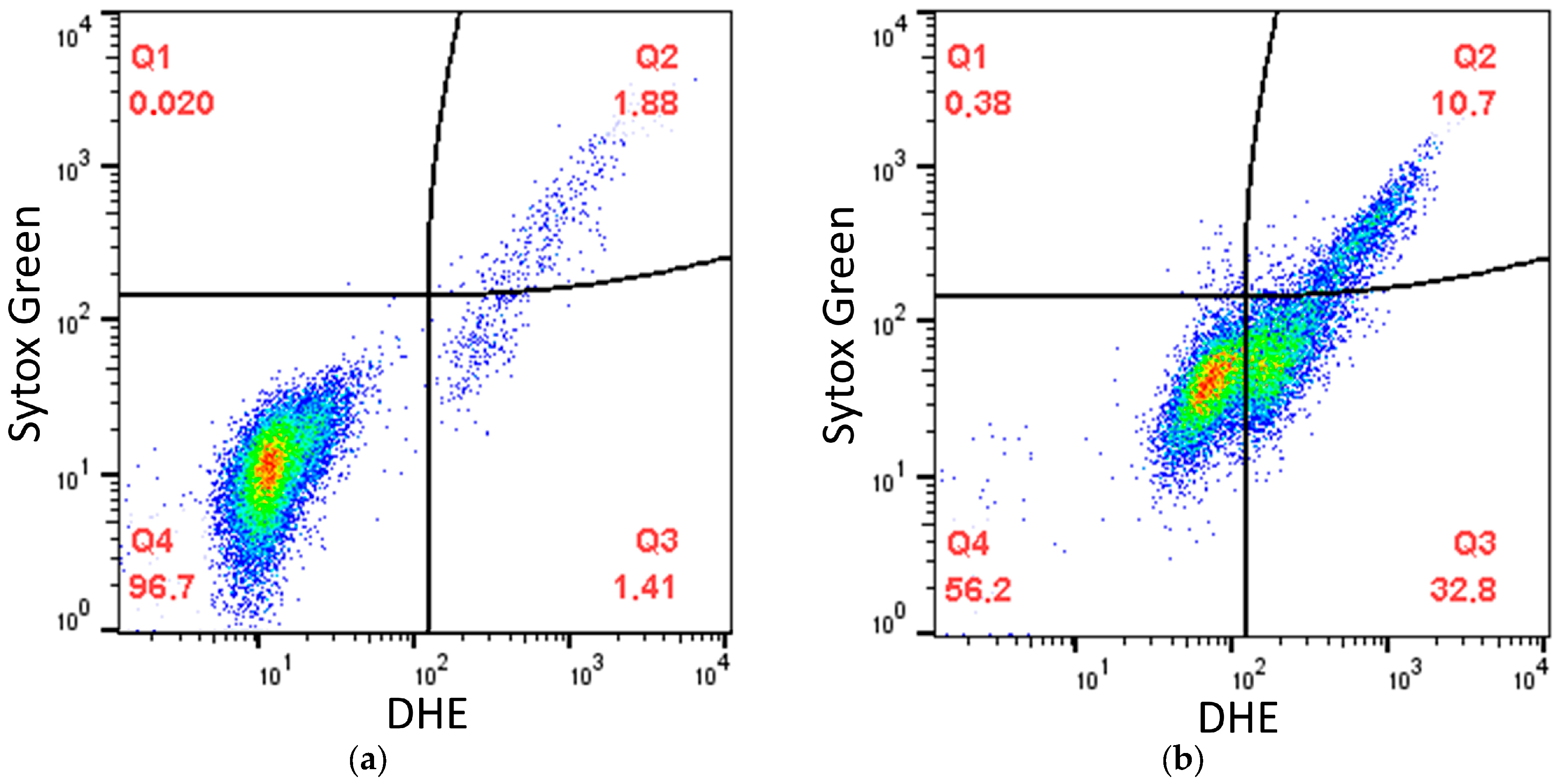
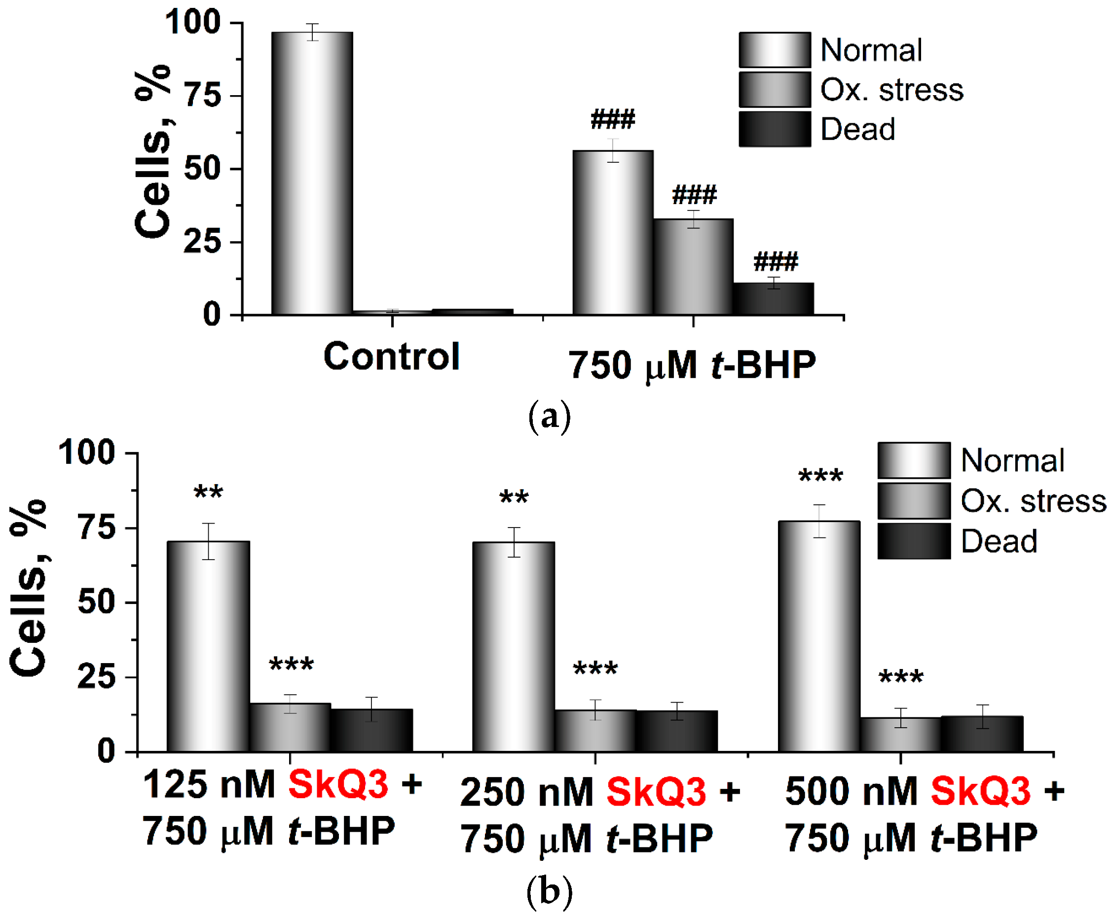
2.2. Effects of SkQ3, SkQ1 and MitoQ Suplementation on D. magnusii Cells

In the second part of this study, we tested the ability of SkQ3, SkQ1 and MitoQ to mitigate the oxidative stress induced by the classic prooxidant tert-butyl hydroperoxide (t-BHP) in D. magnusii cells. Yeast cells harvested in the early exponential growth phase were preincubated with different concentrations of SkQ3, SkQ1 and MitoQ and then transferred to the medium with t-BHP and stained with two dyes: dihydroethidium (DHE), detecting intracellular reactive oxygen species (ROS) (with increased affinity for the superoxide anion radical), and Sytox Green, staining only dead cells with a damaged cytoplasmic membrane. The fluorescence of double-stained yeast cells was examined via flow cytometry. Figure 9 shows (as an example) that the entire cell population was divided into three subpopulations according to the intensity of dye fluorescence. The subpopulation with low fluorescence of both DHE and Sytox Green (Q4) corresponded to living cells with no signs of oxidative stress, not subjected to oxidative stress (for short, “normal” cells); cells with increased DHE fluorescence (Q3) were thought to be alive with oxidative stress; the subpopulation of cells with high fluorescence of both dyes (Q2) represented dead cells (Figure 9 as an example). A two-hour incubation of yeast with t-BHP increased the level of intracellular oxidative stress and cell death.
Figure 10 shows the processed data as histograms. The preincubation of yeast cells with all three mitochondria-targeted antioxidants reduced intracellular oxidative stress and cell death, albeit with different efficacies (Figure 10b–d), with the most effective SkQ3 causing the most pronounced protective action, at lower concentrations.
Normally, D. magnusii cells possess a branched mitochondrial reticulum (Figure 11a, Control), which, however, is fragmented upon the induction of mitochondrial dysfunction (inhibition of respiration and uncoupling) or oxidative stress [46]. The incubation of “normal” D. magnusii cells with SkQ3, SkQ1 and MitoQ did not alter the morphology of mitochondria (Figure 11a, Control). The treatment of cells with low t-BHP concentrations led to complete mitochondrial reticulum fragmentation (Figure 11b, Control). Moreover, fragmented mitochondria did not regenerate into the mitochondrial network even after removing the prooxidant from the incubation medium (Figure 11c, Control). Importantly, cell preincubation with low concentrations of all three tested antioxidants prevented mitochondrial fragmentation when yeast cells were subsequently exposed to the prooxidant (Figure 11b, SkQ3, SkQ1, MitoQ). It is even more astonishing that the addition of low concentrations of SkQ3, SkQ1 and MitoQ to cells with fragmented mitochondria led to the recovery of the mitochondrial reticulum. Based on the data obtained, SkQ3, SkQ1 and MitoQ were effective in preventing and returning the mitochondrial fragmentation caused by oxidative stress. Once again, SkQ3 was the most effective in these two processes, as its preventing or recovering concentrations were the lowest.

3. Discussion

A thorough analysis of the effects of SkQ1, SkQ3 and MitoQ on RLM revealed only minor differences in the decoupling effects (Figure 3), respiratory inhibition (Figure 4), membrane depolarization (Figure 5a), the promotion of mPTP (mitochondrial permeability transition pore) (Figure 6) or impact on the rate of ATP synthesis (Figure 7). More surprisingly, the most pronounced antioxidant effect of SkQ3 on RLM seemed to be shown in the entire range of concentrations studied (Figure 8). Previously, such effects were observed only for SkQT1 ((toluquinonyl)decyltriphenylphosphonium) and SkQThy ((thymoquinonyl)decyltriphenylphosphonium), derivatives of toluquinone and thymoquinone, respectively [27,29], and the absence of a prooxidant effect at high concentrations was considered to be a great advantage of the substance as a potential therapeutic agent [27,29]. However, the main drawbacks of SkQT1 were the relatively low stability and the presence of a mixture of two isoforms in the ratio of 1.4 : 1 [29]. SkQThy is also a mixture of two isoforms [27], which makes it difficult to assess the contribution of each of the isoforms to overall antioxidant activity and their possible application in therapy.
In the tested yeast model, D. magnusii, we were able to not only confirm the data, obtained on mitochondria, but also quantify and compare the antioxidant effects of all three mitochondria-targeted antioxidants used (Figure 10, flow cytometry), as well their ability to prevent mitochondrial fragmentation under induced oxidative stress and, moreover, to restore the mitochondrial reticulum (Figure 11). The optimal concentrations of SkQ3 in these processes were much lower than those of SkQ1 and MitoQ. The cumulative data obtained suggest that SkQ3 is one of the most effective mitochondria-targeted antioxidants and does not have the disadvantages of SkQT1 and SkQThy.
As for the correlation between the quinone fragment structure and antioxidant activity of the examined mitochondria-targeted antioxidants, this is not so clear. MitoQ, a ubiquinone conjugate, had lower antioxidant activity compared to SkQ1 [30], while SkQ1, a plastoquinone conjugate, had lower antioxidant activity compared to the other plastoquinone conjugates SkQT1 and SkQThy [27,29], as well as SkQ3 (Figure 8, this study).
MitoQ, with relatively weak antioxidant activity, and SkQ3 (high activity, this study) both have no substituted atoms in the quinone fragment of the molecule, while SkQThy (high activity) and SkQ1 (moderate activity) have one and SkQT1 (high activity) has two unsubstituted atoms in the quinone ring.
Thus, we can conclude that there is no clear (obvious) correlation between the structure of the quinone ring (more precisely, the number of substituted atoms) in the molecules of the mitochondria-targeted antioxidants and their antioxidant activities, contrary to what has been asserted previously [33].

4. Materials and Methods

4.1. Reagents

ADP, Alamethicin, 3-amino-1,2,4-triazole, P1,P5-di(adenosine-5′)pentaphosphate (Ap5A), Amplex Red, ATP, carbonyl cyanide m-chlorophenylhydrazone (CCCP), cyclosporine A (CsA), EGTA, fatty acid-free BSA, glucose, glucose-6-phosphate dehydrogenase, glutamate, hexokinase, horseradish peroxidase, malate, mannitol, MgCl2, NaCl, NADP, (NH4)2SO4, oligomycin, Phenol Red, phosphoenolpyruvate, pyruvate kinase, rotenone, succinate, sucrose, tert-butyl hydroperoxide and Tris were from Sigma-Aldrich (Saint Louis, MO, USA); Coomassie G-250 was from MP Biomedicals (Santa Ana, CA, USA); CaCl2, KCl, K2HPO4, KH2PO4 and Safranine O were from Merck (Darmstadt, Germany); Dihydroethidium, Mitotracker Green FM, Propidium Iodide and Sytox Green were from Life Technologies (Carlsbad, CA, USA). Other reagents of the highest purity available were from domestic suppliers. MitoQ, SkQ1 and SkQ3 were kindly provided by Dr. D.S. Esipov from the A.N. Belozersky Research Institute of Physico-Chemical Biology, MSU, Moscow, Russia.

4.2. Isolation of RLM

All manipulations with animals were performed in accordance with the Guide for the Care and Use of Laboratory Animals [53] approved by the Local Ethics Committee of the National Research Center “Kurchatov Institute” (protocol no. 1 of 20 December 2022). Two-to-three-month-old (200–250 g) outbred Wistar male rats were used in this study. The animals were fed ad libitum and had full access to tap water. They were killed via decapitation. RLM were isolated via differential centrifugation as described previously [27] with minor modifications. Unless otherwise specified, the homogenization medium contained 0.21 M mannitol, 0.09 M sucrose, 10 mM Tris–HCl buffer, pH 7.2, and 0.5 mM EGTA; the homogenate was centrifuged at 6600× g for 12 min; the mitochondrial pellet was washed with the same buffer and resuspended in a minimum volume of the same buffer. All isolation procedures were conducted at 4 °C. The final mitochondrial preparations were kept on ice and remained fully active for at least 4 h.

4.3. Mitochondrial Protein Assay

The mitochondrial protein was assayed using the Bradford method [19] with BSA as a standard.

4.4. Oxygen Consumption Assay

Oxygen consumption by RLM was monitored amperometrically using a Clark-type oxygen electrode as described in [27]. The basic incubation medium containing 0.21 M mannitol, 0.09 M sucrose and 2 mM Tris–HCl buffer, pH 7.2, was supplemented with 20 mM Tris-succinate + rotenone (2 µg/mg protein) or 20 mM Tris-glutamate + 5 mM Tris-malate, 0.5 mM EGTA and mitochondria (0.5 mg protein/mL). Respiratory control values were calculated according to [54].

4.5. Transmembrane Potential Measurement

The membrane potential (ΔΨ) generated on the inner mitochondrial membrane of RLM was recorded spectrophotometrically using a DU-650 spectrophotometer (Beckman Coulter, Brea, CA, USA) [55]. The basic incubation medium was supplemented with 20 mM Tris-succinate + rotenone (2 µg/mg protein) or 20 mM Tris-glutamate + 5 mM Tris-malate, 0.5 mM EGTA, 20 µM Safranine O (as a ΔΨ-related probe) and mitochondria (0.5 mg protein/mL).

4.6. Swelling of RLM

Mitochondrial swelling was monitored spectrophotometrically using a Cary 300 Bio spectrophotometer (Varian, Palo Alto, CA, USA) as a decrease in the apparent absorbance of the mitochondrial suspension at 540 nm [27]. The basic incubation medium was supplemented with 20 mM Tris-succinate + rotenone (2 µg/mg protein), 40 mM KCl, mitochondria (0.5 mg protein/mL) and, where indicated, with 0.5 mM EGTA and/or 1.8 µM CsA.

4.7. Opening of the Nonspecific Ca2+/Pi-Dependent Pore

Opening of the nonspecific Ca2+/Pi-dependent pore (mPTP) was detected based on a combination of both the membrane potential depolarization and large-scale swelling of mitochondria isolated and incubated in EGTA-free medium, as recommended in [52].

4.8. ATP Synthesis by RLM

The ATP synthesis rate was monitored spectrophotometrically using two methods: based on a pH shift during ADP conversion to ATP using Phenol Red as an indicator with a DU-650 spectrophotometer (Beckman Coulter, Brea, CA, USA) at 557/618 nm or based on NADP reduction at 340 nm in the coupled reactions catalyzed by hexokinase and glucose-6-phosphate dehydrogenase with a Cary 300 Bio spectrophotometer (Varian, Palo Alto, CA, USA) [27]. The basic incubation medium was supplemented with 0.5 mM EGTA, 6 µM Ap5A (the inhibitor of adenylate kinase) and mitochondria (0.5 mg mitochondrial protein/mL). In the first case, additionally added 25 µM Phenol Red (pH-dependent dye) and all stock solutions and additives were carefully adjusted to pH 7.1; the synthesis of ATP was induced by the addition of 500 µM ADP. In the second case, 1 mM glucose, 1 mM NADP, hexokinase (10 IU/mL) and glucose-6-phosphate dehydrogenase (3 IU/mL) were added, and the synthesis of ATP was initiated with 100 µM ADP.

4.9. Hydrogen Peroxide Production by RLM

The rate of hydrogen peroxide generation by RLM was determined fluorometrically with a RF 5301PC fluorescence spectrophotometer (Shimadzu, Kyoto, Japan) based on the Amplex Red/horseradish peroxidase (HRP) assay. The superoxide-anion radical initially formed by mitochondria is converted spontaneously or via the action of superoxide dismutases to hydrogen peroxide, which oxidizes the colorless substance Amplex Red into highly fluorescent resorufin (excitation and emission wavelengths (ex and em) were set to 563 and 587 nm, respectively). The assay was calibrated with known H2O2 concentrations. The basic incubation medium was supplemented with 20 mM Tris-succinate, 5 µM Amplex Red, horseradish peroxidase (9 IU/mL) and 6 mM aminotriazole (an inhibitor of catalase).

4.10. Yeast Cell Culture

Cells of the yeast Dipodascus magnusii, strain VKM Y261, were cultivated in liquid glycerol-containing medium at 28 °C with agitation (220 rpm) [56] and harvested at the early exponential growth phase (OD590 = 1.0).

4.11. Visualization of Mitochondria in Cells

For mitochondria visualization, yeast cells were stained with the fluorescent probe MitoTracker Green FM, which labels mitochondria in a membrane potential-independent manner [57]. Stained cells were observed under a Axioskop 40 fluorescence microscope (Zeiss, Oberkochen, Germany).

4.12. ROS Generation and Determination

Oxidative stress in yeast cells was induced by tert-butyl hydroperoxide (t-BHP), a well-known model prooxidant, and detected with 10 μM dihydroethidium (DHE) as a ROS marker. Cells were incubated with 10 μM DHE for 30 min at room temperature in the dark. The fluorescence of DHE was measured via flow cytometry as described in [58], via flow cytometry with a FACSCalibur flow cytometer (Becton Dickinson, Franklin Lakes, NJ, USA).

4.13. Cell Viability Assay

Cell viability was assayed with 10 μM Sytox Green. The fluorescence of Sytox Green was measured via flow cytometry as described in [59].

4.14. Statistical Analysis

All experiments with yeast mitochondria were performed at least three times. For the analysis of the mitochondrial morphology, at least fifty cells were examined in each trial. Statistical analyses were performed using one-way ANOVA with the post hoc Tukey HSD test. Data are presented as the mean ± S.D. from at least three independent replicates.

Author Contributions

A.G.R., investigation and data curation, writing original draft and editing, formatting and finalizing the text. T.N.G., investigation and data curation, writing original draft and editing. D.A.A., formal analysis, data curation, review and editing. R.A.Z., conceptualization, writing original draft, review and editing, supervision and project administration. All authors have read and agreed to the published version of the manuscript.

Funding

This work was partially supported by the Ministry of Science and Higher Education of the Russian Federation (No. 122041100080-3) and by the National Research Center “Kurchatov Institute” (Thematic plan 1p.4.1 “Structural and functional studies of mitochondria, development of prospective therapeutic agents”).

Institutional Review Board Statement

The animal study protocol was performed in accordance with the NIH Guide for the Care and Use of Laboratory Animals approved by the Local Ethics Committee National Research Center “Kurchatov Institute” (protocol no. 1 of 20 December 2022).

Data Availability Statement

The data used to support the findings of this study are available from the corresponding author upon request.

Acknowledgments

Dedicated to unforgettable academician V.P. Skulachev, the Teacher of several generations of bioenergetics in Russia. We would like to thank D.S. Esipov from the A. N. Belozersky Research Institute of Physico-Chemical Biology, MSU, Moscow, Russian Federation, for providing SkQ1, SkQ3 and MitoQ used in this study.

Conflicts of Interest

The authors declare no conflicts of interest.

References

  1. Russell, O.M.; Gorman, G.S.; Lightowlers, R.N.; Turnbull, D.M. Mitochondrial Diseases: Hope for the Future. Cell 2020, 181, 168–188. [Google Scholar] [CrossRef]
  2. Murphy, M.P.; Hartley, R.C. Mitochondria as a therapeutic target for common pathologies. Nat. Rev. Drug Discov. 2018, 17, 865–886. [Google Scholar] [CrossRef] [PubMed]
  3. Lott, M.T.; Leipzig, J.N.; Derbeneva, O.; Xie, H.M.; Chalkia, D.; Sarmady, M.; Procaccio, V.; Wallace, D.C. mtDNA Variation and Analysis Using Mitomap and Mitomaster. Curr. Protoc. Bioinform. 2013, 44, 1.23.1–1.23.26. [Google Scholar] [CrossRef] [PubMed]
  4. Gorman, G.S.; Chinnery, P.F.; DiMauro, S.; Hirano, M.; Koga, Y.; McFarland, R.; Suomalainen, A.; Thorburn, D.R.; Zeviani, M.; Turnbull, D.M. Mitochondrial diseases. Nat. Rev. Dis. Primers 2016, 2, 16080. [Google Scholar] [CrossRef]
  5. Ahmed, S.T.; Craven, L.; Russell, O.M.; Turnbull, D.M.; Vincent, A.E. Diagnosis and Treatment of Mitochondrial Myopathies. Neurother. J. Am. Soc. Exp. NeuroTherapeutics 2018, 15, 943–953. [Google Scholar] [CrossRef] [PubMed]
  6. Mansouri, A.; Gattolliat, C.H.; Asselah, T. Mitochondrial Dysfunction and Signaling in Chronic Liver Diseases. Gastroenterology 2018, 155, 629–647. [Google Scholar] [CrossRef] [PubMed]
  7. Boyman, L.; Karbowski, M.; Lederer, W.J. Regulation of Mitochondrial ATP Production: Ca(2+) Signaling and Quality Control. Trends Mol. Med. 2020, 26, 21–39. [Google Scholar] [CrossRef] [PubMed]
  8. Khan, N.A.; Nikkanen, J.; Yatsuga, S.; Jackson, C.; Wang, L.; Pradhan, S.; Kivelä, R.; Pessia, A.; Velagapudi, V.; Suomalainen, A. mTORC1 Regulates Mitochondrial Integrated Stress Response and Mitochondrial Myopathy Progression. Cell Metab. 2017, 26, 419–428.e5. [Google Scholar] [CrossRef]
  9. Ma, X.; McKeen, T.; Zhang, J.; Ding, W.X. Role and Mechanisms of Mitophagy in Liver Diseases. Cells 2020, 9, 837. [Google Scholar] [CrossRef]
  10. Wu, Y.; Chen, M.; Jiang, J. Mitochondrial dysfunction in neurodegenerative diseases and drug targets via apoptotic signaling. Mitochondrion 2019, 49, 35–45. [Google Scholar] [CrossRef]
  11. Kim, S.H.; Kim, H. Inhibitory Effect of Astaxanthin on Oxidative Stress-Induced Mitochondrial Dysfunction-A Mini-Review. Nutrients 2018, 10, 1137. [Google Scholar] [CrossRef]
  12. Peoples, J.N.; Saraf, A.; Ghazal, N.; Pham, T.T.; Kwong, J.Q. Mitochondrial dysfunction and oxidative stress in heart disease. Exp. Mol. Med. 2019, 51, 1–13. [Google Scholar] [CrossRef] [PubMed]
  13. Singh, A.; Kukreti, R.; Saso, L.; Kukreti, S. Oxidative Stress: A Key Modulator in Neurodegenerative Diseases. Molecules 2019, 24, 1583. [Google Scholar] [CrossRef] [PubMed]
  14. Ahn, B.; Smith, N.; Saunders, D.; Ranjit, R.; Kneis, P.; Towner, R.A.; Van Remmen, H. Using MRI to measure in vivo free radical production and perfusion dynamics in a mouse model of elevated oxidative stress and neurogenic atrophy. Redox Biol. 2019, 26, 101308. [Google Scholar] [CrossRef]
  15. Fetisova, E.; Chernyak, B.; Korshunova, G.; Muntyan, M.; Skulachev, V. Mitochondria-targeted Antioxidants as a Prospective Therapeutic Strategy for Multiple Sclerosis. Curr. Med. Chem. 2017, 24, 2086–2114. [Google Scholar] [CrossRef] [PubMed]
  16. Demyanenko, I.A.; Zakharova, V.V.; Ilyinskaya, O.P.; Vasilieva, T.V.; Fedorov, A.V.; Manskikh, V.N.; Zinovkin, R.A.; Pletjushkina, O.Y.; Chernyak, B.V.; Skulachev, V.P.; et al. Mitochondria-Targeted Antioxidant SkQ1 Improves Dermal Wound Healing in Genetically Diabetic Mice. Oxidative Med. Cell. Longev. 2017, 2017, 6408278. [Google Scholar]
  17. Lefranc, C.; Friederich-Persson, M.; Palacios-Ramirez, R.; Nguyen Dinh Cat, A. Mitochondrial oxidative stress in obesity: Role of the mineralocorticoid receptor. J. Endocrinol. 2018, 238, R143–R159. [Google Scholar] [CrossRef]
  18. Shabalina, I.G.; Vyssokikh, M.Y.; Gibanova, N.; Csikasz, R.I.; Edgar, D.; Hallden-Waldemarson, A.; Rozhdestvenskaya, Z.; Bakeeva, L.E.; Vays, V.B.; Pustovidko, A.V.; et al. Improved health-span and lifespan in mtDNA mutator mice treated with the mitochondrially targeted antioxidant SkQ1. Aging 2017, 9, 315–339. [Google Scholar] [CrossRef]
  19. Angelova, P.R.; Abramov, A.Y. Role of mitochondrial ROS in the brain: From physiology to neurodegeneration. FEBS Lett. 2018, 592, 692–702. [Google Scholar]
  20. Ekoue, D.N.; He, C.; Diamond, A.M.; Bonini, M.G. Manganese superoxide dismutase and glutathione peroxidase-1 contribute to the rise and fall of mitochondrial reactive oxygen species which drive oncogenesis. Biochim. Biophys. Acta. Bioenerg. 2017, 1858, 628–632. [Google Scholar] [CrossRef]
  21. Cooke, M.S.; Evans, M.D.; Dizdaroglu, M.; Lunec, J. Oxidative DNA damage: Mechanisms, mutation, and disease. FASEB J. 2003, 17, 1195–1214. [Google Scholar] [CrossRef]
  22. Liu, Z.; Zhou, T.; Ziegler, A.C.; Dimitrion, P.; Zuo, L. Oxidative Stress in Neurodegenerative Diseases: From Molecular Mechanisms to Clinical Applications. Oxidative Med. Cell. Longev. 2017, 2017, 2525967. [Google Scholar] [CrossRef]
  23. Tan, B.L.; Norhaizan, M.E.; Huynh, K.; Heshu, S.R.; Yeap, S.K.; Hazilawati, H.; Roselina, K. Water extract of brewers’ rice induces apoptosis in human colorectal cancer cells via activation of caspase-3 and caspase-8 and downregulates the Wnt/β-catenin downstream signaling pathway in brewers’ rice-treated rats with azoxymethane-induced colon carcinogenesis. BMC Complement. Altern. Med. 2015, 15, 205. [Google Scholar]
  24. Radi, E.; Formichi, P.; Battisti, C.; Federico, A. Apoptosis and oxidative stress in neurodegenerative diseases. J. Alzheimer’s Dis. JAD 2014, 42 (Suppl. 3), S125–S152. [Google Scholar] [CrossRef] [PubMed]
  25. Flemming, N.B.; Gallo, L.A.; Forbes, J.M. Mitochondrial Dysfunction and Signaling in Diabetic Kidney Disease: Oxidative Stress and Beyond. Semin. Nephrol. 2018, 38, 101–110. [Google Scholar] [CrossRef] [PubMed]
  26. Chernyak, B.V.; Antonenko, Y.N.; Galimov, E.R.; Domnina, L.V.; Dugina, V.B.; Zvyagilskaya, R.A.; Ivanova, O.Y.; Izyumov, D.S.; Lyamzaev, K.G.; Pustovidko, A.V.; et al. Novel mitochondria-targeted compounds composed of natural constituents: Conjugates of plant alkaloids berberine and palmatine with plastoquinone. Biochem. Biokhimiia 2012, 77, 983–995. [Google Scholar] [CrossRef]
  27. Goleva, T.N.; Rogov, A.G.; Korshunova, G.A.; Trendeleva, T.A.; Mamaev, D.V.; Aliverdieva, D.A.; Zvyagilskaya, R.A. SkQThy, a novel and promising mitochondria-targeted antioxidant. Mitochondrion 2019, 49, 206–216. [Google Scholar] [CrossRef] [PubMed]
  28. Rogov, A.G.; Goleva, T.N.; Trendeleva, T.A.; Ovchenkova, A.P.; Aliverdieva, D.A.; Zvyagilskaya, R.A. New Data on Effects of SkQ1 and SkQT1 on Rat Liver Mitochondria and Yeast Cells. Biochem. Biokhimiia 2018, 83, 552–561. [Google Scholar] [CrossRef] [PubMed]
  29. Severina, I.I.; Severin, F.F.; Korshunova, G.A.; Sumbatyan, N.V.; Ilyasova, T.M.; Simonyan, R.A.; Rogov, A.G.; Trendeleva, T.A.; Zvyagilskaya, R.A.; Dugina, V.B.; et al. In search of novel highly active mitochondria-targeted antioxidants: Thymoquinone and its cationic derivatives. FEBS Lett. 2013, 587, 2018–2024. [Google Scholar] [CrossRef]
  30. Skulachev, V.P. Cationic antioxidants as a powerful tool against mitochondrial oxidative stress. Biochem. Biophys. Res. Commun. 2013, 441, 275–279. [Google Scholar] [CrossRef]
  31. Skulachev, V.P.; Anisimov, V.N.; Antonenko, Y.N.; Bakeeva, L.E.; Chernyak, B.V.; Erichev, V.P.; Filenko, O.F.; Kalinina, N.I.; Kapelko, V.I.; Kolosova, N.G.; et al. An attempt to prevent senescence: A mitochondrial approach. Biochim. Biophys. Acta 2009, 1787, 437–461. [Google Scholar] [CrossRef]
  32. Kelso, G.F.; Porteous, C.M.; Coulter, C.V.; Hughes, G.; Porteous, W.K.; Ledgerwood, E.C.; Smith, R.A.; Murphy, M.P. Selective targeting of a redox-active ubiquinone to mitochondria within cells: Antioxidant and antiapoptotic properties. J. Biol. Chem. 2001, 276, 4588–4596. [Google Scholar] [CrossRef] [PubMed]
  33. Skulachev, V.P. A biochemical approach to the problem of aging: “megaproject” on membrane-penetrating ions. The first results and prospects. Biochem. Biokhimiia 2007, 72, 1385–1396. [Google Scholar] [CrossRef] [PubMed]
  34. Green, D.E. The electromechanochemical model for energy coupling in mitochondria. Biochim. Biophys. Acta 1974, 346, 27–78. [Google Scholar] [CrossRef] [PubMed]
  35. Antonenko, Y.N.; Avetisyan, A.V.; Bakeeva, L.E.; Chernyak, B.V.; Chertkov, V.A.; Domnina, L.V.; Ivanova, O.Y.; Izyumov, D.S.; Khailova, L.S.; Klishin, S.S.; et al. Mitochondria-targeted plastoquinone derivatives as tools to interrupt execution of the aging program. 1. Cationic plastoquinone derivatives: Synthesis and in vitro studies. Biochem. Biokhimiia 2008, 73, 1273–1287. [Google Scholar] [CrossRef] [PubMed]
  36. Anisimov, V.N.; Bakeeva, L.E.; Egormin, P.A.; Filenko, O.F.; Isakova, E.F.; Manskikh, V.N.; Mikhelson, V.M.; Panteleeva, A.A.; Pasyukova, E.G.; Pilipenko, D.I.; et al. Mitochondria-targeted plastoquinone derivatives as tools to interrupt execution of the aging program. 5. SkQ1 prolongs lifespan and prevents development of traits of senescence. Biochem. Biokhimiia 2008, 73, 1329–1342. [Google Scholar] [CrossRef]
  37. Bakeeva, L.E.; Barskov, I.V.; Egorov, M.V.; Isaev, N.K.; Kapelko, V.I.; Kazachenko, A.V.; Kirpatovsky, V.I.; Kozlovsky, S.V.; Lakomkin, V.L.; Levina, S.B.; et al. Mitochondria-targeted plastoquinone derivatives as tools to interrupt execution of the aging program. 2. Treatment of some ROS- and age-related diseases (heart arrhythmia, heart infarctions, kidney ischemia, and stroke). Biochem. Biokhimiia 2008, 73, 1288–1299. [Google Scholar] [CrossRef] [PubMed]
  38. Agapova, L.S.; Chernyak, B.V.; Domnina, L.V.; Dugina, V.B.; Efimenko, A.Y.; Fetisova, E.K.; Ivanova, O.Y.; Kalinina, N.I.; Khromova, N.V.; Kopnin, B.P.; et al. Mitochondria-targeted plastoquinone derivatives as tools to interrupt execution of the aging program. 3. Inhibitory effect of SkQ1 on tumor development from p53-deficient cells. Biochem. Biokhimiia 2008, 73, 1300–1316. [Google Scholar] [CrossRef]
  39. Neroev, V.V.; Archipova, M.M.; Bakeeva, L.E.; Fursova, A.; Grigorian, E.N.; Grishanova, A.Y.; Iomdina, E.N.; Ivashchenko, Z.N.; Katargina, L.A.; Khoroshilova-Maslova, I.P.; et al. Mitochondria-targeted plastoquinone derivatives as tools to interrupt execution of the aging program. 4. Age-related eye disease. SkQ1 returns vision to blind animals. Biochem. Biokhimiia 2008, 73, 1317–1328. [Google Scholar] [CrossRef]
  40. Obukhova, L.A.; Skulachev, V.P.; Kolosova, N.G. Mitochondria-targeted antioxidant SkQ1 inhibits age-dependent involution of the thymus in normal and senescence-prone rats. Aging 2009, 1, 389–401. [Google Scholar] [CrossRef]
  41. Petrosillo, G.; Moro, N.; Ruggiero, F.M.; Paradies, G. Melatonin inhibits cardiolipin peroxidation in mitochondria and prevents the mitochondrial permeability transition and cytochrome c release. Free Radic. Biol. Med. 2009, 47, 969–974. [Google Scholar] [CrossRef]
  42. Kiselevsky, D.B.; Frolova, O.Y.; Solovyev, A.G.; Dorokhov, Y.L.; Morozov, S.Y.; Samuilov, V.D. Plant cell death caused by fungal, bacterial, and viral elicitors: Protective effect of mitochondria-targeted quinones. Biochem. Biokhimiia 2014, 79, 1322–1332. [Google Scholar] [CrossRef] [PubMed]
  43. Sukhanova, E.I.; Trendeleva, T.A.; Zvyagilskaya, R.A. Interaction of yeast mitochondria with fatty acids and mitochondria-targeted lipophilic cations. Biochem. Biokhimiia 2010, 75, 139–144. [Google Scholar] [CrossRef] [PubMed]
  44. Bazhenova, E.N.; Deryabina, Y.I.; Eriksson, O.; Zvyagilskaya, R.A.; Saris, N.E. Characterization of a high capacity calcium transport system in mitochondria of the yeast Endomyces magnusii. J. Biol. Chem. 1998, 273, 4372–4377. [Google Scholar] [CrossRef] [PubMed]
  45. Deryabina, Y.I.; Bazhenova, E.N.; Saris, N.E.; Zvyagilskaya, R.A. Ca(2+) efflux in mitochondria from the yeast Endomyces magnusii. J. Biol. Chem. 2001, 276, 47801–47806. [Google Scholar] [CrossRef] [PubMed]
  46. Rogov, A.G.; Ovchenkova, A.P.; Goleva, T.N.; Kireev, I.I.; Zvyagilskaya, R.A. New yeast models for studying mitochondrial morphology as affected by oxidative stress and other factors. Anal. Biochem. 2018, 552, 24–29. [Google Scholar] [CrossRef]
  47. Snow, B.J.; Rolfe, F.L.; Lockhart, M.M.; Frampton, C.M.; O’Sullivan, J.D.; Fung, V.; Smith, R.A.; Murphy, M.P.; Taylor, K.M. A double-blind, placebo-controlled study to assess the mitochondria-targeted antioxidant MitoQ as a disease-modifying therapy in Parkinson’s disease. Mov. Disord. 2010, 25, 1670–1674. [Google Scholar] [CrossRef]
  48. Brzheskiy, V.V.; Efimova, E.L.; Vorontsova, T.N.; Alekseev, V.N.; Gusarevich, O.G.; Shaidurova, K.N.; Ryabtseva, A.A.; Andryukhina, O.M.; Kamenskikh, T.G.; Sumarokova, E.S.; et al. Results of a Multicenter, Randomized, Double-Masked, Placebo-Controlled Clinical Study of the Efficacy and Safety of Visomitin Eye Drops in Patients with Dry Eye Syndrome. Adv. Ther. 2015, 32, 1263–1279. [Google Scholar] [CrossRef]
  49. Baiula, M.; Spampinato, S. Experimental Pharmacotherapy for Dry Eye Disease: A Review. J. Exp. Pharmacol. 2021, 13, 345–358. [Google Scholar] [CrossRef]
  50. Chance, B.; Williams, G.R. Respiratory enzymes in oxidative phosphorylation. III. The steady state. J. Biol. Chem. 1955, 217, 409–427. [Google Scholar] [CrossRef]
  51. Chance, B.; Williams, G.R.; Holmes, W.F.; Higgins, J. Respiratory Enzymes in Oxidative Phosphorylation, V. a Mechanism for Oxidative Phosphorylation. J. Biol. Chem. 1955, 217, 439–452. [Google Scholar] [CrossRef]
  52. Bernardi, P.; Forte, M. The mitochondrial permeability transition pore. Novartis Found. Symp. 2007, 287, 157–164, discussion 164–169. [Google Scholar] [PubMed]
  53. Committee for the Update of the Guide for the Care and Use of Laboratory Animals; Institute for Laboratory Animal Research. The National Academies Collection: Reports funded by National Institutes of Health. In Guide for the Care and Use of Laboratory Animals; National Academies Press, National Academy of Sciences: Washington, DC, USA, 2011. [Google Scholar]
  54. Chance, B.; Williams, G.R. A simple and rapid assay of oxidative phosphorylation. Nature 1955, 175, 1120–1121. [Google Scholar] [CrossRef]
  55. Goleva, T.N.; Lyamzaev, K.G.; Rogov, A.G.; Khailova, L.S.; Epremyan, K.K.; Shumakovich, G.P.; Domnina, L.V.; Ivanova, O.Y.; Marmiy, N.V.; Zinevich, T.V.; et al. Mitochondria-targeted 1,4-naphthoquinone (SkQN) is a powerful prooxidant and cytotoxic agent. Biochim. Biophys. Acta. Bioenerg. 2020, 1861, 148210. [Google Scholar] [CrossRef] [PubMed]
  56. Zviagil’skaia, R.A.; Zelenshchikova, V.A.; Ural’skaia, L.A.; Kotel’nikova, A.V. Respiratory system of Endomyces magnusii. Properties of mitochondria from cells grown on glycerol. Biokhimiia 1981, 46, 3–10. [Google Scholar]
  57. Agnello, M.; Morici, G.; Rinaldi, A.M. A method for measuring mitochondrial mass and activity. Cytotechnology 2008, 56, 145–149. [Google Scholar] [CrossRef] [PubMed]
  58. Epremyan, K.K.; Rogov, A.G.; Goleva, T.N.; Lavrushkina, S.V.; Zinovkin, R.A.; Zvyagilskaya, R.A. Altered Mitochondrial Morphology and Bioenergetics in a New Yeast Model Expressing Aβ42. Int. J. Mol. Sci. 2023, 24, 900. [Google Scholar] [CrossRef]
  59. Thakur, S.; Cattoni, D.I.; Nollmann, M. The fluorescence properties and binding mechanism of SYTOX green, a bright, low photo-damage DNA intercalating agent. Eur. Biophys. J. 2015, 44, 337–348. [Google Scholar] [CrossRef]
Figure 1. Structures of SkQ3, SkQ1 and MitoQ.
Ijms 25 01107 g001
Figure 2. Amperometric recording of oxygen consumption by RLM oxidizing succinate (a) or glutamate and malate (b). The basic incubation medium containing 0.21 M mannitol, 0.09 M sucrose and 2 mM Tris-phosphate, pH 7.2, was supplemented with 0.5 mM EGTA, 20 mM Tris-succinate + 2 µg rotenone (a) or 20 mM Tris-glutamate + 5mM Tris-malate (b) and mitochondria corresponding to 0.5 mg protein/mL. Where indicated, ADP was added. Respiratory control values for successive ADP additives were (a) 8.3 and 8.9 and (b) 7.3 and 9.8.
Ijms 25 01107 g002
Figure 3. Effect of SkQ3, SkQ1 and MitoQ on oxygen consumption by rat liver mitochondria in the state 4 respiration oxidizing succinate (a) or glutamate and malate (b). The incubation medium was as in Figure 2. Data are presented as the mean ± S.D. from three replicates.
Ijms 25 01107 g003
Figure 4. Effects of SkQ3, SkQ1 and MitoQ on oxygen consumption by RLM in the state 3 (a) or uncoupled state (b) of respiration supported by succinate. The basic incubation medium was supplemented with 0.5 mM EGTA, 20 mM Tris-succinate + 2 µg rotenone, mitochondria corresponding to 0.5 mg protein/mL and either 0.9 mM ADP (a) or 2 μM CCCP (b). State 3 respiration was initiated by adding 0.9 mM ADP, while the uncoupling state was triggered by the addition of 2 μM CCCP; 100% is defined as the respiratory rate before the addition of SkQ1, SkQ3 or MitoQ. Data are presented as the mean ± S.D. from three replicates.
Ijms 25 01107 g004
Figure 5. Effect of SkQ3, SkQ1 and MitoQ on the mitochondrial membrane potential (a) and swelling (b) of RLM oxidizing succinate. The basic incubation medium was supplemented with 0.5 mM EGTA, 20 mM Tris-succinate + 2 µg rotenone, mitochondria corresponding to 0.5 mg protein/mL and either 20 µM Safranine O (a) or 40 µM KCl and 5 µM Alamethicin (b). Where indicated, SkQ1, SkQ3 or MitoQ, respectively, were added. Data in (a) are presented as the mean ± S.D. from three replicates.
Ijms 25 01107 g005
Figure 6. SkQ3, SkQ1 and MitoQ promote mPTP opening in RLM isolated and suspended in EGTA-free media. The basic Incubation medium was supplemented with 20 mM Tris-succinate + 2 µg rotenone, mitochondria corresponding to 0.5 mg protein/mL and either 20 µM Safranine O (a) or 40 µM KCl (b). Where indicated, 30 µM CaCl2, 0.5 mM EGTA or 1.8 µM CsA or 3 µM SkQ1, 3 µM SkQ3 or 3 µM MitoQ were added.
Ijms 25 01107 g006
Figure 7. Effect of SkQ3, SkQ1 and MitoQ on ATP production by RLM oxidizing succinate. The basic incubation medium was supplemented with 0.5 mM EGTA, 20 mM Tris-succinate, rotenone (2 µg/mg), 1 µg/mL Ap5A, 20 µM Phenol Red (a) or 1 mM glucose + 2 mM NADP + 9U/mL hexokinase + 3U/mL glucose-6-phosphate dehydrogenase (b). Data are presented as the mean ± S.D. from three replicates.
Ijms 25 01107 g007
Figure 8. Effect of SkQ3, SkQ1 and MitoQ on peroxide production by RLM oxidizing succinate. The basic incubation medium was supplemented with 0.5 mM EGTA, 20 mM Tris-succinate, rotenone (2 µg/mg), 5 µM Amplex Red, 9 U horseradish peroxidase, 6 mM aminotriazole (an inhibitor of catalase) and mitochondria corresponding to 0.5 mg protein. Data are presented as the mean ± S.D. from three replicates.
Ijms 25 01107 g008
Figure 9. Oxidative stress and cell death in control (untreated) D. magnusii cells (a) and cells treated with t-BHP (b) for 2 h, as measured via flow cytometry. Q1—without cells; Q2—dead cells; Q3—cells with signs of oxidative stress; Q4—live cells with no signs of oxidative stress. The numbers inside the figure are the percentages of the prevailing subpopulation in the entire cellular population.
Ijms 25 01107 g009
Figure 10. The oxidative stress and cell death in D. magnusii yeasts treated with t-BHP for 2 h (a). Effects of SkQ3 (b), SkQ1 (c) and MitoQ (d). Results were obtained via flow cytometry measurements from three independent experiments, presented as histograms. Statistical analyses were carried out via the one-way ANOVA test. Symbols: * marks differences between the results obtained for SkQ-preincubated samples against only t-BHP-treated ones; ***: p < 0.001, **: 0.001 < p < 0.01; # marks differences between the control and t-BHP-treated sample; ###: p < 0.001.
Ijms 25 01107 g010aIjms 25 01107 g010b
Figure 11. t-BHP-induced fragmentation of mitochondria in D. magnusii cells and protective effects of SkQ3, SkQ1 and MitoQ on the integrity of the mitochondrial reticulum. (a) No preincubation with t-BHP; (b) where indicated, cells were preincubated with 250 nM SkQ3, 800 nM SkQ1 or 800 nM MitoQ for 1 h and then incubated with 750 μM t-BHP for 2 h; (c) where indicated, cells were preincubated with 250 μM t-BHP for 2 h and then incubated with 250 nM SkQ3, 800 nM SkQ1 or 800 nM MitoQ for 1 h. Bars are 5 μm.
Ijms 25 01107 g011
Disclaimer/Publisher’s Note: The statements, opinions and data contained in all publications are solely those of the individual author(s) and contributor(s) and not of MDPI and/or the editor(s). MDPI and/or the editor(s) disclaim responsibility for any injury to people or property resulting from any ideas, methods, instructions or products referred to in the content.

