Review

The Multifaceted Functions of TRPV4 and Calcium Oscillations in Tissue Repair

by
Dongsheng Jiang
1,*,†,‡,§,
Ruiji Guo
1,2,†,
Ruoxuan Dai
1,
Samuel Knoedler
1,2,3,
Jin Tao
4,5,
Hans-Günther Machens
2 and
Yuval Rinkevich
1,*
1
Institute of Regenerative Biology and Medicine, Helmholtz Center Munich, 81377 Munich, Germany
2
Department of Plastic and Hand Surgery, Klinikum Rechts der Isar, School of Medicine, Technical University of Munich, 81675 Munich, Germany
3
Division of Plastic Surgery, Brigham and Women’s Hospital, Harvard Medical School, Boston, MA 02152, USA
4
Department of Physiology and Neurobiology and Centre for Ion Channelopathy, Medical College of Soochow University, Suzhou 215123, China
5
Jiangsu Key Laboratory of Neuropsychiatric Diseases, Soochow University, Suzhou 215123, China
*
Authors to whom correspondence should be addressed.
These authors contribute equally to this work.
Current addresses: Precision Research Center for Refractory Diseases, Shanghai General Hospital, Shanghai Jiao Tong University School of Medicine, Shanghai 201620, China.
§
Current addresses: Trauma Medical Center, Shanghai General Hospital, Shanghai Jiao Tong University School of Medicine, Shanghai 201620, China.
Int. J. Mol. Sci. 2024, 25(2), 1179; https://doi.org/10.3390/ijms25021179
Submission received: 29 November 2023 / Revised: 9 January 2024 / Accepted: 16 January 2024 / Published: 18 January 2024
(This article belongs to the Collection Feature Papers in “Molecular Biology”)

Abstract

:
The transient receptor potential vanilloid 4 (TRPV4) specifically functions as a mechanosensitive ion channel and is responsible for conveying changes in physical stimuli such as mechanical stress, osmotic pressure, and temperature. TRPV4 enables the entry of cation ions, particularly calcium ions, into the cell. Activation of TRPV4 channels initiates calcium oscillations, which trigger intracellular signaling pathways involved in a plethora of cellular processes, including tissue repair. Widely expressed throughout the body, TRPV4 can be activated by a wide array of physicochemical stimuli, thus contributing to sensory and physiological functions in multiple organs. This review focuses on how TRPV4 senses environmental cues and thereby initiates and maintains calcium oscillations, critical for responses to organ injury, tissue repair, and fibrosis. We provide a summary of TRPV4-induced calcium oscillations in distinct organ systems, along with the upstream and downstream signaling pathways involved. In addition, we delineate current animal and disease models supporting TRPV4 research and shed light on potential therapeutic targets for modulating TRPV4-induced calcium oscillation to promote tissue repair while reducing tissue fibrosis.

1. Introduction

Transient receptor potential vanilloid type 4 (TRPV4) ion channels are calcium ion (Ca2+) permeable nonselective cation channels that belong to the transient receptor potential vanilloid (TRPV) subfamily of cation channels [1,2].
The original TRP channel was found in Drosophila photoreceptors. It contributes to sustained retinal depolarization when exposed to light. Deletion of the trp gene results in a transient calcium response, which lends its gene name. Unlike in Drosophila, mammalian TRPs serve a wider range of physiological functions [3,4]. In general, TRPs enable cells to detect subtle alterations in the local environment (e.g., fluid shear stress and mechanical forces), perceiving and responding to a variety of external cues through varied sensory perceptions, including vision, smell, taste, hearing, thermosensation, and most importantly, mechanosensation [5]. They are a class of ion channels found in diverse tissues and cell types, widely distributed in human organs, such as the liver, lungs, skin, kidneys, nerves, and intestines. They are permeable to a broad range of cations such as Ca2+, Mg2+, K+, Na+, and others [6]. TRP family members are categorized into two main groups, based on their sequence topology and phylogeny. Group 1 consists of TRPC (canonical), TRPV (vanilloid), TRPM (melastatin), TRPN (nompC), and TRPA (ankyrin), whereas group 2 includes TRPP (polycystin) and TRPML (mucolipin), totaling seven subfamilies [7]. Group 1 TRPs share substantial homology across their transmembrane domain sequence. Most of the group 1 TRP proteins are characterized by a C-terminal TRP domain, composed of 23 to 25 amino acids and located near the sixth transmembrane domain. In contrast, group 2 TRPs feature low sequence similarity with group 1, and an extracellular loop that is interposed between the first two transmembrane domains [2,5].
TRPV channels (TRPV1–TRPV6) exhibit diverse gating mechanisms. These versatile proteins serve as thermal, osmotic, and chemical sensors in vertebrates, hygrosensors, and mechanical sensors in insects, and function as both mechanical and chemical sensors in nematodes [8]. Notably, TRPV1 and TRPV2 are predominantly expressed in neurons but can also be detected in non-neuronal tissues [9,10,11]. Conversely, TRPV3 is expressed in the skin, hair follicles, tongue, nose, sensory ganglia, brain, and colon, while TRPV4 exhibits a broader expression pattern, including the skin [8,12].
TRPV4 was initially named on the basis of its distinct features, such as vanilloid receptor-related osmotically activated channel (VR-OAC) [13,14], osmosensitive transient receptor potential channel 4 (OTRPC4) [15], vanilloid receptor-like protein (VRL-2) [16], and TRP-12 [17]. In mammals, it was first identified as an osmotic detector, contributing to the maintenance of the systemic osmotic equilibrium [14,15,16]. Reduction of extracellular osmolarity conveyed by hypotonic solution, even as little as 10%, could trigger a reversible Ca2+ increase via TRPV4 in TRPV4-transfected HEK293 cells. Vice versa, when exposed to hypertonic environments, a decrease in Ca2+ was observed [15]. Notably, activation via heat stimuli has also been reported [13,18]. Temperature fluctuations between 36 °C and 42 °C were found to induce current fluctuations, while sustained or repeated stimulation over 42 °C resulted in TRPV4 desensitization and a decline in the current amplitude [18,19]. In the last decade, research on TRPV4 has mainly focused on its mechanosensory function, which is involved in various physiological and pathological processes, such as vasoconstriction [20], gastrointestinal motility [21], hair growth cycle [22,23], fibrosis [24,25,26], scleroderma [27], hypertension [28,29], gastric cancer [30,31], rosacea [32,33], brain injury [34], and pancreatitis [35,36].

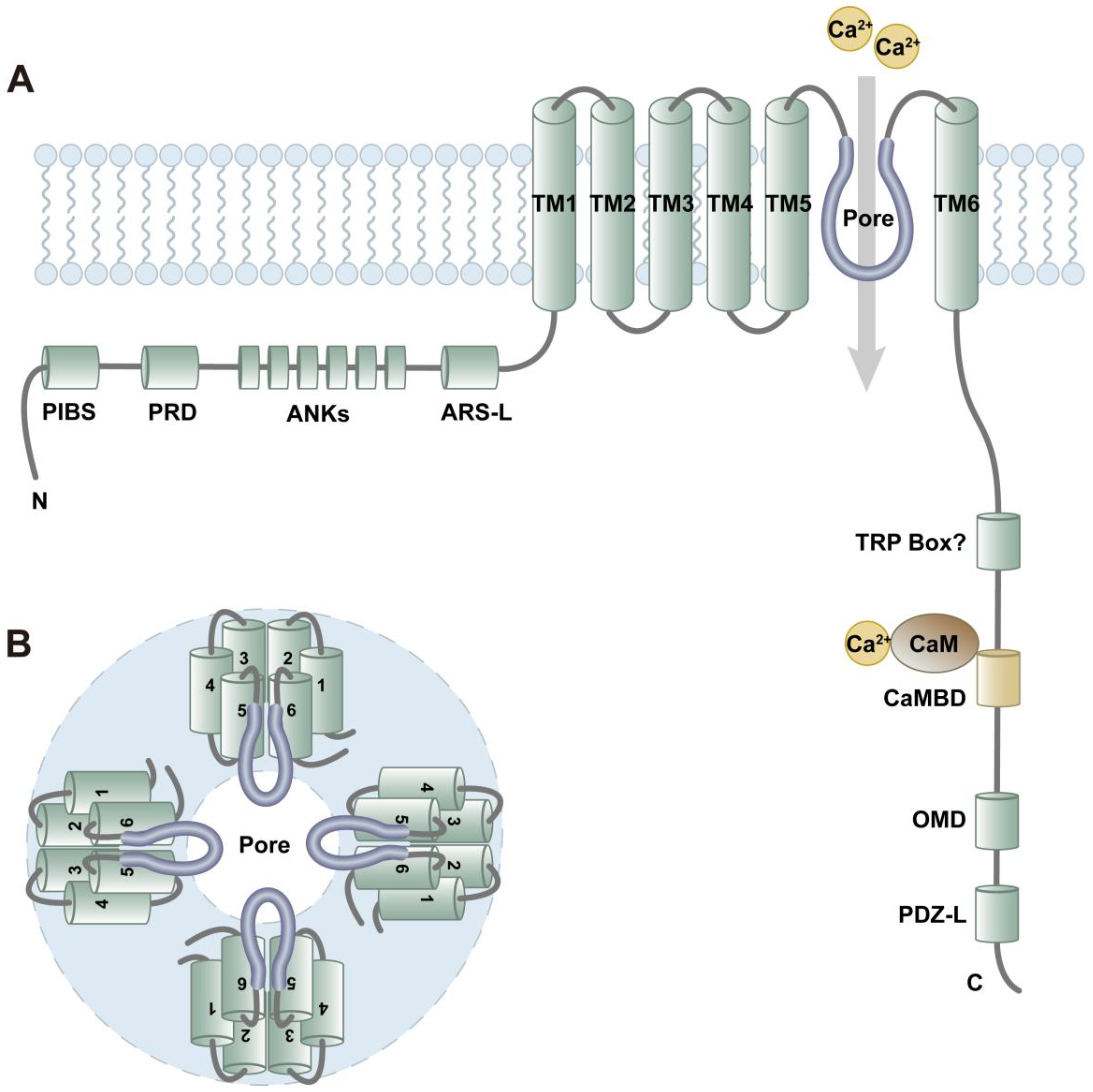
2. Protein and Channel Structure of TRPV4

TRPV4 is a tetrameric non-selective cation channel that is not voltage-gated. Murine TRPV4 is comprised of 871 amino acids (aa). Both N-and C-terminal tails are located intracellularly, with six transmembrane segments spanning the cell membrane and forming the ion channel pore [37]. The structure of TRPV4 (from N to C terminal regions) includes a phosphoinositide-binding site (PIBS, aa121–aa125) [38], a proline-rich domain (PRD, aa132–aa144) [39], six ankyrin repeats (ANK1–6) [40], an arachidonate-like recognition sequence (ARS-L, aa402–aa408) [41], six transmembrane domains (TM1–TM6), a questioned TRP box [42], a calmodulin-binding domain (CaMBD, aa812–aa831) [43], an oligomerization domain (OMD, aa828–aa844) [44], and a PDZ-like domain (PDZ-L, last four aa) [43], which are situated in tandem [25] (Figure 1A). Typically, Ca2+ does not act directly on the channel but rather through attachment to the Ca2+-binding protein calmodulin (CaM). The Ca2+-CaM complex then binds to the C-terminal calmodulin-binding site. Accordingly, the deletion of the calmodulin-binding domain results in lack of Ca2+-dependent potentiation and slower ion channel activation [45]. Previous studies have revealed that inositol 1,4,5-trisphosphate (IP3) sensitizes TRPV4 to mechanical and osmotic stimulation. This sensitization process requires binding of the IP3 receptor (IP3R) to the TRPV4 calmodulin-binding site [43].
Four identical TRPV4 monomers form a functional channel. The transmembrane segments TM1–TM4 form a structural unit surrounding the central ion-conduction pore that is comprised of TM5 and TM6 [37] (Figure 1B). The selectivity filters in TRPV channels generally function through two barriers, namely the lower and upper gates. However, in comparison to other TRPV channels, the diameter at the narrowest point of the upper gate within the TRPV4 pore is larger than that of hydrated cations such as K+, Na+, and Ca2+. This implies non-selective entry/exit of cation ions in TRPV4 [37]. Due to the two negatively charged aspartate residues within the conserved lower gate, it is easier for divalent ions than monovalent ions to pass through TRPV4. As a result, TRPV4 is mainly permeable to Ca2+ rather than other ions, with the following permeability sequence: Ca2+ >> Mg2+ > K+ > Na+, whereby the permeability for Ca2+ is about five times higher than that of Mg2+, and about ten times higher than for Na+ [46,47].

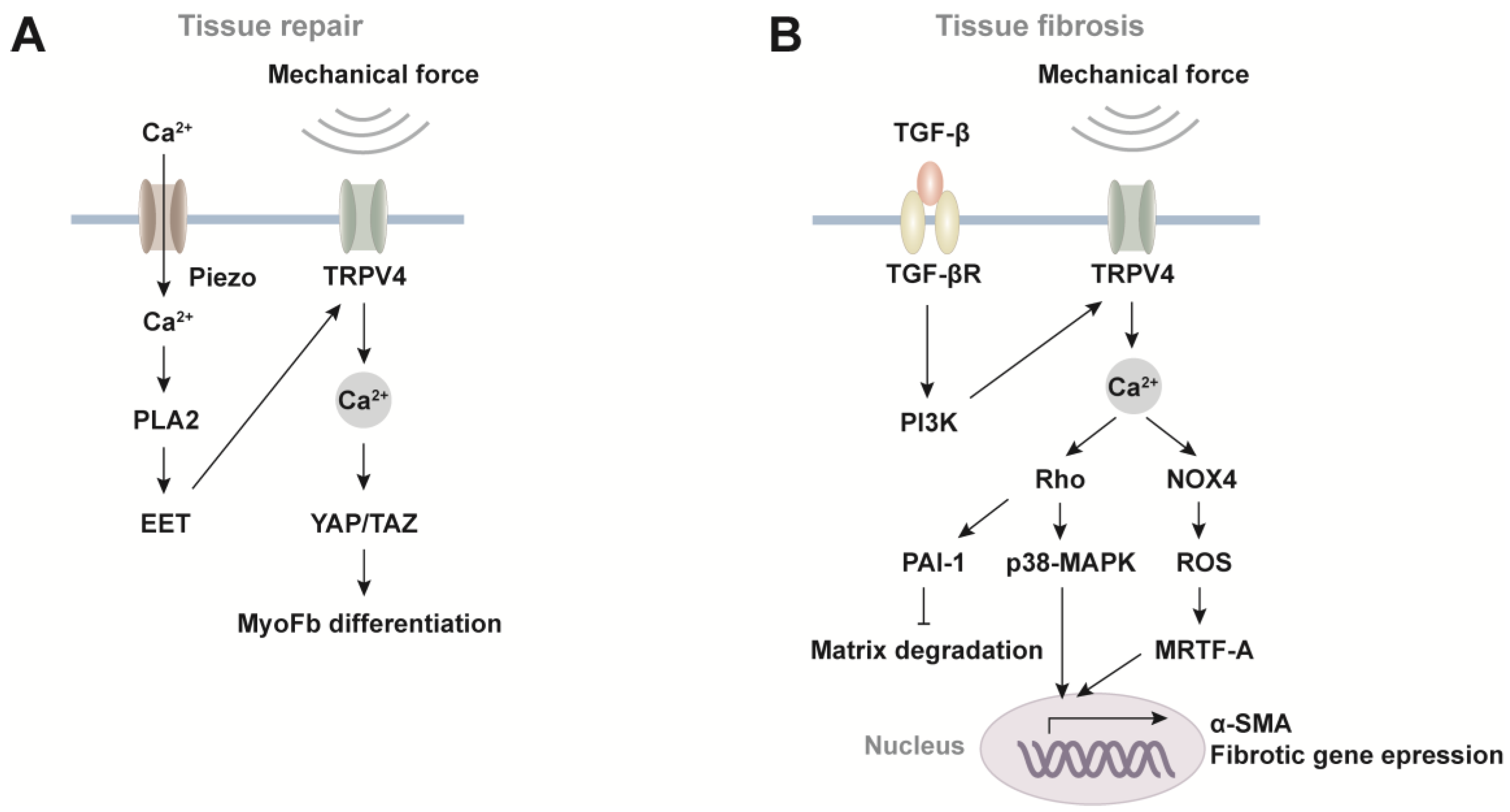
3. TRPV4 Signaling in Mechanotransduction

As a critical mechanotransduction molecule, TRPV4 plays a pivotal role in injury repair by orchestrating a wide array of cellular responses to mechanical stimuli (Figure 2A). For instance, in chondrocytes, TRPV4 is responsible for mechanotransduction in response to mechanical loading, together with Piezo channels. Specifically, TRPV4 and Piezo channels synergistically mediate calcium oscillation induced by different intensities of stretch stimulation. TRPV4-mediated Ca2+ signaling functions in response to low strain levels, whereas Piezo2-mediated Ca2+ signaling is activated in high strain settings, thus specifying the distinct cellular instructions during homeostasis and traumatic states [48,49]. Moreover, TRPV4 and Piezo1 can form a mechanosensory Ca2+ circuit. In pancreatic acinar cells, Piezo1 directly senses fluid shear stress and triggers Ca2+ influx, leading to an increase in phospholipase A2 (PLA2) activity and subsequent production of the arachidonic acid metabolites, particularly epoxieicosatrienoic acids (EET). EET sensitizes TRPV4 to initiate calcium oscillation, thereby amplifying the mechanical stimuli [36,43]. In response to changes in environmental stiffness, yes-associated protein/transcriptional coactivator with PDZ-binding motif (YAP/TAZ) acts as a downstream molecule for TRPV4 activity. This activation leads to myofibroblast differentiation, which plays a significant role in wound healing and injury repair [50,51].
TRPV4 is also involved in tissue fibrosis processes (Figure 2B). The most extensively studied fibrotic pathways mediated by TRPV4 are found in pulmonary and cardiac fibroblasts. Following acute lung injury repair, there is an abundance of TGF-β1 in the environment. TRPV4 activation is sustained in lung fibroblasts through TGF-β1 and its downstream phosphatidylinositol 3-kinase (PI3K) pathway. Consequently, Ca2+ signaling activates Rho family GTPases, which further activates p38-MAPK to upregulate fibrotic gene expression, and induces plasminogen activator inhibitor 1 (PAI-1) to reduce matrix degradation [52,53]. The imbalance of synthesis and degradation leads to the accumulation of extracellular matrix (ECM) and tissue stiffening, which provides a positive feedback loop for TRPV4 activity. In addition, through interaction with NADPH Oxidase 4 (NOX4), TRPV4 in lung fibroblasts potentiates the production of reactive oxygen species (ROS) to promote nuclear translocation of myocardin-related transcription factor A (MRTF-A), a transcription coactivator of α-SMA [26,52]. Therefore, TRPV4 stands at the crossroads of fibrotic signaling pathways that integrate the mechanical, TGF-β1, and ROS signals to promote myofibroblast differentiation of lung fibroblasts, by remodeling both intracellular actomyosin and ECM. Similar responses and signaling pathways have been documented in human ventricular cardiac fibroblasts and Trpv4-deficient mice. TRPV4-mediated Ca2+ influx regulates cardiac fibrotic gene promoter activity and myofibroblast differentiation [54] through the Rho/Rho kinase pathway activation and the mechanosensitive transcription factor MRTF-A [55], and the downstream MAPK/ERK pathway [56]. These findings indicate that TRPV4-mediated Ca2+ signaling is crucial for injury repair, but uncontrolled and persistent signaling results in fibrosis and scarring following the healing process.

4. TRPV4 Triggers Ca2+ Oscillations

Since cytosolic Ca2+ participates in numerous cellular activities, such as migration, contraction, exocytosis, cell growth and cell death [57], Ca2+ is strictly maintained in specific regions, normally at concentrations of less than a few hundred nanomolar. It can reach micromolar concentrations when Ca2+ is mobilized for cellular activities [58]. In essence, there are two reservoirs for Ca2+: (i) external Ca2+ within the extracellular microenvironment and (ii) sequestered Ca2+ within organelles, for instance, endoplasmic reticulum (ER) [58].
Ca2+ influx from outside the cell via TRPV4 evokes Ca2+ release from internal storages, termed “Ca2+-induced Ca2+ release”. This process is mediated through IP3R calcium ion channels in the ER of astrocytic endfeet, causing repetitive Ca2+ oscillations [59] (Figure 3A). When extracellular Ca2+ is removed, intracellular Ca2+ responses in chondrocytes can be completely suppressed, highlighting the necessity of extracellular Ca2+ for the onset of Ca2+ signaling [60]. Of note, the same phenomenon is also found in pulmonary fibroblasts [61]. Under abnormal circumstances, the equilibrium of mitochondrial membrane potential can be disrupted by elevated ATP production, thermogenesis, or mitochondrial dysfunction [62], including factors such as hyperoxia and pharmacologic uncoupling of mitochondrial oxidative phosphorylation [63,64]. These conditions lead to an increase in the levels of reactive oxygen species (ROS) surrounding the mitochondria [65]. Consequently, the release of Ca2+ is triggered through a more intricate pathway (Figure 3B). TRPV4 activation induces ATP release through pannexin hemichannels, whereupon ATP stimulates plasma membrane-bound purinergic receptors (P2Y), which activate phospholipase C (PLC) to produce IP3, eventually culminating in the activation of IP3R Ca2+ channels [64]. Moreover, ATP released through pannexin into the extracellular space undergoes degradation into ADP, AMP, and adenosine by ectonucleoside triphosphate diphosphohydrolase (ENTDPase) and ecto-5′-nucleotidase (ecto-5′-NT) [66,67]. Adenosine, a key player in this process, exerts its effects by binding to four distinct receptor subtypes—A1R, A2AR, A2BR, and A3R—belonging to the G-protein coupled receptor (GPCR) family [68]. When A1R is coupled to the Gi/0 proteins and A2BR is coupled to the Gq/11 proteins, the stimulation of PLC promotes the cleavage of phosphatidylinositol 4,5-bisphosphate (PIP2) into diacylglycerol (DAG) and IP3, which in turn increase intracellular Ca2+ levels [69]. In addition to the modulation of mitochondrial membrane potential, it has been documented that TRPV4 activation in hippocampal neurons leads to the depolarization of the resting membrane potential. Notably, heat stimulation induced depolarization exclusively in TRPV4-positive neurons, while TRPV4-negative or TRPV4-KO neurons did not exhibit such changes [70,71]. Furthermore, TRPV4 activation through hypotonic stimulation or the TRPV4 agonist 4α-PDD initiates NMDA-activated currents in hippocampal CA1 pyramidal neurons, with the inward current demonstrating a dose-dependent relationship to 4α-PDD [72]. Simultaneously, TRPV4 activation inhibits GABA-activated currents and increases glycine-activated currents [73,74].
It is worth noting that cytosolic Ca2+ oscillations correlate with TGF-β [75,76], platelet-derived growth factor (PDGF) [61] and ATP [58]. PDGF also requires PLC and IP3-gated channels in its downstream to induce Ca2+ waves [61]. The oscillatory frequency is associated with growth factor concentrations [76]. More importantly, calcium oscillation is necessary for proper cellular functions, since sustained high levels of Ca2+ induce cell apoptosis via PERK/CHOP/Bcl2 apoptotic pathway [77].
The downstream effects of calcium oscillation are believed to be mediated by second messengers. Yet, to date, only a few second messenger pathways have been identified, and they typically depend on the spatial and temporal context. To achieve its precision in modulation, Ca2+ oscillation—as a common form of Ca2+ signals—is encoded by the amplitude and frequency of Ca2+ spikes [57]. Once the amplitude is modulated, for example if the affinity of the decoder to the Ca2+ binding site is high, it is sensitive to the subtle Ca2+ elevations in the cytoplasm. Conversely, if the decoder’s affinity to the Ca2+ binding site is low, a more significant increase in pathway activation is required. In frequency modulation, Ca2+/calmodulin-dependent protein kinase II (CaMKII), a downstream Ca2+ sensor, fails to respond to low-frequency Ca2+ oscillations. In contrast, when the frequency of calcium oscillations exceeds its threshold, the kinase activity increases in a frequency-dependent manner [78].
The finely synchronized interplay of amplitude and frequency of Ca2+ signals in calcium oscillation is highly relevant in signal transduction and cellular function, with Ca2+ oscillation providing more information compared to a single pulse. For example, the amplitude and frequency of Ca2+ oscillations in chondrocytes are directly linked to the viscoelastic properties during cell swelling on substrates of different stiffness [79]. Lung fibroblasts exhibit recurring Ca2+ transients in response to PDGF, which spread through the cells as Ca2+ waves. There is a sigmoidal relationship between the frequency of Ca2+ wave and PDGF concentration [61]. Trabecular meshwork cells are specialized cells in the eye, responsible for regulating fluid outflow in the anterior portion of the eye. Long-term stimulation on TRPV4 is required to trigger Ca2+ oscillation in trabecular meshwork cells. Via the Ca2+ peak phase, plateau phase, and fluctuation phase, the mechanical signal can be transduced via trabecular meshwork cells to regulate intraocular pressure, thus upholding healthy fluid balance and pressure within the eye [80].

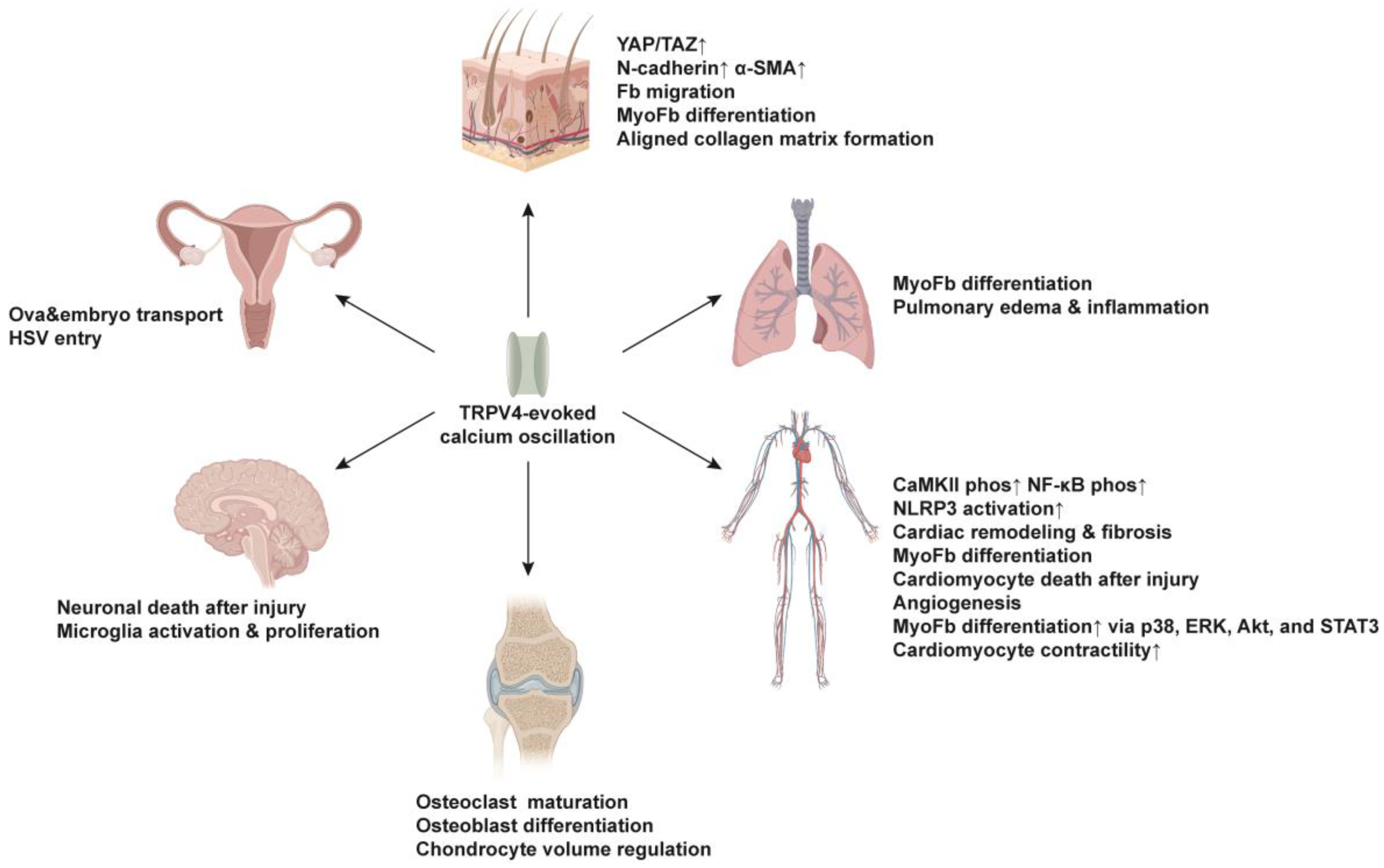
5. Function of TRPV4-Evoked Ca2+ Oscillations in Injury Repair and Fibrosis

TRPV4 mechanosensor is an integral factor in the progression of tissue repair and fibrotic disorders across different organs, including the skin, lungs, liver, kidneys, brain, blood vessels and heart [47]. In this section, we provide a comprehensive summary of the current understanding regarding the functions of TRPV4-induced Ca2+ oscillation in each specific organ (Figure 4). It is noteworthy that the mechanisms underlying cytosolic calcium oscillations in excitable and non-excitable cells significantly differ in the context of TRPV4 activation. Essential details, including whether the cells are excitable or non-excitable, are summarized in Table 1. This review primarily centers on examining the influence of TRPV4-induced Ca2+ oscillation in non-excitable cells during tissue repair and fibrosis.

5.1. Skin

In a bleomycin-induced skin fibrosis model, increasing matrix stiffness was found to activate TRPV4, with TRPV4 being essential for the nuclear translocation of YAP/TAZ in response to matrix stiffness. Genetic ablation of TRPV4 (Trpv4-knockout) or pharmacological inhibition of TRPV4 could ameliorate this fibrotic response. The authors attributed the effect of TRPV4 to its role in regulating the process of epithelial-mesenchymal transition [51]. In this study, it was also found that TRPV4-induced Ca2+ oscillations led to higher expression of N-cadherin and α-SMA on skin fibroblasts [51]. Our group has recently revealed that N-cadherin [109]—together with Connexin-43 [110] and p120-catenin [111]—are critical intercellular adhesion molecules that coordinate the collective migration of fibroblasts at a supracellular level needed for wound repair and subsequent scar formation. Therefore, in the skin fibrosis model, TRPV4′s effect on fibrosis development could also be attributed to its induction of fibroblasts’ collective migration and cell state transition toward myofibroblasts. However, the detailed signaling components and routes between TRPV4-induced Ca2+ oscillations and YAP/TAZ nuclear translocation, and the role of Ca2+ oscillations in synchronizing collective fibroblast migrations are yet to be determined. Of note, TRPV4-induced Ca2+ oscillations seem to be involved in adipogenic differentiation. Knocking down TRPV4 using shRNA significantly reduced adipogenesis in human preadipocytes via reduced phosphorylation of Akt kinase [81].
In addition, TRPV4-mediated Ca2+ oscillations are also implicated in matrix deposition. Gilchrist et al. found that the TRPV4 agonist GSK1016790A induced Ca2+ oscillation in mesenchymal stem cells at low concentrations (1, 10 nM), and is required for collagen matrix assembly in an aligned manner [82]. Interestingly, at a higher concentration (100 nM), the agonist initially activated TRPV4 and yielded a strong Ca2+ influx, but failed to sustain a rhythmic Ca2+ oscillation and regulate the formation of aligned collagen matrix [82].
TRPV4 is expressed in the hair follicles during the growing anagen phase. More specifically, it can be detected mainly in the outer and inner root sheath layers of the hair follicle epithelium, particularly in keratinocytes. The activation of TRPV4 through its agonist GSK1016790A was found to cause a significant reduction in hair shaft elongation and induce morphological and structural changes in anagen hair follicles, pushing them toward the catagen phase [23]. This study indicates that TRPV4-induced Ca2+ influx promotes injury repair and fibrotic processes, yet it may also suppress hair follicle regeneration after wounding. Therefore, targeting TRPV4 could either facilitate skin wound healing by enhancing Ca2+ influx, or stimulate hair follicle regeneration and reduce scarring by blocking its effects.

5.2. Lung

In human pulmonary fibroblasts, activation of TRPV4 assists cell membrane depolarization. When the membrane potential is in the voltage range of voltage-dependent Ca2+ current, the activated Ca2+ influx contributes to Ca2+ oscillations [76]. TRPV4 has also been reported to mediate TGF-β evoked repetitive Ca2+ waves in human lung fibroblasts, leading to the upregulation of ECM genes encoding collagen and fibronectin, and increased phosphorylation of SMAD-2 protein [75]. Via the P2Y receptor, ATP is able to release internal Ca2+ through ryanodine-insensitive channels, thereby exerting its effect on Ca2+ waves [58].
TRPV4 activity has been found to be significantly higher in patients with idiopathic pulmonary fibrosis. Activation of TRPV4 in lung fibroblasts accelerates the differentiation of myofibroblasts and contributes to the progression of pulmonary fibrosis [26]. In this context, it is worth mentioning that TRPV4 expression on murine lung fibroblasts is necessary for TGF-β-induced myofibroblast differentiation. Furthermore, TRPV4 mechanotransduction increases on stiffer matrices or in fibrotic lung tissue, while Trpv4-deficient mice are protected from bleomycin-induced lung fibrosis [26,54]. These findings imply that TRPV4 antagonists may represent potential targets for pulmonary fibrosis disease.
While TRPV4 activation promotes skin wound repair, its activity in lung endothelial cells has been shown to be detrimental in the setting of lung ischemia-reperfusion (IR) injury. In fact, TRPV4 activation in lung endothelial cells increases vascular permeability, thus causing increased lung inflammation and edema after IR [83]. Genetic depletion of TRPV4 using Trpv4−/− mice or pharmacological inactivation of TRPV4 with the TRPV4-specific inhibitor GSK2193874a markedly reduced pulmonary edema and neutrophil infiltration after IR injury, when compared to untreated wild-type mice. To further investigate TRPV4′s role in endothelial cells, Ottolini et al. generated an inducible endothelial Trpv4 knockout line by crossing Trpv4 floxed mice (Trpv4fl/fl) with tamoxifen-inducible VE-Cadherin (Cdh5) Cre mice [84]. The endothelial-specific Trpv4 knockout exhibited the same phenotype as observed in the whole-body Trpv4 knockout, suggesting that TRPV4 activity in endothelial cells contributes to pulmonary dysfunction and edema following IR injury. In line with the IR injury model, inhibition of TRPV4 had a protective effect in a mouse model of resistive breathing (RB)-induced lung injury. During RB, increased mechanical stress is imposed on the lung, thus leading to lung injury. The administration of the TRPV4 antagonist HC-067047 significantly mitigated symptoms by restoring static compliance and ameliorating the lung inflammation by reducing the number of macrophages and neutrophils, and the inflammatory chemokines keratinocyte-chemoattractant (KC) and interleukin (IL)-6 in bronchoalveolar lavage [112].

5.3. Cardiovascular System

The role of TRPV4 in cardiac remodeling has been investigated in the context of myocardial infarction and subsequent fibrosis, with a focus on TRPV4 channels expressed in non-excitable cardiac fibroblasts. Experimental findings demonstrate that while hypotonicity induces significant Ca2+ influx in cardiac fibroblasts from wild-type mice, this response is markedly diminished in cardiac fibroblasts derived from Trpv4-knockout mice, thereby establishing TRPV4′s role as a mechanosensor in fibroblasts [55]. In vitro studies using rat or human cardiac fibroblasts have revealed that TRPV4 activation by specific agonists such as 4αPDD or GSK1016790A leads to Ca2+ influx, which subsequently promotes the differentiation of fibroblasts into myofibroblasts through the p38 and ERK MAPK pathways [56,85]. Consistently, the blockade of TRPV4 by different antagonists or Trpv4-siRNA inhibits TGFβ-induced myofibroblast transformation by inhibiting the p38, Akt, and STAT3 signaling pathways [86,87]. In addition, it has been noticed that TRPV4 protein expression increases in cardiomyocytes of the aged heart. TRPV4-evoked calcium oscillations are directly correlated to the contractility of cardiomyocytes and contribute to tissue damage in the aged heart following ischemia-reperfusion (I/R), a pathological condition associated with cardiomyocyte osmotic stress. TRPV4 antagonist HC-067047 is effective in preventing Ca2+ overload in cardiomyocytes after I/R and prevents hypoosmotic stress-induced cardiomyocyte death and I/R-induced cardiac damage in the elderly population [91,92]. The same observation is documented in the aged Trpv4 knockout mice [93]. Furthermore, TRPV4-induced Ca2+ oscillation has been reported in human cardiac c-kit+ progenitor cells, regulating their migration but not proliferation [88]. In endothelial colony-forming cells, TRPV4-mediated Ca2+ oscillation results in the nuclear translocation of a Ca2+-sensitive transcription factor p65 NF-κB and induces angiogenesis [89].
In addition, TRPV4 channel expression is found to be significantly increased in failing human ventricles, and in murine ventricles using a mouse model of pressure overload-induced cardiac hypertrophy [90]. Compared to wild-type mice, Trpv4 knockout mice showed a remarkable reduction in cardiac hypertrophy, cardiac dysfunction, fibrosis, and inflammation. Those findings imply that TRPV4 is involved in cardiac remodeling and fibrosis after injury. Moreover, the absence of TRPV4 prevented an elevation of CaMKII phosphorylation in excitable cardiomyocytes of a pressure-overloaded left ventricle. It has been proposed that the TRPV4-mediated increase in CaMKII phosphorylation triggers NF-κB phosphorylation and NLRP3 activation, both of which contribute to the pro-inflammatory remodeling observed in these pressure-overloaded hearts [90].
In the vascular system, TRPV4 expression is widely reported in vascular smooth muscle cells (VSMCs), and TRPV4 plays an important role in the regulation of blood pressure and the development of hypertension. In patients with hypertension and in mouse models of hypertension, VSMCs showed elevated TRPV4 activity, resulting in signal amplification for the simulation of α1 adrenergic receptor. Strikingly, VSMC-specific Trpv4-knockout mice were protected from hypertension [95]. TRPV4 expression in VSMCs also contributed to vascular remodeling after injury, whereby TRPV4 was activated by platelet-derived microvesicles and triggered Ca2+ influx from the ECM and subsequent Ca2+ oscillations, thus contributing to the modulation of the cell migration [94].

5.4. Skeletal System

A mounting body of evidence indicates that TRPV4 is required for the normal development and maintenance of bone and cartilage. Osteoclasts are considered the main mediators of bone resorption. Their activity in removing damaged tissue and modulating the bone microenvironment is crucial for subsequent phases of bone healing and the restoration of bone structure and function. The maturation and function of osteoclasts are regulated by intracellular Ca2+ oscillatory signals, which are stimulated by mechanical forces, such as fluid shear stress. TRPV4 was found to be highly expressed in mature osteoclasts and necessary for Ca2+ oscillation induced by fluid shear stress [96]. Ca2+ oscillation affected the bone homeostasis-related gene expression, including sclerostin (Sost), osterix (Sp7), and osteoprotegerin (Tnfrsf11b) [97], and osteoblastic differentiation genes, such as alkaline phosphatase (Alpl), osterix (Sp7), dentin matrix protein 1 (Dmp1), and osteocalcin (Bglap) [98]. Spontaneous Ca2+ oscillation was generated by intracellular Ca2+ release as well as Ca2+ reuptake, and sustained by Ca2+ influx. It was gradually shifted to Ca2+ influx through TRPV4 during osteoclast differentiation, so that Ca2+ oscillations and Ca2+ influx via TRPV4 were sequentially required [99]. Upon exposure to an electromagnetic field, TRPV4 expression was downregulated in osteoclasts, and Ca2+ oscillations were impaired as a result of Ca2+ influx decrease, culminating in reduced activity of the calcium/calmodulin-dependent protein kinase–cyclic AMP response element-binding protein (CaMK–CREB) pathway [113].
In chondrocytes, intracellular Ca2+ oscillations are one of the earliest responses elicited by physical stimuli, such as compressive loading and stretch [60,114]. TRPV4 serves as a major mechanical sensor on chondrocytes by regulating Ca2+ signaling, together with Piezo1 and Piezo2 channels [100]. Evoked Ca2+ oscillation regulates chondrocyte volume in response to the pericellular matrix [79]. In stiff substrates, cytosolic Ca2+ oscillations were enhanced in the cell recovering phase, whereas in soft substrates, the cytosolic Ca2+ oscillations were increased in the cell swelling process. Of note, heterozygous mutations in TRPV4 led to altered chondrocyte Ca2+ oscillation and caused severe metatropic dysplasia [114].
Intracellular Ca2+ oscillation and a sustained Ca2+ response have been documented in annulus fibrosus cells, which form one of the structural components of intervertebral discs. This suggests that TRPV4 is a crucial regulator of the perception of mechanical stimulation in the intervertebral discs of the spine [101]. Furthermore, TRPV4 is also implicated in joint inflammation. Both TRPV4 and TRPV1 are activated in synovial cells by protein kinase A and protein kinase C, leading to increased cell sensitivity to noxious thermal changes and hypoosmotic stress [102,103]. In contrast, in a mouse model of osteoarthritis, knockout Trpv4 in articular chondrocytes (Col2a1CreER;Trpv4fl/fl) induced more severe age-related osteoarthritis [115].

5.5. Nervous System

TRPV4 is expressed in microglia, a type of non-excitable cells within the central nervous system (CNS), and plays a role in microglia activation and proliferation, promoting functional and structural plasticity in excitatory spinal cord neurons through the release of lipocalin-2 [104]. Genetic ablation of Trpv4 in microglia using Cx3cr1CreER;Trpv4fl/fl mice or a pharmacological blockade of TRPV4 with TRPV4 antagonist GSK2193874 markedly attenuated neuropathic pain-like behavior in a mouse model of spared nerve injury [104], highlighting TRPV4 as a potential target for chronic pain treatment.
The functional involvement of TRPV4 in excitable neurons in the spinal cord remains a subject of controversy. Previous studies have reported TRPV4 expression in spinal dorsal horn neurons, ventral horn neurons, and dorsal root ganglion in Sprague-Dawley rats and neonatal mice, indicating a direct influence on the induction of neuropathic pain [116,117,118]. In contrast, recent work by Hu and colleagues challenges this perspective, asserting that TRPV4 is not expressed in NeuN+ spinal cord neurons but rather in IBA1+/Tmem119+ microglia and CD31+ endothelial cells, as revealed by genetic lineage tracing in mice. This suggests that TRPV4 might participate in pain signaling through sustained neurogenic inflammation [104].
TRPV4 also plays a crucial role in neuronal damage during cerebral ischemia. In a rat model of hypoxia/ischemia-induced cerebral injury, the expression of TRPV4 is markedly enhanced in hippocampal astrocytes. The increased activity of TRPV4 channels triggers Ca2+ oscillations in these cells, promoting active cellular proliferation that protects neurons, initiates repair, and correlates with astrogliosis—scar formation in the brain [105]. TRPV4 expression undergoes upregulation in the ipsilateral hippocampus following middle cerebral artery occlusion, contributing to neuronal injury during cerebral ischemia. The neurotoxic effects associated with TRPV4 activation coincide with an elevation in the phosphorylation of the NR2B subunit of the N-methyl-D-aspartate receptor (NMDAR) and a concurrent downregulation of the Akt signaling pathway. Consequently, the TRPV4-specific antagonist HC-067047 has proven effective in reducing cerebral infarction. Conversely, intracerebroventricular injection of the TRPV4 agonist GSK1016790A0 has been shown to induce hippocampal neuronal death [119]. In Trpv4-knockout mice, impaired osmotic sensing and regulation in the CNS have been observed. In this context, TRPV4-induced Ca2+ oscillations, which trigger c-FOS expression in osmotically responsive cells of the CNS, become dysfunctional [120].
Moreover, rapid Ca2+ oscillations are recorded in the subpial endfeet of astrocytes, with oscillatory activities relying on the IP3R type 2 channel correlating with TRPV4 and dystrophin. Astrocytes leveraged the Ca2+ response to effectively respond to hypoosmotic changes in the cerebrospinal fluid [106]. However, this particular function is lost after subarachnoid hemorrhage [121].

5.6. Reproductive System

TRPV4, in conjunction with γ-aminobutyric acid receptors A and B (GABA A/B), has been identified as a contributor to progesterone (P4)-induced ova and embryo transport. In the mouse oviduct, Ca2+ oscillation and an increase in ciliary beat frequency are seen in response to P4 and GABA A/B activation. Importantly, TRPV4-mediated Ca2+ entry is essential for the initiation and maintenance of this oscillatory signal [107]. In addition, during infection in vaginal epithelial cells, Ca2+ fluctuations seem to play a critical role in facilitating the entry of Herpes simplex virus (HSV). TRPV4 expressed in human epithelial cells mediates the influx of Ca2+. Inhibition of TRPV4 by TRPV4 inhibitors, such as GSK2193874 and HC067047, not only reduces HSV-2 infection in human vaginal epithelial cells, but also attenuates the associated inflammatory responses, characterized by the expression of tumor necrosis factor (TNF)-α, IL-6, C-X-C motif chemokine ligand (CXCL)-9, and CXCL-10 [108]. These findings underscore the potential of TRPV4 as a target for therapeutic interventions related to reproductive health and HSV-2 infection.

6. Conclusions and Future Perspectives

The body of evidence summarized herein highlights the multifaceted role of TRPV4-induced Ca2+ influx and of Ca2+ oscillations across different tissues and under different pathological conditions. Altogether, TRPV4-mediated calcium oscillations have emerged as a crucial regulatory mechanism in injury repair and tissue fibrosis. Understanding the precise role of TRPV4 in these processes may accelerate the development of innovative therapeutic approaches in a wide array of disorders.
Notably, the physiological and pathological functions of TRPV4-induced Ca2+ oscillations in skin wound repair and scarring remain relatively unexplored. Given its ramifications in fibroblast maturation, differentiation, cellular migration, ECM remodeling, and inflammation, it is tempting to speculate that TRPV4-mediated Ca2+ oscillations play a pivotal role in wound healing and scarring. Accordingly, future research endeavors should prioritize unraveling the precise underlying mechanisms and exploring targeted strategies that engage TRPV4 to promote effective skin wound healing and mitigate scar formation. In this context, the development of readily transferable research tools such as fibroblast subset-specific (e.g., papillary, reticular, hypodermal, superficial fascia) or fibroblast state-specific (e.g., naïve, proinflammatory, proto-myofibroblast and myofibroblast) TRPV4 knockout animals will be invaluable to decipher TRPV4-evoked Ca2+ oscillations in fibroblast fate determination. These avenues can help bridge the gap between basic science and clinical translation and, ultimately, optimize patient care.
In conclusion, by elucidating the interplay between TRPV4 and other cellular components, such as ion channels, receptors, and intracellular signaling molecules, we can gain a comprehensive understanding of TRPV4′s functional significance in diverse fibroblast subsets and cell states. This knowledge will guide the design and engineering of selective TRPV4 modulators that precisely regulate Ca2+ oscillation, which holds tremendous potential for revolutionizing wound healing treatments. The ability to fine-tune TRPV4-mediated Ca2+ oscillation could lead to improved outcomes and reduced scar formation.

Author Contributions

Writing: original draft preparation, D.J., R.G. and R.D.; writing: review and editing, D.J., R.G., R.D., S.K., J.T., H.-G.M. and Y.R.; funding acquisition: Y.R. All authors have read and agreed to the published version of the manuscript.

Funding

This research was funded by the European Research Council Consolidator Grant (ERC-CoG 819933), the LEO Foundation (LF-OC-21-000835), the European Foundation for the Study of Diabetes (EFSD) Anniversary Fund Program, the German Research Foundation (CRC/TRR359 PILOT), and the European Union Horizon 2020 (Marie Curie 955722 SkinTERM).

Conflicts of Interest

The authors declare no conflict of interest.

References

  1. White, J.P.; Cibelli, M.; Urban, L.; Nilius, B.; McGeown, J.G.; Nagy, I. TRPV4: Molecular Conductor of a Diverse Orchestra. Physiol. Rev. 2016, 96, 911–973. [Google Scholar] [PubMed]
  2. Venkatachalam, K.; Montell, C. TRP channels. Annu. Rev. Biochem. 2007, 76, 387–417. [Google Scholar] [CrossRef] [PubMed]
  3. Moran, M.M. TRP Channels as Potential Drug Targets. Annu. Rev. Pharmacol. Toxicol. 2018, 58, 309–330. [Google Scholar] [PubMed]
  4. Cosens, D.J.; Manning, A. Abnormal electroretinogram from a Drosophila mutant. Nature 1969, 224, 285–287. [Google Scholar] [CrossRef] [PubMed]
  5. Montell, C. The TRP superfamily of cation channels. Sci. STKE 2005, 2005, re3. [Google Scholar] [CrossRef] [PubMed]
  6. Zhang, M.; Ma, Y.; Ye, X.; Zhang, N.; Pan, L.; Wang, B. TRP (transient receptor potential) ion channel family: Structures, biological functions and therapeutic interventions for diseases. Signal Transduct. Target. Ther. 2023, 8, 261. [Google Scholar] [PubMed]
  7. Himmel, N.J.; Cox, D.N. Transient receptor potential channels: Current perspectives on evolution, structure, function and nomenclature. Proc. Biol. Sci. 2020, 287, 20201309. [Google Scholar]
  8. Rosenbaum, T.; Islas, L.D. Molecular Physiology of TRPV Channels: Controversies and Future Challenges. Annu. Rev. Physiol. 2023, 85, 293–316. [Google Scholar] [CrossRef]
  9. Ho, C.Y.; Gu, Q.; Lin, Y.S.; Lee, L.Y. Sensitivity of vagal afferent endings to chemical irritants in the rat lung. Respir. Physiol. 2001, 127, 113–124. [Google Scholar] [CrossRef]
  10. Bohlen, C.J.; Priel, A.; Zhou, S.; King, D.; Siemens, J.; Julius, D. A bivalent tarantula toxin activates the capsaicin receptor, TRPV1, by targeting the outer pore domain. Cell 2010, 141, 834–845. [Google Scholar] [CrossRef]
  11. Shibasaki, K.; Murayama, N.; Ono, K.; Ishizaki, Y.; Tominaga, M. TRPV2 enhances axon outgrowth through its activation by membrane stretch in developing sensory and motor neurons. J. Neurosci. 2010, 30, 4601–4612. [Google Scholar] [CrossRef] [PubMed]
  12. Nilius, B.; Biro, T.; Owsianik, G. TRPV3: Time to decipher a poorly understood family member! J. Physiol. 2014, 592, 295–304. [Google Scholar]
  13. Liedtke, W. TRPV4 plays an evolutionary conserved role in the transduction of osmotic and mechanical stimuli in live animals. J. Physiol. 2005, 567 Pt 1, 53–58. [Google Scholar] [PubMed]
  14. Liedtke, W.; Choe, Y.; Martí-Renom, M.A.; Bell, A.M.; Denis, C.S.; Sali, A.; Hudspeth, A.J.; Friedman, J.M.; Heller, S. Vanilloid receptor-related osmotically activated channel (VR-OAC), a candidate vertebrate osmoreceptor. Cell 2000, 103, 525–535. [Google Scholar] [CrossRef] [PubMed]
  15. Strotmann, R.; Harteneck, C.; Nunnenmacher, K.; Schultz, G.; Plant, T.D. OTRPC4, a nonselective cation channel that confers sensitivity to extracellular osmolarity. Nat. Cell Biol. 2000, 2, 695–702. [Google Scholar] [CrossRef]
  16. Delany, N.S.; Hurle, M.; Facer, P.; Alnadaf, T.; Plumpton, C.; Kinghorn, I.; See, C.G.; Costigan, M.; Anand, P.; Woolf, C.J.; et al. Identification and characterization of a novel human vanilloid receptor-like protein, VRL-2. Physiol. Genom. 2001, 4, 165–174. [Google Scholar] [CrossRef]
  17. Nilius, B.; Prenen, J.; Wissenbach, U.; Bödding, M.; Droogmans, G. Differential activation of the volume-sensitive cation channel TRP12 (OTRPC4) and volume-regulated anion currents in HEK-293 cells. Pflug. Arch. 2001, 443, 227–233. [Google Scholar]
  18. Güler, A.D.; Lee, H.; Iida, T.; Shimizu, I.; Tominaga, M.; Caterina, M. Heat-evoked activation of the ion channel, TRPV4. J. Neurosci. 2002, 22, 6408–6414. [Google Scholar] [CrossRef]
  19. Laing, R.J.; Dhaka, A. ThermoTRPs and Pain. Neuroscientist 2016, 22, 171–187. [Google Scholar] [CrossRef]
  20. Hong, K.; Cope, E.L.; DeLalio, L.J.; Marziano, C.; Isakson, B.E.; Sonkusare, S.K. TRPV4 (Transient Receptor Potential Vanilloid 4) Channel-Dependent Negative Feedback Mechanism Regulates G(q) Protein-Coupled Receptor-Induced Vasoconstriction. Arterioscler. Thromb. Vasc. Biol. 2018, 38, 542–554. [Google Scholar] [CrossRef]
  21. Luo, J.; Qian, A.; Oetjen, L.K.; Yu, W.; Yang, P.; Feng, J.; Xie, Z.; Liu, S.; Yin, S.; Dryn, D.; et al. TRPV4 Channel Signaling in Macrophages Promotes Gastrointestinal Motility via Direct Effects on Smooth Muscle Cells. Immunity 2018, 49, 107–119.e4. [Google Scholar] [PubMed]
  22. Szabó, I.L.; Lisztes, E.; Béke, G.; Tóth, K.F.; Paus, R.; Oláh, A.; Bíró, T. The Phytocannabinoid (-)-Cannabidiol Operates as a Complex, Differential Modulator of Human Hair Growth: Anti-Inflammatory Submicromolar versus Hair Growth Inhibitory Micromolar Effects. J. Investig. Dermatol. 2020, 140, 484–488.e5. [Google Scholar] [PubMed]
  23. Szabó, I.L.; Herczeg-Lisztes, E.; Szegedi, A.; Nemes, B.; Paus, R.; Bíró, T.; Szöllősi, A.G. TRPV4 Is Expressed in Human Hair Follicles and Inhibits Hair Growth In Vitro. J. Investig. Dermatol. 2019, 139, 1385–1388. [Google Scholar] [CrossRef] [PubMed]
  24. She, G.; Hou, M.C.; Zhang, Y.; Zhang, Y.; Wang, Y.; Wang, H.F.; Lai, B.C.; Zhao, W.B.; Du, X.J.; Deng, X.L. Gal-3 (Galectin-3) and K(Ca)3.1 Mediate Heterogeneous Cell Coupling and Myocardial Fibrogenesis Driven by βAR (β-Adrenoceptor) Activation. Hypertension 2020, 75, 393–404. [Google Scholar] [CrossRef]
  25. Zhan, L.; Li, J. The role of TRPV4 in fibrosis. Gene 2018, 642, 1–8. [Google Scholar] [CrossRef] [PubMed]
  26. Rahaman, S.O.; Grove, L.M.; Paruchuri, S.; Southern, B.D.; Abraham, S.; Niese, K.A.; Scheraga, R.G.; Ghosh, S.; Thodeti, C.K.; Zhang, D.X.; et al. TRPV4 mediates myofibroblast differentiation and pulmonary fibrosis in mice. J. Clin. Investig. 2014, 124, 5225–5238. [Google Scholar] [CrossRef] [PubMed]
  27. Goswami, R.; Cohen, J.; Sharma, S.; Zhang, D.X.; Lafyatis, R.; Bhawan, J.; Rahaman, S.O. TRPV4 ION Channel Is Associated with Scleroderma. J. Investig. Dermatol. 2017, 137, 962–965. [Google Scholar] [CrossRef]
  28. Saez, F.; Hong, N.J.; Cabral, P.D.; Garvin, J.L. Stretch-Induced Increases in Intracellular Ca Stimulate Thick Ascending Limb O2) Production and Are Enhanced in Dahl Salt-Sensitive Rats. Hypertension 2020, 75, 431–438. [Google Scholar] [CrossRef]
  29. Greenstein, A.S.; Kadir, S.; Csato, V.; Sugden, S.A.; Baylie, R.A.; Eisner, D.A.; Nelson, M.T. Disruption of Pressure-Induced Ca2+ Spark Vasoregulation of Resistance Arteries, Rather Than Endothelial Dysfunction, Underlies Obesity-Related Hypertension. Hypertension 2020, 75, 539–548. [Google Scholar] [CrossRef]
  30. Tang, B.; Wu, J.; Zhu, M.X.; Sun, X.; Liu, J.; Xie, R.; Dong, T.X.; Xiao, Y.; Carethers, J.M.; Yang, S.; et al. VPAC1 couples with TRPV4 channel to promote calcium-dependent gastric cancer progression via a novel autocrine mechanism. Oncogene 2019, 38, 3946–3961. [Google Scholar] [CrossRef]
  31. Xie, R.; Xu, J.; Xiao, Y.; Wu, J.; Wan, H.; Tang, B.; Liu, J.; Fan, Y.; Wang, S.; Wu, Y.; et al. Calcium Promotes Human Gastric Cancer via a Novel Coupling of Calcium-Sensing Receptor and TRPV4 Channel. Cancer Res. 2017, 77, 6499–6512. [Google Scholar] [PubMed]
  32. Mascarenhas, N.L.; Wang, Z.; Chang, Y.L.; Di Nardo, A. TRPV4 Mediates Mast Cell Activation in Cathelicidin-Induced Rosacea Inflammation. J. Investig. Dermatol. 2017, 137, 972–975. [Google Scholar] [PubMed]
  33. Chen, Y.; Moore, C.D.; Zhang, J.Y.; Hall, R.P., 3rd; MacLeod, A.S.; Liedtke, W. TRPV4 Moves toward Center-Fold in Rosacea Pathogenesis. J. Investig. Dermatol. 2017, 137, 801–804. [Google Scholar] [CrossRef]
  34. Gonzalez-Lopez, A.; Lopez-Alonso, I.; Pickerodt, P.A.; von Haefen, C.; Amado-Rodriguez, L.; Reimann, H.; Niendorf, T.; Kuebler, W.; Albaiceta, G.M.; Francis, R.C.E.; et al. Lung Purinoceptor Activation Triggers Ventilator-Induced Brain Injury. Crit. Care Med. 2019, 47, e911–e918. [Google Scholar] [CrossRef]
  35. Gorelick, F.; Nathanson, M.H. TRPV4 helps Piezo1 put the squeeze on pancreatic acinar cells. J. Clin. Investig. 2020, 130, 2199–2201. [Google Scholar] [CrossRef] [PubMed]
  36. Swain, S.M.; Romac, J.M.; Shahid, R.A.; Pandol, S.J.; Liedtke, W.; Vigna, S.R.; Liddle, R.A. TRPV4 channel opening mediates pressure-induced pancreatitis initiated by Piezo1 activation. J. Clin. Investig. 2020, 130, 2527–2541. [Google Scholar] [CrossRef] [PubMed]
  37. Deng, Z.; Paknejad, N.; Maksaev, G.; Sala-Rabanal, M.; Nichols, C.G.; Hite, R.K.; Yuan, P. Cryo-EM and X-ray structures of TRPV4 reveal insight into ion permeation and gating mechanisms. Nat. Struct. Mol. Biol. 2018, 25, 252–260. [Google Scholar]
  38. Garcia-Elias, A.; Mrkonjic, S.; Pardo-Pastor, C.; Inada, H.; Hellmich, U.A.; Rubio-Moscardó, F.; Plata, C.; Gaudet, R.; Vicente, R.; Valverde, M.A. Phosphatidylinositol-4,5-biphosphate-dependent rearrangement of TRPV4 cytosolic tails enables channel activation by physiological stimuli. Proc. Natl. Acad. Sci. USA 2013, 110, 9553–9558. [Google Scholar]
  39. Cuajungco, M.P.; Grimm, C.; Oshima, K.; D’Hoedt, D.; Nilius, B.; Mensenkamp, A.R.; Bindels, R.J.; Plomann, M.; Heller, S. PACSINs bind to the TRPV4 cation channel. PACSIN 3 modulates the subcellular localization of TRPV4. J. Biol. Chem. 2006, 281, 18753–18762. [Google Scholar] [CrossRef]
  40. Inada, H.; Procko, E.; Sotomayor, M.; Gaudet, R. Structural and biochemical consequences of disease-causing mutations in the ankyrin repeat domain of the human TRPV4 channel. Biochemistry 2012, 51, 6195–6206. [Google Scholar]
  41. Nilius, B.; Watanabe, H.; Vriens, J. The TRPV4 channel: Structure-function relationship and promiscuous gating behaviour. Pflug. Arch. 2003, 446, 298–303. [Google Scholar]
  42. Nilius, B.; Flockerzi, V. Mammalian transient receptor potential (TRP) cation channels. Preface. Handb. Exp. Pharmacol. 2014, 223, v–vi. [Google Scholar] [PubMed]
  43. Garcia-Elias, A.; Lorenzo, I.M.; Vicente, R.; Valverde, M.A. IP3 receptor binds to and sensitizes TRPV4 channel to osmotic stimuli via a calmodulin-binding site. J. Biol. Chem. 2008, 283, 31284–31288. [Google Scholar] [CrossRef] [PubMed]
  44. Becker, D.; Müller, M.; Leuner, K.; Jendrach, M. The C-terminal domain of TRPV4 is essential for plasma membrane localization. Mol. Membr. Biol. 2008, 25, 139–151. [Google Scholar]
  45. Strotmann, R.; Schultz, G.; Plant, T.D. Ca2+-dependent potentiation of the nonselective cation channel TRPV4 is mediated by a C-terminal calmodulin binding site. J. Biol. Chem. 2003, 278, 26541–26549. [Google Scholar] [CrossRef] [PubMed]
  46. Voets, T.; Prenen, J.; Vriens, J.; Watanabe, H.; Janssens, A.; Wissenbach, U.; Bödding, M.; Droogmans, G.; Nilius, B. Molecular determinants of permeation through the cation channel TRPV4. J. Biol. Chem. 2002, 277, 33704–33710. [Google Scholar]
  47. Chaigne, S.; Barbeau, S.; Ducret, T.; Guinamard, R.; Benoist, D. Pathophysiological Roles of the TRPV4 Channel in the Heart. Cells 2023, 12, 1654. [Google Scholar] [CrossRef]
  48. Zhang, M.; Meng, N.; Wang, X.; Chen, W.; Zhang, Q. TRPV4 and PIEZO Channels Mediate the Mechanosensing of Chondrocytes to the Biomechanical Microenvironment. Membranes 2022, 12, 237. [Google Scholar]
  49. Lee, W.; Leddy, H.A.; Chen, Y.; Lee, S.H.; Zelenski, N.A.; McNulty, A.L.; Wu, J.; Beicker, K.N.; Coles, J.; Zauscher, S.; et al. Synergy between Piezo1 and Piezo2 channels confers high-strain mechanosensitivity to articular cartilage. Proc. Natl. Acad. Sci. USA 2014, 111, E5114-22. [Google Scholar]
  50. Batan, D.; Peters, D.K.; Schroeder, M.E.; Aguado, B.A.; Young, M.W.; Weiss, R.M.; Anseth, K.S. Hydrogel cultures reveal Transient Receptor Potential Vanilloid 4 regulation of myofibroblast activation and proliferation in valvular interstitial cells. FASEB J. 2022, 36, e22306. [Google Scholar]
  51. Sharma, S.; Goswami, R.; Zhang, D.X.; Rahaman, S.O. TRPV4 regulates matrix stiffness and TGFβ1-induced epithelial-mesenchymal transition. J. Cell Mol. Med. 2019, 23, 761–774. [Google Scholar] [CrossRef] [PubMed]
  52. Al-Azzam, N.; Teegala, L.R.; Pokhrel, S.; Ghebreigziabher, S.; Chachkovskyy, T.; Thodeti, S.; Gavilanes, I.; Covington, K.; Thodeti, C.K.; Paruchuri, S. Transient Receptor Potential Vanilloid channel regulates fibroblast differentiation and airway remodeling by modulating redox signals through NADPH Oxidase 4. Sci. Rep. 2020, 10, 9827. [Google Scholar] [CrossRef] [PubMed]
  53. Gombedza, F.; Kondeti, V.; Al-Azzam, N.; Koppes, S.; Duah, E.; Patil, P.; Hexter, M.; Phillips, D.; Thodeti, C.K.; Paruchuri, S. Mechanosensitive transient receptor potential vanilloid 4 regulates Dermatophagoides farinae-induced airway remodeling via 2 distinct pathways modulating matrix synthesis and degradation. FASEB J. 2017, 31, 1556–1570. [Google Scholar]
  54. Adapala, R.K.; Katari, V.; Teegala, L.R.; Thodeti, S.; Paruchuri, S.; Thodeti, C.K. TRPV4 Mechanotransduction in Fibrosis. Cells 2021, 10, 3053. [Google Scholar] [CrossRef]
  55. Adapala, R.K.; Kanugula, A.K.; Paruchuri, S.; Chilian, W.M.; Thodeti, C.K. TRPV4 deletion protects heart from myocardial infarction-induced adverse remodeling via modulation of cardiac fibroblast differentiation. Basic. Res. Cardiol. 2020, 115, 14. [Google Scholar] [CrossRef]
  56. Ahn, M.S.; Eom, Y.W.; Oh, J.E.; Cha, S.K.; Park, K.S.; Son, J.W.; Lee, J.W.; Youn, Y.J.; Ahn, S.G.; Kim, J.Y.; et al. Transient receptor potential channel TRPV4 mediates TGF-β1-induced differentiation of human ventricular fibroblasts. Cardiol. J. 2020, 27, 162–170. [Google Scholar] [PubMed]
  57. Parekh, A.B. Decoding cytosolic Ca2+ oscillations. Trends Biochem. Sci. 2011, 36, 78–87. [Google Scholar]
  58. Janssen, L.J.; Farkas, L.; Rahman, T.; Kolb, M.R. ATP stimulates Ca2+-waves and gene expression in cultured human pulmonary fibroblasts. Int. J. Biochem. Cell Biol. 2009, 41, 2477–2484. [Google Scholar] [CrossRef]
  59. Dunn, K.M.; Hill-Eubanks, D.C.; Liedtke, W.B.; Nelson, M.T. TRPV4 channels stimulate Ca2+-induced Ca2+ release in astrocytic endfeet and amplify neurovascular coupling responses. Proc. Natl. Acad. Sci. USA 2013, 110, 6157–6162. [Google Scholar]
  60. Lv, M.; Zhou, Y.; Chen, X.; Han, L.; Wang, L.; Lu, X.L. Calcium signaling of in situ chondrocytes in articular cartilage under compressive loading: Roles of calcium sources and cell membrane ion channels. J. Orthop. Res. 2018, 36, 730–738. [Google Scholar] [CrossRef]
  61. Mukherjee, S.; Duan, F.; Kolb, M.R.; Janssen, L.J. Platelet derived growth factor-evoked Ca2+ wave and matrix gene expression through phospholipase C in human pulmonary fibroblast. Int. J. Biochem. Cell Biol. 2013, 45, 1516–1524. [Google Scholar]
  62. Logan, A.; Pell, V.R.; Shaffer, K.J.; Evans, C.; Stanley, N.J.; Robb, E.L.; Prime, T.A.; Chouchani, E.T.; Cocheme, H.M.; Fearnley, I.M.; et al. Assessing the Mitochondrial Membrane Potential in Cells and In Vivo using Targeted Click Chemistry and Mass Spectrometry. Cell Metab. 2016, 23, 379–385. [Google Scholar]
  63. Sack, M.N. Mitochondrial depolarization and the role of uncoupling proteins in ischemia tolerance. Cardiovasc. Res. 2006, 72, 210–219. [Google Scholar] [PubMed]
  64. Zhang, X.; Lee, M.D.; Buckley, C.; Wilson, C.; McCarron, J.G. Mitochondria regulate TRPV4-mediated release of ATP. Br. J. Pharmacol. 2022, 179, 1017–1032. [Google Scholar] [CrossRef] [PubMed]
  65. Liang, X.; Xu, S.; Zhang, J.; Li, J.; Shen, Q. Cascade Amplifiers of Intracellular Reactive Oxygen Species Based on Mitochondria-Targeted Core-Shell ZnO-TPP@D/H Nanorods for Breast Cancer Therapy. ACS Appl. Mater. Interfaces 2018, 10, 38749–38759. [Google Scholar] [CrossRef] [PubMed]
  66. Bhat, E.A.; Sajjad, N. Human Pannexin 1 channel: Insight in structure-function mechanism and its potential physiological roles. Mol. Cell Biochem. 2021, 476, 1529–1540. [Google Scholar] [CrossRef] [PubMed]
  67. Moesta, A.K.; Li, X.Y.; Smyth, M.J. Targeting CD39 in cancer. Nat. Rev. Immunol. 2020, 20, 739–755. [Google Scholar] [CrossRef]
  68. Domenici, M.R.; Ferrante, A.; Martire, A.; Chiodi, V.; Pepponi, R.; Tebano, M.T.; Popoli, P. Adenosine A(2A) receptor as potential therapeutic target in neuropsychiatric disorders. Pharmacol. Res. 2019, 147, 104338. [Google Scholar] [CrossRef]
  69. Fernanda da Rocha, L.; Sérgio José Macedo, J.; Murilo Luiz, C.; Adair Roberto Soares, S. Pharmacology of Adenosine Receptors and Their Signaling Role in Immunity and Inflammation. In Pharmacology and Therapeutics; Gowder, S.G.T., Ed.; IntechOpen: Rijeka, Croatia, 2014; Chapter 4. [Google Scholar]
  70. Shibasaki, K.; Sugio, S.; Takao, K.; Yamanaka, A.; Miyakawa, T.; Tominaga, M.; Ishizaki, Y. TRPV4 activation at the physiological temperature is a critical determinant of neuronal excitability and behavior. Pflug. Arch. 2015, 467, 2495–2507. [Google Scholar] [CrossRef]
  71. Shibasaki, K.; Suzuki, M.; Mizuno, A.; Tominaga, M. Effects of body temperature on neural activity in the hippocampus: Regulation of resting membrane potentials by transient receptor potential vanilloid 4. J. Neurosci. 2007, 27, 1566–1575. [Google Scholar] [CrossRef]
  72. Li, L.; Qu, W.; Zhou, L.; Lu, Z.; Jie, P.; Chen, L.; Chen, L. Activation of Transient Receptor Potential Vanilloid 4 Increases NMDA-Activated Current in Hippocampal Pyramidal Neurons. Front. Cell Neurosci. 2013, 7, 17. [Google Scholar] [CrossRef] [PubMed]
  73. Hong, Z.; Tian, Y.; Qi, M.; Li, Y.; Du, Y.; Chen, L.; Liu, W.; Chen, L. Transient Receptor Potential Vanilloid 4 Inhibits gamma-Aminobutyric Acid-Activated Current in Hippocampal Pyramidal Neurons. Front. Mol. Neurosci. 2016, 9, 77. [Google Scholar] [CrossRef] [PubMed]
  74. Qi, M.; Wu, C.; Wang, Z.; Zhou, L.; Men, C.; Du, Y.; Huang, S.; Chen, L.; Chen, L. Transient Receptor Potential Vanilloid 4 Activation-Induced Increase in Glycine-Activated Current in Mouse Hippocampal Pyramidal Neurons. Cell Physiol. Biochem. 2018, 45, 1084–1096. [Google Scholar] [CrossRef]
  75. Mukherjee, S.; Kolb, M.R.; Duan, F.; Janssen, L.J. Transforming growth factor-β evokes Ca2+ waves and enhances gene expression in human pulmonary fibroblasts. Am. J. Respir. Cell Mol. Biol. 2012, 46, 757–764. [Google Scholar]
  76. Rahman, M.; Mukherjee, S.; Sheng, W.; Nilius, B.; Janssen, L.J. Electrophysiological characterization of voltage-dependent calcium currents and TRPV4 currents in human pulmonary fibroblasts. Am. J. Physiol. Lung Cell Mol. Physiol. 2016, 310, L603–L614. [Google Scholar] [CrossRef]
  77. Shen, J.; Tu, L.; Chen, D.; Tan, T.; Wang, Y.; Wang, S. TRPV4 channels stimulate Ca2+-induced Ca2+ release in mouse neurons and trigger endoplasmic reticulum stress after intracerebral hemorrhage. Brain Res. Bull. 2019, 146, 143–152. [Google Scholar] [CrossRef]
  78. De Koninck, P.; Schulman, H. Sensitivity of CaM kinase II to the frequency of Ca2+ oscillations. Science 1998, 279, 227–230. [Google Scholar] [CrossRef] [PubMed]
  79. Zhang, M.; Wu, X.; Du, G.; Chen, W.; Zhang, Q. Substrate stiffness-dependent regulatory volume decrease and calcium signaling in chondrocytes. Acta Biochim. Biophys. Sin. 2022, 54, 113–125. [Google Scholar] [CrossRef]
  80. Yarishkin, O.; Phuong, T.T.T.; Vazquez-Chona, F.; Bertrand, J.; van Battenburg-Sherwood, J.; Redmon, S.N.; Rudzitis, C.N.; Lakk, M.; Baumann, J.M.; Freichel, M.; et al. Emergent Temporal Signaling in Human Trabecular Meshwork Cells: Role of TRPV4-TRPM4 Interactions. Front. Immunol. 2022, 13, 805076. [Google Scholar] [CrossRef]
  81. Che, H.; Yue, J.; Tse, H.F.; Li, G.R. Functional TRPV and TRPM channels in human preadipocytes. Pflug. Arch. 2014, 466, 947–959. [Google Scholar] [CrossRef]
  82. Gilchrist, C.L.; Leddy, H.A.; Kaye, L.; Case, N.D.; Rothenberg, K.E.; Little, D.; Liedtke, W.; Hoffman, B.D.; Guilak, F. TRPV4-mediated calcium signaling in mesenchymal stem cells regulates aligned collagen matrix formation and vinculin tension. Proc. Natl. Acad. Sci. USA 2019, 116, 1992–1997. [Google Scholar] [PubMed]
  83. Haywood, N.; Ta, H.Q.; Zhang, A.; Charles, E.J.; Rotar, E.; Noona, S.T.; Salmon, M.; Daneva, Z.; Sonkusare, S.K.; Laubach, V.E. Endothelial Transient Receptor Potential Vanilloid 4 Channels Mediate Lung Ischemia-Reperfusion Injury. Ann. Thorac. Surg. 2022, 113, 1256–1264. [Google Scholar] [CrossRef] [PubMed]
  84. Ottolini, M.; Hong, K.; Cope, E.L.; Daneva, Z.; DeLalio, L.J.; Sokolowski, J.D.; Marziano, C.; Nguyen, N.Y.; Altschmied, J.; Haendeler, J.; et al. Local Peroxynitrite Impairs Endothelial Transient Receptor Potential Vanilloid 4 Channels and Elevates Blood Pressure in Obesity. Circulation 2020, 141, 1318–1333. [Google Scholar] [PubMed]
  85. Hatano, N.; Itoh, Y.; Muraki, K. Cardiac fibroblasts have functional TRPV4 activated by 4alpha-phorbol 12,13-didecanoate. Life Sci. 2009, 85, 808–814. [Google Scholar] [CrossRef]
  86. Liao, J.; Wu, Q.; Qian, C.; Zhao, N.; Zhao, Z.; Lu, K.; Zhang, S.; Dong, Q.; Chen, L.; Li, Q.; et al. TRPV4 blockade suppresses atrial fibrillation in sterile pericarditis rats. JCI Insight 2020, 5, e137528. [Google Scholar] [CrossRef]
  87. Adapala, R.K.; Thoppil, R.J.; Luther, D.J.; Paruchuri, S.; Meszaros, J.G.; Chilian, W.M.; Thodeti, C.K. TRPV4 channels mediate cardiac fibroblast differentiation by integrating mechanical and soluble signals. J. Mol. Cell Cardiol. 2013, 54, 45–52. [Google Scholar] [CrossRef]
  88. Che, H.; Xiao, G.S.; Sun, H.Y.; Wang, Y.; Li, G.R. Functional TRPV2 and TRPV4 channels in human cardiac c-kit(+) progenitor cells. J. Cell Mol. Med. 2016, 20, 1118–1127. [Google Scholar]
  89. Balducci, V.; Faris, P.; Balbi, C.; Costa, A.; Negri, S.; Rosti, V.; Bollini, S.; Moccia, F. The human amniotic fluid stem cell secretome triggers intracellular Ca2+ oscillations, NF-κB nuclear translocation and tube formation in human endothelial colony-forming cells. J. Cell Mol. Med. 2021, 25, 8074–8086. [Google Scholar] [CrossRef]
  90. Zou, Y.; Zhang, M.; Wu, Q.; Zhao, N.; Chen, M.; Yang, C.; Du, Y.; Han, B. Activation of transient receptor potential vanilloid 4 is involved in pressure overload-induced cardiac hypertrophy. eLife 2022, 11, e74519. [Google Scholar] [CrossRef]
  91. Peana, D.; Polo-Parada, L.; Domeier, T.L. Arrhythmogenesis in the aged heart following ischaemia-reperfusion: Role of transient receptor potential vanilloid 4. Cardiovasc. Res. 2022, 118, 1126–1137. [Google Scholar]
  92. Veteto, A.B.; Peana, D.; Lambert, M.D.; McDonald, K.S.; Domeier, T.L. Transient receptor potential vanilloid-4 contributes to stretch-induced hypercontractility and time-dependent dysfunction in the aged heart. Cardiovasc. Res. 2020, 116, 1887–1896. [Google Scholar] [PubMed]
  93. Jones, J.L.; Peana, D.; Veteto, A.B.; Lambert, M.D.; Nourian, Z.; Karasseva, N.G.; Hill, M.A.; Lindman, B.R.; Baines, C.P.; Krenz, M.; et al. TRPV4 increases cardiomyocyte calcium cycling and contractility yet contributes to damage in the aged heart following hypoosmotic stress. Cardiovasc. Res. 2019, 115, 46–56. [Google Scholar] [CrossRef] [PubMed]
  94. Li, S.S.; Gao, S.; Chen, Y.; Bao, H.; Li, Z.T.; Yao, Q.P.; Liu, J.T.; Wang, Y.; Qi, Y.X. Platelet-derived microvesicles induce calcium oscillations and promote VSMC migration via TRPV4. Theranostics 2021, 11, 2410–2423. [Google Scholar] [PubMed]
  95. Chen, Y.L.; Daneva, Z.; Kuppusamy, M.; Ottolini, M.; Baker, T.M.; Klimentova, E.; Shah, S.A.; Sokolowski, J.D.; Park, M.S.; Sonkusare, S.K. Novel Smooth Muscle Ca2+-Signaling Nanodomains in Blood Pressure Regulation. Circulation 2022, 146, 548–564. [Google Scholar] [CrossRef] [PubMed]
  96. Li, P.; Bian, X.; Liu, C.; Wang, S.; Guo, M.; Tao, Y.; Huo, B. STIM1 and TRPV4 regulate fluid flow-induced calcium oscillation at early and late stages of osteoclast differentiation. Cell Calcium 2018, 71, 45–52. [Google Scholar] [CrossRef]
  97. Williams, K.M.; Leser, J.M.; Gould, N.R.; Joca, H.C.; Lyons, J.S.; Khairallah, R.J.; Ward, C.W.; Stains, J.P. TRPV4 calcium influx controls sclerostin protein loss independent of purinergic calcium oscillations. Bone 2020, 136, 115356. [Google Scholar] [CrossRef] [PubMed]
  98. Suzuki, T.; Notomi, T.; Miyajima, D.; Mizoguchi, F.; Hayata, T.; Nakamoto, T.; Hanyu, R.; Kamolratanakul, P.; Mizuno, A.; Suzuki, M.; et al. Osteoblastic differentiation enhances expression of TRPV4 that is required for calcium oscillation induced by mechanical force. Bone 2013, 54, 172–178. [Google Scholar] [PubMed]
  99. Masuyama, R.; Vriens, J.; Voets, T.; Karashima, Y.; Owsianik, G.; Vennekens, R.; Lieben, L.; Torrekens, S.; Moermans, K.; Vanden Bosch, A.; et al. TRPV4-mediated calcium influx regulates terminal differentiation of osteoclasts. Cell Metab. 2008, 8, 257–265. [Google Scholar] [CrossRef]
  100. Du, G.; Li, L.; Zhang, X.; Liu, J.; Hao, J.; Zhu, J.; Wu, H.; Chen, W.; Zhang, Q. Roles of TRPV4 and piezo channels in stretch-evoked Ca2+ response in chondrocytes. Exp. Biol. Med. 2020, 245, 180–189. [Google Scholar] [CrossRef]
  101. Kim, M.K.; Ramachandran, R.; Séguin, C.A. Spatiotemporal and functional characterisation of transient receptor potential vanilloid 4 (TRPV4) in the murine intervertebral disc. Eur. Cell Mater. 2021, 41, 194–203. [Google Scholar] [CrossRef]
  102. Alessandri-Haber, N.; Dina, O.A.; Joseph, E.K.; Reichling, D.; Levine, J.D. A transient receptor potential vanilloid 4-dependent mechanism of hyperalgesia is engaged by concerted action of inflammatory mediators. J. Neurosci. 2006, 26, 3864–3874. [Google Scholar] [CrossRef]
  103. Kochukov, M.Y.; McNearney, T.A.; Yin, H.; Zhang, L.; Ma, F.; Ponomareva, L.; Abshire, S.; Westlund, K.N. Tumor necrosis factor-alpha (TNF-alpha) enhances functional thermal and chemical responses of TRP cation channels in human synoviocytes. Mol. Pain. 2009, 5, 49. [Google Scholar] [CrossRef] [PubMed]
  104. Hu, X.; Du, L.; Liu, S.; Lan, Z.; Zang, K.; Feng, J.; Zhao, Y.; Yang, X.; Xie, Z.; Wang, P.L.; et al. A TRPV4-dependent neuroimmune axis in the spinal cord promotes neuropathic pain. J. Clin. Investig. 2023, 133, e161507. [Google Scholar] [CrossRef] [PubMed]
  105. Butenko, O.; Dzamba, D.; Benesova, J.; Honsa, P.; Benfenati, V.; Rusnakova, V.; Ferroni, S.; Anderova, M. The increased activity of TRPV4 channel in the astrocytes of the adult rat hippocampus after cerebral hypoxia/ischemia. PLoS ONE 2012, 7, e39959. [Google Scholar] [CrossRef] [PubMed]
  106. Eilert-Olsen, M.; Hjukse, J.B.; Thoren, A.E.; Tang, W.; Enger, R.; Jensen, V.; Pettersen, K.H.; Nagelhus, E.A. Astroglial endfeet exhibit distinct Ca2+ signals during hypoosmotic conditions. Glia 2019, 67, 2399–2409. [Google Scholar] [PubMed]
  107. Jung, C.; Fernández-Dueñas, V.; Plata, C.; Garcia-Elias, A.; Ciruela, F.; Fernández-Fernández, J.M.; Valverde, M.A. Functional coupling of GABA(A/B) receptors and the channel TRPV4 mediates rapid progesterone signaling in the oviduct. Sci. Signal 2018, 11, eaam6558. [Google Scholar] [CrossRef]
  108. Jiang, P.; Li, S.S.; Xu, X.F.; Yang, C.; Cheng, C.; Wang, J.S.; Zhou, P.Z.; Liu, S.W. TRPV4 channel is involved in HSV-2 infection in human vaginal epithelial cells through triggering Ca2+ oscillation. Acta Pharmacol. Sin. 2023, 44, 811–821. [Google Scholar] [PubMed]
  109. Jiang, D.; Christ, S.; Correa-Gallegos, D.; Ramesh, P.; Kalgudde Gopal, S.; Wannemacher, J.; Mayr, C.H.; Lupperger, V.; Yu, Q.; Ye, H.; et al. Injury triggers fascia fibroblast collective cell migration to drive scar formation through N-cadherin. Nat. Commun. 2020, 11, 5653. [Google Scholar] [CrossRef]
  110. Wan, L.; Jiang, D.; Correa-Gallegos, D.; Ramesh, P.; Zhao, J.; Ye, H.; Zhu, S.; Wannemacher, J.; Volz, T.; Rinkevich, Y. Connexin43 gap junction drives fascia mobilization and repair of deep skin wounds. Matrix Biol. 2021, 97, 58–71. [Google Scholar] [CrossRef]
  111. Rajendran, V.; Ramesh, P.; Dai, R.; Kalgudde Gopal, S.; Ye, H.; Machens, H.G.; Adler, H.; Jiang, D.; Rinkevich, Y. Therapeutic Silencing of p120 in Fascia Fibroblasts Ameliorates Tissue Repair. J. Investig. Dermatol. 2023, 143, 854–863.e4. [Google Scholar]
  112. Toumpanakis, D.; Chatzianastasiou, A.; Vassilakopoulou, V.; Mizi, E.; Dettoraki, M.; Perlikos, F.; Giatra, G.; Mikos, N.; Theocharis, S.; Vassilakopoulos, T. TRPV4 Inhibition Exerts Protective Effects Against Resistive Breathing Induced Lung Injury. Int. J. Chron. Obs. Pulmon Dis. 2022, 17, 343–353. [Google Scholar] [CrossRef] [PubMed]
  113. Nam, M.H.; Park, H.J.; Seo, Y.K. Reduction of Osteoclastic Differentiation of Raw 264.7 Cells by EMF Exposure through TRPV4 and p-CREB Pathway. Int. J. Mol. Sci. 2023, 24, 3058. [Google Scholar] [CrossRef]
  114. Hurd, L.; Kirwin, S.M.; Boggs, M.; Mackenzie, W.G.; Bober, M.B.; Funanage, V.L.; Duncan, R.L. A mutation in TRPV4 results in altered chondrocyte calcium signaling in severe metatropic dysplasia. Am. J. Med. Genet. A 2015, 167, 2286–2293. [Google Scholar] [CrossRef]
  115. O’Conor, C.J.; Ramalingam, S.; Zelenski, N.A.; Benefield, H.C.; Rigo, I.; Little, D.; Wu, C.L.; Chen, D.; Liedtke, W.; McNulty, A.L.; et al. Cartilage-Specific Knockout of the Mechanosensory Ion Channel TRPV4 Decreases Age-Related Osteoarthritis. Sci. Rep. 2016, 6, 29053. [Google Scholar] [CrossRef] [PubMed]
  116. Cui, Y.Y.; Li, M.Y.; Li, Y.T.; Ning, J.Y.; Gou, X.C.; Shi, J.; Li, Y.Q. Expression and functional characterization of transient receptor potential vanilloid 4 in the dorsal root ganglion and spinal cord of diabetic rats with mechanical allodynia. Brain Res. Bull. 2020, 162, 30–39. [Google Scholar] [PubMed]
  117. Jang, Y.; Jung, J.; Kim, H.; Oh, J.; Jeon, J.H.; Jung, S.; Kim, K.T.; Cho, H.; Yang, D.J.; Kim, S.M.; et al. Axonal neuropathy-associated TRPV4 regulates neurotrophic factor-derived axonal growth. J. Biol. Chem. 2012, 287, 6014–6024. [Google Scholar]
  118. Rodrigues, P.; Ruviaro, N.A.; Trevisan, G. TRPV4 Role in Neuropathic Pain Mechanisms in Rodents. Antioxidants 2022, 12, 24. [Google Scholar] [CrossRef]
  119. Jie, P.; Lu, Z.; Hong, Z.; Li, L.; Zhou, L.; Li, Y.; Zhou, R.; Zhou, Y.; Du, Y.; Chen, L.; et al. Activation of Transient Receptor Potential Vanilloid 4 is Involved in Neuronal Injury in Middle Cerebral Artery Occlusion in Mice. Mol. Neurobiol. 2016, 53, 8–17. [Google Scholar] [CrossRef]
  120. Liedtke, W.; Friedman, J.M. Abnormal osmotic regulation in trpv4-/- mice. Proc. Natl. Acad. Sci. USA 2003, 100, 13698–13703. [Google Scholar]
  121. Koide, M.; Wellman, G.C. Activation of TRPV4 channels does not mediate inversion of neurovascular coupling after SAH. Acta Neurochir. Suppl. 2015, 120, 111–116. [Google Scholar]
Figure 1. Protein and channel structure of TRPV4. (A) Protein structure of TRPV4. TRPV4 contains the N-terminal part, six transmembrane domains, and the C-terminal part. Ca2+-calmodulin complex binds to the calmodulin-binding domain. The fifth and sixth transmembrane domains (TM5, TM6) form the ion-conduction pore. (B) Structure of TRPV4 channel consisting of four monomers. PIBS: phosphoinositide-binding site. PRD, proline-rich domain; ANKs, ankyrin repeats; ARS-L, arachidonate-like recognition sequence; TM1–TM6, transmembrane domains; CaMBD, calmodulin-binding domain; OMD, oligomerization domain; PDZ-L, PDZ-like domain.
Ijms 25 01179 g001
Figure 2. TRPV4 mediated signaling pathways. (A) TRPV4 signaling in tissue repair. Ca2+ influx through Piezo channel activates PLA2 with elevated production of EET. EET sensitizes TRPV4 activation in response to mechanical cues. TRPV4-evoked Ca2+ influx and subsequent Ca2+ oscillation activate downstream YAP/TAZ to promote myofibroblast (MyoFb) differentiation. (B) TRPV4 signaling in tissue fibrosis. TGF-βR activation induces PI3K, leading to Ca2+ influx via TRPV4. Ca2+ signal further activates Rho/Rho kinases, inhibiting matrix degradation via PAI-1, and upregulating fibrotic gene expression via p38-MAPK. TRPV4-induced Ca2+ signal also interacts with NOX4 to promote ROS production and nuclear translocation of MRTF-A, the coactivator of α-SMA expression. PLA2, phospholipase A2; EET, epoxieicosatrienoic acids; YAP/TAZ, yes-associated protein/transcriptional coactivator; MyoFb, Myofibroblast; PI3K, phosphatidylinositol 3-kinase; PAI-1, plasminogen activator inhibitor 1; MAPK, mitogen-activated protein kinases; NOX4, NADPH Oxidase 4; ROS, reactive oxygen species; MRTF-A, myocardin-related transcription factor A.
Ijms 25 01179 g002
Figure 3. TRPV4-evoked calcium oscillations. (A) Ca2+ oscillation induced by TRPV4 under normal circumstances. TRPV4-mediated Ca2+ influx activates IP3R at ER membrane, which controls Ca2+ release and uptake from intracellular Ca2+ storage. (B) When the mitochondrial membrane potential is impaired due to increased ROS level, TRPV4 activation provokes ATP release through pannexin hemichannels. Released ATP binds P2Y receptors or adenosine resulted from ATP degradation bounds to Gi/0-coupled A1R or Gq/11-coupled A2BR, activates PLC, which converts PIP2 to IP3. IP3 binds to IP3R channels on ER initiating calcium oscillation. IP3, 1,4,5-trisphosphate; IP3R, IP3 receptor; P2Y: purinergic receptor; PLC, phospholipase C; PIP2, Phosphatidylinositol 4,5-bisphosphate; A1R: adenosine A1 receptor. A2BR: adenosine A2B receptor.
Ijms 25 01179 g003
Figure 4. Functions of TRPV4-induced Ca2+ oscillations across different organs. TRPV4 is essential for physiological activities in the skin, lung, cardiovascular, skeletal, nervous and reproductive systems. Fb, fibroblast; MyoFb, myofibroblast; Phos, phosphorylation; HSV, Herpes simplex virus. Upward arrows indicate higher expression or activity.
Ijms 25 01179 g004
Table 1. The roles of TRPV4-evoked Ca2+ oscillations in injury repair and fibrosis across organs.
OrganSpeciesTissue/Cell TypeNon-Excitable/ExcitableState of TRPV4Physiological/Pathological FunctionReferences
Skinmouseskin fibroblastsnon-excitableactivationnuclear translocation of YAP/TAZ, N-cadherin and α-SMA↑, myofibroblasts differentiation[51]
humanpreadipocytesnon-excitableactivationphosphorylation of Akt kinase↓, adipogenesis↓[81]
humanmesenchymal stem cellsnon-excitableactivationaligned collagen matrix assembly[82]
Lunghuman lung fibroblastsnon-excitableactivationECM genes↑, collagen and fibronectin↑, phosphorylation of SMAD-2 protein↑ [75,76]
mouselung fibroblastsnon-excitableactivationdifferentiation of myofibroblasts↑, stiffer matrices, pulmonary fibrosis↑[26]
mouselung endothelial cellsnon-excitableactivationvascular permeability↑, lung inflammation and edema↑[83,84]
Cardiovascular systemmousecardiac fibroblastsnon-excitableinhibitionfibrosis↓ after myocardial infarction[55]
rat, humancardiac fibroblastsnon-excitableactivationvia p38 and ERK MAPK pathways, fibroblasts differentiation↑, myofibroblasts↑[56,85]
ratcardiac fibroblastsnon-excitableinhibitionp38, Akt, and STAT3 signaling↓[86,87]
humancardiac c-kit+ progenitor cellsnon-excitableinhibitionmigration↓[88]
humanendothelial colony-forming cellsnon-excitableactivationnuclear translocation of a Ca2+-sensitive transcription factor p65 NF-κB, angiogenesis↑[89]
mouse, rat, humancardiomyocytesexcitableactivationCaMKII phosphorylation↑, NF-κB phosphorylation↑, NLRP3 activation↑[90]
mousecardiomyocytesexcitableactivationcontractility of cardiomyocytes↑[91,92]
mousecardiomyocytesexcitableinhibitionCa2+ overload↓[93]
ratvascular smooth muscle cellsexcitableactivationvascular remodeling after injury, modulation of the cell migration[94]
mouse, human vascular smooth muscle cells excitableinhibitionα1 adrenergic receptor↑, hypertension↓ [95]
Skeletal systemmousemature osteoclastsnon-excitableactivationmaturation and function of osteoclasts[96]
mouseosteocytesnon-excitableactivationbone homeostasis, regulation of Sost, Sp7, Tnfrsf11b[97]
mouseosteoblastsnon-excitableactivationosteoblastic differentiation, regulation of Alpl, Sp7, Dmp1 and Bglap[98]
mouseosteoclast non-excitableactivationosteoclast differentiation[99]
mousechondrocytesnon-excitableactivationmajor mechanical sensor [79,100]
mouseannulus fibrosus cellsnon-excitableactivationregulator of perception of mechanical stimulation in intervertebral discs of spine [101]
humansynovial cellsnon-excitableactivationcell sensitivity↑ to noxious thermal changes and hypoosmotic stress[102,103]
Nervous systemmousemicroglianon-excitableactivationmicroglia activation and proliferation, promoting functional and structural plasticity in excitatory spinal cord neurons via lipocalin-2[104]
ratastrocytesexcitableactivationpromoting proliferation that protects neurons, initiates repair, and correlates with astrogliosis[105]
mousesubpial endfeet of astrocytesexcitableactivationhypoosmotic changes in cerebrospinal fluid [106]
Reproductive systemmouse oviductnon-excitableactivationciliary beat frequency[107]
humanvaginal epithelial cellsnon-excitableactivationfacilitating the entry of Herpes simplex virus (HSV)[108]
Disclaimer/Publisher’s Note: The statements, opinions and data contained in all publications are solely those of the individual author(s) and contributor(s) and not of MDPI and/or the editor(s). MDPI and/or the editor(s) disclaim responsibility for any injury to people or property resulting from any ideas, methods, instructions or products referred to in the content.

