Article

Leveraging SARS-CoV-2 Main Protease (Mpro) for COVID-19 Mitigation with Selenium-Based Inhibitors

1
Department of Biology, Agriculture and Food Sciences, National Research Council (CNR), Institute of Biosciences and Bioresources, 80131 Naples, Italy
2
Neurofarba Department, Pharmaceutical and Nutraceutical Section, Laboratory of Molecular Modeling Cheminformatics & QSAR, University of Florence, Via Ugo Schiff 6, Sesto Fiorentino, 50019 Florence, Italy
3
Department of Chemistry “Ugo Schiff”, University of Florence, Via Della Lastruccia 3-13, Sesto Fiorentino, 50019 Florence, Italy
*
Authors to whom correspondence should be addressed.
These authors equally contributed to the work.
Int. J. Mol. Sci. 2024, 25(2), 971; https://doi.org/10.3390/ijms25020971
Submission received: 7 December 2023 / Revised: 5 January 2024 / Accepted: 9 January 2024 / Published: 12 January 2024
(This article belongs to the Special Issue Editorial Board Members’ Collection Series: "Enzyme Inhibition")

Abstract

:
The implementation of innovative approaches is crucial in an ongoing endeavor to mitigate the impact of COVID-19 pandemic. The present study examines the strategic application of the SARS-CoV-2 Main Protease (Mpro) as a prospective instrument in the repertoire to combat the virus. The cloning, expression, and purification of Mpro, which plays a critical role in the viral life cycle, through heterologous expression in Escherichia coli in a completely soluble form produced an active enzyme. The hydrolysis of a specific substrate peptide comprising a six-amino-acid sequence (TSAVLQ) linked to a p-nitroaniline (pNA) fragment together with the use of a fluorogenic substrate allowed us to determine effective inhibitors incorporating selenium moieties, such as benzoselenoates and carbamoselenoates. The new inhibitors revealed their potential to proficiently inhibit Mpro with IC50-s in the low micromolar range. Our study contributes to the development of a new class of protease inhibitors targeting Mpro, ultimately strengthening the antiviral arsenal against COVID-19 and possibly, related coronaviruses.

1. Introduction

The COVID-19 pandemic caused by the coronavirus SARS-CoV-2 has reached its third year with alarming numbers [1]. During this period, the virus generated over 6.9 million deaths, of which approximately 190,000 were in Italy. However, the scenario was also accompanied by damage to culture, economy, and social life in general [2,3]. The pandemic has dramatically and negatively changed the way we all live [4]. Perhaps initially, science was caught off guard by the violence of the virus’s attack. However, it later performed “miracles” by producing vaccines against the responsible virus in record time, which was unimaginable before the pandemic [5]. Vaccines have remained critical for protecting the global population. However, the continued emergence of variants, which is exceptionally high in the case of SARS-CoV-2, has not allowed the immunological approach to eradicate the virus [6]. It has only resulted in coexistence with an attenuated form of it. Unfortunately, the concept of coexistence, which has become widespread, is a partial victory, not a final victory, because it accepts the genuine risk of the emergence of more pathogenic viral variants that “pierce” immunity, triggering an exhausting race between new variants and new efficient vaccines [7].
Therefore, it is evident that there is a need to associate the development of new drugs capable of eliminating the virus with vaccine prevention, thus definitively ending the threat posed by this virus [8,9]. At the onset of the pandemic, numerous studies focused on researching ways to treat severely ill COVID-19 patients to save lives and alleviate the burden on hospitals [10]. In mid-2020, scientists discovered that a steroid, dexamethasone, suppresses overstimulated immune responses that can contribute to the final stages of severe illness and reduce deaths among this group [11,12]. These steroids remain the most effective treatment for reducing deaths from COVID-19. Other drugs target the virus more directly, but must be administered by healthcare professionals, which limits their use. The field of selenium-containing drugs is in its nascent stage. However, significant evidence supports the impact of selenium properties on the pharmacological activity, toxicity, and biochemical pathways of organoselenium compounds. In recent years, various structurally diverse organoselenium compounds have emerged, exhibiting promising chemopreventive and antioxidant activities [13,14,15,16,17]. The introduction of selenium into small molecules often brings about additional benefits that are closely related to the modulation of the oxidative stress status of mammalian cells [13,14,15,16,17]. In addition, organoselenium compounds have exhibited both anti-SARS-CoV-2 activity and antioxidant/anti-inflammatory properties, positioning them as potential antiviral and anti-COVID-19 agents. Ebselen, for instance, has demonstrated inhibitory effects on both the SARS-CoV-2 proteases, with an IC50 ranging from 0.67 to 2.1 μM [18,19]. This underscores the potential of exploring organoselenium compounds for the development of drugs targeting COVID-19 [20]. Hence, in our study, we sought to address this potential, by synthesizing and investigating novel organoselenium derivatives against the viral recombinant Mpro as potential anti-SARS-CoV-2 agents.
The antiviral drug remdesivir (Veklury), a viral RNA polymerase inhibitor, was administered as an injection and, therefore, until recently, it was only reserved for hospitalized patients with COVID-19 [21,22]. Several companies have developed monoclonal antibodies, which are mass-produced versions of neutralizing antibodies that the immune system synthesizes to bind to SARS-CoV-2 and deactivate it [23,24]. These therapies have offered another avenue for early treatment, and more than 200 monoclonal antibodies are currently in development or awaiting authorization [25,26]. However, they are expensive compared to other treatments, often in short supply, and typically require injection [27]. A recent exception is the long-acting combination of two monoclonal antibodies (mAbs) called Evusheld [28]. This drug can be injected into the muscle and was authorized by the FDA in December to prevent COVID-19 in people at high risk of exposure to SARS-CoV-2 [28]. Over time, attention has shifted towards drugs that can be used outside the hospital environment to treat mild disease, hoping to prevent progression towards a more severe form [29]. Numerous antivirals have been developed to block viral replication by targeting one of its crucial proteins [30]. One such example is molnupiravir, which targets the viral RNA-dependent RNA polymerase [31,32]. Molnupiravir is a prodrug metabolized into an active form, acting as a mutagenic ribonucleoside analog, and inducing mutations in the viral RNA during replication, which can cause errors and prevent the virus from replicating correctly [32]. Other molecules, such as Nirmatrelvir or Bofutrelvir (Figure 1), operate by blocking the main protease (Mpro) of SARS-CoV-2, which is responsible for cutting viral polyproteins into their final functional forms [33,34]. Neither drug is a panacea, as molnupiravir may cause mutations in human DNA, leading regulatory bodies to discourage its use during pregnancy [31]. Meanwhile, the use of Nirmatrelvir in combination with Ritonavir (known as Paxlovid), although leading to a wide range of pharmacological interactions with commonly used drugs, seems to be the most effective clinical avenue to date [35,36]. Recently, among these new protease inhibitors, the one that has made the most progress is S-217622, which is in an advanced stage of clinical testing [37].
Here, we decided to clone, express, and purify the SARS-CoV-2 Mpro. Our data showed that SARS-CoV-2 Mpro may be expressed successfully in a fully soluble form using the Escherichia coli BL21(DE3) strain and with a relatively simple protocol. Affinity chromatography was used for purification of Mpro. This viral protease will be used in our laboratories to discover new, effective, and low-side-effect small molecules that can inhibit the replication of SARS-CoV-2, contributing to the development of future antiviral strategies and therapeutic interventions.

2. Results and Discussion

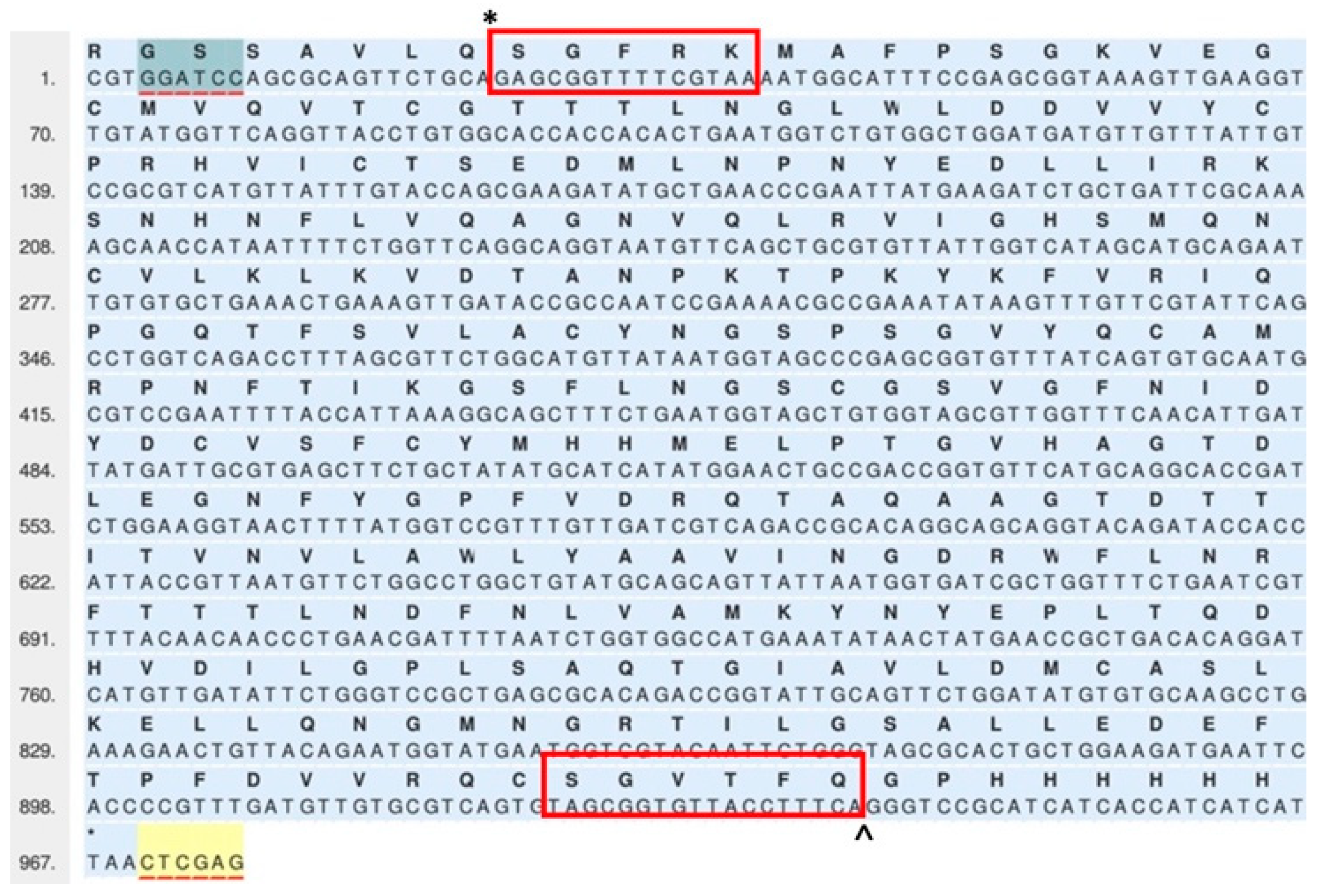
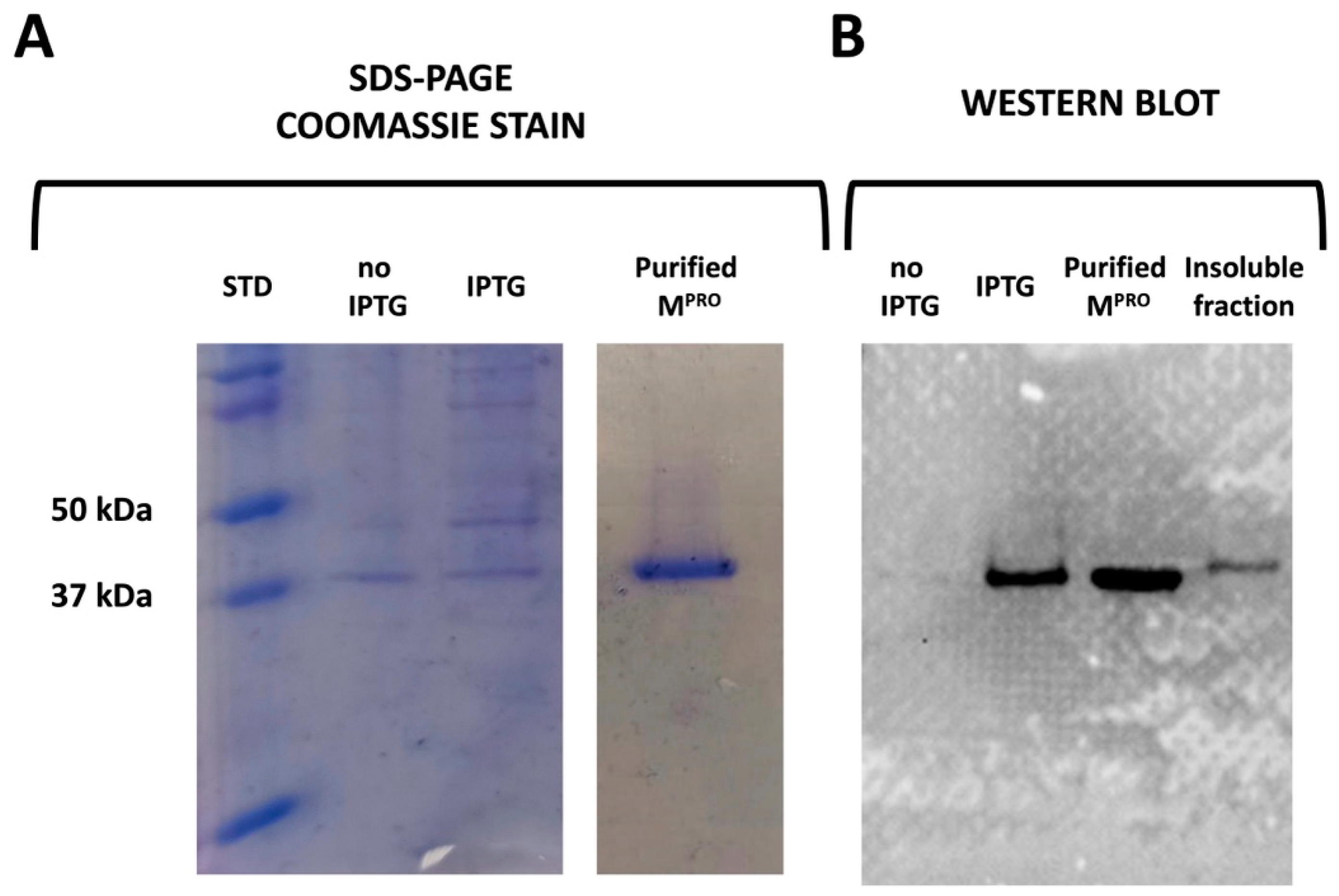
2.1. Cloning, Expression, and Purification

The viral genome of SARS-CoV-2 is composed of a positive-sense, single-stranded RNA molecule (Figure 2).
It consists of several structural and nonstructural genes that encode proteins necessary for viral replication and infection [38,39]. SARS-CoV-2 Mpro is encoded by a specific gene known as ORF1ab, which is located in the open reading frame (ORF) 1ab region of the viral genome (Figure 2). The examination of SARS-CoV-2 Mpro has provided opportunities to develop drugs that can effectively combat coronaviruses [38,39,40,41]. Mpro is known to operate on at least 11 different points on the large polyprotein 1ab, also known as replicase 1ab, which has a size of approximately 790 kDa and is the largest gene in the viral genome [38]. Most of these sites contain a recognition sequence composed of Leu-Gln, followed by a cleavage site that can be either Ser, Ala, or Gly [42]. One advantage of targeting Mpro for inhibition is that its cleavage specificity is unique to coronaviruses and differs from that of human proteases [43]. This specificity arises from the specific amino acid sequence and structural characteristics of the Mpro active site [44,45]. As a result, inhibitors designed to target Mpro are less likely to interfere with human proteases and, therefore, may be less prone to causing toxicity or adverse effects in human cells [45]. Expressing and purifying the SARS-CoV-2 Mpro is crucial for conducting structural and functional research on this protein.
Based on the primary structure of the SARS-CoV-2 Mpro, a synthetic gene was synthesized to encode the Mpro polypeptide chain, which consists of 322 residues (Figure 3).
The gene construct had start and stop codons, and sites placed at appropriate intervals to allow the expression of the native N- and C-terminals of Mpro. The construct contained the Mpro cleavage site SAVLQ*SGFRK (* represents the scissile peptide bond) at the N-terminus. A PreScission cleavage site [46] and a Tag of six histidines (SGVTFQ^GPHHHHHH, the symbol ^ indicates the PreScission cleavage site) were inserted at the C-terminus [47]. In the process of gene expression, the Mpro enzyme undergoes auto-cleavage to generate a native N-terminus, whereas, after treatment with PreScission protease, the original C-terminus is developed. The recombinant mature enzyme was a polypeptide chain of 306 amino acid residues, starting with SGFRK and terminating with SGVTFQ (Figure 3, boxed residues).
The recombinant SARS-CoV-2 Mpro was successfully obtained from soluble fractions of bacterial cell lysates. This was achieved by cultivating bacterial cells containing the SARS-CoV-2 Mpro/pET-100D Topo plasmid under specific conditions, such as a lower temperature (20 °C) and a mild concentration of IPTG (0.5 mM). The expression and purification of the recombinant Mpro were evaluated through SDS-PAGE and Western Blot analysis (Figure 4A,B).
Upon IPTG induction, Mpro is expressed at a relatively high level. Notably, most of the expressed Mpro was found in the soluble fraction of the bacterial cell lysates, which were purified using a HisTrap affinity column (Figure 4A, right side). The expression of His-tagged Mpro was confirmed by Western Blot analysis using an anti-His-tag antibody (Figure 4B). The antibody-detected protein had an apparent molecular weight of approximately 37.0 kDa, whereas its calculated theoretical molecular mass was 34.7 kDa (Figure 4B). Purified His-tagged Mpro was treated with PreScission protease at 4 °C overnight to achieve the desired authentic C-termini for the target protein. Subsequently, PreScission-treated Mpro was passed against the HisTrap affinity column to eliminate the cleaved His-tagged fragments produced by the PreScission protease. A yield of 4 mg of recombinant Mpro (theoretical molecular weight of 33.7 kDa) was obtained from a 1 L culture of bacterial cells.

2.2. Determination of the Mpro Activity Using the Spectrophotometric Method

To evaluate the recombinant Mpro enzymatic activity, the hydrolysis of a specific substrate peptide was investigated. The peptide comprises a sequence of six amino acids (TSAVLQ) connected to a p-nitroaniline (pNA) moiety at the amino terminal part [48]. The substrate exhibits a remarkable susceptibility to precise cleavage by Mpro at a designated cleavage site known as Gln-pNA. The proteolytic activity of Mpro was assayed continuously by monitoring the cleavage of the TSAVLQ-pNA. The enzymatic action executed by Mpro leads to the liberation of pNA, which elicits a noticeable increase in absorbance at the wavelength of 405 nm. By monitoring the hydrolysis of this substrate peptide, we gain valuable insights into the enzymatic proficiency of Mpro. Figure 5 shows the dependence of enzyme activity, expressed as optical density 450 nm/s, as a function of the enzyme concentration, which was in the range of 0.125–2 µM.
Based on the insights provided by Figure 5, it is evident that the purified recombinant enzyme exhibits good activity, with the reaction rate influenced by the enzyme concentration. However, additional increments in enzyme concentration (Mpro > 1.2 µM) cease to impact the reaction rate. This intriguing observation can be attributed to the gradual imposition of a limiting factor due to the substrate’s availability (500 µM in the assay). Moreover, we also investigated the Mpro activity of the recombinant protein with the native N-terminus (SGFRK) but having the PreScission cleavage site and the tag of six histidines at the C-terminal (SGVTFQ^GPHHHHHH). The Mpro with only the native N-terminus resulted in behavior similar to that of the protease with the native N- and C-terminals in the polypeptide chains. This result was expected since the native N-terminal, not the Mpro C-terminal, is crucial for the protease dimerization process [49]. More specifically, the interaction of the Ser in the first position of one monomer with Glu166 present in the adjacent monomer is crucial for the catalytic activity of the viral protease [49].

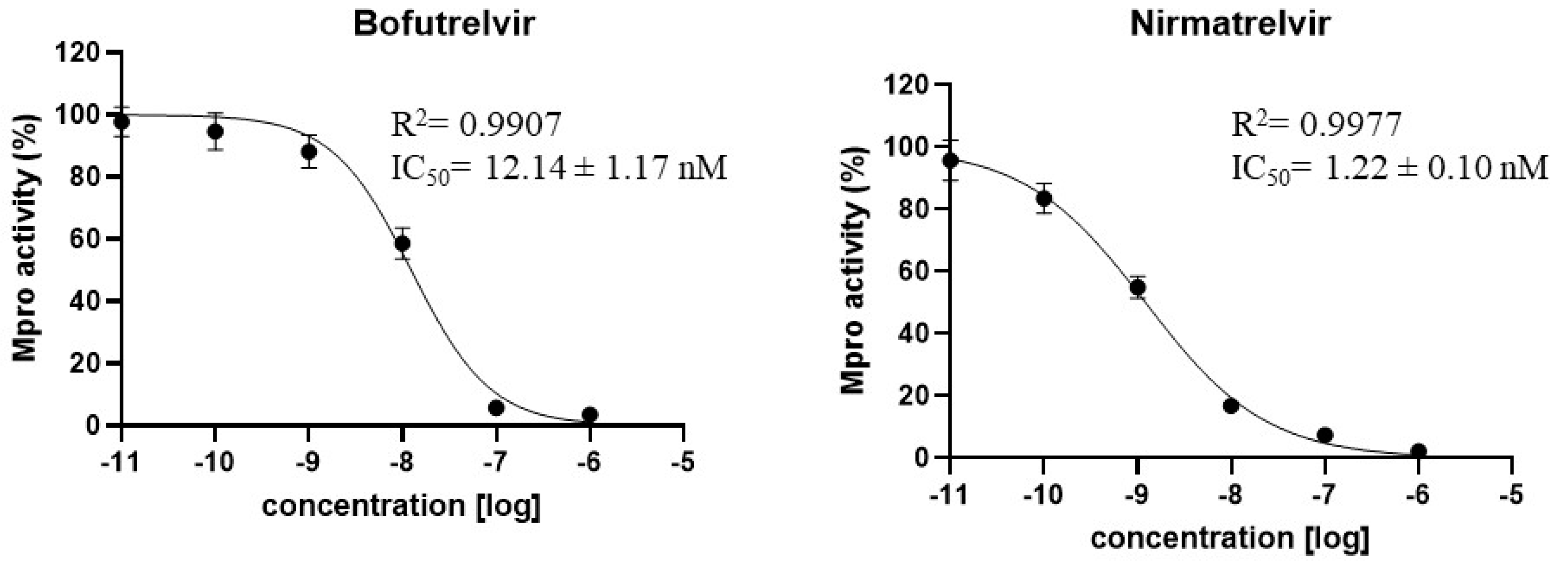
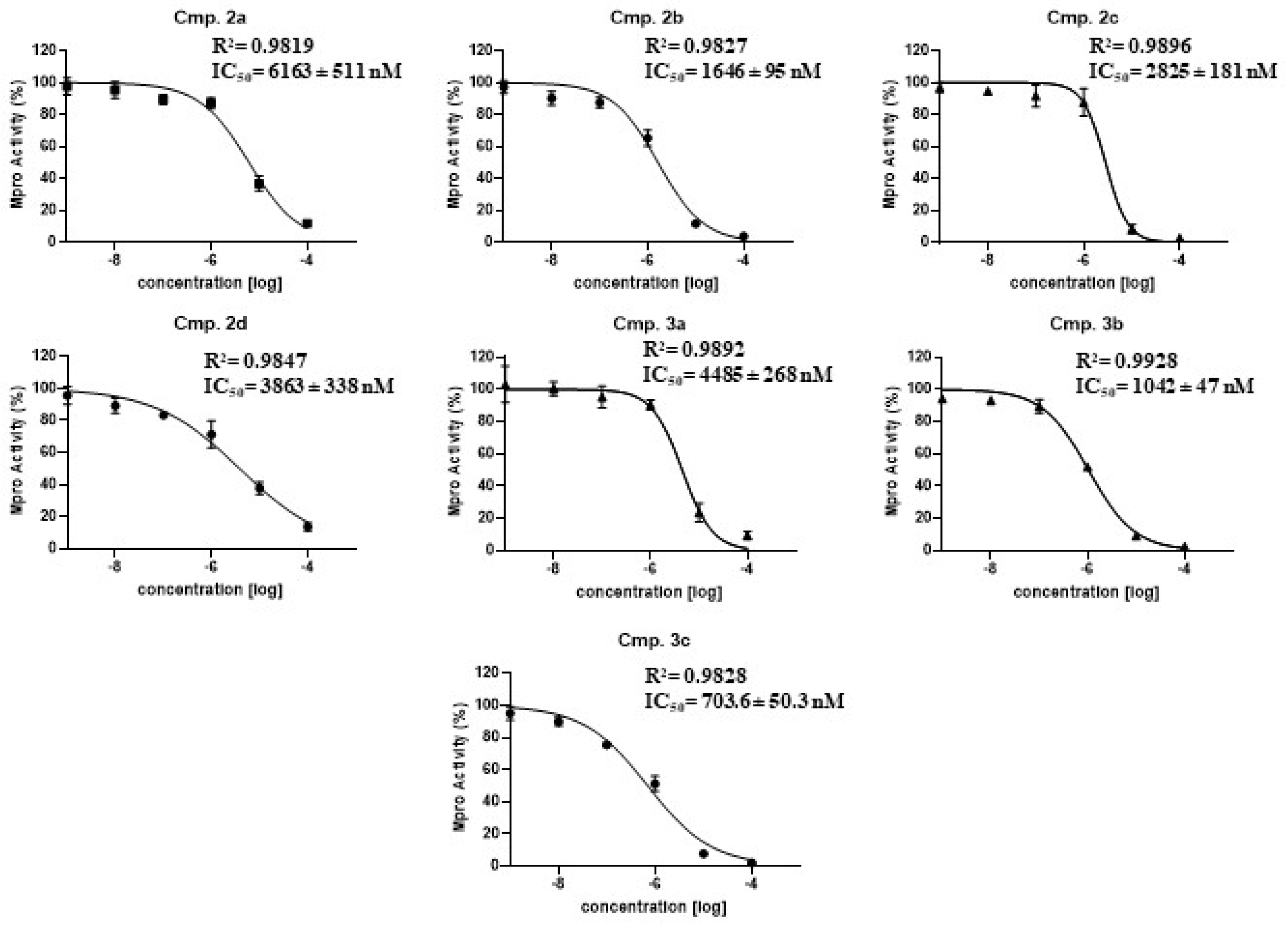
2.3. Determination of the Mpro Half Maximal Inhibitory Concentrations (IC50)

Initially, in our analysis we also employed a colorimetric method to assess Mpro activity and inhibition. However, recognizing the limitations of colorimetric assays, we sought to enhance the accuracy of our method. To this end, we transitioned to a more sensitive approach using a fluorogenic substrate for enzyme inhibition analysis. The fluorogenic substrate was preferred over the colorimetric method due to its higher sensitivity, ability to detect subtle changes in fluorescence intensity, and broader dynamic range for IC50 determination. Moreover, the fluorogenic substrate enabled a more refined evaluation of compound efficacy, particularly for detecting lower concentrations. By mitigating background noise and interference, the fluorogenic substrate also improved the reliability and accuracy of the experimental measurements. A plethora of inhibitors targeting SARS-CoV-2 Mpro have been evaluated, and few have demonstrated low IC50 values [50,51,52]. In the literature it has been reported that Nirmatrelvir and Bofutrelvir (Figure 1) exhibit IC50 values of less than 1 μM when tested against SARS-CoV-2 Mpro [50,51,52]. Nirmatrelvir, a drug marketed under the trade name Paxlovid (in combination with Ritonavir), and Bofutrelvir, are two orally administered antiviral therapeutics specifically developed by Pfizer for the management of COVID-19 [53,54,55]. These two inhibitors were demonstrated to disrupt a critical stage of the viral life cycle, potentially reducing disease progression and alleviating the severity of COVID-19 symptoms. It is essential to note that ongoing clinical investigations and regulatory evaluations continue to assess the efficacy, safety profile, and optimal administration protocols for these and other Mpro inhibitors [53,54,55]. These investigations are crucial for establishing comprehensive therapeutic guidelines and refining treatment strategies to effectively combat COVID-19.
In our study, we determined the IC50 values of Nirmatrelvir and Bofutrelvir standard Mpro inhibitors, which were used as a positive control for the IC50 test. The in vitro experiments demonstrated that Nirmatrelvir and Bofutrelvir had IC50 values of 1.22 and 12.14 nM, respectively. These findings are consistent with those previously reported in the literature (see Figure 6).
Selenium-containing inhibitors 2 and 3 were synthesized by exploiting the exquisite nucleophilic character of selenols, which were demonstrated to selectively react with a wide variety of electrophilic partners [56,57,58,59]. Selenols 1 were prepared following reported procedures via reductive cleavage of the corresponding diselenides [60] or through the ring-opening reaction of the corresponding epoxides with (Me3Si)2Se in the presence of TBAF [59]. The Spectrum of compounds can be seen in the Supplementary Materials.
Selenolesters 2ad were synthesized upon reaction of benzyl- or alkyl-substituted selenols with benzoyl chloride in the presence of triethylamine (Scheme 1, left). Similarly, selenocarbamates 3a,b were obtained via the addition reaction of benzeneselenol 1a to suitable isocyanates (Scheme 1, right) [61,62].
On the other hand, the synthesis of selenocarbamate 3c followed a different synthetic pathway involving the use of the selenating reagent LiAlHSeH [62] with isocyanate and reacted with the appropriate benzoyl bromide, as outlined in Scheme 2.
A fluorogenic assay was conducted to assess the inhibition levels against the Mpro enzyme of selenoesters 2ad and selenocarbamate 3ac derivatives. In Figure 7, inhibitory curves against Mpro and the corresponding IC50 values for each compound are presented. The data unequivocally illustrate that these compounds exhibit substantial Mpro inhibitory activity, with IC50 values in the micromolar range for selenoesters. Notably, derivatives with a hydroxyl group demonstrated better activity than compound 2a without it.
Conversely, selenocarbamates 3ac exhibited superior efficacy in inhibiting the Mpro enzyme, reaching sub-micromolar levels of inhibition (703.6 nM) for compound 3c. These findings indicate a clear on-target interaction of these compounds with Mpro, showcasing significant inhibitory potential against SARS-CoV-2 and suggesting their potential for development as treatments for COVID-19 patients.

2.4. Computational Study

It is noteworthy that computational protein design methods have played a pivotal role in creating novel molecules targeting specific regions of the SARS-CoV-2 virus. Sophisticated computational protein design techniques have been instrumental in generating miniproteins with a high binding affinity for the viral spike protein—an essential element facilitating viral entry into host cells [63]. Additionally, a stapled peptide has been designed, demonstrating both high affinity and specificity for the receptor-binding domain (RBD) of the SARS-CoV-2 spike protein [64]. Considering this, the Mpro inhibition mechanism of selenoesters 2a2d and selenocarbamates 3a3c was simulated by a covalent docking analysis selecting Cys145 as the bond forming residue (Scheme 3).
A series of thioester inhibitors of SARS-CoV-2 Mpro has been described previously and X-ray structures showed a covalent thioester bond with the catalytic Cys145 residue of the protease [65]. Modifying the force field (OPLS4) allowed us here to simulate similar selenium derivatives. The results indicate that a nucleophilic attack of the cysteine thiol to the selenoester or selenocarbamate carbonyl group occurs with the related selenides acting as the leaving groups and leads to the formation of the covalently bound thioester or thiocarbammate adduct (Figure 8). The thioester carbonyl group is predicted to engage H-bonds with the side chain NH moieties of Gly143 and Cys145 (Figure 8A). The benzene ring accommodates in the S2 pocket of the Mpro active site forming VdW contacts with residues nearby. Instead, the thiocarbammate carbonyl group forms a H-bond with the His41 imidazole ring (Figure 8B). The methyl substituted phenyl ring is located in the same enzyme subpocket but shifted towards Gln189 and Met49 due to the greater length of the linker, confirming thus the hypothesized binding proposed in Scheme 3.

3. Materials and Methods

3.1. Construction of Mpro Expression Vector

The Mpro gene for SARS-CoV-2, which covers ORF1ab polyprotein residues 3264–3569 and has a GenBank code of MN908947.3, was designed in our laboratories and produced by GeneArt (Life Technologies, Carlsbad, CA, USA), a company specialized in gene synthesis. The gene was synthesized using Escherichia coli codon usage. The vector pET100D-Topo was used to produce the pET100/Mpro vector, which overexpressed the fusion recombinant viral protease with a Tag of six histidines at the C-terminus of the polypeptide chain. An intricate design was employed for gene construction by incorporating strategic nucleotide sites. At the beginning of the start codon, the nucleotides encoding the amino acids of the Mpro cleavage site SAVLQ*SGFRK were added (* is the Mpro autocleavage). In contrast, the nucleotides encoding amino acids for the PreScission cleavage site and six histidines (SGVTFQ^GPHHHHH) were added before the stop codon (^ is the PreScission cleavage site). This meticulous arrangement ensured the accurate expression of the native N- and C-terminals of the native Mpro through autocleavage and PreScission protease, respectively (see Results and Discussion for more details).

3.2. Mpro Expression and Purification

To overexpress His-Tag Mpro, competent E. coli BL21 (DE3) cells were transformed with the constructs described above. They were grown at 37.0 °C and induced with 0.5 mM isopropyl-thio-b-D-galactoside (IPTG) at an OD600 of 0.6–0.7 nm. After additional growth for 18 h at 20 °C, the cells were harvested by centrifugation and washed three times with PBS 1X. Aliquots of cells were resuspended in 20 mM Tris/HCl, 250 mM NaCl, 2 mM β-Mercaptoethanol, 0.2% Triton, pH 8.5, and disrupted by sonication. The bacterial lysate was centrifuged and purified using a nickel-affinity column (His-Trap FF). HisTrap column (1.0 mL) was equilibrated with 20 mL equilibration buffer (20 mM Tris/HCl, 250 mM NaCl, 2 mM β-Mercaptoethanol, 0.2% Triton, pH 8.5) at 1 mL/min. The supernatant from the cellular lysate was loaded onto the column at 1.0 mL/min, connected with AKTA Prime. The recombinant His-Tag Mpro was eluted from the column with a flow of 0.5 mL/min and the elution buffer composed of 20 mM Tris/HCl, 30 mM NaCl, 2 mM β-Mercaptoethanol, 150 mM imidazole, pH 8.5. The fractions containing target protein were pooled and mixed with PreScission protease and dialyzed into 20 mM Tris/HCl, 150 mM NaCl, 2 mM β-Mercaptoethanol, pH 8.5 at 4 °C overnight, resulting in the target protein with authentic N- and C-termini. PreScission-treated Mpro was applied again to the His-Trap FF nickel columns to remove the PreScission protease, His-tag, and protein with an uncleaved His-tag. The His-tag-free Mpro in the flow through was dialyzed into 50 mM HEPES, 150 mM NaCl, 1 mM DTT, 1 mM EDTA, and 10% glycerol, pH 7.5 at 4 °C overnight.

3.3. SDS-Page and Western Blot

A 12% Sodium Dodecyl Sulfate-polyacrylamide gel electrophoresis (SDS-PAGE), following the method described by Laemmli, was used to load and separate the recovered Mpro at various steps of the purification process. The gel was electrophoresed at 150 V until the dye front completely migrated off the gel and subsequently stained with Coomassie Brilliant Blue-R for visualization. For the subsequent Western Blot analysis, the overexpressed cytoplasmic Mpro was subjected to electrophoretic transfer onto a PVDF membrane using a transfer buffer composed of 25 mM Tris, 192 mM glycine, and 20% methanol using a Trans-Plot SD Cell (Bio-Rad, Hercules, CA, USA). His-tag Western Blot was carried out using the Pierce Fast Western Blot Kit (Thermo Scientific, Waltham, MA, USA). The blotted membrane was immersed in Fast Western 1 Wash Buffer to eliminate any residual transfer buffer. The working dilution of the primary antibody was then applied to the blot and incubated for 30.0 min at room temperature (RT) with gentle agitation. Following this, the membrane was removed from the primary antibody solution and incubated for 10.0 min with Fast Western Optimized HRP Reagent Working Dilution. The membrane was washed twice using approximately 20 mL of Fast Western 1 Wash Buffer. Finally, the membrane was incubated with the Detection Reagent Working Solution for 3.0 min at RT. The chemiluminescent signals were captured using the Invitrogen iBright CL1500 Imaging System, enabling data acquisition and analysis.

3.4. Enzymatic Protease Assay

The enzymatic activity of Mpro was evaluated using a colorimetric assay that measured the peptide cleavage of the peptide substrate TSAVLQ-para-nitroanilide (TQ6-pNA, Merck, Rahway, NJ, USA) [48]. This substrate undergoes cleavage at the Gln-pNA bond, resulting in the release of free pNA and a visible change in the solution color to yellow. The absorbance was continuously monitored at 405 nm using a Varian Cary 50 UV-Vis Spectrophotometer (Palo Alto, CA, USA). Protease activity assay was performed at 22 °C in 20 mM phosphate buffer (pH 7.6). The substrate stock solution was prepared at a concentration of 1 mM and the working concentration used in the assay was 500 µM. The assay was assembled in a quartz cuvette (0.5 mL) containing the enzyme in working protein solutions ranging from 0.125–2 µM.
For the determination of half-maximal inhibitory concentrations (IC50) of inhibitors against Mpro, an AFC-Peptide substrate (SAE0180, Merck) was used for a fluorescence-based cleavage assay [66]. The assay was performed in half area 96-well, black, flat-bottomed microtiter plates (Corstar, Corning, Glendale, CA, USA) with a final volume of 125 μL. Mpro (final concentrations of 5–50 nM) was pre-incubated for 1 h at 25 °C with compounds at different concentrations in the assay buffer (PBS, pH 7.5). The substrate was then added at a final concentration of 12 μM to the reaction mixture and the reaction was incubated for 1 h at 25 °C. The readings for the different concentrations of the inhibitor compounds incubated with the substrate without MPro were measured as a blank. The fluorescence signals (excitation/emission, 400 nm/505 nm) of released AFC were measured using a Spark multimode plate reader (Tecan, Glendale, CA, USA). The results were plotted as dose inhibition curves using nonlinear regression with a variable slope to determine the IC50 values of inhibitor compounds using GraphPad Prism 9.0.

3.5. Chemistry

3.5.1. General

All commercial materials were purchased from Merck—Sigma-Aldrich and used as received, without further purification. Solvents were dried using a solvent purification system (Pure-Solv™). Flash column chromatography purifications were performed with Silica gel 60 (230–400 mesh). Thin layer chromatography was performed with TLC plates Silica gel 60 F254, which was visualized under UV light, or by staining with an ethanolic acid solution of p-anisaldehyde followed by heating. Mass spectra were recorded by Electrospray Ionization (ESI). 1H and 13C NMR spectra were recorded in CDCl3 or DMSO-d6 using Varian Mercury 400 and Bruker 400 Ultrashield spectrometers, operating at 400 MHz for 1H, 100 MHz for 13C, and 76 MHz for 77Se. 1H NMR signals were referenced to nondeuterated residual solvent signals (7.26 ppm for CDCl3 and 2.49 for DMSO-d6). 13C NMR was referenced to CDCl3 or DMSO-d6 signals (77.0 ppm or 39.7 ppm, respectively). (PhSe)2 was used as an external reference for 77Se NMR (δ = 461 ppm). Chemical shifts (δ) are given in parts per million (ppm) and coupling constants (J) are given in Hertz (Hz), rounded to the nearest 0.1 Hz. The 1H NMR data are reported as follows: chemical shift, integration, multiplicity (s = singlet, d = doublet, t = triplet, ap d = apparent doublet, m = multiplet, dd = doublet of doublet, bs = broad singlet, bd = broad doublet, etc.), coupling constant (J) or line separation (ls), and assignment. Spectroscopic data of compounds 2b, 2d, 3a, and 3b matched those reported in the literature.

3.5.2. General Procedure for the Synthesis of Selenolesters (2)

A solution of selenol 1 (1.0 mmol) in dry CH2Cl2 (3 mL) was cooled at 0 °C under inert atmosphere (N2) and treated with Et3N (1.2 mmol). The mixture was stirred for 5 min and then a solution of acyl chloride (1.2 mmol) in dry CH2Cl2 (2 mL) was slowly added. The reaction was allowed to warm to room temperature and stirred for an additional 2 h. Afterwards, a saturated solution of aq. NH4Cl was added and the organic phase was extracted with Et2O (3 × 15 mL), washed with brine (2 × 10 mL) and dried over Na2SO4. The solvent was removed under vacuum and the crude material was purified by flash column chromatography (silica gel) to afford pure selenolesters 2.

3.5.3. Synthesis of Se-Phenethyl Benzoselenoate (2a)

Following the general procedure, phenylmethaneselenol (86 mg, 0.5 mmol) and benzoyl chloride, after purification by column chromatography (petroleum ether/Et2O 15:1), gave 2a as a yellowish oil (74 mg, 51%). 1H NMR (400 MHz, CDCl3), δ (ppm): 4.35 (2H, s), 7.22 (1H, t, J = 7.2 Hz), 7.30 (2H, ap t, ls = 7.5 Hz), 7.38 (2H, ap d, ls = 7.2 Hz), 7.45 (2H, ap t, ls = 7.7 Hz), 7.59 (1H, ap t, ls = 7.4 Hz), 7.90 (2H, ap d, ls = 7.4 Hz). 13C NMR (100 MHz, CDCl3), δ (ppm): 29.0, 127.0, 127.2, 128.6, 128.8, 129.0, 133.7, 138.8, 139.0, 194.5. MS (ESI, positive) m/z: 291.0 [M + H]+.

3.5.4. Synthesis of Se-(2-Hydroxycyclohexyl) Benzoselenoate (2c)

Following the general procedure, 2-hydroselenocyclohexan-1-ol (134 mg, 0.75 mmol) and benzoyl chloride, after purification by column chromatography (petroleum ether/EtOAc 5:1), gave 2c as a yellowish oil (155 mg, 73%). 1H NMR (200 MHz, CDCl3) δ (ppm): 1.31–1.47 (3H, m), 1.68–1.72 (2H, m), 1.80–1.83 (1H, m), 2.16–2.26 (2H, m), 2.52 (1H, bs), 3.58–3.65 (2H, m), 7.42–7.46 (2H, m), 7.56–7.60 (1H, m), 7.88–7.91 (2H, m). 13C NMR (50 MHz, CDCl3) δ (ppm): 24.2, 26.7, 32.9, 35.3, 50.8, 73.4, 127.4, 128.8, 133.8, 139.0, 195.7. MS (ESI, positive) m/z: 285.0 [M + H]+.

3.5.5. Synthesis of Se-(4-Sulfamoylbenzyl) (3,5-Dimethylphenyl) Carbamoselenoate (3c)

Elemental selenium (1 eq.) was added in THF at 0 °C and subsequently added LiAlH4 (1eq.). The reaction mixture was stirred at 0 °C for 30 min, 1-isocyanato-3,5-dimethylbenzene (1 eq.) was added, and the mixture was then stirred for 1 h at 0 °C. Subsequently, 4-(bromomethyl)benzenesulfonamide (1 eq.) was added, the mixture was then stirred for 2 h, quenched with H2O, and extracted with EtOAc. The crude reaction was purified by flash chromatography (EtOAc/Hex 7:3) to afford a yellow solid. Yield 57%. 1H NMR (400 MHz, DMSO-d6 δ(ppm): 8.48 (1H, s), 7.80 (2H, d, J = 8.33 Hz), 7.44 (2H, d, J = 8.35 Hz), 7.37 (2H, s), 7.10 (2H, s), 6.64 (1H, s), 4.07 (2H, s), 2.26 (6H, s); 13C NMR (100 MHz, DMSO-d6) δ(ppm): 153.3, 144.2, 143.5, 140.5, 138.6, 130.2, 126.6, 124.25, 116.8, 31.5, 22.0; 77Se NMR (76 MHz, DMSO-d6) δ(ppm): 406.1; MS (ESI positive) m/z: 399.0 [M + H]+.

3.6. Computational Study

The crystal structure of SARS-CoV-2 MPro (PDB 7CB7) [67] was retrieved from the Protein Data Bank and prepared using the Protein Preparation module implemented in Maestro Schrödinger suite [68], assigning bond orders, adding hydrogens, deleting water molecules, and optimizing H-bonding networks. Energy minimization with a Root Means Square Deviation (RMSD) value of 0.30 was applied using an Optimized Potential for Liquid Simulation (OPLS4) force field. The 3D ligand structures were prepared by Maestro (version 2023-4) and evaluated for their ionization states at pH 7.4 ± 0.5 with Epik, version 2023-4. The OPLS4 force field was modified according to Schrödinger to enable the treatment of selenium derivatives in the docking procedure. The grid for the covalent docking was generated with the center located on the centroid of the cocrystallized ligand and Cys145 was selected as the reacting residue. Covalent docking was performed with Glide Covalent Docking. The best pose for type of compound, evaluated in terms of score, hydrogen bond interactions and hydrophobic contacts, was refined with Prime adopting a VSGB solvation model. Figures were generated with ChimeraX (version 1.7).

4. Conclusions

The study here reported advances our knowledge of viral biochemistry, and may lead to the development of new antivirals belonging to the class of cysteine protease inhibitors. We report conditions for the cloning and purification of Mpro with significant yields, which produced highly catalytically active SARS-CoV-2 enzyme. In our study, we successfully synthesized novel selenoester and selenocarbamate derivatives, and evaluated their potential as Mpro inhibitors. The screening revealed that all synthesized compounds exhibited inhibition activity against the target enzyme, whereas the selenocarbamate 3c has emerged as an effective inhibitor, demonstrating an IC50 value in the sub-micromolar range at 703.6 nM. These findings underscore the promise of organoselenium derivatives as effective Mpro inhibitors, suggesting their potential significance in the development of antiviral therapeutics. Similar to other Mpro inhibitors reported so far [69,70], the new compounds investigated here may react with the SH moiety of the cysteine from the catalytic triad of the enzyme, inactivating it. Thus, a possible inhibition mechanism relies on acyl- or carbamoyl-transfer from the selenium atom to the catalytic cysteine of the enzyme. The good leaving group ability of the formed selenolate anions reasonably moves the equilibrium towards the formation of the enzyme-derived thiolesters or thiocarbamate. Such a reactivity has not yet been reported for cysteine proteases from coronaviruses, and thus selenobenzoates and selenocarbamates constitute new classes of Mpro inhibitors [71,72]. The success of our synthesized compounds in inhibiting Mpro activity contributes valuable insights to the ongoing efforts in identifying and designing compounds for combating viral infections, particularly targeting SARS-CoV-2. Further exploration of the structure–activity relationships and mechanistic studies will enhance our understanding and pave the way for the development of more potent and selective inhibitors with clinical relevance. This study marks a significant stride towards advancing the arsenal of antiviral agents and underscores the potential of organoselenium derivatives in the pursuit of effective treatments against viral infections.

Supplementary Materials

The following supporting information can be downloaded at: https://www.mdpi.com/article/10.3390/ijms25020971/s1.

Author Contributions

Conceptualization, C.C. and C.T.S.; methodology, V.D.L., A.A., A.N., S.P., D.T. and P.G.; validation, A.C. and V.C.; investigation, C.C., V.D.L. and A.N.; data curation, C.C., V.C., A.A. and A.N.; writing—original draft preparation, C.C..; writing—review and editing, C.C. and C.T.S.; supervision, C.C. and C.T.S.; project administration and funding acquisition, C.C. All authors have read and agreed to the published version of the manuscript.

Funding

This research was funded by Italian National Research Council (CNR), Grant COVPI-Tackling coronaviruses-related diseases by developing viral Protease Inhibitors.

Data Availability Statement

Data are available directly from the authors at request.

Acknowledgments

We are grateful to Valentina Brasiello and Francesca Segreti for their excellent technical assistance.

Conflicts of Interest

The authors declare no conflicts of interest.

References

  1. Markov, P.V.; Ghafari, M.; Beer, M.; Lythgoe, K.; Simmonds, P.; Stilianakis, N.I.; Katzourakis, A. The evolution of SARS-CoV-2. Nat. Rev. Microbiol. 2023, 21, 361–379. [Google Scholar] [CrossRef]
  2. Li, Y.; Niu, Y. A commentary on “The socio-economic implications of the coronavirus pandemic (COVID-19): A review”. Int. J. Surg. 2021, 95, 106048. [Google Scholar] [CrossRef] [PubMed]
  3. Sardar, S.; Abdul-Khaliq, I.; Ingar, A.; Amaidia, H.; Mansour, N. ‘COVID-19 lockdown: A protective measure or exacerbator of health inequalities? A comparison between the United Kingdom and India.’ a commentary on “the socio-economic implications of the coronavirus and COVID-19 pandemic: A review”. Int. J. Surg. 2020, 83, 189–191. [Google Scholar] [CrossRef]
  4. Scherf, M.; Matschke, X.; Rieger, M.O. Stock market reactions to COVID-19 lockdown: A global analysis. Financ. Res. Lett. 2022, 45, 102245. [Google Scholar] [CrossRef]
  5. Badiani, A.A.; Patel, J.A.; Ziolkowski, K.; Nielsen, F.B.H. Pfizer: The miracle vaccine for COVID-19? Public Health Pract. 2020, 1, 100061. [Google Scholar] [CrossRef] [PubMed]
  6. Carabelli, A.M.; Peacock, T.P.; Thorne, L.G.; Harvey, W.T.; Hughes, J.; Consortium, C.-G.U.; Peacock, S.J.; Barclay, W.S.; de Silva, T.I.; Towers, G.J.; et al. SARS-CoV-2 variant biology: Immune escape, transmission and fitness. Nat. Rev. Microbiol. 2023, 21, 162–177. [Google Scholar] [CrossRef]
  7. Li, M.; Wang, H.; Tian, L.; Pang, Z.; Yang, Q.; Huang, T.; Fan, J.; Song, L.; Tong, Y.; Fan, H. COVID-19 vaccine development: Milestones, lessons and prospects. Signal Transduct. Target. Ther. 2022, 7, 146–177. [Google Scholar] [CrossRef] [PubMed]
  8. Ghosh, A.K.; Mishevich, J.L.; Mesecar, A.; Mitsuya, H. Recent Drug Development and Medicinal Chemistry Approaches for the Treatment of SARS-CoV-2 Infection and COVID-19. ChemMedChem 2022, 17, e202200440. [Google Scholar] [CrossRef]
  9. Lui, G.; Guaraldi, G. Drug treatment of COVID-19 infection. Curr. Opin. Pulm. Med. 2023, 29, 174–183. [Google Scholar] [CrossRef]
  10. Ravaghi, H.; Naidoo, V.; Mataria, A.; Khalil, M. Hospitals early challenges and interventions combatting COVID-19 in the Eastern Mediterranean Region. PLoS ONE 2022, 17, e0268386. [Google Scholar] [CrossRef]
  11. Sharun, K.; Tiwari, R.; Dhama, J.; Dhama, K. Dexamethasone to combat cytokine storm in COVID-19: Clinical trials and preliminary evidence. Int. J. Surg. 2020, 82, 179–181. [Google Scholar] [CrossRef]
  12. da Silva, S.J.R.; do Nascimento, J.C.F.; Mendes, R.P.G.; Guarines, K.M.; da Silva, C.T.A.; da Silva, P.G.; de Magalhaes, J.J.F.; Vigar, J.R.J.; Silva, A.; Kohl, A.; et al. Two Years into the COVID-19 Pandemic: Lessons Learned. ACS Infect. Dis. 2020, 8, 1758–1814. [Google Scholar] [CrossRef] [PubMed]
  13. Debnath, S.; Agarwal, A.; Kumar, N.R.; Bedi, A. Selenium-Based Drug Development for Antioxidant and Anticancer Activity. Future Pharmacol. 2022, 2, 595–607. [Google Scholar] [CrossRef]
  14. Ali, W.; Benedetti, R.; Handzlik, J.; Zwergel, C.; Battistelli, C. The innovative potential of seleniumcontaining agents for fighting cancer and viral infections. Drug Discov. Today 2021, 26, 256–263. [Google Scholar] [CrossRef] [PubMed]
  15. Hou, W.; Dong, H.; Zhang, X.; Wang, Y.; Su, L.; Xu, H. Selenium as an emerging versatile player in heterocycles and natural products modification. Drug Discov. Today 2022, 27, 2268–2277. [Google Scholar] [CrossRef]
  16. Angeli, A.; Ferraroni, M.; Capperucci, A.; Tanini, D.; Costantino, G.; Supuran, C.T. Selenocarbamates As a Prodrug-Based Approach to Carbonic Anhydrase Inhibition. ChemMedChem 2022, 17, e202200085. [Google Scholar] [CrossRef]
  17. Tanini, D.; Carradori, S.; Capperucci, A.; Lupori, L.; Zara, S.; Ferraroni, M.; Ghelardini, C.; Di Cesare Mannelli, L.; Micheli, L.; Lucarini, E.; et al. Chalcogenides-incorporating carbonic anhydrase inhibitors concomitantly reverted oxaliplatin-induced neuropathy and enhanced antiproliferative action. Eur. J. Med. Chem. 2021, 225, 113793. [Google Scholar] [CrossRef]
  18. Jin, Z.; Du, X.; Xu, Y.; Deng, Y.; Liu, M.; Zhao, Y.; Zhang, B.; Li, X.; Zhang, L.; Peng, C.; et al. Structure of Mpro from SARS-CoV-2 and discovery of its inhibitors. Nature 2020, 582, 289–293. [Google Scholar] [CrossRef]
  19. Ma, C.; Hu, Y.; Townsend, J.A.; Lagarias, P.I.; Marty, M.T.; Kolocouris, A.; Wang, J. Ebselen, Disulfiram, Carmofur, PX-12, Tideglusib, and Shikonin Are Nonspecific Promiscuous SARS-CoV-2 Main Protease Inhibitors. ACS Pharmacol. Transl. Sci. 2020, 3, 1265–1277. [Google Scholar] [CrossRef]
  20. Amporndanai, K.; Meng, X.; Shang, W.; Jin, Z.; Rogers, M.; Zhao, Y.; Rao, Z.; Liu, Z.J.; Yang, H.; Zhang, L.; et al. Inhibition mechanism of SARS-CoV-2 main protease by ebselen and its derivatives. Nat. Commun. 2021, 12, 3061. [Google Scholar] [CrossRef]
  21. Kovacs, T.; Kurtan, K.; Varga, Z.; Nagy, P.; Panyi, G.; Zakany, F. Veklury(R) (remdesivir) formulations inhibit initial membrane-coupled events of SARS-CoV-2 infection due to their sulfobutylether-beta-cyclodextrin content. Br. J. Pharmacol. 2023, 180, 2064–2084. [Google Scholar] [CrossRef] [PubMed]
  22. Pilote, S.; Simard, C.; Drolet, B. Remdesivir (VEKLURY) for Treating COVID-19: Guinea Pig Ex Vivo and In Vivo Cardiac Electrophysiological Effects. J. Cardiovasc. Pharmacol. 2022, 80, 616–622. [Google Scholar] [CrossRef] [PubMed]
  23. Kelley, B.; De Moor, P.; Douglas, K.; Renshaw, T.; Traviglia, S. Monoclonal antibody therapies for COVID-19: Lessons learned and implications for the development of future products. Curr. Opin. Biotechnol. 2022, 78, 102798–102805. [Google Scholar] [CrossRef] [PubMed]
  24. Ravi, G.; Eerike, M.; Konda, V.R.; Bisoi, D.; Raj, G.M.; Priyadarshini, R.; Mali, K.R.; Chaliserry, L.F. Efficacy and Safety of Anti-SARS-CoV-2 Monoclonal Antibodies: An Updated Review. Monoclon. Antibodies Immunodiagn. Immunother. 2023, 42, 77–94. [Google Scholar] [CrossRef]
  25. Harris, E. Nasally Administered Monoclonal Antibody for COVID-19. JAMA-J. Am. Med. Assoc. 2023, 329, 1142–1152. [Google Scholar] [CrossRef] [PubMed]
  26. Kip, K.E.; McCreary, E.K.; Collins, K.; Minnier, T.E.; Snyder, G.M.; Garrard, W.; McKibben, J.C.; Yealy, D.M.; Seymour, C.W.; Huang, D.; et al. Evolving Real-World Effectiveness of Monoclonal Antibodies for Treatment of COVID-19 A Cohort Study. Ann. Intern. Med. 2023, 176, 496–504. [Google Scholar] [CrossRef] [PubMed]
  27. Wynter-Adams, D.; Thomas-Brown, P. COVID-19 vaccine hesitancy in a developing country: Prevalence, explanatory factors and implications for the future. Public Health 2023, 217, 146–154. [Google Scholar] [CrossRef]
  28. Iacobucci, G. COVID-19: Evusheld protects the most vulnerable patients, analysis shows. BMJ-Br. Med. J. 2022, 379, o2690. [Google Scholar] [CrossRef]
  29. Niknam, Z.; Jafari, A.; Golchin, A.; Danesh Pouya, F.; Nemati, M.; Rezaei-Tavirani, M.; Rasmi, Y. Potential therapeutic options for COVID-19: An update on current evidence. Eur. J. Med. Res. 2022, 27, 6–20. [Google Scholar] [CrossRef]
  30. Ng, T.I.; Correia, I.; Seagal, J.; DeGoey, D.A.; Schrimpf, M.R.; Hardee, D.J.; Noey, E.L.; Kati, W.M. Antiviral Drug Discovery for the Treatment of COVID-19 Infections. Viruses 2022, 14, 961. [Google Scholar] [CrossRef]
  31. Kabinger, F.; Stiller, C.; Schmitzova, J.; Dienemann, C.; Kokic, G.; Hillen, H.S.; Hobartner, C.; Cramer, P. Mechanism of molnupiravir-induced SARS-CoV-2 mutagenesis. Nat. Struct. Mol. Biol. 2021, 28, 740–746. [Google Scholar] [CrossRef] [PubMed]
  32. Kamal, L.; Ramadan, A.; Farraj, S.; Bahig, L.; Ezzat, S. The pill of recovery; Molnupiravir for treatment of COVID-19 patients; a systematic review. Saudi Pharm. J. 2022, 30, 508–518. [Google Scholar] [CrossRef] [PubMed]
  33. Buxeraud, J.; Faure, S.; Fougere, E. Nirmatrelvir/ritonavir (Paxlovid(R)), a treatment for COVID-19. Actual. Pharm. 2022, 61, 10–12. [Google Scholar] [PubMed]
  34. Yang, D.W.; Ju, M.J.; Wang, H.; Jia, Y.C.; Wang, X.D.; Fang, H.; Fan, J. Proxalutamide for the treatment of COVID-19 rebound following Paxlovid treatment: Report of four cases and review of the literature. J. Clin. Lab. Anal. 2023, 37, e24880. [Google Scholar] [CrossRef]
  35. Chavda, V.P.; Vuppu, S.; Mishra, T.; Kamaraj, S.; Patel, A.B.; Sharma, N.; Chen, Z.S. Recent review of COVID-19 management: Diagnosis, treatment and vaccination. Pharmacol. Rep. 2022, 74, 1120–1148. [Google Scholar] [CrossRef]
  36. Hashemian, S.M.R.; Sheida, A.; Taghizadieh, M.; Memar, M.Y.; Hamblin, M.R.; Bannazadeh Baghi, H.; Sadri Nahand, J.; Asemi, Z.; Mirzaei, H. Paxlovid (Nirmatrelvir/Ritonavir): A new approach to COVID-19 therapy? Biomed. Pharmacother. 2023, 162, 114367. [Google Scholar] [CrossRef]
  37. Eltayb, W.A.; Abdalla, M.; Rabie, A.M. Novel Investigational Anti-SARS-CoV-2 Agent Ensitrelvir “S-217622”: A Very Promising Potential Universal Broad-Spectrum Antiviral at the Therapeutic Frontline of Coronavirus Species. ACS Omega 2023, 8, 5234–5246. [Google Scholar] [CrossRef]
  38. Yadav, R.; Chaudhary, J.K.; Jain, N.; Chaudhary, P.K.; Khanra, S.; Dhamija, P.; Sharma, A.; Kumar, A.; Handu, S. Role of Structural and Non-Structural Proteins and Therapeutic Targets of SARS-CoV-2 for COVID-19. Cells 2021, 10, 821. [Google Scholar] [CrossRef]
  39. Mori, M.; Capasso, C.; Carta, F.; Donald, W.A.; Supuran, C.T. A deadly spillover: SARS-CoV-2 outbreak. Expert Opin. Ther. Pat. 2020, 30, 481–485. [Google Scholar] [CrossRef]
  40. Capasso, C.; Nocentini, A.; Supuran, C.T. Protease inhibitors targeting the main protease and papain-like protease of coronaviruses. Expert Opin. Ther. Pat. 2021, 31, 309–324. [Google Scholar] [CrossRef]
  41. Deniz, S.; Uysal, T.K.; Capasso, C.; Supuran, C.T.; Ozensoy Guler, O. Is carbonic anhydrase inhibition useful as a complementary therapy of COVID-19 infection? J. Enzym. Inhib. Med. Chem. 2021, 36, 1230–1235. [Google Scholar] [CrossRef] [PubMed]
  42. Citarella, A.; Scala, A.; Piperno, A.; Micale, N. SARS-CoV-2 Mpro: A Potential Target for Peptidomimetics and Small-Molecule Inhibitors. Biomolecules 2021, 11, 607. [Google Scholar] [CrossRef] [PubMed]
  43. Flynn, J.M.; Samant, N.; Schneider-Nachum, G.; Barkan, D.T.; Yilmaz, N.K.; Schiffer, C.A.; Moquin, S.A.; Dovala, D.; Bolon, D.N.A. Comprehensive fitness landscape of SARS-CoV-2 Mpro reveals insights into viral resistance mechanisms. eLife 2022, 11, e77433. [Google Scholar] [CrossRef] [PubMed]
  44. Hu, Q.; Xiong, Y.; Zhu, G.H.; Zhang, Y.N.; Zhang, Y.W.; Huang, P.; Ge, G.B. The SARS-CoV-2 main protease (Mpro): Structure, function, and emerging therapies for COVID-19. MedComm (2020) 2022, 3, e151. [Google Scholar] [CrossRef] [PubMed]
  45. Mengist, H.M.; Dilnessa, T.; Jin, T. Structural Basis of Potential Inhibitors Targeting SARS-CoV-2 Main Protease. Front. Chem. 2021, 9, 622898–622916. [Google Scholar] [CrossRef] [PubMed]
  46. Waugh, D.S. An overview of enzymatic reagents for the removal of affinity tags. Protein Expr. Purif. 2011, 80, 283–293. [Google Scholar] [CrossRef] [PubMed]
  47. Zhang, L.L.; Lin, D.Z.; Sun, X.Y.Y.; Curth, U.; Drosten, C.; Sauerhering, L.; Becker, S.; Rox, K.; Hilgenfeld, R. Crystal structure of SARS-CoV-2 main protease provides a basis for design of improved alpha-ketoamide inhibitors. Science 2020, 368, 409–412. [Google Scholar] [CrossRef] [PubMed]
  48. Razali, R.; Subbiah, V.K.; Budiman, C. Technical Data of Heterologous Expression and Purification of SARS-CoV-2 Proteases Using Escherichia coli System. Data 2021, 6, 99. [Google Scholar] [CrossRef]
  49. Sacco, M.D.; Ma, C.; Lagarias, P.; Gao, A.; Townsend, J.A.; Meng, X.; Dube, P.; Zhang, X.; Hu, Y.; Kitamura, N.; et al. Structure and inhibition of the SARS-CoV-2 main protease reveal strategy for developing dual inhibitors against Mpro and cathepsin L. Sci. Adv. 2020, 6, eabe0751. [Google Scholar] [CrossRef]
  50. Bhardwaj, V.K.; Singh, R.; Das, P.; Purohit, R. Evaluation of acridinedione analogs as potential SARS-CoV-2 main protease inhibitors and their comparison with repurposed anti-viral drugs. Comput. Biol. Med. 2021, 128, 104117–104128. [Google Scholar] [CrossRef]
  51. Ullrich, S.; Nitsche, C. The SARS-CoV-2 main protease as drug target. Bioorganic Med. Chem. Lett. 2020, 30, 127377–127384. [Google Scholar] [CrossRef] [PubMed]
  52. Zhao, Y.; Fang, C.; Zhang, Q.; Zhang, R.; Zhao, X.; Duan, Y.; Wang, H.; Zhu, Y.; Feng, L.; Zhao, J.; et al. Crystal structure of SARS-CoV-2 main protease in complex with protease inhibitor PF-07321332. Protein Cell 2022, 13, 689–693. [Google Scholar] [CrossRef] [PubMed]
  53. Chen, W.; Shao, Y.; Peng, X.; Liang, B.; Xu, J.; Xing, D. Review of preclinical data of PF-07304814 and its active metabolite derivatives against SARS-CoV-2 infection. Front. Pharmacol. 2022, 13, 1035969–1035978. [Google Scholar] [CrossRef]
  54. Jiang, H.; Zou, X.; Zeng, P.; Zeng, X.; Zhou, X.; Wang, J.; Zhang, J.; Li, J. Crystal structures of main protease (Mpro) mutants of SARS-CoV-2 variants bound to PF-07304814. Mol. Biomed. 2023, 4, 23–32. [Google Scholar] [CrossRef] [PubMed]
  55. Li, J.; Lin, C.; Zhou, X.; Zhong, F.; Zeng, P.; McCormick, P.J.; Jiang, H.; Zhang, J. Structural Basis of Main Proteases of Coronavirus Bound to Drug Candidate PF-07304814. J. Mol. Biol. 2022, 434, 167706–167716. [Google Scholar] [CrossRef] [PubMed]
  56. Karniadakis, I.; Mazonakis, N.; Tsioutis, C.; Papadakis, M.; Markaki, I.; Spernovasilis, N. Oral Molnupiravir and Nirmatrelvir/Ritonavir for the Treatment of COVID-19: A Literature Review with a Focus on Real-World Evidence. Infect. Dis. Rep. 2023, 15, 662–678. [Google Scholar] [CrossRef]
  57. Liu, T.H.; Wu, J.Y.; Huang, P.Y.; Hsu, W.H.; Chuang, M.H.; Tsai, Y.W.; Lai, C.C.; Huang, C.Y. Clinical efficacy of nirmatrelvir plus ritonavir in patients with COVID-19 and preexisting cardiovascular diseases. Expert Rev. Anti-Infect. Ther. 2023, 1–8. [Google Scholar] [CrossRef]
  58. Tanini, D.; Capperucci, A. Synthesis and Applications of Organic Selenols. Adv. Synth. Catal. 2021, 363, 5360–5385. [Google Scholar] [CrossRef]
  59. Tanini, D.; Scarpelli, S.; Ermini, E.; Capperucci, A. Seleno-Michael Reaction of Stable Functionalised Alkyl Selenols: A Versatile Tool for the Synthesis of Acyclic and Cyclic Unsymmetrical Alkyl and Vinyl Selenides. Adv. Synth. Catal. 2019, 361, 2337–2346. [Google Scholar] [CrossRef]
  60. Tanini, D.; Tiberi, C.; Gellini, C.; Salvi, P.R.; Capperucci, A. A Straightforward Access to Stable β-Functionalized Alkyl Selenols. Adv. Synth. Catal. 2018, 360, 3367–3375. [Google Scholar] [CrossRef]
  61. Angeli, A.; Tanini, D.; Nocentini, A.; Capperucci, A.; Ferraroni, M.; Gratteri, P.; Supuran, C.T. Selenols: A new class of carbonic anhydrase inhibitors. Chem. Commun. 2019, 55, 648–651. [Google Scholar] [CrossRef]
  62. Angeli, A.; Carta, F.; Donnini, S.; Capperucci, A.; Ferraroni, M.; Tanini, D.; Supuran, C.T. Selenolesterase enzyme activity of carbonic anhydrases. Chem. Commun. 2020, 56, 4444–4447. [Google Scholar] [CrossRef]
  63. Capperucci, A.; Petrucci, A.; Faggi, C.; Tanini, D. Click Reaction of Selenols with Isocyanates: Rapid Access to Selenocarbamates as Peroxide-Switchable Reservoir of Thiol-peroxidase-Like Catalysts. Adv. Synth. Catal. 2021, 363, 4256–4263. [Google Scholar] [CrossRef]
  64. Ishihara, H.; Koketsu, M.; Fukuta, Y.; Nada, F. Reaction of lithium aluminum hydride with elemental selenium: Its application as a selenating reagent into organic molecules. J. Am. Chem. Soc. 2001, 123, 8408–8409. [Google Scholar] [CrossRef] [PubMed]
  65. Cao, L.X.; Goreshnik, I.; Coventry, B.; Case, J.B.; Miller, L.; Kozodoy, L.; Chen, R.E.; Carter, L.; Walls, A.C.; Park, Y.J.; et al. De novo design of picomolar SARS-CoV-2 miniprotein inhibitors. Science 2020, 370, 426–431. [Google Scholar] [CrossRef] [PubMed]
  66. Thakkar, R.; Agarwal, D.K.; Ranaweera, C.B.; Ishiguro, S.; Conda-Sheridan, M.; Gaudreault, N.N.; Richt, J.A.; Tamura, M.; Comer, J. De novo design of a stapled peptide targeting SARS-CoV-2 spike protein receptor-binding domain. RSC Med. Chem. 2023, 14, 1722–1733. [Google Scholar] [CrossRef]
  67. Hoffman, R.L.; Kania, R.S.; Brothers, M.A.; Davies, J.F.; Ferre, R.A.; Gajiwala, K.S.; He, M.; Hogan, R.J.; Kozminski, K.; Li, L.Y.; et al. Discovery of Ketone-Based Covalent Inhibitors of Coronavirus 3CL Proteases for the Potential Therapeutic Treatment of COVID-19. J. Med. Chem. 2020, 63, 12725–12747. [Google Scholar] [CrossRef]
  68. Narayanan, A.; Narwal, M.; Majowicz, S.A.; Varricchio, C.; Toner, S.A.; Ballatore, C.; Brancale, A.; Murakami, K.S.; Jose, J. Identification of SARS-CoV-2 inhibitors targeting Mpro and PLpro using in-cell-protease assay. Commun. Biol. 2022, 5, 169–185. [Google Scholar] [CrossRef]
  69. Wang, Y.C.; Yang, W.H.; Yang, C.S.; Hou, M.H.; Tsai, C.L.; Chou, Y.Z.; Hung, M.C.; Chen, Y. Structural basis of SARS-CoV-2 main protease inhibition by a broad-spectrum anti-coronaviral drug. Am. J. Cancer Res. 2020, 10, 2535–2545. [Google Scholar]
  70. Schrödinger Suite Release 2022-4. (a) Maestro v.13.2; (b) Prime, v.5.5; (c) Epik, v.6.0; (d) Impact, v.9.5; (e) Macromodel v.13.6. (f) Glide, v.9.5; Schrödinger, LLC: New York, NY, USA, 2022. [Google Scholar]
  71. Nocentini, A.; Capasso, C.; Supuran, C.T. Perspectives on the design and discovery of alpha-ketoamide inhibitors for the treatment of novel coronavirus: Where do we stand and where do we go? Expert Opin. Drug Discov. 2022, 17, 547–557. [Google Scholar] [CrossRef]
  72. ElNaggar, M.H.; Elgazar, A.A.; Gamal, G.; Hamed, S.M.; Elsayed, Z.M.; El-Ashrey, M.K.; Abood, A.; El Hassab, M.A.; Soliman, A.M.; El-Domany, R.A.; et al. Identification of sulphonamide-tethered N-((triazol-4-yl)methyl)isatin derivatives as inhibitors of SARS-CoV-2 main protease. J. Enzym. Inhib. Med. Chem. 2023, 38, 2234665–2234675. [Google Scholar] [CrossRef] [PubMed]
Figure 1. Clinically used Mpro inhibitors Nirmatrelvir and Bofutrelvir and S-217622 in clinical trials.
Ijms 25 00971 g001
Figure 2. The genome organization of coronaviruses is depicted schematically. The genome comprises positive-strand RNA with nucleotide positions indicated by numbers. The 5′-UTR (represented by a black line on the left) is followed by ORF1a and b (green and brown boxes), respectively, which encode 16 nonstructural proteins (Nsp1–16). Only the specific nonstructural protein, Mpro (Nsp5), is indicated in this representation. Additionally, the schematic includes the ORF (blue boxes), encoding four viral structural proteins: S (spike), E (envelope), M (membrane), and N (nucleocapsid). The black line on the right side represents the 3′-UTR.
Ijms 25 00971 g002
Figure 3. Amino acid and nucleotide sequences of SARS-CoV-2 Mpro. Legend: amino acid residues are indicated in uppercase bold letters; the nucleotide residues optimized for E. coli expression are reported in uppercase letters; the red underlined nucleotide residues indicate the BamHI restriction site (highlighted in gray) and the XhoI restriction site (highlighted in yellow); SAVLQ*SGFRK, Mpro cleavage site; SGVTFQ^GPHHHHHH, PreScission cleavage site and the Tag of six histidines; * and ^ are the cleavage sites; the two red boxes indicate the native Mpro N- and C-terminals obtained after auto-cleavage and treatment with PreScission protease.
Ijms 25 00971 g003
Figure 4. Recombinant Mpro protein was analyzed using a combination of SDS-PAGE and Western Blotting. Samples without IPTG induction, samples induced with IPTG, and affinity-purified Mpro protein were subjected to SDS-PAGE to assess their protein profiles (Panel A). Furthermore, the samples, including the pellet from the sonication step, were subjected to electroblotting and subsequent incubation with an anti-HisTag antibody (Panel B). Molecular markers were incorporated as a reference standard to ensure accurate size determination. Legend: Lane STD, molecular markers; Lane no IPTG, cells withdrawn before induction; Lane IPTG, cells withdrawn after adding IPTG and 18 h of incubation at 20 °C; Lane, Purified Mpro, viral proteinase after HisTrap affinity column; Lane Insoluble fraction, pellet after sonication and centrifugation.
Ijms 25 00971 g004
Figure 5. Dependence of Mpro activity on enzyme concentration. The enzyme concentrations (from 0.125 µM to 2.0 µM) were incubated with a fixed substrate concentration (500 µM) at a temperature of 22 °C. The protease substrate cleavage was continuously monitored for 40 s. By observing the changes in optical density at 405 nm over time, the graph allowed for the assessment of how varying enzyme concentrations impacted the activity of Mpro in catalyzing the cleavage of the substrate. Data represent the mean of three independent experiments.
Ijms 25 00971 g005
Figure 6. Dose response of Mpro activity with Bofutrelvir and Nirmatrelvir. Data represent the mean ± SD (n = 3).
Ijms 25 00971 g006
Scheme 1. Structure of selenium-containing molecules studied in this work and synthesis of selenolesters 2ad and selenocarbamates 3a,b.
Ijms 25 00971 sch001
Scheme 2. Synthesis of selenocarbamate 3c.
Ijms 25 00971 sch002
Figure 7. Dose response of Mpro activity with organoselenium derivatives 2ad and 3ac. Data represent the mean ± SD (n = 3).
Ijms 25 00971 g007
Scheme 3. Proposed inhibition mechanism with selenoesters of SARS-CoV-2 Mpro by covalent modification of Cys145.
Ijms 25 00971 sch003
Figure 8. Mpro inhibition mechanism predicted by covalent docking for (A) selenoesters 2a2d and (B) selenocarbammates 3a3c.
Ijms 25 00971 g008
Disclaimer/Publisher’s Note: The statements, opinions and data contained in all publications are solely those of the individual author(s) and contributor(s) and not of MDPI and/or the editor(s). MDPI and/or the editor(s) disclaim responsibility for any injury to people or property resulting from any ideas, methods, instructions or products referred to in the content.

