
Nanoparticle-Based Immunotherapy for Reversing T-Cell Exhaustion

1
Institute of Pathogen Biology, School of Basic Medical Sciences, Lanzhou University, Lanzhou 730000, China
2
School of Public Health, Lanzhou University, Lanzhou 730000, China
*
Author to whom correspondence should be addressed.
Int. J. Mol. Sci. 2024, 25(3), 1396; https://doi.org/10.3390/ijms25031396
Submission received: 1 December 2023
/
Revised: 18 January 2024
/
Accepted: 21 January 2024
/
Published: 23 January 2024
(This article belongs to the Special Issue Nanoparticle-Based Drug Targeting)
Abstract
:T-cell exhaustion refers to a state of T-cell dysfunction commonly observed in chronic infections and cancer. Immune checkpoint molecules blockading using PD-1 and TIM-3 antibodies have shown promising results in reversing exhaustion, but this approach has several limitations. The treatment of T-cell exhaustion is still facing great challenges, making it imperative to explore new therapeutic strategies. With the development of nanotechnology, nanoparticles have successfully been applied as drug carriers and delivery systems in the treatment of cancer and infectious diseases. Furthermore, nanoparticle-based immunotherapy has emerged as a crucial approach to reverse exhaustion. Here, we have compiled the latest advances in T-cell exhaustion, with a particular focus on the characteristics of exhaustion that can be targeted. Additionally, the emerging nanoparticle-based delivery systems were also reviewed. Moreover, we have discussed, in detail, nanoparticle-based immunotherapies that aim to reverse exhaustion, including targeting immune checkpoint blockades, remodeling the tumor microenvironment, and targeting the metabolism of exhausted T cells, etc. These data could aid in comprehending the immunopathogenesis of exhaustion and accomplishing the objective of preventing and treating chronic diseases or cancer.
1. Introduction
During acute infections, naive T cells are activated and undergo differentiation into effector T cells and memory T cells [1,2,3]. Effector T cells play a role in eliminating antigens and controlling infections. Following antigen clearance, the majority of effector cells would die by apoptosis [4]. Only about 5–10% of T cells persist and continue to differentiate into memory T cell subsets [5]. These memory T cells have long-term survival and retain the capability of homeostatic proliferation [6,7]. When re-stimulated, memory T cells can generate effector T cells and sustain a recall response [8].
In contrast, during several chronic infections and cancer, due to persistent antigen stimulation, antigen-specific T cells become dysfunctional or even exhausted, which is characterized by a decreased effector function, the sustained expression of various inhibitory receptors, such as PD-1, TIM-3, and cytotoxic T lymphocyte antigen 4 (CTLA-4), and a loss of memory ability [9,10,11]. T-cell exhaustion usually leads to disease progression. Recently, targeted immune checkpoint molecules have been widely studied and can effectively reverse T-cell exhaustion [10]. Although immune checkpoint blockade therapy shows great promise, there are still limitations, such as limited response rates, the possibility of relapse, and toxicity [12,13]. Therefore, it is necessary to summarize the characteristics of exhaustion to explore new therapeutic targets for reversing T-cell exhaustion in chronic infections and cancer.
Nanoparticle (NP)-based delivery systems show promise to act as drug carriers and can enhance the efficiency of antigen delivery [14]. Besides enhanced treatment effectiveness and decreased side effects, these systems possess great potential in immunotherapy due to their targeting abilities and stimulation-responsive properties [15,16]. In addition, combining NPs that target immune checkpoints with multiple therapeutic approaches has been shown to have a better effect on reversing exhaustion. In this review, we summarize the characteristics of exhausted T cells and discuss recent progress in NP-based immunotherapy for exhaustion therapy. Finally, we provide a summary on the existing problems and discuss future challenges and perspectives of nanoparticle application and exhaustion.
2. T-Cell Exhaustion Is Common in Both Infectious Diseases and Cancer
Immune dysfunction that occurs following persistent viral and bacterial infections poses a threat to human health. During chronic pathogen infections, the immune system is unable to quickly eliminate antigens, causing them to persist in the body, resulting in T-cell dysfunction or even exhaustion [17]. T-cell exhaustion is initially observed in the chronic lymphocytic choriomeningitis virus (LCMV) infection [18], as well as in cancer and other chronic infections like the hepatitis B virus (HBV), human immunodeficiency virus (HIV), and Mycobacterium tuberculosis (M. tuberculosis) [9,10,19]. The characteristics of exhausted T cells are a gradual loss of effector function, the excessive expression of multiple inhibitory checkpoints, and alterations in transcriptional programming [11]. Continuous antigen stimulation, hypoxia, and high reactive oxygen species (ROS) levels are the main factors that drive exhaustion [20]. When the immune system becomes “exhausted”, it becomes incapable of effectively resisting the invasion of foreign pathogens, thus losing its ability to eliminate pathogens [21].
When exhaustion occurs, T cell phenotypes change. Exhausted T cells overexpress multiple cell surface inhibitory immune checkpoints such as PD-1, TIM-3, CTLA-4, and lymphocyte activation gene 3 (LAG-3) [22]. In addition, recent studies have found that an immune regulatory molecule, CD39, is also highly expressed on the surface of exhausted T cells. CD39 is a surface-expressed ATP ecto-nucleotidase and is utilized to define exhaustion [23,24]. In general, the more inhibitory checkpoints co-expressed on exhausted T cells, the more severe the exhaustion [11].
T-cell exhaustion is a process of progressively losing their function [25], starting with a loss of cytotoxicity, proliferation potential, and IL-2 secretion, followed by a loss of IFN-γ and TNF-α production, ultimately impairing the ability to confer protection [10,11]. Furthermore, the expression of transcription factors also changes. For instance, the upregulated expression of B lymphocyte-induced maturation protein-1 (Blimp-1) [26] and the nuclear factor of activated T cells (NFAT) [20,27] and the downregulated expression of the T-box-containing protein expressed in T cells (T-bet) [28].
Exhausted T cells are heterogeneous in both phenotype and function [29], and can be classified into two main clusters: progenitor exhausted and terminally exhausted T cells. Progenitor exhausted subsets refer to a population of exhausted T cells that are similar to stem cells but express PD-1 and T-cell factor 1 (TCF-1) [9,30]. This subset has the ability to self-renew and proliferate and shows a good blocking response to the PD-1/PD-L1 pathway [31,32]. In contrast, terminally exhausted T cells exhibit an impaired proliferation ability and they have no response to PD-1 pathway blocking [33]. They have high expression of PD-1 and TIM-3 and a loss of TCF-1 expression [34]. Emerging insight redefines the phenotypic diversity of later-stage exhausted T cells, including terminal exhaustion and a cytotoxic phenotype expressing the killer cell lectin-like receptor [35]. In this article, “exhausted T cells” primarily refers to T cells that are terminally exhausted.
These two types of exhausted cells also exhibit different metabolic characteristics. Progenitor exhausted T cells manifest a catabolic metabolism and mainly utilize mitochondrial fatty acid β-oxidation (FAO) and oxidative phosphorylation (OXPHOS) as sources of fuel energy [36]. Conversely, terminally exhausted T cells primarily depend on glycolysis, with a reduced mitochondrial OXPHOS metabolism and decreased glycolysis [37,38,39,40,41]. In addition, terminally exhausted T cells show a reduced PGC-1α transcription and expression, which is involved in controlling mitochondrial biogenesis [38,39]. In terminally exhausted T cells, mitochondrial dysfunction is mainly manifested by an increased mitochondrial mass and reduced mitochondrial membrane potential, making it impossible for cells to effectively utilize OXPHOS for energy production [37,42]. The impaired mitochondrial OXPHOS restricts T cell proliferation and effector function by limiting ATP synthesis [43].
Targeting immune checkpoints has been extensively described as an efficient way to restore immunity and reinvigorate exhaustion [44]. Additionally, blocking the ligands of immune checkpoint molecules also achieves effective immunotherapy against tumor-induced exhaustion. For instance, Galectin-9 is a TIM-3 ligand that acts as a negative regulator and can induce cell death in the tumor microenvironment [45]. It has been found that blocking Galectin-9 can induce anti-tumor immunity and reverse the exhaustion of effector T cells [45,46]. Furthermore, multi-antibody combination therapy has demonstrated significant efficacy in rejuvenating exhausted T cells, but the reinvigoration remains incomplete and still has numerous limitations [47,48]. For example, the use of antibodies often requires high doses and long-term usage [49]. Additionally, aside from the limited response rates and toxicity, relapse is frequent, and many forms of cancer do not react to a single immune checkpoint blockade [12,13]. Consequently, it is imperative to explore the novel pathogenesis of exhaustion and explore new therapeutic targets to combat T cell exhaustion.
3. Nanoparticle Classification and Application
Nanoparticle (NP)-based delivery approaches can reduce side effects and toxicity in non-target cells in immunotherapy, thereby significantly improving the effectiveness of immunotherapy [50]. NPs have remarkable features, such as adjustable structures, a strong biomolecular loading capacity, abundant surface modification, and controllable release molecules [51,52]. NP-based delivery systems provide extended circulation and active targeting [53,54]. They can target solid tumors by targeting tumor cells, stimulating or reprogramming immune cells, remodeling the tumor microenvironment, and altering immune responses, thereby generating effective antitumor immunity [55]. Moreover, NPs can be easily modified to bind to specific receptors or ligands, thereby enhancing compatibility and efficiency [56]. As a result, NP-based strategies have gained widespread attention in disease treatment.
With the advancement of nanotechnology and materials science, numerous types of NPs have been developed and applied in delivery systems [57]. Nanomaterials primarily consist of organic nanomaterials (such as polymers and lipids), inorganic nanomaterials (such as metals, oxides, and carbon), and hybrid nanomaterials (such as lipid polymers and metal organic) [58,59]. These NPs can be designed and functionalized based on the properties and requirements of different drugs or biomolecules, enabling efficient and safe delivery in vitro. Consequently, lipid, polymeric, and inorganic NPs are engineered and applied to enhance precision therapies [57].
3.1. Lipid-Based NPs
Lipid-based NPs are the most common ones with high safety [60,61]. Lipid-based NPs are typically composed of phospholipids, ionizable lipids, cholesterol, and PEGylated lipids [15]. The most typical form of these NPs is spherical particles, which primarily consist of an internal hydrophilic part and an external lipid molecular layer. Lipid-based NPs as a delivery system have numerous advantages, including self-assembly, simple emulation, the ability to carry large loads, biocompatibility, and adjustable physicochemical properties [62]. The most typical representatives of such NPs are liposomes and lipid NPs (LNPs).
Liposomes, the most numerous types, are nanoparticle vesicles formed by the self-assembly of amphiphilic phospholipid molecules. They have been utilized in various scientific fields, typically for loading and delivering compounds with various properties, such as lipophilic, amphiphilic, or hydrophilic compounds [63]. Liposomes are frequently employed as carriers for gene delivery, enabling the encapsulation of DNA or RNA for gene therapy or gene editing. Due to the rapid absorption of liposomes by the reticuloendothelial system, their application is limited. Typically, the surface modification of liposomes is performed to prolong their circulation and enhance delivery, enabling their clinical use [64]. Additionally, the size of NPs affects cellular uptake. Generally, NPs smaller than 10 nm in diameter are rapidly cleared by the kidneys, while NPs with a diameter exceeding 200 nm are prone to triggering the complement system. Hence, the typical size of NPs used in immunotherapy is usually between 10 and 200 nm [65].
Furthermore, another type of lipid-based NP is LNPs. Recently developed LNPs are mainly composed of ionizable lipids, helper lipids, PEG-lipids, and cholesterol [66,67]. LNPs are extensively used for delivering nucleic acids and resemble liposomes in structure. However, the main distinction lies in the micelle structure formed within the core of LNPs, which can be altered according to the formulation and synthesis parameters [68]. Due to their effectiveness in delivering nucleic acid, with the advantages of a small size, simple synthesis, and serum stability, LNPs play a crucial role in personalized gene therapy applications [69,70]. However, the disadvantages of the LNPs system include a low drug load and limited biological distribution, leading to a high uptake in the liver and spleen [61].
3.2. Polymeric NPs
Polymer NPs are another important class of nanoparticle carriers, which have a variety of compositions and forms [57]. Polymer NPs also possess flexible and controllable delivery capabilities. The therapeutic agent can be encapsulated inside the NPs, encased in the matrix, or chemically coupled to the NPs surface or with the polymer. With this feature, polymer nanoparticles can effectively carry a variety of materials, such as drugs, biomacromolecules, various proteins, and vaccines [71]. Although the transfection efficiency of polymer NPs is relatively low compared with lipid-based NPs, the structure of polymers is more stable and easier to modify. By introducing functional groups such as thiol groups, polymers NPs can respond to stimuli such as ROS, pH, and enzymes. The modification of ligands such as targeting peptides and antibodies promotes the specific targeting of the delivery system [72].
Currently, the nanocapsules and nanospheres are the most common forms of polymer NPs, which are further divided into three categories: polymersomes, micelles, and dendrimers. Polymersomes are a type of artificial synthetic vesicles whose membranes consist of block copolymer amphiphiles [73]. These NPs contain an aqueous inner core surrounded by an outer bilayer membrane, which integrates hydrophobic drugs, while the core can encapsulate hydrophilic drugs, peptides, nucleotides, and enzymes. Moreover, the outer surface of the membrane can be modified to show the surface portion used for targeting [74]. They offer better stability and drug retention efficiency and become effective carriers for delivering therapeutic agents to cytosol [75,76]. Commonly used polymersomes include PEG [77], Poly (lactic-co-glycolic acid) (PLGA) [78], and poly (dimethylsiloxane) (PDMS) [79]. PEG polymers consist of repeated units of ethylene glycol, which can form linear or branched chain structures, with functional groups at one or more ends, enabling various conjugation possibilities and greatly increasing the drug loading [80]. Additionally, PLGA polymers are linear copolymers with repeating units of lactic and glycolic acid and the most widely applied type of particles due to their favorable properties, such as biocompatibility, biodegradability, and controllable drug release profile [81].
Polymer micelles can carry various types of therapeutics, such as small molecules or proteins, and have been widely used in clinical trials to deliver cancer drugs [82,83]. Dendrimers are a type of hyperbranched polymer with complex three-dimensional structures. Their mass, shape, size, and surface chemistry can be controlled in synthesis. Among them, polyethyleneimine (PEI) and poly (amidoamine) (PAMAM) dendrimers are widely used. Dendritic polymers can accommodate various types of therapeutics, and they are most commonly explored for transporting small molecules and nucleic acids [84]. In size, polymer micelles and other particles with a diameter of 10–100 nm are more likely to aggregate in tumors compared to larger liposomes [85]. Therefore, to achieve the most effective tumor permeability, it is crucial to control the particle size. However, polymeric NPs are still limited by the high risk of particle aggregation and toxicity. As a result, only a small amount of polymer nanomedicines has been approved by the Food and Drug Administration (FDA) for clinical use. Currently, polymeric NPs are being tested in a large number of clinical trials [60].
3.3. Inorganic NPs
Inorganic materials like gold, silica, manganese (Mn), and iron have been synthesized and are widely used in various types of delivery. The formulation of these inorganic NPs is highly precise and can be designed into various sizes, geometry shapes, and structures. Currently, the most extensively investigated are gold NPs (AuNPs), which have flexible forms in practical synthesis applications, including nanospheres, nanostars, nanorods, nanoshells, and nanocages [86]. Another common class of inorganic NPs materials is iron oxide, which possesses size-related superparamagnetic properties and has been successfully utilized for drug delivery [87]. Mesoporous silica (MSNs) and calcium phosphate (CaP) are also common inorganic NPs that show promise as emerging nanocarriers for delivering various molecules to various target sites [88]. Manganese dioxide (MnO2) NPs are also among the most stable and functional inorganic nanomaterials, they are widely used as carriers for nucleic acid, protein, and drug delivery [89]. Most inorganic NPs have good biocompatibility and stability, thereby addressing the limitations that organic materials cannot overcome. Nevertheless, their clinical application is restricted due to their low solubility and high toxicity [87,90].
3.4. Other NPs
In addition to the above nanomaterials, others have also been developed for drug delivery, such as cell membrane (CM)-camouflaged nanocarriers and metal–organic frameworks (MOFs), etc.
Recently, CM-camouflage technology has emerged as a new type of nanocarrier that provides NPs with the desired functions and complements the therapeutic efficacy [55,91,92]. For instance, tumor CM-decorated NPs carry abundant tumor antigens, which activate dendritic cells (DCs) and T cells to stimulate and infiltrate the tumor microenvironment, ultimately inhibiting tumor growth [92,93]. Additionally, a T lymphocyte membrane-decorated epigenetic nanoinducer loaded with IFN I inducer ORY-1001 and overexpressing PD-1 could identify and enter PD-L1-expressing cells. This would then provide intratumor IFN supplementation and inhibit its immunosuppressive activities, resulting in improved T cell-mediated antitumor activity [92]. Additionally, T-cell-membrane-coated NPs (TCMNPs) have been developed to target tumors and block immune checkpoint interactions. TCMNPs have shown potential as an alternative to current immunotherapy [94,95]. Moreover, red blood cell (RBC) membrane-coated NPs loaded with an anti-inflammatory Glyburide and monocyte membrane-coated NPs loaded with Gliclazide have also been developed for atherosclerosis therapy, respectively [96,97].
Cell-derived nanovesicles (CDNs) are artificially generated vesicles from the membranes of various immune cells. These vesicles reserve membrane proteins, resulting in low immune recognition [98,99,100]. Unlike extracellular vesicles (EVs), cell-derived nanovesicles overcome challenges such as low yields and can achieve higher yields using methods like mechanical extrusion, ultrasonic, or microfluidic [101,102,103]. Moreover, cell-derived nanovesicles can efficiently load RNA and modify surface proteins [104]. Since they originate from the cell membrane, these vesicles offer the possibility of producing vesicles expressing certain surface molecules [105,106]. Therefore, cell-derived nanovesicles provide a promising approach for enhancing immunomodulation through engineering.
Metal–organic frameworks (MOFs) are a highly versatile enzyme carrier [107,108]. They can encapsulate functional enzymes, potentially preserving their catalytic activity and protecting them from degradation by the surrounding environment [109]. In addition, MOFs offer advantages like a nontoxic or less toxic adjustable structure and pore size, large surface areas, and better biocompatibility, making them a potential delivery carrier for the development of nanoreactors [110,111,112,113].
4. Nanoparticle-Based Immunotherapy in Reversing T-Cell Exhaustion
Besides targeting immune checkpoints, NP-based remodeling of the tumor microenvironment and targeting the T cell metabolism have emerged as methods to reverse T cell exhaustion (Figure 1). The approaches that have been applied up to date are as follows (Table 1).
4.1. Targeting Checkpoint Blockade
Immune checkpoint blockade therapy has shown great promise for overcoming exhaustion. During T-cell activation, PD-1 is induced later and, after binding to PD-L1 or PD-L2, weakens TCR signaling through the recruitment of tyrosine phosphatase [114]. Anti-PD-1 mainly induces the expansion of specific tumor-infiltrating exhaust-like CD8 T cell subsets. Additionally, CTLA-4 is immediately upregulated after competitively binding to the B7 ligand, thereby limiting T-cell activation [115]. Anti-CTLA4 promotes the expansion of ICOS+ Th1-like CD4 effector cell populations [116]. Considering the constitutive expression of CLTA-4 on CD4 regulatory T cells (Treg), anti-CTLA4 mainly depletes inhibitory Treg cells [117]. However, there is still limited efficacy, significant toxicity, limited delivery potential, off-target effects, etc. [118]. NPs can greatly enhance delivery by protecting immunotherapies and enhancing the interaction with immune cells, ultimately enhancing the effectiveness of current immunotherapy approaches [119]. By modifying antibodies and other ligands on the surface of NPs, the specific and efficient uptake of NPs can be induced [120].
Inhibitory receptors are often targeted for NP-based immunotherapy against exhausted T cells. Recently, researchers have successfully transported PD-1 siRNA into T lymphocytes by lipid-coated CaP, improving the cellular uptake of siRNA and reducing PD-1 expression [121]. In addition, the direct delivery of PD-1 siRNA into T cells through AuNPs coated with PAMAM dendrimers could increase PD-1 gene silence and regulate T cell exhaustion. Considering that 2,3-dioxygenase (IDO), an immunosuppressive agent, causes exhaustion and the increased formation of regulatory T cells (Tregs), synergizing with an IDO inhibitor can further improve the tumor immunotherapeutic potency and reverse T cell exhaustion [49]. Meanwhile, some studies have also developed tumor CM-camouflaged NPs, which are edited by a long noncoding RNA (lncRNA). They way in which they synergistically act with anti-TIM-3 could amplify DC inflammasome activation by enhancing antigen cross-presentation and ameliorate exhaustion, showing remarkable efficacy against tumors [122].
Ionizable LNPs have been developed to deliver Epstein–Barr virus (EBV) latent membrane protein 2 (LMP2) mRNA to lymph nodes. Subsequently, LMP2 mRNA is expressed on antigen-presenting cells (APC), which activate CD8+ T cells, combat LMP2-expressing cancer cells, and promote the formation of memory T cells. Additionally, synergistic anti-PD-1 therapy could block the PD-L1 pathway, provoke strong anti-tumor efficacy, and reverse T-cell exhaustion [123]. Furthermore, a nanoparticle vaccine based on CaP, loaded with CpG, which is the Toll-like receptor 9 (TLR9) ligand, and a Gag epitope from a Friend retrovirus-specific CD8+ T cell, is very effective in activating DC and enhancing cell response [124,125,126]. Additionally, combining anti-PD-L1 with a therapeutic vaccination is more effective in reactivating the CD8+ T cell response and eliminating the viral [127].
T-cell-derived nanovesicles also provide an effective strategy to influence T-cell exhaustion. These nanovesicles generated by cytotoxic T cells are continuously extruded through membranes containing micro/nano pores. The surfaces of this nanovesicle are equipped with PD-1 and TGF-β receptors, which can cut off the PD-L1 pathway on cancer cells and clear TGF-β secretion, ultimately killing cancer cells and preventing cytotoxic-T-cell exhaustion [128]. To address the limited therapeutic effectiveness of immune checkpoint blockades, researchers have developed T-cell-membrane-coated NPs (TCMNPs). These TCMNPs contain proteins derived from the T-cell membrane and are modified with adhesion proteins LFA, allowing them to target tumors and block immune checkpoint interactions [94]. Additionally, they are loaded with anticancer drugs, such as dacarbazine, which can be released to kill cancer cells and induce FasL-mediated apoptosis, similar to the way cytotoxic T lymphocytes (CTLs) function. However, TCMNPs do not respond to immunosuppressive molecules like TGF-β1 and PD-L1, as they are capable of clearing them [95].
Besides antibody blockades, various alternatives to antibodies, such as PD-L1 aptamers and nanocarriers, are being developed to reduce the cost of tumor immunotherapy. Some studies have employed PD-L1 aptamer to modify gold nanorods (GNRs) to create a PD-L1-targeting therapy. This novel approach can block immune checkpoints, facilitate nanoparticle accumulation, and generate strong photoacoustic signals within tumors. When combined with concurrent photothermal therapy, this strategy significantly enhances antitumor immunity by activating CD8+ T cells and inhibiting Treg cells, thereby resulting in the suppression of exhaustion [129].
4.2. Remodeling the Tumor Microenvironment
The components within the tumor microenvironment, which participate in regulating the progress of T-cell exhaustion are garnering increasing attention as potential immune targets [41,130,131]. Tumor-associated macrophages (TAMs) are the most abundant types of immune cells in the tumor microenvironment [132,133]. TAMs, a vital component in this microenvironment, can influence tumor development [134]. TAMs are divided into M1 and M2 types. M1-like TAMs are typically considered tumor-killing macrophages, while the M2 type displays immunosuppressive properties, tumor-promoting functions, and distal metastasis [135]. Additionally, the tumor microenvironment also comprises various regulatory immunosuppressive cells, such as Tregs, myeloid-derived suppressor cells (MDSCs), and regulatory DCs. These cells play a role in tumor immune escape and pose a significant challenge in cancer immunotherapy [55]. Therefore, remodeling the tumor microenvironment by targeting TAMs, which includes inducing M2-to-M1 repolarization, inhibiting TAMs recruitment and depleting TAMs, and targeting immunosuppressive cells, has emerged as a strategy for cancer therapy and reversing exhaustion [136].
4.2.1. Targeting TAMs
- (1)
- Inducing TAMs repolarization
The majority of macrophages in the tumor microenvironment exhibit an anti-inflammatory, M2-like phenotype, and the number of M2-like TAMs is associated with poor prognosis and drug resistance [137,138]. Therefore, repolarizing M2-type TAMs to M1-type is beneficial for macrophages to exert tumor-killing effects, prevent tumor metastasis, and improve the immunosuppressive state of the tumor microenvironment. This TAMs reprogramming strategy might be a superior therapeutic approach.
Modulating the tumor microenvironment by consuming lactate and amplifying immunogenic cell death-induced immune responses can enhance the anti-tumor activity of cytotoxic T cells [139]. Lactic acid secreted by cancer cells promotes macrophage polarization (from the M1 to M2 phenotype) and T-cell exhaustion [140]. Wang H, et al., use MOFs to load lactate oxidase and oxaliplatin, which catalytically consume lactic acid and induce immunogenic cell death, respectively, and then coat with the platelet membrane (PM) for targeting tumor sites. This induces M2-to-M1 repolarization and decreases the Treg levels, thereby favoring tumor eradication [139].
Remodeling the tumor microenvironment combined with immune checkpoint blockades provokes a strong anti-tumor effect by reversing T-cell exhaustion [141]. Some studies utilize a sheddable PEG-decorated nanodrug loaded with a STAT6 inhibitor (AS) to induce M2-to-M1 repolarization. This contributes to recruiting effector T cells for tumor infiltration. Additionally, being combined with Galectin-9 blocker enhances the immune response and reduces exhaustion in highly malignant breast cancer [46]. In addition, the delivery of TLR7/8 agonist (R848)-loaded β-cyclodextrin NPs to TAMs in vivo has been demonstrated in multiple tumor models to promote M2-to-M1 repolarization, leading to the inhibition of tumor growth. Similarly, when used in combination with anti-PD-1 therapy, immunotherapy becomes more effective [142]. Moreover, the delivery of another TLR7/8 agonist 3M-052 to TAMs also induces phenotypic changes in TAMs and promotes tumor regression [143].
Targeting the stimulators of interferon genes (STING) pathway in TAMs through optimizing the delivery system is critical for reversing T-cell exhaustion. Cyclic dimeric guanosine monophosphate (c-di-GMP), a STING agonist, initiates a type I interferon (IFN-I) response, which causes CD8+ T cells to accumulate and infiltrate at the tumor sites and triggers an immunogenic response [144,145]. Utilizing novel NPs consisting of neutral cytidinyl lipid DNCA together with cationic lipid CLD to loaded c-di-GMP could stimulate more IFN-β production and promote immunogenic cell death, effectively reversing T-cell exhaustion in tumors [146].
Additionally, altering the phenotype of TAMs can also be achieved by targeting signaling pathways involved in macrophage polarization, such as histone deacetylases (HDACs), phosphoinositide 3-kinase gamma (PI3Kγ) inhibitors, etc. [136]. The high expression of HDAC has been observed in various types of cancers [147]. Recently, the HDAC inhibitor TMP195 has been found to change the TAMs phenotype, leading to a decrease in tumor burden and metastases. Furthermore, the combination of TMP195 and anti-PD-1 has exhibited additive effects on anti-tumor effects [148]. Moreover, PI3Kγ acts to enhance immunosuppressive activity and reduce immunostimulatory activity during inflammation and cancer. PI3Kγ inhibitors (SF1126 and AZD3458) or the genetic deletion of Pik3cg, can also facilitate macrophage reprogramming [149,150]. When AZD3458 is combined with anti-PD-1/anti-PD-L1, it exhibits greater therapeutic efficacy compared to the checkpoint inhibitor alone [136].
- (2)
- Inhibiting TAMs recruitment
Chemokines play a crucial role in regulating the recruitment of TAMs to the inflammation site. Numerous studies have demonstrated that the chemokine ligands secreted by both cancer and stromal cells in the tumor microenvironment are significant in recruiting TAMs [151,152]. Consequently, blocking chemokine signals could be a novel strategy to disrupt the accumulation of TAMs and enhance therapeutic responses.
TAMs-recruiting chemokine (CCL2, CCL3, CCL4, and CCL5), VEGF, and CSF-1 have the potential to enhance TAMs recruitment and serve as therapeutic targets. Among them, CCL2 and its ligand CCR2 (the CCL2–CCR2 axis) are crucial in determining TAMs accumulation [153]. To block this axis, cationic NPs encapsulating siRNA-CCR2 (CNP/siCCR2) have been developed to inhibit CCR2 expression in monocytes. More importantly, by blocking the recruitment of monocytes to tumor tissue, CNP/siCCR2 can reprogram the tumor microenvironment, suppress tumor growth, reduce tumor metastasis, and exert effective anti-tumor effects [154]. Furthermore, CCR2 antagonists (RS504393 and RS102896) have been developed to suppress tumor metastasis [155,156]. In some mouse tumor and metastasis models, CCR2 antagonists synergize with anti-PD-1 therapy to show an enhanced antitumor response [157].
- (3)
- TAMs depletion
The infiltration of TAMs in tumor tissue is significantly negatively correlated with tumor prognosis. TAMs generate signals that support tumor growth and promote cell survival. When TAMs are depleted, the production of these signals is reduced, leading to a decrease in tumor cell proliferation [158]. Therefore, the targeted depletion of M2-like TAMs is a feasible option for immunotherapy. Some studies demonstrate that TAMs induce exhaustion programs while depleting TAMs reduces exhaustion programs and enhance the effectiveness of tumor-infiltrating CD8+ T cells [159]. TAMs are specifically targeted for apoptosis induced by liposomal clodronate due to their active phagocytosis of liposomes [160]. Using liposomes encapsulating clodronate could deplete TAMs, thereby restoring the host’s defenses and eliminating tumor cells [158,160]. The targeted delivery of proapoptotic peptides M2pep to TAMs to selectively reduce the TAMs population has shown improved survival rates and anticancer effects [161].
In addition, the production and activation of TAMs mainly rely on macrophage colony-stimulating factor-1 (CSF-1). Besides clodronate liposome treatment, CSF-1 receptor (CSF-1R) inhibitors BLZ945 and PLX3397 have also been shown to deplete macrophages. This depletion restores T-cell migration and infiltration into tumor islets and improves anti–PD-1 immunotherapy [162,163]. Furthermore, TAMs dual-targeting lipid NPs (M2NPs) loaded with CSF-1R siRNA have also been developed. These NPs have a scavenger receptor targeting the peptide connected to TAMs-targeting peptides (M2pep) on the surface. These dual-targeting NPs significantly reduce the proportion of TAMs, inhibit tumor growth, downregulate the expression of PD-1 and TIM-3, and restore T-cell function [164]. Compared to inhibiting TAMs recruitment, depleting pulmonary TAMs may be a favorable strategy for alleviating lung cancer progression [158]. However, further exploration is still needed.
4.2.2. Targeting Immunosuppressive Cells
Although various engineered NPs have been designed to target effector cells, such as T cells and TAMs, other approaches have also been used to suppress the activity of immunosuppressive cells, such as Treg cells, MDSCs, and regulatory DCs, and indirectly enhance immune responses. For instance, hybrid NPs conjugated with the tLyp1 peptide and loaded with the tyrosine kinase inhibitor imatinib have been used to target the Nrp1 receptor on Treg cells and downregulate their suppression by inhibiting STAT3/STAT5 signaling. When these NPs are combined with anti-CTLA-4 therapy, they show enhanced tumor inhibition [165]. Furthermore, a lipid nanoparticle encapsulating the cyclin-dependent kinase inhibitor dinaciclib and modified with anti-PD-L1 has been designed to deplete MDSCs and attenuate their immunosuppressive functions [166]. When exhaustion occurs, TIM-3 is also expressed on DCs, which inhibits their response upon interaction with their ligands [167].
4.3. Targeting T-Cell Metabolism
NP-based targeting the T-cell metabolism has become a new method for reversing exhaustion. Impaired mitochondrial function and hypoxia are the main metabolic features and drivers of the exhaustion of T cells, along with the low expression of major histocompatibility complex class I (MHC I) on the surface of tumor cells. This deficiency causes the low-efficiency recognition of T cells, which compromises therapeutic outcomes. Zhang D, et al., utilize a tumor CM-decorated vesicle, modified by oxidized sodium alginate and loaded with axitinib, 4-1BB antibody, and PCSK9 inhibitor PF-06446846. Axitinib can alleviate hypoxia, the 4-1BB antibody can enhance T-cell mitochondrial biogenesis, and PF-06446846 increases the expression of MHC I and further enhances the efficiency recognition of T cells. The synergistic effects of these agents significantly revitalize T cell function [168].
Mitochondrial dysfunction is an intrinsic trigger factor for exhaustion. Wu H, et al. demonstrate that mitochondrial dysfunction drives T cells towards terminal exhaustion through maintaining stable levels of hypoxia-inducible factor 1α (HIF-1α) protein expression and the related glycolytic reprogramming [169]. Additionally, HIF-1α also initiates downstream gene PD-L1 transcription [170]. Since hypoxia and ROS are the main drivers in immune exhaustion, ROS-responsive manganese dioxide (MnO2) NPs are developed to carry the HIF-1α inhibitor (acriflavine) to the tumor sites, successfully relieving T-cell exhaustion and activating tumor-specific immune responses [171].
Overall, these data suggest that NP-based methods may have the potential to reverse T-cell exhaustion, but further investigation is still required.

Table 1.
Nanoparticle-based immunotherapy in reversing T-cell exhaustion.
| Strategy | Composition of NPs | Immunomodulators | Target Cells | Intervention Mechanism | Ref. |
|---|---|---|---|---|---|
| Targeting checkpoint blockade | PAMAM dendrimer-entrapped AuNPs | PD-1 siRNA; IDO inhibitor | T cells | Silencing PD-1 gene | [49] |
| lncRNA-edited tumor CM-camouflaged NPs | Anti-TIM-3 | DC cells and CD8+ T cells | Mediating antigen cross-presentation and dampening immunosuppression | [122] | |
| LMP2-mRNA LNPs | Anti-PD-1 | CD8+ T cells | Enhancing memory T-cell formation | [123] | |
| CaP-based nanoparticle vaccine | Anti-PD-L1; CpG; a virus-specific CD8+ T cell epitope | CD8+ T cell | Reactivating CD8+ T cell immunity | [127] | |
| T-cell-derived nanovesicles | PD-1; TGF-β receptor | Cancer cells | Blocking the PD-L1 pathway and eliminating TGF-β | [128] | |
| T-cell-membrane-coated NPs (TCMNPs) | LFA; Dacarbazine | Tumor cells | Blocking immune checkpoint interactions and inducing FasL-mediated apoptosis | [95] | |
| Gold nanorods (GNRs) | PD-L1 aptamer | Tumor cells | Activating CTLs and inhibiting Treg cells | [129] | |
| Remodeling the tumor microenvironment | MOFs coating with PM | Lactate oxidase; Oxaliplatin | TAMs | Promoting M2-to-M1 repolarization and decreasing Treg levels | [139] |
| PEG-decorated NPs | Anti-Galectin-9; AS | TAMs | Promoting M2-to-M1 repolarization and enhancing effector T-cell infiltration | [46] | |
| Cyclodextrin NPs | TLR7/8 agonist (R848); anti-PD-1 | TAMs | Promoting M2-to-M1 repolarization and inhibiting tumor growth | [142] | |
| Neutral cytidinyl lipid DNCA/cationic lipid CLD | c-di-GMP | Cancer cells | Triggering immunogenic cell death and increasing effector T-cell infiltration | [146] | |
| Cationic NPs | CCR2 siRNA | Monocytes | Inhibition of TAMs recruitment | [154] | |
| Liposome | Clodronate | TAMs | TAMs depletion | [160] | |
| Lipid NPs | CSF-1R siRNA; M2pep; a scavenger receptor targeting peptide | TAMs; scavenger receptor | TAMs depletion | [164] | |
| Hybrid NPs | tLyp1 peptide; Imatinib; Anti-CTLA-4 | Tregs | Downregulating Tregs suppression | [165] | |
| Lipid NPs | Dinaciclib; Anti-PD-L1 | MDSCs | Depleting MDSCs and attenuating their immunosuppressive functions | [166] | |
| Targeting T-cell metabolism | Tumor CM decorated vesicle | Axitinib; 4-1BB antibody; PF-06446846 | T cells | Promoting T-cell mitochondrial biogenesis and reducing hypoxia | [168] |
| MnO2 NPs | Acriflavine | Tumor cells | HIF-1α functional inhibition and subsequently activating tumor-specific immune responses | [171] |
5. Conclusions and Perspectives
T-cell exhaustion commonly emerges in numerous pathogens, infections, and cancer. T-cell exhaustion usually leads to disease progression. Although multiple mechanisms may be involved in the occurrence of exhaustion, it is still necessary to dissect how these mechanisms network together to influence the immune response and explore new targets for immunotherapy.
An NP-based programming approach has been studied in the process of rejuvenating T-cell exhaustion. The results demonstrate that the NP-based combination immunotherapy elicits strong T-cell responses and reverses T-cell exhaustion. Despite these investigations enhancing our understanding of the mechanisms of exhaustion and introducing new research on NP-targeted therapies, numerous important questions remain unanswered. For instance, while targeting pathways like PD-1 antibodies have shown some therapeutic effects, we still know very little about the underlying mechanisms. Additionally, when targeting multiple pathways to reverse T-cell exhaustion, we lack a comprehensive molecular understanding of their synergistic effects.
In addition, there still exist several limitations in NPs’ application in the delivery system. For instance, toxicity, low intake, off-targeted, tissue retention-induced immune tolerance, etc., [172]. Cytotoxicity is the most common one [173]. NPs also exhibit immunogenicity and can be easily recognized and cleared by immune cells [174]. Furthermore, the size of NPs also affects cell uptake, as it influences the enthalpy and entropy capabilities that control the adsorption effect of NPs [175]. Therefore, certain-sized non-degradable NPs could be retained in tissue and organs, such as lungs, liver, kidneys, etc., and pose serious hazards [65].
Rationale NP designs are critical for improving precision therapies. This review has discussed numerous NP designs for reversing T-cell exhaustion. The NPs platform offers a range of modifiable characteristics, such as size, shape, surface properties, charge, and responsiveness, which can be selected to optimize specific applications in chronic infection and cancer treatment. For instance, surface modifications are implemented in some NP designs to prevent the side-effect of non-specific distribution. Additionally, many NPs incorporate PEG to avoid rapid excretion. However, the most important issue remains to be that by understanding the characteristics of exhausted T cells and their immunosuppressive microenvironment during the exhaustion process, NPs can be designed for targeted interventions to achieve the best outcomes.
Notably, the NP-based approach has shown impressive and remarkable outcomes during preclinical research, indicating its strong potential for combating cancer and infectious diseases. However, only a few materials have been examined in clinical trials so far, and none have been authorized for use [176]. Additionally, NP-based antigen delivery can induce immune tolerance, which promotes the application of NPs in autoimmunity [177,178]. Therefore, further investigation into the pathogenesis of exhaustion and NP-based immunotherapies is necessary for developing novel interventions against exhaustion.
Author Contributions
Conceptualization, D.C. and F.L.; writing—original draft preparation, F.L. and Y.W.; writing—review and editing, F.L.; visualization, Y.D.; supervision, F.L.; project administration, F.L.; funding acquisition, F.L. All authors have read and agreed to the published version of the manuscript.
Funding
This research was funded by the Gansu Science and Technology Project of China, grant number 23JRRA1100; the Youth Science Fund Project of the National Natural Science Foundation of China, grant number 82001675; and Fundamental Research Funds for the Central Universities of the Lanzhou University, grant number lzujbky-2023-17.
Informed Consent Statement
Not applicable.
Data Availability Statement
Data sharing not applicable.
Acknowledgments
The authors would like to thank the editor and anonymous reviewers for positive and constructive comments and suggestions.
Conflicts of Interest
The authors declare no conflicts of interest.
Abbreviations
PD-1: programmed cell death protein 1; TIM-3: T-cell immunoglobulin and mucin domain 3; CTLA-4: cytotoxic T lymphocyte antigen 4; NPs: nanoparticle; HBV: hepatitis B virus; LCMV: lymphocytic choriomeningitis virus; HIV: human immunodeficiency virus; M. tuberculosis: Mycobacterium tuberculosis; ROS: reactive oxygen species; LAG-3: lymphocyte activation gene 3; Blimp-1: B lymphocyte-induced maturation protein-1; NFAT: nuclear factor of activated T cells; T-bet: T-box–containing protein expressed in T cells; TCF-1: T-cell factor 1; PD-L1: PD-ligand 1; FAO: fatty acid β-oxidation; OXPHOS: oxidative phosphorylation; LNPs: lipid nanoparticles; PEG: polyethylene glycol; PLGA: Poly (lactic-co-glycolic acid); PDMS: poly (dimethylsiloxane); PEI: polyethyleneimine; PAMAM: poly (amidoamine); FDA: Food and Drug Administration; Mn: manganese; AuNPs: gold NPs; MSNs: mesoporous silica nanoparticles; CaP: calcium phosphate; CM: cell membrane; MOFs: metal–organic frameworks; DC: dendritic cell; TCMNPs: T-cell-membrane-coated nanoparticles; CTLs: cytotoxic T lymphocytes; RBC: red blood cell; CDNs: cell-derived nanovesicles; EVs: extracellular vesicle; MOFs: metal–organic frameworks; Treg: regulatory T cell; IDO: indoleamine 2,3-dioxygenase; lncRNA: long noncoding RNA; EBV: Epstein–Barr virus; LMP2: latent membrane protein; APC: antigen-presenting cells; TLR9: Toll-like receptor 9; GNRs: gold nanorods; TAMs: tumor-associated macrophages; PM: platelet membrane; STAT6: signal transducer and activator of transcription 6; AS: STAT6 inhibitor; STING: stimulators of interferon genes; c-di-GMP: cyclic dimeric guanosine monophosphate; HDACs: histone deacetylases; PI3Kγ: phosphoinositide 3-kinase gamma; CNP/siCCR2: siRNA-CCR2-encapsulated cationic NPs; CSF-1: colony-stimulating factor-1; CSF-1R: CSF-1 receptor; M2NPs: TAMs dual-targeting Lipid NPs; M2pep: TAMs targeting peptides; MDSCs: myeloid-derived suppressor cells; IFN-I: type I interferon; MHC I: major histocompatibility complex class I; PCSK9: proprotein convertase subtilisin/kexin type 9; HIF-1α: hypoxia-inducible factor 1α; MnO2: manganese dioxide.
References
- Pace, L. Temporal and epigenetic control of plasticity and fate decision during CD8+ T-cell memory differentiation. Cold Spring Harb. Perspect. Biol. 2021, 13, a037754. [Google Scholar] [CrossRef] [PubMed]
- Tanel, A.; Fonseca, S.G.; Yassine-Diab, B.; Bordi, R.; Zeidan, J.; Shi, Y.; Benne, C.; Sékaly, R.P. Cellular and molecular mechanisms of memory T-cell survival. Expert. Rev. Vaccines 2009, 8, 299–312. [Google Scholar] [CrossRef] [PubMed]
- Restifo, N.P.; Gattinoni, L. Lineage relationship of effector and memory T cells. Curr. Opin. Immunol. 2013, 25, 556–563. [Google Scholar] [CrossRef] [PubMed]
- Kaech, S.M.; Tan, J.T.; Wherry, E.J.; Konieczny, B.T.; Surh, C.D.; Ahmed, R. Selective expression of the interleukin 7 receptor identifies effector CD8 T cells that give rise to long-lived memory cells. Nat. Immunol. 2003, 4, 1191–1198. [Google Scholar] [CrossRef]
- Lugli, E.; Galletti, G.; Boi, S.K.; Youngblood, B.A. Stem, effector, and hybrid states of memory CD8+ T cells. Trends Immunol. 2020, 41, 17–28. [Google Scholar] [CrossRef] [PubMed]
- Kaech, S.M.; Wherry, E.J. Heterogeneity and cell-fate decisions in effector and memory CD8+ T cell differentiation during viral infection. Immunity 2007, 27, 393–405. [Google Scholar] [CrossRef]
- Wherry, E.J.; Ha, S.J.; Kaech, S.M.; Haining, W.N.; Sarkar, S.; Kalia, V.; Subramaniam, S.; Blattman, J.N.; Barber, D.L.; Ahmed, R. Molecular signature of CD8+ T cell exhaustion during chronic viral infection. Immunity 2007, 27, 670–684. [Google Scholar] [CrossRef]
- Masopust, D.; Schenkel, J.M. The integration of T cell migration, differentiation and function. Nat. Rev. Immunol. 2013, 13, 309–320. [Google Scholar] [CrossRef]
- McLane, L.M.; Abdel-Hakeem, M.S.; Wherry, E.J. CD8 T cell exhaustion during chronic viral infection and cancer. Annu. Rev. Immunol. 2019, 37, 457–495. [Google Scholar] [CrossRef]
- Wherry, E.J. T cell exhaustion. Nat. Immunol. 2011, 12, 492–499. [Google Scholar] [CrossRef]
- Wherry, E.J.; Kurachi, M. Molecular and cellular insights into T cell exhaustion. Nat. Rev. Immunol. 2015, 15, 486–499. [Google Scholar] [CrossRef]
- Dyck, L.; Mills, K.H.G. Immune checkpoints and their inhibition in cancer and infectious diseases. Eur. J. Immunol. 2017, 47, 765–779. [Google Scholar] [CrossRef]
- Callahan, M.K.; Postow, M.A.; Wolchok, J.D. Targeting T cell co-receptors for cancer therapy. Immunity 2016, 44, 1069–1078. [Google Scholar] [CrossRef]
- Smith, D.M.; Simon, J.K.; Baker, J.R., Jr. Applications of nanotechnology for immunology. Nat. Rev. Immunol. 2013, 13, 592–605. [Google Scholar] [CrossRef]
- Cheng, R.; Santos, H.A. Smart nanoparticle-based platforms for regulating tumor microenvironment and cancer immunotherapy. Adv. Healthc. Mater. 2023, 12, e2202063. [Google Scholar] [CrossRef]
- Qi, J.; Jin, F.; Xu, X.; Du, Y. Combination cancer immunotherapy of nanoparticle-based immunogenic cell death inducers and immune checkpoint inhibitors. Int. J. Nanomed. 2021, 16, 1435–1456. [Google Scholar] [CrossRef]
- Jin, H.T.; Jeong, Y.H.; Park, H.J.; Ha, S.J. Mechanism of T cell exhaustion in a chronic environment. BMB Rep. 2011, 44, 217–231. [Google Scholar] [CrossRef]
- Zajac, A.J.; Blattman, J.N.; Murali-Krishna, K.; Sourdive, D.J.; Suresh, M.; Altman, J.D.; Ahmed, R. Viral immune evasion due to persistence of activated T cells without effector function. J. Exp. Med. 1998, 188, 2205–2213. [Google Scholar] [CrossRef]
- Lombardi, A.; Villa, S.; Castelli, V.; Bandera, A.; Gori, A. T-cell exhaustion in mycobacterium tuberculosis and nontuberculous mycobacteria Infection: Pathophysiology and therapeutic perspectives. Microorganisms 2021, 9, 2460. [Google Scholar] [CrossRef]
- Scharping, N.E.; Rivadeneira, D.B.; Menk, A.V.; Vignali, P.D.A.; Ford, B.R.; Rittenhouse, N.L.; Peralta, R.; Wang, Y.; Wang, Y.; DePeaux, K.; et al. Mitochondrial stress induced by continuous stimulation under hypoxia rapidly drives T cell exhaustion. Nat. Immunol. 2021, 22, 205–215. [Google Scholar] [CrossRef] [PubMed]
- Yi, J.S.; Cox, M.A.; Zajac, A.J. T-cell exhaustion: Characteristics, causes and conversion. Immunology 2010, 129, 474–481. [Google Scholar] [CrossRef] [PubMed]
- Wherry, E.J.; Blattman, J.N.; Murali-Krishna, K.; van der Most, R.; Ahmed, R. Viral persistence alters CD8 T-cell immunodominance and tissue distribution and results in distinct stages of functional impairment. J. Virol. 2003, 77, 4911–4927. [Google Scholar] [CrossRef] [PubMed]
- Canale, F.P.; Ramello, M.C.; Núñez, N.; Araujo Furlan, C.L.; Bossio, S.N.; Gorosito Serrán, M.; Tosello Boari, J.; Del Castillo, A.; Ledesma, M.; Sedlik, C.; et al. CD39 expression defines cell exhaustion in tumor-infiltrating CD8+ T cells. Cancer Res. 2018, 78, 115–128. [Google Scholar] [CrossRef] [PubMed]
- Martinez-Usatorre, A.; Carmona, S.J.; Godfroid, C.; Yacoub Maroun, C.; Labiano, S.; Romero, P. Enhanced phenotype definition for precision isolation of precursor exhausted tumor-infiltrating CD8 T cells. Front. Immunol. 2020, 11, 340. [Google Scholar] [CrossRef]
- Blank, C.U.; Haining, W.N.; Held, W.; Hogan, P.G.; Kallies, A.; Lugli, E.; Lynn, R.C.; Philip, M.; Rao, A.; Restifo, N.P.; et al. Defining ‘T cell exhaustion’. Nat. Rev. Immunol. 2019, 19, 665–674. [Google Scholar] [CrossRef]
- Shin, H.; Blackburn, S.D.; Intlekofer, A.M.; Kao, C.; Angelosanto, J.M.; Reiner, S.L.; Wherry, E.J. A role for the transcriptional repressor Blimp-1 in CD8+ T cell exhaustion during chronic viral infection. Immunity 2009, 31, 309–320. [Google Scholar] [CrossRef]
- Martinez, G.J.; Pereira, R.M.; Äijö, T.; Kim, E.Y.; Marangoni, F.; Pipkin, M.E.; Togher, S.; Heissmeyer, V.; Zhang, Y.C.; Crotty, S.; et al. The transcription factor NFAT promotes exhaustion of activated CD8+ T cells. Immunity 2015, 42, 265–278. [Google Scholar] [CrossRef]
- Kao, C.; Oestreich, K.J.; Paley, M.A.; Crawford, A.; Angelosanto, J.M.; Ali, M.A.; Intlekofer, A.M.; Boss, J.M.; Reiner, S.L.; Weinmann, A.S.; et al. Transcription factor T-bet represses expression of the inhibitory receptor PD-1 and sustains virus-specific CD8+ T cell responses during chronic infection. Nat. Immunol. 2011, 12, 663–671. [Google Scholar] [CrossRef]
- Hudson, W.H.; Gensheimer, J.; Hashimoto, M.; Wieland, A.; Valanparambil, R.M.; Li, P.; Lin, J.X.; Konieczny, B.T.; Im, S.J.; Freeman, G.J.; et al. Proliferating transitory T cells with an effector-like transcriptional signature emerge from PD-1+ stem-like CD8+ T cells during chronic infection. Immunity 2019, 51, 1043–1058.e4. [Google Scholar] [CrossRef]
- Kagoya, Y. Dissecting the heterogeneity of exhausted T cells at the molecular level. Int. Immunol. 2022, 34, 547–553. [Google Scholar] [CrossRef]
- Siddiqui, I.; Schaeuble, K.; Chennupati, V.; Fuertes Marraco, S.A.; Calderon-Copete, S.; Pais Ferreira, D.; Carmona, S.J.; Scarpellino, L.; Gfeller, D.; Pradervand, S.; et al. Intratumoral Tcf1+PD-1+CD8+ T Cells with Stem-like Properties Promote Tumor Control in Response to Vaccination and Checkpoint Blockade Immunotherapy. Immunity 2019, 50, 195–211.e10. [Google Scholar] [CrossRef] [PubMed]
- Miller, B.C.; Sen, D.R.; Al Abosy, R.; Bi, K.; Virkud, Y.V.; LaFleur, M.W.; Yates, K.B.; Lako, A.; Felt, K.; Naik, G.S.; et al. Subsets of exhausted CD8+ T cells differentially mediate tumor control and respond to checkpoint blockade. Nat. Immunol. 2019, 20, 326–336. [Google Scholar] [CrossRef] [PubMed]
- Im, S.J.; Hashimoto, M.; Gerner, M.Y.; Lee, J.; Kissick, H.T.; Burger, M.C.; Shan, Q.; Hale, J.S.; Lee, J.; Nasti, T.H.; et al. Defining CD8+ T cells that provide the proliferative burst after PD-1 therapy. Nature 2016, 537, 417–421. [Google Scholar] [CrossRef] [PubMed]
- Chen, Z.; Ji, Z.; Ngiow, S.F.; Manne, S.; Cai, Z.; Huang, A.C.; Johnson, J.; Staupe, R.P.; Bengsch, B.; Xu, C.; et al. TCF-1-centered transcriptional network drives an effector versus exhausted CD8 T cell-fate decision. Immunity 2019, 51, 840–855.e5. [Google Scholar] [CrossRef] [PubMed]
- Daniel, B.; Yost, K.E.; Hsiung, S.; Sandor, K.; Xia, Y.; Qi, Y.; Hiam-Galvez, K.J.; Black, M.; Raposo, C.J.; Shi, Q.; et al. Divergent clonal differentiation trajectories of T cell exhaustion. Nat. Immunol. 2022, 23, 1614–1627. [Google Scholar] [CrossRef] [PubMed]
- Adams, W.C.; Chen, Y.H.; Kratchmarov, R.; Yen, B.; Nish, S.A.; Lin, W.W.; Rothman, N.J.; Luchsinger, L.L.; Klein, U.; Busslinger, M.; et al. Anabolism-associated mitochondrial stasis driving lymphocyte differentiation over self-renewal. Cell Rep. 2016, 17, 3142–3152. [Google Scholar] [CrossRef] [PubMed]
- Schurich, A.; Pallett, L.J.; Jajbhay, D.; Wijngaarden, J.; Otano, I.; Gill, U.S.; Hansi, N.; Kennedy, P.T.; Nastouli, E.; Gilson, R.; et al. Distinct metabolic requirements of exhausted and functional virus-specific CD8 T cells in the same host. Cell Rep. 2016, 16, 1243–1252. [Google Scholar] [CrossRef]
- Scharping, N.E.; Menk, A.V.; Moreci, R.S.; Whetstone, R.D.; Dadey, R.E.; Watkins, S.C.; Ferris, R.L.; Delgoffe, G.M. The tumor microenvironment represses T cell mitochondrial biogenesis to drive intratumoral T cell metabolic insufficiency and dysfunction. Immunity 2016, 45, 374–388. [Google Scholar] [CrossRef]
- Bengsch, B.; Johnson, A.L.; Kurachi, M.; Odorizzi, P.M.; Pauken, K.E.; Attanasio, J.; Stelekati, E.; McLane, L.M.; Paley, M.A.; Delgoffe, G.M.; et al. Bioenergetic insufficiencies due to metabolic alterations regulated by the inhibitory receptor PD-1 are an early driver of CD8+ T cell exhaustion. Immunity 2016, 45, 358–373. [Google Scholar] [CrossRef]
- Siska, P.J.; Beckermann, K.E.; Mason, F.M.; Andrejeva, G.; Greenplate, A.R.; Sendor, A.B.; Chiang, Y.J.; Corona, A.L.; Gemta, L.F.; Vincent, B.G.; et al. Mitochondrial dysregulation and glycolytic insufficiency functionally impair CD8 T cells infiltrating human renal cell carcinoma. JCI Insight 2017, 2, e93411. [Google Scholar] [CrossRef]
- McKinney, E.F.; Smith, K.G.C. Metabolic exhaustion in infection, cancer and autoimmunity. Nat. Immunol. 2018, 19, 213–221. [Google Scholar] [CrossRef] [PubMed]
- Pallett, L.J.; Schmidt, N.; Schurich, A. T cell metabolism in chronic viral infection. Clin. Exp. Immunol. 2019, 197, 143–152. [Google Scholar] [CrossRef] [PubMed]
- Vardhana, S.A.; Hwee, M.A.; Berisa, M.; Wells, D.K.; Yost, K.E.; King, B.; Smith, M.; Herrera, P.S.; Chang, H.Y.; Satpathy, A.T.; et al. Impaired mitochondrial oxidative phosphorylation limits the self-renewal of T cells exposed to persistent antigen. Nat. Immunol. 2020, 21, 1022–1033. [Google Scholar] [CrossRef]
- Verdon, D.J.; Mulazzani, M.; Jenkins, M.R. Cellular and molecular mechanisms of CD8+ T cell differentiation, dysfunction and exhaustion. Int. J. Mol. Sci. 2020, 21, 7357. [Google Scholar] [CrossRef] [PubMed]
- Zheng, S.; Song, J.; Linghu, D.; Yang, R.; Liu, B.; Xue, Z.; Chen, Q.; Liu, C.; Zhong, D.; Hung, M.C.; et al. Galectin-9 blockade synergizes with ATM inhibition to induce potent anti-tumor immunity. Int. J. Biol. Sci. 2023, 19, 981–993. [Google Scholar] [CrossRef] [PubMed]
- Huang, T.; Zhang, Q.; Yi, J.; Wang, R.; Zhang, Z.; Luo, P.; Zeng, R.; Wang, Y.; Tu, M. PEG-sheddable nanodrug remodels tumor microenvironment to promote effector T cell infiltration and revise their exhaustion for breast cancer immunotherapy. Small 2023, 19, e2301749. [Google Scholar] [CrossRef] [PubMed]
- Abdel-Hakeem, M.S.; Manne, S.; Beltra, J.C.; Stelekati, E.; Chen, Z.; Nzingha, K.; Ali, M.A.; Johnson, J.L.; Giles, J.R.; Mathew, D.; et al. Epigenetic scarring of exhausted T cells hinders memory differentiation upon eliminating chronic antigenic stimulation. Nat. Immunol. 2021, 22, 1008–1019. [Google Scholar] [CrossRef]
- Tabana, Y.; Moon, T.C.; Siraki, A.; Elahi, S.; Barakat, K. Reversing T-cell exhaustion in immunotherapy: A review on current approaches and limitations. Expert Opin. Ther. Targets 2021, 25, 347–363. [Google Scholar] [CrossRef]
- Gao, Y.; Ouyang, Z.; Yang, C.; Song, C.; Jiang, C.; Song, S.; Shen, M.; Shi, X. Overcoming T cell exhaustion via immune checkpoint modulation with a dendrimer-based hybrid nanocomplex. Adv. Healthc. Mater. 2021, 10, e2100833. [Google Scholar] [CrossRef]
- Wu, P.; Han, J.; Gong, Y.; Liu, C.; Yu, H.; Xie, N. Nanoparticle-based drug delivery systems targeting tumor microenvironment for cancer immunotherapy resistance: Current advances and applications. Pharmaceutics 2022, 14, 1990. [Google Scholar] [CrossRef]
- Xu, C.; Lei, C.; Yu, C. Mesoporous silica nanoparticles for protein protection and delivery. Front. Chem. 2019, 7, 290. [Google Scholar] [CrossRef]
- Chong, G.; Zang, J.; Han, Y.; Su, R.; Weeranoppanant, N.; Dong, H.; Li, Y. Bioengineering of nano metal-organic frameworks for cancer immunotherapy. Nano Res. 2021, 14, 1244–1259. [Google Scholar] [CrossRef] [PubMed]
- Wu, H.; Wang, M.D.; Liang, L.; Xing, H.; Zhang, C.W.; Shen, F.; Huang, D.S.; Yang, T. Nanotechnology for hepatocellular carcinoma: From surveillance, diagnosis to management. Small 2021, 17, e2005236. [Google Scholar] [CrossRef] [PubMed]
- Wicki, A.; Witzigmann, D.; Balasubramanian, V.; Huwyler, J. Nanomedicine in cancer therapy: Challenges, opportunities, and clinical applications. J. Control. Release 2015, 200, 138–157. [Google Scholar] [CrossRef] [PubMed]
- Chen, Y. Nanotechnology for next-generation cancer immunotherapy: State of the art and future perspectives. J. Control. Release 2023, 356, 14–25. [Google Scholar] [CrossRef] [PubMed]
- Li, S.R.; Huo, F.Y.; Wang, H.Q.; Wang, J.; Xu, C.; Liu, B.; Bu, L.L. Recent advances in porous nanomaterials-based drug delivery systems for cancer immunotherapy. J. Nanobiotechnol. 2022, 20, 277. [Google Scholar] [CrossRef] [PubMed]
- Mitchell, M.J.; Billingsley, M.M.; Haley, R.M.; Wechsler, M.E.; Peppas, N.A.; Langer, R. Engineering precision nanoparticles for drug delivery. Nat. Rev. Drug Discov. 2021, 20, 101–124. [Google Scholar] [CrossRef]
- Dai, H.; Fan, Q.; Wang, C. Recent applications of immunomodulatory biomaterials for disease immunotherapy. Exploration 2022, 2, 20210157. [Google Scholar] [CrossRef]
- Liu, R.; Luo, C.; Pang, Z.Q.; Zhang, J.M.; Ruan, S.B.; Wu, M.Y.; Wang, L.; Sun, T.; Li, N.; Han, L.; et al. Advances of nanoparticles as drug delivery systems for disease diagnosis and treatment. Chin. Chem. Lett. 2023, 34, 107518. [Google Scholar] [CrossRef]
- Anselmo, A.C.; Mitragotri, S. Nanoparticles in the clinic: An update. Bioeng. Transl. Med. 2019, 4, e10143. [Google Scholar] [CrossRef] [PubMed]
- Fenton, O.S.; Olafson, K.N.; Pillai, P.S.; Mitchell, M.J.; Langer, R. Advances in biomaterials for drug delivery. Adv. Mater. 2018, 30, e1705328. [Google Scholar] [CrossRef] [PubMed]
- Sercombe, L.; Veerati, T.; Moheimani, F.; Wu, S.Y.; Sood, A.K.; Hua, S. Advances and challenges of liposome assisted drug delivery. Front. Pharmacol. 2015, 6, 286. [Google Scholar] [CrossRef] [PubMed]
- Torchilin, V.P. Multifunctional nanocarriers. Adv. Drug Deliv. Rev. 2006, 58, 1532–1555. [Google Scholar] [CrossRef] [PubMed]
- Fonseca-Santos, B.; Gremião, M.P.; Chorilli, M. Nanotechnology-based drug delivery systems for the treatment of Alzheimer’s disease. Int. J. Nanomed. 2015, 10, 4981–5003. [Google Scholar] [CrossRef] [PubMed]
- Hoshyar, N.; Gray, S.; Han, H.; Bao, G. The effect of nanoparticle size on in vivo pharmacokinetics and cellular interaction. Nanomedicine 2016, 11, 673–692. [Google Scholar] [CrossRef]
- Kim, M.; Jeong, M.; Hur, S.; Cho, Y.; Park, J.; Jung, H.; Seo, Y.; Woo, H.A.; Nam, K.T.; Lee, K.; et al. Engineered ionizable lipid nanoparticles for targeted delivery of RNA therapeutics into different types of cells in the liver. Sci. Adv. 2021, 7, eabf4398. [Google Scholar] [CrossRef]
- Han, J.; Lim, J.; Wang, C.J.; Han, J.H.; Shin, H.E.; Kim, S.N.; Jeong, D.; Lee, S.H.; Chun, B.H.; Park, C.G.; et al. Lipid nanoparticle-based mRNA delivery systems for cancer immunotherapy. Nano Converg. 2023, 10, 36. [Google Scholar] [CrossRef]
- Leung, A.K.; Tam, Y.Y.; Chen, S.; Hafez, I.M.; Cullis, P.R. Microfluidic mixing: A general method for encapsulating macromolecules in lipid nanoparticle systems. J. Phys. Chem. B 2015, 119, 8698–8706. [Google Scholar] [CrossRef]
- Berraondo, P.; Martini, P.G.V.; Avila, M.A.; Fontanellas, A. Messenger RNA therapy for rare genetic metabolic diseases. Gut 2019, 68, 1323–1330. [Google Scholar] [CrossRef]
- Cheng, Q.; Wei, T.; Farbiak, L.; Johnson, L.T.; Dilliard, S.A.; Siegwart, D.J. Selective organ targeting (SORT) nanoparticles for tissue-specific mRNA delivery and CRISPR-Cas gene editing. Nat. Nanotechnol. 2020, 15, 313–320. [Google Scholar] [CrossRef]
- Zhang, L.; Beatty, A.; Lu, L.; Abdalrahman, A.; Makris, T.M.; Wang, G.; Wang, Q. Microfluidic-assisted polymer-protein assembly to fabricate homogeneous functionalnanoparticles. Mater. Sci. Eng. C Mater. Biol. Appl. 2020, 111, 110768. [Google Scholar] [CrossRef]
- Xu, Y.; Ren, X.; Yu, M.; Weng, W.; Liu, Y.; Song, B.; Niu, J.; Qiao, Z.; Lin, Y.; Cao, Y.; et al. Nanotechnology-based drug delivery strategies for cancer therapy. Chin. Sci. Bull. 2023, 68, 4346–4372. (In Chinese) [Google Scholar] [CrossRef]
- Xu, Q.; Wang, Y.; Zheng, Y.; Zhu, Y.; Li, Z.; Liu, Y.; Ding, M. Polymersomes in drug delivery—From experiment to computational modeling. Biomacromolecules 2023. [Google Scholar] [CrossRef]
- Leong, J.; Teo, J.Y.; Aakalu, V.K.; Yang, Y.Y.; Kong, H. Engineering polymersomes for diagnostics and therapy. Adv. Healthc. Mater. 2018, 7, e1701276. [Google Scholar] [CrossRef]
- Rideau, E.; Dimova, R.; Schwille, P.; Wurm, F.R.; Landfester, K. Liposomes and polymersomes: A comparative review towards cell mimicking. Chem. Soc. Rev. 2018, 47, 8572–8610. [Google Scholar] [CrossRef]
- Zelmer, C.; Zweifel, L.P.; Kapinos, L.E.; Craciun, I.; Güven, Z.P.; Palivan, C.G.; Lim, R.Y.H. Organelle-specific targeting of polymersomes into the cell nucleus. Proc. Natl. Acad. Sci. USA 2020, 117, 2770–2778. [Google Scholar] [CrossRef]
- Gautam, M.; Jozic, A.; Su, G.L.; Herrera-Barrera, M.; Curtis, A.; Arrizabalaga, S.; Tschetter, W.; Ryals, R.C.; Sahay, G. Lipid nanoparticles with PEG-variant surface modifications mediate genome editing in the mouse retina. Nat. Commun. 2023, 14, 6468. [Google Scholar] [CrossRef]
- Ramezanpour, S.; Tavatoni, P.; Akrami, M.; Navaei-Nigjeh, M.; Shiri, P. Potential wound healing of PLGA nanoparticles containing a novel L-Carnitine–GHK peptide conjugate. J. Nanomater. 2022, 2022, 6165759. [Google Scholar] [CrossRef]
- Liu, K.; Hou, G.; Mao, J.; Xu, Z.; Yan, P.; Li, H.; Guo, X.; Bai, S.; Zhang, Z.C. Genesis of electron deficient Pt1(0) in PDMS-PEG aggregates. Nat. Commun. 2019, 10, 996. [Google Scholar] [CrossRef]
- Fishburn, C.S. The pharmacology of PEGylation: Balancing PD with PK to generate novel therapeutics. J. Pharm. Sci. 2008, 97, 4167–4183. [Google Scholar] [CrossRef]
- Swider, E.; Koshkina, O.; Tel, J.; Cruz, L.J.; de Vries, I.J.M.; Srinivas, M. Customizing poly(lactic-co-glycolic acid) particles for biomedical applications. Acta Biomater. 2018, 73, 38–51. [Google Scholar] [CrossRef]
- Liu, X.; Li, C.; Lv, J.; Huang, F.; An, Y.; Shi, L.; Ma, R. Glucose and H2O2 dual-responsive polymeric micelles for the self-regulated release of insulin. ACS Appl. Bio. Mater. 2020, 3, 1598–1606. [Google Scholar] [CrossRef]
- Lee, S.W.; Kim, Y.M.; Cho, C.H.; Kim, Y.T.; Kim, S.M.; Hur, S.Y.; Kim, J.H.; Kim, B.G.; Kim, S.C.; Ryu, H.S.; et al. An open-label, randomized, parallel, phase II trial to evaluate the efficacy and safety of a cremophor-free polymeric micelle formulation of paclitaxel as first-line treatment for ovarian cancer: A korean gynecologic oncology group study (KGOG-3021). Cancer Res. Treat. 2018, 50, 195–203. [Google Scholar] [CrossRef]
- Xu, L.; Zhang, H.; Wu, Y. Dendrimer advances for the central nervous system delivery of therapeutics. ACS Chem. Neurosci. 2014, 5, 2–13. [Google Scholar] [CrossRef]
- Yokoyama, M. Drug targeting with nano-sized carrier systems. J. Artif. Organs. 2005, 8, 77–84. [Google Scholar] [CrossRef]
- Lee, K.X.; Shameli, K.; Yew, Y.P.; Teow, S.Y.; Jahangirian, H.; Rafiee-Moghaddam, R.; Webster, T.J. Recent developments in the facile bio-synthesis of gold nanoparticles (AuNPs) and their biomedical applications. Int. J. Nanomed. 2020, 15, 275–300. [Google Scholar] [CrossRef]
- Arias, L.S.; Pessan, J.P.; Vieira, A.P.M.; Lima, T.M.T.; Delbem, A.C.B.; Monteiro, D.R. Iron oxide nanoparticles for biomedical applications: A perspective on synthesis, drugs, antimicrobial activity, and toxicity. Antibiotics 2018, 7, 46. [Google Scholar] [CrossRef]
- Djayanti, K.; Maharjan, P.; Cho, K.H.; Jeong, S.; Kim, M.S.; Shin, M.C.; Min, K.A. Mesoporous silica nanoparticles as a potential nanoplatform: Therapeutic applications and considerations. Int. J. Mol. Sci. 2023, 24, 6349. [Google Scholar] [CrossRef]
- Huang, Y.; Ruan, Y.; Ma, Y.; Chen, D.; Zhang, T.; Fan, S.; Lin, W.; Huang, Y.; Lu, H.; Xu, J.F.; et al. Immunomodulatory activity of manganese dioxide nanoparticles: Promising for novel vaccines and immunotherapeutics. Front. Immunol. 2023, 14, 1128840. [Google Scholar] [CrossRef]
- Manshian, B.B.; Jiménez, J.; Himmelreich, U.; Soenen, S.J. Personalized medicine and follow-up of therapeutic delivery through exploitation of quantum dot toxicity. Biomaterials 2017, 127, 1–12. [Google Scholar] [CrossRef]
- Chugh, V.; Vijaya Krishna, K.; Pandit, A. Cell Membrane-coated mimics: A methodological approach for fabrication, characterization for therapeutic applications, and challenges for clinical translation. ACS Nano 2021, 15, 17080–17123. [Google Scholar] [CrossRef] [PubMed]
- Zhai, Y.; Wang, J.; Lang, T.; Kong, Y.; Rong, R.; Cai, Y.; Ran, W.; Xiong, F.; Zheng, C.; Wang, Y.; et al. T lymphocyte membrane-decorated epigenetic nanoinducer of interferons for cancer immunotherapy. Nat. Nanotechnol. 2021, 16, 1271–1280. [Google Scholar] [CrossRef] [PubMed]
- Zou, M.Z.; Li, Z.H.; Bai, X.F.; Liu, C.J.; Zhang, X.Z. Hybrid vesicles based on autologous tumor cell membrane and bacterial outer membrane to enhance innate immune response and personalized tumor immunotherapy. Nano Lett. 2021, 21, 8609–8618. [Google Scholar] [CrossRef] [PubMed]
- Parodi, A.; Quattrocchi, N.; van de Ven, A.L.; Chiappini, C.; Evangelopoulos, M.; Martinez, J.O.; Brown, B.S.; Khaled, S.Z.; Yazdi, I.K.; Enzo, M.V.; et al. Synthetic nanoparticles functionalized with biomimetic leukocyte membranes possess cell-like functions. Nat. Nanotechnol. 2013, 8, 61–68. [Google Scholar] [CrossRef] [PubMed]
- Kang, M.; Hong, J.; Jung, M.; Kwon, S.P.; Song, S.Y.; Kim, H.Y.; Lee, J.R.; Kang, S.; Han, J.; Koo, J.H.; et al. T-cell-mimicking nanoparticles for cancer immunotherapy. Adv. Mater. 2020, 32, e2003368. [Google Scholar] [CrossRef] [PubMed]
- Karami, Z.; Mehrzad, J.; Akrami, M.; Hosseinkhani, S. Anti-inflammation-based treatment of atherosclerosis using Gliclazide-loaded biomimetic nanoghosts. Sci. Rep. 2023, 13, 13880. [Google Scholar] [CrossRef] [PubMed]
- Karami, Z.; Akrami, M.; Mehrzad, J.; Esfandyari-Manesh, M.; Haririan, I.; Nateghi, S. An anti-inflammatory Glyburide-loaded nanoghost for atherosclerosis therapy: A red blood cell based bio-mimetic strategy. Giant 2023, 16, 100206. [Google Scholar] [CrossRef]
- Hong, J.; Jung, M.; Kim, C.; Kang, M.; Go, S.; Sohn, H.; Moon, S.; Kwon, S.; Song, S.Y.; Kim, B.S. Senescent cancer cell-derived nanovesicle as a personalized therapeutic cancer vaccine. Exp. Mol. Med. 2023, 55, 541–554. [Google Scholar] [CrossRef]
- Rao, L.; Wu, L.; Liu, Z.; Tian, R.; Yu, G.; Zhou, Z.; Yang, K.; Xiong, H.G.; Zhang, A.; Yu, G.T.; et al. Hybrid cellular membrane nanovesicles amplify macrophage immune responses against cancer recurrence and metastasis. Nat. Commun. 2020, 11, 4909. [Google Scholar] [CrossRef]
- Jiang, X.C.; Zhang, T.; Gao, J.Q. The in vivo fate and targeting engineering of crossover vesicle-based gene delivery system. Adv. Drug Deliv. Rev. 2022, 187, 114324. [Google Scholar] [CrossRef]
- Li, Y.J.; Wu, J.Y.; Liu, J.; Xu, W.; Qiu, X.; Huang, S.; Hu, X.B.; Xiang, D.X. Artificial exosomes for translational nanomedicine. J. Nanobiotechnol. 2021, 19, 242. [Google Scholar] [CrossRef] [PubMed]
- Zhou, X.; Miao, Y.; Wang, Y.; He, S.; Guo, L.; Mao, J.; Chen, M.; Yang, Y.; Zhang, X.; Gan, Y. Tumour-derived extracellular vesicle membrane hybrid lipid nanovesicles enhance siRNA delivery by tumour-homing and intracellular freeway transportation. J. Extracell Vesicles 2022, 11, e12198. [Google Scholar] [CrossRef] [PubMed]
- Jo, W.; Kim, J.; Yoon, J.; Jeong, D.; Cho, S.; Jeong, H.; Yoon, Y.J.; Kim, S.C.; Gho, Y.S.; Park, J. Large-scale generation of cell-derived nanovesicles. Nanoscale 2014, 6, 12056–12064. [Google Scholar] [CrossRef] [PubMed]
- Sayyed, A.A.; Gondaliya, P.; Yan, I.K.; Carrington, J.; Driscoll, J.; Moirangthem, A.; Patel, T. Engineering cell-derived nanovesicles for targeted immunomodulation. Nanomaterials 2023, 13, 2751. [Google Scholar] [CrossRef] [PubMed]
- Lee, J.R.; Kyung, J.W.; Kumar, H.; Kwon, S.P.; Song, S.Y.; Han, I.B.; Kim, B.S. Targeted delivery of mesenchymal stem cell-derived nanovesicles for spinal cord injury treatment. Int. J. Mol. Sci. 2020, 21, 4185. [Google Scholar] [CrossRef]
- Harada, T.; Tsuboi, I.; Hino, H.; Yuda, M.; Hirabayashi, Y.; Hirai, S.; Aizawa, S. Age-related exacerbation of hematopoietic organ damage induced by systemic hyper-inflammation in senescence-accelerated mice. Sci. Rep. 2021, 11, 23250. [Google Scholar] [CrossRef]
- Lian, X.; Huang, Y.; Zhu, Y.; Fang, Y.; Zhao, R.; Joseph, E.; Li, J.; Pellois, J.P.; Zhou, H.C. Enzyme-MOF nanoreactor activates nontoxic paracetamol for cancer therapy. Angew. Chem. Int. Ed. Engl. 2018, 57, 5725–5730. [Google Scholar] [CrossRef]
- Gkaniatsou, E.; Sicard, C.; Ricoux, R.; Benahmed, L.; Bourdreux, F.; Zhang, Q.; Serre, C.; Mahy, J.P.; Steunou, N. Enzyme encapsulation in mesoporous metal-organic frameworks for selective biodegradation of harmful dye molecules. Angew. Chem. Int. Ed. Engl. 2018, 57, 16141–16146. [Google Scholar] [CrossRef]
- Wan, X.; Song, L.; Pan, W.; Zhong, H.; Li, N.; Tang, B. Tumor-targeted cascade nanoreactor based on metal-organic frameworks for synergistic ferroptosis-starvation anticancer therapy. ACS Nano 2020, 14, 11017–11028. [Google Scholar] [CrossRef]
- Wan, X.; Zhong, H.; Pan, W.; Li, Y.; Chen, Y.; Li, N.; Tang, B. Programmed release of dihydroartemisinin for synergistic cancer therapy using a CaCO3 mineralized metal-organic framework. Angew. Chem. Int. Ed. Engl. 2019, 58, 14134–14139. [Google Scholar] [CrossRef]
- Zhang, Y.; Wang, F.; Liu, C.; Wang, Z.; Kang, L.; Huang, Y.; Dong, K.; Ren, J.; Qu, X. Nanozyme decorated metal-organic frameworks for enhanced photodynamic therapy. ACS Nano 2018, 12, 651–661. [Google Scholar] [CrossRef] [PubMed]
- Diercks, C.S.; Yaghi, O.M. The atom, the molecule, and the covalent organic framework. Science 2017, 355, eaal1585. [Google Scholar] [CrossRef] [PubMed]
- He, L.; Liu, Y.; Lau, J.; Fan, W.; Li, Q.; Zhang, C.; Huang, P.; Chen, X. Recent progress in nanoscale metal-organic frameworks for drug release and cancer therapy. Nanomedicine 2019, 14, 1343–1365. [Google Scholar] [CrossRef] [PubMed]
- Chemnitz, J.M.; Parry, R.V.; Nichols, K.E.; June, C.H.; Riley, J.L. SHP-1 and SHP-2 associate with immunoreceptor tyrosine-based switch motif of programmed death 1 upon primary human T cell stimulation, but only receptor ligation prevents T cell activation. J. Immunol. 2004, 173, 945–954. [Google Scholar] [CrossRef] [PubMed]
- Cho, H.; Kang, H.; Lee, H.H.; Kim, C.W. Programmed cell death 1 (PD-1) and cytotoxic T lymphocyte-associated antigen 4 (CTLA-4) in viral hepatitis. Int. J. Mol. Sci. 2017, 18, 1517. [Google Scholar] [CrossRef] [PubMed]
- Wei, S.C.; Levine, J.H.; Cogdill, A.P.; Zhao, Y.; Anang, N.A.S.; Andrews, M.C.; Sharma, P.; Wang, J.; Wargo, J.A.; Pe’er, D.; et al. Distinct cellular mechanisms underlie anti-CTLA-4 and anti-PD-1 checkpoint blockade. Cell 2017, 170, 1120–1133.e17. [Google Scholar] [CrossRef] [PubMed]
- Twyman-Saint Victor, C.; Rech, A.J.; Maity, A.; Rengan, R.; Pauken, K.E.; Stelekati, E.; Benci, J.L.; Xu, B.; Dada, H.; Odorizzi, P.M.; et al. Radiation and dual checkpoint blockade activate non-redundant immune mechanisms in cancer. Nature 2015, 520, 373–377. [Google Scholar] [CrossRef]
- Marshall, H.T.; Djamgoz, M.B.A. Immuno-oncology: Emerging targets and combination therapies. Front. Oncol. 2018, 8, 315. [Google Scholar] [CrossRef]
- Urbanavicius, D.; Alvarez, T.; Such, G.K.; Johnston, A.P.R.; Mintern, J.D. The potential of nanoparticle vaccines as a treatment for cancer. Mol. Immunol. 2018, 98, 2–7. [Google Scholar] [CrossRef]
- Ahmad, A.; Khan, F.; Mishra, R.K.; Khan, R. Precision cancer nanotherapy: Evolving Role of multifunctional nanoparticles for cancer active targeting. J. Med. Chem. 2019, 62, 10475–10496. [Google Scholar] [CrossRef]
- Wu, Y.; Gu, W.; Li, L.; Chen, C.; Xu, Z.P. Enhancing PD-1 gene silence in T lymphocytes by comparing the delivery performance of two inorganic nanoparticle platforms. Nanomaterials 2019, 9, 159. [Google Scholar] [CrossRef] [PubMed]
- Zhang, Y.; Liu, F.; Tan, L.; Li, X.; Dai, Z.; Cheng, Q.; Liu, J.; Wang, Y.; Huang, L.; Wang, L.; et al. LncRNA-edited biomimetic nanovaccines combined with anti-TIM-3 for augmented immune checkpoint blockade immunotherapy. J. Control. Release 2023, 361, 671–680. [Google Scholar] [CrossRef] [PubMed]
- Xiang, Y.; Tian, M.; Huang, J.; Li, Y.; Li, G.; Li, X.; Jiang, Z.; Song, X.; Ma, X. LMP2-mRNA lipid nanoparticle sensitizes EBV-related tumors to anti-PD-1 therapy by reversing T cell exhaustion. J. Nanobiotechnol. 2023, 21, 324. [Google Scholar] [CrossRef] [PubMed]
- Knuschke, T.; Bayer, W.; Rotan, O.; Sokolova, V.; Wadwa, M.; Kirschning, C.J.; Hansen, W.; Dittmer, U.; Epple, M.; Buer, J.; et al. Prophylactic and therapeutic vaccination with a nanoparticle-based peptide vaccine induces efficient protective immunity during acute and chronic retroviral infection. Nanomedicine 2014, 10, 1787–1798. [Google Scholar] [CrossRef] [PubMed]
- Knuschke, T.; Rotan, O.; Bayer, W.; Kollenda, S.; Dickow, J.; Sutter, K.; Hansen, W.; Dittmer, U.; Lang, K.S.; Epple, M.; et al. Induction of type I interferons by therapeutic nanoparticle-based vaccination is indispensable to reinforce cytotoxic CD8+ T cell responses during chronic retroviral infection. Front. Immunol. 2018, 9, 614. [Google Scholar] [CrossRef]
- Heße, C.; Kollenda, S.; Rotan, O.; Pastille, E.; Adamczyk, A.; Wenzek, C.; Hansen, W.; Epple, M.; Buer, J.; Westendorf, A.M.; et al. A tumor-peptide-based nanoparticle vaccine elicits efficient tumor growth control in antitumor immunotherapy. Mol. Cancer Ther. 2019, 18, 1069–1080. [Google Scholar] [CrossRef]
- Knuschke, T.; Kollenda, S.; Wenzek, C.; Zelinskyy, G.; Steinbach, P.; Dittmer, U.; Buer, J.; Epple, M.; Westendorf, A.M. A combination of anti-PD-L1 treatment and therapeutic vaccination facilitates improved retroviral clearance via reactivation of highly exhausted T cells. mBio 2021, 12, 10–1128. [Google Scholar] [CrossRef]
- Hong, J.; Kang, M.; Jung, M.; Lee, Y.Y.; Cho, Y.; Kim, C.; Song, S.Y.; Park, C.G.; Doh, J.; Kim, B.S. T-cell-derived nanovesicles for cancer immunotherapy. Adv. Mater. 2021, 33, e2101110. [Google Scholar] [CrossRef]
- Chang, R.; Li, T.; Fu, Y.; Chen, Z.; He, Y.; Sun, X.; Deng, Y.; Zhong, Y.; Xie, Z.; Yang, Y.; et al. A PD-L1 targeting nanotheranostic for effective photoacoustic imaging guided photothermal-immunotherapy of tumor. J. Mater. Chem. B 2023, 11, 8492–8505. [Google Scholar] [CrossRef]
- Ma, X.; Bi, E.; Lu, Y.; Su, P.; Huang, C.; Liu, L.; Wang, Q.; Yang, M.; Kalady, M.F.; Qian, J.; et al. Cholesterol induces CD8+ T cell exhaustion in the tumor microenvironment. Cell Metab. 2019, 30, 143–156.e5. [Google Scholar] [CrossRef]
- Crespo, J.; Sun, H.; Welling, T.H.; Tian, Z.; Zou, W. T cell anergy, exhaustion, senescence, and stemness in the tumor microenvironment. Curr. Opin. Immunol. 2013, 25, 214–221. [Google Scholar] [CrossRef] [PubMed]
- Cassetta, L.; Pollard, J.W. Targeting macrophages: Therapeutic approaches in cancer. Nat. Rev. Drug Discov. 2018, 17, 887–904. [Google Scholar] [CrossRef] [PubMed]
- Mantovani, A.; Marchesi, F.; Malesci, A.; Laghi, L.; Allavena, P. Tumour-associated macrophages as treatment targets in oncology. Nat. Rev. Clin. Oncol. 2017, 14, 399–416. [Google Scholar] [CrossRef] [PubMed]
- Han, S.; Wang, W.; Wang, S.; Yang, T.; Zhang, G.; Wang, D.; Ju, R.; Lu, Y.; Wang, H.; Wang, L. Tumor microenvironment remodeling and tumor therapy based on M2-like tumor associated macrophage-targeting nano-complexes. Theranostics 2021, 11, 2892–2916. [Google Scholar] [CrossRef] [PubMed]
- Cassetta, L.; Kitamura, T. Macrophage targeting: Opening new possibilities for cancer immunotherapy. Immunology 2018, 155, 285–293. [Google Scholar] [CrossRef] [PubMed]
- Lopez-Yrigoyen, M.; Cassetta, L.; Pollard, J.W. Macrophage targeting in cancer. Ann. N. Y. Acad. Sci. 2021, 1499, 18–41. [Google Scholar] [CrossRef]
- Li, R.; Serrano, J.C.; Xing, H.; Lee, T.A.; Azizgolshani, H.; Zaman, M.; Kamm, R.D. Interstitial flow promotes macrophage polarization toward an M2 phenotype. Mol. Biol. Cell 2018, 29, 1927–1940. [Google Scholar] [CrossRef]
- Bai, R.; Li, Y.; Jian, L.; Yang, Y.; Zhao, L.; Wei, M. The hypoxia-driven crosstalk between tumor and tumor-associated macrophages: Mechanisms and clinical treatment strategies. Mol. Cancer 2022, 21, 177. [Google Scholar] [CrossRef]
- Wang, H.; Wu, C.; Tong, X.; Chen, S. A biomimetic metal-organic framework nanosystem modulates immunosuppressive tumor microenvironment metabolism to amplify immunotherapy. J. Control. Release 2023, 353, 727–737. [Google Scholar] [CrossRef]
- Leone, R.D.; Powell, J.D. Metabolism of immune cells in cancer. Nat. Rev. Cancer 2020, 20, 516–531. [Google Scholar] [CrossRef]
- Saeed, M.; Gao, J.; Shi, Y.; Lammers, T.; Yu, H. Engineering nanoparticles to reprogram the tumor immune microenvironment for improved cancer immunotherapy. Theranostics 2019, 9, 7981–8000. [Google Scholar] [CrossRef] [PubMed]
- Rodell, C.B.; Arlauckas, S.P.; Cuccarese, M.F.; Garris, C.S.; Li, R.; Ahmed, M.S.; Kohler, R.H.; Pittet, M.J.; Weissleder, R. TLR7/8-agonist-loaded nanoparticles promote the polarization of tumour-associated macrophages to enhance cancer immunotherapy. Nat. Biomed. Eng. 2018, 2, 578–588. [Google Scholar] [CrossRef] [PubMed]
- Singh, M.; Khong, H.; Dai, Z.; Huang, X.F.; Wargo, J.A.; Cooper, Z.A.; Vasilakos, J.P.; Hwu, P.; Overwijk, W.W. Effective innate and adaptive antimelanoma immunity through localized TLR7/8 activation. J. Immunol. 2014, 193, 4722–4731. [Google Scholar] [CrossRef] [PubMed]
- Wang, Y.; Luo, J.; Alu, A.; Han, X.; Wei, Y.; Wei, X. cGAS-STING pathway in cancer biotherapy. Mol. Cancer 2020, 19, 136. [Google Scholar] [CrossRef] [PubMed]
- Shen, J.; Zhao, W.; Ju, Z.; Wang, L.; Peng, Y.; Labrie, M.; Yap, T.A.; Mills, G.B.; Peng, G. PARPi triggers the STING-dependent immune response and enhances the therapeutic efficacy of immune checkpoint blockade independent of BRCAness. Cancer Res. 2019, 79, 311–319. [Google Scholar] [CrossRef] [PubMed]
- Yu, X.; Yu, J.; Dai, H.; Deng, C.; Sun, X.; Long, S.; Jiang, Z.; Jin, H.; Guan, Z.; Yang, Z. Novel formulation of c-di-GMP with cytidinyl/cationic lipid reverses T cell exhaustion and activates stronger anti-tumor immunity. Theranostics 2022, 12, 6723–6739. [Google Scholar] [CrossRef] [PubMed]
- Glozak, M.A.; Seto, E. Histone deacetylases and cancer. Oncogene 2007, 26, 5420–5432. [Google Scholar] [CrossRef] [PubMed]
- Guerriero, J.L.; Sotayo, A.; Ponichtera, H.E.; Castrillon, J.A.; Pourzia, A.L.; Schad, S.; Johnson, S.F.; Carrasco, R.D.; Lazo, S.; Bronson, R.T.; et al. Class IIa HDAC inhibition reduces breast tumours and metastases through anti-tumour macrophages. Nature 2017, 543, 428–432. [Google Scholar] [CrossRef]
- Kaneda, M.M.; Messer, K.S.; Ralainirina, N.; Li, H.; Leem, C.J.; Gorjestani, S.; Woo, G.; Nguyen, A.V.; Figueiredo, C.C.; Foubert, P.; et al. PI3Kγ is a molecular switch that controls immune suppression. Nature 2016, 539, 437–442. [Google Scholar] [CrossRef]
- Vergadi, E.; Ieronymaki, E.; Lyroni, K.; Vaporidi, K.; Tsatsanis, C. Akt signaling pathway in macrophage activation and M1/M2 polarization. J. Immunol. 2017, 198, 1006–1014. [Google Scholar] [CrossRef]
- Argyle, D.; Kitamura, T. Targeting macrophage-recruiting chemokines as a novel therapeutic strategy to prevent the progression of solid tumors. Front. Immunol. 2018, 9, 2629. [Google Scholar] [CrossRef] [PubMed]
- Nagarsheth, N.; Wicha, M.S.; Zou, W. Chemokines in the cancer microenvironment and their relevance in cancer immunotherapy. Nat. Rev. Immunol. 2017, 17, 559–572. [Google Scholar] [CrossRef] [PubMed]
- Kumari, N.; Choi, S.H. Tumor-associated macrophages in cancer: Recent advancements in cancer nanoimmunotherapies. J. Exp. Clin. Cancer Res. 2022, 41, 68. [Google Scholar] [CrossRef] [PubMed]
- Shen, S.; Zhang, Y.; Chen, K.G.; Luo, Y.L.; Wang, J. Cationic polymeric nanoparticle delivering CCR2 siRNA to inflammatory monocytes for tumor microenvironment modification and cancer therapy. Mol. Pharm. 2018, 15, 3642–3653. [Google Scholar] [CrossRef] [PubMed]
- Schmall, A.; Al-Tamari, H.M.; Herold, S.; Kampschulte, M.; Weigert, A.; Wietelmann, A.; Vipotnik, N.; Grimminger, F.; Seeger, W.; Pullamsetti, S.S.; et al. Macrophage and cancer cell cross-talk via CCR2 and CX3CR1 is a fundamental mechanism driving lung cancer. Am. J. Respir. Crit. Care Med. 2015, 191, 437–447. [Google Scholar] [CrossRef]
- Han, R.; Gu, S.; Zhang, Y.; Luo, A.; Jing, X.; Zhao, L.; Zhao, X.; Zhang, L. Estrogen promotes progression of hormone-dependent breast cancer through CCL2-CCR2 axis by upregulation of Twist via PI3K/AKT/NF-κB signaling. Sci. Rep. 2018, 8, 9575. [Google Scholar] [CrossRef] [PubMed]
- Tu, M.M.; Abdel-Hafiz, H.A.; Jones, R.T.; Jean, A.; Hoff, K.J.; Duex, J.E.; Chauca-Diaz, A.; Costello, J.C.; Dancik, G.M.; Tamburini, B.A.J.; et al. Inhibition of the CCL2 receptor, CCR2, enhances tumor response to immune checkpoint therapy. Commun. Biol. 2020, 3, 720. [Google Scholar] [CrossRef]
- Fritz, J.M.; Tennis, M.A.; Orlicky, D.J.; Lin, H.; Ju, C.; Redente, E.F.; Choo, K.S.; Staab, T.A.; Bouchard, R.J.; Merrick, D.T.; et al. Depletion of tumor-associated macrophages slows the growth of chemically induced mouse lung adenocarcinomas. Front. Immunol. 2014, 5, 587. [Google Scholar] [CrossRef]
- Kersten, K.; Hu, K.H.; Combes, A.J.; Samad, B.; Harwin, T.; Ray, A.; Rao, A.A.; Cai, E.; Marchuk, K.; Artichoker, J.; et al. Spatiotemporal co-dependency between macrophages and exhausted CD8+ T cells in cancer. Cancer Cell 2022, 40, 624–638.e9. [Google Scholar] [CrossRef]
- Hiraoka, K.; Zenmyo, M.; Watari, K.; Iguchi, H.; Fotovati, A.; Kimura, Y.N.; Hosoi, F.; Shoda, T.; Nagata, K.; Osada, H.; et al. Inhibition of bone and muscle metastases of lung cancer cells by a decrease in the number of monocytes/macrophages. Cancer Sci. 2008, 99, 1595–1602. [Google Scholar] [CrossRef]
- Cieslewicz, M.; Tang, J.; Yu, J.L.; Cao, H.; Zavaljevski, M.; Motoyama, K.; Lieber, A.; Raines, E.W.; Pun, S.H. Targeted delivery of proapoptotic peptides to tumor-associated macrophages improves survival. Proc. Natl. Acad. Sci. USA 2013, 110, 15919–15924. [Google Scholar] [CrossRef] [PubMed]
- Peranzoni, E.; Lemoine, J.; Vimeux, L.; Feuillet, V.; Barrin, S.; Kantari-Mimoun, C.; Bercovici, N.; Guérin, M.; Biton, J.; Ouakrim, H.; et al. Macrophages impede CD8 T cells from reaching tumor cells and limit the efficacy of anti-PD-1 treatment. Proc. Natl. Acad. Sci. USA 2018, 115, E4041–E4050. [Google Scholar] [CrossRef] [PubMed]
- Yan, D.; Kowal, J.; Akkari, L.; Schuhmacher, A.J.; Huse, J.T.; West, B.L.; Joyce, J.A. Inhibition of colony stimulating factor-1 receptor abrogates microenvironment-mediated therapeutic resistance in gliomas. Oncogene 2017, 36, 6049–6058. [Google Scholar] [CrossRef]
- Qian, Y.; Qiao, S.; Dai, Y.; Xu, G.; Dai, B.; Lu, L.; Yu, X.; Luo, Q.; Zhang, Z. Molecular-targeted immunotherapeutic strategy for melanoma via dual-targeting nanoparticles delivering small interfering RNA to tumor-associated macrophages. ACS Nano 2017, 11, 9536–9549. [Google Scholar] [CrossRef]
- Ou, W.; Thapa, R.K.; Jiang, L.; Soe, Z.C.; Gautam, M.; Chang, J.H.; Jeong, J.H.; Ku, S.K.; Choi, H.G.; Yong, C.S.; et al. Regulatory T cell-targeted hybrid nanoparticles combined with immuno-checkpoint blockage for cancer immunotherapy. J. Control Release 2018, 281, 84–96. [Google Scholar] [CrossRef] [PubMed]
- Zhang, P.; Miska, J.; Lee-Chang, C.; Rashidi, A.; Panek, W.K.; An, S.; Zannikou, M.; Lopez-Rosas, A.; Han, Y.; Xiao, T.; et al. Therapeutic targeting of tumor-associated myeloid cells synergizes with radiation therapy for glioblastoma. Proc. Natl. Acad. Sci. USA 2019, 116, 23714–23723. [Google Scholar] [CrossRef] [PubMed]
- Das, M.; Zhu, C.; Kuchroo, V.K. Tim-3 and its role in regulating anti-tumor immunity. Immunol. Rev. 2017, 276, 97–111. [Google Scholar] [CrossRef] [PubMed]
- Zhang, D.; Li, Q.; Chen, X.; Nie, X.; Xue, F.; Xu, W.; Luan, Y. An injectable hydrogel to modulate T cells for cancer immunotherapy. Small 2022, 18, e2202663. [Google Scholar] [CrossRef]
- Wu, H.; Zhao, X.; Hochrein, S.M.; Eckstein, M.; Gubert, G.F.; Knöpper, K.; Mansilla, A.M.; Öner, A.; Doucet-Ladevèze, R.; Schmitz, W.; et al. Mitochondrial dysfunction promotes the transition of precursor to terminally exhausted T cells through HIF-1α-mediated glycolytic reprogramming. Nat. Commun. 2023, 14, 6858. [Google Scholar] [CrossRef]
- Shurin, M.R.; Umansky, V. Cross-talk between HIF and PD-1/PD-L1 pathways in carcinogenesis and therapy. J. Clin. Investg. 2022, 132, e159473. [Google Scholar] [CrossRef]
- Meng, L.; Cheng, Y.; Tong, X.; Gan, S.; Ding, Y.; Zhang, Y.; Wang, C.; Xu, L.; Zhu, Y.; Wu, J.; et al. Tumor oxygenation and hypoxia inducible factor-1 functional inhibition via a reactive oxygen species responsive nanoplatform for enhancing radiation therapy and abscopal effects. ACS Nano 2018, 12, 8308–8322. [Google Scholar] [CrossRef] [PubMed]
- Neetika; Sharma, M.; Thakur, P.; Gaur, P.; Rani, G.M.; Rustagi, S.; Talreja, R.K.; Chaudhary, V. Cancer treatment and toxicity outlook of nanoparticles. Environ. Res. 2023, 237, 116870. [Google Scholar]
- Labouta, H.I.; Asgarian, N.; Rinker, K.; Cramb, D.T. Meta-analysis of nanoparticle cytotoxicity via data-mining the literature. ACS Nano 2019, 13, 1583–1594. [Google Scholar] [CrossRef] [PubMed]
- Dobrovolskaia, M.A.; Aggarwal, P.; Hall, J.B.; McNeil, S.E. Preclinical studies to understand nanoparticle interaction with the immune system and its potential effects on nanoparticle biodistribution. Mol. Pharm. 2008, 5, 487–495. [Google Scholar] [CrossRef] [PubMed]
- Elias, D.R.; Poloukhtine, A.; Popik, V.; Tsourkas, A. Effect of ligand density, receptor density, and nanoparticle size on cell targeting. Nanomedicine 2013, 9, 194–201. [Google Scholar] [CrossRef]
- Grimaldi, A.M.; Incoronato, M.; Salvatore, M.; Soricelli, A. Nanoparticle-based strategies for cancer immunotherapy and immunodiagnostics. Nanomedicine 2017, 12, 2349–2365. [Google Scholar] [CrossRef]
- Getts, D.R.; Shea, L.D.; Miller, S.D.; King, N.J. Harnessing nanoparticles for immune modulation. Trends Immunol. 2015, 36, 419–427. [Google Scholar] [CrossRef]
- Getts, D.R.; Martin, A.J.; McCarthy, D.P.; Terry, R.L.; Hunter, Z.N.; Yap, W.T.; Getts, M.T.; Pleiss, M.; Luo, X.; King, N.J.; et al. Microparticles bearing encephalitogenic peptides induce T-cell tolerance and ameliorate experimental autoimmune encephalomyelitis. Nat. Biotechnol. 2012, 30, 1217–1224. [Google Scholar] [CrossRef]
Figure 1.
NP-based immune-targeting methods to reverse T cell exhaustion. These include targeting inhibitory checkpoint blockades, remodeling the tumor microenvironment, and targeting the metabolism of exhausted T cells, etc. NPs are used to deliver antibodies like anti-PD-1, anti-TIM-3, anti-Galectin-9, PD-1 siRNA, PD-L1 aptamer, etc., which can block immune checkpoints. Additionally, c-di-GMP and small-molecule-targeting therapeutics like oxaliplatin, AS, TLR7/8 agonist (R848), CCR2 siRNA, and clodronate can induce M2-to-M1 macrophage repolarization and trigger efficient immunity. NP-loaded imatinib and dinaciclib are designed to target immunosuppressive cells, such as Tregs and MDSCs. Acriflavine, axitinib, or 4-1BB antibody can alter T cell metabolism, promoting T cell mitochondrial biogenesis or reducing hypoxia, thus effectively relieving T-cell exhaustion. Furthermore, NP-based combination therapies elicit strong antitumor responses.
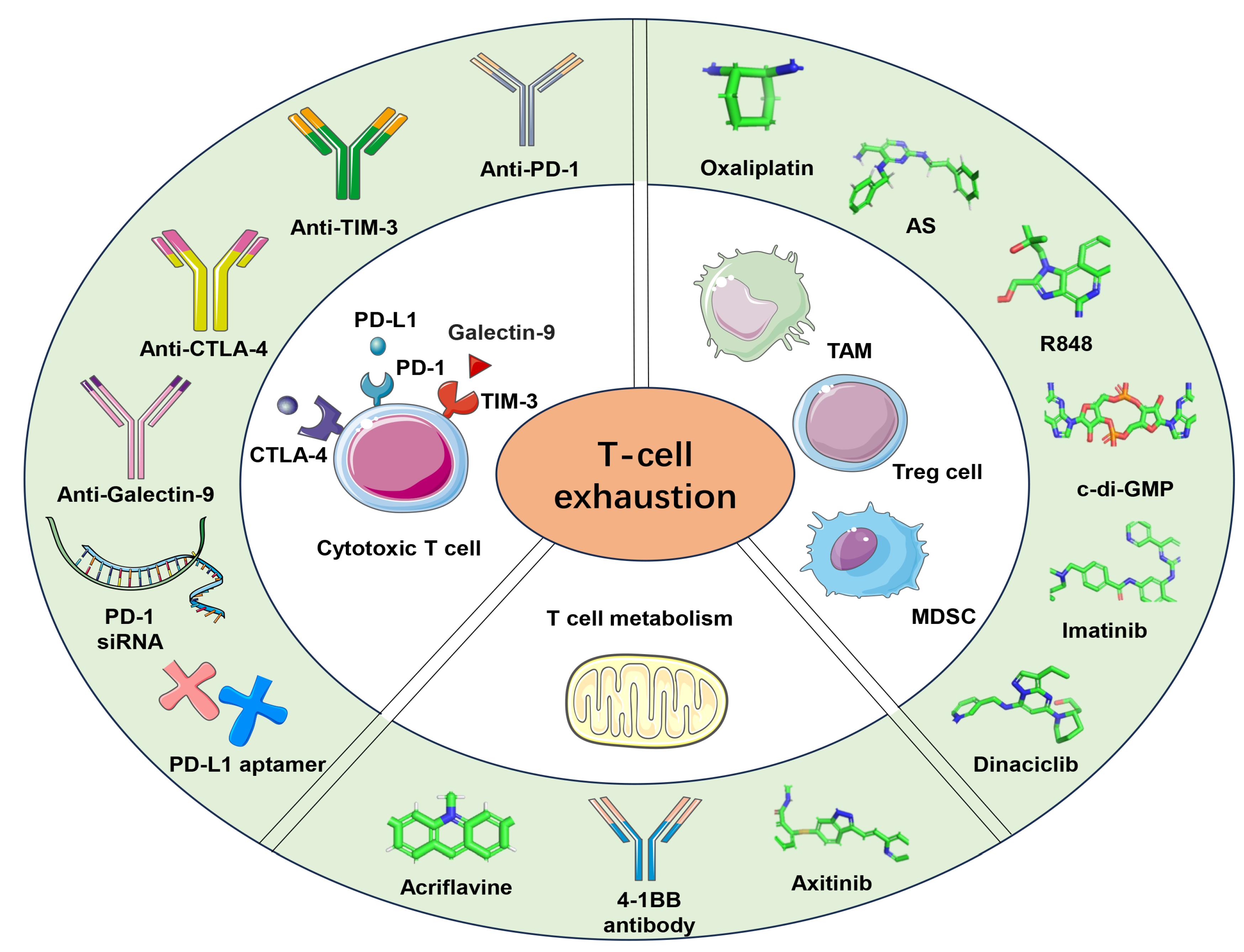
Disclaimer/Publisher’s Note: The statements, opinions and data contained in all publications are solely those of the individual author(s) and contributor(s) and not of MDPI and/or the editor(s). MDPI and/or the editor(s) disclaim responsibility for any injury to people or property resulting from any ideas, methods, instructions or products referred to in the content. |
© 2024 by the authors. Licensee MDPI, Basel, Switzerland. This article is an open access article distributed under the terms and conditions of the Creative Commons Attribution (CC BY) license (https://creativecommons.org/licenses/by/4.0/).
Share and Cite
MDPI and ACS Style
Li, F.; Wang, Y.; Chen, D.; Du, Y. Nanoparticle-Based Immunotherapy for Reversing T-Cell Exhaustion. Int. J. Mol. Sci. 2024, 25, 1396. https://doi.org/10.3390/ijms25031396
AMA Style
Li F, Wang Y, Chen D, Du Y. Nanoparticle-Based Immunotherapy for Reversing T-Cell Exhaustion. International Journal of Molecular Sciences. 2024; 25(3):1396. https://doi.org/10.3390/ijms25031396
Chicago/Turabian StyleLi, Fei, Yahong Wang, Dandan Chen, and Yunjie Du. 2024. "Nanoparticle-Based Immunotherapy for Reversing T-Cell Exhaustion" International Journal of Molecular Sciences 25, no. 3: 1396. https://doi.org/10.3390/ijms25031396
Note that from the first issue of 2016, this journal uses article numbers instead of page numbers. See further details here.