Article

Hydrophilic Reduction-Resistant Spin Labels of Pyrrolidine and Pyrroline Series from 3,4-Bis-hydroxymethyl-2,2,5,5-tetraethylpyrrolidine-1-oxyl

by
Mikhail S. Usatov
1,2,
Sergey A. Dobrynin
1,
Yuliya F. Polienko
1,
Denis A. Morozov
1,
Yurii I. Glazachev
3,
Sergey V. An’kov
1,
Tatiana G. Tolstikova
1,
Yuri V. Gatilov
1,
Irina Yu. Bagryanskaya
1,
Arthur E. Raizvikh
1,4,
Elena G. Bagryanskaya
1 and
Igor A. Kirilyuk
1,*
1
N. N. Vorozhtsov Novosibirsk Institute of Organic Chemistry SB RAS, Lavrentiev Ave. 9, Novosibirsk 630090, Russia
2
Department of Natural Sciences, Novosibirsk State University, Pirogova Str. 1, Novosibirsk 630090, Russia
3
Voevodsky Institute of Chemical Kinetics and Combustion SB RAS, Institutskaya 3, Novosibirsk 630090, Russia
4
Department of Physics, Novosibirsk State University, Pirogova Str. 1, Novosibirsk 630090, Russia
*
Author to whom correspondence should be addressed.
Int. J. Mol. Sci. 2024, 25(3), 1550; https://doi.org/10.3390/ijms25031550
Submission received: 28 November 2023 / Revised: 18 December 2023 / Accepted: 24 December 2023 / Published: 26 January 2024
(This article belongs to the Topic Advances in Chemistry and Chemical Engineering)

Abstract

:
Highly resistant to reduction nitroxides open new opportunities for structural studies of biological macromolecules in their native environment inside living cells and for functional imaging of pH and thiols, enzymatic activity and redox status in living animals. 3,4-Disubstituted nitroxides of 2,2,5,5-tetraethylpyrrolidine and pyrroline series with a functional group for binding to biomolecules and a polar moiety for higher solubility in water and for more rigid attachment via additional coordination to polar sites were designed and synthesized. The EPR spectra, lipophilicities, kinetics of the reduction in ascorbate-containing systems and the decay rates in liver homogenates were measured. The EPR spectra of all 3,4-disubstituted pyrrolidine nitroxides showed additional large splitting on methylene hydrogens of the ethyl groups, while the spectra of similar pyrroline nitroxides were represented with a simple triplet with narrow lines and hyperfine structure of the nitrogen manifolds resolved in oxygen-free conditions. Both pyrrolidine and pyrroline nitroxides demonstrated low rates of reduction with ascorbate, pyrrolidines being a bit more stable than similar pyrrolines. The decay of positively charged nitroxides in the rat liver homogenate was faster than that of neutral and negatively charged radicals, with lipophilicity, rate of reduction with ascorbate and the ring type playing minor role. The EPR spectra of N,N-dimethyl-3,4-bis-(aminomethyl)-2,2,5,5-tetraethylpyrrolidine-1-oxyl showed dependence on pH with pKa = 3, ΔaN = 0.055 mT and ΔaH = 0.075 mT.

1. Introduction

Nitroxides (aminoxyl radicals) are widely used as molecular probes and labels for the investigation of complex molecular systems using various methods of EPR and NMR spectroscopy and imaging [1,2,3,4,5,6,7,8,9,10]. Biophysics and structural biology are the areas where application of nitroxide-based spectroscopic methods have been especially fruitful. Special approaches and techniques have been developed for structural studies of biological macromolecules in their native environment inside living cells [2,11] and for functional imaging of pH and thiols [2,12], enzyme activity [13] and redox status [14] in living animals. The stability of the nitroxide radical is of crucial importance for these applications. Early studies showed that reduction of a nitroxide group to hydroxylamine one with enzymatic systems and low-molecular biogenic antioxidants is a major pathway of nitroxide decay in biological systems [15]. To slow down a radical decay, so-called “sterically shielded” pyrroline and pyrrolidine nitroxides with four ethyl (or more bulky alkyl) substituents adjacent to the N-O group were designed [16,17]. These nitroxides showed much higher stability in biological samples and in cells than conventional tetramethyl-substituted nitroxides [18,19,20,21]. A number of reduction-resistant spin labels for in-cell applications have been prepared from these nitroxides [20,21,22]. However, bulky alkyl substituents make these spin labels lipophilic and poorly soluble in water. Stock solutions were usually prepared in DMSO or in water–DMSO mixtures, see for example [20,23]. Recent studies revealed that an alternative metabolic pathway implying cytochrome P450 activity may significantly contribute to the decay of sterically shielded nitroxides in subcellular systems [24,25]. These data coincide with our observations that decay of 2,2,5,5-tetraethylpyrrolidine nitroxides in homogenates of liver and kidney, which are known to be rich of cytochrome P450 enzymes, proceeds much faster than in homogenates of brain, heart or in blood [22]. According to some estimates, hydrophilic nitroxides, with a negative charge and/or minimal hydrophilicity are required to slow down transformation by P450 enzymes, either via reductive or oxidative metabolism [24].
We recently described convenient procedures for synthesis of 3,4-bis-hydroxymethyl-2,2,5,5-tetraethylpyrrolidine-1-oxyl (1) (Scheme 1) [26,27]. Pyrrolidine nitroxide 1 can be converted into pyrroline derivatives, e.g., 2 [28]. However, synthetic potential of 1 and 2 was not seriously studied yet. It should be noted that similar bifunctional derivatives of 2,2,5,5-tetramethyl-substituted pyrrolidine or pyrroline nitroxides attracted much attention, although they are not as readily available, see for example [29,30,31,32]. Spin labels of pyrroline series with additional functional groups in a neighboring position of the heterocycle are known for especially rigid binding to the target biomolecules allowing for more accurate distance measurements using pulsed EPR methods [33,34,35].
The aim of this work was to find a convenient synthetic approach to 2,2,5,5-tetraethyl-3,4-disubstituted pyrrolidine and pyrroline nitroxides with two different functional groups: one for selective binding to biopolymers or other target molecules (X) and a polar charged moiety that makes the nitroxide hydrophilic and water soluble (Y). We have focused on the preparation of hydrophilic analogs of the known reduction-resistant maleimide spin labels, which are used for selective binding to thiol groups of cysteine residues [20,21], and nitroxide azides, which can be used inside the cells for bioorthogonal spin labeling of proteins modified with unnatural alkyne-containing amino acids using copper (I)-catalyzed azide–alkyne cycloaddition (CuAAC) [36] or strain-promoted azide–alkyne cycloadditions (SPAAC) [37]. Since 1 is a readily available compound, novel hydrophilic nitroxides can be produced on a large scale not only for SDSL-EPR studies but also for the development of specific probes/contrast agents for in vivo EPRI/OMRI/MRI studies. The parameters of EPR spectra of the new nitroxides, their lipophilicities and decay in liver homogenate and in model ascorbate-containing systems were studied.

2. Results and Discussion

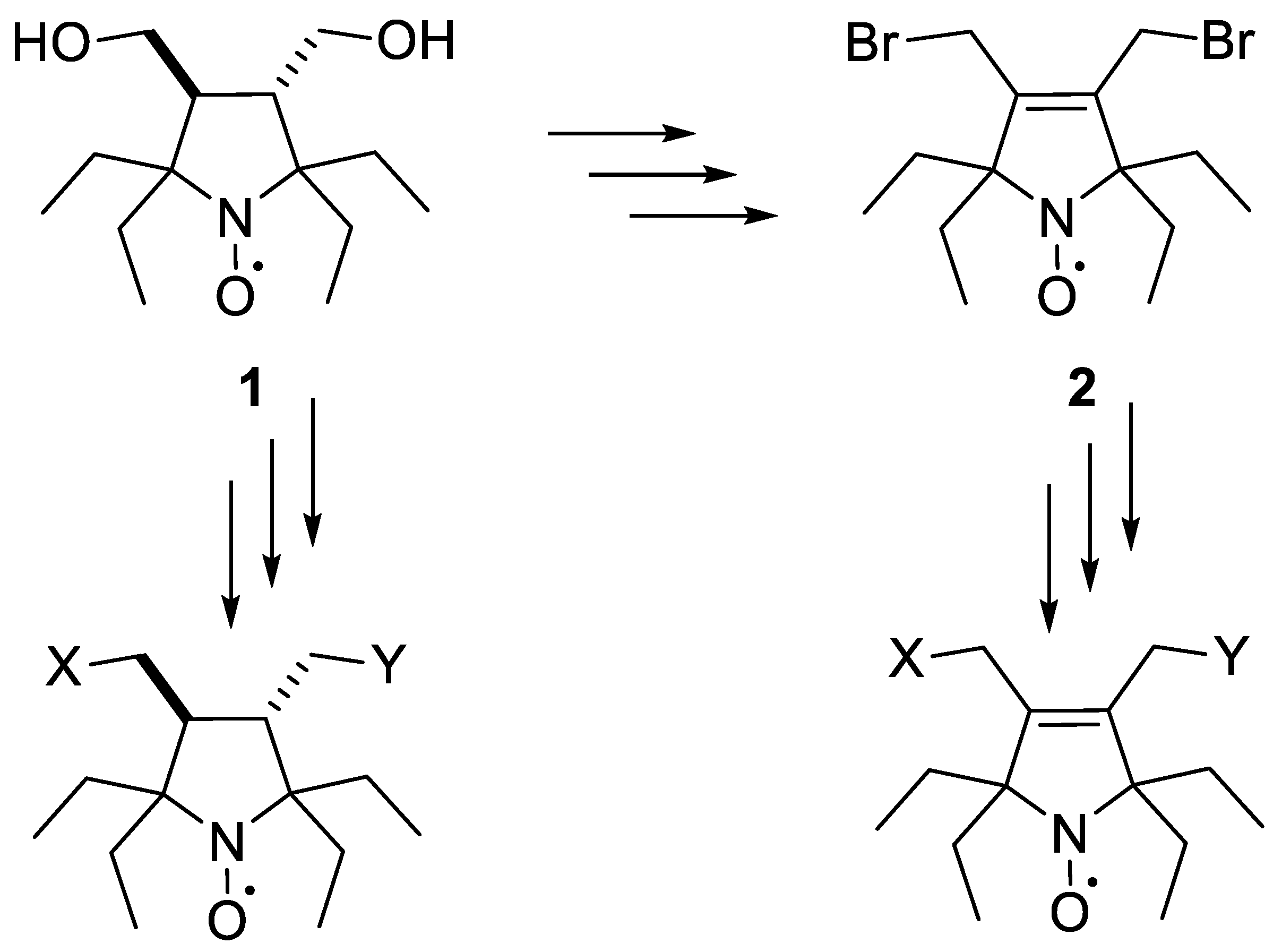
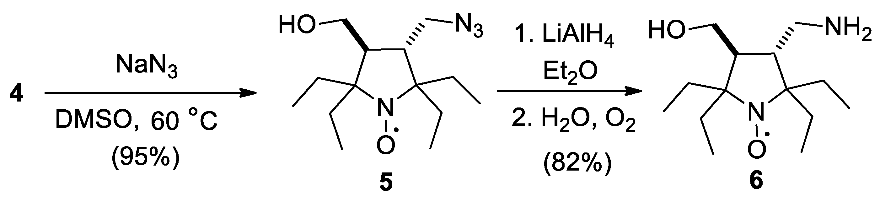
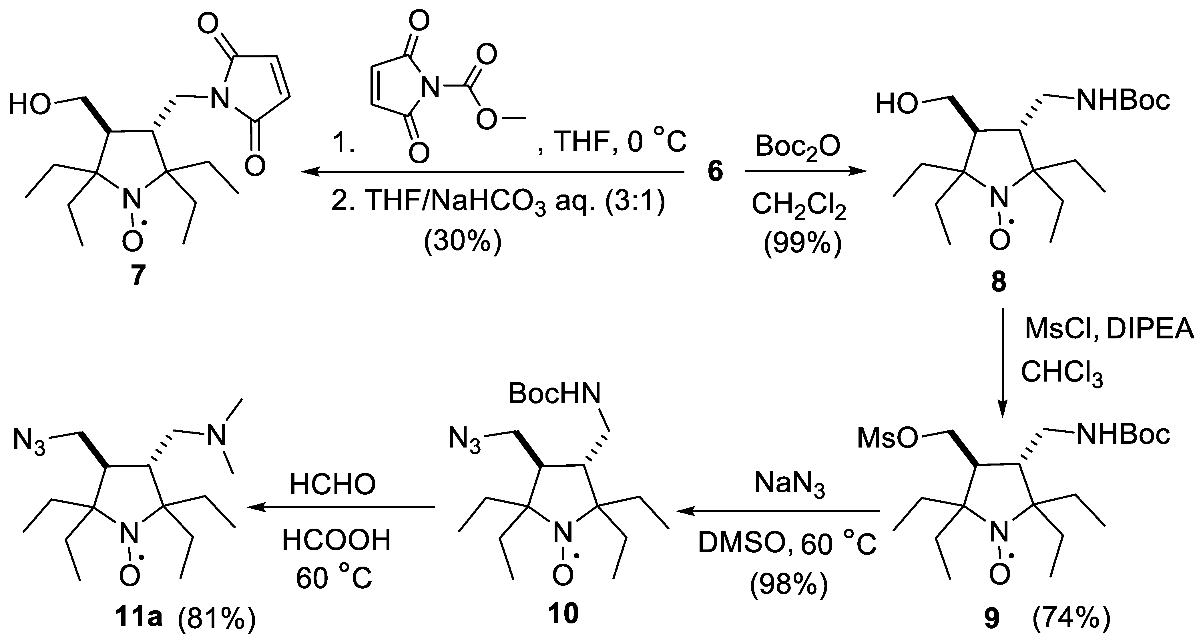
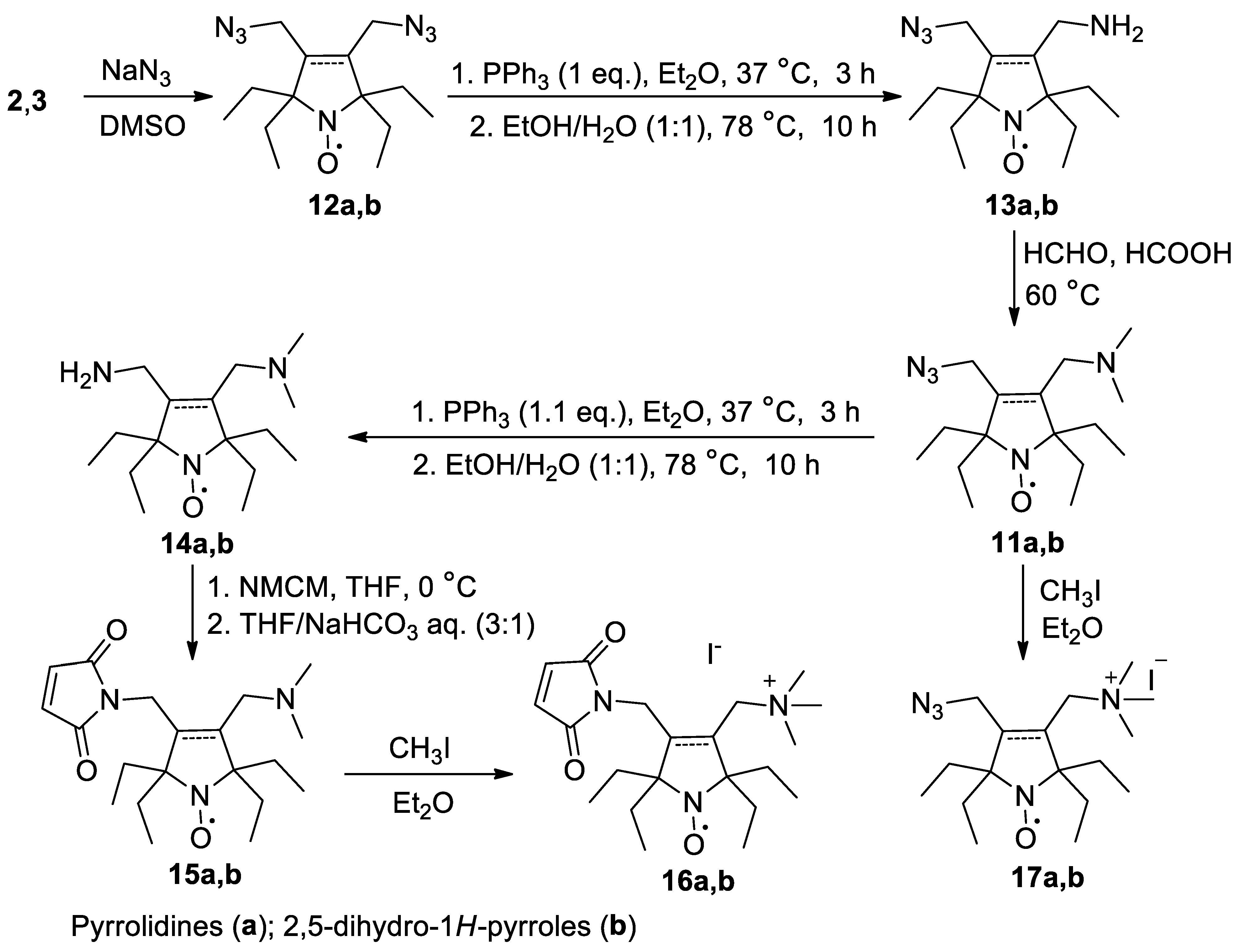
2.1. Synthesis of the Hydrophilic Nitroxides

To convert a symmetric structure, such as 1, into heterofunctional spin labels one should find a convenient way to modify only one of the two identical functional groups with a satisfactory yield. Reactions on one of two hydroxyl groups in 1 seems to be the shortest pathway to heterobifunctional derivatives of 2,2,5,5-tetraethylpyrrolidine-1-oxyl. The subsequent selective modification of the substituents in the position 3 and 4 can be used to prepare various derivatives. We have showed earlier that the reaction of 1 with excess of methanesulfonyl chloride (MsCl) in the presence of diisopropylethylamine (DIPEA) afforded 3 (Scheme 2) [28], and the latter was used in the synthesis of 2. Notably, we did not observe any significant conversion in the absence of a base, while the reaction with MsCl in the presence of excess of the base caused accumulation of dark-colored products. Therefore, we used slow addition of DIPEA to control the reaction in the presence of excess of MsCl. The addition of a smaller amount of reagents gives a mixture of di- and monomesylated products 3 and 4. The optimal preparative yield of 4 64–68% (75–80% with conversion ca. 85%) was achieved upon slow addition of DIPEA (1.4 eq.) to a solution of 1 and MsCl (1.58 eq.). The higher excess of the reagents required for nearly quantitative conversion of 1 with yields of 3 and 4 are 35 and 55%, correspondingly. Both 3 and 4 were isolated as crystalline compounds. Structure assignments were done on the basis of NMR spectra after nitroxide reduction with Zn/CF3COOH [22], IR spectra, element analyses (see Section 3 and Supplementary Materials pp. S4, S10) and single crystal X-ray data (Figure 1). An unexpectedly high yield of 4 may result from stabilization of anionic form of 1 via hydrogen bonding with the neighboring hydroxy group, which makes the rate of mesylation of 1 much higher than that of 4.
Mesyloxy derivatives are convenient precursors for a variety of spin labels. Heating of 4 with an excess of sodium azide in DMSO afforded 5 with nearly quantitative yield (Scheme 3). Azido group was then reduced with LiAlH4 to give aminoalcohol 6 which was isolated as crystalline solid. The structure of 6 was confirmed by IR and NMR spectra and single crystal X-ray analyses data (Figure 1 and Supplementary Materials pp. S5, S12).
A reaction with N-methoxycarbonyl maleimide (NMCM) was chosen to convert amine 6 into corresponding spin label (Scheme 4). The use of this reagent made it possible to keep the hydroxy group intact, although the yield of spin label 7 was low. In this and all subsequent similar syntheses of maleimide spin labels, we used a modified protocol based on literature methods [38,39]. Primary amine was slowly added dropwise to a solution of NMCM in dry THF at 0 °C under argon, and then the reaction mixture was diluted with a saturated aqueous solution of NaHCO3. This two-step procedure prevents the addition of unreacted amine to the maleimide formed, since the elimination of methylcarbamate to form maleimide occurs in the second stage, when the amine has already been consumed.
Despite the presence of a hydrophilic hydroxy group, radicals 5 and 7 are still poorly soluble in water. The introduction of quaternary ammonium groups, such as trimethylammonium, is a simple and effective way to make a compound highly hydrophilic. It is more convenient to introduce such groups at the final steps of synthesis. For this reason, amino group in 6 was protected with tert-butyloxycarbonyl moiety to give 8. The latter was carefully treated with methanesulfonyl chloride in basic conditions (excess of DIPEA) to give 9 and subsequent heating with NaN3 according to the procedure described above afforded 10. The structures of 8 and 10 were assigned on the basis of IR spectra and HRMS (see Section 3 and Supplementary Materials pp. S11–S12) and confirmed with single-crystal X-ray analysis data (Figure 2). Under Eschweiler–Clarke reaction conditions deprotection and alkylation occur with formation of 11a as a single product.
We have found that 11a can also be prepared from 3 (Scheme 5) with a higher yield and in fewer steps (45% overall yield from 1 in 4 steps versus 35–36% in 7 steps via Scheme 1, Scheme 2, Scheme 3 and Scheme 4). It is noteworthy that a similar radical of the pyrroline series, 11b, can also be obtained from 2 using this scheme. It is difficult to obtain a good yield of the monoazido derivative by treating 2 or 3 with sodium azide, see for example literature data for similar 2,2,5,5-tetramethyl-substituted nitroxide [40]. In contrast, the corresponding diazides 12a,b were readily obtained by heating of 2 and 3 with excess of NaN3 in DMSO. These nitroxides were isolated as orange crystals with 93–99% yield and characterized with IR spectra, HRMS and element analyses (see Section 3 and Supplementary Materials pp. S11–S12). The structure of 12a was confirmed by single crystal X-ray data (Figure 2). The subsequent Staudinger reaction with one equivalent of triphenylphosphine afforded 13a,b in 64–73% yield. The resulting aminoazides were converted to 11a,b by Eschweiler–Clarke alkylation.
For the preparation of maleimide spin labels from 11a,b we used the Staudinger reaction followed by treatment with NMCM in analogy to the above procedures. The structures of 15a,b were confirmed by single crystal X-ray analysis (Figure 3). The addition of methyl iodide to the solutions of the spin labels 11a,b and 15a,b in dry diethyl ether leads to precipitation of the corresponding trimethylammonium iodides 16a,b and 17a,b.
Diazide 12a can also be converted into dianionic spin label. A Huisgen cycloaddition reaction with one equivalent of dimethyl acetylenedicarboxylate (DMAD) gave a mixture of mono- and bis-triazoles 18 and 19 (Scheme 6). After alkaline hydrolysis of the ester groups in 18, the monosodium salt 20 was isolated. The structure of this nitroxide was confirmed by single-crystal X-ray diffraction data (Figure 3).

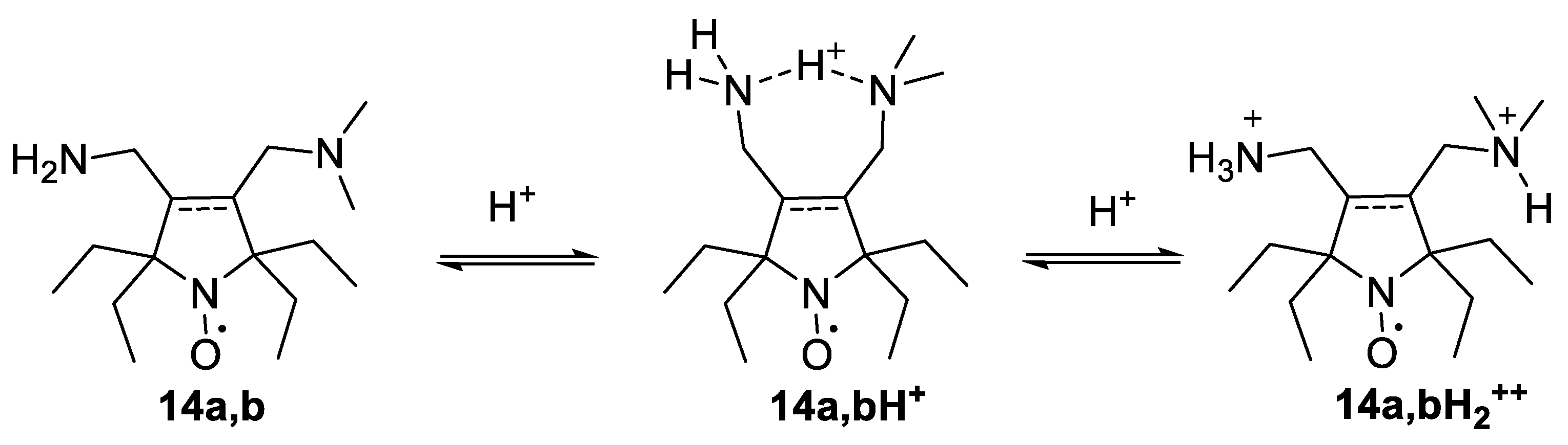
2.2. EPR Studies of the New Nitroxides

The CW-EPR spectra (X-band) were recorded for a broad selection of the new radicals (see Supplementary Materials pp. S23–S26). The parameters of the EPR spectra are given in Table 1. For comparison parameters of the spectra of nitroxides 1, 3-carboxy-2,2,5,5-tetraethylpyrrolidine-1-oxyl (21) [22] and 2,2,5,5-tetraethyl-3,4-bis(hydroxymethyl)-2,5-dihydro-1H-pyrrol-1-oxyl (22) [28] (Figure 4) are shown. In addition, the nitroxide 23 was synthesized for this study by heating 3 with sodium cyanide followed by alkaline hydrolysis of the cyano groups (Scheme 7).
The EPR spectra of 3,4-disubstituted pyrrolidine nitroxides correspond to the pattern described for 1, with broad lines and two large additional doublet splittings on two hydrogen atoms of the methylene groups of the ethyl substituents [26,41] (Figure 5a). The value of these constants depends on the size of the neighboring substituent at position 3 or 4. For example, the introduction of the bulky maleimido group results in up to a 50% increase in one of two aH (cf. 14a and 15a; 16a and 17a). We have showed earlier that high hfc with one of methylene hydrogens of ethyl group is observed in nitroxides with the pseudoaxial position of this ethyl group and CH2-CH3 bond nearly parallel to the N-O bond axis [41]. Apparently, the repulsion of the large substituent and the neighboring ethyl group (cis relative to each other) increases the contribution of this conformation.
In contrast, EPR spectra of the pyrroline nitroxides showed classical triplet with no additional large splittings (Figure 5b). In oxygen-free solution, hyperfine structure of the spectral lines was resolved, which is typical for the nitroxides with planar heterocycle (pyrroline, 3-imidazoline and isoindoline) (Figure 5d) [4]. The simulation gives four aH = 0.0611 mT and four aH = 0.0317 mT, apparently due to hfc with methylene hydrogens of ethyl groups.
The EPR spectra of nitroxides with ionizable groups may be dependent on pH. Similarly to that described for 3-aminomethyl-proxyl (25) [42] (Figure 4), spectra of 3-aminomethyl- and 3-dimethylaminomethyl nitroxides 6, 11a,b, 13a,b and 15a,b demonstrate small dependence on pH with ΔaN ca. 0.02 mT and pK ca. 9. Titration of the nitroxides 14a and 14b revealed two transitions at pH ca. 3 and ca. 11 (Figure 6), which apparently correspond to two protonation steps with formation of mono- and dication. Similarly to the above pattern protonation of free bases 14a,b to form monocations leads to minor changes in nitrogen hfc, ΔaN = 0.02 mT, however more significant changes were observed upon the second protonation, ΔaN = 0.044–0.055 mT. Surprisingly, the constants on the hydrogen atoms of the methylene fragments of ethyl groups in 14a turned out to be more sensitive to pH changes (Figure 6). While formation of monocation 14aH+ leads to decrease of apparent aH by 0.02 mT, the spectrum of 14aH2++ (pH = 1) shows increase of the hydrogen hfc constant by 0.075 mT. Presumably the monocations 14aH+ and 14bH+ are stabilized due to coordination (hydrogen bonding) with the neighboring amino group forming 7-membered rings (Scheme 8).
This stabilization partly accounts for higher pK of the first protonation of 14a,b. Formation of these cyclic structures apparently decreases the repulsion of the ethyl groups and the substituents in the position 3 and 4. A noticeable decrease of aH upon the first protonation of 14a confirms the assumption of the formation of these cyclic structures. In contrast, the second protonation leads to opening of the 7-membered ring and strong conformational changes because of electrostatic repulsion of the neighboring cationic groups. These changes result in pronounced growth of aH in the spectra of 14a. In the spectra of 14b, the hyperfine structure of the triplet manifolds was not resolved, but growth of aH upon the second protonation can be followed by the inhomogeneous line broadening which leads to notable decrease of the peak height in the spectrum (Figure 6c).

2.3. Lipophilicity

Partition in octanol-water mixtures for ionizable compounds may depend on pH, and the pH changes may result from addition of the compound. Therefore, phosphate-citrate-borate buffer (5 mM, pH 7.4) was used for Kp measurements to make the partition closer to physiological conditions. The data is listed in Table 1. The new set of spin labels contains highly hydrophilic nitroxides, such as the positively charged spin labels 16a,b and 17a,b and negatively charged 20, which were not detected in octanol after shaking with buffer. Although monoamino derivatives 11 and 13 mainly exist in protonated form at pH 7.4, they still remain lipophilic and, therefore, should permeate cells. Amino groups are known to provide higher retention/intracellular accumulation due to transmembrane potential or ion-trapping effect [44]. This effect was exploited in design of nitroxide contrast agents for MRI and radioprotectors [45]. Cellular accumulation of spin labels like 11 and 13 may help to increase efficacy spin labeling using CuAAC or SPAAC.

2.4. Reduction Rates in Ascorbate-Containing Systems

Rate constants of the nitroxides decay in the ascorbate-containing systems are given in Table 1. All the pyrrolidine radicals demonstrated very high stability to reduction, exceeding that of 21. Reduction rates of pyrroline nitroxides are always expectedly higher than those of similar nitroxides of pyrrolidine series, but they were never higher than that of 21 by more than 40%.

2.5. Decay in the Rat Liver Homogenates

For this research we used a selection of nitroxides, containing cationic groups 17a,b, ionizable groups existing at pH 7.4 in cationic 14a,b and anionic 20, 21, 23 forms, and non-ionizable nitroxides with hydroxyl groups 1, 22. We avoided using maleimido derivatives because they could react with proteins of the homogenate, therefore, the results would be hard to interpret. The nitroxide solutions were mixed with rat liver homogenate at 37 °C and decay of the EPR signal was followed. The kinetics of decay are shown in Figure 7.
Surprisingly, the decay rates showed no correlation with the reduction rates with ascorbate, with the lipophilicity values or with the nitroxide heterocyclic ring type. The compounds can be divided into three categories in accordance with the type of the functional groups. The decay of cationic nitroxides and amino derivatives was significantly faster than that of carboxylic acids and diols. In our previous study, we showed that the decay of positively charged 2,2,5,5-tetraethylpyrrolidine-1-oxyls in homogenates of kidney and liver proceeds much faster than in blood and in homogenates of brain and heart [22]. Therefore, a relatively high rate of decay of cationic nitroxides in liver homogenates does not mean that they cannot be used as spin labels in living cells, since the cells normally used in such studies do not have such a high activity towards xenobiotics. However, the above data show that negatively charged or non-charged polar nitroxides may perform better in EPR/NMR imaging studies in vivo. This coincides with the conclusions of the recent paper by Peyrot, et al. [24]. Despite much similarity of our results to literature data, we are far from the assignment of the observed decay of nitroxides to certain enzyme activity and from elucidation of the decay mechanism without special investigation.

3. Materials and Methods

3.1. General Information

The compounds 1 [26,27] and 2 [28] were prepared according to the literature. The IR spectra were recorded on a Bruker Vector 22 FT-IR spectrometer (Bruker, Billerica, MA, USA) in KBr pellets (1:150 ratio) or in neat samples (see the Supplementary Materials in this article pp. S10–S18) and are reported in wave numbers (cm−1). UV spectra were acquired on a HP Agilent 8453 spectrometer (Agilent Technologies, Santa Clara, CA, USA) in ethanol solutions (concentration~10−4 M). 1H NMR spectra were recorded on a Bruker AV 300 (300.132 MHz), AV 400 (400.134 MHz) and DRX 500 (500.130 MHz) spectrometers (Bruker, Billerica, MA, USA). 13C NMR spectrum was recorded on a Bruker AV 300 (75.467 MHz) (see the Supplementary Materials in this article pp. S4–S9). All the NMR spectra were acquired for 5–10% solutions in CDCl3 or CD3OD at 300 K using the signal of the solvent as a standard. To confirm the structure of stable nitroxides, 1H NMR spectra were recorded of the solutions of corresponding hydroxylamines prepared via reduction of the nitroxide samples (10–20 mg) with Zn powder in a CD3OD–CF3COOH 10:1 mixture [22]. HRMS analyses were performed using a High-Resolution Mass Spectrometer DFS (Thermo Electron, Waltham, MA, USA).
Reactions were monitored by TLC on precoated TLC sheets ALUGRAM Xtra SIL G/UV254 ((Macherey-Nagel GmbH & Co. KG, Düren, Germany) using UV light 254 nm, 1% aqueous permanganate, 10% solution of phosphomolybdic acid in ethanol and Dragendorff reagent as visualizing agents. Kieselgel60 (Macherey-Nagel GmbH & Co. KG) was utilized as an adsorbent for column chromatography.
CCDC 2308349 (3), 2308350 (4), 2309021 (6), 2308351 (8), 2308352 (10), 2308353 (12a), 2308354 (15a), 2308355 (15b), 2308356 (20) contain the supplementary crystallographic data for this paper. These data can be obtained free of charge from The Cambridge Crystallographic Data Centre https://www.ccdc.cam.ac.uk/structures/ (accessed on 17 November 2023).

3.2. Synthesis

3.2.1. A Reaction of Hydroxymethyl Nitroxides 1 and 8 with Methanesulfonyl Chloride

Method A: N,N-diisopropylethylamine (DIPEA) was added dropwise within 6 h to a stirred solution of 1 and methanesulfonyl chloride 1.4 g in dry chloroform, the amount and ratio of the reagents is shown in Table 2. The mixture was stirred overnight, washed with water and dried with MgSO4. The solution was concentrated in a vacuum and separated using column chromatography on silica gel, eluent chloroform; yields of 3 and 4 are shown in the Table 2.
2,2,5,5-Tetraethyl-3,4-bis((methylsulfonyloxy)methyl)pyrrolidin-1-oxyl (3), yellow crystals, m.p. 73–75 °C (hexane). IR (KBr) νmax: 1348, 1170 (OMs). 1H NMR (300 MHz; CD3OD, Zn/CF3COOH, δ): 1.06 (t, Jt = 7.4 Hz, 6H), 1.07 (t, Jt = 7.4 Hz, 6H), 1.88 (dq, Jd = 14.7 Hz, Jq = 7.4 Hz, 2H), 2.00 (q, Jq = 7.4 Hz, 4H), 2.06 (dq, Jd = 14.7 Hz, Jq = 7.4 Hz, 2H), 2.67–2.78 (m, 2H), 3.18 (s, 6H), 4.44–4.55 (m, 4H). Anal. Calcd for C16H32NO7S2: C, 46.36; H, 7.78; N, 3.38; S, 15.47; found: C, 46.49; H, 7.50; N, 3.57.
2,2,5,5-Tetraethyl-3-(hydroxymethyl)-4-((methylsulfonyloxy)methyl)pyrrolidin-1-oxyl (4), yellow crystals, m.p. 66–67 °C (hexane). IR (KBr) νmax: 3386 (OH), 1359, 1180 (OMs). 1H NMR (300 MHz; CD3OD, Zn/CF3COOH, δ): 1.04 (t, Jt = 7.4 Hz, 6H), 1.05 (t, Jt = 7.4 Hz, 3H), 1.06 (t, Jt = 7.4 Hz, 3H), 1.77–2.12 (m, 8H), 2.32 (ddd, Jd1 = 11.8 Hz, Jd2 = 5.6, Jd3 = 4.9 Hz, 1H), 2.68 (ddd, Jd1 = 11.8 Hz, Jd2 = 7.3 Hz, Jd3 = 3.6 Hz, 1H), 3.14 (s, 3H), 3.78 (dd, Jd1 = 11.7 Hz, Jd2 = 4.9 Hz, 1H), 3.83 (dd, Jd1 = 11.7 Hz, Jd2 = 5.6 Hz, 1H), 4.43 (dd. Jd1 = 11.0 Hz, Jd2 = 7.3 Hz, 1H), 4.54 (dd, Jd1 = 11.0 Hz, Jd2 = 3.6 Hz, 1H). 13C{1H} (75 MHz; CD3OD, Zn/CF3COOH, δ): 6.71, 6.77, 23.5, 23.7, 28.8, 28.9, 35.3, 48.1, 49.9, 58.42, 66.5, 69.7, 70.1. Anal. Calcd for C15H30NO5S: C, 53.54; H, 8.99; N, 4.16; S, 9.53; found: C, 53.38; H, 8.96; N, 4.20; S, 9.50.
Method B: Methanesulfonyl chloride (227 mg, 1.98 mmol) was added dropwise to a stirred solution of 8 (274 mg, 0.57 mmol) and DIPEA (335 mg, 2.59 mmol)) in dry chloroform (10 mL). The mixture was stirred at ambient temperature for 3 days (TLC control on silica gel, chloroform—methanol 100:1, visualization with UV-254), washed with water and dried with Na2CO3. The solution was concentrated in vacuum. The residue was purified using column chromatography on silica gel, eluent chloroform—methanol 100:1 to give 9 (247mg, 74%).
3-(((tert-Butoxycarbonyl)amino)methyl)-2,2,5,5-tetraethyl-4-(((methylsulfonyl)oxy)methyl)pyrrolidin-1-oxyl (9), yellow crystals, m.p. 135–136 °C (hexane—chloroform 25:1). IR (KBr) νmax: 3363, 1513 (NH), 1675 (C=O), 1358, 1178 (OMs). UV (EtOH) λmax (log ε): 239 (3.34). Anal. Calcd for C20H39N2O6S: C, 55.15; H, 9.02; N, 6.43; S, 7.36; found: C, 54.89; H, 9.06; N, 6.38; S, 7.19. HRMS (EI/DFS) m/z: [M]+ calcd for C20H39N2O6S 435.2523, found 435.2525.

3.2.2. Reaction of Mesylates with Sodium Azide (General Method)

A mixture of a mesylate 3, 4, or 9 (12 mmol), NaN3 (5 g, 77 mmol) and DMSO (50 mL) was heated to 60 °C and stirred for 4–6 h. The conversion was controlled using TLC (silica gel, chloroform, UV 254). The mixture was diluted with H2O (200 mL) and extracted with hexane-diethyl ether mixture 1:1 (3 × 100 mL). The extract was washed with water and dried with MgSO4. The solvent was distilled off and residue was purified as described below.
3-(Azidomethyl)-2,2,5,5-tetraethyl-4-(hydroxymethyl)pyrrolidin-1-oxyl (5). Crude nitroxide was dissolved in the mixture hexane–toluene 20:1 and the solution was cooled to −15 °C. The crystalline precipitate of 5 was filtered off and washed with cold hexane, yield 90%, m.p. 35–38 °C. IR (KBr) νmax: 3406 (OH), 2098 (N3). UV (EtOH) λmax (log ε): 223 (3.35). Anal. Calcd for C14H27N4O2: C, 59.34; H, 9.60; N, 19.77; found: C, 59.34; H, 9.86; N, 19.81.
3-(Azidomethyl)-4-((tert-butoxycarbonylamino)methyl)-2,2,5,5-tetraethylpyrrolidin-1-oxyl (10). Yield 98%, yellow oil. IR (KBr) νmax: 3353 (NH), 2100, 2090 (N3), 1708 (C=O). UV (EtOH) λmax (log ε): 223 (3.43). HRMS (EI/DFS) m/z: [M]+ calcd for C19H36O3N5 382.2813, found 382.2817.
3,4-Bis(azidomethyl)-2,2,5,5-tetraethylpyrrolidin-1-oxyl (12a). Yield 99%, m.p. 62–65 °C (hexane). IR (KBr) νmax: 2193, 2096 (N3). Anal. Calcd for C14H26N7O: C, 54.52; H, 8.50; N, 31.79; found: C, 54.60; H, 8.46; N, 31.50.
3,4-Bis(azidomethyl)-2,2,5,5-tetraethyl-2,5-dihydro-1H-pyrrol-1-oxyl (12b). Crude nitroxide was purified using column chromatography on silica gel, hexane—ethyl acetate mixture 4:1 to give 12, yield 93%, yellow crystals, m.p. 44–46 °C (hexane). IR (KBr) νmax: 2098 (N3). Anal. Calcd for C14H24N7O: C, 54.88; H, 7.90; N, 32.00; found: C, 54.70; H 8.00; N, 32.12.
3-(Aminomethyl)-2,2,5,5-tetraethyl-4-(hydroxymethyl)pyrrolidin-1-oxyl (6). A solution of nitroxide azide 5 (243 mg, 0.86 mmol) in absolute ethyl ether (5 mL) was added dropwise to a stirred solution of lithium aluminum hydride (211 mg, 5.55 mmol) in absolute ethyl ether (20 mL). The conversion was controlled using TLC (silica gel, hexane—ether 1:1, UV 254). The flask was cooled in an ice bath and carefully quenched with 10 mL of 2.5 M aqueous sodium hydroxide. The organic layer was separated via decantation, the wet precipitate was washed with diethyl ether (10 mL × 3 times) and the combined extract was evaporated under reduced pressure. The residue was purified by column chromatography on silica gel (eluent chloroform—methanol—aqueous ammonia 190:10:1). Yield of 6 187 mg (82%), m.p. 43–44 °C (dec). IR (KBr) νmax: 3463, 3380, 3359, 3288, 3193 (OH, NH2). UV (EtOH) λmax (log ε): 238 (3.29). 1H NMR (300 MHz; CD3OD, Zn/CF3COOH, δ): 0.91 (t, Jt = 7.4 Hz, 3H), 0.96 (t, Jt = 7.4 Hz, 3H), 0.97 (t, Jt = 7.4 Hz, 3H), 0.98 (t, Jt = 7.4 Hz, 3H), 1.16–2.03 (m, 8H), 2.32 (dd, Jd1 = 11 Hz, Jd2 = 10 Hz, 1H), 2.46 (dd, Jd1 = 11 Hz, Jd2 = 10 Hz, 1H), 2.90 (dd, Jd1 = 12 Hz, Jd2 = 11 Hz, 1H), 3.16 (d, Jd = 12 Hz, 1H), 3.52 (dd, Jd1 = 11 Hz, Jd2 = 10 Hz, 1H), 3.87 (d, Jd1 = 10 Hz, 1H). Anal. Calcd for C14H29N2O2: C, 65.33; H, 11.36; N, 10.88; found: C, 65.11; H 11.38; N, 10.67. HRMS (EI/DFS) m/z: [M]+ calcd for C14H29O2N2 257.2224, found 257.2225.
3-(((tert-Butoxycarbonyl)amino)methyl)-2,2,5,5-tetraethyl-4-(hydroxymethyl)pyrrolidin-1-oxyl (8). A solution of Boc2O (505 mg, 2.31 mmol) in dichloromethane (5 mL) was cooled in an ice bath and the solution of nitroxide 6 (520 mg, 2.02 mmol), triethylamine (0.83 mL, 5.95 mmol) in dichloromethane (1 mL) was added dropwise. The reaction mixture was allowed to stay under stirring at the ambient temperature for 24 h. The conversion was controlled using TLC (silica gel, chloroform—methanol—aqueous ammonia 190:10:1, UV 254). The mixture was diluted with 10% aqueous solution of ammonium chloride (10 mL) and extracted with ethyl acetate (3 × 10 mL). The extract was washed with brine and dried with Na2SO4. The solvent was distilled off and residue was purified by column chromatography on silica gel (eluent chloroform—methanol 200:1 gradient enhanced to 50:1). Yield 776 mg (99%), yellow crystals, m.p. 82–85 °C. IR (KBr) νmax: 3376, 3320 (OH, NH), 1720, 1691 (C=O), 1540 (NH). UV (EtOH) λmax (log ε): 238 (3.37). Anal. Calcd for C19H38N2O4: C, 63.83; H, 10.43; N, 7.84; found: C, 63.58; H 10.53; N, 7.94. HRMS (EI/DFS) m/z: [M]+ calcd forC19H38O4N2 357.2748, found 357.2753.

3.2.3. The Eschweiler–Clarke Methylation of Nitroxides 10 and 13a,b (General Method)

The mixture of nitroxide 10, 13a or 13b (0.06 mmol), 21% aqueous solution of formaldehyde (0.16 mL, 1.05 mmol) and formic acid (95%) (525 mg, 10.84 mmol) was heated in an oil bath at 60 °C for 36 h. The conversion was controlled using TLC (silica gel, chloroform—methanol—aqueous ammonia 100:10:1, UV 254). The reaction mixture was diluted with water, basified (Na2CO3) to pH ~ 8, and extracted with ether (3 × 10 mL). An organic extract was dried over Na2CO3, and the solvent was removed under reduced pressure.
3-(Azidomethyl)-4-((dimethylamino)methyl)-2,2,5,5-tetraethylpyrrolidin-1-oxyl (11a). Yield 81% (from 10) and 80% (from 13a), yellow crystals, m.p. 45–46 °C. IR (KBr) νmax: 2821, 2769 (N-CH3), 2098 (N3). UV (EtOH) λmax (log ε): 236 (3.43). Anal. Calcd for C16H32N5O: C, 61.90; H, 10.39; N, 22.56; found: C, 61.74; H, 10.43; N, 22.59. HRMS (EI/DFS) m/z: [M]+ calcd for C16H32ON5 310.2601, found 310.2599.
3-(Azidomethyl)-4-((dimethylamino)methyl)-2,2,5,5-tetraethyl-2,5-dihydro-1H-pyrrol-1-oxyl (11b). Yield 56%, yellow oil. IR (neat) νmax: 2817, 2769 (N-CH3), 2094 (N3). Anal. Calcd for C16H30N5O: C, 62.30; H, 9.80; N, 22.71; found: C, 62.17; H, 9.78; N, 22.57. HRMS (EI/DFS) m/z: [M]+ calcd for C16H30ON5 308.2445, found 308.2443.

3.2.4. General Procedure for Quaternization of Dimethylamino Group

Iodomethane (120 mg, 0.85 mmol) was added to the solution of nitroxide 11a,b or 15a,b (0.1 mmol) in dry diethyl ether (0.5 mL) and the resulting mixture was allowed to stay at ambient temperature for 72 h. The precipitate formed was collected on a filter and washed with dry diethyl ether.
3-((2,5-Dioxo-2,5-dihydro-1H-pyrrol-1-yl)methyl)-2,2,5,5-tetraethyl-4-((trimethylammonio)methyl)pyrrolidin-1-oxyl monoiodide (16a). Yield 41%, yellow crystals, m.p. 155–156 °C (dec). IR (KBr) νmax: 1768, 1705 (C=O). Anal. Calcd for C21H37IN3O3: C, 49.80; H, 7.36; N, 8.30; found: C, 49.75; H, 7.60; N, 8.17.
3-((2,5-Dioxo-2,5-dihydro-1H-pyrrol-1-yl)methyl)-2,2,5,5-tetraethyl-4-((trimethylammonio)methyl)-2,5-dihydro-1H-pyrrol-1-oxyl monoiodide (16b). Yield 20%, yellow crystals, m.p. 204–205 °C. IR (KBr) νmax: 1772, 1712 (C=O). Anal. Calcd for C21H35IN3O3: C, 50.00; H, 6.99; N, 8.33; I, 25.16; found: C, 50.11; H, 7.14; N, 8.40; I, 25.30.
3-(Azidomethyl)-2,2,5,5-tetraethyl-4-((trimethylammonio)methyl)pyrrolidin-1-oxyl monoiodide (17a). Yield 42%, yellow crystals, m.p. 91–92 °C (dec). IR (KBr) νmax: 2100 (N3). UV (EtOH) λmax (log ε): 219 (4.09). Anal. Calcd for C17H35N5OI: C, 45.13; H, 7.80; N, 15.48; I, 28.05; found: C, 44.99; H, 7.63; N, 15.67; I, 28.02.
3-(Azidomethyl)-2,2,5,5-tetraethyl-4-((trimethylammonio)methyl)-2,5-dihydro-1H-pyrrol-1-oxyl monoiodide (17b). Yield 85%, yellow crystals, m.p. 148–149 °C. IR (KBr) νmax: 2098 (N3). Anal. Calcd for C17H33N5OI: C, 45.34; H, 7.39; N, 15.55; I, 28.18; found: C, 45.48; H, 7.60; N, 15.75; I, 28.24.

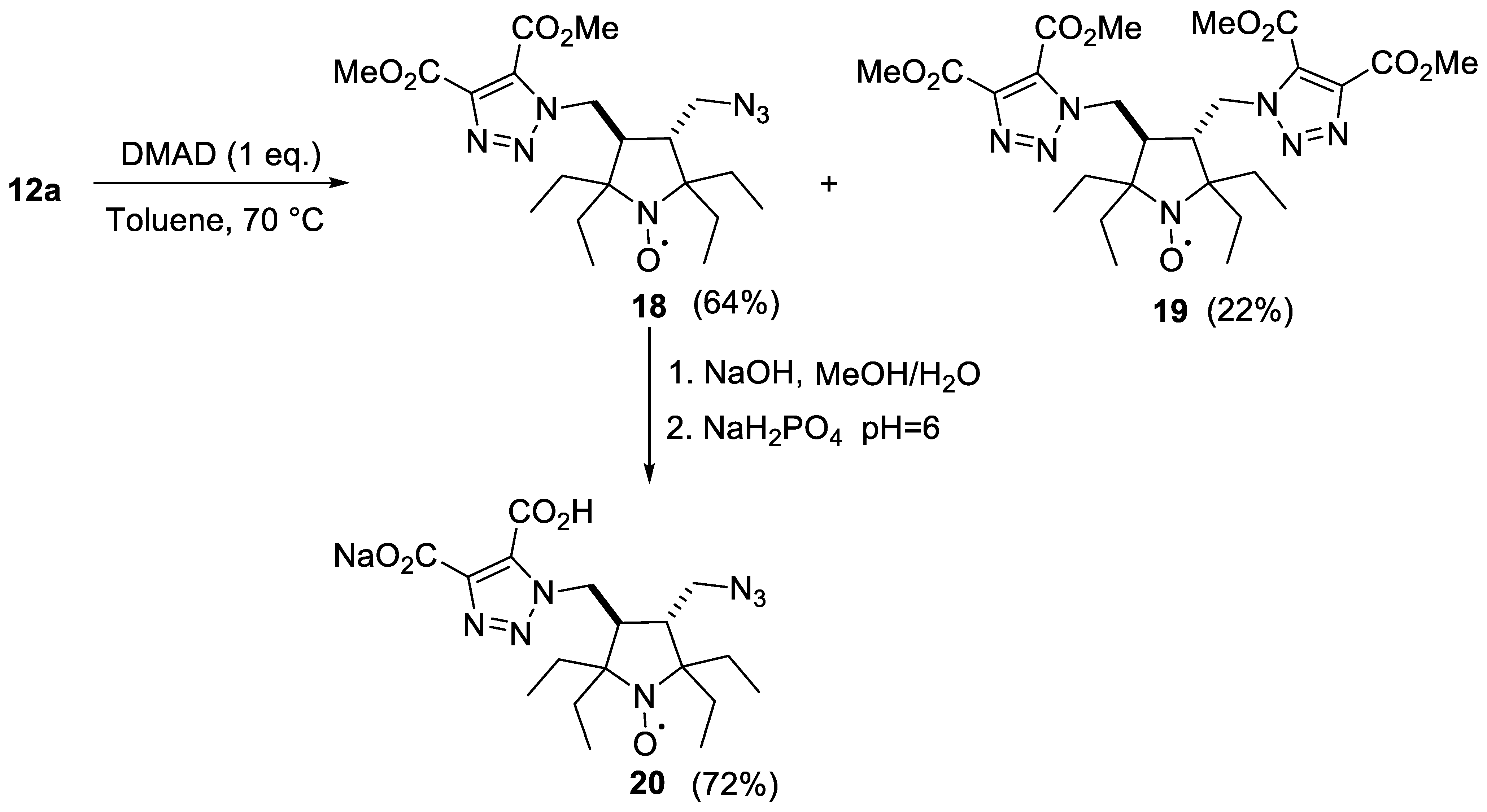
3.2.5. Reaction of Nitroxide 12a with Dimethyl Acetylenedicarboxylate (DMAD)

A solution of DMAD (149 mg, 1.00 mmol) in toluene (2 mL) was added to the solution of nitroxide 12a (305 mg, 1.00 mmol) in toluene (2 mL). The resulting mixture was stirred and heated in an oil bath at 70 °C for 24 h. The conversion was controlled using TLC (silica gel, chloroform—methanol 200:1, UV 254). The solvent was distilled off and residue was separated by column chromatography on silica gel (eluent hexane—ethyl acetate 1:1) to give 18 (285 mg, 64%) and 19 (129 mg, 22%).
3-(Azidomethyl)-4-((4,5-bis(methoxycarbonyl)-1H-1,2,3-triazol-1-yl)methyl)-2,2,5,5-tetraethylpyrrolidin-1-oxyl (18). Yellow crystals, m.p. 113–114 °C. IR (KBr) νmax: 2200, 2098 (N3), 1733 (C=O). Anal. Calcd for C20H32N7O5: C, 53.32; H, 7.16; N, 21.76; found: C, 53.47; H, 7.06; N, 21.94. HRMS (EI/DFS) m/z: [M]+ calcd for C20H32O5N7 450.2459, found 450.2461.
3,4-Bis((4,5-bis(methoxycarbonyl)-1H-1,2,3-triazol-1-yl)methyl)-2,2,5,5-tetraethylpyrrolidin-1-oxyl (19). Yellow crystals, m.p. 178–179 °C (dec). IR (KBr) νmax: 1731 (C=O). Anal. Calcd for C26H38N7O9: C, 52.69; H, 6.46; N, 16.54; found: C, 52.87; H, 6.38; N, 16.26. HRMS (EI/DFS) m/z: [M]+ calcd for C26H38O9N7 592.2726, found 592.2721.

3.2.6. General Procedure of the Synthesis of Amines 13a,b and 14a,b with PPh3

Triphenylphosphine (1 eq. for 12a,b or 1.1 eq. for 11a,b) was added to the solution of azide 11a,b or 13a,b (1 eq., 3 mmol) in dry diethyl ether (10 mL). The reaction mixture was stirred and heated under reflux for 3 h. The solvent was evaporated under reduced pressure and 50% aqueous ethanol (10 mL) was added to the residue. The resulted mixture was heated under reflux with stirring for 10 h. The ethanol was distilled off under reduced pressure and the water layer was acidified with 1M aqueous NaHSO4 to pH = 2 and extracted with chloroform (3 × 20 mL). The water layer was basified with sodium carbonate to pH~9–10, extracted with chloroform (5 × 20 mL), the extract was dried with sodium carbonate, and the solvent was evaporated under reduced pressure.
3-(Aminomethyl)-4-(azidomethyl)-2,2,5,5-tetraethylpyrrolidin-1-oxyl (13a). Yield 64%, yellow crystals, m.p. 36–40 °C. IR (KBr) νmax: 3390, 3360 (NH), 2177, 2098 (N3). Anal. Calcd for C14H28IN5O: C, 59.54; H, 9.99; N, 24.80; found: C, 59.53; H, 9.94; N, 24.57. HRMS (EI/DFS) m/z: [M]+ calcd for C14H28ON5 282.2288, found 282.2291.
3-(Aminomethyl)-4-(azidomethyl)-2,2,5,5-tetraethyl-2,5-dihydro-1H-pyrrol-1-oxyl (13b). Yield 73%, yellow oil. IR (neat) νmax: 3380, 3311 (NH), 2096 (N3). HRMS (EI/DFS) m/z: [M]+ calcd for C14H26ON5 280.2132, found 280.2131.
3-(Aminomethyl)-4-((dimethylamino)methyl)-2,2,5,5-tetraethylpyrrolidin-1-oxyl (14a). Yield 90%, yellow oil. IR (neat) νmax: 3361, 3270 (NH), 2821, 2767 (N-CH3). NMR sample was prepared as follows: after the procedure [21] a solution was evaporated, the residue was mixed with 10% NaOH in MeOH (1 mL) and again evaporated, the residue was triturated with CDCl3 and the solution was filtered into the NMR tube. 1H NMR (300 MHz; CDCl3, δ): 0.80 (t, Jt = 7.3 Hz, 3H), 0.81 (t, Jt = 7.4 Hz, 6H), 0.82 (t, Jt = 7.3 Hz, 3H), 1.14–1.32 (m, 7H), 1.32–1.51 (m, 3H), 1.76 (ddd, Jd1 = 11.2 Hz, Jd2 = 9.2 Hz, Jd3 = 2.6 Hz, 1H), 1.92 (ddd, Jd1 = 11.2 Hz, Jd2 = 10.6 Hz, Jd3 = 1.8 Hz, 1H), 2.00 (dd, Jd1 = 12.5 Hz, Jd2 = 1.8 Hz, 1H), 2.17 (s, 6H), 2.35 (dd, Jd1 = 12.5 Hz, Jd2 = 10.6 Hz, 1H), 2.53 (dd, Jd1 = 12.8 Hz, Jd2 = 9.2 Hz, 1H), 2.71 (dd, Jd1 = 12.8 Hz, Jd2 = 2.6 Hz, 1H). 13C{1H} NMR (75 MHz; CDCl3, δ): 7.4, 7.5, 8.2, 8.6, 28.2, 28.4, 29.2, 29.6, 30.8, 42.1, 45.5, 46.4, 54.3, 60.4, 62.5, 62.6. Anal. Calcd for C16H34N3O: C, 67.56; H, 12.05; N, 14.77; found: C, 67.72; H, 11.77; N, 14.60. HRMS (EI/DFS) m/z: [M]+ calcd for C16H34ON3 284.2696, found 284.2698.
3-(Aminomethyl)-4-((dimethylamino)methyl)-2,2,5,5-tetraethyl-2,5-dihydro-1H-pyrrol-1-oxyl (14b). Yield 60%, yellow oil. IR (neat) νmax: 3376 (NH), 2815, 2765 (N-CH3). 1H NMR (300 MHz; CD3OD, Zn/CF3COOH, δ): 1.06 (t, Jt = 7.4 Hz, 6H), 1.07 (t, Jt = 7.4 Hz, 6H), 1.94 (dq, Jd = 14.7 Hz, Jq = 7.4 Hz, 2H), 1.97 (dq, Jd = 14.7 Hz, Jq = 7.4 Hz, 2H), 2.15 (dq, Jd = 14.7 Hz, Jq = 7.4 Hz, 2H), 2.17 (dq, Jd = 14.7 Hz, Jq = 7.4 Hz, 2H), 2.71–2.94 (m, 6H), 3.71–3.99 (m, 4H). HRMS (EI/DFS) m/z: [M]+ calcd for C16H32ON3 282.2540, found 282.2539.

3.2.7. General Procedure of the Synthesis of Maleimides 7 and 15a,b

A solution of N-methoxycarbonyl maleimide NMCM (51 mg, 0.33 mmol) in absolute THF (10 mL) was cooled in an ice bath and the solution of nitroxide 6 or 14a,b (0.30 mmol) in absolute THF (5 mL) was added dropwise. The reaction mixture was allowed to stay under stirring for 5 min. The conversion was controlled using TLC (silica gel, methanol—ethyl acetate 1:9, UV 254). The mixture was diluted with saturated aqueous solution of NaHCO3 (10 mL) and allowed to stay under stirring for 1 h. The conversion was controlled using TLC (silica gel, hexane—ethyl acetate 1:1, UV 254). The mixture was diluted with water and extracted with ethyl acetate (3 × 10 mL). The extract was washed with brine and dried with Na2SO4. The solvent was distilled off and residue was purified by column chromatography on silica gel (eluent hexane—ethyl acetate 1:1).
3-((2,5-Dioxo-2,5-dihydro-1H-pyrrol-1-yl)methyl)-2,2,5,5-tetraethyl-4-(hydroxymethyl)pyrrolidin-1-oxyl (7). Yield 29%, yellow crystals, m.p. 44–55 °C. IR (KBr) νmax: 3546, 3419 (OH), 1768, 1706 (C=O). UV (EtOH) λmax (log ε): 219 (4.03). Anal. Calcd for C18H29N2O4: C, 64.07; H, 8.66; N, 8.30; found: C, 64.17; H, 8.59; N, 8.31.
3-((Dimethylamino)methyl)-4-((2,5-dioxo-2,5-dihydro-1H-pyrrol-1-yl)methyl)-2,2,5,5-tetraethylpyrrolidin-1-oxyl (15a). Yield 72%, yellow crystals, m.p. 90–91 °C (dec). IR (KBr) νmax: 2861, 2821 (N-CH3), 1768, 1706 (C=O). Anal. Calcd for C20H34N3O3: C, 65.90; H, 9.40; N, 11.53; found: C, 65.78; H 2.23; N, 11.39. HRMS (EI/DFS) m/z: [M]+ calcd for C20H34O3N3 364.2595, found 364.2597.
3-((Dimethylamino)methyl)-4-((2,5-dioxo-2,5-dihydro-1H-pyrrol-1-yl)methyl)-2,2,5,5-tetraethyl-2,5-dihydro-1H-pyrrol-1-oxyl (15b). Yield 73%, yellow crystals, m.p. 96–101 °C. IR (KBr) νmax: 2813, 2763 (N-CH3), 1766, 1706 (C=O). UV (EtOH) λmax (log ε): 202 (4.24), 217 (4.16). Anal. Calcd for C20H32N3O3: C, 66.27; H, 8.90; N, 11.59; found: C, 66.08; H 8.79; N, 11.38. HRMS (EI/DFS) m/z: [M]+ calcd for C20H32O3N3 362.2438, found 362.2435.
3-(Azidomethyl)-4-((4,5-dicarboxy-1H-1,2,3-triazol-1-yl)methyl)-2,2,5,5-tetraethylpyrrolidin-1-oxyl monosodium salt (20). A solution of 0.5 M aqueous NaOH (10 mL) was added to the solution of nitroxide 18 (197 mg, 0.44 mmol) in methanol (10 mL). The reaction mixture was allowed to stay at ambient temperature for 72 h. The conversion was controlled using TLC (silica gel, chloroform—methanol 200:1, UV 254). The mixture was diluted with 0.5 M aqueous solution of NaH2PO4 (20 mL) and evaporated. The residue was triturated with a mixture of chloroform—methanol (60:1), filtered and again evaporated (this procedure was repeated several times). RSA sample was prepared by slow evaporation of the solvent (chloroform—methanol 5:1) in the cold place. Yield 72%, yellow crystals, m.p. 106–107 °C (dec). IR (KBr) νmax: 2102 (N3), 1724 (C=O), 1631 (COO). Anal. Calcd for C18H27N7NaO5: C, 48.64; H, 6.12; N, 22.06; found: C, 48.89; H 5.95; N, 21.91.
3,4-Bis(carboxymethyl)-2,2,5,5-tetraethylpyrrolidin-1-oxyl (23). A solution of 24 (1.5 g, 5.42 mmol) in ethanol (20 mL) was heated to boiling upon stirring and a solution of sodium hydroxide (5 g, 125 mmol) in water (50 mL) was added portionwise within 4 h. The ethanol/water mixtures were allowed to distill off upon stirring until the volume of the solution reached 40 mL and the mixture was stirred under reflux for 48 h. The solution was allowed to cool down to ambient temperature and extracted with diethyl ether. The aqueous phase was separated, acidified to pH = 2 and extracted with ethyl acetate (5 × 30 mL). The combined extracts were dried with MgSO4. The solvent was distilled off in vacuum to give 23 as a light-yellow powder, yield 1.5 g (88%), m.p. 193–194 °C (ethyl acetate). IR (KBr) νmax: 1706 (C=O). 1H NMR (300 MHz; CD3OD, Zn/CF3COOH, δ): 1.00 (t, Jt = 7.4 Hz, 6H), 1.05 (t, Jt = 7.4 Hz, 6H), 1.75 (dq, Jd = 15.1 Hz, Jq = 7.4 Hz, 2H), 1.87 (dq, Jd = 14.2 Hz, Jq = 7.4 Hz, 2H), 1.92 (dq, Jd = 14.2 Hz, Jq = 7.4 Hz, 2H), 1.96 (dq, Jd = 15.1 Hz, Jq = 7.4 Hz, 2H), 2.43–2.70 (m, 6H); 13C{1H} NMR (75 MHz; CD3OD, Zn/CF3COOH, δ): 8.4, 8.6, 25.9, 30.4, 34.1, 49.1, 71.9, 175.3. Anal. Calcd for C16H28NO5: C, 61.12; H, 8.98; N, 4.46; found: C, 61.17; H 8.62; N, 4.29.
3,4-Bis(cyanomethyl)-2,2,5,5-tetraethylpyrrolidin-1-oxyl (24). A mixture of 3 (2.260 g, 5.45 mmol), NaCN (1.5 g, 30.6 mmol) and DMSO (25 mL) was stirred at 60 °C for 18 h. The mixture was diluted with brine (50 mL), extracted with ethyl acetate (3 × 20 mL) and the combined extracts were washed with water and dried with MgSO4. The solvent was distilled off in vacuum to give 24 as a yellow crystals, yield 1.5 g (99%), m.p. 94–98 °C (hexane). IR (KBr) νmax: 2244 (C≡N). 1H NMR (400 MHz; CD3OD, Zn/CF3COOH, δ): 1.07 (t, Jt = 7.4 Hz, 6H), 1.10 (t, Jt = 7.4 Hz, 6H), 1.90 (dq, Jd = 14.7 Hz, Jq = 7.4, 2H), 1.99 (dq, Jd = 14.2 Hz, Jq = 7.4 Hz, 2H), 2.03 (dq, Jd = 14.2 Hz, Jq = 7.4 Hz, 2H), 2.06 (dq, Jd = 14.7 Hz, Jq = 7.4 Hz, 2H), 2.53–2.62 (m, 2H), 2.88–2.96 (m, 4H); 13C{1H} NMR (100 MHz; CD3OD, Zn/CF3COOH, δ): 7.1, 7.2, 15.5, 23.9, 28.7, 47.1, 70.1, 117.7. HRMS (EI/DFS) m/z: [M]+ calcd for C16H26ON3 276.2070, found 276.2068.

3.3. EPR Measurements and Kinetics

The stock solutions of the nitroxides in DMSO (20 mM) were diluted 100-fold with 5 mM phosphate-citrate-borate buffer pH 7.4. EPR spectra were recorded on Elexsys E540 X-band spectrometer (Bruker Corporation, Billerica, MA, USA) in a 50 µL glass capillary for 0.2 mM solutions, with the following spectrometer settings: frequency, 9.87 GHz; centerfield, 350.6 mT; sweep range, 10 mT; microwave power, 2.0 mW; modulation amplitude, 0.05 mT; time constant, 10.24 ms; and conversion time, 20.48 ms. Simulations of solution electron spin resonance lines were carried out in the EasySpin software (5.2.35), which is available at http://www.easypin.org (accessed on 18 May 2023). The spectra with resolved hyperfine structure were obtained for degassed solutions using the following settings: microwave power, 2.0 mW; modulation amplitude, 0.005 and 0.0025 mT; modulation frequency 100 KHz.
The pH-dependences of the EPR spectra were investigated using the 0.2 mM solutions of the nitroxides in 5 mM phosphate-citrate-borate buffer on a Bruker ER-200D-SRC spectrometer in 50 µL glass capillary for 0.2 mM radical solutions. Spectrometer settings: frequency, 9.87 GHz; microwave power, 5.0 mW; modulation amplitude, 0.1 mT; time constant, 50–100 ms; and conversion time, 50.12 ms. Small portions of 0.1–1 N HCl or 0.1–1 N NaOH were added to the stirred solution of a nitroxide in buffer at 25 °C, while the pH was controlled using pH-meter. After the equilibrium was established, the samples were taken from the solution and the EPR spectra were measured. The experimental points were fitted with the Equation (1) [43] to obtain the pKa values.
a N ( p H ) = ( p 1 + p 2 × 10 pK 1 pH + p 3 × 10 pK 1 + pK 2 2 × pH ) 1 + 10 pK 1 pH + 10 pK 1 + pK 2 2 × pH
For kinetic measurements in water, stock solutions of nitroxide, ascorbic acid and glutathione in phosphate-citrate-borate buffer (5 mM, pH 7.4) were prepared, and pH was adjusted to 7.4 with NaOH or HCl. All the solutions were deoxygenated with argon, carefully and quickly mixed in a small tube to attain final concentrations (nitroxide, 0.2–04 mM; GSH, 5 mM; and ascorbate, 100–300 mM) and were placed into an EPR capillary (50 μL). The choice of acsorbate concentration is a compromise between the low reduction rate, which requires very long experiment time and deviations of the kinetics from linearity at higher ascorbate concentrations due to ionic strength effects. Since the ascorbate oxidation is slow under these conditions, the 5 mM concentration of GSH was sufficient to suppress the reverse reaction of ascorbate radical with the hydroxylamine, because the rate of the ascorbate radical formation was low. The increase in GSH concentration produced little effect on the final reduction rate constant values. The capillary was sealed on both sides and placed into the EPR resonator. The decay of amplitude of the low-field component of the EPR spectrum was followed to obtain the kinetics. The initial part of the decay curves (up to 20 min) was used for fitting. Kinetics of the decay was fitted to a monoexponential function to calculate the first-order rate constants. Then, these constants were divided by the concentration of ascorbic acid to calculate the second-order reaction constants. Partition coefficients in a water–octanol mixture were estimated from the amplitudes of the EPR spectra of a nitroxide in water after extensive shaking with different portions of added octanol and separation using centrifugation.
The measurements of kinetics of nitroxide decay in liver homogenates were performed using Bruker ER-200D-SRC spectrometer in a 50 µL glass capillary for 0.2 mM solutions, with the following spectrometer settings: frequency, 9.87 GHz; centerfield, 350.6 mT; sweep range, 10 mT; microwave power, 2.0 mW; modulation amplitude, 0.1 mT; time constant, 10.24 ms; and conversion time, 20.48 ms. Before measurement, 10 mM solutions of the nitroxides in PBS were prepared ex tempore and mixed with thawed homogenates (final concentration 0.2 mM). Homogenates were then loaded into 50 μL glass capillaries, which were then sealed with plasticine and inserted into the chamber of the EPR spectrometer. The time elapsed between thawing and beginning of the recording was 7 min. The decay of low-field component of the EPR spectrum was followed to obtain the kinetics.

3.4. Animals

Male Wistar rats weighing 220–250 g were kept under standard conditions with unlimited access to water and pelleted food. All manipulations with animals were carried out in a strict accordance with the legislation of the Russian Federation, GOST 33044-2014 “Principles of Good Laboratory Practice”, GOST 33647-2015 “Principles of Good Laboratory Practice (GLP). Terms and definitions”, Decision “On approval of the Rules of Good Laboratory Practice of the Eurasian Economic Union in the field of circulation of medicines” dated 3 November 2016 No. 81 and the provisions of Directive 2010/63/EU of the EU Parliament and the Council of the European Union dated 22 September 2010 on the protection of animals used for scientific purposes. The animal study protocol was approved by the Ethics Committee of the Institute of Organic Chemistry N.N. Vorozhtsov SB RAS (protocol No. P-05-06.2022-14, approved 5 June 2022).
Animals were sacrificed by decapitation, organ tissue (liver) was excised on ice, rinsed with ice-cold 10 mM PBS (pH 7.4), sheared with scissors and homogenized in PBS (1/1 by weight) on ice using electric homogenizer fitted with a Teflon head. The homogenates were snap-frozen in liquid nitrogen.

4. Conclusions

In this work, we tested various methods of synthesis heterofunctional derivatives of the sterically shielded nitroxide 1. The resulting maleimide and azide spin labels of both pyrrolidine and pyrroline series demonstrated high resistance to reduction. Besides that, they contain hydrophilic moieties, making them soluble in water and allowing for additional coordination with target biomolecules for more precise distance measurements. The new pyrroline spin labels are especially valuable due to simple EPR spectra with relatively narrow lines.
The decay of the new nitroxides in rat liver homogenates was studied. The results revealed higher stability of negatively charged and neutral nitroxides as compared to the positively charged ones. This information will be used in design of the spin probes for in vivo EPRI and MRI applications.
Another important finding in this work is the unexpectedly high sensitivity of the EPR spectra of 14a to pH changes. It is not only the case that the nitrogen hfc is decreasing by 0.055 mT upon second protonation (pKa 3.0), but also hydrogen hfc constants are increasing by 0.075 mT. Unless these effects are somewhat lower than pH-sensitivity of widely used spin probes [4] and pK is far from physiologically important range, this is the first example of a functional spin probe where variation of hfc on γ-hydrogens due to conformational changes is used. We believe that similar structures can be designed where comparable changes can result, for example, from a reaction with thiols (see [46]). High stability to reduction makes further investigation of these nitroxides very promising.

Supplementary Materials

The following supporting information can be downloaded at: https://www.mdpi.com/article/10.3390/ijms25031550/s1, NMR spectra of 3, 4, 6, 14a, 14b, 23 and 24; IR spectra of all new compounds, X-ray diffraction data for 3, 4, 6, 8, 10, 12a, 15a, 15b, and 20, EPR spectra of 510, 11a, 11b, 13a,b17a,b and 2023.

Author Contributions

Conceptualization, S.A.D. and I.A.K.; validation, M.S.U., S.A.D., Y.F.P., D.A.M. and I.A.K.; formal analysis, Y.I.G., S.V.A., T.G.T., Y.V.G., I.Y.B., A.E.R. and E.G.B.; investigation, M.S.U., S.A.D., Y.F.P., D.A.M., Y.I.G., S.V.A., T.G.T., Y.V.G., I.Y.B., A.E.R., E.G.B. and I.A.K.; resources, S.V.A. and T.G.T.; writing—original draft preparation, S.A.D., Y.F.P. and I.A.K.; writing—review and editing, Y.F.P. and I.A.K.; visualization, Y.I.G., Y.V.G., I.Y.B., A.E.R. and E.G.B.; supervision, I.A.K.; project administration, Y.F.P.; funding acquisition, I.A.K. All authors have read and agreed to the published version of the manuscript.

Funding

This work has been supported by the grants the Russian Science Foundation, RSF 23-13-00178.

Institutional Review Board Statement

The animal study protocol was approved by the Ethics Committee of the Institute of Organic Chemistry. N.N. Vorozhtsov SB RAS (protocol No. P-05-06.2022-14, approved 5 June 2022).

Informed Consent Statement

Not applicable.

Data Availability Statement

All the data are in the text and the Supplementary Information in this article.

Acknowledgments

We thank the personnel of the Multi-Access Center of SB RAS for recording the IR, UV, NMR, and HRMS spectra and elemental analyses.

Conflicts of Interest

The authors declare no conflicts of interest.

References

  1. Galazzo, L.; Bordignon, E. Electron paramagnetic resonance spectroscopy in structural-dynamic studies of large protein complexes. In Progress in Nuclear Magnetic Resonance Spectroscopy; Neuhaus, D., Bodenhausen, G., Eds.; Elsevier BV: Amsterdam, The Netherlands, 2023; Volume 134–135, pp. 1–19. [Google Scholar] [CrossRef]
  2. Bonucci, A.; Ouari, O.; Guigliarelli, B.; Belle, V.; Mileo, E. In-Cell EPR: Progress towards Structural Studies Inside Cells. ChemBioChem 2020, 21, 451–460. [Google Scholar] [CrossRef]
  3. Klug, C.S.; Lerch, M.T.; Feix, J.B. Applications of Nitroxide Spin Labels to Structural Biology. In Nitroxides: Synthesis, Properties and Applications; Ouari, O., Gigmes, D., Eds.; The Royal Society of Chemistry: London, UK, 2021; Chapter 10; pp. 392–419. [Google Scholar] [CrossRef]
  4. Khramtsov, V.V. Spin probes and imaging using nitroxides. In Nitroxides: Synthesis, Properties and Applications; Ouari, O., Gigmes, D., Eds.; The Royal Society of Chemistry: London, UK, 2021; Chapter 4; pp. 147–186. [Google Scholar] [CrossRef]
  5. Kovaleva, E.; Molochnikov, L. pH-Sensitive Nitroxide Radicals for Studying Inorganic and Organo-Inorganic Materials and Systems. In Nitroxides: Applications in Chemistry, Biomedicine and Material Science; Likhtenshtein, G.I., Yamauchi, J., Nakatsuji, S., Smirnov, A., Tamura, R., Eds.; Wiley—VCH Verlag, GmbH & Co. KGaA: Weinheim, Germany, 2008; Chapter 7; pp. 211–246. [Google Scholar] [CrossRef]
  6. Voinov, M.A.; Smirnov, A.I. Spin labels and spin probes for measurements of local pH and electrostatics by EPR. In Electron Paramagnetic Resonance; Gilbert, B.C., Murphy, D.M., Chechik, V., Eds.; RSC Publishing: Cambridge, UK, 2010; Volume 22, pp. 71–106. [Google Scholar]
  7. Menzildjian, G.; Schlagnitweit, J.; Casano, G.; Ouari, O.; Gajan, D.; Lesage, A. Polarizing agents for efficient high field DNP solid-state NMR spectroscopy under magic-angle spinning: From design principles to formulation strategies. Chem. Sci. 2023, 14, 6120–6148. [Google Scholar] [CrossRef]
  8. Jaudzems, K.; Polenova, T.; Pintacuda, G.; Oschkinat, H.; Lesage, A. DNP NMR of biomolecular assemblies. J. Struct. Biol. 2019, 206, 90–98. [Google Scholar] [CrossRef] [PubMed]
  9. Ichikawa, K.; Yasukawa, K. Imaging in vivo redox status in high spatial resolution with OMRI. Free Radic. Res. 2012, 46, 1004–1010. [Google Scholar] [CrossRef] [PubMed]
  10. Boudries, D.; Massot, P.; Parzy, E.; Seren, S.; Mellet, P.; Franconi, J.-M.; Miraux, S.; Bezançon, E.; Marque, S.R.A.; Audran, G.; et al. A system for in vivo on-demand ultra-low field Overhauser-enhanced 3D-Magnetic resonance imaging. J. Magn. Reson. 2023, 348, 107383. [Google Scholar] [CrossRef] [PubMed]
  11. Pierro, A.; Drescher, M. Dance with Spins: Site-Directed Spin Labeling Coupled to Electron Paramagnetic Resonance Spectroscopy Directly inside Cells. Chem. Commun. 2023, 59, 1274–1284. [Google Scholar] [CrossRef] [PubMed]
  12. Nakaoka, R.; Kato, K.; Yamamoto, K.; Yasui, H.; Matsumoto, S.; Kirilyuk, I.A.; Khramtsov, V.V.; Inanami, O.; Hirata, H. Electron Paramagnetic Resonance Implemented with Multiple Harmonic Detections Successfully Maps Extracellular pH In Vivo. Anal. Chem. 2023, 95, 3940–3950. [Google Scholar] [CrossRef] [PubMed]
  13. Audran, G.; Bosco, L.; Brémond, P.; Franconi, J.-M.; Koonjoo, N.; Marque, S.R.A.; Massot, P.; Mellet, P.; Parzy, E.; Thiaudiere, E. Enzymatically Shifting Nitroxides for EPR Spectroscopy and Overhauser-Enhanced Magnetic Resonance Imaging. Angew. Chem. Int. Ed. 2015, 54, 13379–13384. [Google Scholar] [CrossRef]
  14. Kosem, N.; Naganuma, T.; Ichikawa, K.; Morales, N.P.; Yasukawa, K.; Hyodo, F.; Yamada, R.; Utsumi, H. Whole-body kinetic image of a redox probe in mice using Overhauser-enhanced MRI. Free Rad. Biol. Med. 2012, 53, 328–336. [Google Scholar] [CrossRef]
  15. Kocherginsky, N.; Swartz, H. Nitroxide Spin Labels—Reactions in Biology and Chemistry; CRC Press, Inc.: Boca Raton, FL, USA, 1995. [Google Scholar]
  16. Paletta, J.T.; Pink, M.; Foley, B.; Rajca, S.; Rajca, A. Synthesis and Reduction Kinetics of Sterically Shielded Pyrrolidine Nitroxides. Org. Lett. 2012, 14, 5322–5325. [Google Scholar] [CrossRef]
  17. Wang, Y.; Paletta, J.T.; Berg, K.; Reinhart, E.; Rajca, S.; Rajca, A. Synthesis of unnatural amino acids functionalized with sterically shielded pyrroline nitroxides. Org. Lett. 2014, 16, 5298–5300. [Google Scholar] [CrossRef] [PubMed]
  18. Jagtap, A.P.; Krstic, I.; Kunjir, N.C.; Hänsel, R.; Prisner, T.F.; Sigurdsson, S.T. Sterically Shielded Spin Labels for In-Cell EPR Spectroscopy: Analysis of Stability in Reducing Environment. Free Radic. Res. 2015, 49, 78–85. [Google Scholar] [CrossRef] [PubMed]
  19. Ketter, S.; Dajka, M.; Rogozhnikova, O.; Dobrynin, S.A.; Tormyshev, V.M.; Bagryanskaya, E.G.; Joseph, B. In situ distance measurements in a membrane transporter using maleimide functionalized orthogonal spin labels and 5-pulse electron-electron double resonance spectroscopy. J. Magn. Reson. Open 2022, 10, 100041. [Google Scholar] [CrossRef]
  20. Bleicken, S.; Assafa, T.E.; Zhang, H.; Elsner, C.; Ritsch, I.; Pink, M.; Rajca, S.; Jeschke, G.; Rajca, A.; Bordignon, E. gem-Diethyl Pyrroline Nitroxide Spin Labels: Synthesis, EPR Characterization, Rotamer Libraries and Biocompatibility. ChemistryOpen 2019, 8, 1035. [Google Scholar] [CrossRef] [PubMed]
  21. Karthikeyan, G.; Bonucci, A.; Casano, G.; Gerbaud, G.; Abel, S.; Thomé, V.; Kodjabachian, L.; Magalon, A.; Guigliarelli, B.; Belle, V.; et al. Bioresistant Nitroxide Spin Label for In-Cell EPR Spectroscopy: In Vitro and In Oocytes Protein Structural Dynamics Studies. Angew. Chem. Int. Ed. Engl. 2018, 57, 1366–1370. [Google Scholar] [CrossRef] [PubMed]
  22. Dobrynin, S.A.; Usatov, M.S.; Zhurko, I.F.; Morozov, D.A.; Polienko, Y.F.; Glazachev, Y.I.; Parkhomenko, D.A.; Tyumentsev, M.A.; Gatilov, Y.V.; Chernyak, E.I.; et al. A simple method of synthesis of 3-carboxy-2,2,5,5-tetraethylpyrrolidine-1-oxyl and preparation of reduction-resistant spin labels and probes of pyrrolidine series. Molecules 2021, 26, 5761. [Google Scholar] [CrossRef] [PubMed]
  23. Ovcherenko, S.S.; Chinak, O.A.; Chechushkov, A.V.; Dobrynin, S.A.; Kirilyuk, I.A.; Krumkacheva, O.A.; Richter, V.A.; Bagryanskaya, E.G. Uptake of Cell-Penetrating Peptide RL2 by Human Lung Cancer Cells: Monitoring by Electron Paramagnetic Resonance and Confocal Laser Scanning Microscopy. Molecules 2021, 26, 5442. [Google Scholar] [CrossRef] [PubMed]
  24. Rančić, A.; Babić, N.; Orio, M.; Peyrot, F. Structural features governing the metabolic stability of tetraethyl-substituted nitroxides in rat liver microsomes. Antioxidants 2023, 12, 402. [Google Scholar] [CrossRef]
  25. Babić, N.; Orio, M.; Peyrot, F. Unexpected rapid aerobic transformation of 2,2,6,6-tetraethyl-4-oxo(piperidin-1-yloxyl) radical by cytochrome P450 in the presence of NADPH: Evidence against a simple reduction of the nitroxide moiety to the hydroxylamine. Free Radic. Biol. Med. 2020, 156, 144–156. [Google Scholar] [CrossRef]
  26. Dobrynin, S.A.; Glazachev, Y.I.; Gatilov, Y.V.; Chernyak, E.I.; Salnikov, G.E.; Kirilyuk, I.A. Synthesis of 3,4-bis(hydroxymethyl)-2,2,5,5-tetraethylpyrrolidin-1-oxyl via 1,3-dipolar cycloaddition of azomethine ylide to activated alkene. J. Org. Chem. 2018, 83, 5392–5397. [Google Scholar] [CrossRef]
  27. Dobrynin, S.A.; Gulman, M.M.; Morozov, D.A.; Zhurko, I.F.; Taratayko, A.I.; Sotnikova, Y.S.; Glazachev, Y.I.; Gatilov, Y.V.; Kirilyuk, I.A. Synthesis of Sterically Shielded Nitroxides Using the Reaction of Nitrones with Alkynylmagnesium Bromides. Molecules 2022, 27, 7626. [Google Scholar] [CrossRef] [PubMed]
  28. Dobrynin, S.; Kutseikin, S.; Morozov, D.; Krumkacheva, O.; Spitsyna, A.; Gatilov, Y.; Silnikov, V.; Angelovski, G.; Bowman, M.K.; Kirilyuk, I. Human serum albumin labelled with sterically-hindered nitroxides as potential MRI contrast agents. Molecules 2020, 25, 1709. [Google Scholar] [CrossRef] [PubMed]
  29. Tsai, P.; Cao, G.L.; Merkel, T.J.; Rosen, G.M. Spin labelling of Bacillus anthracis endospores: A model for in vivo tracking by EPR imaging. Free Radic. Res. 2008, 42, 49–56. [Google Scholar] [CrossRef] [PubMed]
  30. Hideg, K.; Sár, C.P.; Hankovszky, O.H.; Tamás, T.; Jerkovich, G. Synthesis of new 3,4-disubstituted 2,5-dihydro-1H-pyrrol-1-yloxyl spin label reagents via allylic rearrangements. Synthesis 1993, 1993, 390–394. [Google Scholar] [CrossRef]
  31. Dikalov, S.I.; Dikalova, A.E.; Morozov, D.A.; Kirilyuk, I.A. Cellular accumulation and antioxidant activity of acetoxymethoxycarbonyl pyrrolidine nitroxides. Free Radic. Res. 2018, 52, 339–350. [Google Scholar] [CrossRef] [PubMed]
  32. Kálai, T.; Bognár, B.; Zsolnai, D.; Berente, Z.; Hideg, K. Synthesis of nitroxide-annulated carbocycles and heteroicycles. Synthesis 2012, 44, 3655–3660. [Google Scholar] [CrossRef]
  33. Fawzi, N.L.; Fleissner, M.R.; Anthis, N.J.; Kálai, T.; Hideg, K.; Hubbell, W.L.; Clore, G.M. A rigid disulfide-linked nitroxide side chain simplifies the quantitative analysis of PRE data. J. Biomol. NMR. 2011, 51, 105–114. [Google Scholar] [CrossRef] [PubMed]
  34. Columbus, L.; Kálai, T.; Jekö, J.; Hideg, K.; Hubbell, W.L. Molecular motion of spin labeled side chains in alpha-helices: Analysis by variation of side chain structure. Biochemistry 2001, 40, 3828–3846. [Google Scholar] [CrossRef]
  35. Lösel, R.M.; Philipp, R.; Kálai, T.; Hideg, K.; Trommer, W.E. Synthesis and application of novel bifunctional spin labels. Bioconjug. Chem. 1999, 10, 578–582. [Google Scholar] [CrossRef]
  36. Widder, P.; Schuck, J.; Summerer, D.; Drescher, M. Combining site-directed spin labeling in vivo and in-cell EPR distance determination. Phys. Chem. Chem. Phys. 2020, 22, 4875–4879. [Google Scholar] [CrossRef]
  37. Chang, P.V.; Prescher, J.A.; Sletten, E.M.; Baskin, J.M.; Miller, I.A.; Agard, N.J.; Lo, A.; Bertozzi, C.R. Copper-free click chemistry in living animals. Proc. Natl. Acad. Sci. USA 2010, 107, 1821–1826. [Google Scholar] [CrossRef] [PubMed]
  38. De Bruin, B.; Kuhnast, B.; Hinnen, F.; Yaouancq, L.; Amessou, M.; Johannes, L.; Samson, A.; Boisgard, R.; Tavitian, B.; Dollé, F. 1-[3-(2-[18F]fluoropyridin-3-yloxy)propyl]pyrrole-2,5-dione: Design, synthesis, and radiosynthesis of a new [18F]fluoropyridine-based maleimide reagent for the labeling of peptides and proteins. Bioconjug. Chem. 2005, 16, 406–420. [Google Scholar] [CrossRef] [PubMed]
  39. Keller, O.; Rudinger, J. Preparation and some properties of maleimido acids and maleoyl derivatives of peptides. Helv. Chim. Acta 1975, 58, 531–541. [Google Scholar] [CrossRef] [PubMed]
  40. Kálai, T.; Hubbell, W.L.; Hideg, K. Click Reactions with Nitroxides. Synthesis 2009, 2009, 1336–1340. [Google Scholar] [CrossRef]
  41. Polienko, Y.F.; Dobrynin, S.A.; Lomanovich, K.A.; Brovko, A.O.; Bagryanskaya, E.G.; Kirilyuk, I.A. Origin of Long-Range Hyperfine Couplings in the EPR Spectra of 2,2,5,5-Tetraethylpyrrolidine-1-oxyls. ACS Omega 2023, 8, 38723–38732. [Google Scholar] [CrossRef] [PubMed]
  42. Kirilyuk, I.; Polovyanenko, D.; Semenov, S.; Grigor’ev, I.; Gerasko, O.; Fedin, V.; Bagryanskaya, E. Inclusion Complexes of Nitroxides of Pyrrolidine and Imidazoline Series with Cucurbit[7]uril. J. Phys. Chem. B 2010, 114, 1719–1728. [Google Scholar] [CrossRef] [PubMed]
  43. Kirilyuk, I.A.; Bobko, A.A.; Khramtsov, V.V.; Grigor’ev, I.A. Nitroxides with two pK values–Useful spin probes for pH monitoring within a broad range. Org. Biomol. Chem. 2005, 3, 1269–1274. [Google Scholar] [CrossRef] [PubMed]
  44. Goldman, S.D.B.; Funk, R.S.; Rajewski, R.A.; Krise, J.P. Mechanisms of amine accumulation in, and egress from, lysosomes. Bioanalysis 2009, 1, 1445–1459. [Google Scholar] [CrossRef]
  45. Davis, R.M.; Sowers, A.L.; DeGraff, W.; Bernardo, M.; Thetford, A.; Krishna, M.C.; Mitchell, J.B. A novel nitroxide is an effective brain redox imaging contrast agent and in vivo radioprotector. Free Radic. Biol. Med. 2011, 51, 780–790. [Google Scholar] [CrossRef]
  46. Kálai, T.; Balog, M.; Jekö, J.; Hideg, K. Synthesis and Reactions of a Symmetric Paramagnetic Pyrrolidine Diene. Synthesis 1999, 1999, 973–980. [Google Scholar] [CrossRef]
Scheme 1. General approach to the synthesis of hydrophilic reduction-resistant spin labels.
Ijms 25 01550 sch001
Scheme 2. The reaction of 1 with MsCl in the presence of DIPEA.
Ijms 25 01550 sch002
Figure 1. The structure of 3, 4 and 6 according to single-crystal X-ray diffraction data (hydrogen atoms are not shown).
Ijms 25 01550 g001
Scheme 3. Synthesis of 6.
Ijms 25 01550 sch003
Scheme 4. Conversion of 6 into spin label 7 and synthesis of 11a.
Ijms 25 01550 sch004
Figure 2. The structure of 8, 10 and 12a according to single-crystal X-ray diffraction data (hydrogen atoms are not shown).
Ijms 25 01550 g002
Scheme 5. Synthesis of cationic 2,5-dihydro-1H-pyrrole and pyrrolidine spin labels.
Ijms 25 01550 sch005
Figure 3. The structure of 15a,b and 20 according to single-crystal X-ray diffraction data (hydrogen atoms are not shown).
Ijms 25 01550 g003
Scheme 6. Synthesis of hydrophilic spin label 20 using Huisgen 1,3-dipolar cycloaddition reaction.
Ijms 25 01550 sch006
Figure 4. Structure of nitroxides 21, 22 and 25.
Ijms 25 01550 g004
Scheme 7. Synthesis of 23.
Ijms 25 01550 sch007
Figure 5. EPR spectra of 11a,b in 50 mM phosphate buffer under aerobic conditions, 11a (a) and 11b (b) and in the oxygen-free solutions (middle component), 11a (c) and 11b (d).
Ijms 25 01550 g005
Figure 6. Dependence of the EPR spectra of 14a,b on pH. Titration curves, 14a, aN versus pH (blue squares), aH versus pH (red circles), 14b, aN versus pH (green diamonds) (a). The fitting with modified Henderson–Hasselbalch equation [43] is shown with solid lines. EPR spectra of 14a at pH 1.0, 7.0 and 13.4 (b); of 14b at pH 1.7 and 6.8 (c).
Ijms 25 01550 g006
Scheme 8. Protonation of 14a,b.
Ijms 25 01550 sch008
Figure 7. Decay of nitroxides in rat liver homogenate.
Ijms 25 01550 g007
Table 1. Parameters of the EPR spectra of nitroxides in 50 mM phosphate buffer (pH 7.4) with the addition of 1% DMSO at concentration of 0.2 mM (aNhfc constant on nitrogen, aHhfc constant on methylene hydrogens of ethyl groups, Hp-p–peak to peak linewidth), partition coefficients (Kp) in octanol/aqueous 5 mM phosphate-citrate-borate buffer pH 7.4 mixtures, and rates of reduction with ascorbate (kred) (100–300 mM) in 5 mM phosphate-citrate-borate buffer, pH 7.4, 25 °C in the presence of glutathione 5 mM. For spectrometer settings see Section 3.
NitroxideaN, mT
±0.005
aH, mT
±0.005
Hp-p, mT
±0.005
g-Factor
±0.00002
Kpkred × 104, M−1 s−1
11.570.22; 0.220.1252.00563 [27]41 ± 23.3 ± 0.8 [26]
51.520.22; 0.240.272.00562
61.510.22; 0.230.262.00562
71.520.24; 0.260.282.0056284 ± 2
81.520.24; 0.250.272.00562 6.0 ± 2.0
91.510.23; 0.270.282.00562 3.0 ± 1.0
101.500.23; 0.250.272.00562 3.0 ± 1.0
11a1.490.23; 0.270.262.0056352 ± 54.0 ± 1.0
11b1.51 0.212.0056215 ± 115 ± 4.0
13a1.490.22; 0.280.262.00563350 ± 356.3 ± 0.9
pH = 1.5 *1.51
pH = 13 *1.53
13b1.51 0.182.0056245 ± 27.6 ± 0.3
pH = 1.5 *1.53
pH = 13 *1.55
14a1.510.20; 0.280.262.00562 6.4 ± 1.0
pH = 7.4 *1.520.23; 0.23
pH = 1.5 *1.460.31; 0.31
pH = 13 *1.540.25; 0.25
14b1.52 0.192.005637 ± 0.38 ± 1.0
pH = 7.4 *1.53
pH = 1.5 *1.49
pH = 13 *1.55
15a1.480.26; 0.31 2.00562
15b1.51 0.232.00563
16a1.470.23; 0.360.292.005620
16b1.48 0.252.00562
17a1.480.22; 0.270.262.0056103.0 ± 1.0
17b1.49 0.232.00562016 ± 3.0
201.500.24; 0.310.282.0056105.9 ± 1.2
211.550.190.262.0056327012.5 ± 0.3 [22]
221.54 0.162.0056337 ± 23.4 ± 0.1
231.530.24; 0.290.272.00562
* in 5 mM phosphate-citrate-borate buffer, for details see Section 3.
Table 2. Reaction conditions, conversion and yields of 3 and 4.
No.1,
g/mmol
MsCl,
g/eq.
DIPEA,
g/eq.
CHCl3, mLConversion of 1, %Yield
3, %4, %
12/7.751.4/1.581.4/1.450851268
24/15.53/1.683/1.550>953555
34/15.55/2.84.8/2.250100>950
Disclaimer/Publisher’s Note: The statements, opinions and data contained in all publications are solely those of the individual author(s) and contributor(s) and not of MDPI and/or the editor(s). MDPI and/or the editor(s) disclaim responsibility for any injury to people or property resulting from any ideas, methods, instructions or products referred to in the content.

