Article

Implementing a New Algorithm for Reinterpretation of Ambiguous Variants in Genetic Dilated Cardiomyopathy

by
Alexandra Pérez-Serra
1,2,†,
Rocío Toro
3,4,†,
Estefanía Martinez-Barrios
5,6,7,
Anna Iglesias
1,2,
Anna Fernandez-Falgueras
1,2,8,
Mireia Alcalde
1,2,
Mónica Coll
1,2,
Marta Puigmulé
1,2,
Bernat del Olmo
1,2,
Ferran Picó
1,2,
Laura Lopez
1,2,
Elena Arbelo
2,5,9,
Sergi Cesar
5,6,7,
Coloma Tiron de Llano
8,
Alipio Mangas
3,4,10,
Josep Brugada
2,5,9,
Georgia Sarquella-Brugada
5,6,7,11,
Ramon Brugada
1,2,8,11,*,‡ and
Oscar Campuzano
1,2,11,*,‡
1
Cardiovascular Genetics Center, Institut d’Investigació Biomèdica de Girona (IDIBGI-CERCA), Parc Hospitalari Martí i Julià, Edifici M2, 17190 Salt, Spain
2
Centro de Investigación Biomédica en Red Enfermedades Cardiovasculares (CIBERCV), 28029 Madrid, Spain
3
Medicine Department, School of Medicine, Cadiz University, 11003 Cadiz, Spain
4
Research Unit, Biomedical Research and Innovation Institute of Cadiz (INiBICA), Puerta del Mar University Hospital, 11009 Cadiz, Spain
5
European Reference Network for Rare, Low Prevalence and Complex Diseases of the Heart (ERN GUARD-Heart), 1105 AZ Amsterdam, The Netherlands
6
Pediatric Arrhythmias, Inherited Cardiac Diseases and Sudden Death Unit, Cardiology Department, Sant Joan de Déu Hospital de Barcelona, 08950 Barcelona, Spain
7
Arrítmies Pediàtriques, Cardiologia Genètica i Mort Sobtada, Malalties Cardiovasculars en el Desenvolupament, Institut de Recerca Sant Joan de Déu, Esplugues de Llobregat, 08950 Barcelona, Spain
8
Cardiology Service, Hospital Josep Trueta, University of Girona, 17007 Girona, Spain
9
Arrhythmias Unit, Hospital Clinic, University of Barcelona-IDIBAPS, 08036 Barcelona, Spain
10
Internal Medicine Department, Puerta del Mar University Hospital, School of Medicine, University of Cadiz, 11009 Cadiz, Spain
11
Medical Science Department, School of Medicine, University of Girona, 17003 Girona, Spain
*
Authors to whom correspondence should be addressed.
Both equally contributed as co-first authors.
Both equally contributed as co-senior authors.
Int. J. Mol. Sci. 2024, 25(7), 3807; https://doi.org/10.3390/ijms25073807
Submission received: 19 February 2024 / Revised: 13 March 2024 / Accepted: 27 March 2024 / Published: 29 March 2024
(This article belongs to the Special Issue Advances in Human Hereditary Diseases: Genetics and Genomics Research)

Abstract

:
Dilated cardiomyopathy is a heterogeneous entity that leads to heart failure and malignant arrhythmias. Nearly 50% of cases are inherited; therefore, genetic analysis is crucial to unravel the cause and for the early identification of carriers at risk. A large number of variants remain classified as ambiguous, impeding an actionable clinical translation. Our goal was to perform a comprehensive update of variants previously classified with an ambiguous role, applying a new algorithm of already available tools. In a cohort of 65 cases diagnosed with dilated cardiomyopathy, a total of 125 genetic variants were classified as ambiguous. Our reanalysis resulted in the reclassification of 12% of variants from an unknown to likely benign or likely pathogenic role, due to improved population frequencies. For all the remaining ambiguous variants, we used our algorithm; 60.9% showed a potential but not confirmed deleterious role, and 24.5% showed a potential benign role. Periodically updating the population frequencies is a cheap and fast action, making it possible to clarify the role of ambiguous variants. Here, we perform a comprehensive reanalysis to help to clarify the role of most of ambiguous variants. Our specific algorithms facilitate genetic interpretation in dilated cardiomyopathy.

1. Introduction

Dilated cardiomyopathy (DCM) is an entity characterized by left ventricular (LV) or biventricular dilatation in the absence of hypertension, valve disease, or coronary artery disease [1]. The heterogeneous etiology of DCM includes wide-ranging genetic and acquired disorders, leading to heart failure and the main cause of transplantation in young people worldwide.
Nowadays, a genetic origin of DCM (G-DCM) can be found in 20–30% of patients with DCM, increasing to 30–50% in those with a family history, mainly following an autosomal dominant pattern of inheritance [2]. DCM can manifest during adolescence or in young adults despite penetrance being age-dependent, and careful diagnostic work-up should be performed to identify the underlying cause and then consider an etiology-oriented management [1]. Nowadays, several rare alterations in more than 50 genes have been associated with G-DCM, despite comprehensive analysis concluding that many of these genes have limited or disputed evidence of causality [2,3]. Truncating variants in the titin gene (TTN) are found in up to 20% of G-DCM cases, being the most frequent cause of DCM in adults, so far. In pediatric populations, G-DCM is usually also associated with rare variants of TTN, but aggressive phenotypes have been also associated with deleterious variants in MYH7, TNNT2, RBM20, and LMNA. Additional deleterious variants related to G-DCM have also been identified in the following genes, although only a definite/robust association should be taken into account in clinical diagnosis: ACTC1, ACTN2, BAG3, DES, DSP, FLNC, JPH2, LMNA, MYH7, NEXN, PLN, RBM20, SCN5A, TNNC1, TNNI3, TNNT2, TPM1, and VCL [2,4,5].
In 2015, the American College of Medical Genetics and Genomics (ACMG) structured standard terminology for classifying sequence variants that help clinical translation [6]. Identification of an alteration definitively classified as pathogenic or likely pathogenic (P/LP) following ACMG recommendations confirms the genetic origin and helps distinguish G-DCM from other cardiomyopathies or overlapped phenotypes. A recent study has shown an increased risk of developing cardiomyopathy in individuals harboring a deleterious genetic variant compared with non-carriers [7]. In this way, those patients who harbor a rare variant classified as P/LP in a DCM-related gene had an increased risk of suffering the most severe phenotypes [8]. Recently, Stroecks et al. suggested that a variant classified as ambiguous (variant unknown significance, VUS) in a robust DCM-associated gene is associated with an adverse prognosis, reinforcing the use of diagnostic gene panels only limited to definite DCM-associated genes [9]. These recent data highlight the crucial role of performing a genetic analysis with a suitable genetic interpretation. In addition, identification of a P/LP alteration allows familial screening to identify carriers, and consequently, those at risk of disease. Therefore, updating variants classified as not following ACMG recommendations is highly recommended [10]. In addition, a close periodic cardiac follow-up should be performed on relatives who carry the deleterious variant for early detection of the phenotype and the adoption of personalized measures [11].
Current guidelines recommend genetic diagnosis, even in the absence of familial context or associated clinical features [2,12,13]. However, it is not always conclusive, due to lack of variant identification in any of the currently known genes or due to the identification of a VUS following ACMG recommendations. The patient’s clinical history and family information are essential for the interpretation of variants but a lack of data or controversial available data impedes a conclusive classification [14]. Due to dynamic, continuous improvements in the data concerning rare variants, their reinterpretation may modify previous classifications, clarifying their clinical role, especially in rare variants previously classified as VUS. Thus, it may alter patient diagnosis and treatment, prenatal diagnosis or pre-implantation genetic diagnosis, and the screening of at-risk family members.
Up to now, few studies have focused on how often reanalysis is necessary and what the management implications are in inherited arrhythmogenic syndromes [15,16], as well as familial cardiomyopathies [17]. In DCM, few studies focused on variant reevaluation are published nowadays, showing nearly a 30% decrease in ambiguity in VUSs classified more than ten years ago [18]. However, a large burden of ambiguous variants remains. The development of improved tools to clarify the role of VUS is necessary [8,19]. In this approach, we focused on stringent gene selection for DCM genetic testing, helping to reduce the number of VUSs. We move one step forward and propose a new algorithm including definite-gene DCM, and also the parameters of population frequencies which aim to clarify this critical point, facilitating accurate genetic classification in G-DCM patients.

2. Results

Our retrospective study included 65 non-related Caucasian patients with a definitive clinical diagnosis of DCM. Patients were diagnosed between 2016 and 2019, according to the clinical guidelines available at the time of the study. Exhaustive genetic analysis of all genes related to DCM identified at least one rare variant classified as a VUS, following ACMG recommendations. We reinterpreted all rare variants according to the present available data.
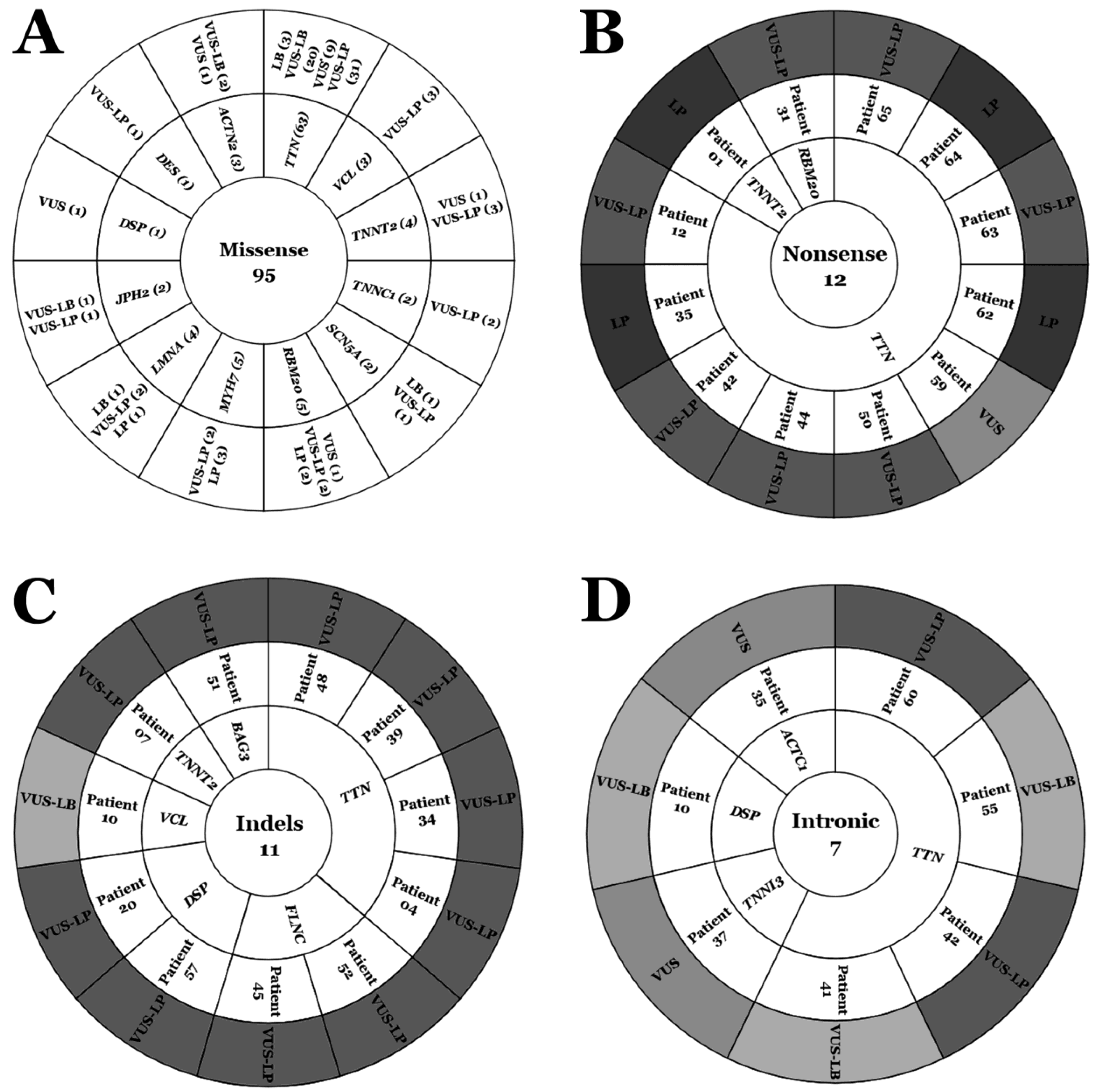
A total of 125 rare variants were located in 16 different genes with a definite association with DCM: one in ACTC1 (0.8%), three in ACTN2 (2.4%), one in BAG3 (0.8%), one in DES (0.8%), four in DSP (3.2%), two in FLNC (1.6%), two in JPH2 (1.6%), four in LMNA (3.2%), five in MYH7 (4%), six in RBM20 (4.8%), two in SCN5A (1.6%), two in TNNC1 (1.6%), one in TNNI3 (0.8%), six in TNNT2 (4.8%), eighty-one in TTN (64.8%), and four in VCL (3.2%). Most of the rare variants were missense (95 variants, 76%), and eleven indels (8.8%) including one gross deletion, seven intronic variants (5.6%), and twelve nonsense variants (9.5%), including one stoploss (Table 1; Figure 1).
Our comprehensive analysis showed that 15 of the 125 rare variants (12%) were reclassified; five variants downgraded their role to LB and ten upgraded their level to LP. All rare variants downgraded the level of pathogenicity were missense (one in LMNA, one in SCN5A, and three in TTN) and modifications were due to increased MAF from the previous to current classification. Concerning the ten rare variants upgraded to LP, four were nonsense (one in TNNT2, three in TTN) and six missenses (one in LMNA, three in MYH7, two in RBM20). The 110 rare variants previously classified as VUS remain currently as VUS. At this point, we developed three subgroups within the VUS category, as mentioned in the Methods section. We observed that 27 (24.54%) variants were reclassified as VUS-LB and 67 (60.9%) as VUS-LP. The remaining 16 (14.54%) rare variants remain as VUSs (Table 1 and Table 2; Figure 1 and Figure 2).
We also performed an analysis by gene (ACTC1, ACTN2, BAG3, DES, DSP, FLNC, JPH2, LMNA, MYH7, RBM20, SCN5A, TNNC1, TNNI3, TNNT2, TTN, and VCL) and type of variant (missense, intronic, indels, and nonsense), as well as each year of previous classification (2016, 2017, 2018, and 2019) compared to current classification. Firstly, concerning years of previous classification, we observed that the further away that year was (2016 in our study), the more variability we found compared to 2023, after applying our algorithm. Additionally, the closer the year of classification to now, the greater the proportion of VUSs showed a tendency toward potential pathogenicity, as well as a lower tendency to play a potential benign role in DCM. Therefore, out of 70 VUS in 2016, five (7.14%) decreased to LB, and seven increased their pathogenicity to LP (10%). Inside the group of the remaining 58 VUSs, 33 seemed to show a tendency for increasing their potential deleterious role (56.89%) and 16 showed a tendency for decreasing their role to benignity (27.58%). In 2017, one of 35 increased its definitive role to LP (2.85). Of the remaining 34 VUSs, 21/34 seemed to show a tendency for the potential deleterious role (61.76%) and 9/34 a tendency for benignity (26.47%). In 2018, from the fourteen VUSs, nine seemed to show a tendency for a potential deleterious role (64.3%) and two a tendency for being not-deleterious (14.3%). In 2019, the last year included in our study, two VUSs showed a tendency toward LP (33.33%). The remaining four VUSs showed a tendency for a potential deleterious role (VUS-LP). Regarding the type of variant, most of those previously classified as VUSs that nowadays showed a tendency to increase their deleterious role to LP were missense (4.8%) and nonsense (3.2%). A group of sixty-seven variants (forty-eight missense, ten indels, seven nonsense and two intronic) showed a tendency to be deleterious (VUS-LP) after applying our algorithm (Table 1 and Table 2; Figure 1 and Figure 2).
Additionally, we performed an exhaustive analysis focused on TTN, the major gene currently associated with DCM. We identified that 3 of 81 rare variants changed from VUS to LB (3.7%). These three variants were missense and were previously classified in 2016. Three more rare variants upgraded to LP, which were all nonsense. After applying our algorithm to the remaining 75 VUSs, 22 (29.33%) showed a tendency to not be deleterious (VUS-LB), 20 of which were missense and 2 intronic, and 43 (57.33%) seemed to have a potential deleterious role despite not being confirmed (VUS-LP), of which thirty-one were missense, six nonsense, four indels, and two intronic. Concerning the proportion of potential pathogenicity, indels and nonsense showed high levels (100% and 90%, respectively), whereas missense and intronic showed medium rates (63.49% and 50%, respectively) (Table 1 and Table 2; Figure 1 and Figure 2).
Finally, we identified ten patients harboring an LP variant (six harboring only an LP variant and four more harboring one LP variant plus at least one VUS-LP). All these deleterious variants were located in LMNA (one variant), MYH7 (three variants), RBM20 (one variant), TNNT2 (one variant), and TTN (six variants) genes. Our algorithm showed that 44 patients carried at least one rare variant classified as VUS-LP. A total of sixty-three variants with a highly potential deleterious role were identified (forty-four missense, eleven indels, seven nonsense, and one intronic). These rare variants with a potential deleterious role were located in the same genes abovementioned, as well as BAG3, DES, DSP, FLNC, JPH2, SCN5A, TNNC1, and VCL (Table 1 and Table 2; Figure 2).

3. Discussion

In the era of precision medicine, conclusive molecular genetic tests inform diagnoses, prognoses, and risk assessments for patients and their relatives in G-DCM [1]. The use of current ACMG recommendations helps to obtain a classification of rare variants [6]. However, an implicit stringent variant adjudication approach and a lack of sufficient scientific data, or available but controversial data, lead to many rare variants remaining categorized as VUSs. This ambiguous role does not provide conclusive data on the cause of disease and cannot be disregarded [5]. Continuous data improvement may modify a previous ambiguous classification, helping to gain certainty and, therefore, playing an actionable clinical role [2]. This fact highlights the importance of periodic reanalysis of VUS, at least in a timeframe from 2 to 5 years, as suggested by our group in previous studies [10,15,17,20]. Therefore, revising and clarifying the roles of VUSs is necessary for obtaining a comprehensive diagnosis [5]. In G-DCM, a sole study focused on variant reevaluation has been published to date, showing a near 30% decrease in ambiguity in VUS classified more than 10 years ago and without following ACMG recommendations [18]. A high percentage of reclassification suggests an immediate update of variants classified not following current ACMG recommendations, as already suggested by our group [10]. Another study showed a reduction in VUSs using analyses including only genes with a definitive-MCD association [19], demonstrating that the prior choice of genes to study is a key point, in our view, before performing a genetic analysis. Our study moved one step forward and designed an easy-to-use algorithm, helping to unravel the tendency of VUSs in G-DCM.
In our study, 12% of VUSs definitively modified their first classification, helping, thus, to clarify their role in G-DCM. This modification has been performed mainly by updating population frequencies; a simple and quick action. We suggest that it should be mandatory at least to update MAF periodically in public databases. We identified that 4% of VUSs decreased their ambiguous roles to benign. In concordance with previous studies, reducing the ambiguity of a VUS is an important advantage of a comprehensive reanalysis because these VUSs can be disregarded as the main cause of disease in each patient [17,20]. The 8% of VUSs increased the pathogenic potential to LP in G-DCM due to the reduction in MAF and, in a few cases, additional functional data since the first classification. All these variants are located in genes with a current definite/strong association with DCM [2]. However, no family segregation data were available in our study, impeding a conclusive translation into families. At this point, it is important to highlight the necessity of performing comprehensive large family correlation analysis to gain certainty. Another main item is functional studies. The use of this basic approach can help to unravel the pathophysiological mechanism of a rare variant, helping to conclude a definite pathogenic role. In recent years, the use of iPSC-derived cardiomyocytes, especially, has allowed researchers to demonstrate the molecular and cellular pathways involved in the pathogenicity of a rare variant. Both kinds of studies are crucial to help with clinical diagnosis, due to the fact that patients harboring a deleterious variant in any cardiomyopathy-associated gene showed a higher risk of mortality and a significantly increased risk of developing cardiomyopathy [7,8].
We identified that most of the rare variants remain classified as VUSs to date, following the current ACMG recommendations. This implies no impact on clinical management [1]. However, a recent study suggests that a VUS in a robust DCM-associated gene is associated with an adverse prognosis [21]. To shed light on the ambiguity implicit in VUSs, subgroups were categorized according to the abovementioned algorithm (please see methods section) already used in other inherited arrhythmogenic diseases [17,20], but now explicit for G-DCM, according to definite gene–disease association, lack of data or available contradictory data, and extremely rare population frequencies. Despite already being published, we suggest the additional use of our algorithm in other centers, and large cohorts in order to corroborate proper VUS subclassification. Hence, of the 110 rare variants that remain as VUSs after reclassification following the ACMG recommendations, 60.90% increase their potential pathogenic role (VUS-LP), 14.54% remain as VUSs, and 24.54% potentially decrease their ambiguous role (VUS-LB). It is important to remark that despite a tendency toward a benign or deleterious role in G-DCM, all these variants remain as VUSs and no clinical translation and adoption of therapeutic measures should be performed [1,2]. Unfortunately, for the VUS analyzed in our study, neither a complete family segregation nor a functional analysis is currently available, impeding a conclusive role following the current ACMG recommendations. It is important to mention a recent study suggesting that a VUS can be associated with an increased risk of cardiac mortality and heart failure hospitalization, especially those VUSs located in definite genes (ACTC1, BAG3, DSP, FLNC, LMNA, MYH7, NEXN, PLN, RBM20, TNNC1, TNNT2, TPM1, TTN, and VCL) [21]. In our study, 94.02% of reclassified VUS-LP variants were located in any of these genes with a robust association with G-DCM, supporting a potential deleterious role and suggesting a close follow-up of patients harboring any of these variants.
The reinterpreted variants were previously classified in different years, from 2016 to 2019, both inclusive. The percentage of reclassified variants was higher in 2016 than in 2019, in concordance with studies published several years ago in the field of reclassification of VUS associated with inherited arrhythmogenic syndromes [10,15,17,20]. This is mainly due to the constant updating of population frequencies, which allows us to have more precise data that brings us closer to being able to classify the variants more precisely, as occurred in previous studies [15,17,20]. Therefore, the number of variants remaining steady as the number of VUSs decreases constantly in comparison to previous years and, for this reason, the percentage of variants reclassified in 2019 was lower than in 2016, at least in our study, which is also in concordance with previous studies of our group in other inherited arrhythmogenic syndromes [15,17,20]. In this way, while being focused on updating population frequencies, the algorithm we propose also allows us to observe that VUSs with a tendency to be deleterious are increasingly classified today, while more variants with a tendency to have a genetic background without a main causative role can be ruled out. In G-DCM, we observed this pattern with a smaller number of VUSs each year, as we move closer to the present. In addition, and following specific algorithms for unravelling the role of VUSs in each disease, their classification is more precise, with a tendency to be deleterious, as reported in other inherited arrhythmogenic diseases [15,17,20]. It is important to mention that obtaining precise classifications occurs mainly due to updates in population frequencies, without including functional studies or genotype-phenotype correlations of large families for more than three generations. Despite these limitations, as already mentioned, the inclusion of these additional studies is necessary to be able to conclude a definitive pathogenic role in these rare variants in G-DCM [1,5].
Finally, it is important to remark that most of deleterious or potentially deleterious variants were identified in the TTN gene, the main gene associated with G-DCM in adult and pediatric population. Re-evaluation was also higher in TNNT2, RBM20, MYH7, and LMNA, in concordance with reports related to aggressive phenotypes, especially in the pediatric population [22,23]. Therefore, if a variant is re-analyzed in a pediatric patient, it should be also taken into account. Focused on TTN, the major gene currently associated with DCM [1,2], despite this ratio being based mainly on radical variants (nonsense, indels…) [24]. Most of the reported VUSs in the TTN gene are missense [25], as observed in our study. The role of TTN missense variants in G-DCM has been difficult to elucidate because of a lack of data concerning this gene, which encodes the largest protein in the human body and is associated with several skeletal muscular diseases [26]. This lack of data impedes a conclusive classification of most rare variants, clarifying a definite role of only three missense variants to LB and three nonsense variants to LP. After applying our specific algorithm for G-DCM, and updating only population frequencies, practically all nonsense and indel variants increased their potential deleterious role to VUS-LP in contrast to nearly 50% of intronic and missense VUSs. We moves one step forward in the prediction of missense VUS in TTN, contrary to a study suggesting that TTN missense variants should be classified as likely benign due to lack of data [27]. Our approach suggests a tendency for a potential deleterious role, but it is necessary to include functional and large family segregation studies to clarify the role of missense variants in TTN, according to a recent study [28]. For this reason, we suggest that the adoption of measures based on missense variants in TTN should be undertaken with caution and in a personalized way.
There are some limitations to mention. The reclassification and reinterpretation of rare genetic variants in our cohort have important limitations. Firstly, all cases included in our cohort may carry additional rare variants in other genes not included in our panel. However, we have analyzed all genes currently associated with G-DCM. In addition, a lack of available functional data for most rare variants impedes a more accurate interpretation. In this same way, a lack of family segregation, which is also crucial to clarify the role of a rare variant, in our view, is a key point to finally conclude the potential deleterious role of a rare variant. To support our results, reclassifications/reinterpretations of all variants should be corroborated in additional larger cohorts of G-DCM patients, to clarify their potential actionable role in clinical practice. At this point, it is important to take into account the ethnicity of the patient, due to genetic differences from diverse ancestries, to guide testing interpretation [29]. Finally, who should assume to perform the reclassification/reinterpretation and the implicit economic cost are controversial points not assessed in our study.

4. Materials and Methods

4.1. Study Cohort

Our retrospective study included 65 patients (all adults, >18 years old; mean age: 43.2 years old; 61.23% men; all Caucasian) with a definitive clinical diagnosis of DCM, determined following the available clinical guidelines [1,30,31]. Suspected cases with an inconclusive diagnosis were not included, to avoid bias in the reclassification of genetic variants.
All patients were genetically analyzed (please see Section 4.2). All patients included in our study carried at least one rare variant classified as a VUS, according to the ACMG recommendations [6].

4.2. Genetic Analysis

Genetic analysis was approved by the Ethics Committee of Hospital Josep Trueta (Girona, Catalonia, Spain), following the World Medical Association Declaration of Helsinki. Clinical and genetic data on all the patients were anonymized and kept confidential. Written informed consent was obtained from all the patients before genetic analysis.
A resequencing custom-made panel included all the main genes associated with G-DCM [2,5] (ACTC1, ACTN2, BAG3, DES, DSP, FLNC, JPH2, LMNA, MYH7, NEXN, PLN, RBM20, SCN5A, TNNC1, TNNI3, TNNT2, TPM1, TTN, and VCL). Isoforms of all the genes are described in Ensembl 75 (www.ensembl.org/), linked to a RefSeq code (www.ncbi.nlm.nih.gov/refseq/) or CCDS (www.ncbi.nlm.nih.gov/CCDS/). The bioinformatic analysis included adaptor and low-quality base trimming of the FASTQ files. GEM III was used for map-trimmed reads. The output was sorted, and uniquely and properly mapped read pairs were selected. SAMtools v.1.2 was used for variant calling from the cleaned BAM files. The final annotation steps provided information included in public databases. Rare genetic variants (with a minor allele frequency—MAF—of <1%) were confirmed by Sanger sequencing. The exons and exon–intron boundaries of each gene were amplified in both directions.
The SeqScape v2.7 software (Applied Biosystems, Waltham, MA, USA) was used to reanalyze all sequences obtained. No new rare variants were identified in any of the analyzed genes. The original classification compared rare variants with DNA sequences from 350 healthy Spanish individuals (individuals not related to any index case and of the same ethnicity) as control cases. Sequence variants were described following the Human Genome Variation Society (HGVS) rules (www.hgvs.org/). Currently, all rare variants in any of the genes analyzed were contrasted in the Genome Aggregation Database (gnomAD) (https://gnomad.broadinstitute.org/). All rare variants were also consulted in ClinVar (www.ncbi.nlm.nih.gov/clinvar/intro/), VarSome (www.varsome.com/), CardioClassifier (www.cardioclassifier.org/), InterVar (https://wintervar.wglab.org/), and CardioVAI (https://cardiovai.engenome.com/).

4.3. Data Sources

An exhaustive review of the available data on each rare variant was performed independently by five authors; subsequently, the reviews were compared and verified. Data were collected from the Human Gene Mutation Database (HGMD) (www.hgmd.org), ClinVar (https://www.ncbi.nlm.nih.gov/clinvar/intro/), the National Center for Biotechnology Information single-nucleotide polymorphism (SNP) database (https://www.ncbi.nlm.nih.gov/snp), Google Scholar (https://www.scholar.google.com), Springer Link (https://www.springer.com), and Science Direct (www.sciencedirect.com).

4.4. Classification/Interpretation

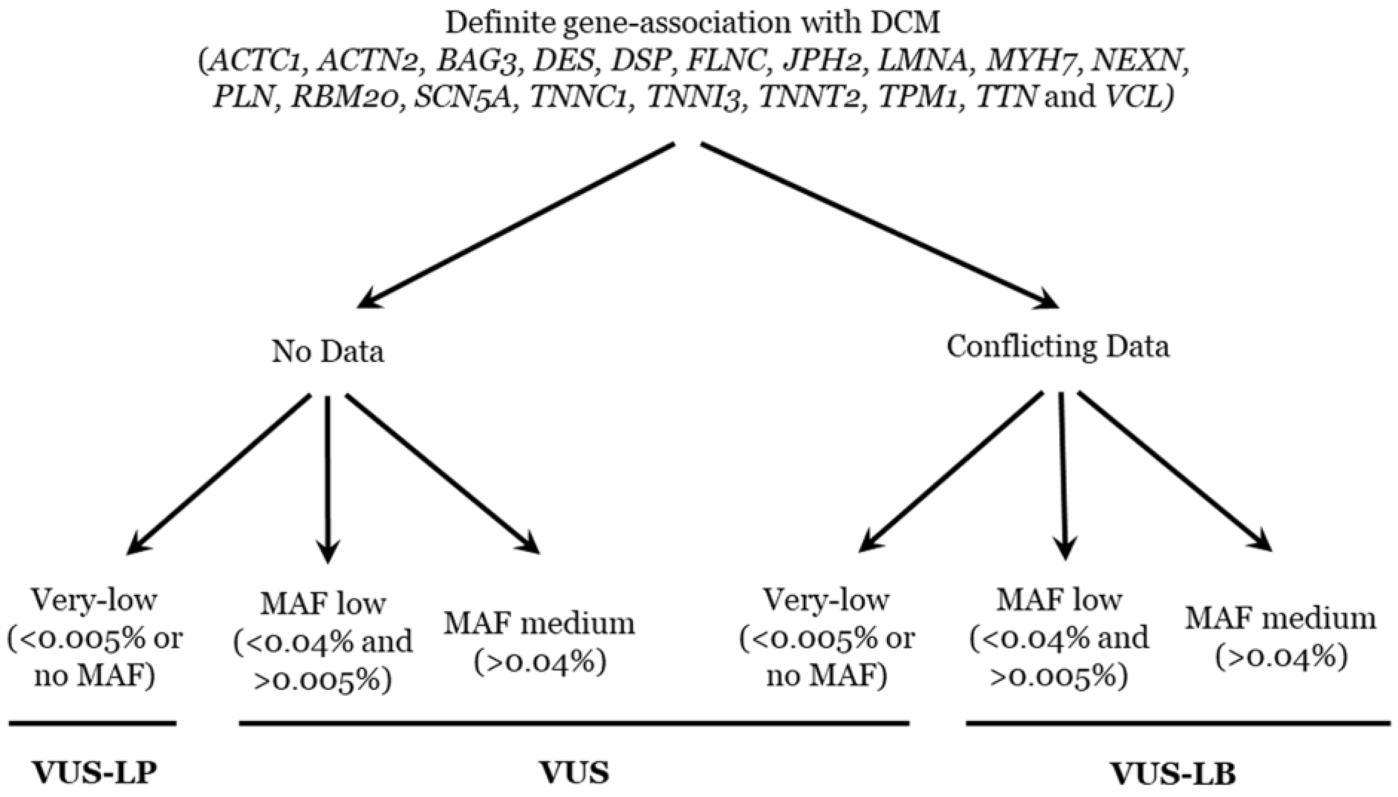
From 2016 to 2019, all rare variants in genes associated with DCM were classified according to the ACMG recommendations [6]. Now, the same rare variants have also been reclassified and the same ACMG recommendations also followed, but updates are included: the PM2 item was considered fulfilled if the MAF in the relevant population databases was ≤0.01% [32]. In addition, all reported deleterious variants currently classified as definitively pathogenic in IASs following ACMG recommendations are very rare (MAF < 0.005%) [33]. Other recent updates, according to gene-specific [34] and disease-specific associations, have also been taken into account [35,36]. The final classification of a rare variant as a VUS may be due to a lack of data or incongruences in the available data [37,38]. The current prevalence for familial DCM is approximately 1/2500 (MAF: 0.04%). Taking into account these two items, we propose a subclassification of VUSs into three subgroups: VUS-LB, VUS-LP, or remaining VUS (please see Figure 3). The first point is a definite gene-association with DCM (ACTC1, ACTN2, BAG3, DES, DSP, FLNC, JPH2, LMNA, MYH7, NEXN, PLN, RBM20, SCN5A, TNNC1, TNNI3, TNNT2, TPM1, TTN, and VCL) [2]. Then, the existence or not of data (if available, contradictory data). Finally, the use of the MAF (prevalence of familial DCM is the threshold), being very low (<0.005% or no MAF), low (>0.005% and <0.04%), or medium (>0.04%). We have already implemented this approach in previously published manuscripts focused on other IAS [16,20,21]. Genetic data were independently evaluated and classified by five authors, who are specialists in the genetics of inherited arrhythmias, to avoid bias. All investigators agreed on the final classification of all the rare variants included in our study.

5. Conclusions

In summary, a definite genetic diagnosis of rare variants related to G-DCM has actionable consequences for patients and their relatives. This emphasizes the importance of cautious genetic interpretation of variants and highlights the necessity of periodic reclassification/reinterpretation if a variant remains as a VUS, at least once every five years. Part of VUS clarifies its function after an update of population frequencies; a simple, cheap, and fast action. Due to a lack of functional studies as well as comprehensive clinical studies of large familial cases, most rare variants remain as VUSs in G-DCM following the stringent criteria suggested in ACMG recommendations. Identifying a deleterious or benevolent tendency of a VUS help with follow-up patients and their relatives despite no clinically actionable, so far. We propose a specific algorithm to help the interpretation of VUSs in G-DCM.

Author Contributions

O.C., G.S.-B., J.B. and R.B. developed the concept. A.P.-S., E.M.-B., O.C., A.F.-F., M.C., A.I., S.C., E.A., M.P., M.A., B.d.O., L.L., F.P., C.T.d.L., A.M., R.T. and G.S.-B. acquired, pre-processed, and analyzed the data. A.P.-S., O.C., R.T. and G.S.-B. prepared the manuscript. O.C., R.T. and R.B. supervised the study. All authors have read and agreed to the published version of the manuscript.

Funding

This work was supported by Obra Social “La Caixa Foundation” (LCF/PR/GN16/50290001 and LCF/PR/GN19/50320002). Co-funded by Instituto de Salud Carlos III (FIS PI16/01203 and FIS PI21/00094) and by ERDF/ESF, “Investing in Your Future” and Fundació La Marató TV3 (245/U/2020). CIBERCV is an initiative of the ISCIII, Spanish Ministry of Economy and Competitiveness. IDIBGI is a “CERCA Programme/Generalitat de Catalunya”. The funders played no role in study design, data collection, data analysis, interpretation, or writing of the report.

Institutional Review Board Statement

The analysis was approved by the ethics committee of Hospital Josep Trueta (Girona, Catalonia, Spain, Approval Code: CEiM2012097) according to the World Medical Association Declaration of Helsinki. Written informed consent was obtained from all patients before genetic analysis.

Informed Consent Statement

Not applicable.

Data Availability Statement

All data are included in the manuscript.

Conflicts of Interest

The authors declare that they have no competing interests.

References

  1. Arbelo, E.; Protonotarios, A.; Gimeno, J.R.; Arbustini, E.; Barriales-Villa, R.; Basso, C.; Bezzina, C.R.; Biagini, E.; A Blom, N.; A de Boer, R.; et al. 2023 ESC Guidelines for the management of cardiomyopathies. Eur. Heart J. 2023, 44, 3503–3626. [Google Scholar] [CrossRef] [PubMed]
  2. Wilde, A.A.M.; Semsarian, C.; Marquez, M.F.; Shamloo, A.S.; Ackerman, M.J.; Ashley, E.A.; Sternick, E.B.; Barajas-Martinez, H.; Behr, E.R.; Bezzina, C.R.; et al. European Heart Rhythm Association (EHRA)/Heart Rhythm Society (HRS)/Asia Pacific Heart Rhythm Society (APHRS)/Latin American Heart Rhythm Society (LAHRS) Expert Consensus Statement on the State of Genetic Testing for Cardiac Diseases. Europace 2022, 24, 1307–1367. [Google Scholar] [CrossRef]
  3. Muller, R.D.; McDonald, T.; Pope, K.; Cragun, D. Evaluation of Clinical Practices Related to Variants of Uncertain Significance Results in Inherited Cardiac Arrhythmia and Inherited Cardiomyopathy Genes. Circ. Genom. Precis. Med. 2020, 13, e002789. [Google Scholar] [CrossRef] [PubMed]
  4. Jordan, E.; Peterson, L.; Ai, T.; Asatryan, B.; Bronicki, L.; Brown, E.; Celeghin, R.; Edwards, M.; Fan, J.; Ingles, J.; et al. An Evidence-Based Assessment of Genes in Dilated Cardiomyopathy. Circulation 2021, 144, 7–19. [Google Scholar] [CrossRef]
  5. Landstrom, A.P.; Chahal, A.A.; Ackerman, M.J.; Cresci, S.; Milewicz, D.M.; Morris, A.A.; Sarquella-Brugada, G.; Semsarian, C.; Shah, S.H.; Sturm, A.C.; et al. Interpreting Incidentally Identified Variants in Genes Associated with Heritable Cardiovascular Disease: A Scientific Statement from the American Heart Association. Circ. Genom. Precis. Med. 2023, 16, e000092. [Google Scholar] [CrossRef] [PubMed]
  6. Richards, S.; Aziz, N.; Bale, S.; Bick, D.; Das, S.; Gastier-Foster, J.; Grody, W.W.; Hegde, M.; Lyon, E.; Spector, E.; et al. Standards and guidelines for the interpretation of sequence variants: A joint consensus recommendation of the American College of Medical Genetics and Genomics and the Association for Molecular Pathology. Genet. Med. 2015, 17, 405–424. [Google Scholar] [CrossRef]
  7. Asatryan, B.; Shah, R.A.; Sharaf Dabbagh, G.; Landstrom, A.P.; Darbar, D.; Khanji, M.Y.; Lopes, L.R.; van Duijvenboden, S.; Muser, D.; Lee, A.M.; et al. Predicted Deleterious Variants in Cardiomyopathy Genes Prognosticate Mortality and Composite Outcomes in UK Biobank. JACC Heart Fail. 2023. ahead of print. [Google Scholar] [CrossRef]
  8. Hofmeyer, M.; Haas, G.J.; Jordan, E.; Cao, J.; Kransdorf, E.; Ewald, G.A.; Morris, A.A.; Owens, A.; Lowes, B.; Stoller, D.; et al. Rare Variant Genetics and Dilated Cardiomyopathy Severity: The DCM Precision Medicine Study. Circulation 2023, 148, 872–881. [Google Scholar] [CrossRef]
  9. Stroeks, S.; Verdonschot, J.A.J. The next step toward personalized recommendations for genetic cardiomyopathies. Eur. J. Hum. Genet. 2023, 31, 1201–1203. [Google Scholar] [CrossRef]
  10. Campuzano, O.; Sarquella-Brugada, G.; Fernandez-Falgueras, A.; Coll, M.; Iglesias, A.; Ferrer-Costa, C.; Cesar, S.; Arbelo, E.; García-Álvarez, A.; Jordà, P.; et al. Reanalysis and reclassification of rare genetic variants associated with inherited arrhythmogenic syndromes. EBioMedicine 2020, 54, 102732. [Google Scholar] [CrossRef]
  11. Towbin, J.A.; McKenna, W.J.; Abrams, D.J.; Ackerman, M.J.; Calkins, H.; Darrieux, F.C.C.; Daubert, J.P.; de Chillou, C.; DePasquale, E.C.; Desai, M.Y.; et al. 2019 HRS expert consensus statement on evaluation, risk stratification, and management of arrhythmogenic cardiomyopathy. Heart Rhythm 2019, 16, e301–e372. [Google Scholar] [CrossRef] [PubMed]
  12. Hershberger, R.E.; Givertz, M.M.; Ho, C.Y.; Judge, D.P.; Kantor, P.F.; McBride, K.L.; Morales, A.; Taylor, M.R.G.; Vatta, M.; Ware, S.M.; et al. Genetic evaluation of cardiomyopathy: A clinical practice resource of the American College of Medical Genetics and Genomics (ACMG). Genet. Med. 2018, 20, 899–909. [Google Scholar] [CrossRef] [PubMed]
  13. Musunuru, K.; Hershberger, R.E.; Day, S.M.; Klinedinst, N.J.; Landstrom, A.P.; Parikh, V.N.; Prakash, S.; Semsarian, C.; Sturm, A.C.; on behalf of the American Heart Association Council on Genomic and Precision Medicine; et al. Genetic Testing for Inherited Cardiovascular Diseases: A Scientific Statement from the American Heart Association. Circ. Genom. Precis. Med. 2020, 13, e000067. [Google Scholar] [CrossRef] [PubMed]
  14. Stiles, M.K.; Wilde, A.A.; Abrams, D.J.; Ackerman, M.J.; Albert, C.M.; Behr, E.R.; Chugh, S.S.; Cornel, M.C.; Gardner, K.; Ingles, J.; et al. 2020 APHRS/HRS expert consensus statement on the investigation of decedents with sudden unexplained death and patients with sudden cardiac arrest, and of their families. Heart Rhythm 2021, 18, e1–e50. [Google Scholar] [CrossRef] [PubMed]
  15. Sarquella-Brugada, G.; Fernandez-Falgueras, A.; Cesar, S.; Arbelo, E.; Coll, M.; Perez-Serra, A.; Puigmulé, M.; Iglesias, A.; Alcalde, M.; Vallverdú-Prats, M.; et al. Clinical impact of rare variants associated with inherited channelopathies: A 5-year update. Hum. Genet. 2022, 141, 1579–1589. [Google Scholar] [CrossRef] [PubMed]
  16. Bennett, J.S.; Bernhardt, M.; McBride, K.L.; Reshmi, S.C.; Zmuda, E.; Kertesz, N.J.; Garg, V.; Fitzgerald-Butt, S.; Kamp, A.N. Reclassification of Variants of Uncertain Significance in Children with Inherited Arrhythmia Syndromes is Predicted by Clinical Factors. Pediatr. Cardiol. 2019, 40, 1679–1687. [Google Scholar] [CrossRef] [PubMed]
  17. Vallverdú-Prats, M.; Alcalde, M.; Sarquella-Brugada, G.; Cesar, S.; Arbelo, E.; Fernandez-Falgueras, A.; Coll, M.; Pérez-Serra, A.; Puigmulé, M.; Iglesias, A.; et al. Rare Variants Associated with Arrhythmogenic Cardiomyopathy: Reclassification Five Years Later. J. Pers. Med. 2021, 11, 162. [Google Scholar] [CrossRef] [PubMed]
  18. Quiat, D.; Witkowski, L.; Zouk, H.; Daly, K.P.; Roberts, A.E. Retrospective Analysis of Clinical Genetic Testing in Pediatric Primary Dilated Cardiomyopathy: Testing Outcomes and the Effects of Variant Reclassification. J. Am. Heart Assoc. 2020, 9, e016195. [Google Scholar] [CrossRef]
  19. Stroeks, S.; Hellebrekers, D.; Claes, G.R.F.; Tayal, U.; Krapels, I.P.C.; Vanhoutte, E.K.; van den Wijngaard, A.; Henkens, M.T.H.M.; Ware, J.S.; Heymans, S.R.B.; et al. Clinical impact of re-evaluating genes and variants implicated in dilated cardiomyopathy. Genet. Med. 2021, 23, 2186–2193. [Google Scholar] [CrossRef]
  20. Martínez-Barrios, E.; Sarquella-Brugada, G.; Pérez-Serra, A.; Fernández-Falgueras, A.; Cesar, S.; Coll, M.; Puigmulé, M.; Iglesias, A.; Alcalde, M.; Vallverdú-Prats, M.; et al. Discerning the Ambiguous Role of Missense TTN Variants in Inherited Arrhythmogenic Syndromes. J. Pers. Med. 2022, 12, 241. [Google Scholar] [CrossRef]
  21. Martinez-Barrios, E.; Sarquella-Brugada, G.; Perez-Serra, A.; Fernandez-Falgueras, A.; Cesar, S.; Alcalde, M.; Coll, M.; Puigmulé, M.; Iglesias, A.; Ferrer-Costa, C.; et al. Reevaluation of ambiguous genetic variants in sudden unexplained deaths of a young cohort. Int. J. Leg. Med. 2023, 137, 345–351. [Google Scholar] [CrossRef] [PubMed]
  22. Ware, S.M.; Bhatnagar, S.; Dexheimer, P.J.; Wilkinson, J.D.; Sridhar, A.; Fan, X.; Shen, Y.; Tariq, M.; Schubert, J.A.; Colan, S.D.; et al. The genetic architecture of pediatric cardiomyopathy. Am. J. Hum. Genet. 2022, 109, 282–298. [Google Scholar] [CrossRef]
  23. Stroeks, S.L.V.M.; Hellebrekers, D.; Claes, G.R.F.; Krapels, I.P.C.; Henkens, M.H.T.M.; Sikking, M.; Vanhoutte, E.K.; Enden, A.H.-V.D.; Brunner, H.G.; Wijngaard, A.v.D.; et al. Diagnostic and prognostic relevance of using large gene panels in the genetic testing of patients with dilated cardiomyopathy. Eur. J. Hum. Genet. 2023, 31, 776–783. [Google Scholar] [CrossRef] [PubMed]
  24. McAfee, Q.; Chen, C.Y.; Yang, Y.; Caporizzo, M.A.; Morley, M.; Babu, A.; Jeong, S.; Brandimarto, J.; BediJr, K.C.; Flam, E.; et al. Truncated titin proteins in dilated cardiomyopathy. Sci. Transl. Med. 2021, 13, eabd7287. [Google Scholar] [CrossRef]
  25. Begay, R.L.; Graw, S.; Sinagra, G.; Merlo, M.; Slavov, D.; Gowan, K.; Jones, K.L.; Barbati, G.; Spezzacatene, A.; Brun, F.; et al. Role of Titin Missense Variants in Dilated Cardiomyopathy. J. Am. Heart Assoc. 2015, 4, e002645. [Google Scholar] [CrossRef] [PubMed]
  26. Rich, K.A.; Moscarello, T.; Siskind, C.; Brock, G.; Tan, C.A.; Vatta, M.; Winder, T.L.; Elsheikh, B.; Vicini, L.; Tucker, B.; et al. Novel heterozygous truncating titin variants affecting the A-band are associated with cardiomyopathy and myopathy/muscular dystrophy. Mol. Genet. Genom. Med. 2020, 8, e1460. [Google Scholar] [CrossRef] [PubMed]
  27. Akinrinade, O.; Heliö, T.; Deprez, R.H.L.; Jongbloed, J.D.H.; Boven, L.G.; Berg, M.P.v.D.; Pinto, Y.M.; Alastalo, T.-P.; Myllykangas, S.; van Spaendonck-Zwarts, K.; et al. Relevance of Titin Missense and Non-Frameshifting Insertions/Deletions Variants in Dilated Cardiomyopathy. Sci. Rep. 2019, 9, 4093. [Google Scholar] [CrossRef] [PubMed]
  28. Domínguez, F.; Lalaguna, L.; Martínez-Martín, I.; Piqueras-Flores, J.; Rasmussen, T.B.; Zorio, E.; Giovinazzo, G.; Prados, B.; Ochoa, J.P.; Bornstein, B.; et al. Titin Missense Variants as a Cause of Familial Dilated Cardiomyopathy. Circulation 2023, 147, 1711–1713. [Google Scholar] [CrossRef] [PubMed]
  29. Rosamilia, M.B.; Markunas, A.M.; Kishnani, P.S.; Landstrom, A.P. Underrepresentation of Diverse Ancestries Drives Uncertainty in Genetic Variants Found in Cardiomyopathy-Associated Genes. JACC 2024, 3, 100767. [Google Scholar] [CrossRef]
  30. Priori, S.G.; Blomstrom-Lundqvist, C. 2015 European Society of Cardiology Guidelines for the management of patients with ventricular arrhythmias and the prevention of sudden cardiac death summarized by co-chairs. Eur. Heart J. 2015, 36, 2757–2759. [Google Scholar] [CrossRef]
  31. Al-Khatib, S.M.; Stevenson, W.G.; Ackerman, M.J.; Bryant, W.J.; Callans, D.J.; Curtis, A.B.; Deal, B.J.; Dickfeld, T.; Field, M.E.; Fonarow, G.C.; et al. 2017 AHA/ACC/HRS guideline for management of patients with ventricular arrhythmias and the prevention of sudden cardiac death: Executive summary: A Report of the American College of Cardiology/American Heart Association Task Force on Clinical Practice Guidelines and the Heart Rhythm Society. Heart Rhythm 2018, 15, e190–e252. [Google Scholar] [PubMed]
  32. Kobayashi, Y.; Yang, S.; Nykamp, K.; Garcia, J.; Lincoln, S.E.; Topper, S.E. Pathogenic variant burden in the ExAC database: An empirical approach to evaluating population data for clinical variant interpretation. Genome Med. 2017, 9, 13. [Google Scholar] [CrossRef] [PubMed]
  33. Jordan, D.M.; Kiezun, A.; Baxter, S.M.; Agarwala, V.; Green, R.C.; Murray, M.F.; Pugh, T.; Lebo, M.S.; Rehm, H.L.; Funke, B.H.; et al. Development and validation of a computational method for assessment of missense variants in hypertrophic cardiomyopathy. Am. J. Hum. Genet. 2011, 88, 183–192. [Google Scholar] [CrossRef] [PubMed]
  34. Kelly, M.A.; Caleshu, C.; Morales, A.; Buchan, J.; Wolf, Z.; Harrison, S.M.; Cook, S.; Dillon, M.W.; Garcia, J.; Haverfield, E.; et al. Adaptation and validation of the ACMG/AMP variant classification framework for MYH7-associated inherited cardiomyopathies: Recommendations by ClinGen’s Inherited Cardiomyopathy Expert Panel. Genet. Med. 2018, 20, 351–359. [Google Scholar] [CrossRef] [PubMed]
  35. Bains, S.; Dotzler, S.M.; Krijger, C.; Giudicessi, J.R.; Ye, D.; Bikker, H.; Rohatgi, R.K.; Tester, D.J.; Bos, J.M.; Wilde, A.A.; et al. A phenotype-enhanced variant classification framework to decrease the burden of missense variants of uncertain significance in type 1 long QT syndrome. Heart Rhythm 2022, 19, 435–442. [Google Scholar] [CrossRef] [PubMed]
  36. Giudicessi, J.R.; Lieve, K.V.V.; Rohatgi, R.K.; Koca, F.; Tester, D.J.; van der Werf, C.; Martijn Bos, J.; Wilde, A.A.M.; Ackerman, M.J. Assessment and Validation of a Phenotype-Enhanced Variant Classification Framework to Promote or Demote RYR2 Missense Variants of Uncertain Significance. Circ. Genom. Precis. Med. 2019, 12, e002510. [Google Scholar] [CrossRef] [PubMed]
  37. Arbustini, E.; Behr, E.R.; Carrier, L.; van Duijn, C.; Evans, P.; Favalli, V.; van der Harst, P.; Haugaa, K.H.; Jondeau, G.; Kääb, S.; et al. Interpretation and actionability of genetic variants in cardiomyopathies: A position statement from the European Society of Cardiology Council on cardiovascular genomics. Eur. Heart J. 2022, 43, 1901–1916. [Google Scholar] [CrossRef]
  38. Arbustini, E.; Urtis, M.; Elliott, P. Interpretation of genetic variants depends on a clinically guided integration of phenotype and molecular data. Eur. Heart J. 2022, 43, 2638–2639. [Google Scholar] [CrossRef]
Figure 1. Global number/percentage of VUSs. (A) Global reclassification of variants. (B) Reclassification each year. LB: likely benign. VUS: variant of unknown significance. VUS-LP: variant of unknown significance—likely pathogenic. VUS-LB: variant of unknown significance—likely benign.
Ijms 25 03807 g001
Figure 2. Number, type, and classification of variants. (A) Missense variants. (B) Nonsense variants. (C) Indel variants. (D) Intronic variants. LB: likely benign. VUS: variant of unknown significance. VUS-LP: variant of unknown significance—likely pathogenic. VUS-LB: variant of unknown significance—likely benign.
Ijms 25 03807 g002
Figure 3. Algorithm of classification for VUS. MAF: minor allele frequency. VUS: variant of unknown significance. VUS-LP: variant of unknown significance—likely pathogenic. VUS-LB: variant of unknown significance—likely benign.
Ijms 25 03807 g003
Table 1. Genetic data of variants.
PatientGeneNucleotideProteindbSNPClinVarGnomAD (MAF%)Classification (Year)Classification 2023
1TNNT2c.860G>Ap.Trp287Terrs727504247LPNAVUS (2016)LP
1TTNc.47951G>Ap.Arg15984Hisrs201774108VUSc0.0001VUS (2016)VUS
2TTNc.73195G>Ap.Val24399Ilers1257567608NANAVUS (2016)VUS (VUS-LP)
2TTNc.57388C>Tp.Arg19130Cysrs72646861LB0.827VUS (2016)LB
2TTNc.53117C>Tp.Pro17706Leurs72646845LB0.369VUS (2016)LB
3ACTN2c.2051A>Tp.Asn684Ilers576783493VUSc0.0001VUS (2016)VUS
3TTNc.93125G>Ap.Gly31042Asprs373754986VUS0.007VUS (2016)VUS (VUS-LP)
3TTNc.76456G>Cp.Asp25486Hisrs780958039VUS0.0008VUS (2016)VUS (VUS-LP)
4ACTN2c.1426G>Ap.Ala476Thrrs142943120VUSc0.027VUS (2016)VUS (VUS-LB)
4TTNc.57978delp.Val19327PhefsTer10NANANAVUS (2016)VUS (VUS-LP)
4TTNc.76559G>Ap.Ser25520Asnrs200450022VUSc0.061VUS (2016)VUS (VUS-LB)
4TTNc.72764G>Cp.Gly24225Alars114071241VUS0.001VUS (2016)VUS (VUS-LP)
4TTNc.17066G>Cp.Gly5689Alars200118743VUSc0.063VUS (2016)VUS (VUS-LB)
4TTNc.4675G>Ap.Val1559Ilers538451328NA0.0003VUS (2016)VUS (VUS-LP)
5TTNc.98971G>Cp.Glu32991Glnrs199632397VUSc0.042VUS (2016)VUS (VUS-LB)
5TTNc.27659G>Ap.Arg9220Glnrs727504757VUS0.003VUS (2016)VUS (VUS-LP)
6TTNc.45392G>Ap.Arg15131Hisrs72646808VUSc0.185VUS (2016)LB
7TNNT2c.629_631delp.Lys210delrs45578238VUSNAVUS (2016)VUS (VUS-LP)
8MYH7c.1106G>Ap.Arg369Glnrs397516089LP0.00006VUS (2016)LP
9TTNc.73967A>Gp.Asn24656Serrs368443217VUSc0.008VUS (2016)VUS (VUS-LB)
9TTNc.65012T>Ap.Met21671Lysrs750298083VUSc0.004VUS (2016)VUS
9TTNc.51830G>Ap.Arg17277Hisrs201457934VUSc0.01VUS (2016)VUS (VUS-LB)
9TTNc.9247G>Ap.Glu3083LysNANANAVUS (2016)VUS (VUS-LP)
9MYH7c.2711G>Ap.Arg904Hisrs397516165LP0.0001VUS (2016)LP
10DSPc.1266+6G>TNArs73375345LB0.037VUS (2016)VUS (VUS-LB)
10TTNc.20920A>Gp.Ser6974Glyrs72648980VUSc0.062VUS (2016)VUS (VUS-LB)
10VCLc.2862_2864delp.Leu955delrs397517237VUSc0.021VUS (2016)VUS (VUS-LB)
11SCN5Ac.6010T>Cp.Phe2004Leurs41311117VUSc0.198VUS (2016)LB
12TTNc.72970C>Tp.Gln24324TerNANANAVUS (2016)VUS (VUS-LP)
13TTNc.49249G>Ap.Asp16417Asnrs1244503464NA0.0004VUS (2016)VUS (VUS-LP)
14TTNc.12814G>Tp.Asp4272Tyrrs72648940VUSc0.0005VUS (2016)VUS
15TTNc.77076A>Cp.Glu25692Asprs370547473NA0.004VUS (2016)VUS (VUS-LP)
16TTNc.32932C>Ap.Pro10978Thrrs1393076582VUScNAVUS (2016)VUS
17DESc.179C>Tp.Ser60Leurs868853251NA0.0001VUS (2016)VUS (VUS-LP)
17TTNc.46877G>Ap.Gly15626Asprs201802447VUS0.007VUS (2016)VUS
17TTNc.1066G>Cp.Glu356Glnrs144531477VUSc0.015VUS (2016)VUS (VUS-LB)
18TTNc.95137A>Gp.Ile31713Valrs758945559NA0.0008VUS (2016)VUS (VUS-LP)
18TTNc.30484C>Ap.Pro10162Thrrs532102837VUSc0.058VUS (2016)VUS (VUS-LB)
18DSPc.6799A>Tp.Thr2267Serrs181378432LB0.009VUS (2016)VUS
18LMNAc.1621C>Tp.Arg541Cysrs56984562LP0.0001VUS (2016)LP
19RBM20c.1900C>Tp.Arg634Trprs796734066LPNAVUS (2016)LP
20DSPdel ex. 21_24NANANANAVUS (2016)VUS (VUS-LP)
21MYH7c.1371A>Gp.Ile457MetNANANAVUS (2016)VUS (VUS-LP)
22TTNc.94724T>Cp.Met31575Thrrs397517786VUSc0.007VUS (2016)VUS (VUS-LB)
22TTNc.62687G>Tp.Gly20896Valrs549938348VUS0.0004VUS (2016)VUS (VUS-LP)
23RBM20c.1906C>Tp.Arg636Cysrs267607002VUSc0.0001VUS (2016)VUS
24JPH2c.1736C>Tp.Pro579Leurs953353202VUS0.001VUS (2016)VUS (VUS-LP)
24RBM20c.1980C>Ap.Ser660ArgNANA0.00007VUS (2016)VUS (VUS-LP)
25RBM20c.2200C>Gp.Arg734GlyNANANAVUS (2016)VUS (VUS-LP)
26TTNc.57212G>Ap.Arg19071Glnrs373282633VUS0.003VUS (2016)VUS
26TTNc.48221T>Ap.Leu16074Glnrs140714512VUSc0.051VUS (2016)VUS (VUS-LB)
26TTNc.29645A>Cp.Lys9882Thrrs760742068VUS0.001VUS (2016)VUS (VUS-LP)
27LMNAc.1930C>Tp.Arg644Cysrs1420000963VUSc0.201VUS (2016)LB
27TTNc.77188C>Tp.Arg25730Trprs779581886VUS0.002VUS (2016)VUS (VUS-LP)
27TTNc.60581T>Cp.Leu20194Prors1359881893VUS0.0008VUS (2016)VUS (VUS-LP)
28RBM20c.1913C>Tp.Pro638Leurs267607003LP0.0003VUS (2016)LP
28TTNc.74942C>Tp.Ala24981Valrs749950083NANAVUS (2016)VUS (VUS-LP)
28TTNc.73501C>Gp.Pro24501Alars770542451NA0.0004VUS (2016)VUS (VUS-LP)
28TTNc.1333G>Ap.Ala445Thrrs142414432VUSc0.021VUS (2016)VUS (VUS-LB)
29LMNAc.1949A>Gp.Asn650Serrs775728847VUS0.0003VUS (2016)VUS (VUS-LP)
29TTNc.38807A>Gp.Asn12936Serrs1184631064VUS0.0008VUS (2016)VUS (VUS-LP)
30ACTN2c.1040C>Tp.Thr347Metrs727504590VUSc0.009VUS (2016)VUS (VUS-LB)
31RBM20c.3684A>Gp.Ter1228TrpextTer33rs1845123103NA0.0001VUS (2016)VUS (VUS-LP)
31TTNc.40001G>Ap.Gly13334Glurs561284948VUS0.001VUS (2016)VUS (VUS-LP)
31TTNc.20863C>Tp.Pro6955Serrs1438804317NANAVUS (2016)VUS (VUS-LP)
32MYH7c.602T>Cp.Ile201Thrrs397516258LP0.0001VUS (2016)LP
32TTNc.71188G>Ap.Gly23730Argrs72648205VUSc0.034VUS (2016)VUS (VUS-LB)
33TTNc.82400G>Ap.Arg27467Hisrs199895320VUS0.002VUS (2016)VUS (VUS-LP)
33TTNc.75575A>Tp.Asn25192IIers200714263VUS0.002VUS (2016)VUS (VUS-LP)
34TTNc.9139_9150delp.Ser3047_Thr3050delNANANAVUS (2016)VUS (VUS-LP)
35ACTC1c.455-7C>TNArs768363857LB0.003VUS (2017)VUS
35TTNc.53791C>Tp.Arg17931Terrs869312112LPNAVUS (2017)LP
35TTNc.33568T>Gp.Cys11190GlyNANANAVUS (2017)VUS (VUS-LP)
36TTNc.58846G>Ap.Gly19616Serrs1262240030NANAVUS (2017)VUS (VUS-LP)
36TTNc.58590G>Cp.Gln19530HisNANANAVUS (2017)VUS (VUS-LP)
36TTNc.8320G>Ap.Glu2774Lysrs763666119LB0.0003VUS (2017)VUS (VUS-LP)
37TNNI3c.-8G>ANArs773513015VUSc0.001VUS (2017)VUS
37TTNc.40796G>Ap.Arg13599Glnrs778774812VUS0.001VUS (2017)VUS (VUS-LP)
38TTNc.57709G>Tp.Val19237Leurs1397460981NANAVUS (2017)VUS (VUS-LP)
39TTNc.95967dupp.Arg31990ThrfsTer10NANANAVUS (2017)VUS (VUS-LP)
40TTNc.12389G>Ap.Cys4130Tyrrs375577529VUSc0.008VUS (2017)VUS (VUS-LB)
40VCLc.2905G>Ap.Ala969Thrrs199751261VUS0.002VUS (2017)VUS (VUS-LP)
41LMNAc.253C>Gp.Leu85ValNANANAVUS (2017)VUS (VUS-LP)
41TTNc.78602G>Ap.Gly26201Asprs756222422NA0.0004VUS (2017)VUS (VUS-LP)
41TTNc.64137G>Cp.Lys21379Asnrs56019808VUSc0.014VUS (2017)VUS (VUS-LB)
41TTNc.36844+9A>GNArs372725070LB0.013VUS (2017)VUS (VUS-LB)
41TTNc.21355G>Tp.Ala7119Serrs200972189VUSc0.02VUS (2017)VUS (VUS-LB)
42TTNc.92472G>Cp.Lys30824AsnNANANAVUS (2017)VUS (VUS-LP)
42TTNc.25810C>Tp.Arg8604TerNANANAVUS (2017)VUS (VUS-LP)
42TTNc.28738+7T>GNANANANAVUS (2017)VUS (VUS-LP)
42TTNc.14656A>Tp.Thr4886Serrs794727816VUSNAVUS (2017)VUS (VUS-LP)
43TNNC1c.400G>Ap.Glu134Lysrs1553651640VUSNAVUS (2017)VUS (VUS-LP)
43TTNc.95173A>Gp.Lys31725Glurs72629783VUSc0.021VUS (2017)VUS (VUS-LB)
43TTNc.31709T>Cp.Ile10570Thrrs72650057VUSc0.013VUS (2017)VUS (VUS-LB)
44TTNc.56541G>Ap.Trp18847TerNALPNAVUS (2017)VUS (VUS-LP)
45FLNCc.3612delp.His1205ThrfsTer65NANANAVUS (2017)VUS (VUS-LP)
46TTNc.19570G>Ap.Asp6524Asnrs72648973VUSc0.075VUS (2017)VUS (VUS-LB)
47TNNT2c.476G>Ap.Arg159Glnrs45501500VUScNAVUS (2017)VUS
48TTNc.51369_51384delp.Asp17123GlufsTer4NANANAVUS (2017)VUS (VUS-LP)
49JPH2c.572C>Gp.Pro191Argrs554853074LB0.045VUS (2017)VUS (VUS-LB)
49TNNT2c.230C>Tp.Pro77Leurs144900708VUS0.003VUS (2017)VUS (VUS-LP)
50TTNc.86270C>Ap.Ser28757TerNALPNAVUS (2017)VUS (VUS-LP)
50TTNc.62666A>Tp.Asp20889Valrs535816123NA0.002VUS (2017)VUS (VUS-LP)
50TTNc.47435T>Cp.Ile15812Thrrs72646819VUSc0.007VUS (2017)VUS (VUS-LB)
50TTNc.16066A>Gp.Thr5356Alars530353051VUS0.002VUS (2017)VUS
51BAG3c.903delp.Arg301SerfsTer6NANANAVUS (2018)VUS (VUS-LP)
51TTNc.29327A>Gp.Tyr9776Cysrs72650035VUSc0.02VUS (2018)VUS (VUS-LB)
52FLNCc.1414delp.Cys472ValfsTer20NANANAVUS (2018)VUS (VUS-LP)
53MYH7c.5452C>Tp.Arg1818Trprs763073072VUS0.0005VUS (2018)VUS (VUS-LP)
54TNNC1c.394G>Ap.Asp132Asnrs397516846VUS0.0003VUS (2018)VUS (VUS-LP)
55TTNc.24617A>Gp.Asn8206SerNANANAVUS (2018)VUS
55TTNc.12889+7A>TNArs10200398VUSc0.07VUS (2018)VUS (VUS-LB)
56TTNc.20839G>Ap.Glu6947Lysrs201326258VUS0.003VUS (2018)VUS
56VCLc.1620T>Gp.Asp540Glurs533622785NA0.0006VUS (2018)VUS (VUS-LP)
57DSPc.5673_5674dupp.Lys1892ArgfsTer38NANANAVUS (2018)VUS (VUS-LP)
57SCN5Ac.2924G>Ap.Arg975Glnrs753149586VUS0.005VUS (2018)VUS (VUS-LP)
58TNNT2c.835G>Ap.Gly279Argrs757664792VUS0.0004VUS (2018)VUS (VUS-LP)
59TTNc.9220C>Tp.Arg3074Terrs780706937VUScNAVUS (2018)VUS
60TTNc.81493+1G>TNANANANAVUS (2018)VUS (VUS-LP)
61TNNT2c.391C>Gp.Arg131Glyrs74315380LPNAVUS (2019)VUS (VUS-LP)
62TTNc.78412C>Tp.Arg26138Terrs794729384LP0.0004VUS (2019)LP
63TTNc.67528G>Tp.Glu22510TerNANANAVUS (2019)VUS (VUS-LP)
64TTNc.39790C>Tp.Arg13264Terrs751746401LP0.0004VUS (2019)LP
65TTNc.59787G>Ap.Trp19929TerNANANAVUS (2019)VUS (VUS-LP)
65VCLc.158A>Gp.Asn53Serrs751938777VUS0.001VUS (2019)VUS (VUS-LP)
Genetic data of variants. LB: Likely Benign. LP: Likely Pathogenic. MAF: Minor Allele Frequency. NA: Not Available. VUS: Variant of Unknown Significance. VUSc: Variant of unknown significance with Controversial/Conflicting data. VUS-LP: Variant of Unknown Significance-Likely Pathogenic. VUS-LB: Variant of Unknown Significance-Likely Benign.
Table 2. Type, and classification of genetic variants.
2023IntronicIndelsNonsenseMissenseTotal
B00000
LB0005 (4%)5 (4%)
VUSVUS-LB3 (2.4%)1 (0.8%)023 (18.4%)27 (21.6%)110 (88%)
VUS2 (1.6%)01 (0.8%)13 (10.4%)16 (12.8%)
VUS-LP2 (1.6%)10 (8%)7 (5.6%)48 (38.4%)67 (53.6%)
LP004 (3.2%)6 (4.8%)10 (8%)
P00000
Total7 (5.6%)11 (8.8%)12 (9.6%)95 (76%)125 (100%)
Type and classification of genetic variants. LB: likely benign. LP: likely pathogenic. VUS: variant of unknown significance. VUS-LP: variant of unknown significance—likely pathogenic. VUS-LB: variant of unknown significance—likely benign.
Disclaimer/Publisher’s Note: The statements, opinions and data contained in all publications are solely those of the individual author(s) and contributor(s) and not of MDPI and/or the editor(s). MDPI and/or the editor(s) disclaim responsibility for any injury to people or property resulting from any ideas, methods, instructions or products referred to in the content.