Article

Testosterone Enhances KV Currents and Airway Smooth Muscle Relaxation Induced by ATP and UTP through P2Y4 Receptors and Adenylyl Cyclase Pathway

by
Abril Carbajal-García
1,
Jorge Reyes-García
1,
Verónica Díaz-Hernández
2,
María F. Casas-Hernández
1,
Francisco Javier Flores-Murrieta
3,4 and
Luis M. Montaño
1,*
1
Departamento de Farmacología, Facultad de Medicina, Universidad Nacional Autónoma de México, Mexico City 04510, Mexico
2
Departamento de Embriología, Facultad de Medicina, Universidad Nacional Autónoma de México, Mexico City 04510, Mexico
3
Unidad de Investigación en Farmacología, Instituto Nacional de Enfermedades Respiratorias Ismael Cosío Villegas, Mexico City 14080, Mexico
4
Sección de Estudios de Posgrado e Investigación, Escuela Superior de Medicina, Instituto Politécnico Nacional, Mexico City 11340, Mexico
*
Author to whom correspondence should be addressed.
Int. J. Mol. Sci. 2024, 25(9), 4652; https://doi.org/10.3390/ijms25094652
Submission received: 29 February 2024 / Revised: 7 April 2024 / Accepted: 17 April 2024 / Published: 24 April 2024
(This article belongs to the Special Issue Ion Movements and Membrane Proteins)

Abstract

:
Numerous studies suggest the involvement of adenosine-5′-triphosphate (ATP) and similar nucleotides in the pathophysiology of asthma. Androgens, such as testosterone (TES), are proposed to alleviate asthma symptoms in young men. ATP and uridine-5′-triphosphate (UTP) relax the airway smooth muscle (ASM) via purinergic P2Y2 and P2Y4 receptors and K+ channel opening. We previously demonstrated that TES increased the expression of voltage-dependent K+ (KV) channels in ASM. This study investigates how TES may potentiate ASM relaxation induced by ATP and UTP. Tracheal tissues treated with or without TES (control group) from young male guinea pigs were used. In organ baths, tracheas exposed to TES (40 nM for 48 h) showed enhanced ATP- and UTP-evoked relaxation. Tetraethylammonium, a K+ channel blocker, annulled this effect. Patch-clamp experiments in tracheal myocytes showed that TES also increased ATP- and UTP-induced K+ currents, and this effect was abolished with flutamide (an androgen receptor antagonist). KV channels were involved in this phenomenon, which was demonstrated by inhibition with 4-aminopyridine. RB2 (an antagonist of almost all P2Y receptors except for P2Y2), as well as N-ethylmaleimide and SQ 22,536 (inhibitors of G proteins and adenylyl cyclase, respectively), attenuated the enhancement of the K+ currents induced by TES. Immunofluorescence and immunohistochemistry studies revealed that TES did not modify the expression of P2Y4 receptors or COX-1 and COX-2, while we have demonstrated that this androgen augmented the expression of KV1.2 and KV1.5 channels in ASM. Thus, TES leads to the upregulation of P2Y4 signaling and KV channels in guinea pig ASM, enhancing ATP and UTP relaxation responses, which likely limits the severity of bronchospasm in young males.

1. Introduction

Extracellular adenosine 5′-triphosphate (ATP) and other related nucleotides are involved in the pathogenesis of respiratory diseases such as chronic obstructive pulmonary disease (COPD) and asthma through abnormal purinergic signaling [1,2,3,4,5,6]. Patients with COPD have increased ATP levels in bronchoalveolar lavage fluid (BALF), which are linked to poor lung function, and show an increase in neutrophilic inflammation due to the upregulation of ATP-activated purinergic receptors [7,8]. Moreover, induction of the purinergic pathway can stimulate human neutrophils to produce interleukin (IL)-8, a key mediator associated with asthma and COPD [9,10,11]. In the case of asthma, ATP plays a multifaceted role in causing airway inflammation, bronchoconstriction, mucus secretion, and airway remodeling [12,13,14,15]. Increased ATP levels have been detected in the airways of asthmatics and mouse models of experimentally induced asthma after allergen exposure [1,13,16]. Elevated ATP and uridine 5′-triphosphate (UTP) exert chemotactic effects on airway dendritic cells that induce allergic sensitization [13,17]. Several cell types, including epithelial cells, inflammatory cells, and neuronal terminals, release ATP when inflammatory and hypoxic conditions prevail, which is the case in most severe acute asthma attacks [18,19,20,21,22]. ATP sustains and enhances airway inflammation in asthma via activating dendritic cells and producing Th2 cytokines [13,16]. This nucleotide can also induce airway remodeling in asthmatic mice via the upregulation of P2X4 receptors [23]. In addition, purinergic signaling molecules, such as adenosine, ATP, and uridine 5′-triphosphate (UTP), are key mediators in the modulation of airway smooth muscle (ASM) tone [14,24,25,26,27,28].
Purinergic signaling involves the activation of purinergic receptors. These proteins are classified into two major subtypes: P1 (adenosine) and P2 receptors. P2 receptors are divided into P2X ionotropic receptors (P2X1-7) and P2Y metabotropic receptors (P2Y1,2,4,6,11–14) [29]. P2X receptors are only activated by ATP, leading to rapid cation movement across the cell membrane. On the other hand, activation of P2Y receptors by ATP, UTP, ADP, or UDP triggers intracellular signaling cascades via G protein coupling [30]. Although ATP is associated with airway hyperresponsiveness (AHR) and bronchospasm, some studies have shown that this nucleotide triggers a biphasic response in the tone of the ASM [27,31]. Initially, ATP induces the ASM contraction via intracellular Ca2+ increments [14,24]. This purinergic mediator is also released alongside acetylcholine as a co-transmitter from the parasympathetic nerves [32,33]. It causes mast cells to degranulate [34] and results in the release of histamine [35] and leukotrienes [28], thereby enhancing bronchoconstriction. However, once the maximal contraction is reached, relaxation of the tissue takes place [27]. The evidence postulates that the synthesis of prostaglandin E2 (PGE2) and the desensitization of purinergic receptors are responsible for the relaxation phase [27,36,37]. Using guinea pig tracheal tissue, we have characterized the role of P2Y2 and P2Y4 receptors in mediating smooth muscle relaxation after stimulation with ATP or UTP [27]. The activation of these receptors triggers signaling cascades leading to the opening of K+ channels, particularly voltage-dependent delayed K+ (KV) channels and high-conductance Ca2+-activated K+ (BKCa) channels, facilitating membrane hyperpolarization and subsequent ASM relaxation [27]. This complex process, and particularly the opening of K+ channels, is modulated by adenylyl cyclase (AC), cyclic adenosine monophosphate (cAMP), and the protein kinase A (PKA) signaling pathway (this enzyme phosphorylates K+ channels and triggers their opening) [38,39,40,41], highlighting the intricate regulatory mechanisms involved in ATP-induced ASM relaxation [27,37].
Androgens, particularly testosterone (TES), have garnered increased attention for their potential protective factor against asthma. This sex hormone reduces airway inflammation, promotes larger airway caliber, decreases ASM hyperresponsiveness, and relieves asthma symptoms, especially in teenage boys and young men, when plasma TES levels rise from 6 to 50 nanomolar (nM) [42,43,44,45]. Androgens utilize both genomic and non-genomic mechanisms to elicit their physiological effects. In a temporal spectrum spanning hours to days, these hormones cross the plasma membrane and interact with the androgen receptor (AR) located in the cytosol, causing genomic responses characterized by transcriptional changes and protein synthesis [46]. In contrast, the non-genomic effects of TES manifest independently of its binding to the androgen receptor and occur rapidly, within a time frame of seconds to minutes [47,48]. Several studies have provided evidence that TES has direct non-genomic effects on ASM relaxation [48,49,50,51,52]. These effects are mainly related to the blockade of membrane Ca2+ channels, including L-type voltage-dependent Ca2+ channels (L-VDCCs) and store-operated Ca2+ channels (SOCCs) [49,51,52]. Blocking Ca2+ channels results in a reduction in the intracellular Ca2+ concentration ([Ca2+]i), consequently inducing the relaxation of the ASM [53,54,55].
In addition to Ca2+ channels, K+ channels play a primary role in regulating smooth muscle cells’ membrane potential and excitability, influencing airway tone and contractility [27,56,57]. Our recent studies demonstrated that TES can directly modulate the expression of KV1.5 and KV1.2 in guinea pig ASM and enhance the relaxation response to salbutamol and theophylline via a genomic pathway [40,58]. Moreover, this androgen augments the expression of the β2 adrenergic receptor (β2-AR) in the ASM [40]. Based on our previous studies, we aimed to investigate the influence of TES on ATP- and UTP-induced relaxation and the role of K+ channels in this response, using guinea pig tracheal rings as an experimental model. We also explored other possible molecular mechanisms underlying the TES-induced enhancement of ATP- and UTP-evoked ASM relaxation, focusing on purinergic signaling pathways and regulation of KV channels. Our experimental results provide new insights into the interactions between TES, purinergic signaling pathways, and the activity of K+ channels in the ASM, which can lead to potential therapeutic targets for airway diseases such as asthma.

2. Results

2.1. Testosterone Increases the Relaxation Response to ATP and UTP in Guinea Pig Tracheal Rings

The cumulative addition of ATP or UTP resulted in a concentration-dependent relaxation of guinea pig tracheal rings. ATP caused a stronger relaxation response, reaching up to 66.98 ± 8.6% of the histamine contraction response. In comparison, UTP only reached 40.9 ± 4% of relaxation with respect to the tissue contraction induced by 10 µM histamine (Figure 1). The exposure of tracheal rings to 40 nM TES for 48 h markedly enhanced the relaxation response induced by ATP and UTP. Significant differences were observed when 32, 100, 320, and 1000 µM ATP (Figure 1A) or UTP (Figure 1B) were added to the organ bath chambers. The maximum concentration of ATP used (1000 µM) in the presence of TES reached 89.15 ± 7.1% of relaxation, while UTP 1000 µM induced the relaxation of the tracheal tissues up to 53.21 ± 4.77% in the presence of the androgen. The nonspecific blocker of K+ channels, tetraethylammonium (TEA, 1 mM), completely prevented the effect of TES. These results suggest that the androgen action involves the regulation of K+ channels, as the TES-induced enhancement of relaxation is abolished in the presence of TEA.

2.2. Testosterone Increases ATP- and UTP-Induced K+ Currents in Airway Myocytes through a Genomic Pathway

Since K+ channels are implicated in the ATP and UTP relaxation responses [27], we conducted patch-clamp experiments to investigate whether TES could modify the K+ currents induced by these nucleotides. In these experiments, single myocytes were subjected to a depolarizing-pulse protocol from −60 to +50 mV. This protocol generated outward K+ currents that, under control conditions, peaked at ~0.6 nanoamperes (nA) at the maximum voltage tested (Figure 2A and Figure 3A, closed circle). The addition of increasing concentrations of ATP or UTP (1, 10, 100, and 1000 µM) over the depolarization step protocol augmented the K+ currents in a concentration-dependent manner (Figure 2A and Figure 3A). The highest ATP concentration tested produced a K+ current of approximately 3.3 nA (Figure 2A,H), while UTP 1000 µM reached a peak of ~2 nA (Figure 3A,H). Exposure of tracheal myocytes to TES 40 nM markedly increased the K+ currents generated by perfusion of ATP or UTP (Figure 2B and Figure 3B). The androgen receptor antagonist flutamide (Flu) reversed the enhancement of ATP- and UTP-induced K+ currents (Figure 2C and Figure 3C). To analyze the effects of TES and Flu appropriately, the increase in the K+ currents was compared concerning the tested concentration of ATP or UTP. Figure 2D and Figure 3D illustrate that TES markedly augmented the K+ currents induced by the protocol of depolarizing pulses alone from −10 mV and −30 mV ahead, respectively, and flutamide annulled this effect. These findings indicate a direct regulation of the androgen over K+ channels. Adding ATP or UTP at several concentrations (1, 10, 100, and 1000 µM) increased the K+ currents over the depolarizing steps protocol (Figure 2E–H and Figure 3E–H). Moreover, TES significantly improved the ATP- and UTP-induced K+ currents from −50 mV (Figure 2H), −40 mV (Figure 3E,G), −30 mV (Figure 2E,F and Figure 3H), or −20 mV ahead (Figure 2G and Figure 3F). Flutamide reversed this effect. Overall, these results confirm the genomic nature of the influence of TES on ATP- and UTP-induced K+ currents and the importance of P2Y receptors for generating K+ currents.

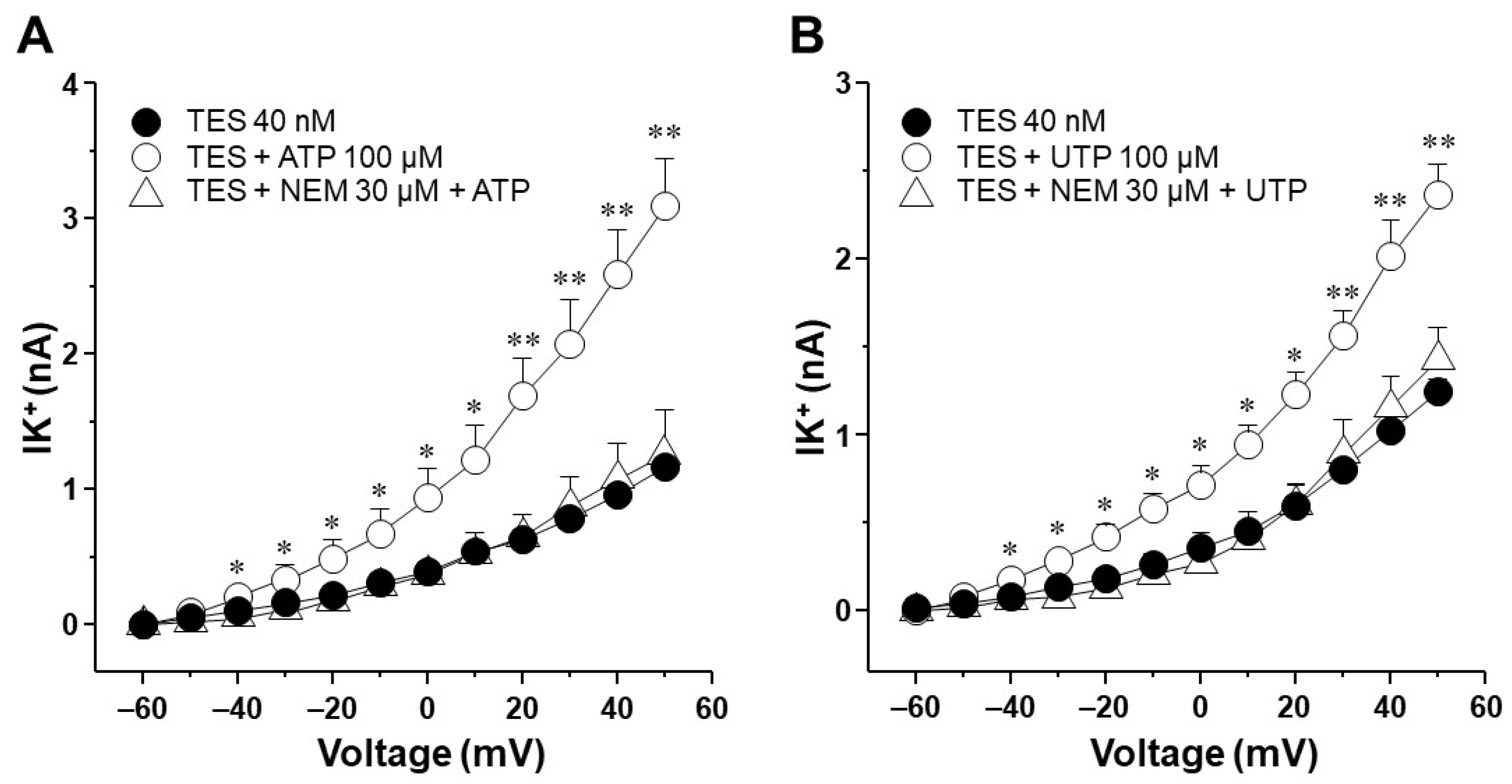
2.3. Voltage-Dependent Delayed Rectifier K+ (KV) Channels Are Responsible for Testosterone Enhancement of ATP- and UTP-Induced K+ Currents

To find out which type of K+ channel is involved in the TES effect, we used blockers of the KV and the BKCa channels on the ATP- and UTP-induced K+ currents. In control tracheal smooth muscle cells, ATP and depolarizing pulses increased the K+ currents, which peaked at approximately 2.1 nA (Figure 4A, open triangle). The perfusion of 4-aminopyridine (4-AP, 3 mM, a blocker of KV channels) partially decreased the ATP 100 µM-induced K+ currents from −20 mV ahead (open circle), but it did not reach the values of the control K+ currents caused by depolarizing pulses (closed circle). The summative addition of iberiotoxin (IBTX, 100 nM, the selective blocker of BKCa channels) decreased the remaining ATP-induced K+ currents from 30 mV forward (Figure 4A) below the control trace. These results confirm that the K+ current generated by ATP (along with the depolarizing pulses) is due to the opening of KV and BKCa channels, as we previously demonstrated [27]. Moreover, as shown in Figure 2, TES 40 nM (for 48 h) significantly improved the ATP-induced K+ currents. 4-AP almost entirely suppressed the resulting K+ currents from −10 mV onwards, and IBTX blocked the remaining K+ currents (Figure 4B). The results were similar when the K+ currents were induced by UTP. Under control conditions, UTP 100 µM generated a K+ current that peaked at approximately 1.6 nA, and 4-AP partially reduced this K+ current from −20 mV ahead. At the same time, the consecutive addition of IBTX abolished the K+ currents elicited by UTP (Figure 5A). As shown in Figure 3, TES-treated myocytes showed a significantly augmented UTP-induced K+ current (Figure 5B), which was almost abolished by 4-AP from −30 mV forward. The consecutive addition of IBTX annulled the UTP-induced K+ currents. These results indicate that the effect of TES appears to be mediated mainly through the action of KV channels.

2.4. In Tracheal Myocytes, KV Currents but Not BKCa Currents Are Upregulated by Testosterone Treatment

Because the KV subtype seems to be more involved in the enhancement exerted by TES on ATP- and UTP-induced K+ currents compared to BKCa subtype, we decided to investigate the influence of this androgen on K+ currents generated only by the protocol of depolarizing pulses. In cultured tracheal myocytes without TES, depolarizing steps from −60 to +50 mV generated K+ currents that reached approximately 0.6 nA (Figure 6A). Administration of 4-AP blocked the K+ currents from −40 mV forward, and the subsequent addition of IBTX almost completely blocked the K+ currents from 0 mV forward (Figure 6A). Further administration of TEA annulled the K+ currents. These results indicate that although KV and BKCa are the primary subtypes of K+ channels in guinea pig tracheal smooth muscle cells, KV subtype is the major contributor to total K+ currents. Analysis of the area under the curve (AUC) showed that under the protocol of depolarizing steps, KV was responsible for 62.47% and BKCa for 37.52% of the K+ currents mediated by these two subtypes of K+ channels. Additionally, the treatment of cultured myocytes with TES 40 nM increased the K+ currents generated by the depolarizing steps, as previously shown in Figure 2D and Figure 3D. Perfusion of 4-AP virtually abolished the enhancement of K+ currents generated by TES, and IBTX and TEA practically abolished the total K+ currents (Figure 6B). In this case, analysis of AUC showed that in myocytes treated with TES, the proportional current for KV increased to 69.86%. In comparison, BKCa reduced its proportion to 30.13%. Therefore, the effect of the androgen relates exclusively to KV channels by upregulating the function or the expression of these proteins.

2.5. Testosterone Increases the Expression of KV1.2 and KV1.5

Previous immunofluorescence assays confirmed that the expression of two subtypes of KV, KV1.2 and KV1.5, in the ASM of guinea pigs increased when tracheal rings were exposed to 40 nM TES for 48 h (Figure 7 and Figure 8) [58]. These results were analyzed by measurements of mean fluorescence intensity illustrated in Figure 7D and Figure 8D. Negative controls, including incubation with the corresponding blocking peptide or no primary antibody incubation, confirmed antibodies’ specificity against KV1.2 and KV1.5 proteins. No fluorescence signal was detected in the negative controls. The colocalization of the two K+ channels in the ASM was confirmed by detecting smooth muscle α-actin in Figure 7A,B and Figure 8A,B.

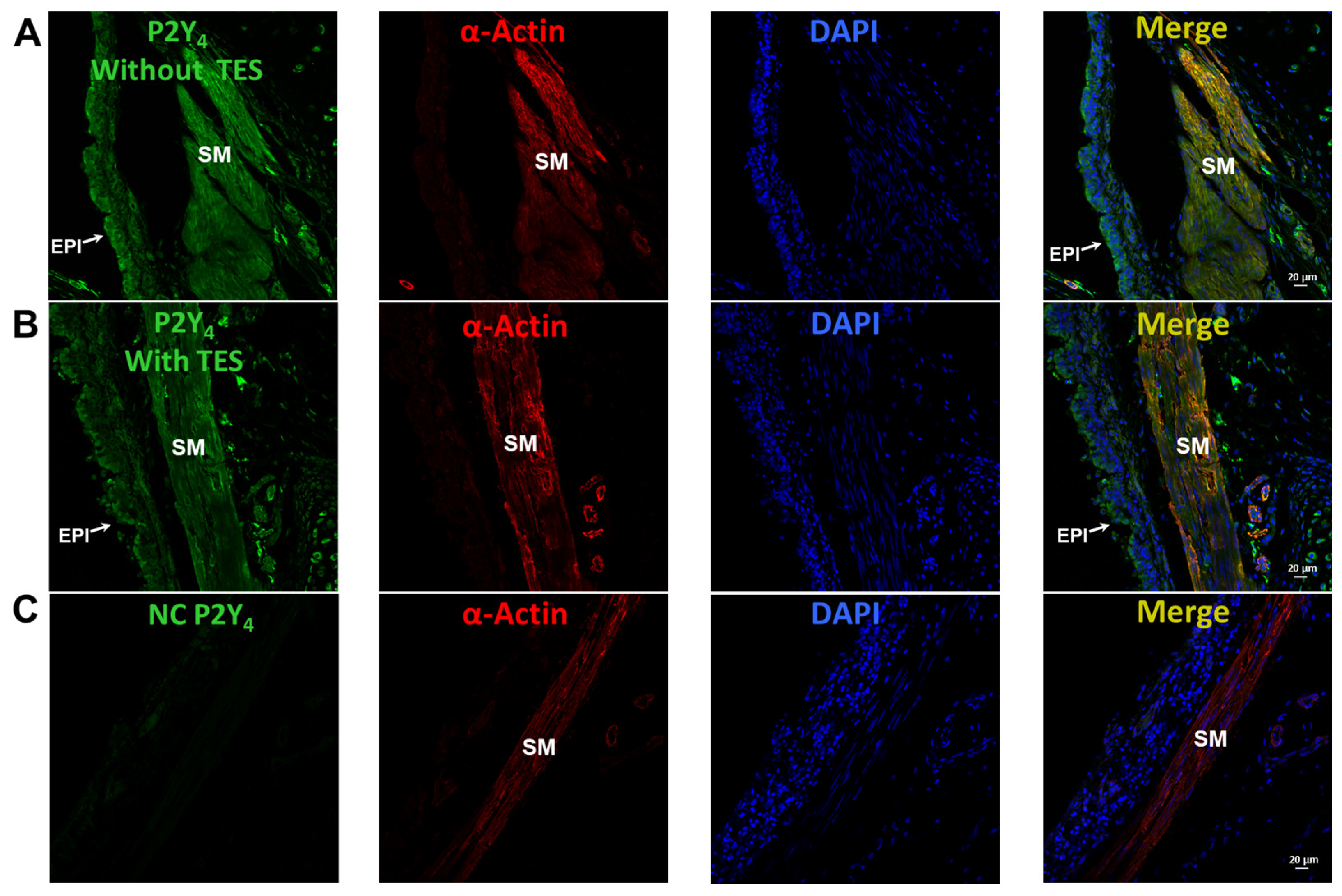
2.6. UTP Stimulates P2Y2 and P2Y4 Receptors in Airway Myocytes, and Testosterone Increases P2Y4-Mediated K+ Currents but Does Not Increase the Expression of P2Y4 Receptors

In control tracheal myocytes, the antagonist for almost all P2Y receptors except P2Y2, RB2 100 μM [59,60,61,62], partially decreased the K+ currents induced by UTP (100 µM). Consecutive addition of AR-C118925XX (10 µM, selective antagonist of the P2Y2 receptors) [63] abolished the remaining UTP-induced K+ currents (Figure 9A). TES 40 nM enhanced the UTP-induced K+ currents, RB2 completely blocked this improvement, and AR-C118925XX did not modify these currents (Figure 9B). Based on these findings, we investigated whether TES could increase the expression of P2Y4 receptors in the ASM. However, immunofluorescence experiments revealed that TES did not improve the expression of P2Y4 receptors (Figure 10).

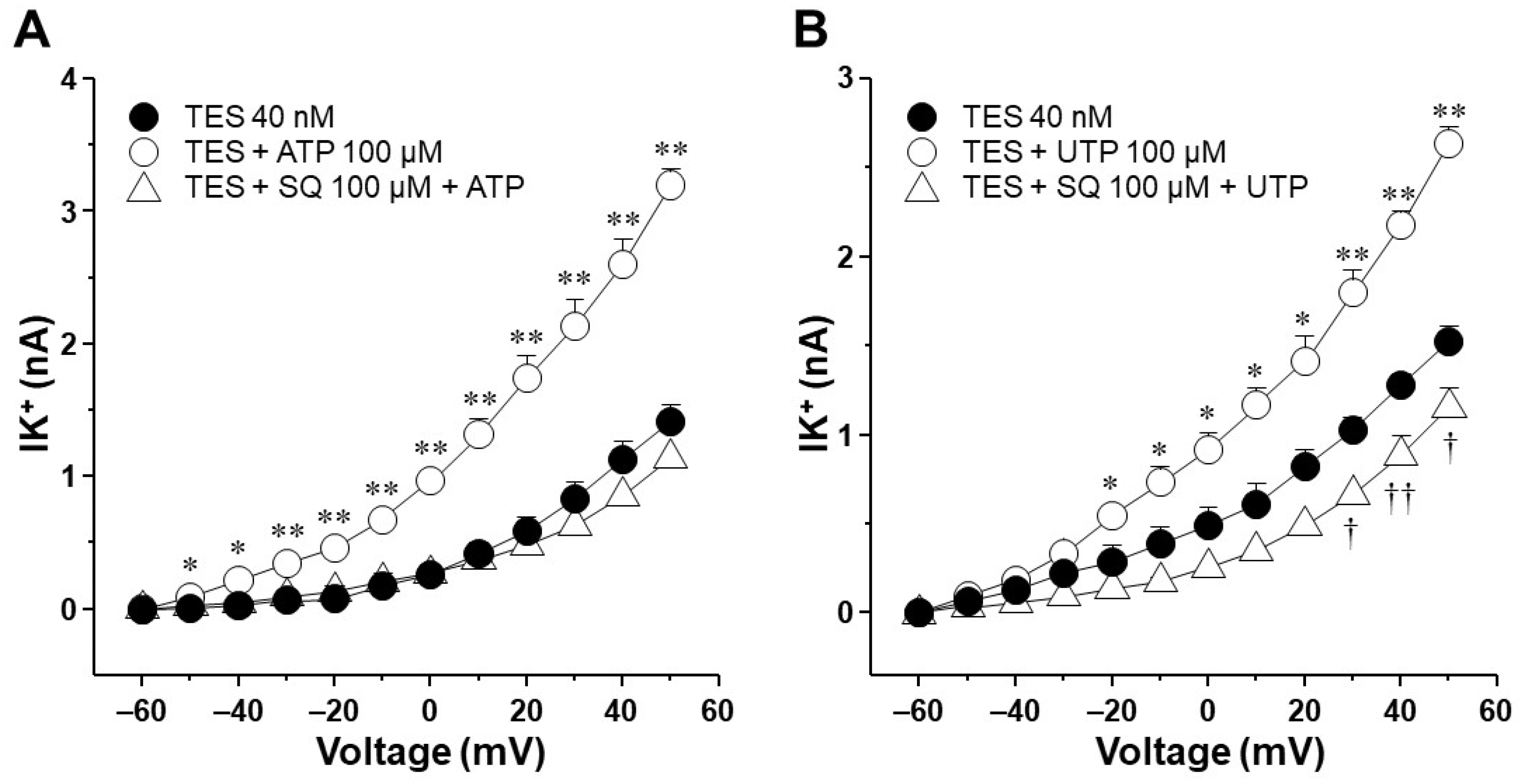
2.7. The Enhancement of ATP- and UTP-Mediated K+ Currents Depends on the GS Protein–Adenylyl Cyclase Axis

It has been postulated that the ATP- and UTP-induced K+ current increase and ASM relaxation are mediated by the production of prostaglandins in the smooth muscle, which in turn enhances cAMP signaling [27,36,37]. However, by performing immunostaining assays, we found that TES did not affect the expression of cyclooxygenase (COX)-1 or COX-2 (Figure 11), suggesting that prostaglandins are not involved in the action of the androgen. N-ethylmaleimide (NEM, 30 μM), a drug that uncouples G proteins from several receptors, blocked the increase in K+ currents elicited by ATP or UTP in tracheal myocytes previously incubated with TES, confirming that this effect occurs via receptors coupled to G proteins (Figure 12). Moreover, in another set of experiments, 100 μM SQ 22,536, a cell-permeable adenylyl cyclase inhibitor, completely abolished the enhanced ATP- and UTP-evoked K+ current increase (Figure 13). Overall, all these results point out that ATP and UTP in TES-treated myocytes stimulate P2Y4 receptors, which in turn activate a G protein, probably GS, and adenylyl cyclase to mediate the enhanced relaxation of the ASM.

3. Discussion

According to our findings, the chronic exposure of tracheal rings to 40 nM TES enhanced ATP- and UTP-induced relaxation involving the activation of K+ channels. TES increased ATP- and UTP-induced K+ currents in tracheal myocytes, and flutamide reversed this increase, confirming the genomic nature of these findings. Moreover, the effect of TES on ATP- and UTP-induced K+ currents was abolished by 4-AP, indicating that KV channels are involved in the androgenic potentiation effect. The upregulation of KV currents elicited by depolarizing pulses in myocytes chronically exposed to TES is explained by the increased expression of KV1.2 and KV1.5 induced by the androgen. RB2, the antagonist of almost all P2Y receptors (except for P2Y2), inhibited the UTP-induced increase in K+ currents, suggesting that P2Y4 receptors are implicated in the action of the androgen. However, TES did not affect P2Y4 receptor expression or COX-1 and COX-2 in the ASM. These last findings ruled out the involvement of prostaglandins. The stimulation of P2Y4 receptors with ATP or UTP triggers the G protein–adenylyl cyclase pathway, which TES could positively regulate. Consequently, the probability of K+ channels opening increases, leading to membrane hyperpolarization and smooth muscle relaxation.
ATP relaxes the ASM of several species, including mice, rats, rabbits, guinea pigs, and humans [31,36,37,64]. Moreover, we have demonstrated that ATP-induced ASM relaxation is dependent on P2Y2 and P2Y4 receptors signaling [27]. In the present study, we confirmed that ATP and UTP relaxed the histamine-pre-contracted ASM of a guinea pig in a concentration-dependent manner (Figure 1). We also found that a physiological concentration of TES (40 nM) significantly increased the relaxation response for both agonists (Figure 1). Furthermore, we observed that ATP caused a greater relaxation of tracheal tissues compared to UTP in both the control and TES groups. The difference in the relaxation of the ASM triggered by ATP or UTP may be due to the involvement of the hydrolysis of ATP and the activation of other purinergic receptors [27]. For example, adenosine, produced by the degradation of ATP by ectonucleotidases, activates A2 receptors, a subgroup of the purinergic P1 receptor family. This process facilitates the relaxation of the ASM pre-contracted with KCl or histamine [65,66]. Our previous work has shown that blocking ectonucleotidases in guinea pig tracheal rings with ARL 67156 shifted the concentration–response curve to ATP to the left; i.e., the relaxation response was more intense [27]. This shift indicates that ATP is enzymatically degraded under our experimental conditions. Moreover, we also showed a remanent relaxation induced by ATP even when P2Y2 and P2Y4 receptors were blocked [27]. These previous results suggest that the higher relaxation induced by ATP than UTP may be mediated by adenosine acting through P1 receptors [67].
Several studies have convincingly demonstrated the direct influence of TES on the relaxation of ASM. There are different mechanisms through which this hormone can affect ASM tension, including the inhibition of membrane Ca2+ channels [45]. For example, androgens induce the relaxation of pre-contracted guinea pigs and bovine tracheal smooth muscle by blocking L-type voltage-dependent Ca2+ channels (L-VDCCs) and store-operated Ca2+ channels (SOCCs) [51,52]. In addition, androgenic hormones enhance the relaxation of ASM caused by β adrenergic agonists [49,50], and TES induces ASM relaxation via an epithelial-dependent mechanism that is related to nitric oxide production [48]. All these reported effects are mediated through non-genomic pathways triggered by the androgens. On the other hand, we recently demonstrated the genomic effects of TES on ASM relaxation. This androgen significantly augmented the relaxation caused by the clinically used bronchodilators, salbutamol and theophylline, in guinea pig tracheal rings by upregulating the β2-AR and K+ channels [40,58,68]. In the experiments shown in the present work, a 48 h incubation of the androgen was performed, suggesting that the enhanced relaxation evoked by ATP and UTP may be due to a genomic pathway. Moreover, because K+ channels are implicated in the purinergic-mediated guinea pig ASM relaxation [27], we investigated whether these channels are involved in the increased ATP- and UTP-induced relaxation. Thus, we blocked the K+ channels with TEA, which reversed the effect of the androgen (Figure 1).
Next, we explored the influence of TES on K+ currents triggered by ATP and UTP. We found that myocytes chronically exposed to TES (40 nM) showed increased K+ currents elicited by depolarizing pulses (Figure 2D and Figure 3D) and by increasing concentrations of ATP (Figure 2E–H) and UTP (Figure 3E–H). The fact that TES markedly augmented the K+ currents during depolarizing pulses alone indicates the direct regulation of K+ channels by the androgen. Moreover, the enhanced ATP- and UTP-evoked K+ currents suggest a modulation of the purinergic signaling. To confirm the possible genomic mechanism of TES, we used flutamide, an androgen receptor antagonist, which abolished the sex-hormone-induced effects on the K+ currents (Figure 2 and Figure 3). Thus, it is conceivable that TES upregulates the expression of proteins involved in purinergic responses, including P2Y receptors and K+ channels. The major populations of K+ channels involved in the modulation of the tone and relaxation of the ASM are the KV and BKCa [27,40,56,58,69]. Therefore, we used 4-AP and IBTX to investigate which type of K+ channel is responsible for the TES effect on ATP- and UTP-induced K+ currents. As shown in Figure 4A and Figure 5A, ATP- and UTP-induced K+ currents in control myocytes (without TES) were partially decreased by 4-AP, and the remaining K+ currents were abolished by the addition of IBTX. Consistent with the results in Figure 2 and Figure 3, the K+ currents were greater when cells were incubated with TES (Figure 4B and Figure 5B). Notably, 4-AP completely blocked the TES-induced enhancement of K+ currents, highlighting the importance of KV channels in this phenomenon. Given these findings, we decided to investigate the effects of this androgen on the K+ currents produced only by the protocol of depolarizing steps. First, we demonstrated that KV and BKCa channels were involved in the K+ currents by 62.47% and 37.52%, respectively, under control conditions (Figure 6A). However, exposure to TES increased the contribution of KV channels (69.86%) but decreased that of BKCa channels (30.13%) (Figure 6B). These last results of Figure 6 confirm the positive regulation of KV channels by the androgen. In this context, we had previously shown that TES increased the expression of KV1.2 and KV1.5 isoforms, but not BKCa, in the ASM (Figure 7B and Figure 8B) [58]. In this context, there is evidence of the genomic effects of TES on K+ channels in cardiomyocytes, where this hormone improved the expression of KV7.1 [70]. On the other hand, there are no reports of non-genomic effects of TES on K+ channels in ASM. However, in vascular smooth muscle, the acute application of TES resulted in vasodilation associated with opening KV and BKCa channels [71,72]. Further research is needed to determine whether TES can trigger non-genomic mechanisms affecting K+ channels in the ASM.
Additionally, we investigated the possible effects of TES on P2Y2 and P2Y4 receptors, as ATP exerts its relaxing effect via their activation [27]. In patch-clamp studies, the K+ currents elicited by UTP were prevented in control cells by pharmacologically blocking P2Y4 and P2Y2 receptors (Figure 9A). We used RB2 as an antagonist of P2Y4 receptors because this chemical compound can block the action of almost all P2Y receptors, including P2Y4, except for P2Y2 [59,61]. AR-C118925XX was employed as a selective antagonist of P2Y2 [63]. We focused on UTP as an agonist of P2Y receptors to avoid the nonspecific effects of ATP on other purinergic receptors [61,62,73,74,75]. Long-term TES exposure significantly increased the UTP-evoked K+ currents, and RB2 antagonized this effect (Figure 9B). Therefore, we investigated the possibility that TES might increase the expression of this receptor in the ASM, but no changes in the immunofluorescence were observed (Figure 10). It is most likely that the increase in ATP- and UTP-induced K+ currents is exclusively due to the upregulation of K+ channels, specifically KV1.2 and KV1.5.
Previous evidence postulates that ATP-induced relaxation of ASM is caused by intermediate biosynthesis of PGE2 following stimulation of P2Y2 and P2Y4 receptors [27,36]. The activation of these receptors triggers heterotrimeric Gq proteins [30,76,77] leading to intracellular Ca2+ increases [14,78]. Ca2+ mobilization could activate phospholipase A2 (PLA2), which favors PGE2 production [79]. Because COX is the main pathway that catalyzes the conversion of arachidonic acid to prostaglandins, we investigated whether TES could enhance the expression of this enzyme. Chronic exposure to TES in tracheal slices did not increase the expression of constitutive COX-1 and inducible COX-2 in ASM (Figure 11), indicating that the androgen effect is not related to PGE2 synthesis.
We then decided to examine the signaling pathway that modulates the increase in K+ currents triggered by ATP and UTP in myocytes exposed to TES. Perfusion of NEM (a G protein uncoupler) and SQ 22,536 (an adenylyl cyclase inhibitor) reversed the effect of TES on the K+ currents generated with ATP and UTP (Figure 12 and Figure 13). In general, P2Y4 receptors trigger the Gq-mediated signaling pathway with the subsequent activation of phospholipase Cβ and the intracellular Ca2+ increase [14,80]. Nevertheless, it has been reported that several P2Y receptors can couple to different G proteins [80]. For instance, the P2Y11 receptor that becomes active upon ATP binding forms connections with members of the Gq and Gs subfamilies [80,81,82]. Gs proteins initiate the activation of adenylyl cyclase, leading to augmented cAMP levels. The interaction between the P2Y11 receptor and Gs proteins holds significance in regulating various physiological processes, including cell migration and apoptosis [83,84,85]. In this context, we hypothesize that the P2Y4 receptor in the ASM couples to a Gs protein after TES exposure which would lead to the formation of cAMP. The synthesis of cAMP catalyzed by AC promotes the activation of PKA and the phosphorylation of the KV, increasing its activity [41,86,87]. This might explain why NEM and SQ 22,536 abated the effect of TES. Nonetheless, this hypothesis requires further corroboration. In addition, it has been demonstrated that P2Y4 can couple to the βγ-subunits of Gi/o proteins in rat sympathetic neurons [77]. Gi activation classically leads to reduced cAMP synthesis and inhibition of Gs-stimulated AC activity [88,89,90]. However, it has been suggested that the βγ-subunits of the Gi protein can indirectly trigger the activity of the AC (type VI) in HEK cells [91]. This AC isoform is the predominant one in ASM, but we previously showed that the expression of this enzyme was not altered in guinea pig tracheal tissues exposed to TES [40]. Collectively, our results indicate that TES enhances the P2Y4 signaling pathway (probably via a GS protein) and increases the expression of KV channels. Activation of this purinergic receptor induces the formation of cAMP and the opening of KV channels, which are enhanced by TES, resulting in augmented ASM relaxation.
In the context of airway inflammation in asthma patients, ATP is significantly released into the airways [13]. In this regard, platelets are an important source of ATP during the asthmatic process, as they can store up to 1 M in their granules [92]. There is evidence that allergen exposure triggers intravascular platelet activation in patients with allergic asthma [93] and in the pulmonary capillaries of ovalbumin-sensitized mice [94], emphasizing the significant role of circulating activated platelets in the progression of allergic airway inflammation following exposure to allergens. Our studies with guinea pig platelets have confirmed this phenomenon by demonstrating the release of ATP during antigen challenge [3]. In addition to platelets, other entities, such as airway epithelial cells [21] and ASM cells [28], are also involved in ATP release during inflammation. Extracellular ATP exerts direct effects on the ASM by inducing contraction and facilitating the recruitment of inflammatory cells into the airways [17,28,95,96]. These cells can subsequently release bronchoconstrictor substances such as histamine, thromboxane, leukotrienes, and acetylcholine [97,98,99,100,101]. Given the pre-existing smooth muscle contraction during asthma, ATP probably promotes relaxation. Thus, ATP may serve as a regulating factor that prevents excessive bronchoconstriction and maintains airway patency during an asthma attack, particularly in young men when TES levels are elevated. Furthermore, studies in animal and human tissues have shown that K+ channel openers can induce hyperpolarization of ASM cells, resulting in bronchodilation and attenuation of airway hyperresponsiveness [57,102,103,104,105,106,107,108]. Therefore, KV channels might be a promising pharmacologic target for a novel and effective therapeutic approach in the treatment of asthma in young men.

4. Materials and Methods

4.1. Experimental Animals

Male Hartley guinea pigs weighing between 350 and 400 g and aged 4–6 weeks were used. The animals were reared under standard institutional conditions, i.e., filtered air, 21 ± 1 °C, 50–70% humidity, sterile bedding, and feed consisting of Harlan® pellets and sterilized water. The General Health Law for Health Research (NOM-062-Z00-1999) and the Mexican National Animal Welfare Laws establish guidelines that were followed when handling the animals. The scientific and bioethical committees of the National Autonomous University of Mexico approved the project under the number FM/DI/003/2020.

4.2. Organ Baths

Pentobarbital sodium (50 mg/kg) was used to anesthetize the guinea pigs before exsanguination. The tracheas were dissected and placed in a dissection chamber filled with Krebs-Ringer solution containing [mM] 118 NaCl, 25 NaHCO3, 11 glucose, 4.6 KCl, 2 CaCl2, 1.2 KH2PO4, and 1.2 MgSO4. The solution was bubbled with a carbogen mixture of 95% O2 and 5% CO2 for 30 min, keeping the pH constant in the physiological range (7.3 to 7.4). Afterward, the fatty and connective tissue was removed from the trachea. The tracheas were divided into seven segments, each with four cartilage rings. The tissues were incubated without and with TES (40 nM) for 48 h at 9 °C in separate Eppendorf tubes containing 1 mL of Krebs-Ringer solution bubbled for 1 h with 5% CO2 in oxygen. After 48 h of incubation, each tissue was hung on a transducer at a resting tension of 1 g in separate organ baths containing 5 mL Krebs-Ringer solution at 37 °C and continuously bubbled with 5% CO2 in oxygen. A CyberAmp 380 signal integrator system (Axon Instruments, Foster City, CA, USA) and a Digidata 1440A interface (Axon Instruments) were used to record isometric force with a model FT03 strain transducer (Grass Instruments, West Warwick, RI, USA). Guinea pig tracheal smooth muscle contraction was recorded, and data were acquired on a computer using Axoscope software version 10.2 (Axon Instruments, Foster City, CA, USA). The rings were in resting tension for 1 h before the experimental procedures were performed. The contractile apparatus was optimized, and tissue responses were normalized by stimulating the rings with KCl (60 mM). The preparations with or without TES were pre-contracted with histamine (10 µM). Once maximal contraction was achieved, a cumulative concentration–response curve was generated with ATP or UTP (1, 3.2, 10, 32, 100, 320, and 1000 µM) to elicit and study ASM relaxation. In one experimental group, the tracheal rings were incubated with TEA (1 mM), 15 min before the addition of ATP or UTP.

4.3. Patch-Clamp Studies

The guinea pig trachea was removed as described in the procedure for organ baths. The connective tissue and epithelium were removed from the tracheal smooth muscle and subjected to enzymatic disruption by placing it in 5 mL of Hanks solution (GIBCO, Waltham, MA, USA) containing 0.05 U/mL papain (Worthington Biochemical Corporation, Lakewood, NJ, USA) and 2 mg L-cysteine. The pH of the solution was adjusted to 7.4 with 1 M NaHCO3, and the tissues were incubated at 37 °C for 10 min. The muscle strip was then washed with Leibovitz solution (L15, GIBCO) to remove excess enzymes and then placed in Hanks solution containing 1 mg/mL collagenase type I (Worthington Biochemical Corporation) and 1 mg/mL collagenase type II (Worthington Biochemical Corporation) for 10 min at 37 °C. The tissue was then aspirated approximately 25 times with a Pasteur pipette to allow the dissociation of the myocytes. The enzymatic activity was stopped with the L15 solution, and the cells were centrifuged at 600 rpm for 6 min. The supernatant was removed, and the last step was repeated once. The cell pellet was re-suspended in 6 mL minimal essential medium (MEM, GIBCO) containing 5% fetal bovine serum, 10 mM glucose, 2 mM L-glutamine, 10 µg/mL streptomycin, and 10 U/mL penicillin. Myocytes were cultured on coverslips impregnated with sterile rat tail collagen placed in 6-well plates. A group of myocytes was incubated with 40 nM TES (48 h) or 3.2 µM flutamide 30 min prior to the addition of TES. The cells were stored in a 5% CO2 incubator at 37 °C for 48 h before patch-clamp experiments were performed.
The coverslip containing the cultured tracheal myocytes was placed in a chamber that was continuously perfused with a volume of 0.5–1 mL of external solution at a flow rate of 1.5–2 mL/min. For K+ currents measurement, the external solution was adjusted to pH = 7.4 with NaOH and had the following composition (mM): 130 NaCl; 10 HEPES; 10 glucose; 5 KCl; 3 NaHCO3; 1.2 KH2PO4; 1 CaCl2; 0.5 MgCl2; 0.1 niflumic acid. The perfusion chamber was adapted to an inverted microscope (Zeiss model IP03). The patch pipettes for voltage recording were made of borosilicate glass (1B200F-6, World Precision Instruments, Sarasota, FL, USA) using a micropipette puller (P-87, Sutter Instruments Co., Novato, CA, USA). The pipette resistance was between 3 and 6 MΩ. K+ currents measurements were recorded with an Axopatch amplifier (model 200A, Axon Instruments) and performed with the 1–5 KHz current filters at a frequency of 10 KHz. The composition of the patch pipette internal solution was (mM) 140 K+ gluconate, 5 NaCl, 5 HEPES, 5 ATP, 1 EGTA, 0.1 leupeptin, and 0.1 GTP, adjusted with KOH to pH = 7.3.
Outward K+ currents were recorded using pClamp v10.2 software (Axon Instruments) with a protocol consisting of depolarizing pulses from −60 to +50 mV in steps of 10 mV with a holding potential of −60 mV for 500 ms at 1 Hz. Myocytes were perfused with cumulative concentrations of ATP or UTP (1, 10, 100, and 1000 µM) and received the protocol of depolarizing pulses. To characterize K+ channels in a group of experiments, cells were perfused with 3 mM 4-AP and 100 nM IBTX, which block KV and BKCa, respectively. TEA was used to block K+ channels nonspecifically. Some airway myocytes were perfused with Reactive Blue 2 (RB2, 100 μM) and AR-C118925XX (10 µM) before the addition of 100 µM UTP. In a series of experiments, 30 μM N-ethylmaleimide (NEM; a compound that uncouples G proteins) and SQ 22,536 (100 μM, adenylyl cyclase inhibitor) were perfused for 10 min before the addition of 100 µM ATP or UTP.

4.4. Double Immunofluorescence

Tracheal tissues were fixed in 4% paraformaldehyde in phosphate-buffered saline (PBS) for 4 h after gradual dehydration in 30% sucrose/PBS for 17 h before being included in O.C.T. compound (Tissue Tek, Torrance, CA, USA) and frozen at 60 °C. Frozen sections (10 nm thick) were obtained with a cryostat (Ecoshel ECO-1900, Pharr, TX, USA) and mounted on gelatinized slides. Afterward, slides were transferred into a Sequenza slide rack (Thermo Fisher Scientific, Rockford, IL, USA), washed with TBS, and permeabilized with 0.001% Tween 20 in TBS. To block nonspecific binding to proteins, cryosections were treated with 10% horse serum for 2 h at room temperature. Then, the sections were incubated with the primary antibody anti-P2Y4 diluted 1:75 overnight at 4 °C (Alomone Labs, APR-006, Jerusalem, Israel). The secondary antibody Alexa488 donkey anti-rabbit IgG (Life Technologies, Foster City, CA, USA) was incubated for 20 min (1:500). Anti-α-actin (1A4) Alexa Fluor 546 (sc-32251 AF546, Santa Cruz Biotechnology, Dallas, TX, USA), diluted 1:1200, was incubated overnight at 4 °C as the next primary antibody. The nuclei in all tissue slices were counterstained with 4′, 6-diamidino-2-phenylindole (DAPI, Life Technologies). Slides were maintained with Fluoromount aqueous mounting medium (Merck, Darmstadt, Germany). Immunofluorescence was observed using an LSM 880 Zeiss confocal microscope (Oberkochen, Germany). For display purposes, images in which P2Y4 is green, actin is red, and nuclei are blue were merged.

4.5. Immunochemistry

Tracheas from guinea pigs were obtained as described above and then incubated with and without TES 40 nM for 48 h at 9 °C. Then, tracheal tissue was fixed in 4% paraformaldehyde for 4 h and placed in 30% ethanol until their inclusion in paraplast plus (Sigma Aldrich Co., St. Louis, MO, USA). Once paraffinized, tissue blocks were cut into thin slices (5 μm) and placed over gelatinized slides. Paraffin was removed by incubation at 60 °C and placed in xylol, followed by graded alcohols, and then slices were washed with Tris-buffered saline (TBS). Slices were transferred into a Sequenza slide rack, washed with TBS, and permeabilized with 0.5% Triton/TBS. To block nonspecific binding to proteins, 10% horse serum was applied to the slices for 2 h at 32 °C. The sections were incubated with Bloxall blocking solution (Vector Laboratories, Burlingame, CA, USA) for 20 min at room temperature. The slides were incubated overnight at 4 °C with the rabbit polyclonal antibody to COX-1 (sc-7950, Santa Cruz Biotechnology, CA, USA) or COX-2 (sc-7951,) both diluted 1:25. For negative control, the primary antibody was omitted. After washing with TBS, the sections were incubated with donkey anti-rabbit IgG HRP (Thermo Fisher Scientific, Rockford, IL, USA) for 20 min and washed with TBS. Finally, the Betazoid DAB Chromogen Kit (Biocare Medical Pike Lane, CA, USA) was added to each section for 4 min. All slices were counterstained with hematoxylin (Biocare Medical Pike Lane, CA, USA) and maintained with a Sub-X mounting medium (Electron Microscopy Sciences, Hatfield, PA, USA). Images were collected and processed with a Nikon eclipse Ni microscope (Tokyo, Japan). For display purposes, images in which COX-1 or COX-2 is brown and nuclei are blue were merged.

4.6. Drugs and Chemicals

Testosterone (17β-hydroxy-4-androsten-3-one, TES), flutamide, histamine, ATP, UTP, TEA, RB2, N-ethylmaleimide, L-cysteine, and SQ 22,536 were purchased from Sigma Chem. Co. (St. Louis, MO, USA). 4-aminopyridine was purchased from Research Chemical Ltd. (Word Hill, MA, USA), iberiotoxin from Enzo Life Sciences (Farmingdale, NY, USA), and AR-C118925XX from Tocris Bioscience (Avonbridge Trading Estate, Bristol, UK). TES, flutamide, and 4-aminopyridine were diluted in absolute ethanol, and the highest percentage used was 0.1% v/v of vehicle. AR-C118925XX was dissolved in dimethyl sulfoxide (DMSO) 0.1% v/v.

4.7. Statistical Analysis

In organ baths, Dunnett’s multiple comparison test was performed after a one-way analysis of variance to examine these data. K+ currents were analyzed using a one-way analysis of variance, followed by Student–Newman–Keuls test or Dunnett’s test. In the experiments, each value of “n” refers to a different animal. Data are presented as mean ± the standard error of the mean (S.E.M). Statistical significance was set at p < 0.05.

5. Conclusions

In summary, our studies demonstrate that chronic exposure of guinea pig ASM to a physiological concentration of TES leads to a ~50% increase in ATP- and UTP-elicited K+ currents. This increase is associated with the enhanced signaling of P2Y4 receptors and augmented expression of KV channels. These mechanisms result in the improvement of ATP- and UTP-induced ASM relaxation by 22.17% and 12.31%, respectively. The enhanced ASM relaxation may limit the severity of bronchospasm in teenage boys and men. To test this hypothesis, further experiments using plethysmography to measure airway resistance and evaluate bronchorelaxation induced by ATP are needed. However, these experiments are beyond the scope of the present work. Additionally, the activation of GS proteins after P2Y4 receptor stimulation suggested in this article should be further corroborated.

Author Contributions

Conceptualization, L.M.M., A.C.-G., and J.R.-G.; Methodology, A.C.-G., J.R.-G., V.D.-H., and M.F.C.-H.; Software, J.R.-G., A.C.-G., V.D.-H., and M.F.C.-H.; Validation, L.M.M. and J.R.-G.; Formal Analysis, L.M.M., A.C.-G., J.R.-G., V.D-H., and F.J.F.-M. Investigation, L.M.M. A.C.-G., J.R.-G., and F.J.F.-M.; Resources, L.M.M., J.R.-G., and V.D.-H.; Data Curation, J.R.-G., A.C.-G., M.F.C.-H., and V.D.-H.; Writing—Original Draft Preparation, L.M.M., A.C.-G., and J.R.-G.; Writing—Review and Editing, L.M.M., A.C.-G., J.R.-G., and F.J.F.-M.; Visualization, L.M.M. and J.R.-G.; supervision, L.M.M. and J.R.-G.; Project administration, L.M.M. and J.R.-G.; Funding acquisition, L.M.M., V.D.-H., and J.R.-G. All authors have read and agreed to the published version of the manuscript.

Funding

This research was funded by grants from Dirección General de Asuntos del Personal Académico (DGAPA), Universidad Nacional Autónoma de México (IN200522) to L.M. Montaño, (IA201322 and IA203924) to J. Reyes-García, and (IN200924) to V. Díaz-Hernández and from the Consejo Nacional de Ciencia y Tecnología, México (CF/2019-137725) to L.M. Montaño and (CBF2023-24-1074) to J. Reyes-García.

Institutional Review Board Statement

The animal study protocol was approved by the Institutional Ethics Committee of the Facultad de Medicina, Universidad Nacional Autónoma de México (protocol code FM/DI/003/2020, 5 May 2020).

Informed Consent Statement

Not applicable.

Data Availability Statement

Data are available upon request.

Acknowledgments

María F. Casas-Hernández is grateful to the Programa de Maestría en Ciencias de la Salud, Escuela Superior de Medicina, Instituto Politécnico Nacional, for the instruction received during her M.Sc. degree studies. She received a fellowship from the Consejo Nacional de Ciencia y Tecnología, México (CVU: 912413).

Conflicts of Interest

The authors declare no conflicts of interest.

References

  1. Arzola-Martinez, L.; Benavente, R.; Vega, G.; Rios, M.; Fonseca, W.; Rasky, A.J.; Morris, S.; Lukacs, N.W.; Villalon, M.J. Blocking ATP-releasing channels prevents high extracellular ATP levels and airway hyperreactivity in an asthmatic mouse model. Am. J. Physiol. Lung Cell. Mol. Physiol. 2021, 321, L466–L476. [Google Scholar] [CrossRef] [PubMed]
  2. Chávez, J.; Vargas, M.H.; Martinez-Zuniga, J.; Falfan-Valencia, R.; Ambrocio-Ortiz, E.; Carbajal, V.; Sandoval-Roldan, R. Allergic sensitization increases the amount of extracellular ATP hydrolyzed by guinea pig leukocytes. Purinergic Signal. 2019, 15, 69–76. [Google Scholar] [CrossRef] [PubMed]
  3. Chávez, J.; Vargas, M.H.; Rebollar-Ayala, D.C.; Diaz-Hernandez, V.; Cruz-Valderrama, J.E.; Flores-Soto, E.; Flores-Garcia, M.; Jimenez-Vargas, N.N.; Barajas-Lopez, C.; Montaño, L.M. Inhibition of extracellular nucleotides hydrolysis intensifies the allergic bronchospasm. A novel protective role of ectonucleotidases. Allergy 2013, 68, 462–471. [Google Scholar] [CrossRef]
  4. Liu, T.T.; Wang, Y.L.; Zhang, Z.; Jia, L.X.; Zhang, J.; Zheng, S.; Chen, Z.H.; Shen, H.H.; Piao, C.M.; Du, J. Abnormal adenosine metabolism of neutrophils inhibits airway inflammation and remodeling in asthma model induced by Aspergillus fumigatus. BMC Pulm. Med. 2023, 23, 258. [Google Scholar] [CrossRef] [PubMed]
  5. Pelleg, A.; Xu, F.; Zhuang, J.; Undem, B.; Burnstock, G. DT-0111: A novel drug-candidate for the treatment of COPD and chronic cough. Ther. Adv. Respir. Dis. 2019, 13, 1753466619877960. [Google Scholar] [CrossRef]
  6. Thompson, R.J.; Sayers, I.; Kuokkanen, K.; Hall, I.P. Purinergic Receptors in the Airways: Potential Therapeutic Targets for Asthma? Front. Allergy 2021, 2, 677677. [Google Scholar] [CrossRef] [PubMed]
  7. Lommatzsch, M.; Cicko, S.; Muller, T.; Lucattelli, M.; Bratke, K.; Stoll, P.; Grimm, M.; Durk, T.; Zissel, G.; Ferrari, D.; et al. Extracellular adenosine triphosphate and chronic obstructive pulmonary disease. Am. J. Respir. Crit. Care Med. 2010, 181, 928–934. [Google Scholar] [CrossRef]
  8. Chen, Y.; Corriden, R.; Inoue, Y.; Yip, L.; Hashiguchi, N.; Zinkernagel, A.; Nizet, V.; Insel, P.A.; Junger, W.G. ATP release guides neutrophil chemotaxis via P2Y2 and A3 receptors. Science 2006, 314, 1792–1795. [Google Scholar] [CrossRef] [PubMed]
  9. Mortaz, E.; Braber, S.; Nazary, M.; Givi, M.E.; Nijkamp, F.P.; Folkerts, G. ATP in the pathogenesis of lung emphysema. Eur. J. Pharmacol. 2009, 619, 92–96. [Google Scholar] [CrossRef]
  10. Nocker, R.E.; Schoonbrood, D.F.; van de Graaf, E.A.; Hack, C.E.; Lutter, R.; Jansen, H.M.; Out, T.A. Interleukin-8 in airway inflammation in patients with asthma and chronic obstructive pulmonary disease. Int. Arch. Allergy Immunol. 1996, 109, 183–191. [Google Scholar] [CrossRef]
  11. Warringa, R.A.; Mengelers, H.J.; Raaijmakers, J.A.; Bruijnzeel, P.L.; Koenderman, L. Upregulation of formyl-peptide and interleukin-8-induced eosinophil chemotaxis in patients with allergic asthma. J. Allergy Clin. Immunol. 1993, 91, 1198–1205. [Google Scholar] [CrossRef] [PubMed]
  12. Vultaggio-Poma, V.; Falzoni, S.; Salvi, G.; Giuliani, A.L.; Di Virgilio, F. Signalling by extracellular nucleotides in health and disease. Biochim. Biophys. Acta Mol. Cell Res. 2022, 1869, 119237. [Google Scholar] [CrossRef] [PubMed]
  13. Idzko, M.; Hammad, H.; van Nimwegen, M.; Kool, M.; Willart, M.A.; Muskens, F.; Hoogsteden, H.C.; Luttmann, W.; Ferrari, D.; Di Virgilio, F.; et al. Extracellular ATP triggers and maintains asthmatic airway inflammation by activating dendritic cells. Nat. Med. 2007, 13, 913–919. [Google Scholar] [CrossRef] [PubMed]
  14. Flores-Soto, E.; Carbajal, V.; Reyes-García, J.; Garcia-Hernandez, L.M.; Figueroa, A.; Checa, M.; Barajas-Lopez, C.; Montaño, L.M. In airways ATP refills sarcoplasmic reticulum via P2X smooth muscle receptors and induces contraction through P2Y epithelial receptors. Pflug. Arch. 2011, 461, 261–275. [Google Scholar] [CrossRef] [PubMed]
  15. Shin, C.Y.; Kim, K.C.; Lee, W.J.; Jo, M.J.; Park, K.H.; Dalby, R.; Ko, K.H. Inhaled ATP causes mucin release from goblet cells of intact rats. Exp. Lung Res. 2000, 26, 1–11. [Google Scholar] [CrossRef] [PubMed]
  16. Zhang, F.; Su, X.; Huang, G.; Xin, X.F.; Cao, E.H.; Shi, Y.; Song, Y. Adenosine Triphosphate Promotes Allergen-Induced Airway Inflammation and Th17 Cell Polarization in Neutrophilic Asthma. J. Immunol. Res. 2017, 2017, 5358647. [Google Scholar] [CrossRef] [PubMed]
  17. Idzko, M.; Dichmann, S.; Ferrari, D.; Di Virgilio, F.; la Sala, A.; Girolomoni, G.; Panther, E.; Norgauer, J. Nucleotides induce chemotaxis and actin polymerization in immature but not mature human dendritic cells via activation of pertussis toxin-sensitive P2Y receptors. Blood 2002, 100, 925–932. [Google Scholar] [CrossRef] [PubMed]
  18. McFadden, E.R., Jr.; Lyons, H.A. Arterial-blood gas tension in asthma. N. Engl. J. Med. 1968, 278, 1027–1032. [Google Scholar] [CrossRef]
  19. Tai, E.; Read, J. Blood-gas tensions in bronchial asthma. Lancet 1967, 1, 644–646. [Google Scholar] [CrossRef]
  20. Eckert, D.J.; Catcheside, P.G.; Smith, J.H.; Frith, P.A.; McEvoy, R.D. Hypoxia suppresses symptom perception in asthma. Am. J. Respir. Crit. Care Med. 2004, 169, 1224–1230. [Google Scholar] [CrossRef]
  21. Okada, S.F.; Ribeiro, C.M.; Sesma, J.I.; Seminario-Vidal, L.; Abdullah, L.H.; van Heusden, C.; Lazarowski, E.R.; Boucher, R.C. Inflammation promotes airway epithelial ATP release via calcium-dependent vesicular pathways. Am. J. Respir. Cell Mol. Biol. 2013, 49, 814–820. [Google Scholar] [CrossRef] [PubMed]
  22. De Ita, M.; Vargas, M.H.; Carbajal, V.; Ortiz-Quintero, B.; Lopez-Lopez, C.; Miranda-Morales, M.; Barajas-Lopez, C.; Montaño, L.M. ATP releases ATP or other nucleotides from human peripheral blood leukocytes through purinergic P2 receptors. Life Sci. 2016, 145, 85–92. [Google Scholar] [CrossRef] [PubMed]
  23. Chen, H.; Xia, Q.; Feng, X.; Cao, F.; Yu, H.; Song, Y.; Ni, X. Effect of P2X4R on airway inflammation and airway remodeling in allergic airway challenge in mice. Mol. Med. Rep. 2016, 13, 697–704. [Google Scholar] [CrossRef] [PubMed]
  24. Oguma, T.; Ito, S.; Kondo, M.; Makino, Y.; Shimokata, K.; Honjo, H.; Kamiya, K.; Kume, H. Roles of P2X receptors and Ca2+ sensitization in extracellular adenosine triphosphate-induced hyperresponsiveness in airway smooth muscle. Clin. Exp. Allergy 2007, 37, 893–900. [Google Scholar] [CrossRef] [PubMed]
  25. Bergner, A.; Sanderson, M.J. ATP stimulates Ca2+ oscillations and contraction in airway smooth muscle cells of mouse lung slices. Am. J. Physiol. Lung Cell. Mol. Physiol. 2002, 283, L1271–L1279. [Google Scholar] [CrossRef] [PubMed]
  26. Govindaraju, V.; Martin, J.G.; Maghni, K.; Ferraro, P.; Michoud, M.C. The effects of extracellular purines and pyrimidines on human airway smooth muscle cells. J. Pharmacol. Exp. Ther. 2005, 315, 941–948. [Google Scholar] [CrossRef] [PubMed]
  27. Montaño, L.M.; Cruz-Valderrama, J.E.; Figueroa, A.; Flores-Soto, E.; García-Hernández, L.M.; Carbajal, V.; Segura, P.; Mendez, C.; Díaz, V.; Barajas-Lopez, C. Characterization of P2Y receptors mediating ATP induced relaxation in guinea pig airway smooth muscle: Involvement of prostaglandins and K+ channels. Pflug. Arch. 2011, 462, 573–585. [Google Scholar] [CrossRef] [PubMed]
  28. Bonvini, S.J.; Birrell, M.A.; Dubuis, E.; Adcock, J.J.; Wortley, M.A.; Flajolet, P.; Bradding, P.; Belvisi, M.G. Novel airway smooth muscle-mast cell interactions and a role for the TRPV4-ATP axis in non-atopic asthma. Eur. Respir. J. 2020, 56, 1901458. [Google Scholar] [CrossRef]
  29. Ai, Y.; Wang, H.; Liu, L.; Qi, Y.; Tang, S.; Tang, J.; Chen, N. Purine and purinergic receptors in health and disease. MedComm 2023, 4, e359. [Google Scholar] [CrossRef]
  30. Huang, Z.; Xie, N.; Illes, P.; Di Virgilio, F.; Ulrich, H.; Semyanov, A.; Verkhratsky, A.; Sperlagh, B.; Yu, S.G.; Huang, C.; et al. From purines to purinergic signalling: Molecular functions and human diseases. Signal Transduct. Target. Ther. 2021, 6, 162. [Google Scholar] [CrossRef]
  31. Mounkaila, B.; Marthan, R.; Roux, E. Biphasic effect of extracellular ATP on human and rat airways is due to multiple P2 purinoceptor activation. Respir. Res. 2005, 6, 143. [Google Scholar] [CrossRef] [PubMed]
  32. Burnstock, G. The journey to establish purinergic signalling in the gut. Neurogastroenterol. Motil. 2008, 20 (Suppl. 1), 8–19. [Google Scholar] [CrossRef] [PubMed]
  33. Zhang, M.; Zhong, H.; Vollmer, C.; Nurse, C.A. Co-release of ATP and ACh mediates hypoxic signalling at rat carotid body chemoreceptors. J. Physiol. 2000, 525 Pt 1, 143–158. [Google Scholar] [CrossRef] [PubMed]
  34. Kong, X.; Bennett, W.C.; Jania, C.M.; Chason, K.D.; German, Z.; Adouli, J.; Budney, S.D.; Oby, B.T.; van Heusden, C.; Lazarowski, E.R.; et al. Identification of an ATP/P2X7/mast cell pathway mediating ozone-induced bronchial hyperresponsiveness. JCI Insight 2021, 6, e140207. [Google Scholar] [CrossRef] [PubMed]
  35. Schulman, E.S.; Glaum, M.C.; Post, T.; Wang, Y.; Raible, D.G.; Mohanty, J.; Butterfield, J.H.; Pelleg, A. ATP modulates anti-IgE-induced release of histamine from human lung mast cells. Am. J. Respir. Cell Mol. Biol. 1999, 20, 530–537. [Google Scholar] [CrossRef]
  36. Aksoy, M.O.; Kelsen, S.G. Relaxation of rabbit tracheal smooth muscle by adenine nucleotides: Mediation by P2-purinoceptors. Am. J. Respir. Cell Mol. Biol. 1994, 10, 230–236. [Google Scholar] [CrossRef] [PubMed]
  37. Fortner, C.N.; Breyer, R.M.; Paul, R.J. EP2 receptors mediate airway relaxation to substance P, ATP, and PGE2. Am. J. Physiol. Lung Cell. Mol. Physiol. 2001, 281, L469–L474. [Google Scholar] [CrossRef] [PubMed]
  38. Wang, Z.W.; Kotlikoff, M.I. Activation of KCa channels in airway smooth muscle cells by endogenous protein kinase A. Am. J. Physiol. 1996, 271, L100–L105. [Google Scholar] [CrossRef] [PubMed]
  39. Campos-Bedolla, P.; Vargas, M.H.; Segura, P.; Carbajal, V.; Calixto, E.; Figueroa, A.; Flores-Soto, E.; Barajas-López, C.; Mendoza-Patiño, N.; Montaño, L.M. Airway smooth muscle relaxation induced by 5-HT2A receptors: Role of Na+/K+-ATPase pump and Ca2+-activated K+ channels. Life Sci. 2008, 83, 438–446. [Google Scholar] [CrossRef]
  40. Carbajal-García, A.; Reyes-García, J.; Casas-Hernández, M.F.; Flores-Soto, E.; Díaz-Hernández, V.; Solis-Chagoyán, H.; Sommer, B.; Montaño, L.M. Testosterone augments β2 adrenergic receptor genomic transcription increasing salbutamol relaxation in airway smooth muscle. Mol. Cell. Endocrinol. 2020, 510, 110801. [Google Scholar] [CrossRef]
  41. Mason, H.S.; Latten, M.J.; Godoy, L.D.; Horowitz, B.; Kenyon, J.L. Modulation of Kv1.5 currents by protein kinase A, tyrosine kinase, and protein tyrosine phosphatase requires an intact cytoskeleton. Mol. Pharmacol. 2002, 61, 285–293. [Google Scholar] [CrossRef] [PubMed]
  42. Laffont, S.; Blanquart, E.; Guery, J.C. Sex Differences in Asthma: A Key Role of Androgen-Signaling in Group 2 Innate Lymphoid Cells. Front. Immunol. 2017, 8, 1069. [Google Scholar] [CrossRef] [PubMed]
  43. Ripoll, J.G.; Guo, W.; Andersen, K.J.; Baker, S.E.; Wiggins, C.C.; Shepherd, J.R.A.; Carter, R.E.; Welch, B.T.; Joyner, M.J.; Dominelli, P.B. Sex differences in paediatric airway anatomy. Exp. Physiol. 2020, 105, 721–731. [Google Scholar] [CrossRef] [PubMed]
  44. Sathish, V.; Martin, Y.N.; Prakash, Y.S. Sex steroid signaling: Implications for lung diseases. Pharmacol. Ther. 2015, 150, 94–108. [Google Scholar] [CrossRef] [PubMed]
  45. Montaño, L.M.; Flores-Soto, E.; Sommer, B.; Solis-Chagoyan, H.; Perusquia, M. Androgens are effective bronchodilators with anti-inflammatory properties: A potential alternative for asthma therapy. Steroids 2020, 153, 108509. [Google Scholar] [CrossRef] [PubMed]
  46. Li, J.; Al-Azzawi, F. Mechanism of androgen receptor action. Maturitas 2009, 63, 142–148. [Google Scholar] [CrossRef] [PubMed]
  47. Lucas-Herald, A.K.; Alves-Lopes, R.; Montezano, A.C.; Ahmed, S.F.; Touyz, R.M. Genomic and non-genomic effects of androgens in the cardiovascular system: Clinical implications. Clin. Sci. 2017, 131, 1405–1418. [Google Scholar] [CrossRef] [PubMed]
  48. Kouloumenta, V.; Hatziefthimiou, A.; Paraskeva, E.; Gourgoulianis, K.; Molyvdas, P.A. Non-genomic effect of testosterone on airway smooth muscle. Br. J. Pharmacol. 2006, 149, 1083–1091. [Google Scholar] [CrossRef]
  49. Bordallo, J.; de Boto, M.J.; Meana, C.; Velasco, L.; Bordallo, C.; Suarez, L.; Cantabrana, B.; Sanchez, M. Modulatory role of endogenous androgens on airway smooth muscle tone in isolated guinea-pig and bovine trachea; involvement of β2 adrenoceptors, the polyamine system and external calcium. Eur. J. Pharmacol. 2008, 601, 154–162. [Google Scholar] [CrossRef]
  50. Foster, P.S.; Goldie, R.G.; Paterson, J.W. Effect of steroids on β-adrenoceptor-mediated relaxation of pig bronchus. Br. J. Pharmacol. 1983, 78, 441–445. [Google Scholar] [CrossRef]
  51. Montaño, L.M.; Espinoza, J.; Flores-Soto, E.; Chavez, J.; Perusquia, M. Androgens are bronchoactive drugs that act by relaxing airway smooth muscle and preventing bronchospasm. J. Endocrinol. 2014, 222, 1–13. [Google Scholar] [CrossRef] [PubMed]
  52. Perusquia, M.; Flores-Soto, E.; Sommer, B.; Campuzano-Gonzalez, E.; Martinez-Villa, I.; Martinez-Banderas, A.I.; Montaño, L.M. Testosterone-induced relaxation involves L-type and store-operated Ca2+ channels blockade, and PGE2 in guinea pig airway smooth muscle. Pflug. Arch. 2015, 467, 767–777. [Google Scholar] [CrossRef] [PubMed]
  53. Bai, Y.; Sanderson, M.J. Airway smooth muscle relaxation results from a reduction in the frequency of Ca2+ oscillations induced by a cAMP-mediated inhibition of the IP3 receptor. Respir. Res. 2006, 7, 34. [Google Scholar] [CrossRef] [PubMed]
  54. Reyes-Garcia, J.; Flores-Soto, E.; Carbajal-Garcia, A.; Sommer, B.; Montano, L.M. Maintenance of intracellular Ca2+ basal concentration in airway smooth muscle (Review). Int. J. Mol. Med. 2018, 42, 2998–3008. [Google Scholar] [CrossRef] [PubMed]
  55. Flores-Soto, E.; Reyes-García, J.; Carbajal-García, A.; Campuzano-Gonzalez, E.; Perusquia, M.; Sommer, B.; Montaño, L.M. Sex steroids effects on guinea pig airway smooth muscle tone and intracellular Ca2+ basal levels. Mol. Cell. Endocrinol. 2017, 439, 444–456. [Google Scholar] [CrossRef] [PubMed]
  56. Adda, S.; Fleischmann, B.K.; Freedman, B.D.; Yu, M.; Hay, D.W.; Kotlikoff, M.I. Expression and function of voltage-dependent potassium channel genes in human airway smooth muscle. J. Biol. Chem. 1996, 271, 13239–13243. [Google Scholar] [CrossRef] [PubMed]
  57. Gao, J.; Yin, H.; Dong, Y.; Wang, X.; Liu, Y.; Wang, K. A Novel Role of Uricosuric Agent Benzbromarone in BK Channel Activation and Reduction of Airway Smooth Muscle Contraction. Mol. Pharmacol. 2023, 103, 241–254. [Google Scholar] [CrossRef] [PubMed]
  58. Reyes-García, J.; Díaz-Hernández, V.; Carbajal-García, A.; Casas-Hernández, M.F.; Sommer, B.; Montaño, L.M. Theophylline-induced relaxation is enhanced after testosterone treatment via increased KV1.2 and KV1.5 protein expression in guinea pig tracheal smooth muscle. Int. J. Mol. Sci. 2023, 24, 5884. [Google Scholar] [CrossRef] [PubMed]
  59. Van Crombruggen, K.; Van Nassauw, L.; Timmermans, J.P.; Lefebvre, R.A. Inhibitory purinergic P2 receptor characterisation in rat distal colon. Neuropharmacology 2007, 53, 257–271. [Google Scholar] [CrossRef]
  60. Wildman, S.S.; Unwin, R.J.; King, B.F. Extended pharmacological profiles of rat P2Y2 and rat P2Y4 receptors and their sensitivity to extracellular H+ and Zn2+ ions. Br. J. Pharmacol. 2003, 140, 1177–1186. [Google Scholar] [CrossRef]
  61. Jacobson, K.A.; Costanzi, S.; Joshi, B.V.; Besada, P.; Shin, D.H.; Ko, H.; Ivanov, A.A.; Mamedova, L. Agonists and antagonists for P2 receptors. Novartis Found. Symp. 2006, 276, 58–68; discussion 68–72, 107–112, 275–281. [Google Scholar] [CrossRef] [PubMed]
  62. von Kugelgen, I. Pharmacological profiles of cloned mammalian P2Y-receptor subtypes. Pharmacol. Ther. 2006, 110, 415–432. [Google Scholar] [CrossRef] [PubMed]
  63. Neumann, A.; Attah, I.; Al-Hroub, H.; Namasivayam, V.; Muller, C.E. Discovery of P2Y2 Receptor Antagonist Scaffolds through Virtual High-Throughput Screening. J. Chem. Inf. Model. 2022, 62, 1538–1549. [Google Scholar] [CrossRef] [PubMed]
  64. Kamikawa, Y.; Shimo, Y. Mediation of prostaglandin E2 in the biphasic response to ATP of the isolated tracheal muscle of guinea-pigs. J. Pharm. Pharmacol. 1976, 28, 294–297. [Google Scholar] [CrossRef] [PubMed]
  65. Ali, S.; Metzger, W.J.; Olanrewaju, H.A.; Mustafa, S.J. Adenosine receptor-mediated relaxation of rabbit airway smooth muscle: A role for nitric oxide. Am. J. Physiol. 1997, 273, L581–L587. [Google Scholar] [CrossRef] [PubMed]
  66. Breschi, M.C.; Blandizzi, C.; Fogli, S.; Martinelli, C.; Adinolfi, B.; Calderone, V.; Camici, M.; Martinotti, E.; Nieri, P. In vivo adenosine A2B receptor desensitization in guinea-pig airway smooth muscle: Implications for asthma. Eur. J. Pharmacol. 2007, 575, 149–157. [Google Scholar] [CrossRef]
  67. Piper, A.S.; Hollingsworth, M. ATP and β, γ-methylene ATP produce relaxation of guinea-pig isolated trachealis muscle via actions at P1 purinoceptors. Eur. J. Pharmacol. 1996, 307, 183–189. [Google Scholar] [CrossRef] [PubMed]
  68. Carbajal-García, A.; Reyes-García, J.; Montaño, L.M. Androgen Effects on the Adrenergic System of the Vascular, Airway, and Cardiac Myocytes and Their Relevance in Pathological Processes. Int. J. Endocrinol. 2020, 2020, 8849641. [Google Scholar] [CrossRef] [PubMed]
  69. Boyle, J.P.; Tomasic, M.; Kotlikoff, M.I. Delayed rectifier potassium channels in canine and porcine airway smooth muscle cells. J. Physiol. 1992, 447, 329–350. [Google Scholar] [CrossRef]
  70. Masuda, K.; Takanari, H.; Morishima, M.; Ma, F.; Wang, Y.; Takahashi, N.; Ono, K. Testosterone-mediated upregulation of delayed rectifier potassium channel in cardiomyocytes causes abbreviation of QT intervals in rats. J. Physiol. Sci. 2018, 68, 759–767. [Google Scholar] [CrossRef]
  71. Cairrao, E.; Alvarez, E.; Santos-Silva, A.J.; Verde, I. Potassium channels are involved in testosterone-induced vasorelaxation of human umbilical artery. Naunyn Schmiedebergs Arch. Pharmacol. 2008, 376, 375–383. [Google Scholar] [CrossRef] [PubMed]
  72. Deenadayalu, V.P.; White, R.E.; Stallone, J.N.; Gao, X.; Garcia, A.J. Testosterone relaxes coronary arteries by opening the large-conductance, calcium-activated potassium channel. Am. J. Physiol. Heart Circ. Physiol. 2001, 281, H1720–H1727. [Google Scholar] [CrossRef]
  73. Burnstock, G. Purine and purinergic receptors. Brain Neurosci. Adv. 2018, 2, 1–10. [Google Scholar] [CrossRef]
  74. Communi, D.; Motte, S.; Boeynaems, J.M.; Pirotton, S. Pharmacological characterization of the human P2Y4 receptor. Eur. J. Pharmacol. 1996, 317, 383–389. [Google Scholar] [CrossRef]
  75. Nicholas, R.A.; Watt, W.C.; Lazarowski, E.R.; Li, Q.; Harden, K. Uridine nucleotide selectivity of three phospholipase C-activating P2 receptors: Identification of a UDP-selective, a UTP-selective, and an ATP- and UTP-specific receptor. Mol. Pharmacol. 1996, 50, 224–229. [Google Scholar]
  76. Abbracchio, M.P.; Burnstock, G.; Boeynaems, J.M.; Barnard, E.A.; Boyer, J.L.; Kennedy, C.; Knight, G.E.; Fumagalli, M.; Gachet, C.; Jacobson, K.A.; et al. International Union of Pharmacology LVIII: Update on the P2Y G protein-coupled nucleotide receptors: From molecular mechanisms and pathophysiology to therapy. Pharmacol. Rev. 2006, 58, 281–341. [Google Scholar] [CrossRef] [PubMed]
  77. Filippov, A.K.; Simon, J.; Barnard, E.A.; Brown, D.A. Coupling of the nucleotide P2Y4 receptor to neuronal ion channels. Br. J. Pharmacol. 2003, 138, 400–406. [Google Scholar] [CrossRef]
  78. Henriquez, M.; Fonseca, M.; Perez-Zoghbi, J.F. Purinergic receptor stimulation induces calcium oscillations and smooth muscle contraction in small pulmonary veins. J. Physiol. 2018, 596, 2491–2506. [Google Scholar] [CrossRef]
  79. Tanaka, H.; Watanabe, K.; Tamaru, N.; Yoshida, M. Arachidonic acid metabolites and glucocorticoid regulatory mechanism in cultured porcine tracheal smooth muscle cells. Lung 1995, 173, 347–361. [Google Scholar] [CrossRef] [PubMed]
  80. Erb, L.; Weisman, G.A. Coupling of P2Y receptors to G proteins and other signaling pathways. Wiley Interdiscip. Rev. Membr. Transp. Signal. 2012, 1, 789–803. [Google Scholar] [CrossRef]
  81. Burnstock, G. Purinergic signalling: Its unpopular beginning, its acceptance and its exciting future. Bioessays 2012, 34, 218–225. [Google Scholar] [CrossRef] [PubMed]
  82. Burnstock, G. Historical review: ATP as a neurotransmitter. Trends Pharmacol. Sci. 2006, 27, 166–176. [Google Scholar] [CrossRef] [PubMed]
  83. Vaughan, K.R.; Stokes, L.; Prince, L.R.; Marriott, H.M.; Meis, S.; Kassack, M.U.; Bingle, C.D.; Sabroe, I.; Surprenant, A.; Whyte, M.K. Inhibition of neutrophil apoptosis by ATP is mediated by the P2Y11 receptor. J. Immunol. 2007, 179, 8544–8553. [Google Scholar] [CrossRef] [PubMed]
  84. Kaufmann, A.; Musset, B.; Limberg, S.H.; Renigunta, V.; Sus, R.; Dalpke, A.H.; Heeg, K.M.; Robaye, B.; Hanley, P.J. “Host tissue damage” signal ATP promotes non-directional migration and negatively regulates toll-like receptor signaling in human monocytes. J. Biol. Chem. 2005, 280, 32459–32467. [Google Scholar] [CrossRef] [PubMed]
  85. Nguyen, T.D.; Meichle, S.; Kim, U.S.; Wong, T.; Moody, M.W. P2Y11, a purinergic receptor acting via cAMP, mediates secretion by pancreatic duct epithelial cells. Am. J. Physiol. Gastrointest. Liver Physiol. 2001, 280, G795–G804. [Google Scholar] [CrossRef] [PubMed]
  86. Huang, X.Y.; Morielli, A.D.; Peralta, E.G. Molecular basis of cardiac potassium channel stimulation by protein kinase A. Proc. Natl. Acad. Sci. USA 1994, 91, 624–628. [Google Scholar] [CrossRef] [PubMed]
  87. Jonas, E.A.; Kaczmarek, L.K. Regulation of potassium channels by protein kinases. Curr. Opin. Neurobiol. 1996, 6, 318–323. [Google Scholar] [CrossRef]
  88. Chiba, Y.; Sakai, H.; Misawa, M. Possible involvement of Gi3 protein in augmented contraction of bronchial smooth muscle from antigen-induced airway hyperresponsive rats. Biochem. Pharmacol. 2001, 61, 921–924. [Google Scholar] [CrossRef] [PubMed]
  89. Joshi, S.; Abebe, W.; Agrawal, D.K. Identification of guanine nucleotide binding regulatory proteins in bovine tracheal smooth muscle. Mol. Cell. Biochem. 1996, 154, 179–184. [Google Scholar] [CrossRef]
  90. Barnes, P.J. Pharmacology of airway smooth muscle. Am. J. Respir. Crit. Care Med. 1998, 158, S123–S132. [Google Scholar] [CrossRef]
  91. Thomas, J.M.; Hoffman, B.B. Isoform-specific sensitization of adenylyl cyclase activity by prior activation of inhibitory receptors: Role of beta gamma subunits in transducing enhanced activity of the type VI isoform. Mol. Pharmacol. 1996, 49, 907–914. [Google Scholar]
  92. Gordon, J.L. Extracellular ATP: Effects, sources and fate. Biochem. J. 1986, 233, 309–319. [Google Scholar] [CrossRef]
  93. Kowal, K.; Pampuch, A.; Kowal-Bielecka, O.; DuBuske, L.M.; Bodzenta-Lukaszyk, A. Platelet activation in allergic asthma patients during allergen challenge with Dermatophagoides pteronyssinus. Clin. Exp. Allergy 2006, 36, 426–432. [Google Scholar] [CrossRef]
  94. Yoshida, A.; Ohba, M.; Wu, X.; Sasano, T.; Nakamura, M.; Endo, Y. Accumulation of platelets in the lung and liver and their degranulation following antigen-challenge in sensitized mice. Br. J. Pharmacol. 2002, 137, 146–152. [Google Scholar] [CrossRef] [PubMed]
  95. Bours, M.J.; Swennen, E.L.; Di Virgilio, F.; Cronstein, B.N.; Dagnelie, P.C. Adenosine 5′-triphosphate and adenosine as endogenous signaling molecules in immunity and inflammation. Pharmacol. Ther. 2006, 112, 358–404. [Google Scholar] [CrossRef] [PubMed]
  96. Singanayagam, A.; Footitt, J.; Marczynski, M.; Radicioni, G.; Cross, M.T.; Finney, L.J.; Trujillo-Torralbo, M.B.; Calderazzo, M.; Zhu, J.; Aniscenko, J.; et al. Airway mucins promote immunopathology in virus-exacerbated chronic obstructive pulmonary disease. J. Clin. Investig. 2022, 132, 20220305364. [Google Scholar] [CrossRef] [PubMed]
  97. Gosens, R.; Zaagsma, J.; Meurs, H.; Halayko, A.J. Muscarinic receptor signaling in the pathophysiology of asthma and COPD. Respir. Res. 2006, 7, 73. [Google Scholar] [CrossRef] [PubMed]
  98. Curry, J.J. The Action of Histamine on the Respiratory Tract in Normal and Asthmatic Subjects. J. Clin. Investig. 1946, 25, 785–791. [Google Scholar] [CrossRef]
  99. Gelfand, E.W. Role of histamine in the pathophysiology of asthma: Immunomodulatory and anti-inflammatory activities of H1-receptor antagonists. Am. J. Med. 2002, 113 (Suppl. 9A), 2S–7S. [Google Scholar] [CrossRef]
  100. Hallstrand, T.S.; Henderson, W.R., Jr. An update on the role of leukotrienes in asthma. Curr. Opin. Allergy Clin. Immunol. 2010, 10, 60–66. [Google Scholar] [CrossRef]
  101. Chen, L.C.; Tseng, H.M.; Kuo, M.L.; Chiu, C.Y.; Liao, S.L.; Su, K.W.; Tsai, M.H.; Hua, M.C.; Lai, S.H.; Yao, T.C.; et al. Levels of 15-HETE and TXB2 in exhaled breath condensates as markers for diagnosis of childhood asthma and its therapeutic outcome. Pediatr. Allergy Immunol. 2021, 32, 1673–1680. [Google Scholar] [CrossRef] [PubMed]
  102. Pelaia, G.; Gallelli, L.; Vatrella, A.; Grembiale, R.D.; Maselli, R.; De Sarro, G.B.; Marsico, S.A. Potential role of potassium channel openers in the treatment of asthma and chronic obstructive pulmonary disease. Life Sci. 2002, 70, 977–990. [Google Scholar] [CrossRef] [PubMed]
  103. Malerba, M.; Radaeli, A.; Mancuso, S.; Polosa, R. The potential therapeutic role of potassium channel modulators in asthma and chronic obstructive pulmonary disease. J. Biol. Regul. Homeost. Agents 2010, 24, 123–130. [Google Scholar] [PubMed]
  104. Nielsen-Kudsk, J.E. Potassium channel modulation: A new drug principle for regulation of smooth muscle contractility. Studies on isolated airways and arteries. Dan. Med. Bull. 1996, 43, 429–447. [Google Scholar]
  105. Small, R.C.; Berry, J.L.; Burka, J.F.; Cook, S.J.; Foster, R.W.; Green, K.A.; Murray, M.A. Potassium channel activators and bronchial asthma. Clin. Exp. Allergy 1992, 22, 11–18. [Google Scholar] [CrossRef]
  106. Small, R.C.; Berry, J.L.; Foster, R.W. Potassium channel opening drugs and the airways. Braz. J. Med. Biol. Res. 1992, 25, 983–998. [Google Scholar] [PubMed]
  107. Snetkov, V.A.; Ward, J.P. Ion currents in smooth muscle cells from human small bronchioles: Presence of an inward rectifier K+ current and three types of large conductance K+ channel. Exp. Physiol. 1999, 84, 835–846. [Google Scholar] [CrossRef]
  108. Morley, J. K+ channel openers and suppression of airway hyperreactivity. Trends Pharmacol. Sci. 1994, 15, 463–468. [Google Scholar] [CrossRef]
Figure 1. Effect of testosterone (TES) and tetraethylammonium (TEA) on ATP- and UTP-induced relaxation of guinea pig tracheal rings. (A) The guinea pig airway smooth muscle (ASM), previously constricted with histamine (His, 10 µM), was relaxed after the cumulative addition of ATP (1, 3.2, 10, 32, 100, 320, and 1000 µM). The concentration–response curve shifted to the left, indicating that a 48 h incubation with TES (40 nM) improved the relaxation response to ATP. Compared to the control group, tissues exposed to TES showed enhanced ASM relaxation at ATP 32, 100, 320, and 1000 µM. The effect of TES was abolished by the addition of TEA (1 mM), a nonspecific K+ channel blocker, suggesting that these channels are involved in the potentiation of smooth muscle relaxation. Symbols represent the mean values ± the standard error of the mean (S.E.M). ** p < 0.01, * p < 0.05. A one-way analysis of variance followed by a Dunnett’s test was performed (n = 9). (B) In a concentration-dependent manner, stimulation with cumulative concentrations of UTP (1, 3.2, 10, 32, 100, 320, and 1000 µM) relaxed tracheal tissues that had been pre-contracted with His (10 µM). Incubation with 40 nM TES for 48 h shifted the UTP relaxation curve to the left. TEA (1 mM) inhibited the increase in UTP-induced ASM relaxation, indicating the involvement of K+ channels in the effect of TES. * p < 0.05. A one-way analysis of variance was performed followed by a Dunnett’s test (n = 7).
Ijms 25 04652 g001
Figure 2. Testosterone (TES) enhances ATP-induced K+ currents through a genomic effect in guinea pig tracheal myocytes. Serial depolarization steps from −60 to +50 mV in increments of 10 mV were applied to single cells to elicit outward K+ currents (IK+). (A) Guinea pig tracheal myocytes perfused with increasing concentrations of ATP 1 µM, 10 µM, 100 µM, and 1000 µM showed an increase in the IK+ (n = 6). (B) The ATP-induced IK+ were higher in smooth muscle cells treated with 40 nM TES for 48 h than in the control group (n = 7). (C) Flutamide (Flu 3.2 µM, androgen receptor antagonist; n = 7) abolished this effect. Statistical analysis of the individual ATP concentrations tested in the three experimental groups (control cells without TES, cells exposed to TES, and cells incubated with Flu and TES) is shown in (DH), respectively. The data indicate that the cells chronically exposed to TES 40 nM had a higher increase in the IK+ caused by ATP and depolarizing pulses. Flu annulated this effect. Different scales were used for the Y-axis in (DH) to show the statistical differences. An analysis of variance followed by Dunnett’s tests was performed. Symbols represent the mean values ± S.E.M. In (D), * p < 0.05, ** p < 0.01 comparing TES group vs. control or Flu groups. In (EH), * p < 0.05, ** p < 0.01 comparing TES + ATP group vs. ATP or Flu + TES + ATP group.
Ijms 25 04652 g002
Figure 3. The agonist of P2Y receptors, UTP, generates K+ currents that are amplified by testosterone (TES) via the androgen receptor signaling. (A) Depolarizing pulses ranging from −60 mV to +50 mV were applied to guinea pig tracheal myocytes. These pulses generated outward K+ currents (IK+) which displayed an increase in response to UTP (1 µM, 10 µM, 100 µM, and 1000 µM) in a concentration-dependent manner; n = 6. (B) The treatment of smooth muscle cells with TES 40 nM for 48 h enhanced the UTP-induced IK+ (n = 6), while flutamide (Flu, 3.2 µM, the androgen receptor antagonist) abolished this effect, n = 6, (C). Figures (DH) display the statistical analysis of the individual UTP concentrations used in the different experimental groups (control without TES, TES, and Flu + TES). Notice that Flu hindered the TES-induced potentiation of IK+ when they were triggered by depolarizing pulses and/or UTP. Figures (DH) use different Y-axis scales to illustrate statistical significance. Note that the IK+ produced by each tested UTP concentration is smaller than the IK+ elicited by ATP. Symbols represent the mean values ± S.E.M. Analysis of variance followed by Dunnett’s test was used for statistical analysis. In (D), * p < 0.05, ** p < 0.01 comparing TES group vs. control or Flu groups; in (EH), * p < 0.05, ** p < 0.01 comparing TES + UTP vs. UTP or Flu + TES + UTP groups.
Ijms 25 04652 g003
Figure 4. Voltage-dependent delayed rectifier K+ (KV) channels are involved in the potentiating effect of testosterone (TES) on ATP-induced K+ currents. (A) Depolarizing pulses from −60 to +50 mV in 10 mV increments produced an outward K+currents (IK+) that peaked at approximately 0.7 nanoamperes (nA) (control, closed circle). The addition of 100 µM ATP increased the IK+ to values around 2.2 nA, while 4-aminopyridine (4-AP, 3 mM), a blocker of KV channels, significantly reduced the ATP-induced IK+ from −20 mV ahead. Subsequent addition of iberiotoxin (IBTX, 100 nM), a specific blocker of high-conductance Ca2+-activated K+ (BKCa) channels, almost abolished the IK+ (n = 5). (B) When tracheal myocytes were exposed to TES (40 nM, for 48 h), the ATP-induced IK+ were higher than those under control conditions. This effect was completely blocked by 4-AP, suggesting an important role of KV channels in the TES potentiation effect. Further addition of IBTX (100 nM) almost annulled the remaining IK+ (n = 5). Symbols represent mean values ± S.E.M. A one-way analysis of variance followed by the Student–Newman–Keuls multiple comparison test was performed. In panel (A), * p < 0.05, ** p < 0.01 comparing ATP vs. control, 4-AP + ATP or 4-AP + IBTX + ATP groups; † p < 0.05 comparing control vs. 4-AP + ATP group; ¶ p < 0.05 comparing control vs. 4-AP + IBTX + ATP group. In panel (B), * p < 0.05, ** p < 0.01 comparing TES + ATP vs. TES, TES + 4-AP + ATP or TES + 4-AP + IBTX + ATP groups; ¶ p < 0.05, ¶¶ p < 0.01 comparing TES vs. TES + 4-AP + IBTX + ATP group.
Ijms 25 04652 g004
Figure 5. Testosterone (TES) enhancement of UTP-induced K+ currents involves delayed rectifier K+ (KV) channels. (A) In guinea pig tracheal myocytes, UTP 100 µM increased the outward K+currents (IK+) over the depolarizing pulses reaching about 1.6 nA (open triangle). UTP-evoked IK+ were markedly reduced by 4-aminopyridine (4-AP, 3 mM), a KV channels blocker, from −20 mV onwards (n = 5). The remaining IK+ were abolished by the administration of iberiotoxin (IBTX, 100 nM), a specific blocker of high-conductance Ca2+-activated K+ (BKCa) channels from +40 mV ahead. (B) Myocytes incubated with TES for 48 h showed an enhanced UTP-induced IK+ that were annulled with the perfusion of 4-AP 3 mM. Blockade of UTP-elicited IK+ with 4-AP demonstrates the involvement of KV channels in the androgenic potentiation effect. Moreover, when IBTX 100 nM was added, the IK+ were almost attenuated (n = 5). The symbols represent the mean values ± S.E.M. After a one-way analysis of variance was performed, the Student–Newman–Keuls multiple comparison test was performed. In panel (A), * p < 0.05, ** p < 0.01 comparing UTP vs. control, 4-AP + UTP or 4-AP + IBTX + UTP groups; † p < 0.05 comparing control vs. 4-AP + UTP group; ¶ p < 0.05 comparing control vs. 4-AP + IBTX + UTP group. In figure (B), * p < 0.05, ** p < 0.01 comparing TES + UTP vs. TES, TES + 4-AP + UTP or TES + 4-AP + IBTX + UTP groups; ¶ p < 0.05, ¶¶ p < 0.01 comparing TES vs. TES + 4-AP + IBTX + UTP group.
Ijms 25 04652 g005
Figure 6. High-conductance Ca2+-activated K+ (BKCa) channels and voltage-dependent delayed rectifier K+ (KV) channels are involved in the K+ currents induced by depolarizing pulses. In contrast, only KV channels play a role when airway myocytes are incubated with testosterone (TES). (A) The protocol with depolarizing pulses from −60 to +50 mV in 10 mV increments produced outward K+ currents (IK+) that peaked at approximately 0.6 nA. 4-aminopyridine (4-AP, 3 mM), a KV channels blocker, partially inhibited these currents. Consecutive addition of iberiotoxin (IBTX, 100 nM), a specific BKCa channels blocker, almost completely reduced the IK+. The remaining K+ currents were abolished by tetraethylammonium (TEA, 3 mM), a nonspecific blocker of K+ channels (n = 6). The area under the curve (AUC) illustrates in the light gray area that the KV subtype accounts for 62.47% of the IK+ elicited by depolarizing steps, and BKCa for 37.52% (dark gray area). (B) Myocytes incubated with 40 nM TES (48 h) showed higher IK+ triggered by depolarizing pulses. The increase in IK+ by TES was prevented by 4-AP, and the addition of IBTX and TEA abolished the outward currents (n = 6). These results point out that TES is augmenting the expression of KV channels. The AUC shows in the light gray area that the KV subtype is responsible for 69.86% of the TES-induced enhancement of IK+, and BKCa for 30.13% (dark gray area). Symbols represent the mean ± S.E.M. For figures (A,B), a one-way analysis of variance followed by a Student–Newman–Keuls multiple comparison test was performed. In panel (A), * p < 0.05, ** p < 0.01 comparing control group vs. 4-AP, 4-AP + IBTX or 4-AP + IBTX + TEA groups; † p < 0.05, †† p < 0.01 comparing 4-AP vs. 4-AP + IBTX group; ¶ p < 0.05 comparing 4-AP + IBTX vs. 4-AP + IBTX + TEA group. In panel (B), * p < 0.05, ** p < 0.01 comparing TES group vs. control, TES + 4-AP, TES + 4-AP + IBTX or TES + 4-AP + IBTX + TEA groups; † p < 0.05, †† p < 0.01 comparing TES + 4-AP vs. TES + 4-AP + IBTX group; ¶ p < 0.05, ¶¶ p < 0.01 comparing TES + 4-AP + IBTX vs. TES + 4-A P+ IBTX + TEA group.
Ijms 25 04652 g006
Figure 7. Testosterone increases the expression of KV1.2 in guinea pig airway smooth muscle. The first column of the figure displays KV1.2 immunofluorescence in guinea pig tracheal tissues without (A) and with (B) 40 nM TES (incubated for 48 h). The green color indicates the presence of KV1.2, and it can be noticed that exposure of tracheal tissues to TES augmented the fluorescence. In contrast, incubating the blocking peptide (BP) with anti-KV1.2 produced no fluorescence (C). The second column displays the smooth muscle α-actin in red, and the third column shows cell nuclei stained with DAPI in blue. In the fourth column, the images of the first three panels are merged. Additionally, the figure presents the quantification of KV1.2 expressed as mean fluorescence (D). The data show a significant increase in KV1.2 in the airway smooth muscle due to TES exposure. The bars in the graph represent the mean values ± the S.E.M. The significance was determined using an unpaired two-tailed t-test (** p < 0.01, n = 5). EPI stands for epithelium, and SM stands for smooth muscle. This figure was taken from our previous work [58].
Ijms 25 04652 g007
Figure 8. Testosterone induces the upregulation of KV1.5 in the airway smooth muscle of guinea pigs. The first column illustrates in green the immunofluorescence of KV1.5 in non-exposed guinea pig tracheal preparations (A) and tracheal tissues exposed to TES 40 nM for 48 h (B). TES-treated tracheal preparations showed increased fluorescence, whereas no signal was detected without a primary antibody for KV1.5 (NC = negative control) in (C). The second column shows smooth muscle α-actin in red, and the third column shows cell nuclei in blue. The first three panels are merged in the fourth column. The bar graph in (D) illustrates the quantification of KV1.5 expressed as mean fluorescence, n = 5. The bars in the graph represent the mean values ± the S.E.M. Significance was determined using an unpaired two-tailed t-test (* p < 0.05). The bar graph shows a significant increase in the levels of KV1.5 in response to TES. SM stands for muscle, and EPI stands for epithelium. This immunofluorescence was taken from our previous work [58].
Ijms 25 04652 g008
Figure 9. Role of P2Y4 receptors in the improvement of UTP-induced K+ currents caused by testosterone (TES). (A) Outward K+ currents (IK+) were elicited by depolarizing steps from −60 to +50 mV (control, closed circle). The IK+ increased in response to 100 µM UTP. This effect was partially attenuated by 100 µM RB2, which antagonizes the action of P2Y1,4,6,11,12,13 receptors except for P2Y2, and completely abolished by 10 µM AR-C118925XX, a specific P2Y2 receptor antagonist (n = 5). (B) In tracheal myocytes, UTP-induced IK+ increased significantly after 48 h of exposure to 40 nM TES (open triangle). These currents were blocked by 100 µM RB2, and the subsequent addition of 10 µM AR-C118925XX did not modify the IK+ (n = 5). These results suggest that the TES-mediated enhanced response to UTP involves P2Y4 receptors. Symbols represent mean values ± S.E.M. In figure (A), * p < 0.05, ** p < 0.01 comparing UTP vs. control, RB2 + UTP and RB2 + ARC + UTP groups; † p < 0.05, †† p < 0.01 comparing control vs. RB2 + UTP group. In figure (B), * p < 0.05, ** p < 0.01 comparing TES + UTP vs. TES, TES + RB2 + UTP and TES + RB2 + ARC + UTP groups; ¶ p < 0.05 comparing TES vs. TES + RB2 + ARC + UTP group. A one-way analysis of variance was performed, followed by a Student–Newman–Keuls test.
Ijms 25 04652 g009
Figure 10. Testosterone (TES) does not modify the expression of P2Y4 receptors in airway smooth muscle. The first column shows preparations from guinea pig trachea, in which immunofluorescence for P2Y4 receptors in airway muscle is shown in green without (A) and with 40 nM TES incubated for 48 h (B). (C) The P2Y4 receptors did not fluoresce when no antibody was used (NC = negative control). The second column shows the α-actin of the smooth muscle (red), while the third column shows the cell nuclei (blue). Merged images from the first three panels are shown in the fourth column indicating the colocalization of P2Y4 receptors in the smooth muscle. SM = smooth muscle, EPI = epithelium.
Ijms 25 04652 g010
Figure 11. Chronic exposure to testosterone (TES) has no effect on cyclooxygenase (COX)-1 and COX-2 expression in guinea pig tracheal smooth muscle, as revealed by immunohistochemical staining. Tracheal rings exposed to 40 nM TES for 48 h showed no changes in the expression of COX-1 and COX-2 in airway smooth muscle (second column) compared to control tissues (first column). EPI = epithelium, SM = smooth muscle.
Ijms 25 04652 g011
Figure 12. Importance of the activation of G proteins in the effect of testosterone (TES) on ATP- and UTP-induced K+ currents. Tracheal myocytes were subjected to step depolarizing pulses ranging from −60 to +50 mV in 10 mV increments. (A) ATP (100 µM) caused a significant increase in the outward K+ currents (IK+) in airway smooth muscle cells pre-treated with TES (40 nM). The addition of N-ethylmaleimide (NEM; 30 µM), which uncouples several receptors from G proteins, nullified this effect (n = 4). (B) Similarly, the myocytes pre-treated with TES showed an increase in the IK+ upon exposure to UTP (100 µM), which was also abolished by the addition of 30 µM NEM (n = 4). Symbols represent mean values ± S.E.M. In panel (A), * p < 0.05, ** p < 0.01 comparing TES vs. TES + ATP group. In panel (B), * p < 0.05, ** p < 0.01 when comparing TES vs. TES + UTP group. Repeated measure analyses of variance were performed, followed by Dunnett’s multiple comparison tests.
Ijms 25 04652 g012
Figure 13. The improvement of K+ currents induced by ATP and UTP after incubation with testosterone (TES) requires adenylyl cyclase activity. Myocytes were subjected to step depolarization pulses ranging from −60 to +50 mV. (A) After 48 h of treatment with 40 nM TES, myocytes showed a significant increase in outward K+ currents (IK+) in response to 100 µM ATP. SQ 22,536 (SQ; 100 μM), an inhibitor of adenylyl cyclase, abolished this effect (n = 6). (B) The enhancement of the IK+ in myocytes pre-treated with TES 40 nM upon exposure to 100 µM UTP was blocked by the addition of 100 µM SQ, n = 7. Symbols represent mean ± S.E.M. In panel (A), * p < 0.05, ** p < 0.01 comparing the group of TES vs. TES + ATP group. In panel (B), * p < 0.05, ** p < 0.01 when comparing TES vs. TES + UTP group; † p < 0.05, †† p < 0.01 comparing the group of TES vs TES + SQ + UTP group. An analysis of variance followed by a Dunnett’s test was used for statistical analysis.
Ijms 25 04652 g013
Disclaimer/Publisher’s Note: The statements, opinions and data contained in all publications are solely those of the individual author(s) and contributor(s) and not of MDPI and/or the editor(s). MDPI and/or the editor(s) disclaim responsibility for any injury to people or property resulting from any ideas, methods, instructions or products referred to in the content.

