Inhibition of Acetylcholinesterase in Different Structures of the Rat Brain Following Soman Intoxication Pretreated with Huperzine A

Abstract
:1. Introduction
2. Results and Discussion
2.1 Mortality
2.2 Biochemical
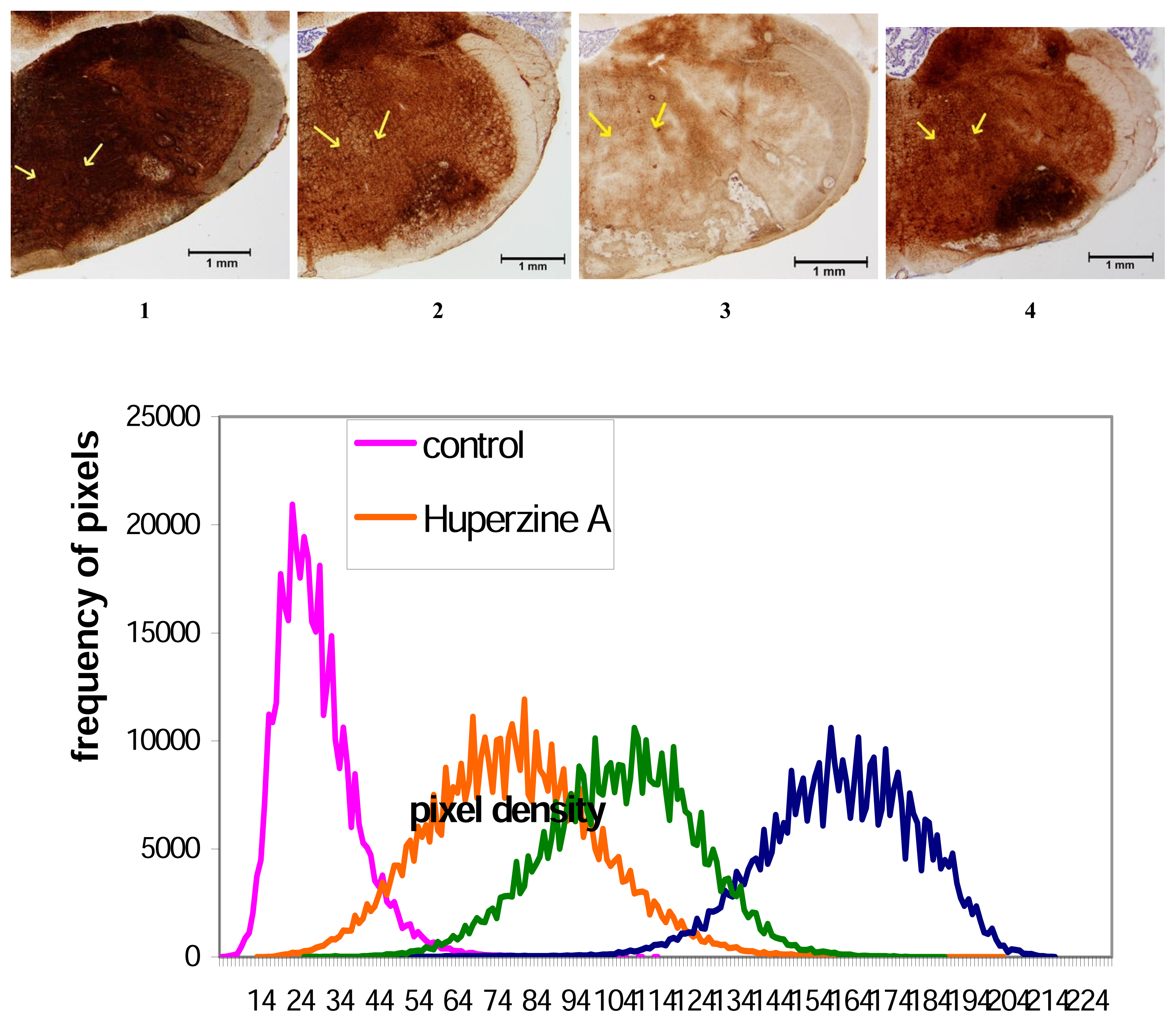
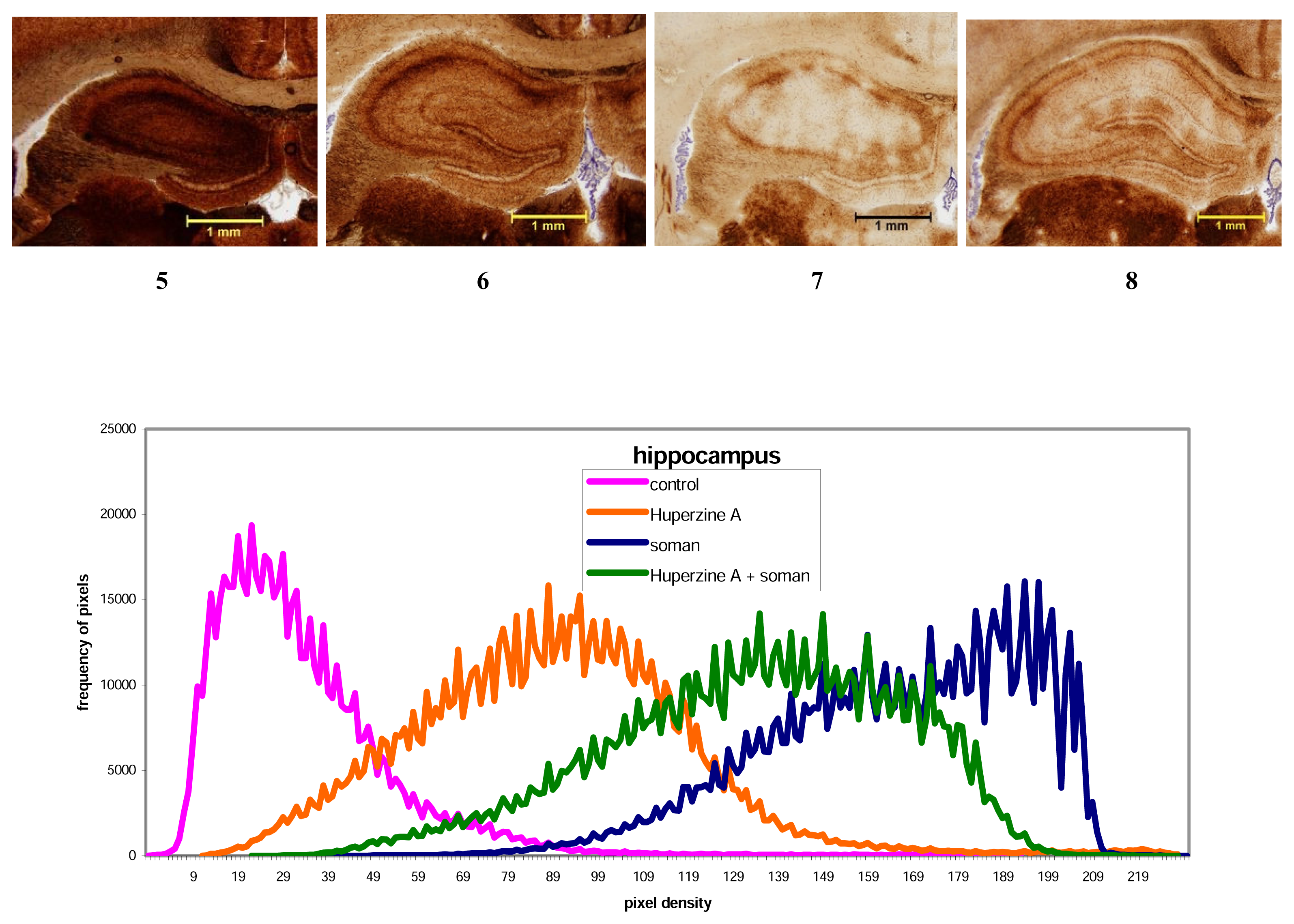
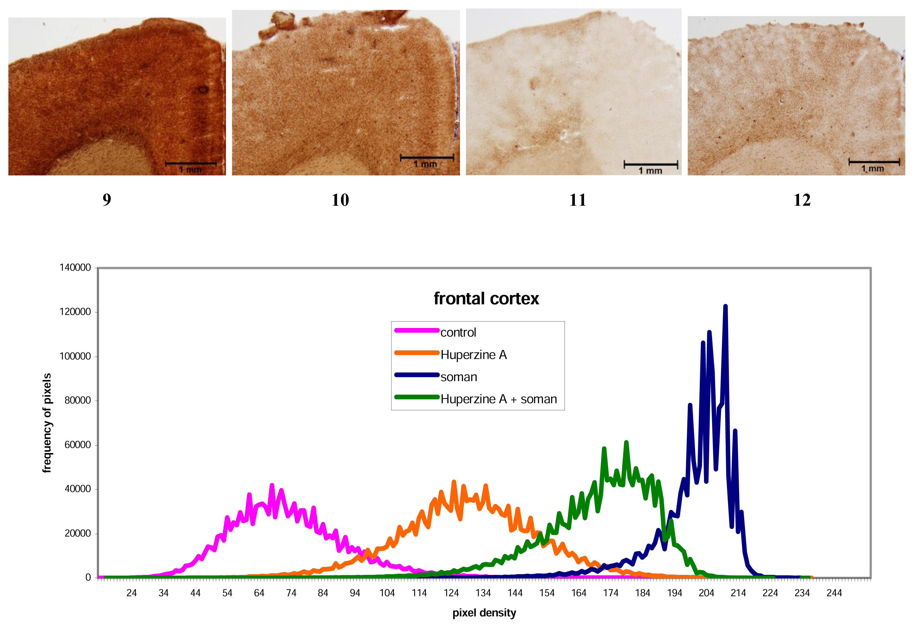
2.3 Histochemical
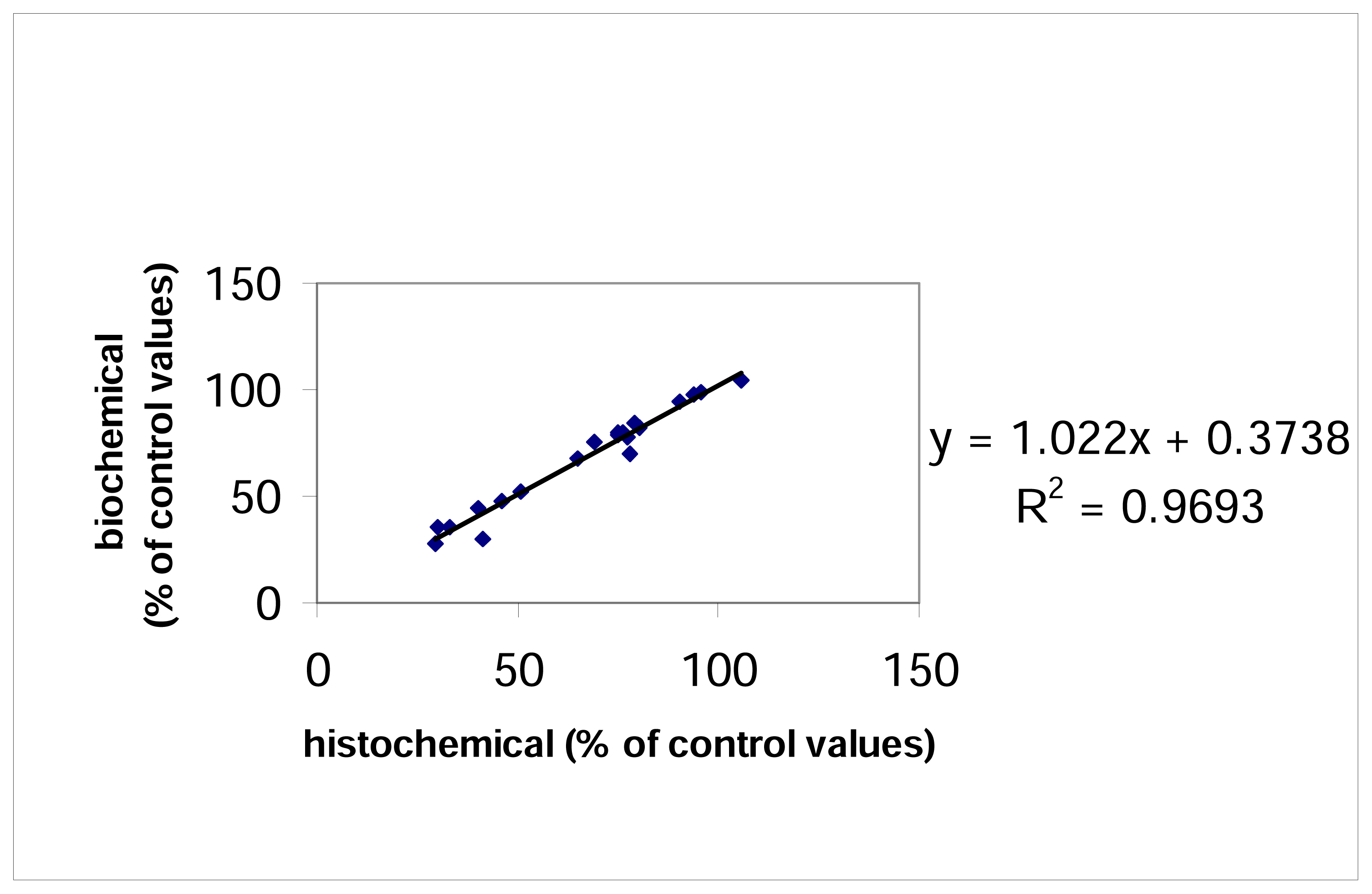
Correlation between results obtained using histochemical and biochemical methods
Discussion
3. Experimental Section
3.1 Animals
3.2 Chemicals
3.3 Intoxication and pretreatment
3.4 Histochemical determination of AChE
3.5 Biochemical determination of AChE
3.6 Statistical evaluation


| Group | CONTROL | SOMAN | HUPERZINE | HUPERZINE+SOMAN | |||||
|---|---|---|---|---|---|---|---|---|---|
| Area | H | B | H | B | H | B | H | B | |
| a | 181.1 | 253.0±13.3 | 53.4 | 70.1±10.5 | 125.6 | 190.2±12.1 | 83.8 | 121.4±12.4 | |
| FC | % | 100 | 100 | 29.V | 27.7c | 69.3 | 75.2a,d | 46.3 | 48.0 b,c,d |
| a | 221.45 | 301.6±15.3 | 89.5 | 135.5±14.7 | 165.9 | 241.3±14.6 | 112.5 | 158.2±13.8 | |
| Hipp | % | 100 | 100 | 40.4 | 44.9 | 74.9 | 80.0a,d | 50.8 | 52.5 b,d |
| a | 203.4 | 262.0±14.3 | 66.8 | 92.7±13.8 | 184.2 | 249.1±14.2 | 154.7 | 209.6±14.3 | |
| HTh | % | 100 | 100 | 32.8 | 35.4c | 90.6 | 95.0d | 76.0 | 80.0 b,c,d |
| a | 187.9 | 251.3±15.3 | 141.05 | 198.3±14.3 | 198.35 | 261.4±13.9 | 176.4 | 246.0±14.1 | |
| NR | % | 100 | 100 | 75.1 | 78.9c | 105.6 | 104.0 | 93.9 | 97.9 c |
| a | 226.9 | 385.0±13.3 | 63.7 | 101.5±12.3 | 176.5 | 269.5±12.9 | 147.4 | 261.8±13.5 | |
| F.ret | % | 100 | 100 | 28.I | 26.4c | 77.8 | 70.0a | 65.0 | 68.0 b,c |
| a | 192.7 | 251.2±12.3 | 57.7 | 89.6±10.8 | 152.3 | 213.5±13.0 | 154.7 | 205.8±13.1 | |
| DSep | % | 100 | 100 | 29.IX | 35.7c | 79.0 | 85.0a | 80.3 | 81.9 b,c |
| a | 223.9 | 325.3±14.0 | 74.1 | 114.8±13.7 | 214.1 | 322.0±13.5 | 172.6 | 253.2±14.2 | |
| Th | % | 100 | 100 | 33.1 | 35.3c | 95.6 | 99.0d | 77.1 | 77.8b,c,d |
Acknowledgements
References and Notes
- Bajgar, J. Organophosphates/nerve agent poisoning: mechanism of action, diagnosis, prophylaxis and treatment. Adv. Clin. Chem 2004, 38, 151–216. [Google Scholar]
- Eyer, P. The role of oximes in the menagement of organophosphorus pesticides poisoning. Toxicol. Rev 2003, 22, 165–190. [Google Scholar]
- Rotenberg, J.S.; Newmark, J. Nerve attacks on children: diagnosis and management. Pediatrics 2003, 112, 648–658. [Google Scholar]
- Wiener, S.W.; Hoffman, R.S. Nerve agents: a comprehensive review. J. Int. Care. Med 2004, 19, 22–37. [Google Scholar]
- Bajgar, J. Prophylaxis against organophosphorus poisoning. J. Med. Chem. Def 2003, 1, 1–15. [Google Scholar]
- Bajgar, J.; Fusek, J.; Sevelova, L.; Kassa, J. Complex prophylaxis against nerve agent intoxication. The Eighth International Symposium on Protection against Chemical and Biological Warfare Agents, Goteburg, Sweden, 2–6 June 2004; Abstracts. 2004; p. 51. [Google Scholar]
- Jiang, H.L.; Luo, X.M.; Bai, D.L. Progress in clinical, pharmacological, chemical and structural biological studies of huperzine A: a drug of traditional Chinese medicine origin for the tratment of Alzheimer’s disease. Curr. Med. Chem 2003, 10, 2231–2252. [Google Scholar]
- Lallement, G.; Foquin, A.; Dorandeu, F.; Baubichon, D.; Aubriot, S.; Carpentier, P. Subchronic administration of various pretreatments of nerve agent poisoning. I. Protection of blood and central cholinesterases, innocuousness towards blood-brain barrier permeability. Drug Chem. Toxicol 2001, 24, 151–164. [Google Scholar]
- Lallement, G.; Foquin, A.; Dorandeu, F.; Baubichon, D.; Carpentier, P. Subchronic administration of various pretreatments of nerve agent poisoning. II. Compared efficacy against soman toxicity. Drug Chem. Toxicol 2001, 24, 165–180. [Google Scholar]
- Lallement, G.; Demoncheaux, J.P.; Foquin, A.; Baubichon, D.; Galonnier, M.; Clarencon, D.; Dorandeu, F. Subchronic administration of pyridostigmine or huperzine to primates: compared efficacy against soman toxicity. Drug Chem. Toxicol 2002, 25, 309–320. [Google Scholar]
- Lallement, G.; Taille, V.; Baubichon, D.; Carpentier, P.; Collombet, J.M.; Filliat, P.; Foquin, A.; Four, E.; Masqueliez, C.; Testylier, G.; Tondulli, L.; Dorandeu, F. Review of the value of huperzine as pretreatment of organophosphate poisoning. Neurotoxicology 2002, 23, 1–5. [Google Scholar]
- Grunwald, J.; Raveh, L.; Doctor, B.P.; Ashani, Y. Huperzine-A as a pretreatment candidate drug against nerve agent toxicity. Life Sci 1994, 54, 991–997. [Google Scholar]
- Albuquerque, E.X.; Pereira, E.F.R.; Aracava, Y.; Fawcett, W.P.; Oliveira, M.; Randall, W.R.; Hamilton, T.A.; Kan, R.K.; Romano, J.A.; Adler, M. Effective countermeasures against poisoning by organophosphorus insecticides and nerve agents. Proc. Nat. Acad. Sci. USA 2006, 103, 13220–13225. [Google Scholar]
- Ashani, Z.; Peggins, J.O.; Doctor, B.P. Mechanism of inhibition of cholinesterase by Huperzine A. Biochem. Biophys. Res. Commun 1992, 184, 719–726. [Google Scholar]
- Gordon, R.K.; Haigh, J.R.; Garcia, G.E.; Feaster, S.R.; Riel, M.A.; Lenz, D.E.; Aisen, P.S.; Doctor, B.P. Oral administration of pyridostigmine bromide and huperzine A protects human whole blood cholinesterases from ex in vivo exposure to soman. Chem-Biol. Interact 2005, 157, 239–246. [Google Scholar]
- Patocka, J.; Kassa, J. Huperzine A – prospective prophylactic antidote against organophosphate warfare agent poisoning. ASA Newslett 1999, 99–2, 16–19. [Google Scholar]
- Wang, R.; Yan, H.; Tang, X.C. Progress in studies of huperzine A, a natural cholinesterase inhibitor from Chinese herbal medicine. Acta Pharmacol. Sin 2006, 27, 1–26. [Google Scholar]
- Eckert, S.; Eyer, P.; Muckter, H.; Worek, F. Kinetic analysis of the protection afforded by reversible inhibitors against irreversible inhibition of acetylcholinesterase by highly toxic organophosphorus compounds. Biochem. Pharmacol 2006, 72, 344–357. [Google Scholar]
- Gupta, R.C. Brain regional heterogeneity and toxicological mechanisms of organophosphates and carbamates. Toxicol. Mech. Methods 2004, 14, 103–143. [Google Scholar]
- Bajgar, J.; Hajek, P.; Slizova, D.; Krs, O.; Fusek, J.; Kuca, K.; Jun, D.; Bartosova, L.; Blaha, V. Changes of acetylcholinesterase activity in different brain areas following intoxication with nerve agents: biochemical and histochemical study. Chem-Biol. Interact 2007, 165, 14–21. [Google Scholar]
- Paxinos, G.; Watson, C. The rat brain in stereotactic coordinates; Academic Press: New York, 1987. [Google Scholar]
- Lojda, Z.; Gossrau, R.R.; Schiebler, T.H. Enzyme Histochemistry – A Laboratory Manual; Springer: New York, 1979. [Google Scholar]
- Benali, A.; Leefken, I.; Eysel, U.; Weile, E. A computerized image analysis system for quantitative analysis of cells in histological brain sections. J. Neurosci. Meth 2003, 125, 33–43. [Google Scholar]
- Hammond, P.I.; Jelacic, T.; Padilla, S.; Brimijoin, S. Quantitative, video-based histochemistry to measure regional effects of anticholinesterase pesticides in rat brain. Anal. Biochem 1996, 241, 82–92. [Google Scholar]
- Ellman, G.L.; Courtney, D.K.; Andres, V.; Featherstone, R.M. A new and rapid colorimetric determination of acetylcholinesterase activity. Biochem. Pharmacol 1961, 7, 88–95. [Google Scholar]
- Giacobini, E.; Pepeu, G. Cholinesterases in human brain: the effect of cholinesterase inhibitors on Alzheimer’s disease and related disorders. In The Brain Cholinergic System in Health and Disease; Informa Healthcare: Abingdon, UK, 2006; pp. 235–264. [Google Scholar]
- Gupta, R.C.; Patterson, G.T.; Dettbarn, W.D. Biochemical and histochemical alterations following acute soman intoxication in the rat. Toxicol. Appl. Pharmacol 1987, 87, 393–402. [Google Scholar]
- Shih, T.-M.; Kan, R.K.; McDonough, J.H. In vivo cholinesterase inhibitory specificity of organophosphorus nerve agents. Chem-Biol. Interact 2005, 157. [Google Scholar]
- Pope, C.N. Central nervous system effects and neurotoxicity. In Toxicology of Organophosphate and Carbamate Compounds; Elsevier-Academic Press: Amsterdam, 2006; pp. 271–291. [Google Scholar]
- Sungur, M.; Guven, M. Intensive care management of organophosphate insecticide poisoning. Crit. Care 2001, 5, 211–215. [Google Scholar]
- Goswany, R.; Chaudhuri, A.; Mahashur, A.A. Study of respiratory failure in organophosphate and carbamate poisoning. Heart Lung 1994, 23, 466–472. [Google Scholar]
- Stewart, W.C.; Anderson, E.A. Effect of a cholinesterase inhibitor when injected into the medulla of the rabbit. J. Pharmacol. Exp. Ther 1968, 162, 309–318. [Google Scholar]
- Bird, S.B.; Gaspari, R.J.; Dickson, E.W. Early death due to severe organophosphate poisoning is a centrally mediated process. Acad. Emerg. Med 2003, 10, 295–298. [Google Scholar]
© 2007 by MDPI Reproduction is permitted for noncommercial purposes.
Share and Cite
Bajgar, J.; Hajek, P.; Karasova, J.; Slizova, D.; Krs, O.; Kuca, K.; Jun, D.; Fusek, J.; Capek, L. Inhibition of Acetylcholinesterase in Different Structures of the Rat Brain Following Soman Intoxication Pretreated with Huperzine A. Int. J. Mol. Sci. 2007, 8, 1165-1176. https://doi.org/10.3390/i8111165
Bajgar J, Hajek P, Karasova J, Slizova D, Krs O, Kuca K, Jun D, Fusek J, Capek L. Inhibition of Acetylcholinesterase in Different Structures of the Rat Brain Following Soman Intoxication Pretreated with Huperzine A. International Journal of Molecular Sciences. 2007; 8(11):1165-1176. https://doi.org/10.3390/i8111165
Chicago/Turabian StyleBajgar, Jiri, Petr Hajek, Jana Karasova, Dasa Slizova, Otakar Krs, Kamil Kuca, Daniel Jun, Josef Fusek, and Lukas Capek. 2007. "Inhibition of Acetylcholinesterase in Different Structures of the Rat Brain Following Soman Intoxication Pretreated with Huperzine A" International Journal of Molecular Sciences 8, no. 11: 1165-1176. https://doi.org/10.3390/i8111165