Article

DNA-Binding Study of Tetraaqua-bis(p-nitrobenzoato)cobalt(II) Dihydrate Complex: [Co(H2O)4(p-NO2C6H4COO)2]·2H2O

1
Department of Chemistry, Faculty of Arts and Sciences, Kafkas University, 36300 Kars, TurkeyDepartment of Chemistry, Faculty of Arts and Sciences, Kafkas University, 36300 Kars, Turkey
2
Department of Biochemistry, Faculty of Veterinary Medicine, Kafkas University, Kars, Turkey
*
Author to whom correspondence should be addressed.
Int. J. Mol. Sci. 2007, 8(7), 564-571; https://doi.org/10.3390/i8070564
Submission received: 27 March 2007 / Revised: 25 May 2007 / Accepted: 4 June 2007 / Published: 27 June 2007
(This article belongs to the Section Biochemistry)

Abstract

:
The interaction of [Co(H2O)4(p-NO2C6H4COO)2]· 2H2O with sheep genomic DNA has been investigated by spectroscopic studies and electrophoresis measurements. The interaction between cobalt(II) p-nitrobenzoate and DNA has been followed by gel electrophoresis while the concentration of the complex was increased from 0 to 14 mM. The spectroscopic study and electrophoretic experiments support the fact that the complex binds to DNA by intercalation via p-nitrobenzoate into the base pairs of DNA. The mobility of the bands decreased as the concentration of complex was increased, indicating that there was increase in interaction between the metal ion and DNA.

1. Introduction

The interaction of transition metal complexes with DNA has been extensively studied in the past few years. The carcinogenicity of cobalt, nickel, cadmium, and arsenic compounds have been known for the long time. Even though only weakly mutagenic, several kinds of cellular damage have been identified, related with their carcinogenic potentials [1]. They were put in the induction of oxidative potential interactions with the removal of different kinds of DNA damage. DNA damage and epigenetic alterations, like gene silencing, change in DNA methylation patterns as shown for particulate nickel compounds and arsenite [1]. Additionally, there is accumulating evidence that metal ions interfere with distinct steps of diverse DNA repair systems. First indications that metal ions may diminish DNA repair processes were obtained by pronounced comutagenic effects of arsenic(III), cobalt(II), nickel(II) and cadmium(II) in bacteria as well as in mammalian cells. Based on these observations, detailed studies have been conducted to oxidative elucidate potential interactions with the removal of different types of DNA damage [1].
These studies are also important for the development of probes nucleic acid structure [25] as well as for determining the mechanism of metal ion toxicity [68]. There has been major interest in the rational design of novel transition metal complexes which bind and cleave duplex DNA with high sequence or structure selectivity [911]. Indeed, there is already a considerable literature involving the practical use of transition metal complexes as chemical nucleases [1224]. Many useful applications of these complexes require that the complex bind to DNA through an intercalative mode with the ligand intercalating into the adjacent base pairs of DNA. However, most of these complexes contain only planar aromatic ligands and investigations of such complexes with ligands containing substituents as DNA-binding reagents have been relatively few. In fact, some of these complexes also exhibit interesting properties upon binding to DNA [2529]. The features common to these complexes are that the molecule has a high affinity for double-stranded DNA, and that the molecule also binds a redox-active metal ion cofactor. The ligands or the metal in these complexes can be varied in an easily controlled manner to facilitate an individual application. All the studies reveal that modification of the metal or ligands would lead to subtle or substantial changes in the binding modes, location and affinity [7,8], giving rise to changes to explore various valuable conformation or site-specific DNA probes and potential chemotherapeutical agents. Currently, much attention has been paid to the complexes of Ru(II) [915]. But, the other metal ion complexes have attracted much less attraction than ruthenium(II) complexes.
In this paper, we chose to concentrate our work on the complex tetraaqua-bis(p-nitrobenzoato)cobalt(II) dihydrate, [Co(H2O)4(p-NO2C6H4COO)2]·2H2O, (CoPNB), which has interesting characteristics and DNA binding properties, but which has not received as much attention as the Ru(II) systems [6,1619]. The binding properties to genomic DNA of this cobalt(II) complex, containing a ligand with nitro substituents at the 5-position of the phenyl group, was studied using absorption spectroscopy and electrophoreis measurements. These studies are necessary for the further comprehension of binding of transition metal complexes to DNA; they can also serve as complementary studies for the corresponding ruthenium complexes.

2. Results and Discussion

The interaction of the complex with DNA was investigated using absorption spectra. The absorption spectra of complex in the absence and presence of genomic DNA (at a constant concentration of the complex) was studied. Figure 1 represents the absorption spectra of CoPNB in the absence and presence of increasing amounts of genomic DNA. In the UV region, the intense absorption bands were seen. The Co(II) complex is believed to be the intraligand transition of the coordinated groups [29]. Addition of increasing amounts of the complex results in hypochromism and bathochromic shift in the UV spectra of CoPNB [29]. The hypochromism in the intraligand band reaches as high as at around 270 nm with red shift at the ratio of [DNA]/[Co]. These spectral characteristics suggest the complex binds to DNA by intercalation. After intercalating the base pairs of DNA, the π-π* orbital of the intercalated ligand can couple with the π orbital of the base pairs, due to the decrease the π-π* transition energy which results in the bathochromism [29].
The interaction of tetraaqua-bis(p-nitrobenzoato)cobalt(II) dihydrate complex, [Co(H2O)4(p-NO2C6H4COO)2]·2H2O, (CoPNB) with DNA was studied by electrophoresis. Figure 2 shows that the complex interacts with DNA.
When genomic DNA was allowed to interact with the complex at pH 7.3, it was seen that although the untreated DNA band appeared, there was no change in the brightness of untreated DNA. There was a substantial increase in intensities of the bands for the concentrations of CoPNB. The real changes in intensity of the bands with the increase in concentrations of the complex were as follows. First of all, in the case of untreated DNA, it was found that the band (lane 1) at 0.14 mM CoPNB concentration had a clearer appreance than that of untreated DNA (Figure 2). In the next 1.4 mM concentration, the band (lane 2) was brighter than that of first band and unteated DNA, and in case of third next highest concentration at 14 mM, it was found that the band (lane 3) was the brightest of first two bands and the band of untreated DNA. they had almost the same brightness and intensities (Figure 2). The electrophoretic mobilities of the bands were seen to decrease as the concentrations of the complex were increased from 0.14, 1.4 to 14 mM (Figure 2).
The results suggest that the covalent binding of the CoPNB complex caused a change in the conformation of the DNA such as more of ethiumdium bromide intercalated, and thus an increase in the intensity of the bands were generally observed.

3. Experimental

3.1 Chemicals

All common solvents and reagents were purchased commercially from Aldrich or Sigma and used without further purification. All experiments involving the interaction of the complex with DNA, were conducted in distilled buffer (Tris–HCl, NaCl, pH 7.4). A solution of genomic DNA in the buffer gave a ratio of UV absorbance at 260 and 280 nm, showing that the DNA was sufficiently free of protein [13].

3.2. DNA Isolation

Blood samples (6–9 mL) were gathered into EDTA-tubes from cattle, and DNA samples were separeted from the leukocytes with a commercial kit (MBI Fermantas®-Genomic DNA Purification Kit #K0512, USA) using the salting out DNA extraction method. Isolated DNA concentrations were measured spectrophotometrically (Spectramax® Plus 384, Molecular Devices, USA), and DNA samples were concentrated at 100 ng/μL prior to processing.

3. 3. Measurements

UV–Vis spectra were recorded on a Heyλios Range UV-Visible Spectrophotometer. For the absorption spectra, an equal solution of DNA was added to both complex solution and reference solution to eliminate the absorbance of DNA itself. For the gel electrophoresis experiments, genomic DNA was treated with Co(II) complex in 30 mM Tris–HCl, 15 mM NaCl buffer, pH 7.3 Agarose gel (1.5 % w/v) in TBE buffer (45 mM Tris, 45 mM boric acid, and 1 mM EDTA, pH 7.3), containing 0.5 μml−1 of ethidium bromide, was prepared. Then, 15 μL of each of the incubated complex-DNA mixtures was loaded on the gel, and electrophoresis, was carried out under TBE buffer system at 50 V for 24 h. At the end of electrophoresis, the gel was visualised under UV light using a Bio-Rad Trans illuminator. The illuminated gel was photographed by using a Polaroid camera (a red filter and Polaroid film were used) [30].

3.4 Synthesis

The complex, [Co(H2O)4(p-NO2C6H4COO)2]·2H2O, tetraaqua-bis(p-nitrobenzoato)cobalt(II) dihydrate, was sythesized (Figure 3) [31]. The title compound was prepared from the reaction CoSO4·7H2O (0.01 mol) with sodium p-nitrobenzoate solution (0.02 mol). The mixture was filtered and set aside to be crystallized at ambient temperature for a few days. Pink crystals were obtained (Figure 4) [31], and Figure 5 shows octahedral environment of the cobalt atom [31].

3. 5 Methods

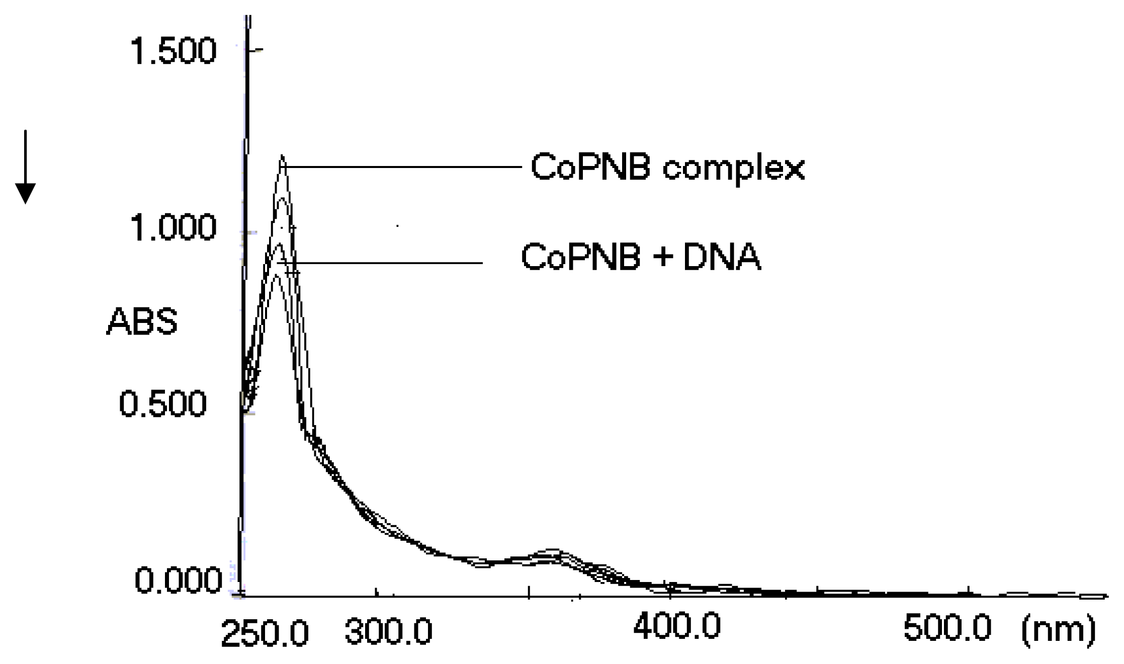
3.5.1 Cobalt(II) p-nitrobenzoate (CoPNB) complex-Genomic DNA Binding Experiment

The DNA was allowed to interact with the metal complex. The DNA-binding experiments were performed at the room temperature. For these series of experiments, cobalt(II) p-nitrobenzoate complex was used as the source of reactive. The solutions of complex were prepared in MiliQ water and sterilized by passing through Milipore filter. The pH of the solutions was fixed to 7.3 by adding slowly NaOH solution. The solution of genomic DNA in the buffer, consisting of 1 mM Tris-HCI at pH 7.3, 1 mM NaCI and 1 mM EDTA, was used [30]. Appropriate volume of the complex was added to 5 μl of genomic DNA, and the total volume was made up to 100 μl by adding MilliQ water, so that the concentration of the complex ranged from 0 to 14 mM while that of DNA remained unchanged in terms of nucleotide. The mixtures were incubated for 24 h, and then the reaction was stopped by rapid cooling at 0 ºC. At the end of incubation, 6μl of loading dye (0.25 % bromophenol blue in 40 % sucrose solution) was added to the mixtures [30]. In order to compare to the effect of interaction of the metal complex between DNA and the complex, the sets of electrophoretic assay were performed.

Conclusions

Complex [cobalt(II) p-nitrobenzoate] was synthesized and characterized. The results from optical experiments together with electrophoretic measurement support complex binds to DNA via electrostatic interaction as well as to partial intercalation via the ring into the base pairs of DNA. The results suggest that the covalent binding of the metal complex caused a change in the conformation of genomic DNA such as more of intercalated and thus an increase in intensity of the band was generally observed. The results described in this study show that changing the ligand environment can modulate the binding property of the complex with DNA [30].
Figure 1. Absorption spectra of CoPNB (14 mM) in the absence and presence upon addition of genomic DNA (0–10 mM). Arrow represents the absorbance changing when concentration of genomic DNA is increased.
Ijms 08 00564f1
Figure 2. Interaction between cobalt(II) p-nitrobenzoate and genomic DNA in presence of TAE buffer at pH 7.3 in air, and 24 h incubation. Lane 1–3: DNA + cobalt(II) p-nitrobenzoate with 0.14, 1.4 to 14 mM, respectively; lane 4: untreated genomic DNA in in TAE buffer at pH 7.3 in air, and incubation for 24 h.
Ijms 08 00564f2
Figure 3. Chemical structure of the complex ([Co(p-O2NC6H4COO)2(H2O)4]·2H2O).
Ijms 08 00564f3
Figure 4. Crystal structure of Cobalt(II) p-nitrobenzoate complex [31].
Ijms 08 00564f4
Figure 5. Octahedral environment of the cobalt atom [31].
Ijms 08 00564f5

Acknowledgements

The Authors thank Assistant Professor Hanife Ozbay Biology Department of the School of Arts and Sciences at Kafkas University, for her help.

Abbreviations

[Co(H2O)4 (p-NO2C6H4COO)2]·2H2O
tetraaqua-bis(p-nitrobenzoato)cobalt(II) Dihydrate
Tris
tris(hydroxymethyl)aminomethane
UV/VIS
UV-visible
CoPNB
Cobalt(II) p-nitrobenzoate

References

  1. Hartwig, A.; Schwerdtle, T. Interactions by carcinogenic metal compounds with DNA repair processes: toxicological implications. Toxicology Letters 2002, 127, 47–58. [Google Scholar]
  2. Burrows, C.J; Rokita, S.E. Recognition of Guanine Structure in Nucleic Acids by Nickel Complexes. Acc. Chem. Res. 1994, 27, 295–203. [Google Scholar]
  3. Pyle, A.M.; Barton, J.K. Lippard, S.J., Ed.; Progress in Inorganic Chemistry; Wiley: New York, 1990; Volume 38, p. 413. [Google Scholar]
  4. Tullius, T.D. Metal-DNA Chemistry; ACS Symposium Series; Volume 402, American Chemical Society: Washington, DC, 1989. [Google Scholar]
  5. Banerjee, A.R.; Jaeger, J.A.; Turner, D.H. Thermal unfolding of a group I ribozyme: the low-temperature transition is primarily disruption of tertiary structure. Biochemistry 1993, 32, 153–160. [Google Scholar]
  6. Klein, C.B.; Frenkel, K.; Costa, M. The role of oxidative processes in metal carcinogenesis. Chem. Res. Toxico 1991, 4, 592–598. [Google Scholar]
  7. Ksaprzak, K.S. DNA deaminating ability and genotoxicity. Chem. Res. Toxicol 1991, 4, 604–612. [Google Scholar]
  8. Vijayalakshmi, R.; Kanthimathi, M.; Subramanian, V.; Nair, B.U. Interaction of DNA with [Cr(Schiff base)(H2O)2]CIO4. Biochim. Biophys. Acta 2000, 1475, 157–164. [Google Scholar]
  9. Geierstanger, B.H.; Marksich, M.; Dervan, P.B.; Wemmer, D.E. Recognition of DNA by designed ligands at subnanomolar concentrations. Science 1994, 266, 646–652. [Google Scholar]
  10. Liu, C.; Zhou, J.; Li, Q.; Wang, L; Liao, Z.; Xu, H. Synthesis, DNA-binding and cleavage studies of macrocyclic copper. J. Inorg. Biochem. 1996, 75, 233–240. [Google Scholar]
  11. Prativiel, G.; Bernadou, J.; Meunier, B. Structure, and DNA Binding Studies of Copper(II). Angew. Chem. Int. Ed. Engl 1995, 34, 746–753. [Google Scholar]
  12. Sigman, D.S.; Mazumder, A.; Perrin, D.M. Chemical nucleases. Chem. Rev 1993, 93, 2295–2304. [Google Scholar]
  13. Vijayalakshmi, R.; Kanthimathi, M.; Subramanian, V.; Nair, B. DNA cleavage by a Chromium(III) complex. Biochem. Biophys. Res. Commun 2000, 271, 731–743. [Google Scholar]
  14. Frey, S.T.; Sun, H.H.J.; Murthy, N.N.; Karlin, K.D. A New Complexes and Its Reactions with Plasmid DNA. Inorg. Chim. Acta 1996, 242, 329–337. [Google Scholar]
  15. Ganesan, V.V.; Nair, B.U. Synthesis, Characterization, and DNA Binding Studies of a Chromium(III) Complex Containing a Tridentate Ligand. J. Inorg. Biochem 2003, 93, 271–280. [Google Scholar]
  16. Barton, J.K. Metals and DNA: molecular left-handed complements. Science 1986, 233, 727–734. [Google Scholar]
  17. Carter, M.; Rodriguez, M.; Bard, A.J. Intercalative ligands upon interaction with double-helical. DNA. J. Am. Chem. Soc 1989, 111, 8901–8910. [Google Scholar]
  18. Morgan, R.J.; Chatterjee, S.; Baker, A.D.; Strekas, T.C. Effects of Ligand Binding of Ru(2,2′-bipyridine)2L (2+) to Calf Thymus DNA. Inorg. Chem 1991, 30, 2687–2693. [Google Scholar]
  19. Sitlani, A.S.; Long, E.C.; Pyle, A.M.; Barton, J.K. Oxidative DNA damage through long-range electron transfer. J. Am. Chem. Soc 1992, 114, 2303–2311. [Google Scholar]
  20. Turro, C.; Bossman, S.H.; Jenkins, Y.; Barton, J.K.; Turro, N.J. Proton Transfer Quenching of the MLCT Excited State of Ru(pehn)2dppz2+ in Homogeneous Solution and Bound to DNA. J. Am. Chem. Soc 1995, 117, 9026–9032. [Google Scholar]
  21. Zou, X.H.; Ye, B.H.; Li, H.; Liu, J.G.; Xiong, Y.; Ji, L.N. Mono- and bi-nuclear ruthenium(II) complexes containing a new asymmetric ligand 3-(pyrazin-2-yl)-as-triazino[5,6-f]1,10-phenanthroline: synthesis, characterization and DNA-binding properties. J. Chem. Soc, Dalton Trans 1999, 1423–1430. [Google Scholar]
  22. Xiong, Y.; Ji, L.N. Coord. Synthesis, DNA-binding and DNA-mediated luminescence quenching of Ru(II)polypyridine complexes. Chem. Rev 1999, 711, 185–191. [Google Scholar]
  23. Xiong, Y.; He, X.F.; Zou, X.H.; Wu, J.Z.; Chen, X.M.; Ji, L.N.; Li, R.H.; Zhou, J.Y.; Yu, K.B. Interaction of polypyridyl ruthenium(II) complexes containing non-planar ligands with DNA. J. Chem. Soc. Dalton Trans 1999, 19. [Google Scholar]
  24. Liu, J.G.; Ye, B.H; Zhang, Q.L.; Zhou, X.H.; Tian, X.; Ji, L.N. Functions and applications of polypyridyl complexes in DNA. J. Biol. Inorg. Chem. 2000, 5, 119–123. [Google Scholar]
  25. Rehmann, J.P.; Barton, J.K. 1H NMR studies of tris(phenanthroline) metal complexes bound to oligonucleotides: characterization of binding modes. Biochemistry 1990, 29, 1701–1712. [Google Scholar]
  26. Arounaguiri, S.; Maiya, B.G. Dipyridophenazine Complexes of Cobalt(III) and Nickel(II): DNABinding and Photocleavage Studies. Inorg. Chem 1996, 35, 4267–4272. [Google Scholar]
  27. Jin, L.; Yang, P. Sythesis and DNA binding studies of C(III) mixed-ligand complex containing dipyrido phenazine and phen. Polyhedron 1997, 16, 3395–3399. [Google Scholar]
  28. He, X.F.; Wang, L.; Chen, H.; Xu, L.; Ji, L.N. Synthesis, characterization and DNA binding study of polypyridyl mixed-ligand complexes. Polyhedron 1998, 17, 3161–3172. [Google Scholar]
  29. Zhang, QL.; Liu, J.G.; Chao, H.; Xue, G.Q.; Ji, L.N. DNA-binding and photocleavage studies of cobalt(III) polypyridyl complexes. J. Inorg. Biochem 2001, 83, 49–58. [Google Scholar]
  30. Arslantas, A.; Devrim, A.K.; Kaya, N.; Necefoglu, N. Studies on the Interaction between Zinc-Hydroxybenzoite Complex and Genomic DNA. Int. J. Mol. Sci 2006, 7, 111–119. [Google Scholar]
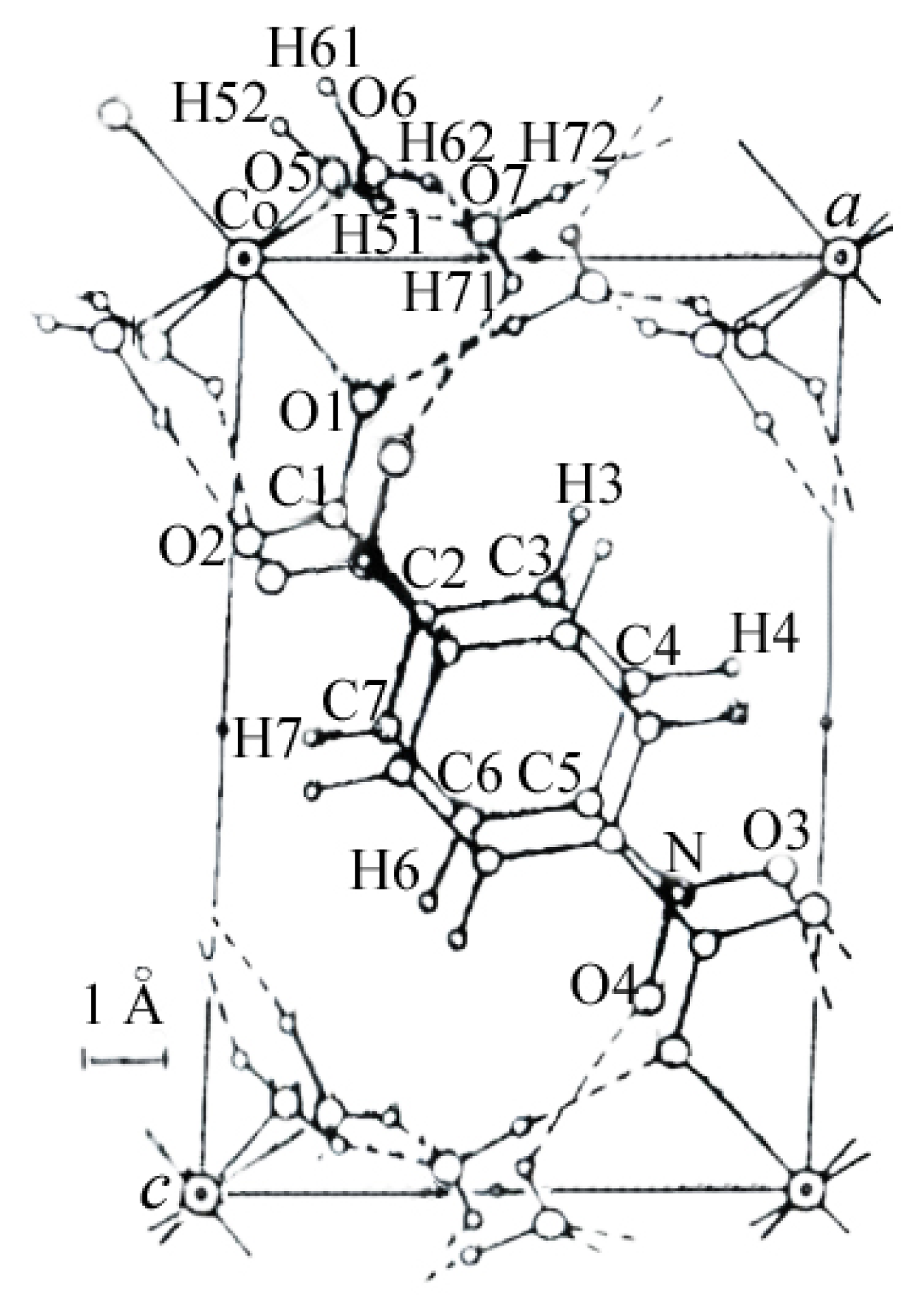
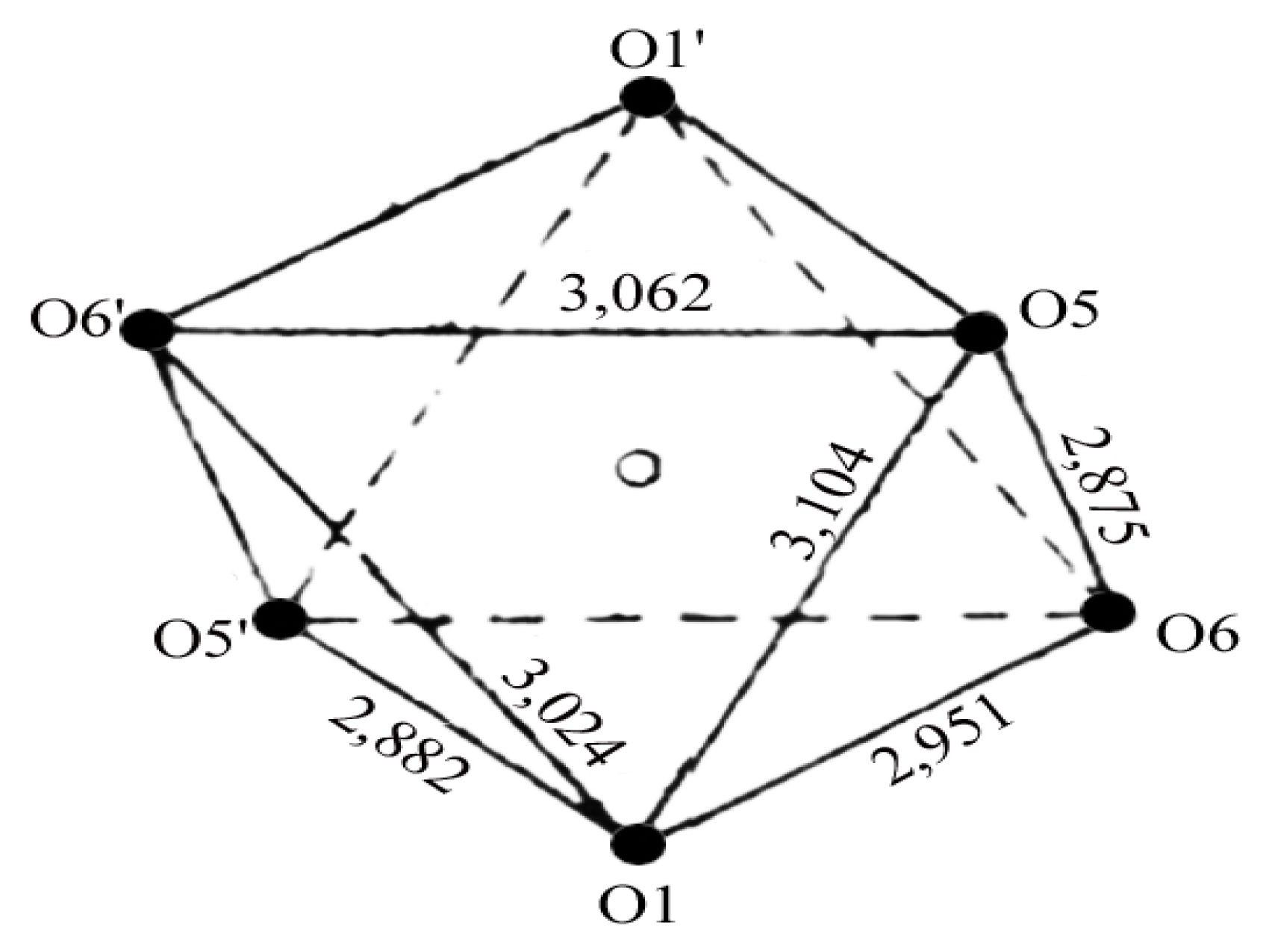
  31. Nadzhafov, G.N.; Shnulin, A.N.; Mamedov, K.S. Crystal and Molecular structure of Di(pnitrobenzoato) tetraaquaocobalt(II) dihydrate. J. Struct. Chem 1981, 22, 898, –902. 686–687. [Google Scholar]

