Article

Three Dimensional Pharmacophore Modelling of Monoamine oxidase-A (MAO-A) inhibitors

by
Kalapatapu V.V.M. Sairam
1,
Roop K. Khar
1,*,
Rama Mukherjee
2 and
Swatantra K. Jain
3
1
Department of Pharmaceutics, Faculty of Pharmacy, Jamia Hamdard (Hamdard University), New Delhi-110062, India
2
Dabur Research Foundation, 22 Site IV, Sahibabad, Ghaziabad - 201010 U.P, India
3
Department of Biotechnology, Faculty of Science, Jamia Hamdard (Hamdard University), New Delhi - 110062, India
*
Author to whom correspondence should be addressed.
Int. J. Mol. Sci. 2007, 8(9), 894-919; https://doi.org/10.3390/i8090894
Submission received: 2 May 2007 / Revised: 25 July 2007 / Accepted: 30 July 2007 / Published: 3 September 2007
(This article belongs to the Special Issue The Chemical Bond and Bonding)

Abstract

:
Flavoprotein monoamine oxidase is located on the outer membrane of mitochondria. It catalyzes oxidative deamination of monoamine neurotransmitters such as serotonin, norepinephrine and dopamine and hence is a target enzyme for antidepressant drugs. MAO (mono amine oxidase) has two isoforms, namely MAO-A and MAO-B. MAO-A isoform has higher affinity for serotonin and norepinephrine, while; MAO-B preferentially deaminates phenylethylamine and benzylamine. These important properties determine the clinical importance of MAO inhibitors. Selective MAO-A inhibitors are used in the treatment of neurological disorders such as depression. In this article we have developed a Hypogen pharmacophore for a set of 64 coumarin analogs and tried to analyze the intermolecular H-bonds with receptor structure.

1. Introduction

Monoamine oxidase is a flavoprotein located at the outer membrane of mitochondria in neuronal, glial and other cells. It catalyzes oxidative deamination of monoamine neurotransmitters such as serotonin, norepinephrine and dopamine and hence is a target enzyme for antidepressant drugs. In addition, it is also responsible for the biotransformation of 1-methyl-4-phenyl-1,2,3,6-tetrahydropyridine into 1-methyl-4-phenylpyridinium a Parkinsonian producing neurotoxin [1]. MAO may also have a role in apoptotic processes.
MAO exists in two forms, namely MAO-A and MAO-B. Cloning and sequencing of MAO-A and MAO-B has great impact in understanding their molecular properties respectively. This has established the fact that both the enzymes are separate and share many similar properties such as covalent link between FAD and cysteine residue, Cys 406 in MAO-A and Cys 397 in MAO-B, through an 8α-(cysteinyl)-riboflavin. In spite of these similarities, two enzymes have separate but overlapping biological functions. Therefore, design of specific inhibitors would lead to little or no side effects which most of existing inhibitors suffer from.
Specific substrates and inhibitors characterize both MAO subtypes. MAO-A has a higher affinity for serotonin and norepinephrine, while; MAO-B preferentially deaminates phenylethylamine and benzylamine. These properties determine the clinical importance of MAO inhibitors. Selective MAO-A inhibitors are used in the treatment of neurological disorders such as depression, whereas the MAO-B inhibitors are useful in the treatment of Parkinson’s and Alzheimer’s disease. In the light of these facts, we ventured into developing a pharmacophore for a set of 64 coumarin analogs [2,3], with close comparisons from structure based interactions.

2. Computational Method

The three-dimensional pharmacophore was developed using the CATALYST 4.1 software [4]. This is an integrated commercially available software package that generates pharmacophores, commonly referred to as hypotheses. It enables the use of structure and activity data for a set of lead compounds to create a hypothesis, thus characterizing the activity of the lead set. At the heart of the software is the HypoGen algorithm that allows identification of hypotheses that are common to the “active” molecules in the training set but at the same time not present in the “inactives”. Structures of 64 monoamine oxidase inhibitors [2,3] of the training and test set were built and energy minimized using smart minimize [5] module from the Cerius2. The CATALYST model treats molecular structures as templates comprising chemical functions localized in space that will bind effectively with complementary functions on the respective binding proteins. The most relevant chemical features are extracted from a small set of compounds that cover a broad range of activity. The best searching procedure was applied to select representative conformers within 20 kcal/mol from the global minimum. The conformational model of the training set was used for hypothesis (pharmacophore) generation within CATALYST, which aims to identify the best 3-dimensional arrangement of chemical functions explaining the activity variations among the compounds in the training set. The automatic generation procedure using the HypoGen algorithm in CATALYST was adopted for generation of the hypotheses. In order to obtain a reliable model, which adequately describes the interaction of ligands with high predictability, the method recommends a collection of training set with biological activity covering 4–5 orders of magnitude for the training set.
The pharmacophore/hypotheses are described by a set of functional features such as hydrophobic, hydrogen-bond donor, hydrogen-bond acceptor, and positive and negative ionizable sites distributed over a 3D space. The hydrogen-bonding features are vectors, whereas all other functions are points. The statistical relevance of the obtained hypotheses is assessed on the basis of their cost relative to the null hypothesis and their correlation coefficient.
Pharmacophore generation was carried out with the 64 monoamine oxidase-A inhibitors (Table 1) by setting the default parameters in the automatic generation procedure in CATALYST such as function weight 0.302, mapping coefficient 0, resolution 10 pm (due to smaller size of the inhibitor set considered for the study), and activity uncertainty 3. As uncertainty Δ in the CATALYST paradigm indicates an activity value lying somewhere in the interval from “activity divided by Δ” to “activity multiplied by Δ”. Hypotheses approximating the pharmacophore of the MAO-A inhibitors are described as a set of aromatic hydrophobic, hydrogen-bond acceptor, hydrogen bond acceptor lipid, positively and negatively ionizable sites distributed within a 3D space. The statistical relevance of various generated hypothesis is assessed on the basis of the cost relative to the null hypothesis and the correlation coefficients. The hypothesis is then used to estimate the activities of the training set. These activities are derived from the best conformation generation mode of the conformers displaying the smallest root-mean-square (RMS) deviations when projected onto the hypothesis. HypoGen considers a pharmacophore that contain features with equal weights and tolerances. Each feature (e.g., hydrogen-bond acceptor, hydrogen-bond donor, hydrophobic, positive ionizable group, etc.) contributes equally to estimate the activity. Similarly, each chemical feature in the HypoGen pharmacophore requires a match to a corresponding ligand atom to be within the same distance (tolerance). Thus, two parameters such as the fit score and conformational energy costs are crucial for estimation of predicted activity of the compounds.

Docking

The crystal structure of monoamine oxidase A with clorgyline (Scheme 1) was used in the study. Protein was prepared as per standard protocol. Docking was carried out using GOLD, which used the genetic algorithm (GA) with default values. Docking was carried out using GOLD, which uses the genetic algorithm (GA). For each of the 10 independent GA runs, a maximum number of 100,000 GA operations were performed on a set of five groups with a population size of 100 individuals. Operator weights for crossover, mutation, and migration were set to 95, 95, and 10, respectively. Default cutoff values of 2.5 Å (dH-X) for hydrogen bonds and 4.0 Å for van der Waals were employed. Hydrophobic fitting points were calculated to facilitate the correct starting orientation of the ligand for docking by placing the hydrophobic ligand atoms appropriately in the corresponding areas of the active site. When the top three solutions attained root-mean-square deviation (RMSD) values within 1.5 Å, GA docking was terminated. The first ranked solutions of the ligands were taken for further analysis.

3. Results and Discussion

We developed a HypoGen 3D-QSAR pharmacophore model for monoamine oxidase-A inhibitory activity from a set of 64 inhibitors [2,3] (Table I). These compounds display a broad range of inhibitory activity against MAO-A with experimental IC50 values ranging from 2.0 × 10⁻⁸ to 1.0 × 10⁻⁴ (Table II). On the basis of the structure-activity relationships within this set of compounds, pharmacophore models of MAO-A inhibition were generated using the CATALYST procedure. The best model produced by CATALYST consisted of the spatial arrangement of five functional groups: two hydrogen-bond acceptors, three hydrophobic groups (Figure 1). The model shows a good correlation (R = 0.95) between experimental and predicted inhibitory activity of the MAO-A inhibitors of the training set. The CATALYST procedure resulted in the generation of 10 alternative pharmacophores describing the MAO-A inhibitory activity of the training set compounds. These pharmacophore models were then evaluated by using them to estimate the inhibitory activity of the training set compounds. The correlation between the estimated and experimental values ranges between 0.95 and 0.66, and the RMS value is 1.2. The statistical significance of the pharmacophores (hypotheses) falls within the recommended range of values in CATALYST. The difference between the fixed and the null cost is found to be 54.4 bits, indicating the robustness of the correlation. The cost difference between the first and the tenth hypothesis is 13.294 bits, closer to the fixed costs than the null costs. All these calculated cost differences were found to be well within the recommended acceptable limits in the cost analysis of the CATALYST procedure. The best pharmacophore model is characterized by two hydrogen bond acceptor functions, three hydrophobic functions (Figure 1) and is also statistically the most relevant model.
The estimated activity values along with the experimental IC50 values for MAO-A inhibition are presented in Table II. Experimentally determined IC50 values versus the calculated activities demonstrate a good correlation (R = 0.95) within the range of uncertainty 3, indicating a good predictive power of the model. The most potent compound in the training set, molecule 62 maps well to the functional features of the pharmacophore with all five features of the molecule mapped well with hypotheses generated, whereas the least potent members of the series, molecule 26, maps poorly with the pharmacophore with only four of five features are mapped missing out the second acceptor feature (Figure 2a, b). Inspection of Figure 2B clearly shows that molecule 26 fails to map the second hydrogen bond acceptor feature of the pharmacophore. Thus, it appears that the second hydrogen bond acceptor feature may be specific requirement for binding to MAO-A.
The test set considered contains three molecules 20, 32 and 34. Activity predicted for the test is also found deviated. Analysis of these test set molecules suggests that bulky substitutions in the 6 and 7 positions of the coumarin ring could be responsible for this deviation in their activity. Estimated activities were calculated by scoring the pharmacophore model on the test set and comparing with the experimental IC50 values.

Enrichment factor analysis
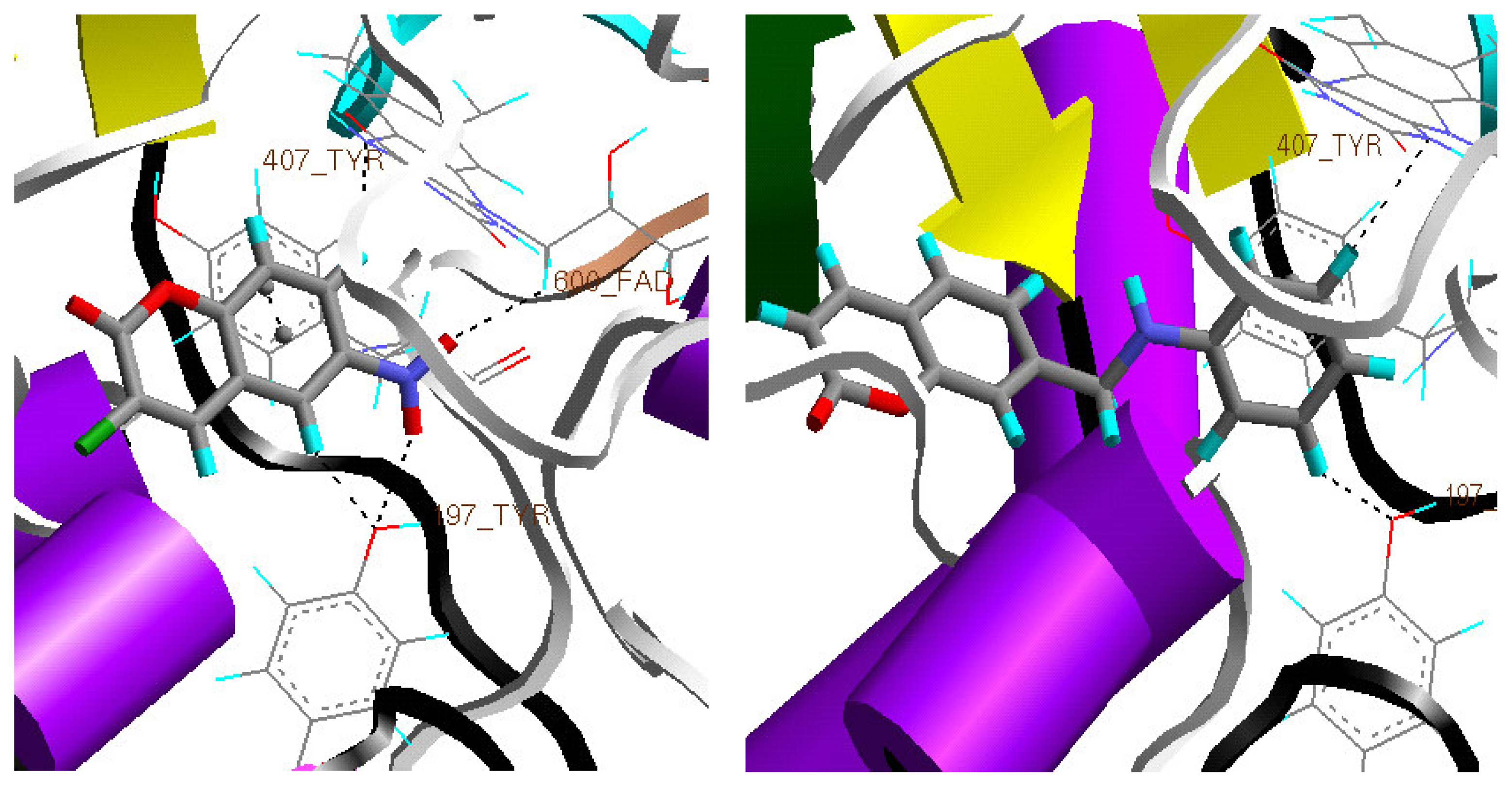
The enrichment factor “ideal” for a set of 125 dataset of compounds that contain 24 monoamine oxidase specific inhibitors and 101 nonspecific inhibitors is 5.35. Of these 24 specific monoamine oxidase inhibitors the pharmacophore generated has picked 17 molecules and within 100 nonspecific inhibitors dataset, model has picked six molecules. Hence enrichment factor calculated over this model generated to 125 dataset is around 4. These numbers speak of evaluation of the model and ability to pick the MAO specific inhibitors in the dataset. Details of 125 dataset are provided in the supporting information.

Docking

Coumarin derivatives were docked into the active site of MAO-A and docking analysis was performed (Figure 3). It was observed that a π interaction with Phe 407 seems to be crucial in its bound ligand clorgyline as well as coumarin analogs docked7. Clorgyline methyl group has a CH···π interaction with phenyl ring of Tyr 407 and CH···N interaction with N1 of FAD (cofactor). This aromatic sandwich with Tyr 407 is important in stabilizing substrate within active site and is also crucial for MAO-A catalytic activity [1]. Pharmacophore model generated has three hydrophobic points which also confirms that aromatic interactions to be very important for stabilizing interactions. Santana et al., in their recent report on the QSAR of coumarin analogs confirms that hydrophobicity along with polarizability to be important physical factor [6]. One of the highest active molecule 64 has a strong OH···O interaction between –OH group of Tyr 197 and oxygen of –NO2, two weak CH···O and one CH···N interactions with Tyr 197 and FAD (phenyl CH of ligand has a CH···O interaction with hydroxy oxygen of Tyr 197, CH···N with N1 of FAD and another CH···O between oxygen of nitro group of the ligand with CH of FAD). Lowest active molecule 4 still maintains a π···π interaction with Tyr 407. This molecule has very fewer interactions and perhaps the low activity can be attributed to this lack of H-bond interactions.

4. Conclusion

This study suggests that it would be difficult for the active site with 7Å width [7] to accommodate bigger molecules, in this case molecules 32 and 34. The same is being reflected in the phamacophore hypothesis. This study partly tries to address the inactivity of the some of the coumarin analogs using analog based methods like pharmacophore hypothesis and structure based methods like docking. A detailed study on each class of compounds would throw light on the optimization aspect in a series of analogs and structural requirements in order to have optimum binding. Understanding of topology and H-bond interactions aiding molecular recognition with receptor govern the biological activity of this set of molecules.

Supplementary Information

Table S1. Following table contains the list of compounds and citation used as a database to find out enrichment factor (measure of validation of the model).
StructureReference
1MAOB specificIjms 08 00894f71Eur. J. Med. Chem.1995, 30, 471–482
2Ijms 08 00894f72Eur. J. Med. Chem.1995, 30, 471–482
3Ijms 08 00894f73Eur. J. Med. Chem.1995, 30, 471–482
4Ijms 08 00894f74J. Enzyme Inhib. Med. Chem.2003, 18, 339–347
5Ijms 08 00894f75J. Med. Chem.1992, 35, 3705–3713
6Ijms 08 00894f76J. Med. Chem.1993, 36, 1157–1167
7Ijms 08 00894f77J. Med. Chem.1993, 36, 1157–1167
8Ijms 08 00894f78J. Med. Chem.1993, 36, 1157–1167
9Ijms 08 00894f79J. Med. Chem.1993, 36, 1157–1167
10Ijms 08 00894f80J. Med. Chem.1995, 38, 4786–4792
11MAOA SpecificIjms 08 00894f81J. Med. Chem.2004, 47, 3455–3461
12Ijms 08 00894f82J. Med. Chem.1994, 37, 2085–2089
13Ijms 08 00894f83J. Med. Chem.1994, 37, 2085–2089
14Ijms 08 00894f84J. Med. Chem.1994, 37, 2085–2089
15Ijms 08 00894f85J. Med. Chem.1993, 36, 1157–1167
16Ijms 08 00894f86J. Med. Chem.1991, 34, 2931–2933
17Ijms 08 00894f87J. Med. Chem.1991, 34, 2931–2933
18Ijms 08 00894f88J. Med. Chem.2005, 48, 2407–2419
19Ijms 08 00894f89Bioorg. Med. Chem. Lett.1994, 4, 1195–1198
20Ijms 08 00894f90J. Med. Chem.1998, 41, 2118–2125
21Ijms 08 00894f91J. Med. Chem.1998, 41, 2118–2125
22Ijms 08 00894f92Eur. J. Med. Chem.1999, 34, 137–151
23Ijms 08 00894f93J. Med. Chem.1997, 40, 2466–2473
24Ijms 08 00894f94J. Med. Chem.1996, 39, 1857–1863
25MixedIjms 08 00894f95J. Med. Chem.1996, 39, 1857–1863
26Ijms 08 00894f96J. Med. Chem.2002, 45, 5260–5279
27Ijms 08 00894f97J. Med. Chem.1994, 37, 2085–2089
28Ijms 08 00894f98J. Med. Chem.1994, 37, 2085–2089
29Ijms 08 00894f99J. Med. Chem.1994, 37, 2085–2089
30Ijms 08 00894f100J. Enzyme Inhib. Med. Chem.2003, 18, 339–347
31Ijms 08 00894f101J. Enzyme Inhib. Med. Chem.2003, 18, 339–347
32Ijms 08 00894f102J. Enzyme Inhib. Med. Chem.2003, 18, 339–347
33Ijms 08 00894f103J. Med. Chem.2004, 47, 3455–3461
34Ijms 08 00894f104J. Med. Chem.2004, 47, 3455–3461
35Ijms 08 00894f105J. Med. Chem.2005, 48, 664–670
36Ijms 08 00894f106J. Med. Chem.2005, 48, 664–670
37Ijms 08 00894f107J. Med. Chem.2005, 48, 2407–2419
38Ijms 08 00894f108J. Med. Chem.2005, 48, 2407–2419
39Ijms 08 00894f109J. Med. Chem.2005, 48, 2407–2419
40Ijms 08 00894f110J. Med. Chem.2005, 48, 2407–2419
41Ijms 08 00894f111J. Med. Chem.2005, 48, 2407–2419
42Ijms 08 00894f112J. Med. Chem.2005, 48, 2407–2419
43Ijms 08 00894f113J. Med. Chem.2005, 48, 2407–2419
44Ijms 08 00894f114J. Med. Chem.2005, 48, 2407–2419
45Ijms 08 00894f115J. Med. Chem.2005, 48, 2407–2419
46Ijms 08 00894f116J. Med. Chem.2005, 48, 2407–2419
47Ijms 08 00894f117J. Med. Chem.2005, 48, 2407–2419
48Ijms 08 00894f118Bioorg. Med. Chem. Lett.1994, 4, 1195–1198
49Ijms 08 00894f119Bioorg. Med. Chem. Lett.1994, 4, 1195–1198
50Ijms 08 00894f120Bioorg. Med. Chem. Lett.1994, 4, 1195–1198
51Ijms 08 00894f121Bioorg. Med. Chem. Lett.1994, 4, 1195–1198
52Ijms 08 00894f122J. Med. Chem.2002, 45, 5260–5279
53Ijms 08 00894f123J. Med. Chem.2002, 45, 5260–5279
54Ijms 08 00894f124J. Med. Chem.2002, 45, 5260–5279
55Ijms 08 00894f125J. Med. Chem.2002, 45, 5260–5279
56Ijms 08 00894f126J. Med. Chem.2002, 45, 5260–5279
57Ijms 08 00894f127J. Med. Chem.1995, 38, 3874–3883
58Ijms 08 00894f128J. Med. Chem.1995, 38, 3874–3883
59Ijms 08 00894f129Bazinaprine
60Ijms 08 00894f130J. Med. Chem.1994, 37, 2085–2089
61Ijms 08 00894f131J. Med. Chem.1993, 36, 1157–1167
62Ijms 08 00894f132J. Med. Chem.1993, 36, 1157–1167
63Ijms 08 00894f133J. Med. Chem.1993, 36, 1157–1167
64Ijms 08 00894f134J. Med. Chem.1993, 36, 1157–1167
65Ijms 08 00894f135J. Med. Chem.1993, 36, 1157–1167
66Ijms 08 00894f136J. Med. Chem.1992, 35, 3705–3713
67Ijms 08 00894f137J. Med. Chem.1992, 35, 3705–3713
68Ijms 08 00894f138J. Med. Chem.1992, 35, 3705–3713
69Ijms 08 00894f139J. Med. Chem.2004, 47, 1760–1766
70Ijms 08 00894f140J. Med. Chem.2004, 47, 3455–3461
71Ijms 08 00894f141J. Med. Chem.2004, 47, 3455–3461
72Ijms 08 00894f142J. Med. Chem.2004, 47, 3455–3461
73Ijms 08 00894f143J. Med. Chem.2004, 47, 3455–3461
74Ijms 08 00894f144J. Med. Chem.2004, 47, 3455–3461
75Ijms 08 00894f145J. Med. Chem.2004, 47, 3455–3461
76Ijms 08 00894f146J. Med. Chem.2004, 47, 3455–3461
77Ijms 08 00894f147J. Med. Chem.2004, 47, 3455–3461
78Ijms 08 00894f148J. Med. Chem.2004, 47, 3455–3461
79Ijms 08 00894f149J. Med. Chem.2005, 48, 664–670
80Ijms 08 00894f150J. Med. Chem.2005, 48, 664–670
81Ijms 08 00894f151J. Med. Chem.2006, 49, 3743–3747
82Ijms 08 00894f152Bioorg. Med. Chem. Lett.1996, 6, 115–120
83Ijms 08 00894f153Bioorg. Med. Chem. Lett.1996, 6, 115–120
84Ijms 08 00894f154Bioorg. Med. Chem. Lett.1996, 6, 115–120
85Ijms 08 00894f155Bioorg. Med. Chem. Lett.1996, 6, 115–120
86Ijms 08 00894f156Bioorg. Med. Chem. Lett.1996, 6, 115–120
87Ijms 08 00894f157Bioorg. Med. Chem. Lett.1996, 6, 115–120
88Ijms 08 00894f158Bioorg. Med. Chem. Lett.1996, 6, 115–120
89Ijms 08 00894f159Bioorg. Med. Chem. Lett.1996, 6, 115–120
90Ijms 08 00894f160Bioorg. Med. Chem. Lett.1996, 6, 115–120
91Ijms 08 00894f161Bioorg. Med. Chem. Lett.1996, 6, 115–120
92Ijms 08 00894f162Bioorg. Med. Chem. Lett.1996, 6, 115–120
93Ijms 08 00894f163Bioorg. Med. Chem. Lett.1996, 6, 115–120
94Ijms 08 00894f164Bioorg. Med. Chem. Lett.1994, 4, 1195–1198
95Ijms 08 00894f165Bioorg. Med. Chem. Lett.1994, 4, 1195–1198
96Ijms 08 00894f166Bioorg. Med. Chem. Lett.1994, 4, 1195–1198
97Ijms 08 00894f167Bioorg. Med. Chem. Lett.1994, 4, 1195–1198
98Ijms 08 00894f168Bioorg. Med. Chem. Lett.1994, 4, 1195–1198
99Ijms 08 00894f169Bioorg. Med. Chem. Lett.1994, 4, 1195–1198
100Ijms 08 00894f170Bioorg. Med. Chem. Lett.1994, 4, 1195–1198
101Ijms 08 00894f171Bioorg. Med. Chem. Lett.1994, 4, 1195–1198
102Ijms 08 00894f172Bioorg. Med. Chem. Lett.2001, 11, 2715–2717
103Ijms 08 00894f173Bioorg. Med. Chem. Lett.2001, 11, 2715–2717
104Ijms 08 00894f174Bioorg. Med. Chem. Lett.2001, 11, 2715–2717
105Ijms 08 00894f175Bioorg. Med. Chem. Lett.2001, 11, 2715–2717
106Ijms 08 00894f176Bioorg. Med. Chem. Lett.2001, 11, 2715–2717
107Ijms 08 00894f177Bioorg. Med. Chem. Lett.2001, 11, 2715–2717
108Ijms 08 00894f178Bioorg. Med. Chem. Lett.2002, 12, 2121–2123
109Ijms 08 00894f179Bioorg. Med. Chem. Lett.2002, 12, 2121–2123
110Ijms 08 00894f180Bioorg. Med. Chem. Lett.2002, 12, 2121–2123
111Ijms 08 00894f181Bioorg. Med. Chem. Lett.2002, 12, 2121–2123
112Ijms 08 00894f182Bioorg. Med. Chem. Lett.2002, 12, 2121–2123
113Ijms 08 00894f183J. Med. Chem.1998, 41, 2118–2125
114Ijms 08 00894f184J. Med. Chem.1998, 41, 3812–3820
115Ijms 08 00894f185J. Med. Chem.1995, 38, 3874–3883
116Ijms 08 00894f186J. Med. Chem.1998, 41, 3812–3820
117Ijms 08 00894f187J. Med. Chem.1998, 41, 3812–3820
118Ijms 08 00894f188Bioorg. Med. Chem.2004, 12, 273–279
119Ijms 08 00894f189Bioorg. Med. Chem.2005, 13, 773–783
120Ijms 08 00894f190Eur. J. Med. Chem.1992, 27, 45–52
121Ijms 08 00894f191Eur. J. Med. Chem.1992, 27, 45–52
122Ijms 08 00894f192Eur. J. Med. Chem.1992, 27, 939–948
123Ijms 08 00894f193Eur. J. Med. Chem.1995, 30, 471–482
124Ijms 08 00894f194Eur. J. Med. Chem.1995, 30, 471–482
125Ijms 08 00894f195Eur. J. Med. Chem.1995, 30, 471–482
Figure 1. Active molecule 64 fitted to the pharmacophore model developed.
Ijms 08 00894f1
Figure 2. A) Active molecule 64 in training set B) Low active molecule 26 in the training test set.
Ijms 08 00894f2
Figure 3. Left figure shows the one of the highest active molecule 64 and interactions with active site amino acids, while figure on the right side shows one of the lowest active molecule 4 in the monoamine oxidase-A active site and fewer weak hydrogen bond interactions with Tyr197 and FAD cofactor.
Ijms 08 00894f3
Scheme 1.
Scheme 1.
Ijms 08 00894f4
Table 1. All the molecules in the training and test set are shown. Test set molecules are given in bold
Ijms 08 00894f5
No.StructuresNo.Structures
SS01Ijms 08 00894f6SS09Ijms 08 00894f7
SS02Ijms 08 00894f8SS10Ijms 08 00894f9
SS03Ijms 08 00894f10SS11Ijms 08 00894f11
SS04Ijms 08 00894f12SS12Ijms 08 00894f13
SS05Ijms 08 00894f14SS13Ijms 08 00894f15
SS06Ijms 08 00894f16SS16Ijms 08 00894f17
SS07Ijms 08 00894f18SS17Ijms 08 00894f19
SS08Ijms 08 00894f20SS18aIjms 08 00894f20
SS19Ijms 08 00894f21SS20Ijms 08 00894f22
SS21Ijms 08 00894f23SS22Ijms 08 00894f24
Table 1. All the molecules in the training and test set are shown. Test set molecules are given in bold
Ijms 08 00894f25Ijms 08 00894f26
No.StructuresNo.Structures
SS14Ijms 08 00894f27SS24Ijms 08 00894f28
SS15Ijms 08 00894f29SS25Ijms 08 00894f30
SS23Ijms 08 00894f31SS26Ijms 08 00894f32
Table 1. All the molecules in the training and test set are shown. Test set molecules are given in bold
Ijms 08 00894f33
No.StructuresNo.Structures
SS27Ijms 08 00894f34SS33Ijms 08 00894f35
SS28Ijms 08 00894f36SS34Ijms 08 00894f37
SS29Ijms 08 00894f38SS35Ijms 08 00894f39
SS30Ijms 08 00894f40SS36Ijms 08 00894f41
SS31Ijms 08 00894f42SS37Ijms 08 00894f43
SS32Ijms 08 00894f44SS38Ijms 08 00894f45
SS39Ijms 08 00894f46SS40Ijms 08 00894f47
SS41Ijms 08 00894f48SS42Ijms 08 00894f49
SS43Ijms 08 00894f50SS44Ijms 08 00894f51
SS45Ijms 08 00894f52SS47Ijms 08 00894f53
SS47Ijms 08 00894f54SS48Ijms 08 00894f55
SS49Ijms 08 00894f56
Table 1. All the molecules in the training and test set are shown. Test set molecules are given in bold
Ijms 08 00894f57
No.StructuresNo.Structures
SS50Ijms 08 00894f58SS51Ijms 08 00894f59
SS52Ijms 08 00894f60SS53Ijms 08 00894f61
SS54Ijms 08 00894f62SS55Ijms 08 00894f63
SS56Ijms 08 00894f64SS57Ijms 08 00894f65
SS60Ijms 08 00894f66SS61Ijms 08 00894f67
SS62Ijms 08 00894f68SS63Ijms 08 00894f69
SS64Ijms 08 00894f70
a8-Methyl derivative
Table II. All the molecules in the training and test set with observed and estimated activities are shown. All test set molecules are given in bold.
Sl. No.Observed ActivityEstimated Activity

10.00004070.000039
20.000006760.000021
30.0000003890.000022
40.00004170.00002
50.000001950.0000022
60.000001950.000018
70.00002340.00000031
80.0000001260.0000022
90.0000001920.0000023
100.000001580.00000041
110.0000010.000002
120.000003550.0000014
130.000001580.0000016
140.000001580.00003
150.00001620.000026
160.000006760.0000014
170.000001380.00000036
180.00000007580.0000026
190.0000005620.00000018
200.00000007070.000000096
210.0001120.0000022
220.000009330.0000023
230.0000004070.000012
240.00002180.000027
250.00003980.000023
260.000003630.000027
270.0001020.00000013
280.000002290.00000046
290.0000004160.00000025
300.000003310.00000034
310.0000004160.00000024
320.000001510.000000066
330.000005880.00000037
340.0000010.000000064
350.000007070.00000045
360.0000005750.00000044
370.000001220.00000029
380.000001910.00000011
390.0000002180.00000026
400.00000007580.00000012
410.000003710.00000021
420.0000001230.000000093
430.0000001230.00000011
440.00000010.00000036
450.0000008310.00000044
460.0000001230.00000065
470.0000006760.0000002
480.0000001140.00000059
490.0000001810.00000045
500.0000006910.00000031
510.0000002290.00000027
520.000000190.0000002
530.0000002080.0000002
540.0000005010.00000018
550.0000002570.000039
560.00001510.00003
570.0000120.00003
600.00000006760.00000007
610.00000006020.000000039
620.00000002880.000000037
630.00000001990.000000036
640.00000003010.000000016

Acknowledgement

KVVMS thanks CSIR for financial support and Prof. G. R. Desiraju, School of Chemistry, University of Hyderabad, Hyderabad, India for encouragement and guidance.

Reference

  1. Geha, R.M.; Chen, K.; Wouters, J.; Ooms, F.; Shih, J.C. Analysis of conserved active site residues in monoamine oxidase A and B and their three-dimensional molecular modeling. J. Biol. Chem 2002, 277, 17209–17216. [Google Scholar]
  2. Gnerre, C.; Catto, M.; Leonetti, F.; Weber, P.; Carrupt, P.A.; Altomare, C.; Carotti, A.; Testa, B. Inhibition of monoamine oxidases by functionalized coumarin derivatives: biological activities, QSARs, and 3D-QSARs. J. Med. Chem 2000, 43, 4747–4758. [Google Scholar]
  3. Chimenti, F.; Secci, D.; Bolasco, A.; Chimenti, P.; Granese, A.M; Befani, O.; Turini, P.; Alcaro, S.; Ortuso, F. Inhibition of monoamine oxidases by coumarin-3-acyl derivatives: biological activity and computational study. Bioorg. Med. Chem. Lett 2004, 14, 3697–3703. [Google Scholar]
  4. CATALYST; Accelrys Inc: San Diego, CA.
  5. Cerius2; Accelrys Inc: San Diego, CA.
  6. Santana, L.; Uriarte, E.; Diaz, H. G.; Zagotto, G.; Otero, R.S.; Alvarez, E.M. A QSAR model for in silico screening of MAO-A inhibitors. Prediction, synthesis, and biological assay of novel coumarins. J. Med. Chem 2006, 49, 1149–1156. [Google Scholar]
  7. Parada, M.R.; Fierro, A.; Vasquez, P. I.; Cassels, B.K. A QSAR model for in silico screening of MAO-A inhibitors. Prediction, synthesis, and biological assay of novel coumarins. Curr. Enz. Inhib 2005, 1, 85–95. [Google Scholar]

