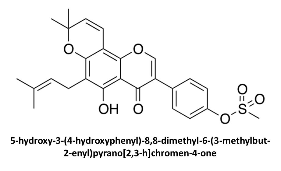
5-Hydroxy-3-(4-hydroxyphenyl)-8,8-dimethyl-6-(3-methylbut-2-enyl)pyrano[2,3-h]chromen-4-one

Department of Pharmaceutical and Pharmacological Sciences, University of Padova, Via Marzolo 5, 35131 Padova, Italy
*
Author to whom correspondence should be addressed.
Molbank 2018, 2018(3), M1004; https://doi.org/10.3390/M1004
Submission received: 18 June 2018
/
Revised: 6 July 2018
/
Accepted: 6 July 2018
/
Published: 9 July 2018
(This article belongs to the Section Natural Products)
Abstract
:Natural and semi-synthetic compounds are being studied as novel phosphodiesterase 5 (PDE5) inhibitors for the treatment of erectile dysfunction, pulmonary hypertension, and lower urinary symptoms. Maclura pomifera is a source of flavonoids, one of the main classes of molecules investigated for these purposes. The extraction of the natural isoflavone osajin and its modification to obtain a semi-synthetic derivative are described in this short note. 1H and 13C-nuclear magnetic resonance spectroscopy (NMR), mass spectrometry, high-performance liquid chromatography (HPLC) and spectroscopic characterization of the title compound are also hereby provided. Two-dimensional (2D) nuclear Overhauser effect spectroscopy (NOESY) NMR, supported by in silico conformational studies, was used to achieve a complete assignment of the proton signals, assessing the correct chemical structure of the compound. Heteronuclear single quantum coherence spectroscopy (HSQC) and heteronuclear multiple bond correlation (HMBC) NMR experiments were performed to assign 13C chemical shifts. Calculated chemical properties and preliminary in silico docking suggest that this molecule might be a promising candidate as PDE5 inhibitor.

1. Introduction
The inhibitors of phosphodiesterase 5 (PDE5) are currently used in the clinical practice for the treatment of erectile dysfunction, pulmonary hypertension, and lower urinary symptoms [1], but several other potential therapeutic applications are emerging [1,2]. Scientific literature is particularly prolific for what concerns natural compounds or nature-inspired molecules showing inhibitory activity on PDE5 [3,4,5,6]. Flavonoids extracted from plants are among the most well-studied compounds in this context [3,4,7]. We previously reported the positive in silico and in vitro results of our studies on natural isoflavones from Maclura pomifera and their semi-synthetic derivatives as PDE5 inhibitors [8,9]. Thus, we improved the extraction conditions of the natural scaffold and expanded the variety of chemical modifications with the aim of further improving the efficacy against PDE5. The procedures for the extraction of osajin (1) from Maclura pomifera and the reaction conditions for the isolation of compound 2 are here described. NMR, mass spectrometry, HPLC and ultra-violet (UV) data are also reported. Particular attention was dedicated to the complete assignment of the proton and 13C-NMR signals, achieved by using 2D NOESY, heteronuclear single quantum coherence spectroscopy (HSQC) and heteronuclear multiple bond correlation (HMBC) experiments. Physico-chemical properties and preliminary in silico results are also briefly discussed.
2. Results and Discussion
2.1. Extraction of Osajin (1) and Synthesis of 2
As discussed in previous papers, M. pomifera is a rich source of isoflavones, some of which are “chemically constitutional isomers” [10]. Besides for its activity on PDE5, the prenylated isoflavone osajin (1) has also been studied as an antioxidant, an anticancer and antibacterial agent [11,12,13]. The extraction of osajin (1) was performed according to a procedure that we previously reported [8,9]. Briefly, the compound was isolated from the dried fruits using a soxhlet apparatus. The procedure consisted of a preparative step (degreasing process, 48 h), followed by 24 h of isoflavones extraction. More in detail, in this study three different combinations of degreasing/extracting solvents were tested: petroleum ether/diethyl ether, cyclohexane/diethyl ether, and cyclohexane/ethyl acetate. The best combination of solvents among the tested resulted to be petroleum ether/diethyl ether. Starting from 100 g of dried fruits, 1.78 g of osajin (1), and 0.32 g of pomiferin were isolated. Lower yields and purities were obtained using the other extraction conditions. The attribution of the correct chemical structure of the extracted compounds was achieved using one-dimensional (1D) and 2D NMR and mass spectrometry [9,14].
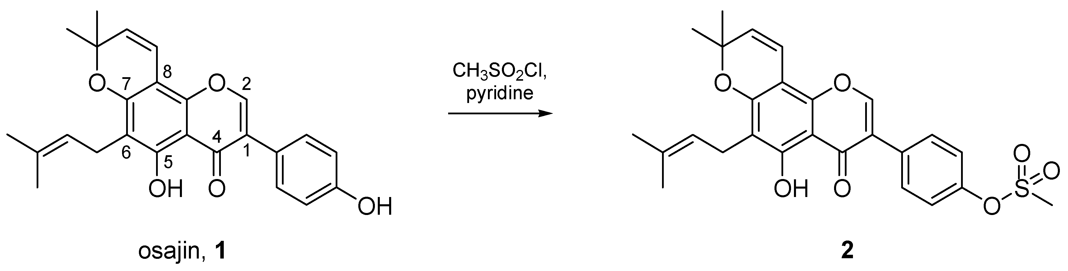
With the aim of preparing novel PDE5 inhibitors, the synthesis of the methanesulfonate derivative of osajin was carried out to explore the effects of extending the bulkiness of the molecule in the phenyl region, and to shade the hydroxyl groups, thus introducing novel hydrogen bond acceptors with a different orientation. Moreover, the introduction of the -SO2-group provides to compound 2 further structural similarity with sildenafil, the well-known PDE5 inhibitor. Moreover, this hypothesis was supported by our previous experimental evidences which suggest that the protections/substitution of hydroxyl groups in the isoflavone scaffold may lead to a highest inhibitory activity (lower IC50) towards PDE5 in vitro [8]. The preparation of the methanesulfonate derivative of osajin (1) was straightforwardly carried out adopting and modifying a procedure described by Wolfrom et al. [15]. In their work, the authors proposed the preparation of the 4-methylbenzenesulfonate derivatives of osajin (1) and pomiferin, another compound extracted from M. pomifera, and of their cyclized isoosajin and isopomiferin analogues. Starting from here, we expanded the set of molecules by introducing on osajin (1) the methanesulfonate moiety under similar experimental conditions. The preparation of such compound was carried out by reacting osajin (1) with methanesulfonyl chloride in pyridine, according to the procedure described in the experimental section and in Scheme 1. The reaction is regioselective and leads to the mono-methanesulfonate derivative. As highlighted by mass spectrometric data and, most importantly, by NMR analysis, only the hydroxyl group of the 4-hydroxyphenyl residue is affected by the reaction, as the other hydroxyl group in the molecule (5-hydroxy) is involved in a strong H-bond interaction and suffers from steric hindrance. The presence of such strong interaction is also testified by the high chemical shift value for that hydrogen atom in the 1H-NMR spectrum (see Supplementary material for complete spectra). As a result, this group is extremely poorly reactive towards the substitution.
2.2. NMR-Based Structural Elucidation and Conformational Studies
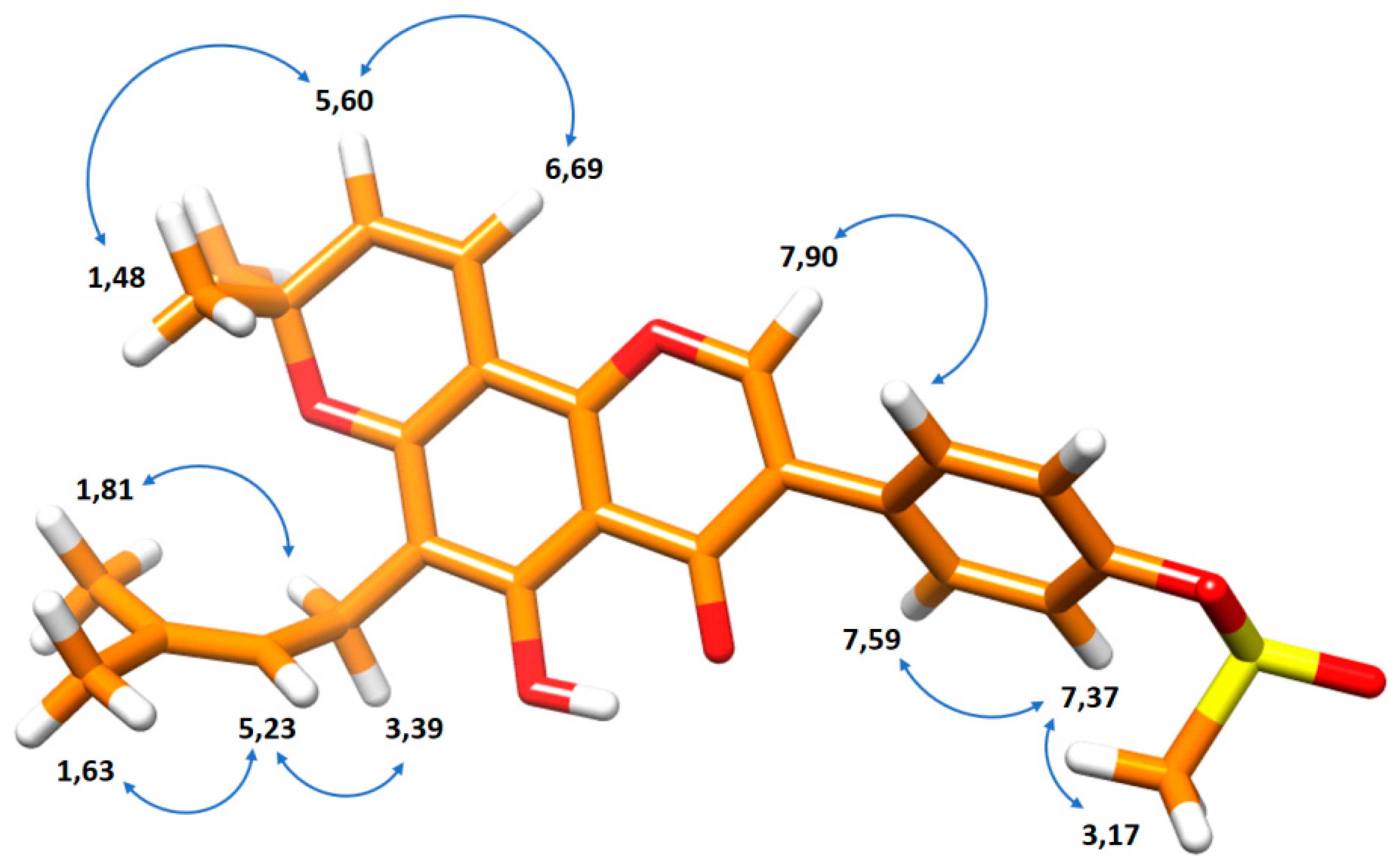
The in silico minimized structure of compound 2 is reported in Figure 1 (force field: MMFF94). By interpreting the results of 2D NOESY NMR experiments, we performed a complete attribution of the signals observed in the proton spectrum (Table 1). Figure 1 shows the correlations observed in the NOESY NMR experiment (see Supplementary material for complete spectra) which confirm that the minimized pose is realistic.
Heteronuclear single quantum coherence spectroscopy (HSQC) and heteronuclear multiple bond correlation (HMBC) were used to perform a complete assignment of 13C signals as reported in Table 1 (see Supplementary material for 2D spectra).
2.3. In Silico Studies: Chemical Properties and Preliminary Molecular Docking
Predicted chemical properties were calculated using Molinspiration Cheminformatics software (Molinspiration Cheminformatics, Slovensky Grob, Slovak Republic, https://www.molinspiration.com). According to these predictions, compound 2 showed a miLogP value of 5.64 and a total polar surface area (TPSA) of 103.05 Å2. Moreover, with five rotable bonds, seven H-bond acceptors, and one H-bond donor this compound possesses promising chemical and pharmacokinetic properties. Moreover, in a preliminary in silico study, we docked osajin (1), compound 2, and sildenafil to the crystal structure of PDE5. The results of this experiment confirmed our initial hypothesis that led us to the introduction of the methanesulfonate moiety on the scaffold of osajin. The residues involved in the protein-ligand interaction (<5 Å) were examined and, in general, compound 2, and sildenafil show a nearly complete superimposition of their scaffolds (Table S1, see Supplementary material). Moreover, as demonstrated by the 3D model reported in Figure S14 (see Supplementary material), the additional H-bond acceptor group that was introduced in the osajin scaffold, allows compound 2 to form a H-bond with Ser663, thus maintaining the main interaction with the residues of the catalytic site of PDE5 which are peculiar of sildenafil (e.g., hydrophobic interaction with Phe820). As a result, compound 2 proved to be the best performing molecule of the set in terms of estimated ΔG (kcal/mol), which is an expression of the energetic efficiency of the interaction between a ligand and the macromolecule (i.e., stronger binding), for the computed interactions (see Supplementary material for complete docking data and 3D models). To be more specific, the more negative value of estimated ΔG (kcal/mol) of 2 enables the possibility of a higher affinity for the catalytic site of the enzyme.
3. Materials and Methods
3.1. Chemistry
Commercially available chemicals were purchased from Sigma-Aldrich (Saint Louis, MO, USA) and used as received, unless otherwise stated. 1H and 13C{1H} NMR spectra were recorded on an Avance III 400 MHz spectrometer (Bruker, Billerica, MA, USA). All spectra were recorded at room temperature; the solvent for each spectrum is given in parentheses. Chemical shifts are reported in ppm and are relative to TMS internally referenced to the residual solvent peak. Datasets were edited with TopSpin (Bruker), and iNMR software (Nucleomatica, Molfetta, Italy). The multiplicity of signals is reported as singlet (s), doublet (d), triplet (t), quartet (q), multiplet (m), broad (br), or a combination of any of these. High-resolution mass spectra (HRMS) were recorded on a Mariner ESI–TOF spectrometer (Applied Biosystems, Foster City, CA, USA). The purity profile was assayed by HPLC using a Pro-Star system (Varian, Palo Alto, CA, USA) equipped with a 1706 UV–VIS detector (254 nm, Bio-rad, Hercules, CA, USA) and an C-18 column (5 μm, 4.6 × 250 mm, Agilent Technologies, Santa Clara, CA, USA). An appropriate ratio of water (A) and acetonitrile (B) was used as mobile phase with an overall flow rate of 1 mL/min; the general method for the analyses is reported here: 0 min (95% A–5% B), 5 min (95% A–5% B), 15 min (5% A–95% B), 20 min (5% A–95% B), and 22 min (95% A–5% B).
3.1.1. Extraction of Osajin (1) from M. pomifera
The fruits of M. pomifera were cut and dried in an oven, and then ground and put in a soxhlet (100 g of dried fruits). A first extraction with 600 mL of petroleum ether was performed to degrease, and remove secondary products. This extract was discarded. A second extraction was performed with 600 mL of diethyl ether to obtain osajin (1), and the other isoflavone pomiferin. The mixture obtained was evaporated at reduced pressure to almost dryness, and then diluted with 500 mL of ethanol. 50 mL of Pb(II) acetate 0.1 M in MeOH solution was added. The resulting precipitate was filtered and the solid obtained was dissolved in acetic acid to disrupt the Pb(II) complex. The solution was poured in 1 L of ice-cold water and filtered obtaining osajin (1) as a yellow solid (1.78 g).
3.1.2. Synthesis of 5-Hydroxy-3-(4-hydroxyphenyl)-8,8-dimethyl-6-(3-methylbut-2-enyl)pyrano[2,3-h]chromen-4-one (2)
A solution of CH3SO2Cl (0.53 mL, 6.88 mmol, 14 eq.) in 3 mL of pyridine was added to a solution of osajin (1, 200 mg, 0.49 mmol, 1 eq.) in 2 mL of pyridine under stirring. The mixture was stirred overnight at room temperature (r.t.) until the reaction was concluded (TLC hexane/ethyl acetate 3:1). Subsequently, it was poured in an equal volume of ice-cold water and the formed precipitate was filtered and washed with water. The product was recrystallized from EtOH (112 mg, 46%). δH (400 MHz, CDCl3) 1.48 (6H, s, Me), 1.68 (3H, s, Me), 1.81 (3H, s, Me), 3.17 (3H, s, -SO2CH3), 3.35 (2H, d, J = 7.3 Hz, CH2), 5.23 (1H, t, J = 7.3 Hz, C=CH), 5.60 (1H, d, J = 10.1 Hz, C=CH), 6.69 (1H, d, J = 10.1 Hz, C=CH), 7.37 (2H, AA′BB′, J = 8.1 Hz, PhH), 7.59 (2H, AA′BB′, J = 8.1 Hz, PhH), 7.90 (1H, s, C=CH), 13.14 (1H, s, OH). δC (100 MHz, CDCl3) 17.9, 21.3, 25.8, 28.2, 37.4, 78.0, 100.9, 105.5, 113.2, 114.8, 121.8, 122.2, 122.5, 127.4, 130.5, 130.6, 131.7, 149.2, 150,4, 152.9, 157.5, 159.3, 180.4. HMRS (ESI) found 483.1693 (C26H26O7S, [M + H]+), calc. 483.1479.
3.2. In Silico Studies
Conformational studies were carried out using Chimera (MMFF94 force field) and images were obtained with the same software [16]. Predicted chemical properties were calculated using Molinspiration Cheminformatics software (Molinspiration Cheminformatics, Slovensky Grob, Slovak Republic, https://www.molinspiration.com). The crystal structure of the target protein was obtained from the Protein Data Bank (PDB, https://www.rcsb.org). Protein and ligands were prepared and then docking experiments were performed using Autodock Vina (Molecular Graphics Laboratory, Department of Integrative Structural and Computational Biology, The Scripps Research Institute, La Jolla, CA, USA) [17] on the 2H42 PDB structure. Docking results and interaction poses were analyzed and visualized with Chimera [16]. The docking experiments were performed on osajin (1), compound 2, and sildenafil as a reference in accordance to our previous in silico studies on the same target [8].
4. Conclusions
The one-step preparation of a potential semi-synthetic PDE5 inhibitor candidate was presented. The chemical structure of the synthesized compound was fully assessed using mass spectrometry and 2D NOESY, HSQC and HMBC NMR. The extraction of osajin (1), the natural precursor, is straightforward and inexpensive, and the regioselective modification introduced to obtain the title compound lead to a more efficient interaction with the enzyme, according to our preliminary in silico data. We aim at expanding the class of semi-synthetic derivatives of isoflavones from M. pomifera, and at supporting our preliminary in silico data with in vitro testing.
Supplementary Materials
The following are available online, Figures S1–S10: 1D and 2D NMR spectra, Figure S11: mass spectrum, Figure S12: HPLC chromatogram, Figure S13: UV spectrum, Figure S14: molecular docking, Table S1: molecular docking results.
Author Contributions
Conceptualization, G.R. and G.Z.; Investigation, A.O. and G.R.; Writing-Original Draft Preparation, A.O. and G.R.; Writing-Review & Editing, G.Z.; Supervision, G.Z.
Funding
This research was funded by University of Padova.
Acknowledgments
The authors are grateful to Sergio Bova (University of Padova) for his kind and friendly support.
Conflicts of Interest
The authors declare no conflict of interest.
References
- Andersson, K.-E. PDE5 Inhibitors—Pharmacology and Clinical Applications 20 Years after Sildenafil Discovery. Br. J. Pharmacol. 2018, 175, 2554–2565. [Google Scholar] [CrossRef] [PubMed]
- Ribaudo, G.; Pagano, M.A.; Bova, S.; Zagotto, G. New Therapeutic Applications of Phosphodiesterase 5 Inhibitors (PDE5-Is). Curr. Med. Chem. 2016, 23, 1239–1249. [Google Scholar] [CrossRef] [PubMed]
- Zhang, J.; Wang, Y.-B.; Ma, C.-G.; Liu, T.; Li, W.-R.; Gong, Y.-Q.; Xin, Z.-C. Icarisid II, a PDE5 Inhibitor from Epimedium Wanshanense, Increases Cellular cGMP by Enhancing NOS in Diabetic ED Rats Corpus Cavernosum Tissue. Andrologia 2012, 44, 87–93. [Google Scholar] [CrossRef] [PubMed]
- Pavan, V.; Mucignat-Caretta, C.; Redaelli, M.; Ribaudo, G.; Zagotto, G. The Old Made New: Natural Compounds against Erectile Dysfunction. Arch. Pharm. 2015, 348, 607–614. [Google Scholar] [CrossRef] [PubMed]
- Yin, C.; Deng, Y.; Gao, J.; Li, X.; Liu, Y.; Gong, Q. Icariside II, a Novel Phosphodiesterase-5 Inhibitor, Attenuates Streptozotocin-Induced Cognitive Deficits in Rats. Neuroscience 2016, 328, 69–79. [Google Scholar] [CrossRef] [PubMed]
- Li, F.; Du, B.-W.; Lu, D.-F.; Wu, W.-X.; Wongkrajang, K.; Wang, L.; Pu, W.-C.; Liu, C.-L.; Liu, H.-W.; Wang, M.-K.; et al. Flavonoid Glycosides Isolated from Epimedium Brevicornum and Their Estrogen Biosynthesis-Promoting Effects. Sci. Rep. 2017, 7, 7760. [Google Scholar] [CrossRef] [PubMed]
- Adefegha, S.A.; Oboh, G.; Fakunle, B.; Oyeleye, S.I.; Olasehinde, T.A. Quercetin, Rutin, and Their Combinations Modulate Penile Phosphodiesterase-5′, Arginase, Acetylcholinesterase, and Angiotensin-I-Converting Enzyme Activities: A Comparative Study. Comp. Clin. Pathol. 2018, 27, 773–780. [Google Scholar] [CrossRef]
- Ribaudo, G.; Pagano, M.A.; Pavan, V.; Redaelli, M.; Zorzan, M.; Pezzani, R.; Mucignat-Caretta, C.; Vendrame, T.; Bova, S.; Zagotto, G. Semi-Synthetic Derivatives of Natural Isoflavones from Maclura Pomifera as a Novel Class of PDE-5A Inhibitors. Fitoterapia 2015, 105, 132–138. [Google Scholar] [CrossRef] [PubMed]
- Ribaudo, G.; Vendrame, T.; Bova, S. Isoflavones from Maclura Pomifera: Structural Elucidation and in Silico Evaluation of Their Interaction with PDE5. Nat. Prod. Res. 2017, 31, 1988–1994. [Google Scholar] [CrossRef] [PubMed]
- Kupeli, E.; Orhan, I.; Toker, G.; Yesilada, E. Anti-Inflammatory and Antinociceptive Potential of Maclura Pomifera (Rafin.) Schneider Fruit Extracts and Its Major Isoflavonoids, Scandenone and Auriculasin. J. Ethnopharmacol. 2006, 107, 169–174. [Google Scholar] [CrossRef] [PubMed]
- Mahmoud, Z.F. Antimicrobial Components from Maclura Pomifera Fruit. Planta Med. 1981, 42, 299–301. [Google Scholar] [CrossRef] [PubMed]
- Tsao, R.; Yang, R.; Young, J.C. Antioxidant Isoflavones in Osage Orange, Maclura Pomifera (Raf.) Schneid. J. Agric. Food Chem. 2003, 51, 6445–6451. [Google Scholar] [CrossRef] [PubMed]
- Son, I.H.; Chung, I.-M.; Lee, S.I.; Yang, H.D.; Moon, H.-I. Pomiferin, Histone Deacetylase Inhibitor Isolated from the Fruits of Maclura Pomifera. Bioorg. Med. Chem. Lett. 2007, 17, 4753–4755. [Google Scholar] [CrossRef] [PubMed]
- Orazbekov, Y.; Ibrahim, M.A.; Mombekov, S.; Srivedavyasasri, R.; Datkhayev, U.; Makhatov, B.; Chaurasiya, N.D.; Tekwani, B.L.; Ross, S.A. Isolation and Biological Evaluation of Prenylated Flavonoids from Maclura Pomifera. Evid. Based Complement. Altern. Med. ECAM 2018, 2018, 1370368. [Google Scholar] [CrossRef] [PubMed]
- Wolfrom, M.L.; Benton, F.L.; Gregory, A.S.; Hess, W.W.; Mahan, J.E.; Morgan, P.W. Osage Orange Pigments. II. Isolation of a New Pigment, Pomiferin. J. Am. Chem. Soc. 1939, 61, 2832–2836. [Google Scholar] [CrossRef]
- Pettersen, E.F.; Goddard, T.D.; Huang, C.C.; Couch, G.S.; Greenblatt, D.M.; Meng, E.C.; Ferrin, T.E. UCSF Chimera? A Visualization System for Exploratory Research and Analysis. J. Comput. Chem. 2004, 25, 1605–1612. [Google Scholar] [CrossRef] [PubMed]
- Trott, O.; Olson, A.J. AutoDock Vina: Improving the Speed and Accuracy of Docking with a New Scoring Function, Efficient Optimization, and Multithreading. J. Comput. Chem. 2010, 31, 455–461. [Google Scholar] [CrossRef] [PubMed]
Sample Availability: Samples of the compounds are available from the authors. |
Scheme 1.
Synthetic procedure for the preparation of 2.

Figure 1.
Minimized three-dimensional (3D) structure of compound 2 (force field: MMFF94). Chemical shifts are reported close to the corresponding nuclei and the observed nuclear Overhauser effect spectroscopy (NOESY) correlations are represented as blue arrows.
Figure 1.
Minimized three-dimensional (3D) structure of compound 2 (force field: MMFF94). Chemical shifts are reported close to the corresponding nuclei and the observed nuclear Overhauser effect spectroscopy (NOESY) correlations are represented as blue arrows.


Table 1.
1H and 13C-nuclear magnetic spectroscopy (NMR) chemical shifts and structure of 2.

| 1H Chemical Shift (ppm) | 13C Chemical Shift (ppm) | Assignment |
|---|---|---|
| 1.81 | 17.9 | O |
| 3.39 | 21.3 | L |
| 1.63 | 25.8 | P |
| 1.48 | 28.2 | K |
| 3.17 | 37.4 | E |
| - | 78.0 | 8 |
| - | 100.9 | J |
| - | 105.5 | 1 |
| - | 113.2 | G |
| 6.69 | 114.8 | H |
| 5.23 | 121.8 | M |
| - | 122.2 | C |
| 7.37 | 122.5 | B |
| 5.60 | 127.4 | I |
| - | 130.5 | 6 |
| 7.59 | 130.6 | A |
| - | 131.7 | N |
| - | 149.2 | D |
| - | 150.4 | F |
| 7.90 | 152.9 | 2 |
| - | 157.5 | 7 |
| - | 159.3 | 5 |
| - | 180.4 | 4 |
| 13.14 | - | -OH |
© 2018 by the authors. Licensee MDPI, Basel, Switzerland. This article is an open access article distributed under the terms and conditions of the Creative Commons Attribution (CC BY) license (http://creativecommons.org/licenses/by/4.0/).
Share and Cite
MDPI and ACS Style
Ribaudo, G.; Ongaro, A.; Zagotto, G. 5-Hydroxy-3-(4-hydroxyphenyl)-8,8-dimethyl-6-(3-methylbut-2-enyl)pyrano[2,3-h]chromen-4-one. Molbank 2018, 2018, M1004. https://doi.org/10.3390/M1004
AMA Style
Ribaudo G, Ongaro A, Zagotto G. 5-Hydroxy-3-(4-hydroxyphenyl)-8,8-dimethyl-6-(3-methylbut-2-enyl)pyrano[2,3-h]chromen-4-one. Molbank. 2018; 2018(3):M1004. https://doi.org/10.3390/M1004
Chicago/Turabian StyleRibaudo, Giovanni, Alberto Ongaro, and Giuseppe Zagotto. 2018. "5-Hydroxy-3-(4-hydroxyphenyl)-8,8-dimethyl-6-(3-methylbut-2-enyl)pyrano[2,3-h]chromen-4-one" Molbank 2018, no. 3: M1004. https://doi.org/10.3390/M1004
Note that from the first issue of 2016, this journal uses article numbers instead of page numbers. See further details here.