
N,N′-Diarylformamidine Dithiocarbamate Ag(I) Cluster and Coordination Polymer


1
School of Chemistry and Physics, Westville Campus, University of Kwazulu-Natal, Private Bag X54001, Durban 4000, South Africa
2
Department of Chemical Sciences, Olabisi Onabanjo University, P.M.B 2002, Ago-Iwoye 120107, Nigeria
3
School of Chemistry and Physics, Pietermaritzburg Campus, University of Kwazulu-Natal, Private Bag X01, Scottsville 3209, South Africa
*
Author to whom correspondence should be addressed.
Molbank 2022, 2022(1), M1327; https://doi.org/10.3390/M1327
Submission received: 29 November 2021
/
Revised: 6 January 2022
/
Accepted: 8 January 2022
/
Published: 28 January 2022
(This article belongs to the Section Structure Determination)
Abstract
:An Ag(I)formamidine cluster Ag6L16 (1) and an Ag(I)formamidine coordination polymer Ag7(L2)2 2 (L1 = N,N′-bis(2,6-disopropylphenyl) formamidine dithiocarbamate and L2 = N,N′-mesityl formamidine dithiocarbamate) have been synthesized from the reactions of L1 and L2 with AgNO3 respectively. The complexes were characterized using spectroscopic and analytical methods, including single-crystal X-ray diffraction. In the structure of 1, a six vertex distorted square bi-pyramidal octahedron is formed from an Ag6 core. The N,N′-bis(2,6-disopropylphenyl) formamidine dithiocarbamate ligands stabilize this core through two main –CS2 bridging modes giving a propeller like structure. In the structure of 2, each of the two Ag(I) centers are bridged by two N,N′-mesityl formamidine dithiocarbamate ligands forming 8-member Ag2(CS2)2 metallacycles with an inversion center in the middle of the Ag—Ag argentophilic bond. The metallacycles are connected through Ag—S bonds forming ribbons in the crystallographic a-axis. The Ag(I) centers are coordinated to two N,N′-mesitylformamidine dithiocarbamates through the dithiocarbamate S atoms. The thermal decomposition of complexes 1 and 2 had similar thermograms with one major weight loss activity and the formation of elemental silver particles thereafter.
1. Introduction
Silver(I) complexes have been applied in various fields, therefore, attracting great attention due to their structural features and functional considerations [1,2,3]. They have been tested as fluorescent materials [4], semiconductors [5,6], source of nanomaterials [7] and biological imaging agents [8] due to their interesting and fascinating physicochemical properties. Ag(I) complexes have also shown potential medical applications, most especially as antibacterial [9,10], antifungal [11], anticancer [12], and antimalarial agents [13]. Argentophilic bonding interactions which exist between seemingly closed-shell silver(I) atoms center have been reported to be responsible for their significant physical properties as well as their structural details [2]. Structurally, they possess coordination geometries ranging from two-coordinate (linear) to eight-coordinate (tetragonal prism) [14]. Factors, such as reaction conditions, as well as alkyl substitution effects might be responsible for the formation of a variety of complexes with unprecedented structures [15,16]. In the past decades, monomeric, dimeric, hexameric and polymeric structures have been reported [17].
Dithiocarbamates (R2CNS2−) belong to a class of mono anionic 1,1-dithiolate ligands, and they are often prepared by nucleophilic addition reaction of primary or secondary amines and carbon disulfide in the presence of a base as a proton acceptor [18]. Dithiocarbamates have been reported to react with silver(I) ions to form Ag(I) complexes of various geometries [19,20] and also serve as efficient stabilizers for silver nanoparticles [21]. Silver(I) dithiocarbamates, [Ag(S2CNR2)]n have been known as far back as the 1950s, but little work had been done on them probably due to their low solubility [17]. They have been widely used as a precursor to acanthite (α-Ag2S), a potential material for microelectronics [22,23]. Early crystallographic studies revealed the hexameric nature of Ag-S2CNR2 (R = Et, Pr, and n-Bu) in the solid states. They have a polymeric chain structure, in which the silver atom is bonded in a distorted manner by three dithiocarbamate ligand, two acting as µ2-bridges and one acting as a chelate [15,24].
Yin et al. reported the crystal structure of poly [(µ3-N,N-dibenzyldithiocarbamato-κ4S,S’:S:S’) silver(I)] [17]. Each Ag(I) cation in this complex was bonded to two pairs of sulfur atoms from three N,N′-dibenzyldithiocarbamate ligands, conforming to distorted tetrahedral geometry. The crystal structure of hexakis (µ3-N,N-diisopropyldithiocarbamato) hexasilver(I) had also been reported by Yin and co-workers [25]. The metal center of the complex was centrosymmetric, and its hexanuclear structure entails two cyclohexane-like Ag3S3 units which are joined together by the S atoms of the thiocarbamate groups with the Ag...Ag distances ranging from 3.0382(5) to 3.0985(5) Å. Herein, we report the synthesis, characterization and thermal analysis of silver(I) dithiocarbamate complexes derived from symmetrical N,N′-diarylformidine dithiocarbamate ligands.
2. Materials and Instrumentation
All solvents (ACS reagent grades ≥ 99.5%) were obtained from Sigma-Aldrich (Johannesburg, South-Africa) and used as purchased without further purification. Reagents: 2,6-diisopropylaniline (97%), 2,4,6-trimethylaniline (98%), triethyl orthoformate (99%), and carbon disulfide were also obtained from Sigma-Aldrich. AgNO3 (98%) and KOH (85%) were obtained from Promark Chemicals, Johannesburg, South Africa.
1H and 13C NMR spectra were recorded at 25 °C on a Bruker AvanceIII 400MHz spectrometer (Karlsruhe, German) at the University of KwaZulu-Natal, Westville Campus. Both 1H NMR and 13C NMR data were measured in parts per million relative to the residual solvent signals (chloroform-d for 1H with δ = 7.26 ppm and 13C NMR δ = 77.00 ppm). IR spectra were obtained on a PerkinElmer Universal ATR spectrum 100 FTIR spectrometer (Shelton, CT, USA), UV-Vis absorption spectra were recorded on Shimadzu UV-Vis-NIR spectrophotometer (Kyoto, Japan), and Q seriesTM Thermal Analyzer DSC/TGA (Q600) (Newcastle, CA, USA), was used to determine the stability of the complexes.
3. General Synthesis Methods
3.1. Preparation of Complexes
The synthesis of N,N′-bis(2,6-disopropylphenyl) formamidine dithiocarbamate and N,N′-mesityl formamidine dithiocarbamate salts have been reported in our previous work [26]. The complexes were synthesized by dissolving two equivalents of the potassium dithiocarbamate salts in 15 mL of acetonitrile into which a solution 1 equivalent of AgNO3 in 5 mL of H2O was added drop-wise with stirring for 30 min at room temperature. The resultant yellow solids were collected by filtration washed three times with diethyl ether, and dried in the oven at 50 °C.
3.1.1. Ag6(L1)6 1
The reaction of L1 (0.3 g, 0.6 mmol) with AgNO3 (0.05 g, 0.3 mmol) in a mixture of acetonitrile and water gave complex 1 as a yellow powder in 79% yield. m.p. 214–216 °C. 1H NMR (CDCl3, 400 MHz) δ (ppm): 1.04(d, 66H, JH,H = 6.68 Hz, CH3-CH), 1.15(d, 42H, JH,H = 5.92 Hz, CH3-CH), 1.21(d, 36H, JH,H = 5.92 Hz, CH3-CH), 2.70(m, 12H, JH,H = 6.0 Hz, CH-CH3), 2.81(m, 12H, JH,H = 6.56 Hz, CH- CH3), 7.06(s, 19, Ar-H), 7.16(d, 11H, JH,H = 7.28 Hz, Ar-H), 7.35(t, 6H, JH,H = 7.76 Hz, Ar-H), 9.38(s, 6H, -C(H)=N). 13C NMR (CDCl3, 100 MHz) δ (ppm): 23.95, 24.09, 25.56, 27.47, 29.16, 123.04, 124.26, 124.54, 129.63, 138.76, 144.87, 151.65 and 214.51. IR υ (cm−1): 2960, 1639, 1474, 1252, 982, 844, 481. UV-Vis (CHCl3, λmax, nm): 238 and 314.
3.1.2. Ag2(L2)2 2
The reaction of L2 (0.3 g, 0.80 mmol) and AgNO3 (0.07 g, 0.4 mmol) in acetonitrile furnished complex 2 as a yellow powder. Yield 64% Melting point 220–222 °C. 1H NMR (CDCl3, 400 MHz) δ (ppm): 1.99(s, 9H, CH3), 2.11(s, 9H, CH3), 2.22(m, 18H, JH,H = 5.92 Hz, CH3), 6.77(s, 3H, Ar-H), 6.87(s, 2H, Ar-H), 6.91(s, 1H, Ar-H), 6.97(s, 2H, Ar-H), 9.43(s, 2H, -CH=N). 13C NMR (CDCl3, 100 MHz) δ (ppm): 17.84, 18.06, 18.58, 18.67, 20.67, 20.73, 20.76, 21.28, 127.54, 128.41, 128.73, 128.86, 128.99, 129.35, 132.18, 133.89, 134.51, 136.05, 138.16, 138.50, 142.49, 144.79, 147.24, 151.27 and 214.01. IR υ (cm−1): 2916, 1634, 1476, 1246, 982, 845, 482. UV-Vis (CHCl3, λmax, nm): 238 and 312.
All the spectra for complexes 1 and 2 are given in the supplementary materials.
3.2. Single Crystal X-ray Diffraction
The structure refinement parameter, as well as the crystallographic data for complexes 1 and 2, are given in Table 1. Evaluation of crystals and collection of data was done on a Bruker Smart APEXII diffractometer with Mo Kα radiation (I = 0.71073 Å) equipped with an Oxford Cryostream low-temperature apparatus operating at 101 K for all samples. Reflections were collected at different starting angles, and the APEXII program suite was used to index the reflections [27]. Reduction of data was carried out using the SAINT software [28], and the absorption corrections and scaling were applied using the SADABS multi-scan technique [29]. The two structures were solved by the direct method using the SHELXS program and refined using the SHELXL program [30]. Graphics of the crystal structures were drawn using Mercury software [31]. Non-hydrogen atoms were first refined isotropically and then by anisotropic refinement with the full-matrix least square method based on F2 using SHELXL. All hydrogen atoms were positioned geometrically, allowed to ride on their parent atoms, and refined isotropically. The crystallographic data and structure refinement parameters for complexes 1 and 2 are given in Table 1.
4. Results and Discussion
4.1. Synthesis of N,N′-Diarylformamidines Dithiocarbamate Ag(I) Complexes
Complexes 1 and 2 were synthesized by reacting acetonitrile potassium salt solutions of L1 and L2 (Figure 1) with aqueous solutions of AgNO3 in a 2:1 ratio. Both complexes were obtained as thermally stable yellow solids with melting points ranging between 214 and 222 °C, 1 having a lower melting point than 2. Both complexes are soluble in dichloromethane, chloroform, toluene and benzene.
4.2. Spectroscopic Studies
The 1H and 13C NMR spectra for complexes 1 and 2 obtained in chloroform had signature peaks for the methane proton of the L1 and L2 as confirmed using their 2D NMR spectra. The azomethine proton of 1 and 2 was observed at 9.38 and 9.43 ppm in the spectra of L1 and L2, respectively, an upfield shift from 10.15 and 9.92 ppm [26]. The signals of aliphatic protons in the spectra of the complexes shifted noticeably downfield. For example, methyl protons in 2 appeared at 1.94, 2.03, and 2.15 ppm but were at 1.99, 2.11, and 2.22 ppm in L2. The downfield shift is a result of the drifts of electron density towards the metal ion center [32,33]. There were similar observations in the 13C-NMR spectra of 1 and 2, and an upfield shift of the carbon atom of -NCS2 moiety to 214.15 and 214.01 ppm from 220.94 and 218.95 ppm in the spectra of L1 and L2, corroborating coordination of the S atom to Ag.
The FT-IR spectra of complexes I and 2 showed a strong absorption band at 1474 and 1476 cm−1, stretching bands for the C—N bond of the thiouride group. These stretching frequencies are intermediate of a typical C—N single bond (1250–1360 cm−1) and a double bond (1640 cm−1), an indication of a partial double bond character of the thiouride bond [34,35]. The two vibrational bands around 1252–1246 cm−1 and 982–983 cm−1 in the spectra of 1 and 2 are assigned to the asymmetric C—(S)—S and the symmetric C—(S)—S moiety confirming the asymmetric linking of the sulfur atoms to the silver atoms [36]. The vibrational bands for υ(C=Nstr) of the azomethine (C(H)=N) in the formamidine backbone of the complexes were observed around 1634–1639 cm−1.
In the electronic spectra of 1 and 2, two bands were observed at 238 and 312, and 314 nm and these are attributed to intraligand π→π* transition associated with N—C=S and π→π* transition within S—C=S groups of the coordinated dithiocarbamate ligands [37].
4.3. X-ray Structural Analysis
Suitable crystals for single-crystal X-ray diffraction analysis were obtained by slow diffusion of hexane into a chloroform solution of complexes 1 and 2, each. While complex 1 is an Ag(I) diarylformamidines dithiocarbamate cluster, 2 is an Ag(I) diarylformamidines dithiocarbamate coordination polymer. Complex 1 is a polymorph of the previously reported structure of Ag6[CS2(2,6-iPr2C6H3NC(H)=NC6H3-iPr2)]6 which crystallized in the Ia3 cubic space group (Table 2). The previously reported structure was synthesized by the insertion of CS2 into a dinuclear Ag(I) complex of Ag2[2,6-iPr2C6H3N)2C-(H)]2 using toluene as a solvent for the reaction [38].
Complex 1 is assembled through six Ag(I) centers and six diarylformamidines dithiocarbamate ligands (1:1 ratio). The asymmetric unit contains one-half of the cluster and is made up of three Ag(I) centers, coordinated through bridging by three diarylformamidines dithiocarbamate molecules (Figure 2a). The other half is generated through an inversion center. In the crystal, a six-vertex distorted square bipyramid octahedron is formed in which Ag3 forms the apexes of the pyramid while Ag12Ag22 (red dashed lines in Figure 2b) for the base of the bipyramid (Figure 2b). The square bipyramid is stabilized by six L1 ligands which have the coordination modes μ1- μ1- for four of the ligands while for the other two have a μ1- μ2- coordination mode. With the CS2 moieties added on, a propeller like core is formed (Figure 2c).
Comparison of 1 to Ag6[CS2(2,6- iPr2C6H3NC(H)=NC6H3- iPr2)]6
The literature structure is solvated with one molecule of chloroform, while 1 is not. The Ag(I) octahedron core of the literature structure forms a perfect square bipyramid, whereas that of 1 is slightly distorted. The Ag—Ag distances in 1 range between 2.9204(4) and 3.3452(4) Å while the distances are between 3.0001(3) and 3.3178(3) Å for literature structure. In both complexes, the Ag—Ag argentophilic distances are all less than the sum of the van der Waals radii of two Ag atoms, 3.44 Å [23,24].
Each Ag(I) center in 1 is coordinating to three sulfur atoms in a κ1κ1κ1-S fashion, a geometry around Ag(I) that can be described as distorted trigonal pyramidal in which the Ag(I) center serves as the apex of the pyramid. The S—Ag—S bond angles range between 107.07(4) and 123.40(3)° in 1 while it ranges between 109.29(3) and 127.14(3) in the literature structure. The Ag—S bond distances are 2.4857(11), 2.5231(10), and 2.5322(9) in comparison to 2.4779(3) and 2.5008(2) for the literature structure.
In the crystal of complex 2, the asymmetric unit consists of two diarylformamidines dithiocarbamate ligands (L2) and two Ag(I) centers (Figure 3a). The Ag(I) centers are bridged by two diarylformamidine dithiocarbamate units in such a way that two Ag1s are paired and likewise two Ag2s. In doing so, each bridged Ag(I) pairs with an 8-member bimetallocycle, and an inversion center is formed. The metallacycles are connected through two centrosymmetrically related Ag—S bonds leading to a ribbon that runs in the a-crystallographic axis (Figure 3b).
Just like in 1, the Ag(I) center is coordinated to three S atoms giving a trigonal pyramidal geometry around the metal center. This differs from the coordination polymer of [Ag(dibenzyldithiocarbamate)]∞ [17] where each Ag(I) cation is bonded to two pairs of sulfur atoms from three dithiocarbamate ligands resulting in a distorted tetrahedral geometry. The S—Ag—S bond angles range from 102.58(3)°—129.07(4)° and these are similar to the ones for coordination polymer of [{Ag(diethyldithiocarbamate)}3]∞ which ranges from 109.49(4)°—128.56(4)° [24].
The Ag—S bond distances are in the range of 2.4757(10)–2.5716(12), i.e., Ag(2)—S(1), Ag(2)—S(2), and Ag(2)—S(3) are 2.5716(12), 2.4757(10), and 2.5660(10) respectively (Table 3) and comparatively, they are similar to those previously reported related complexes [39,40]. The Ag—Ag bond lengths in complex 2 are identical to those observed in 1. The C—S bond distances in the dithiocarbamate ligand (L2) backbone of complex 2, fall between ideal single and double C—S bonds indicating partial delocalization of π-electron density over the entire S2CN fragments in the complex [26,41].
4.4. Thermal Decomposition Studies
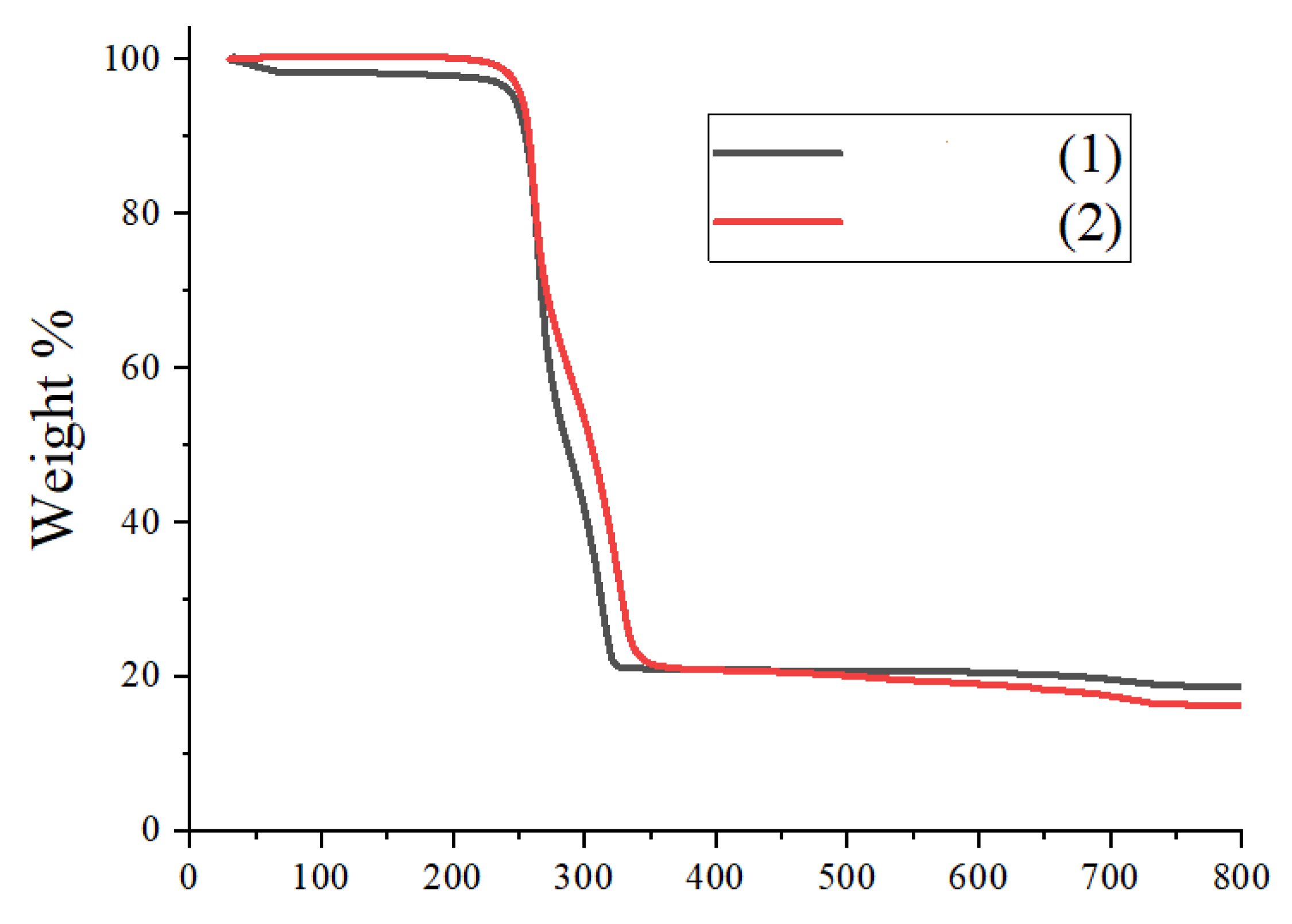
The thermal decomposition of the two silver complexes was examined by thermogravimetric (TG) analysis, and the superimposed TG graphs are represented in Figure 4. It is observed from the TGA curve that the thermal decomposition of the complexes shows one step that is equivalent to about 78% weight loss associated with the decomposition of the dithiocarbamate backbone ligand and the dominant weight loss of complex 1 occur in the temperature region of 250–325 °C while the one for 2 occurs at 250–350 °C. In both complexes, there is almost no weight loss below 100 °C and above 350 °C. The decomposition of 1 and 2 led to the formation of elemental silver residue with about 22% residue, which was about 2% more than the calculated value of 20% for 1 and 1% less than the calculated value of 23% for 2.
5. Conclusions
Conclusively, Ag(I)formamidine cluster and Ag(I)formamidine coordination polymer has been synthesized and characterized by means of thermogravimetric analysis together with FT-IR, UV-vis, 1H, and 13C NMR spectroscopy. X-ray structural analysis showed that the Ag6 core of structure 1 formed a six vertex distorted square bi-pyramidal octahedron while in structure 2, each of the two Ag(I) centers are bridged by two N,N′-mesityl formamidine dithiocarbamate ligands to form an 8-member Ag2(CS2)2 metallacycles with an inversion center in the middle of the Ag—Ag argentophilic bond. The thermal analysis of complexes 1 and 2 led to the formation of the elemental silver residue.
Supplementary Materials
The following supporting information is online. Figure S1: 1H-NMR spectrum for L1; Figure S2: 1H-NMR spectrum for L2; Figure S3: 1H-NMR spectrum for 1; Figure S4: 1H-NMR spectrum for 2; Figure S5: 13C-NMR spectrum for L1; Figure S6: 13C-NMR spectrum for L2; Figure S7: 13C-NMR spectrum for 1; Figure S8: 13C-NMR spectrum for 2; Figure S9: FT-IR spectrum for L1; Figure S10: FT-IR spectrum for L2; Figure S11: UV-Visible spectra of 1 and 2. CCDC 1976274 and CCDC 1976275 contain the supplementary crystallographic data for complexes 1 and 2. These data can be obtained free of charge via http://www.ccdc.cam.ac.uk/conts/retrieving.html, or from the Cambridge Crystallographic Data Centre, 12 Union Road, Cambridge CB2 1EZ, UK; fax: (+44)1223-336-033; or via e-mail: [email protected].
Author Contributions
Conceptualization, S.D.O. and B.O.; methodology, S.D.O.; software, S.D.O. and B.O.; validation, S.D.O. and B.O.; formal analysis, S.D.O. and B.O.; resources, B.O.; data curation, S.D.O. and B.O.; writing—original draft preparation, S.D.O. and B.O.; writing—review and editing, S.D.O. and B.O.; visualization, S.D.O. and B.O.; supervision, B.O.; project administration, B.O.; funding acquisition, S.D.O. and B.O. All authors have read and agreed to the published version of the manuscript.
Funding
This research was funded by the National Research Foundation of South Africa (Grant number: 119342) and the APC was funded by the National Research Foundation of South Africa.
Institutional Review Board Statement
Not applicable.
Informed Consent Statement
Not applicable.
Data Availability Statement
Not applicable.
Acknowledgments
The authors would like to thank the College of Agriculture, Science, and Engineering, the University of Kwazulu-Natal and the National Research Foundation (NRF), South Africa for financial support (Grant number: 119342).
Conflicts of Interest
The authors declare no competing financial interest.
References
- Young, A.G.; Hanton, L.R. Square planar silver(I) complexes: A rare but increasing observed stereochemistry for silver(I). Coord. Chem. Rev. 2008, 252, 1346–1386. [Google Scholar] [CrossRef]
- Schmidbaur, H.; Schier, A. Argentophilic interactions. Angew. Chem. Int. Ed. 2014, 54, 746–784. [Google Scholar] [CrossRef] [PubMed]
- Adeleke, A.A.; Zamisa, S.J.; Omondi, B. Ag(I) complexes of imine derivatives of unexpected 2-thiophenemethyl homo-coupling and Bis-(E)-N-(furan-2-ylmethyl)-1-(quinolin-2-yl)methanimine. Molbank 2021, 2, 1235. [Google Scholar] [CrossRef]
- Castiñeiras, A.; Pedrido, R. Novel fluorescent cationic silver thiosemicarbazone clusters containing different eight-membered Ag4S4 metallacycles. Inorg. Chem. 2009, 48, 4847–4855. [Google Scholar] [CrossRef]
- Su, W.; Hong, M.; Weng, J.; Cao, R.; Lu, S. A Semiconducting Lamella Polymer [{Ag (C5H4NS)} n] with a Graphite-Like Array of Silver (I) Ions and Its Analogue with a Layered Structure. Angew. Chem. Int. Ed. 2000, 39, 2911–2914. [Google Scholar] [CrossRef]
- Zhong, J.C.; Misaki, Y.; Munakata, M.; Kuroda-Sowa, T.; Maekawa, M.; Suenaga, Y.; Konaka, H. Silver (I) Coordination Polymer of 2, 5-Bis-(4‘, 5‘-bis (methylthio)-1‘, 3‘-dithiol-2‘-ylidene)-1, 3, 4, 6-tetrathiapentalene (TTM-TTP) and Its Highly Conductive Iodine Derivative. Inorg. Chem. 2001, 40, 7096–7098. [Google Scholar] [CrossRef] [PubMed]
- Safin, D.A.; Mdluli, P.S.; Revaprasadu, N.; Ahmad, K.; Afzaal, M.; Helliwell, M.; O’Brien, P.; Shakirova, E.R.; Babashkina, M.G.; Klein, A. Nanoparticles and thin films of silver from complexes of derivatives of N-(diisopropylthiophosphoryl) thioureas. Chem. Mater. 2009, 21, 4233–4240. [Google Scholar] [CrossRef]
- Gao, S.; Chen, D.; Li, Q.; Ye, J.; Jiang, H.; Amatore, C.; Wang, X. Near-infrared fluorescence imaging of cancer cells and tumors through specific biosynthesis of silver nanoclusters. Sci. Rep. 2015, 4, 4384. [Google Scholar] [CrossRef]
- Oladipo, S.D.; Tolufashe, G.F.; Mocktar, C.; Omondi, B. Ag(I) symmetrical N,N′-diarylformamidine dithiocarbamate PPh3 complexes: Synthesis, structural characterization, quantum chemical calculations and in vitro biological studies. Inorganica Chim. Acta 2021, 520, 120316. [Google Scholar] [CrossRef]
- Walker, M.; Parsons, D. The biological fate of silver ions following the use of silver-containing wound care products–a review. Int. Wound J. 2014, 11, 496–504. [Google Scholar]
- Mosconi, N.; Giulidori, C.; Velluti, F.; Hure, E.; Postigo, A.; Borthagaray, G.; Back, D.F.; Torre, M.H.; Rizzotto, M.J.C. Antibacterial, Antifungal, Phytotoxic, and Genotoxic Properties of Two Complexes of AgI with Sulfachloropyridazine (SCP): X-ray Diffraction of [Ag (SCP)] n. ChemMedChem 2014, 9, 1211–1220. [Google Scholar] [CrossRef]
- Iqbal, M.A.; Haque, R.A.; Ahamed, M.B.K.; Majid, A.A.; Al-Rawi, S.S. Synthesis and anticancer activity of para-xylyl linked bis-benzimidazolium salts and respective Ag (I) N-heterocyclic carbene complexes. Med. Chem. Res. 2013, 22, 2455–2466. [Google Scholar] [CrossRef]
- Hemmert, C.; Fabié, A.; Fabre, A.; Benoit-Vical, F.; Gornitzka, H. Synthesis, structures, and antimalarial activities of some silver (I), gold (I) and gold (III) complexes involving N-heterocyclic carbene ligands. Eur. J. Med. Chem. 2013, 60, 64–75. [Google Scholar] [CrossRef] [PubMed]
- Steel, P.J.; Fitchett, C.M. Metallosupramolecular silver (I) assemblies based on pyrazine and related ligands. Coord. Chem. Rev. 2008, 252, 990–1006. [Google Scholar] [CrossRef]
- Zhang, W.G.; Zhong, Y.; Tan, M.Y.; Liu, W.S.; Su, C.Y. A Novel Octahedral Hexasilver (I) Cluster: Ag6[SSCN (n-C4H9) 2]6. Chin. J. Chem. 2010, 20, 420–423. [Google Scholar] [CrossRef]
- Liu, N.; Fan, J.; Zhang, W.-G.; Yin, X.; Xie, M.-B. Bis (μ-N, N-diethyldithiocarbamato-κ3S, S′: S′) bis [(μ-N, N-diethyldithiocarbamato-κ2S, S′) silver (II)]. Acta Crystallogr. Sect. E 2006, 62, m2588–m2590. [Google Scholar] [CrossRef]
- Yin, X.; Xie, M.-B.; Zhang, W.-G.; Fan, J. Poly [(μ3-N, N-dibenzyldithiocarbamato-κ4S, S′: S: S′) silver (I)]. Acta Crystallogr. Sect. E 2007, 63, m2273. [Google Scholar] [CrossRef]
- Oladipo, S.D.; Omondi, B.; Mocktar, C. Synthesis and structural studies of Ni(II) and Cu(II) N,N′-diarylformamidine dithiocarbamate complexes as antimicrobial and antioxidant agents. Polyhedron 2019, 170, 712–722. [Google Scholar] [CrossRef]
- Kishore, P.V.; Liao, J.-H.; Hou, H.-N.; Lin, Y.-R.; Liu, C.W. Ferrocene-functionalized Cu (I)/Ag (I) dithiocarbamate clusters. Inorg. Chem. 2016, 55, 3663–3673. [Google Scholar] [CrossRef]
- Yamaguchi, H.; Kido, A.; Uechi, T.; Yasukouchi, K. The Crystal and Molecular Structure of Silver (I) N, N-Diethyldithiocarbamate. Bull. Chem. Soc. Jpn. 1976, 49, 1271–1276. [Google Scholar] [CrossRef]
- Tong, M.C.; Chen, W.; Sun, J.; Ghosh, D.; Chen, S. Dithiocarbamate-capped silver nanoparticles. J. Phys. Chem. B 2006, 110, 19238–19242. [Google Scholar] [CrossRef] [PubMed]
- Hussain, S.T.; Bakar, S.A.; Saima, B.; Muhammad, B. Low temperature deposition of silver sulfide thin films by AACVD for gas sensor application. Appl. Surf. Sci. 2012, 258, 9610–9616. [Google Scholar] [CrossRef]
- Ehsan, M.A.; Khaledi, H.; Tahir, A.A.; Ming, H.N.; Wijayantha, K.U.; Mazhar, M. Synthesis and characterization of silver diethyldithiocarbamate cluster for the deposition of acanthite (Ag2S) thin films for photoelectrochemical applications. Thin Solid Film. 2013, 536, 124–129. [Google Scholar] [CrossRef]
- Song, Y.-W.; Yu, Z.; Zhang, Q.-F. γ-Modification of poly [(N, N-diethyldithiocarbamato) silver (I)]. Acta Crystallogr. Sect. C 2006, 62, m214–m216. [Google Scholar] [CrossRef] [PubMed]
- Yin, X.; Xie, M.-B.; Zhang, W.-G.; Fan, J.; Zeller, M. Hexakis (μ3-N, N-diisopropyldithiocarbamato) hexasilver (I)(6 Ag—Ag). Acta Crystallogr. Sect. E 2007, 63, m2063–m2064. [Google Scholar] [CrossRef]
- Oladipo, S.D.; Olotu, F.A.; Soliman, M.; Mocktar, C.; Omondi, B. Formamidibe-based thiuram disulfides: Synthesis, structural characterization, biological studies and preliminary cheminformatics evaluation. J. Mol. Struct. 2020, 1219, 128553. [Google Scholar] [CrossRef]
- Bruker. APEXII; Bruker AXS Inc.: Madison, WI, USA, 2009. [Google Scholar]
- Bruker. SAINT; Bruker AXS Inc.: Madison, WI, USA, 2009. [Google Scholar]
- Bruker. SADABS; Bruker AXS Inc.: Madison, WI, USA, 2009. [Google Scholar]
- Sheldrick, G.M. A short history of SHELX. Acta Crystallogr. Sect. A: Found. Crystallogr. 2008, 64, 112–122. [Google Scholar] [CrossRef] [Green Version]
- Bruno, C.F.I.J.; Chisholm, J.A.; Edgington, P.R.; McCabe, P.; Pidcock, E.; Rodriguez-Monge, L.; Taylor, R.; Streek, J.V.D.; Wood, P.A. Mercury 2.0-new features for the visualization and investigation of crystal structures. J. Appl. Crystallogr. 2008, 41, 466–470. [Google Scholar]
- Mohammad, A.; Varshney, C.; Nami, S.A. Synthesis, characterization and antifungal activities of 3d-transition metal complexes of 1-acetylpiperazinyldithioc arbamate, M (acpdtc) 2. Spectrochim. Acta Part A: Mol. Biomol. Spectrosc. 2009, 73, 20–24. [Google Scholar] [CrossRef]
- Siddiqi, K.; Khan, S.; Nami, S.A.; El-Ajaily, M. Polynuclear transition metal complexes with thiocarbohydrazide and dithiocarbamates. Spectrochim. Acta Part A: Mol. Biomol. Spectrosc. 2007, 67, 995–1002. [Google Scholar] [CrossRef]
- Oladipo, S.D.; Omondi, B. Mercury(II) N,N′-diarylformamidine dithiocarbamate as single-source precursors for the preparation of oleylamine-capped HgS nanoparticles. Transit. Met. Chem. 2020, 45, 391–402. [Google Scholar] [CrossRef]
- Gölcü, A. Transition metal complexes of propranolol dithiocarbamate: Synthesis, characterization, analytical properties and biological activity. Transit. Met. Chem. 2006, 31, 405–412. [Google Scholar] [CrossRef]
- Korneeva, E.; Ivanov, A.; Gerasimenko, A.; Loseva, O.; Novikova, E.; Ivanov, M. Hexanuclear Silver (I) Hexamethylene Dithiocarbamate Cluster [Ag 6{S 2CN(CH2)6} 6]·2CH 2 Cl 2: Preparation, Molecular Structure (Manifestation of Argentophilic Interaction), and Thermal Behavior. Russ. J. Gen. Chem. 2019, 89, 1642–1648. [Google Scholar] [CrossRef]
- Oladipo, S.D.; Omondi, B.; Mocktar, C. Co (III) N, N′-diarylformamidine dithiocarbamate complexes: Synthesis, characterization, crystal structures and biological studies. Appl. Organomet. Chem. 2020, 34, e5610. [Google Scholar] [CrossRef]
- Lane, A.C.; Vollmer, M.V.; Laber, C.H.; Melgarejo, D.Y.; Chiarella, G.M.; Fackler, J.P.; Yang, X.; Baker, G.A.; Walensky, J.R. Multinuclear Copper(I) and Silver(I) Amidinate Complexes: Synthesis, Luminescence, and CS2 Insertion Reactivity. Inorg. Chem. 2014, 53, 11357–11366. [Google Scholar] [CrossRef]
- Huang, Z.; Lei, X.; Hong, M.; Liu, H. Synergism in a transition metal cluster compound. Crystal and molecular structure of a polysilver cluster molecule with an unusual bridging sulfur atom, Ag11S (Et2dtc) 9. Inorg. Chem. 1992, 31, 2990–2991. [Google Scholar] [CrossRef]
- Liu, C.; Liao, P.-K.; Fang, C.-S.; Saillard, J.-Y.; Kahlal, S.; Wang, J.-C. An eleven-vertex deltahedron with hexacapped trigonal bipyramidal geometry. Chem. Commun. 2011, 47, 5831–5833. [Google Scholar] [CrossRef] [Green Version]
- Prakasam, B.A.; Lahtinen, M.; Peuronen, A.; Muruganandham, M.; Kolehmainen, E.; Haapaniemi, E.; Sillanpää, M. Spectral and structural studies on Ni(II) dithiocarbamates: Nickel sulfide nanoparticles from a dithiocarbamate precursor. Inorg. Chim. Acta 2015, 425, 239–246. [Google Scholar] [CrossRef]
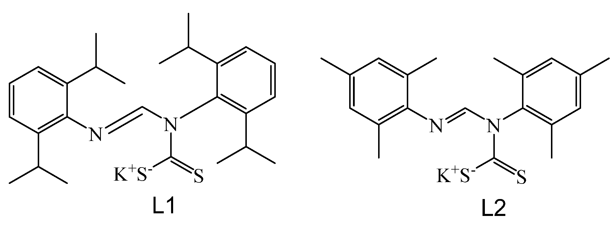
Figure 1.
General structures of ligands L1 and L2.

Figure 2.
(a) Asymmetric unit of complex 1. Hydrogen atoms and 2,6-diisopropyl units of the ligands are omitted for the sake of clarity (b) Octahedral arrangement of Ag atoms in 1 (c) Propeller like core of CS2 moieties in 1. Thermal displacement ellipsoids are drawn for all atoms at the 50 % probability level.


Figure 3.
(a) Asymmetric unit of complex 2 drawn at 50% thermal ellipsoids probability (b) Complex 2, showing the bridging of Ag(I) two diarylformamidine dithiocarbamate units.
Figure 3.
(a) Asymmetric unit of complex 2 drawn at 50% thermal ellipsoids probability (b) Complex 2, showing the bridging of Ag(I) two diarylformamidine dithiocarbamate units.

Figure 4.
Superimposed thermogravimetric analysis (TGA) profile for complexes 1 and 2.


Table 1.
The summary of X-ray crystal data collection and structure refinement parameters for complexes 1 and 2.
Table 1.
The summary of X-ray crystal data collection and structure refinement parameters for complexes 1 and 2.
| 1 | 2 | |
|---|---|---|
| Empirical formula | C156H210Ag6N12S12 | C40H46Ag2N4S4 |
| Formula weight | 3285.29 | 926.79 |
| Crystal system | Trigonal | Triclinic |
| Space group | R-3: H | P-1 |
| a/Å | 30.0941(6) | 8.1650(5) |
| b/Å | 30.0941(6) | 15.5778(9) |
| c/Å | 50.6555(11) | 16.2365(10) |
| α/° | 90 | 77.7000(10) |
| β/° | 90 | 87.3590(2) |
| γ/° | 120 | 83.1580(3) |
| Space group no. | 148 | 2 |
| Volume/Å3 | 39,730(2) | 2002.9(2) |
| Z | 9 | 2 |
| ρcalcg/cm3 | 1.236 | 1.537 |
| μ/mm−1 | 0.841 | 1.220 |
| F(000) | 15,336 | 944 |
| Crystal size/mm3 | 0.320 × 0.210 × 0.140 | 0.32 × 0.19 × 0.11 |
| 2θ range for data collection/° | 1.614 × 26.389 | 1.655 to 26.999 |
| Index ranges | −31 ≤ h ≤ 37 | −9 ≤ h ≤ 10 |
| −37 ≤ h ≤ 24 | −19 ≤ k ≤ 18 | |
| −62 ≤ l ≤ 61 | −20 ≤ l ≤ 12 | |
| Reflections collected | 91,820 | 6320 |
| Independent reflections | 17,643 | 8656 |
| Data/restraints/parameters | 17,643/0/862 | 8657/7/1474 |
| Goodness-of-fit on F2 | 0.935 | 0.983 |
| Final R indexes [I > = 2σ(I)] | R1 = 0.0409 wR2 = 0.0868 | R1 = 0.0610 wR2 = 0.110 |
| Final R indexes [all data] | R1 = 0.0869, wR2 = 0.1009 | R1 = 0.0610, wR2 = 0.1196 |
| Largest diff peak & hole (e Å−3) | 1.597 and −0.799 | 1.551 and 0.879 |
Table 2.
Comparison of some selected X-ray crystal data collection and structure refinement parameters for complex 1 and its polymorph.
Table 2.
Comparison of some selected X-ray crystal data collection and structure refinement parameters for complex 1 and its polymorph.
| 1 | Polymorph | |
|---|---|---|
| Empirical formula | C156H210Ag6N12S12 | C174H262Ag6N12S12 |
| Formula weight | 3285.29 | 3553.88 |
| Crystal system | Trigonal | Cubic |
| Space group | R | Ia |
| a/Å | 30.0941(6) | 32.283(4) |
| b/Å | 30.0941(6) | 32.283(4) |
| c/Å | 50.6555(11) | 32.283(4) |
| α/° | 90 | 90 |
| β/° | 90 | 90 |
| γ/° | 120 | 90 |
| Volume/Å3 | 39,730(2) | 33,644.8(2) |
| Z | 9 | 8 |
| ρcalcg/cm3 | 1.236 | 1.403 |
| Largest diff. peak & hole (e Å−3) | 1.597 and −0.799 | 1.551 and 0.879 |
Table 3.
Selected bond length (Å) and angles (°) for complexes 1 and 2.
| Bond distances (Å) | 1 | Polymorph | 2 |
|---|---|---|---|
| Ag—Ag | 2.9545(4) | 3.0001(1) | 2.8829(6) |
| Ag—Ag | 2.9695(4) | 3.0001(1) | 3.1932(5) |
| Ag—Ag | 3.3452(4) | 3.0001(1) | 3.0644(5) |
| Ag—S | 2.4857(11) | 2.4777(1) | 2.5029(9) |
| Ag—S | 2.5322(9) | 2.5007 (1) | 2.4805(10) |
| C—S | 1.681(4) | 1.6962(1) | 1.682(5) |
| C—S | 1.739(4) | 1.7404(1) | 1.723(4) |
| C—N | 1.362(5) | 1.3632(1) | 1.366(4) |
| C—N | 1.371(4) | 1.4639(1) | 1.406(5) |
| Bond angles (°) | |||
| Ag—S—Ag | 85.03(3) | 82.20 | 74.38(3) |
| S—Ag—S | 107.07(4) | 109.29 | 111.97(4) |
| Ag—Ag—S | 77.81(2) | 80.27 | 88.90(2) |
Publisher’s Note: MDPI stays neutral with regard to jurisdictional claims in published maps and institutional affiliations. |
© 2022 by the authors. Licensee MDPI, Basel, Switzerland. This article is an open access article distributed under the terms and conditions of the Creative Commons Attribution (CC BY) license (https://creativecommons.org/licenses/by/4.0/).
Share and Cite
MDPI and ACS Style
Oladipo, S.D.; Omondi, B. N,N′-Diarylformamidine Dithiocarbamate Ag(I) Cluster and Coordination Polymer. Molbank 2022, 2022, M1327. https://doi.org/10.3390/M1327
AMA Style
Oladipo SD, Omondi B. N,N′-Diarylformamidine Dithiocarbamate Ag(I) Cluster and Coordination Polymer. Molbank. 2022; 2022(1):M1327. https://doi.org/10.3390/M1327
Chicago/Turabian StyleOladipo, Segun D., and Bernard Omondi. 2022. "N,N′-Diarylformamidine Dithiocarbamate Ag(I) Cluster and Coordination Polymer" Molbank 2022, no. 1: M1327. https://doi.org/10.3390/M1327
Note that from the first issue of 2016, this journal uses article numbers instead of page numbers. See further details here.