Communication

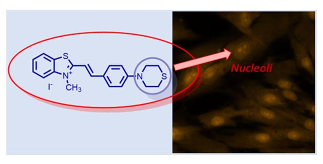
Styryl Hemicyanine Dye (E)-3-Methyl-2-(4-thiomorpholinostyryl)benzo[d]thiazol-3-ium Iodide for Nucleic Acids and Cell Nucleoli Visualization

1
Faculty of Chemistry and Pharmacy, University of Sofia “St. Kliment Ohridski”, 1 J. Bourchier Ave., 1164 Sofia, Bulgaria
2
Department of Medical Chemistry and Biochemistry, Medical Faculty, Medical University-Sofia, 2 Zdrave Str., 1431 Sofia, Bulgaria
*
Author to whom correspondence should be addressed.
Molbank 2022, 2022(2), M1392; https://doi.org/10.3390/M1392
Submission received: 31 May 2022 / Revised: 15 June 2022 / Accepted: 15 June 2022 / Published: 20 June 2022
(This article belongs to the Collection Heterocycle Reactions)

Abstract

:
(E)-3-Methyl-2-(4-thiomorpholinostyryl)benzo[d]thiazol-3-ium iodide 1 was prepared by a convenient and reliable reaction procedure. The slight molar excess of the starting benzaldehyde and the mixture of ethanol: ethyl acetate in the ratio 3:1 as a solvent afforded a pure reaction product. The photophysical properties of the dye in a TE buffer in the absence and presence of double-stranded DNA (dsDNA) were elucidated. The low intrinsic fluorescence of 1 in TE buffer is followed by an increase in the fluorescence after dsDNA binding. The dye is nontoxic for stem cells from apical papilla and the most concentrated fluorescence is detected in the cell nucleoli.

Graphical Abstract

1. Introduction

The styryl dyes are a class of the so called polymethine dyes or cyanine dyes. After the discovery of the styryl dyes in 1920 [1] by König, many new representatives were synthesized. The later discovery of polyacrylic fibers led to the application of styryl dyes on the dyeing of such fibers with good dyeing properties [2]. Styryl dyes are usually more photostable than the classic cyanine dyes. In the past styryl dyes were mainly used as sensitizers and additives in the photographic industry [3,4,5,6,7,8], and in optical recording media in laser disks [9] as flexible dyes [10] and laser dyes [11] and as optical sensitizers in various other fields [12,13,14,15]. A very extensive range of styryl dyes have, therefore, been developed since these compounds were first synthesized. The synthesis, applications and photoluminescence properties of a wide variety of styryl dyes had been reviewed in detail by the end of 1990s [2,16] and by our group [17] about ten years later. For more than sixty years the main application of the styryl cyanine dyes were mainly as sensitizers in photographic emulsions until the systematic work done by Yarmoluk and co-workers [18] who demonstrated the ability of the styryl cyanine dyes to bind different nucleic acids as intercalators or mainly as groove binders [19,20,21]. The investigated styryl dyes demonstrated an increase by several times of the fluorescence in a presence of nucleic acids. In the meantime, Li and Chang published protocols for the preparation of novel styryl dyes and their RNA binding and live cell staining [22]. Later, Lu et al. described [23] the synthesis and the photo physical properties of a new RNA-selective fluorescent dye integrated with a thiazole orange and a p-(methylthio)styryl moiety. The authors demonstrated that the new thiol containing styryl dye had better nucleolus RNA staining and imaging performance in living cells than the commercial stains [23]. It also exhibits excellent photostability, cell tolerance and counterstain compatibility with 4′,6-diamidino-2-phenylindole for specific RNA–DNA colocalization in bioassays [23]. The interesting and useful research described above have stimulated us to search for new fluorogenic biolabeling reagents for nucleic acids and living cells visualization.

2. Results and Discussion

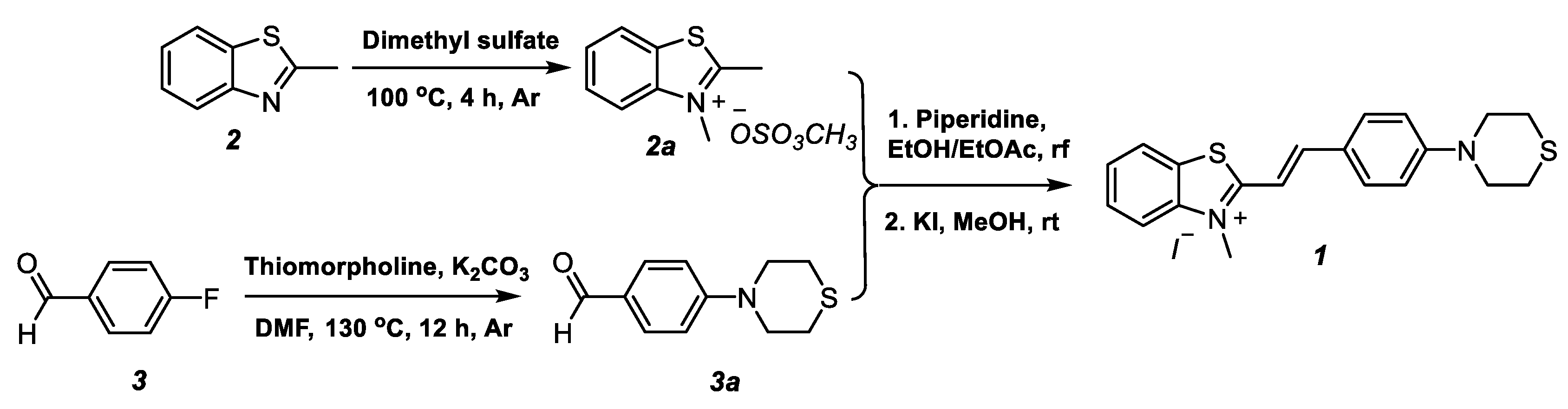
2.1. Synthesis of Monomethyne Cyanine Dye 1

Monomethyne cyanine dye 1 was prepared through a two-step synthetic procedure, shown in Scheme 1. The starting materials—2-methylbenzo[d]thiazolium salt (2a) and 4-thiomorpholinobenzaldehyde (3a) were synthesized according to a method reported in the literature, which provides higher yields and satisfactory purity of the compounds [24,25].
The condensation of 2a and 3a was achieved in the presence of piperidine as a base, replacing ethanol—the solvent usually used in this type of reaction with a mixture of solvents (ethanol and ethyl acetate) taken in a 3:1 ratio. The addition of ethyl acetate to the reaction mixture reduces the solubility of the target dye in ethanol and allows it to be isolated as a methyl sulfate in a high yield and sufficiently pure form after the end of the reaction. The replacement of the methyl sulfate ion with an iodide ion was carried out with potassium iodide in methanol at room temperature, and the target styryl dye 1 was obtained in 96% yield without requiring any additional purification.

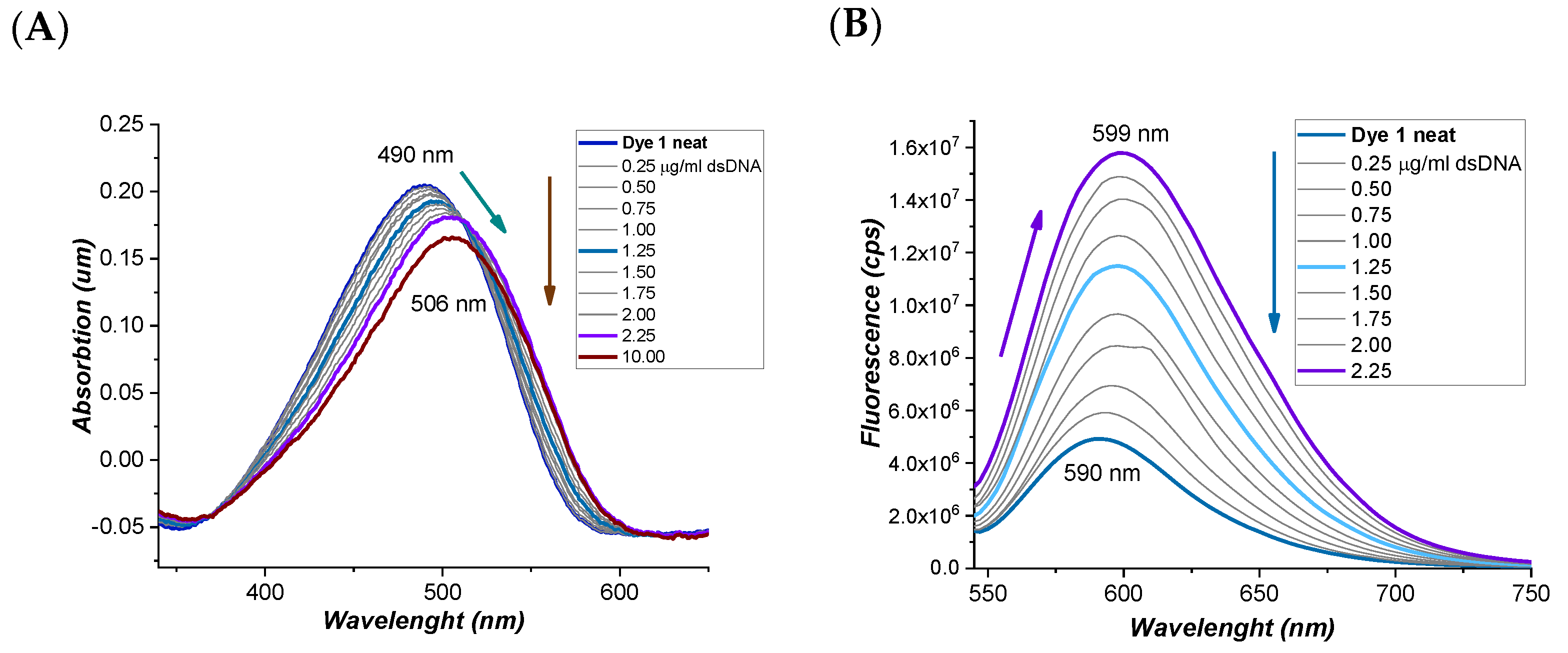
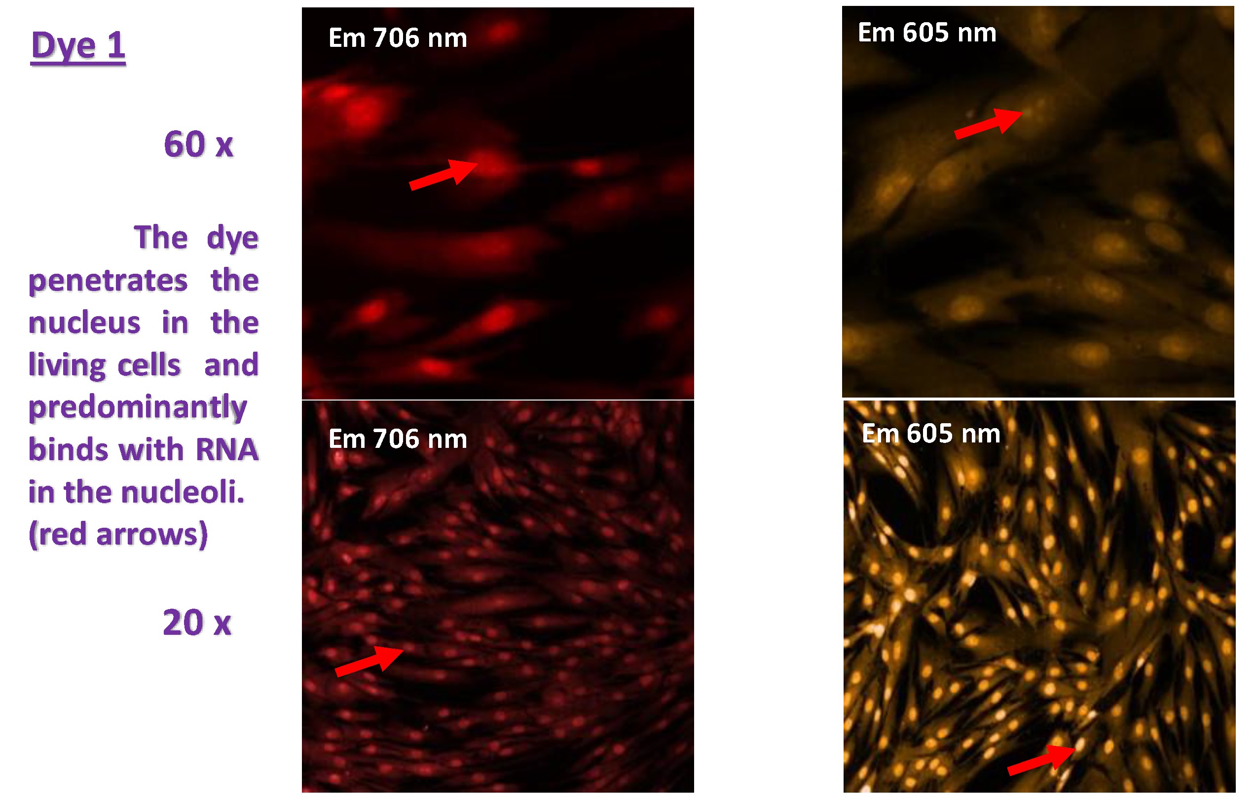
2.2. Photophysical Properties of Dye 1 in the Absence and in the Presence of dsDNA

To verify that dye 1, which was obtained by us, was suitable for a fluorogenic marker for labeling nucleic acids, we examined its photophysical properties in a TE buffer pure and in the presence of dsDNA. Figure 1A shows the change in intensity of the longest wavelength absorption band after the addition of double-stranded DNA in 0.25 μg/mL increments. The bathochromic shift of the absorption maxima from 490 nm to 506 nm combined with the hypochromic one is evidence for the formation of the dye–dsDNA complex and for the probable intercalation mode of binding [26].
The addition of the dsDNA with the above-mentioned step (0.25 µg/mL) leads to a significant increase (almost ten times) in the fluorescence of the dye 1–dsDNA complex (Figure 1B). The promising photo physical properties of the dye made us ambitious to research its behavior in living cells.
We used mesenchymal stem cells from apical papilla (SCAP) between the 3rd and 5th passages for the experiments. SCAP treated with newly synthesized fluorescent dye 1 were examined on an InCell Analyzer 6000 with high throughput analysis. Figure 2 shows a representative sample of the fluorescent paint method code. This analysis establishes the process of penetration, intracellular binding and metabolism of the dye. It was found that dye 1 passes freely through the cell membrane and does not cause cell death. Afterwards, the dye targets the nucleoli and the endoplasmic reticulum, and we speculate it has RNA affinity in the cells. This interesting finding provides an idea for further applications of the dye as the marker of retrovirus activity in living cells or even for its use as a theragnostic reagent. The investigations in this field are in progress and will be published elsewhere.

3. Materials and Methods

3.1. General

All reagents and solvents are commercially available and used as supplied. The 2,3-Dimethylbenzo[d]thiazol-3-ium methyl iodide (2a) [24] and 4-thiomorpholinobenzaldehyde (3a) [24] were synthesized starting from 2-methyl benzo[d]thiazole (2) and 4-fluorobenzaldehyde (3), respectively, according to the previously published research methods in the literature [25]. The progress of the chemical reactions was monitored by thin layer chromatography (TLC) ALUGRAM® SIL G/UV 254-60 Macherey-Nagel, ready-to-use plates with thickness of the silica layer at 0.2 mm. Melting points of 1, 2a and 3a were determined on Kruess M5000 melting point meter for automatic measurements. NMR spectra (1H-, 13C-NMR) were obtained on a Bruker Avance II+ NMR spectrometer operating at 500 MHz for 1H- and 125 MHz for 13C-NMR in DMSO-d6 as a solvent (Supplementary Materials). The chemical shifts are given in ppm (δ) using tetramethylsilane (TMS) as an internal standard. The mass spectra of dye 1 was obtained on an Advion expression CMS mass spectrometer in “High temperature and low fragmentation” regime and analyzed by using Advion CheMS Express software version 5.1.0.2. IR spectra is obtained on Specord 71 (Carl-Zeiss Jena) spectrometer in Nujol as a solvent. UV-VIS spectra were measured on a Unicam 530 UV-VIS spectrophotometer (with concentration of dye 1 C = 1 × 10⁻⁵ M) and the fluorescence spectra were obtained on a Fluorolog 2 (C = 1 × 10⁻⁶ M.) fluorescence spectrophotometer in quartz cuvettes and in TE buffer (10 mM TRIS, 1 mM EDTA, pH 8). The nucleic acid used is dsDNA (49 μg/mL) (Salmon sperm dsDNA, AppliChem, 64291 Darmstadt, Germany). The dye investigated was slightly soluble in redistilled water and DMSO, therefore, fresh stock solutions (1 mM) were prepared in DMF, and further diluted with TE-buffer. The apical papilla of teeth without fully developed roots were gently separated from the root and pulp. The tissue sample was digested with 3 mg/mL collagenase I/4 mg/mL dispase for 1 h at 37 °C and 4% CO2. After reaching 80%, confluence cells were transferred to 48 well plates. After 24 h, the dyes in their respective concentrations were added to every well for another 24 h. Unstained cells were used for determining auto-fluorescence. InCell Analyzer 6000 was used for visualization of the native cellular permeability for the dyes.

3.2. Synthesis of (E)-3-Methyl-2-(4-thiomorpholinostyryl)benzo[d]thiazol-3-ium iodide (1)

The 2,3-Dimethylbenzo[d]thiazol-3-ium methyl sulfate (2a, 0.2 g, 0.77 mmol), in slight excess of 4-thiomorpholinobenzaldehyde (3a, 0.183 g, 0.89 mmol, 1.15 eq) and piperidine (0.1 mL) were dissolved in 8 mL of ethanol/ethyl acetate mixture (3:1) and were refluxed for 4 h. After the end of the reaction, the resulting precipitate from (E)-3-methyl-2-(4-thiomorpholinostyryl)benzo[d]thiazol-3-ium methyl sulfate was filtered off and washed with ethanol. The crude product was obtained as methyl sulfate salt was dissolved in methanol at room temperature and converted into the corresponding iodide 1 adding potassium iodide (0.128 g, 0.77 mmol), which was dissolved in a minimal amount of water. Yield of 1: 0.355 g (96%), mp 225–226 °C. 1H–NMR (DMSO-d6, δ (ppm)): 2.67 (brs, 4H, H–CH2), 3.89 (brs, 4H, H–CH2), 4.26 (s, 3H, H–N+CH3), 7.06 (d, 2H, H–Ph, 3J = 8.5 Hz), 7.69–7.72 (m, 2H, H–Ar, H–St), 7.81 (t, 1H, H–Ar, 3J = 8.0 Hz), 7.92 (d, 2H, H–Ph, 3J = 8.5 Hz), 8.08 (d, 1H, H–St, 3J = 15.0 Hz), 8.13 (d, 1H, H–Ar, 3J = 8.0 Hz), 8.33 (d, 1H, H–Ar, 3J = 8.0 Hz). 13C–NMR δ = 172.07 (C), 152.81 (C), 150.02 (CH), 142.45 (C), 133.30 (2CH), 129.45 (CH), 128.13 (CH), 127.48 (C), 124.37 (CH), 123.19 (C), 116.62 (CH), 114.34 (CH), 108.12 (CH), 49.95 (2CH2), 36.23 (CH3), 25.69 (2CH2). 13C–DEPT–135 δ = 150.02 (CH), 133.30 (2CH), 129.45 (CH), 128.13 (CH), 124.37 (CH), 116.62 (CH), 114.34 (CH), 108.12 (CH), 49.95 (2CH2), 36.23 (CH3), 25.69 (2CH2). IR (ν = cm−1): 605 (C–S), 705 (CH2–S), 750 (C–S), 802 (C=C), 935 (C=C), 1105 (C–N), 1175 (C–N), 1280 (=C–N), 1295 (=C–H), 1340 (=C–H), 1380 (=C–H), 1397 (=C–H), 1440 (=C–H), 1500 (=C–H), 1565 (C=C). ESI-MS: calc. 339.5, found 339.0.

4. Conclusions

A new styryl dye with thiomorpholine functionality for DNA and cell components visualization was prepared by an improved, convenient and reliable reaction procedure. The dye demonstrated low intrinsic fluorescence in the TE buffer and a significant increase in the fluorescence after dsDNA binding. The dye is nontoxic for stem cells from apical papilla and is suitable for cell nucleoli visualization.

Supplementary Materials

The following data are available online: 1H-NMR, 13C-NMR, IR spectra and mass spectra of 1.

Author Contributions

The dye 1 molecule was designed by A.A.V., M.I.K. performed the chemical synthesis. The interpretation of the analytical data and structure characterization of 1 were made by M.I.K. Detailed analysis of the photophysical data was carried out by A.A.V. The manuscript is written by M.I.K. and A.A.V., M.M., N.I., M.D. and L.T. performed the extraction and separation of the apical papilla from teeth and performed the scientific investigations with tissue samples. All authors have read and agreed to the published version of the manuscript.

Funding

This research received no external funding.

Institutional Review Board Statement

Not applicable.

Informed Consent Statement

Not applicable.

Data Availability Statement

Not applicable.

Acknowledgments

This work was supported by the Bulgarian National Science Fund (BNSF) project RNAVision (KP-06-DK1/4) from 29.03.2021.

Conflicts of Interest

The authors declare no conflict of interest.

References

  1. Hammer, F. Cyanine Dyes and Related Compounds; Interscience Publisher: London, UK; New York, NY, USA, 1964; p. 398. [Google Scholar]
  2. Raue, R. Ullmann’s Encyclopedia of Industrial Chemistry; Elvers, B., Hawkins, S., Weinheim, G.S., Eds.; Wiley VCH Verlag: Weinheim, Germany, 1990; p. 487. Volume 16. [Google Scholar]
  3. Banerji, J.; Mandal, A.; Banerje, B. Methine chain substituted styryl cyanine dyes—Part I. Dyes Pigments 1982, 3, 273–280. [Google Scholar] [CrossRef]
  4. Jha, B.; Banerji, J. Chromophoric chain β-aryl-substituted styryl cyanines: Effect of substituents on visible absorption spectra and photosensitisation properties. Dyes Pigments 1985, 6, 213–226. [Google Scholar] [CrossRef]
  5. Jha, B.; Jha, R.; Banerji, J. Chromophoric Chain β-(styryl-substituted styryl and phenyl-substituted butadienyl) cyanines: Effect of β-substituents and chain lengthening on the. Dyes Pigments 1986, 7, 133–152. [Google Scholar] [CrossRef]
  6. Heilbron, K.; Walter, B. XCVIII.—Styrylbenzopyrylium salts. Part V. Distyryl derivatives of 7-hydroxy-2: 4-dimethylbenzopyrylium chloride. J. Chem. Soc. 1925, 127, 690–693. [Google Scholar] [CrossRef]
  7. Nakazawa, Y.; Nakamura, Y.; Sueyoshi, T.; Sato, A. DE2147586A1. Fuji Photo Film Co. Ltd., 13 April 1972. [Google Scholar]
  8. Nakazawa, Y.; Nakamura, Y.; Nakajima, Y.; Matsuyama, J.; Sato, A. DE2146444A1. Fuji Photo Film Co. Ltd., 13 April 1972. [Google Scholar]
  9. Park, K.; Lee, C.; Song, D.; Kim, J.; Huh, Y.; Min, K. Optical Recording Properties of Styryl Derivatives for Digital Versatile Disc-Recordable (DVD-R). Mol. Crystals Liq. Crystals Sci. Tech. Sect. A Mol. Cryst. Liq. Crystals 2001, 370, 165–168. [Google Scholar] [CrossRef]
  10. Antonious, M. Solvent polarity indicators: Flexible styrylpyridinium and quinolinium fluorescence probes for medium free-volume. Spectrochim. Acta A 1997, 53, 317–324. [Google Scholar] [CrossRef]
  11. Yang, J.; Xuexiao, G.; Xuebao, H. Synthesis of Tetramethine-Styryl Cyanine Dyes and Their Lasering Properties. Chem. J. Chin. Univ. 1990, 3, 286–290. [Google Scholar]
  12. Keller, J. US3888668A. Itek Corp., 10 June 1975. [Google Scholar]
  13. Oehlschaeger, H.; Ghys, T.; Verhille, K.; Kampfer, H. DE2102175. Agfa-Gevaert, 18 January 1971. [Google Scholar]
  14. Manhardt, J.; Nashua, N. US3630733A. Itek Corp., 28 December 1971. [Google Scholar]
  15. Tsuhahara, H. JPS48828Y1·. SANKYO COMPANY LTD., 10 January 1973. [Google Scholar]
  16. Mishra, A.; Behera, R.; Behera, P.; Mishra, B.; Behera, G. Cyanines during the 1990s:  A Review. Chem. Rev. 2000, 100, 1973–2012. [Google Scholar] [CrossRef] [PubMed]
  17. Deligeorgiev, T.; Vasilev, A.; Kaloyanova, S.; Vaquero, J.J. Styryl dyes—Synthesis and applications during the last 15 years. Color. Technol. 2010, 126, 55–80. [Google Scholar] [CrossRef]
  18. Kovalska, V.; Kryvorotenko, D.; Balanda, A.; Losytskyy, M.; Tokar, V.; Yarmoluk, S. Fluorescent homodimer styrylcyanines: Synthesis and spectral-luminescent studies in nucleic acids and protein complexes. Dyes Pigments 2005, 67, 47–54. [Google Scholar] [CrossRef]
  19. Kovalska, V.; Losytskyy, M.; Kryvorotenko, D.; Balanda, A.; Tokar, V.; Yarmoluk, S. Synthesis of novel fluorescent styryl dyes based on the imidazo[1,2-a]pyridinium chromophore and their spectral-fluorescent properties in the presence of nucleic acids and proteins. Dyes Pigments 2006, 68, 39–45. [Google Scholar] [CrossRef]
  20. Volkova, K.; Kovalska, V.; Tatarets, A.; Patsenker, L.; Kryvorotenko, D.; Yarmoluk, S. Spectroscopic study of squaraines as protein-sensitive fluorescent dyes. Dyes Pigments 2007, 72, 285–292. [Google Scholar] [CrossRef]
  21. Balanda, A.; Volkova, K.; Kovalska, V.; Losytskyy, M.; Tokar, V.; Prokopets, V.; Yarmoluk, S. Synthesis and spectral–luminescent studies of novel 4-oxo-4,6,7,8-tetrahydropyrrolo[1,2-a]thieno[2,3-d]pyrimidinium styryls as fluorescent dyes for biomolecules detection. Dyes Pigments 2007, 75, 25–31. [Google Scholar] [CrossRef]
  22. Li, Q.; Chang, Y.-T. A protocol for preparing, characterizing and using three RNA-specific, live cell imaging probes: E36, E144 and F22. Nat. Protocols. 2006, 1, 2922–2932. [Google Scholar] [CrossRef] [PubMed]
  23. Lu, Y.-J.; Deng, Q.; Hu, D.-P.; Wang, Z.-Y.; Huang, B.-H.; Du, Z.-Y.; Xiong Fang, Y.; Wong, W.-L.; Zhang, K.; Chow, C.-F. A molecular fluorescent dye for specific staining and imaging of RNA in live cells: A novel ligand integration from classical thiazole orange and styryl compounds. Chem. Commun. 2015, 51, 15241–15244. [Google Scholar] [CrossRef] [PubMed]
  24. Vasilev, A.; Kandinska, M.; Stoyanov, S.; Yordanova, S.; Sucunza, D.; Vaquero, J.; Castaño, O.; Baluschev, S.; Angelova, S.E. Halogen-containing thiazole orange analogues—New fluorogenic DNA stains. Beilstein J. Org. Chem. 2017, 13, 2902–2914. [Google Scholar] [CrossRef] [PubMed] [Green Version]
  25. Zhang, J.; Poongavanam, V.; Kang, D.; Bertagnin, C.; Lu, H.; Kong, X.; Ju, H.; Lu, X.; Gao, P.; Tian, Y.; et al. Optimization of N-Substituted Oseltamivir Derivatives as Potent Inhibitors of Group-1 and -2 Influenza A Neuraminidases, Including a Drug-Resistant Variant. J. Med. Chem. 2018, 61, 6379–6397. [Google Scholar] [CrossRef] [PubMed]
  26. Ustimova, M.; Chernikova, P.; Shepel, N.; Fedorov, Y.; Fedorova, O. Effect of N-substituent in 4-styrylpyridinium dyes on their binding to DNA. Mendeleev Commun. 2020, 30, 217–219. [Google Scholar] [CrossRef]
Scheme 1. Synthesis of (E)-3-methyl-2-(4-thiomorpholinostyryl)benzo[d]thiazol-3-ium iodide (1).
Molbank 2022 m1392 sch001
Figure 1. (A) Absorption spectra of dye 1 in TE buffer neat and in presence of dsDNA; (B) Fluorescence spectra of dye 1 in TE buffer neat and in presence of dsDNA.
Molbank 2022 m1392 g001
Figure 2. Fluorescence of dye 1 with cell organelles in stem cells from apical papilla.
Molbank 2022 m1392 g002
Publisher’s Note: MDPI stays neutral with regard to jurisdictional claims in published maps and institutional affiliations.

