

Synthesis, Characterization, and Structure of Mixed-Ligand Cobalt (II) Complex with N, O Donor Sites

1
Department of Chemistry, Sidho-Kanho-Birsha University, Purulia 723104, West Bengal, India
2
Department of Chemistry, Shiv Nadar University, Gautam Buddha Nagar 201314, Uttar Pradesh, India
*
Authors to whom correspondence should be addressed.
Molbank 2022, 2022(3), M1447; https://doi.org/10.3390/M1447
Submission received: 23 July 2022
/
Revised: 10 September 2022
/
Accepted: 13 September 2022
/
Published: 16 September 2022
(This article belongs to the Topic Organocatalysis and Transition-Metal Catalysis: Key Trends in Synthetic Chemistry and Challenges)
Abstract
:A mononuclear octahedral mixed-ligand cobalt (II) complex [Co(H2L)(PhCOO)2] (1) has been prepared by using H2L (N,N′-dimethyl-N,N′-bi(2-hydroxy-3,5-di methyl benzyl)-ethylenediamine) as a facially coordinating tetradentate ligand with a N2O2 donor center along with sodium benzoate as an ancillary ligand. Complex 1 has been characterized by a single-crystal X-ray diffraction study, as well as by other spectroscopic tools. The complex crystallizes in the monoclinic space group C2 with a = 31.73(3) Å, b = 7.868(3) Å, c = 19.131(15) Å, and β = 125.25(3)°. The single-crystal X-ray diffraction study shows that in the mononuclear cobalt (II) complex [Co(H2L)(PhCOO)2] (1), the metal center adopts an octahedral environment.
1. Introduction
Transition metal complexes with the tetradentate amine-bis(phenolate) ligand have been widely studied as catalysts [1,2,3,4,5,6,7], models of reaction centers of metalloenzymes [8,9], nonlinear optical materials [10], and biological models to understand the structures of biomolecules and biological processes [11]. Recently, research in transition metal complexes with bis(phenolate) ligands has been steadily increasing. A series of mono-, bi- and trinuclear cobalt (III) complexes with amine-bis(phenolate) ligands have been reported [12,13,14,15]. The catalytic activity of mononuclear cobalt (II/III) complexes with an amine-bis(phenolate) ligand for the coupling of CO2 with propylene oxide to give cyclic propylene carbonate have been investigated [16]. At the same time, the catechol oxidase activity of mononuclear cobalt complexes with amine-bis(phenolate) ligands have been reported [17].
Inspired by the above knowledge, we report a mononuclear octahedral mixed-ligand cobalt (II) complex [Co(H2L)(PhCOO)2] (1). The molecular structure of the complex was established by a single-crystal X-ray diffraction study, as well as by other spectroscopic tools. The crystal structure reveals that the cobalt ion in this mononuclear complex is surrounded by a tetradentate ligand (H2L) with a N2O2 donor site along with two benzoate ligands as ancillary ligands.
2. Results
2.1. General: Synthesis of 1
A sterically hindered tetradentate phenol-based N2O2 ligand (H2L) has been used to synthesize a mononuclear mixed-ligand cobalt complex [Co(H2L)(PhCOO)2] (1). The tetradentate ligand N,N′-dimethyl-N,N′-bis(2-hydroxy-3,5-dimethylbenzyl)ethylenediamine (H2L) was prepared according to a reported method [12,18]. The reactions are carried out in acetonitrile in the presence of sodium benzoate as the ancillary ligand and Co(ClO4)2.6H2O as the metal ion precursor. The tetradentate ligand H2L acts as a capping ligand in complex 1 because of its enormous flexibility, thus allowing benzoate ions to form a coordinate bond around the cobalt center in the mononuclear complex [19]. The compound is air stable in the solid state and also fairly stable in solution. The compound is also stable in solution for several days and no aerial oxidation takes place in solution in open air. The detailed strategy is summarized in Scheme 1. The structure of 1 was confirmed by a single-crystal X-ray diffraction study (Figure 1).
IR spectra of the complexes (1) display characteristic bands of the coordinated tetradentate ligand. IR spectra of the compound have a prominent band at the ca. 1183 cm−1 region due to the ν(C-O/phenolate) stretching mode [20]. In addition, the carboxylato complex (1) displays a pair of strong bands at ca. the 1674 and 1453 cm−1 regions due to symmetric and antisymmetric ν(COO−) vibrations, respectively. The difference in frequency (Δν) in all three cases is ca. 220 cm−1, thus indicating a monodentate mode of carboxylate coordination [20].
2.2. X-ray Structure
The asymmetric unit of the crystal structure consists of two independent molecules with two units of solvent of crystallization, as shown in Figure 2. The central metal ion Co(II) is hexa-coordinated, forming an octahedral geometry, and is surrounded by two protonated phenolic O (Co1-O1, 2.163(4) Å; Co2-O3, 2.115(4) Å), two carboxylate O (Co1-O2, 2.059(5) Å; Co2-O4, 2.073(5) Å) and two N (Co1-N1, 2.213(6) Å; Co2-N2, 2.226(6) Å). The phenolic O are opposite to each other (O1-Co1-O1A, 170.47(15)°; O4-Co2-O4A, 170.90(16)°), whereas the tertiary amine N is opposite to the carboxylate O (N1-Co1-O2, 167.55(16)°, N1A-Co1-O2A, 167.55(16)°; N2-Co2-O5, 167.08(16)°, N2A-Co1-O5A, 167.08(16)°).
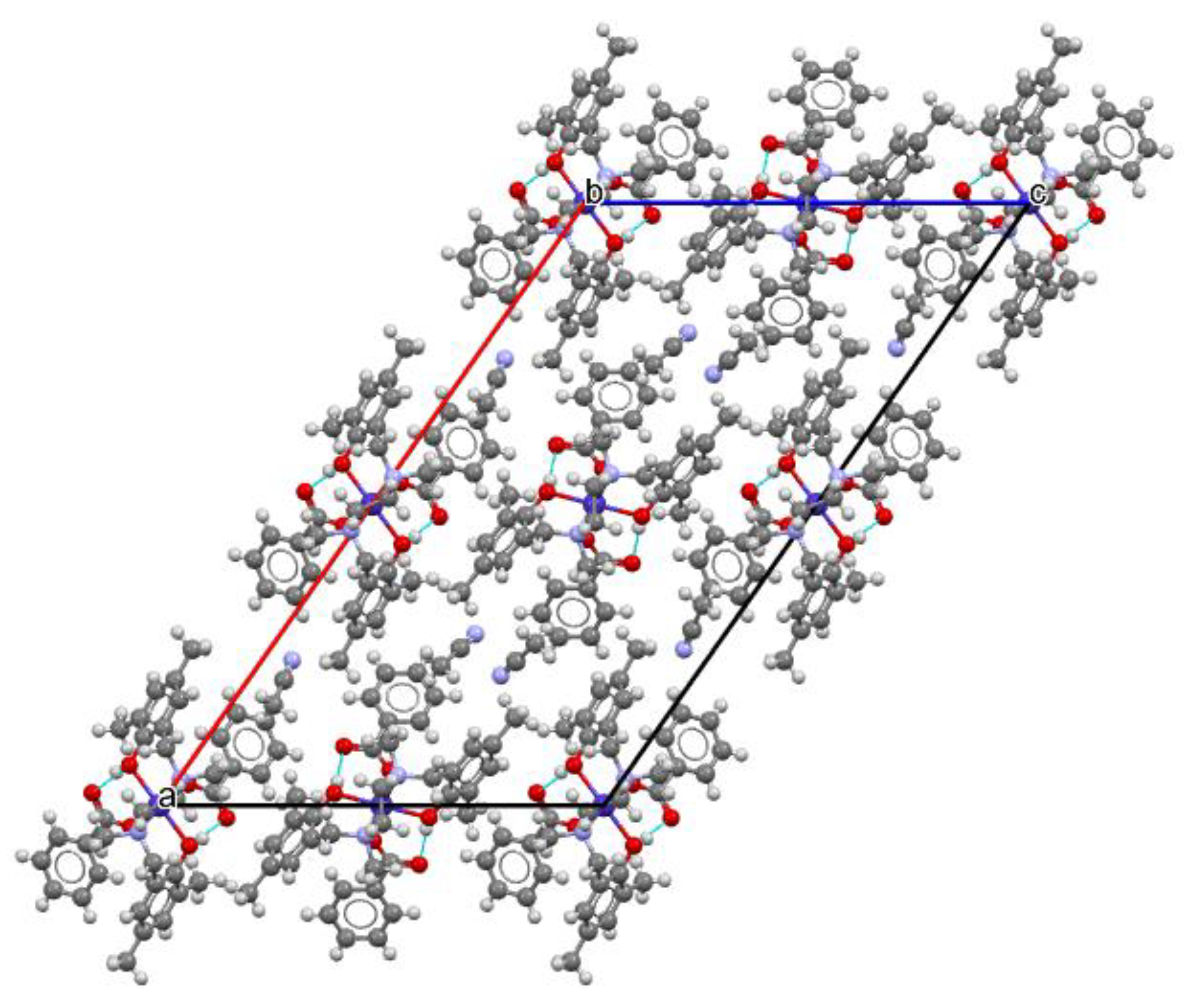
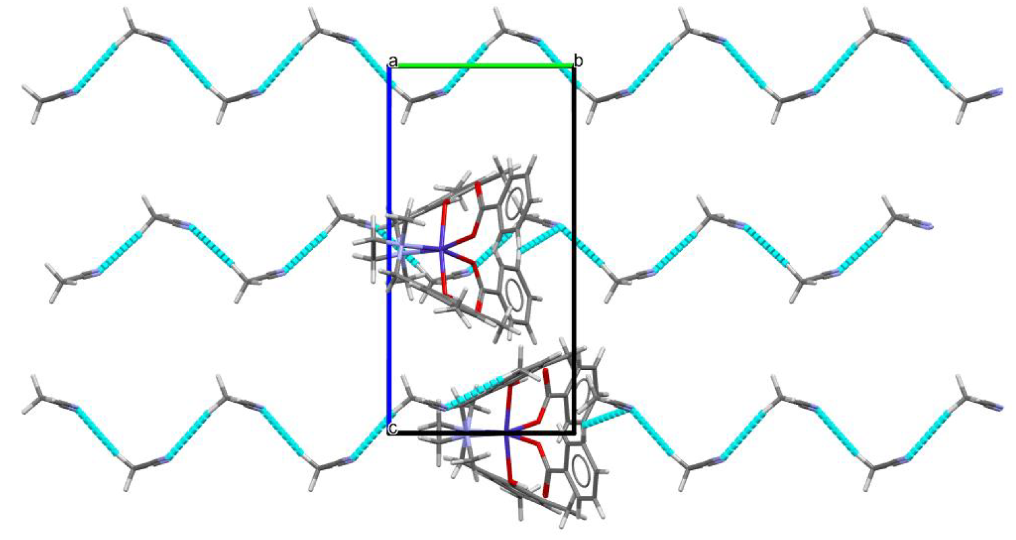
The phenolic proton participates in strong to moderately strong intra-molecular H-bonding (as shown in the packing structure, Figure 3). The H-bond parameters are given in Table 1. In addition to intra-molecular H-bonds, the solvent, acetonitrile, also exhibits weak H-bonding to form a 1-D zig-zag chain along the b-axis (as shown in Figure 4). The contact distance between acetonitrile N and phenolic H is 2.70 Å with the bond angle of 147.85° (N4-H40-C40).
Phenolic O also participates in intermolecular H-bonding of moderate strength with the H of the adjacent phenyl ring.
2.3. Hirschfeld Surface Analysis
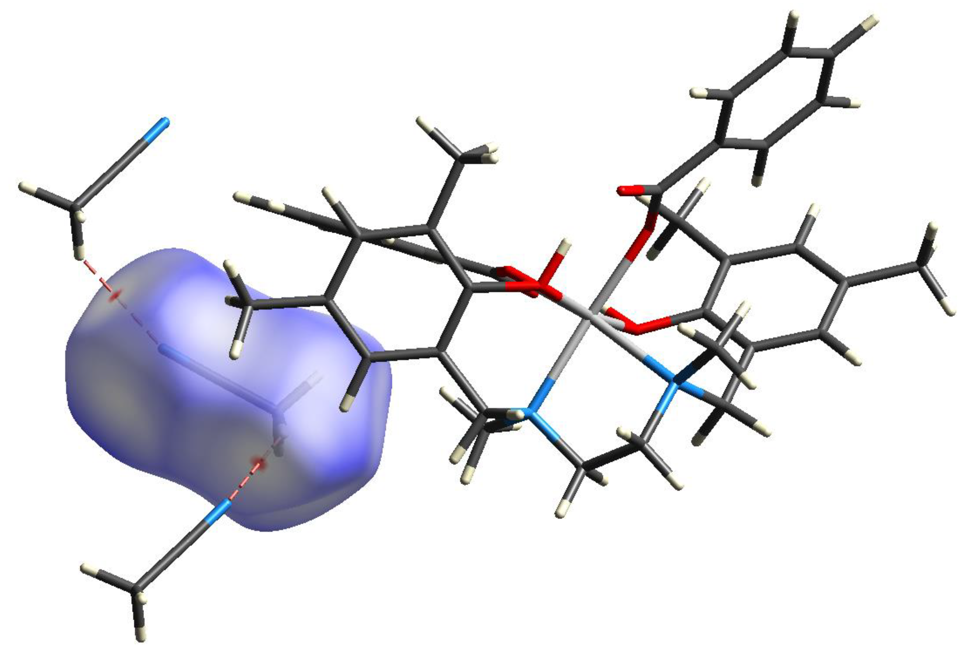
A Hirshfeld surface analysis was performed using Crystal Explorer [21]. The Hirshfeld surface was mapped using an iso-value of 0.5, with red contours indicating a contact less than the sum of the van der Waals radii of the respective elements, whereas blue and white contours indicate that the nearest external atom is at a distance greater than or equal to the sum of the van der Waals radii, respectively, from the atomic coordinate, as shown in Figure 2.
The Hirshfeld analysis of 1 also reveals an array of the H-interaction ranging from a strong H-bond that is intra-molecular (H1O....O3, 1.60(9) Å; H2O....O6, 1.77(10) Å) in nature to a moderate intermolecular H-bond involving the protonated phenolic oxygen atom, as shown in Figure 5. A 1-D zig-zag chain formed by the acetonitrile solvent is shown in Figure 6.
3. Materials and Methods
3.1. General
The tetradentate ligand N,N′-dimethyl-N,N′-bis(2-hydroxy-3,5-dimethylbenzyl)ethylenediamine (H2L) was prepared according to a reported method [12,18]. Solvents were purified by taking suitable drying agents that were distilled under nitrogen prior to their use [22]. All reagents were purchased from E Merck, ( Bangalore, India). Chemicals were used without further purification.
UV–visible spectra of the complex were recorded on a Perkin-Elmer 950 UV/vis/NIR spectrophotometer (San Diego, CA, USA) and infrared spectra were taken on a Nicolet Magna 750 FT-IR spectrometer (San Diego, CA, USA), series II with samples prepared as KBr pellets.
3.2. Experimental: Synthesis of 1
The ligand H2L (0.18 g, 0.5 mmol) was added to a stirred solution of cobalt (II) perchlorate hexahydrate (0.18 g, 0.5 mmol) in acetonitrile (25 mL). The stirring was continued for ca. 15 min and then solid sodium benzoate (0.14 g, 1 mmol) was portion-wise added to it. The resulting solution was refluxed for another 1 h. Then, the solution was filtered, and the filtrate was kept in open air for crystallization. A light-pink crystalline compound, along with diffraction-quality single crystals, were obtained by slow evaporation. It was collected by filtration and washed with cold diethyl ether (2 × 5 mL). Yield: 0.21 g (70%). Anal. Calcd for C36H40CoN2O6: C, 66.15; H, 6.12; N, 4.28. Found: C, 66.05; H, 5.98; N, 4.08%. IR (KBr disk, cm−1): 3415, 1674, 1453, 1307, 1183, 751, 1076, 754 (See Supplementary Materials). UV–Vis [λmax/nm (ε/M−1 cm−1)]: 312 (1090) (See Supplementary Materials).
The intensity data for a blue-block crystal, 0.12 × 0.14 × 0.20 mm, of the cobalt (II) complex (1) was collected at 296 K (23 °C) by mounting the crystal onto a quartz fiber on a Bruker D8 Quest diffractometer (Bruker, Karlsruhe, Germany) with an Incoatec microfocus source of Mo Kα radiation (λ = 0.71073 Å). APEX4 software [23] was used for the preliminary determination of the unit cell. Determination of integrated intensities and unit cell refinement was performed using SAINT [24]. The structure was solved with SHELXS [25] and subsequent structure refinements were performed with SHELX2019/1 [26]. Absorption corrections were performed by SADABS [27]. The crystal exhibits inversion twinning as confirmed by the Flack test [28]. Platon [29] was employed to obtain the twin law [−1 0 0 0 −1 0 0.69 0 1]. The final structure was refined with hkl5.
4. Conclusions
The sterically constrained tetradentate ligand H2L has been used to synthesize mononuclear cobalt complexes in the presence of carboxylate as an ancillary ligand. The complex [Co(H2L)(PhCOO)2] (1) has been characterized by a single-crystal X-ray diffraction study. The geometry of the complex is octahedral in nature and it is a mixed-ligand complex.
Supplementary Materials
The following supporting information can be downloaded online, Figure S1: Electronic absorption spectrum of complex (1) in acetonitrile. Figure S2: IR Spectra of complex (1).
Author Contributions
Conceptualization: R.G. and D.M.; methodology: D.M., A.H. and P.C.; X-ray crystal structures: R.G.; investigation: D.M., A.H. and P.C.; writing—original draft preparation: P.C. and A.H.; writing—review and editing: R.G. and D.M. All authors have read and agreed to the published version of the manuscript.
Funding
Financial support received from the Department of Science and Technology, Government of West Bengal, Kolkata (Project memo no. 1071(Sanc)/ST/P/S&T/15G-4/2015 dated 23 February 2016) and Sidho-Kanho-Birsha University (R/847/SKBU/22).
Data Availability Statement
CCDC 2183749 contains the supplementary crystallographic data for compound 1. These data can be obtained free of charge via http://www.ccdc.cam.ac.uk/conts/retrieving.html, accessed on 12 September 2022, or from the Cambridge Crystallographic Data Center, 12 Union Road, Cambridge CB2 1EZ, UK; Fax: +44-1223-336-033; or e-mail: [email protected].
Acknowledgments
We are grateful for the instrumental support from the Department of Inorganic Chemistry, Indian Association for the Cultivation of Science, Kolkata, India.
Conflicts of Interest
The authors declare no conflict of interest.
Sample Availability
Samples of the compounds are available from the authors.
References
- Katsuki, T. Catalytic asymmetric oxidations using optically active (salen)manganese(III) complexes as catalysts. Coord. Chem. Rev. 1995, 140, 189–214. [Google Scholar] [CrossRef]
- Cozzi, P.G. Metal–Salen Schiff base complexes in catalysis: Practical aspects. Chem. Soc. Rev. 2004, 33, 410–421. [Google Scholar] [CrossRef] [PubMed]
- Ziessel, R. Schiff-based bipyridine ligands. Unusual coordination features and mesomorphic behaviour. Coord. Chem. Rev. 2001, 216, 195–223. [Google Scholar] [CrossRef]
- Bermejo, M.R.; Castiñeiras, A.; Garcia-Monteagudo, J.C.; Rey, M.; Sousa, A.; Watkinson, M.; McAuliffe, C.A.; Pritchard, R.G.; Beddoes, R.L. Electronic and steric effects in manganese Schiff-base complexes as models for the water oxidation complex in photosystem II. The isolation of manganese-(II) and -(III) complexes of 3- and 3,5-substituted N,N′-bis(salicylidene)ethane-1,2-diamine (H2salen) ligands. J. Chem. Soc. Dalton Trans. 1996, 14, 2935–2944. [Google Scholar]
- Mandal, B.; Majee, M.C.; Mandal, D.; Ganguly, R. Synthesis, structure, Hirshfeld surface analysis and catecholase activity of Ni(II) complex with sterically constrained phenol based ligand. J. Mol. Struct. 2020, 1202, 127340. [Google Scholar] [CrossRef]
- Mandal, B.; Majee, M.C.; Rakshit, T.; Banerjee, S.; Mitra, P.; Mandal, D. Synthesis, structure, DFT study and catechol oxidase activity of Cu(II) complex with sterically constrained phenol based ligand. J. Mol. Struct. 2019, 1193, 265–273. [Google Scholar] [CrossRef]
- Mandal, B.; Chakraborty, T.; Ali, I.; Mondal, D.; Majee, M.C.; Raha, S.; Ghosh, K.; Mandal, P.M.D. Synthesis, structure, catechol oxidase activity and antibacterial studies of MnIII complex with sterically constrained phenol-based N2O2 ligand. J. Indian Chem. Soc. 2017, 94, 1079–1087. [Google Scholar]
- Tsou, T.T.; Loots, M.; Halpern, J. Kinetic determination of transition metal-alkyl bond dissociation energies: Application to organocobalt compounds related to B12 coenzymes. J. Am. Chem. Soc. 1982, 104, 623–624. [Google Scholar] [CrossRef]
- Summers, M.F.; Marzilli, L.G.; Bresciani-Pahor, N.; Randaccio, L. Unusual bond lengths, conformations, and ligand exchange rates in B12 models with the bis(salicylidene)-o-phenylenediamine equatorial ligand. J. Am. Chem. Soc. 1984, 106, 4478–4485. [Google Scholar] [CrossRef]
- Bella, S.D.; Fragalà, I. Synthesis and second-order nonlinear optical properties of bis(salicylaldiminato)M(II) metalloorganic materials. Synth. Met. 2000, 115, 191–196. [Google Scholar] [CrossRef]
- Lacroix, P.G. Second-Order Optical Nonlinearities in Coordination Chemistry: The Case of Bis(salicylaldiminato)metal Schiff Base Complexes. Eur. J. Inorg. Chem. 2001, 2, 339–348. [Google Scholar] [CrossRef]
- Velusamy, M.; Palaniandavar, M.; Gopalan, R.S.; Kulkarni, G.U. Iron(III) complexes of certain meridionally coordinating tridentate ligands as models for non-heme iron enzymes: The role of carboxylate coordination. Inorg. Chem. 2003, 42, 8283–8293. [Google Scholar] [CrossRef] [PubMed]
- Merkel, M.; Muller, F.K.; Krebs, B. Novel iron (III) complexes with phenolate containing tripodal tetradentate ligands as model systems for catechol 1, 2-dioxygenases. Inorg. Chim. Acta 2002, 337, 308–316. [Google Scholar] [CrossRef]
- Viswanathan, R.; Palaniandavar, M.; Balasubramanian, T.; Muthiah, T.P. Functional Models for Catechol 1,2-Dioxygenase. Synthesis, Structure, Spectra, and Catalytic Activity of Certain Tripodal Iron(III) Complexes. Inorg. Chem. 1998, 37, 2943–2951. [Google Scholar] [CrossRef]
- Mialane, P.; Anxolabéhère-Mallart, E.; Blondin, G.; Nivorojkine, A.; Guilhem, J.; Tchertanova, L.; Cesario, M.; Ravi, N.; Bominaar, E.; Girerd, J.J.; et al. Structure and electronic properties of (N,N′-bis(4-methyl-6-tert-butyl-2-methyl-phenolato)-N,N′-bismethyl-1,2-diaminoethaneFeIII (DBSQ). Spectroelectrochemical study of the red-ox properties. Relevance to intradiol catechol dioxygenases. Inorg. Chim. Acta 1997, 263, 367–378. [Google Scholar] [CrossRef]
- Saunders, L.N.; Ikpo, N.; Petten, C.F.; Das, U.K.; Dawe, L.N.; Kozak, C.M.; Kerton, F.M. Coupling of carbon dioxide with neat propylene oxide catalyzed by aminebisphenolato cobalt(II)/(III) complexes and ionic co-catalysts. Catal. Commun. 2012, 18, 165–167. [Google Scholar] [CrossRef]
- Ali, I.; Mandal, B.; Saha, R.; Ghosh, R.; Majee, M.C.; Mondal, D.; Mitra, P.; Mandal, D. Mono and tri-nuclear cobalt(III) complexes with sterically constrained phenol based N2O2 ligand: Synthesis, structure and catechol oxidase activity. Polyhedron 2020, 180, 114429. [Google Scholar] [CrossRef]
- Chen, G.J.J.; McDonald, J.W.; Newton, W.E. Reactions of oxobis(N,N-dialkyldithiocarbamato) molybdenum(IV) with unsaturated organic molecules and their biochemical implications. Inorg. Chem. 1976, 15, 2612–2615. [Google Scholar] [CrossRef]
- Mandal, D.; Chatterjee, P.B.; Bhattacharya, S.; Choi, K.Y.; Clerac, R.; Chaudhury, M. Tetra-, Tri-, and Mononuclear Manganese(II/III) Complexes of a Phenol-Based N2O2 Capping Ligand: Use of Carboxylates as Ancillary Ligands in Tuning the Nuclearity of the Complexes. Inorg. Chem. 2009, 48, 1826–1835. [Google Scholar] [CrossRef]
- Nakamoto, K. Infrared and Raman Spectra of Inorganic and Coordination Compounds, 3rd ed.; Wiley-Interscience: New York, NY, USA, 1978. [Google Scholar]
- Spackman, P.R.; Turner, M.J.; McKinnon, J.J.; Wolff, S.K.; Grimwood, D.J.; Jayatilaka, D.; Spackman, M.A. CrystalExplorer: A program for Hirshfeld surface analysis, visualization and quantitative analysis of molecular crystals. J. Appl. Cryst. 2021, 54, 1006–1011. [Google Scholar] [CrossRef]
- Perrin, D.D.; Armarego, W.L.F.; Perrin, D.R. Purification of Laboratory Chemicals, 2nd ed.; Peragamon: Oxford, UK, 1980. [Google Scholar]
- APEX4, Version 2021.10-0; Bruker AXS Inc.: Karlsruhe, Germany, 2021.
- SAINT, Version 8.40B; Bruker AXS Inc.: Madison, WI, USA, 2013.
- Sheldrick, G.M. SADABS. Version 2012/1; Bruker AXS Inc.: Madison, WI, USA, 2012.
- Sheldrick, G.M. XS Version 2013/1 (A short history of SHELX). Acta Cryst. 2008, A64, 112–122. [Google Scholar] [CrossRef] [PubMed]
- Sheldrick, G.M. Crystal structure refinement with SHELXL. Acta Cryst. 2015, C71, 3–8. [Google Scholar] [CrossRef]
- Parsons, S.; Flack, H.D.; Wagner, T. Use of intensity quotients and differences in absolute structure refinement. Acta Cryst. B 2013, 69, 249–259. [Google Scholar] [CrossRef] [PubMed]
- Spek, A.L. Single-crystal structure validation with the program PLATON. J. Appl. Cryst. 2003, 36, 7–11. [Google Scholar] [CrossRef] [Green Version]
Scheme 1.
Preparation of complex [Co(H2L)(PhCOO)2] (1).

Figure 1.
ORTEP representation of 1 showing 50% probability atomic displacement parameters. Asymmetric unit contains 2 symmetry-independent molecules.
Figure 1.
ORTEP representation of 1 showing 50% probability atomic displacement parameters. Asymmetric unit contains 2 symmetry-independent molecules.

Figure 2.
Superposition of two symmetry-independent molecules of 1 (red/blue).

Figure 3.
Crystal structure packing viewed down the b axis with hydrogen-bonding contacts shown as blue dashed lines.
Figure 3.
Crystal structure packing viewed down the b axis with hydrogen-bonding contacts shown as blue dashed lines.

Figure 4.
The solvent, acetonitrile, forms 1-D zig-zag chain along b-axis, as shown by blue dashed line.
Figure 4.
The solvent, acetonitrile, forms 1-D zig-zag chain along b-axis, as shown by blue dashed line.

Figure 5.
Hirshfeld surface analysis of complex 1 (red contours indicating close contacts).

Figure 6.
A 1-D chain formed by the acetonitrile solvents as shown by Hirshfeld surface analysis of complex 1 (red contours indicating close contacts).
Figure 6.
A 1-D chain formed by the acetonitrile solvents as shown by Hirshfeld surface analysis of complex 1 (red contours indicating close contacts).


Table 1.
Hydrogen-bonding geometries (Å and °) for 1.
| Donor --- H....Acceptor | D - H | H...A | D...A | D - H...A | Type (Symmetry) |
|---|---|---|---|---|---|
| O1---H1O....O2 | 0.91(9) | 2.48(8) | 2.901(7) | 109(5) | Intra |
| O1---H1O....O3 | 0.91(9) | 1.60(9) | 2.486(7) | 163(8) | Intra |
| O4---H2O....O5 | 0.73(10) | 2.42(9) | 2.864(7) | 121(7) | Intra |
| O4---H2O....O6 | 0.73(10) | 1.77(10) | 2.477(8) | 163(8) | Intra |
| C17--- H17.... O4 | 0.93 | 2.58 | 3.422(9) | 150.00 | Inter (1 + x, 1 + y, z) |
Publisher’s Note: MDPI stays neutral with regard to jurisdictional claims in published maps and institutional affiliations. |
© 2022 by the authors. Licensee MDPI, Basel, Switzerland. This article is an open access article distributed under the terms and conditions of the Creative Commons Attribution (CC BY) license (https://creativecommons.org/licenses/by/4.0/).
Share and Cite
MDPI and ACS Style
Mandal, D.; Haldar, A.; Chakraborty, P.; Ganguly, R. Synthesis, Characterization, and Structure of Mixed-Ligand Cobalt (II) Complex with N, O Donor Sites. Molbank 2022, 2022, M1447. https://doi.org/10.3390/M1447
AMA Style
Mandal D, Haldar A, Chakraborty P, Ganguly R. Synthesis, Characterization, and Structure of Mixed-Ligand Cobalt (II) Complex with N, O Donor Sites. Molbank. 2022; 2022(3):M1447. https://doi.org/10.3390/M1447
Chicago/Turabian StyleMandal, Debdas, Anwesha Haldar, Priyanka Chakraborty, and Rakesh Ganguly. 2022. "Synthesis, Characterization, and Structure of Mixed-Ligand Cobalt (II) Complex with N, O Donor Sites" Molbank 2022, no. 3: M1447. https://doi.org/10.3390/M1447
Note that from the first issue of 2016, this journal uses article numbers instead of page numbers. See further details here.