Article

Synthesis, Characterization, and Sensor Applications of Spinel ZnCo2O4 Nanoparticles

by
Juan Pablo Morán-Lázaro
1,*,
Florentino López-Urías
2,
Emilio Muñoz-Sandoval
2,
Oscar Blanco-Alonso
3,
Marciano Sanchez-Tizapa
4,
Alejandra Carreon-Alvarez
4,
Héctor Guillén-Bonilla
5,
María De la Luz Olvera-Amador
6,
Alex Guillén-Bonilla
1 and
Verónica María Rodríguez-Betancourtt
7
1
Department of Computer Science and Engineering, CUValles, University of Guadalajara, Ameca, Jalisco 46600, Mexico
2
Advanced Materials Department, IPICYT, San Luis Potosí, S.L.P. 78216, Mexico
3
Department of Physics, CUCEI, University of Guadalajara, Guadalajara, Jalisco 44410, Mexico
4
Department of Natural and Exact Sciences, CUValles, University of Guadalajara, Ameca, Jalisco 46600, Mexico
5
Department of Project Engineering, CUCEI, University of Guadalajara, Guadalajara, Jalisco 44410, Mexico
6
Department of Electrical Engineering (SEES), CINVESTAV-IPN, Mexico City, DF 07360, Mexico
7
Department of Chemistry, CUCEI, University of Guadalajara, Guadalajara, Jalisco 44410, Mexico
*
Author to whom correspondence should be addressed.
Sensors 2016, 16(12), 2162; https://doi.org/10.3390/s16122162
Submission received: 7 October 2016 / Revised: 7 December 2016 / Accepted: 12 December 2016 / Published: 17 December 2016
(This article belongs to the Special Issue Gas Nanosensors)

Abstract

:
Spinel ZnCo2O4 nanoparticles were synthesized by means of the microwave-assisted colloidal method. A solution containing ethanol, Co-nitrate, Zn-nitrate, and dodecylamine was stirred for 24 h and evaporated by a microwave oven. The resulting solid material was dried at 200 °C and subsequently calcined at 500 °C for 5 h. The samples were characterized by scanning electron microscopy (SEM), transmission electron microscopy (TEM), X-ray diffraction (XRD), and Raman spectroscopy, confirming the formation of spinel ZnCo2O4 nanoparticles with average sizes between 49 and 75 nm. It was found that the average particle size decreased when the dodecylamine concentration increased. Pellets containing ZnCo2O4 nanoparticles were fabricated and tested as sensors in carbon monoxide (CO) and propane (C3H8) gases at different concentrations and temperatures. Sensor performance tests revealed an extremely high response to 300 ppm of CO at an operating temperature of 200 °C.

1. Introduction

For a long time, pollutant gases from industry and internal combustion engines have been responsible for many human health issues, and to a large extent, for global climate change. Facing this, several groups have focused on the research and development of new sensor materials for the detection and monitoring of polluting gases such as CO and C3H8. In particular, sensor materials made from semiconductor oxides have been a valuable choice because they possess good chemical stability, low price, and an easy integration into electronic circuits. However, it is still necessary to improve sensor parameters such as selectivity, sensitivity, and operating temperature.
Zinc cobaltite (ZnCo2O4) is a p-type semiconductor material with a spinel-type structure. This material has attracted the attention of several research groups due to its potential applications as an electrode for Li-ion batteries [1,2,3,4,5], as a catalyst [6,7,8,9], and in supercapacitors [10,11,12,13]. In the gas sensors field, sensor devices based on ZnCo2O4 nanoparticles have displayed an excellent sensitivity to liquefied petroleum gas [14,15,16,17], H2S [18], ethanol [19,20], acetone [21], and formaldehyde [22,23], probably due to their high surface area. Additionally, ZnCo2O4 has also been used as Cl2, NO2, CO2, H2, NH3, CH3COOH, SO2, CO, C3H8, ethylene, xylene, toluene, and methanol gas sensors [15,16,18,19,20,21,22]. Specifically, when ZnCo2O4 nanoparticles were exposed to CO and C3H8 concentrations, a poor sensitivity was exhibited at working temperatures in the range of 175 to 350 °C [15,16,19,21]. In contrast, sensors based on nanowire-assembled hierarchical ZnCo2O4 microstructures showed a good sensitivity towards CO and C3H8 at an operating temperature of 300 °C [22]. From these studies, it is fair to assume that the gas sensing properties depend on the shape and size of the nanoparticles [24]. Therefore, developing a ZnCo2O4-based gas sensor, with a high sensitivity and a low operating temperature, is of great interest to many people. Several synthesis methods of ZnCo2O4 nanoparticles have been reported, such as combustion [1], hydrothermal [3,12], thermal decomposition [14], co-precipitation/digestion [15], water-in-oil (W/O) microemulsion [18], sol-gel [25], and surfactant-mediated [26] methods. In recent years, the microwave-assisted colloidal method has been an efficient and low-cost synthesis process for obtaining micro and nanostructures of oxide materials [27,28,29]. In fact, microwave is a simple technique, which plays an important role in colloidal synthesis since it provides a rapid evaporation of the precursor solvent and a short reaction time [30,31,32,33].
To the best of our knowledge, there are not reports on the synthesis and use of faceted ZnCo2O4 nanoparticles for gas sensing applications, such as the ones presented in this investigation, but in recent works, sensors based on faceted SnO2 and ZnSn(OH)6 nanoparticles exhibited high response towards toluene and ethanol, respectively [34,35]. We believe that materials based on faceted nanoparticles can be strong candidates for gas sensing applications due to their large surface areas.
In this paper, ZnCo2O4 nanoparticles are synthesized via a microwave-assisted colloidal method [27,36]. The experimental method and the different steps to synthesize ZnCo2O4 nanoparticles are described in detail. In the following, the results of SEM, XRD, Raman, and TEM characterizations are shown. In addition, sensitivity results for sensors based on ZnCo2O4 nanoparticles are thoroughly discussed.

2. Materials and Methods

Zinc nitrate hydrate (Zn(NO3)2•xH2O, Sigma Aldrich 99.99%), cobalt(II) nitrate hexahydrate (Co(NO3)2•6H2O, Sigma Aldrich 98%), dodecylamine (C12H27N, Sigma Aldrich 98%), and ethanol (C2H6O, Golden Bell 98%) were used as reagents. 5 mmol (0.947 g) of Zn(NO3)2•xH2O, 10 mmol (2.91 g) of (Co(NO3)2•6H2O, and 5.4 mmol (1 g) of C12H27N were separately dissolved in 5 mL of ethanol. Additional syntheses were made with 10.8 mmol (2 g) and 16.2 mmol (3 g) of dodecylamine in order to obtain additional nanoparticle sizes. After 20 min of vigorous stirring on magnetic dishes at room temperature, the cobalt nitrate solution was added dropwise to the dodecylamine solution and kept stirring for 1 h. The zinc nitrate solution was then slowly added to the cobalt and dodecylamine mixture yielding a wine-color solution (the solutions with 10.8 and 16.2 mmol of dodecylamine were greenish-blue) with pH = 2 and a final volume of approximately 16 mL. This final solution was covered with a watch glass to avoid contamination and kept under stirring for 24 h, losing ~2 mL by evaporation. The solution (~14 mL) was evaporated afterwards using a microwave oven (General Electric JES769WK) at a low power (~140 W). The microwave radiation was applied over the solution for periods of 1 min in order to avoid splashing. The total time of evaporation was 3 h. The resulting solid was dried in air with a muffle (Novatech) at 200 °C. Finally, the calcination of the powder was done at 500 °C for 5 h in an alumina crucible with a cover at a heating rate of 100 °C/h. The sample was kept in the furnace for cooling at room temperature. The samples made with 5.4, 10.8, and 16.2 mmol of dodecylamine were labeled as A, B, and C, respectively.
The calcined samples were characterized by SEM using a FEI-Helios Nanolab 600 system operated at 20 kV. XRD characterizations were performed with a PANalytical Empyrean system with CuKα and λ = 1.546 Å for phase identification. XRD patterns were obtained at room temperature in the range 2θ = 10–70° with steps of 0.02°, lasting 30 s for each step. The ZnCo2O4 crystallite size was calculated by Scherrer’s equation [37] using the plane (311) at 2θ = 36.8°:
Cristallite size = 0.89 λ β cos θ 180 ° π
where λ is the X-ray wavelength and β is the full width at half maximum (FWHM). Raman spectroscopy characterization was performed using a Thermo Scientific DXR confocal Raman microscope with a 633 nm excitation source. The Raman spectra were measured from 150 to 800 cm−1 at room temperature, using an exposure time of 60 s and a Laser power of 5 mW. TEM, high-resolution transmission electron microscopy (HRTEM), energy-dispersive X-ray (EDS) and high-angle annular dark-field/scanning transmission electron microscopy (HAADF-STEM) were performed by means of a FEI Tecnai-F30 system operated at 300 kV. The gas sensing measurements were carried out on pellets of ZnCo2O4 with a thickness of 0.5 mm and a diameter of 12 mm. The pellets were prepared with 0.350 g of ZnCo2O4 powders employing a manual pressure machine (Simple Ital Equip) at 20 tons for 120 min. A TM20 Leybold detector was used to control the gas concentration and the partial pressure. The sensing response was investigated by measuring the electric resistance using a digital-multimeter (Keithley). The ZnCo2O4 pellets were exposed to several concentrations (1, 5, 50, 100, 200, and 300 ppm) of CO and C3H8. A schematic diagram of the gas sensing measurement system is shown in Figure 1. The gas sensing response, or sensitivity, was defined as S = Ra/Rg for the reducing gas, where Ra and Rg refer to the resistances measured in air and gas, respectively [21,38,39].

3. Results and Discussion

SEM images of samples A, B, and C are shown in Figure 2. Sample A exhibited a high concentration of nanoparticles with irregular shape and sizes in the range of 50–110 nm (see Figure 2a,d). Agglomerates of nanoparticles were also observed (Figure 2d). SEM images of sample B revealed a large amount of nanoparticles with diameters in the range from 40–85 nm (see Figure 2b,e). Sample C contained nanoparticles with diameters ranging from 25–70 nm (Figure 2c,d,f). The particle-size distribution of the three ZnCo2O4 samples can be seen in Figure 3. From SEM, it is clear that the dodecylamine concentration plays a key role in the morphologies and the particle sizes [40]: an increment of the dodecylamine concentration produced a large amount of nanoparticles and a decreasing particle size, which suggests that the dodecylamine inhibits the growth of the ZnCo2O4 nanoparticles.
The formation of the ZnCo2O4 nanoparticles follows the principles of nucleation and growth established by LaMer and Dinegar [41]. In our synthesis, the nucleation process could occur when the zinc nitrate solution was added to the cobalt and dodecylamine solution [30], and the particle growth continued developing by diffusion of the nuclei during agitation of the final solution [42]. As previously discussed, dodecylamine plays an important role in the microstructure of the ZnCo2O4 particles. In this research, it seems that the ethanol and the dodecylamine are working synergically to reduce cations to metals because ethanol alone is not capable of doing so (i.e., in order to produce ultrafine metal nanoparticles), but just mixed with strong reducing agents such as microwave radiation, other chemical compounds, etc. [43,44,45]. We have observed that the dodecylamine concentration is the rate-determining factor; therefore, we propose the following reaction mechanism.
Dodecylamine is a primary amine with properties of a weak base because of the nitrogen’s unshared electron pair. This kind of amine has nucleophilic behavior and can be used as reducing and surfactant agents. The reaction of dodecylamine with Co2+ cations could occur in two ways: (i) the attraction of the unshared electron pair to the nucleus of the cations in a nucleophilic reaction; (ii) the formation of an electrostatic bond between the nitrogen of the dodecylamine and the electrons of the outer shell of Co2+, because of the strong electronegativity of the nitrogen (= 3.04). The result of these interactions is the formation of the dodecylamine-Co2+ complex [46]:
Co 2 + + NH 2 - ( CH 2 ) 11 NH 3   Co 2 + NH 2 - ( CH 2 ) 11 NH 3
In this reaction, dodecylamine is working as the surfactant; therefore, nitrogen’s electrons could participate as reducing agents, reducing the cationic Co2+ to metallic nanoparticles of Co0.
Co 2 + + 2 e -   Co 0
For 5.4 mmol of dodecylamine, the molar ratio of dodecylamine: Co2+ was around 1:2, which suggests that half of the Co2+ cations are not participating in the formation of complexes and should be reduced in some way by the solvent. When the dodecylamine-ethanol-Co2+ solution is mixed with the ethanol-Zn2+ solution, and once the Co2+ and Zn2+ have been reduced, Zn could attract two atoms of Co to form ZnCo2, as Zn is slightly more electronegative than Co (1.90 vs. 1.65, respectively). Since 5.4 mmol of dodecylamine are not enough to complex all of the Co2+ cations, there are several available cations to be reduced by the solvent, and as a result the ZnCo2 grains are the larger ones. For 10.8 mmol of dodecylamine, most of the Co2+ should be forming a complex, and the growing of ZnCo2 grains is restricted through the slow release-reduction process of Co2+. For 16.2 mmol of dodecylamine, the Co2+ cations could be completely complexed; therefore, the excess of dodecylamine should form complexes even with Zn2+, restricting even more the growing of ZnCo2 grains. Summarizing, the role of dodecylamine is as a surfactant and as a reducing agent; however, it seems that the function of surfactant, through electrostatic interactions, is the dominant one, reducing the particle size and modifying the particles’ microstructure as the dodecylamine concentration increases. With heat treatment, the dodecylamine was finally removed from the ZnCo2O4.
Figure 4 depicts the XRD patterns of the samples. In them, the typical peaks corresponding to the cubic ZnCo2O4 spinel-structure were identified. The well-defined narrow peaks are an indication of the good crystallinity of the ZnCo2O4 samples. Table 1 shows the crystallite size of every sample. From these results, an increase in crystallite size was produced when the dodecylamine concentration was increased. A slight increase in the FWHM was also observed, indicating a particle size reduction as was confirmed by SEM.
The formation of the oxide ZnCo2O4 was also confirmed by Raman characterization. According to the group theory for oxides with a spinel structure, five active Raman bands were expected: A1g + Eg + 3F2g [47]. Figure 5 shows the Raman spectrum indicating the main vibrational bands produced by the ZnCo2O4 spinel crystal structure. The bands labeled by v1, v3, v4, v5, and v6 correspond to 182, 475, 516, 613, and 693 cm−1, respectively. These bands are assigned to the F2g, Eg, F2g, F2g, and A1g symmetry species. These results are consistent with those reported for ZnCo2O4 spinel structures [48]. Interestingly, the band located at 204 cm−1 (v2) is a vibrational mode that could be attributed to a Co3O4 spinel structure [49]. The formation of Co3O4 is likely due to the cation disorder (substitution of Zn2+ by Co2+) in the spinel structure of the zinc cobaltite. Notice that the Co3O4 possesses a spinel structure similar to that of ZnCo2O4. Therefore, the XRD patterns between Co3O4 and ZnCo2O4 are almost indistinguishable.
Figure 6 shows TEM images of the samples. The presence of faceted nanoparticles with a pockmarked structure was clearly identified (Figure 6a,c,e). The estimated average particle size was approximately 75, 61, and 49 nm for samples A, B, and C, respectively; these measurements are consistent with the SEM analysis. The standard deviation was ±12.64, ±8.78, and ±8.36 nm, respectively. Figure 6b,d,f shows HRTEM images of the selected area marked with a black rectangle. These images confirmed the presence of faceted ZnCo2O4 nanoparticles as was done by the XRD patterns and the Raman characterization. Fringe spacings of 0.467 and 0.286 nm are clearly observed, which are attributed to the planes (111) and (220) of the ZnCo2O4 spinel structure, respectively.
To investigate the nanoparticle composition, an EDS line scan was performed on sample A (see Figure 7). Figure 7a shows a HAADF-STEM image of the ZnCo2O4 nanoparticles. The image confirms the presence of faceted nanoparticles with a pockmarked structure, which is consistent with the TEM images. In the EDS line scan, zinc, cobalt, and oxygen are observed across the linear mapping, confirming the presence of the expected elements, as seen in Figure 7b. In the central region p2, a decrease of the element composition is observed in comparison to point p1, which can be due to the irregular surface of the nanoparticle (pockmarked zone). Similar EDS elemental line scan spectra were obtained for samples B and C. Figure 7c shows an EDS microanalysis on p2, where the individual elements Zn, Co, and O can easily be seen. The atomic ratio of Zn:Co is approximately 1:1.96, which is consistent with the composition of ZnCo2O4. This EDS spectrum is in agreement with the literature [3,5].
To investigate the sensing properties of the ZnCo2O4 oxide, pellets of the material were made and tested in CO and C3H8 atmospheres. Figure 8 shows the oxide’s response vs. CO concentration of sensors made from samples A, B, and C. As shown in Figure 8a,c, no response variation was measured for A and C at 100 °C. On the contrary, the sensor made from B exhibited response values of 1, 1.03, 1.21, 1.48, 2.50, and 5.68 for CO concentrations of 1, 5, 50, 100, 200, and 300 ppm, respectively (Figure 8b). At 200 and 300 °C, the response of the three sensors increased with an increase of the CO concentration. In the whole concentration range (1–300 ppm CO), the sensor made from C exhibited a high response at 200 °C, better than the sensors based on A and B. At this temperature, the response values of the sensor based on C were 2.56, 2.66, 3.18, 1274.29, 2622.22, and 2950 for CO concentrations of 1, 5, 50, 100, 200, and 300 ppm, respectively. The sensors made from A and B comparatively also showed a good response to 300 ppm of CO (305.07 and 19.37, respectively) at 300 °C. From these results, it is clear that the ZnCo2O4 sensors are highly sensitive to concentrations of carbon monoxide and working temperatures. As expected, the material’s gas response increased due to the raising of the gas concentration and operation temperature. The raise of the response is associated to an increased oxygen desorption at high temperatures. Some authors report that the response of a semiconductor material depends on the adsorption of several oxygen species as a function of temperature [50,51]. The mechanism to explain the interaction between the CO molecules and a semiconductor oxide like the one used in this work is based on the accumulation layer's modulation due to the chemisorption of oxygen [33,52]. Therefore, in the tests at temperatures above 100 °C, the oxygen species O and O2− (ionic form) that adsorb chemically on the sensor are more reactive than other oxygen species that adsorb at temperatures below 100 °C (like O2) [29,36,37]. It means that below 100 °C, the thermal energy is not enough to produce the desorption reactions of the oxygen, and therefore, an electrical response does not occur regardless of the gas concentration. By contrast, above 100 °C (in this case, 200 and 300 °C), the formation of oxygen species occurs causing an increase in the gas-solid interaction in the presence of CO [33,52,53].
The response of the ZnCo2O4 sensors in propane atmospheres at different operating temperatures is shown in Figure 9. Such as in the case of CO, the response rose with the increasing of temperature and propane concentration. However, at temperatures below 100 °C, no changes were observed in the response. At 100 °C, a sensing response value of ~1 was calculated for the sensors A and B in the range of 1–300 ppm of C3H8 (Figure 9a,c). At the same temperature, the sensor B registered values of 1, 1.03, 1.13, 1.49, 1.76, and 2.62 for C3H8 concentrations of 1, 5, 50, 100, 200, and 300 ppm, respectively (as seen in Figure 9b). Again, the sensor based on sample C also exhibited a higher response than those of A and B at a working temperature of 200 °C. The response values for this sensor C were 1, 1.03, 1.18, 1.56, 1.94, and 8.99 at C3H8 concentrations of 1, 5, 50, 100, 200, and 300 ppm, respectively. The sensors based on A and B also exhibited good response to 300 ppm of C3H8: 5.89 at 200 °C, and 8 at 300 °C, respectively. Additionally, the three ZnCo2O4 sensors showed a decrease in gas response when the test gases were removed from the vacuum chamber.
As discussed above, the gas detection ability of a material such as the one used in this work depends on the microstructure obtained during the synthesis process [28,29,33,36]. If the particle size is nanometric, the response of the material is substantially improved [54]. It has been established that by reducing the particle size of the materials, their performance (i.e., their sensitivity) to detect different concentrations of gases is boosted [36,55,56,57], like in our case. Again, the most accepted mechanism to explain the response of the ZnCo2O4 is based on changes in the electrical resistance (or conductance) due to the adsorption and desorption of oxygen species on the surface [29,54,58,59,60]. Depending on the semiconductor type, the concentration of surface charge carriers can increase or decrease [59,61]. This is because during the chemical adsorption of oxygen molecules, a hole accumulation layer (space charge layer) is generated [52], provoking a chemical reaction between the gas and the surface of the ZnCo2O4 and resulting in changes in the electrical resistance of the material (i.e., a high sensitivity is recorded) [38,57]. Additionally, the ZnCo2O4 response is strongly related to the crystallite size (D), which is less than the thickness of the space charge layer, Ls, defined as [29,54,55,59]:
L s = L D e V s 2 k T
where LD is the Debye length, e is the electron charge, Vs is the surface potential, k is the Boltzmann constant, and T is the absolute temperature. Generally speaking, Ls has a value between 1 and 100 nm [56]. Therefore, the conductivity mechanism is associated with the crystallite size and the space charge layer [29,54,56,62]: if D >> 2Ls, the conductivity is limited by the Schottky barrier at the particle border; thus, gas detection does not depend on the size of the particle; if D = 2Ls, the conductivity and the gas sensing depend on the growing of necks formed by crystallites; and when D < 2Ls, the conductivity depends on the size of the crystallites. In our case, the latter condition occurs while detecting the gases, since the average particle size is less than 100 nm; that is the reason why the conduction of the charge carriers (holes) takes place on the nanoparticles’ surface [56,63].
Comparing the efficiency of our ZnCo2O4 nanoparticles (a maximum sensitivity of ~2950 and ~8.99 in 300 ppm of CO and C3H8, respectively, at 200 °C both) with similar semiconductor oxides, we found in our case greater sensitivity, stability, and efficiency to detect CO and C3H8 at different temperatures. For example, references [51,64] reported that LaCoO3 has a maximum sensitivity in the range of 10 to 15 and approximately 42 for CO and C3H8 concentrations of 200 and 300 ppm, respectively, at 350 °C. For the SnO2 oxide, they reported a sensitivity of ~0.6 and ~0.7 at 300 °C in a concentration of 500 ppm for both gases. Reference [33] reported that the oxide ZnSb2O6 showed a maximum sensitivity of ~6.66 and ~1.2 at 250 °C for CO and C3H8, respectively. Reference [54] reported that the oxide CoSb2O6 had a sensitivity of ~4.8 at 350 °C in 300 ppm of C3H8. In the case of zinc-cobaltites, our ZnCo2O4 shows a superior gas sensing response than those synthesized by Vijayanand et al. [15], which had a sensitivity of ~1 in 50 ppm of CO at 350 °C; those by Zhou et al. (using sensors based on ZnO/ZnCo2O4 composites) [21], who obtained a response of ~1.1 in 100 ppm of CO at 275 °C; those by Liu et al. (using a sensor based on porous ZnCo2O4 nano/microspheres) [20], who obtained a response of ~2 in 100 ppm of CO at 175 °C; and recently, those by Long et al. [22], who reported a sensitivity of ~29 and ~13 at 300 °C in 10 ppm of CO and in 5000 ppm of C3H8, respectively.

4. Conclusions

In this work, we successfully synthesized ZnCo2O4 faceted nanoparticles with a size between 49 and 75 nm by means of a simple, economical, and efficient route: the microwave-assisted colloidal method using dodecylamine as a surfactant agent and a calcination temperature of 500 °C. Sensors prepared with these nanoparticles exhibited an excellent response (~2950 with sample C) at a relatively low operating temperature (200 °C) for the detection of CO, and they were capable of detecting up to 300 ppm of C3H8 at 200 °C, which is comparatively (with similar oxides) a very good performance. Hence, ZnCo2O4 is a promising material for applications as a gas sensor, especially in the detection of CO and C3H8.

Acknowledgments

The authors are grateful to G. J. Labrada-Delgado, B. A. Rivera-Escoto, K. Gomez, Miguel Ángel Luna-Arias, and Sergio Oliva for their technical assistance. CONACYT-Mexico grants: the National Laboratory for Nanoscience and Nanotechnology Research (LINAN). J. P. Morán acknowledges financial support from F-PROMEP-39/Rev-04 SEP-23-005 and PROFOCIE 2016.

Author Contributions

J.P.M.-L. designed the research, performed the synthesis of the material and the structural characterization, and wrote the manuscript. F.L.-U. carried out the morphological characterizations and contributed to the preparation of the manuscript. E.M.-S. gave many helpful suggestions about characterizations. O.B.-A., M.S.-T. and A.C.-A. provided a contribution on data interpretation. H.G.-B. and M.L.O.-A. supported with sensing measurements. A.G.-B. and V.M.R.-B. performed the Raman experiments.

Conflicts of Interest

The authors declare no conflict of interest.

References

  1. Sharma, Y.; Sharma, N.; Subba-Rao, G.V.; Chowdari, B.V.R. Nanophase ZnCo2O4 as a high performance anode material for Li-ion batteries. Adv. Funct. Mater. 2007, 17, 2855–2861. [Google Scholar] [CrossRef]
  2. Du, N.; Xu, Y.; Zhang, H.; Yu, J.; Zhai, C.; Yang, D. Porous ZnCo2O4 nanowires synthesis via sacrificial templates: High-performance anode materials of Li-ion batteries. Inorg. Chem. 2011, 50, 3320–3324. [Google Scholar] [CrossRef] [PubMed]
  3. Huang, L.; Waller, G.H.; Ding, Y.; Chen, D.; Ding, D.; Xi, P.; Wang, Z.L.; Liu, M. Controllable interior structure of ZnCo2O4 microspheres for high-performance lithium-ion batteries. Nano Energy 2015, 11, 64–70. [Google Scholar] [CrossRef]
  4. Bai, J.; Li, X.; Liu, G.; Qian, Y.; Xiong, S. Unusual formation of ZnCo2O4 3D hierarchical twin microspheres as a high-rate and ultralong-life lithium-ion battery anode material. Adv. Funct. Mater. 2014, 24, 3012–3020. [Google Scholar] [CrossRef]
  5. Liu, B.; Zhang, J.; Wang, X.; Chen, G.; Chen, D.; Zhou, C.; Shen, G. Hierarchical three-dimensional ZnCo2O4 nanowire arrays/carbon cloth anodes for a novel class of high-performance flexible lithium-ion batteries. Nano Lett. 2012, 12, 3005–3011. [Google Scholar] [CrossRef] [PubMed]
  6. Kim, T.W.; Woo, M.A.; Regis, M.; Choi, K.-S. Electrochemical synthesis of spinel type ZnCo2O4 electrodes for use as oxygen evolution reaction catalysts. J. Phys. Chem. Lett. 2014, 5, 2370–2374. [Google Scholar] [CrossRef] [PubMed]
  7. Wang, S.; Ding, Z.; Wang, X. A stable ZnCo2O4 cocatalyst for photocatalytic CO2 reduction. Chem. Commun. 2015, 51, 1517–1519. [Google Scholar] [CrossRef] [PubMed]
  8. Wang, H.; Song, X.H.; Wang, H.Y.; Bi, K.; Liang, C.; Lin, S.; Zhang, R.; Du, Y.X.; Liu, J.; Fan, D.Y.; et al. Synthesis of hollow porous ZnCo2O4 microspheres as high-performance oxygen reduction reaction electrocatalyst. Int. J. Hydrog. Energy 2016, 41, 13024–13031. [Google Scholar] [CrossRef]
  9. Zhang, J.; Zhang, D.; Yang, Y.; Ma, J.; Cui, S.; Li, Y.; Yuan, B. Facile synthesis of ZnCo2O4 mesoporous structures with enhanced electrocatalytic oxygen evolution reaction properties. RSC Adv. 2016, 6, 92699–92704. [Google Scholar] [CrossRef]
  10. Zhou, G.; Zhu, J.; Chen, Y.; Mei, L.; Duan, X.; Zhang, G.; Chen, L.; Wang, T.; Lu, B. Simple method for the preparation of highly porous ZnCo2O4 nanotubes with enhanced electrochemical property for supercapacitor. Electrochim. Acta 2014, 123, 450–455. [Google Scholar] [CrossRef]
  11. Wu, C.; Cai, J.; Zhang, Q.; Zhou, X.; Zhu, Y.; Li, L.; Shen, P.; Zhang, K. Direct growth of urchin-like ZnCo2O4 microspheres assembled from nanowires on nickel foam as high-performance electrodes for supercapacitors. Electrochim. Acta 2015, 169, 202–209. [Google Scholar] [CrossRef]
  12. Fu, W.; Li, X.; Zhao, C.; Liu, Y.; Zhang, P.; Zhou, J.; Pan, X.; Xie, E. Facile hydrothermal synthesis of flower like ZnCo2O4 microspheres as binder-free electrodes for supercapacitors. Mater. Lett. 2015, 169, 1–4. [Google Scholar] [CrossRef]
  13. Chang, S.-K.; Zainal, Z.; Tan, K.-B.; Yusof, N.A.; Yusoff, W.M.D.W.; Prabaharan, S.R.S. Recent development in spinel cobaltites for supercapacitor application. Ceram. Int. 2015, 41, 1–14. [Google Scholar] [CrossRef]
  14. Mariappan, C.R.; Kumara, R.; Vijaya, P. Functional properties of ZnCo2O4 nano-particles obtained by thermal decomposition of a solution of binary metal nitrates. RSC Adv. 2015, 5, 26843–26849. [Google Scholar] [CrossRef]
  15. Vijayanand, S.; Joy, P.A.; Potdar, H.S.; Patil, D.; Patil, P. Nanostructured spinel ZnCo2O4 for the detection of LPG. Sens. Actuators B Chem. 2011, 152, 121–129. [Google Scholar] [CrossRef]
  16. Gawande, K.B.; Gawande, S.B.; Thakare, S.R.; Mate, V.R.; Kadam, S.R.; Kale, B.B.; Kulkarni, M.V. Effect of zinc: Cobalt composition in ZnCo2O4 spinels for highly selective liquefied petroleum gas sensing at low and high temperatures. RSC Adv. 2015, 5, 40429–40436. [Google Scholar] [CrossRef]
  17. Bangale, S.V.; Khetre, S.M.; Patil, D.R.; Bamane, S.R. Simple Synthesis of ZnCo2O4 nanoparticles as gas-sensing materials. Sens. Transducers J. 2011, 134, 95–106. [Google Scholar]
  18. Niu, X.; Du, W.; Du, W. Preparation and gas sensing properties of ZnM2O4 (M = Fe, Co, Cr). Sens. Actuators B Chem. 2004, 99, 405–409. [Google Scholar] [CrossRef]
  19. Zhang, G.-Y.; Guo, B.; Chen, J. MCo2O4 (M = Ni, Cu, Zn) nanotubes: Template synthesis and application in gas sensors. Sens. Actuators B Chem. 2006, 114, 402–409. [Google Scholar] [CrossRef]
  20. Liu, T.; Liu, J.; Liu, Q.; Song, D.; Zhang, H.; Zhang, H.; Wang, J. Synthesis, characterization and enhanced gas sensing performance of porous ZnCo2O4 nano/microspheres. Nanoscale 2015, 7, 19714–19721. [Google Scholar] [CrossRef] [PubMed]
  21. Zhou, X.; Feng, W.; Wang, C.; Hu, X.; Li, X.; Sun, P.; Shimanoe, K.; Yamazoe, N.; Lu, G. Porous ZnO/ZnCo2O4 hollow spheres: Synthesis, characterization, and applications in gas sensing. J. Mater. Chem. A. 2014, 2, 17683–17690. [Google Scholar] [CrossRef]
  22. Long, H.; Harley-Trochimczyk, A.; Cheng, S.; Hu, H.; Chi, W.S.; Rao, A.; Carraro, C.; Shi, T.; Tang, Z.; Maboudian, R. Nanowire-assembled hierarchical ZnCo2O4 microstructure integrated with a low-power microheater for highly sensitive formaldehyde detection. ACS Appl. Mater. Interfaces 2016, 8, 31764–31771. [Google Scholar] [CrossRef] [PubMed]
  23. Park, H.J.; Kim, J.; Choi, N.-J.; Song, H.; Lee, D.-S. Nonstoichiometric Co-rich ZnCo2O4 hollow nanospheres for high performance formaldehyde detection at ppb levels. ACS Appl. Mater. Interfaces 2016, 8, 3233–3240. [Google Scholar] [CrossRef] [PubMed]
  24. Yamazoe, N. New approaches for improving semiconductor gas sensors. Sens. Actuators B Chem. 1991, 5, 7–19. [Google Scholar] [CrossRef]
  25. Wei, X.; Chen, D.; Tang, W. Preparation and characterization of the spinel oxide ZnCo2O4 obtained by sol-gel method. Mater. Chem. Phys. 2007, 103, 54–58. [Google Scholar] [CrossRef]
  26. Wang, Y.; Wang, M.; Chen, G.; Dong, C.; Wang, Y.; Fan, L.-Z. Surfactant-mediated synthesis of ZnCo2O4 powders as a high-performance anode material for li-ion batteries. Ionics 2015, 21, 623–628. [Google Scholar] [CrossRef]
  27. Morán-Lázaro, J.P.; Blanco, O.; Rodríguez-Betancourtt, V.M.; Reyes-Gómez, J.; Michel, C.R. Enhanced CO2-sensing response of nanostructured cobalt aluminate synthesized using a microwave-assisted colloidal method. Sens. Actuators B Chem. 2016, 226, 518–524. [Google Scholar] [CrossRef]
  28. Guillen-Bonilla, H.; Reyes-Gomez, J.; Guillen-Bonilla, A.; Pozas-Zepeda, D.; Guillen-Bonilla, J.T.; Gildo-Ortiz, L.; Flores-Martinez, M. Synthesis and characterization of MgSb2O6 trirutile-type in low presence concentrations of ethylenediamine. J. Chem. Chem. Eng. 2013, 7, 395–401. [Google Scholar]
  29. Guillén-Bonilla, A.; Rodríguez-Betancourtt, V.-M.; Flores-Martínez, M.; Blanco-Alonso, O.; Reyes-Gómez, J.; Gildo-Ortiz, L.; Guillén-Bonilla, H. Dynamic response of CoSb2O6 trirutile-type oxides in a CO2 atmosphere at low-temperatures. Sensors 2014, 14, 15802–15814. [Google Scholar] [CrossRef] [PubMed]
  30. Blanco, O.; Morán-Lázaro, J.P.; Rodríguez-Betancourtt, V.M.; Reyes-Gómez, J.; Barrera, A. Colloidal synthesis of CoAl2O4 nanoparticles using dodecylamine and their structural characterization. Superficies y Vacío 2016, 29, 78–82. [Google Scholar]
  31. Mirzaei, A.; Neri, G. Microwave-assisted synthesis of metal oxide nanostructures for gas sensing application: A review. Sens. Actuators B Chem. 2016, 237, 749–775. [Google Scholar] [CrossRef]
  32. Hu, S.-Y.; Lee, Y.-C.; Chen, B.-J. Characterization of calcined CuInS2 nanocrystals prepared by microwave-assisted synthesis. J. Alloys Compd. 2017, 690, 15–20. [Google Scholar] [CrossRef]
  33. Guillen-Bonilla, H.; Rodríguez-Betancourtt, V.M.; Guillén-Bonilla, J.T.; Reyes-Gómez, J.; Gildo-Ortiz, L.; Flores-Martínez, M.; Olvera-Amador, M.L.; Santoyo-Salazar, J. CO and C3H8 sensitivity behavior of zinc antimonate prepared by a microwave-assisted solution method. J. Nanomater. 2015, 2015. [Google Scholar] [CrossRef]
  34. Bing, Y.; Zeng, Y.; Liu, C.; Qiao, L.; Zheng, W. Synthesis of double-shelled SnO2 nano-polyhedra and their improved gas sensing properties. Nanoscale 2015, 7, 3276–3284. [Google Scholar] [CrossRef] [PubMed]
  35. Zhang, H.; Song, P.; Han, D.; Yan, H.; Yang, Z.; Wang, Q. Controllable synthesis of novel ZnSn(OH)6 hollow polyhedral structures with superior ethanol gas-sensing performance. Sens. Actuators B Chem. 2015, 209, 384–390. [Google Scholar] [CrossRef]
  36. Guillén-Bonilla, H.; Flores-Martínez, M.; Rodríguez-Betancourtt, V.M.; Guillén-Bonilla, A.; Reyes-Gómez, J.; Gildo-Ortiz, L.; Olvera-Amador, M.L.; Santoyo-Salazar, J. A novel gas sensor based on MgSb2O6 nanorods to indicate variations in carbon monoxide and propane concentrations. Sensors 2016, 16, 177. [Google Scholar] [CrossRef] [PubMed]
  37. Ji, Y.; Zhao, Z.; Duan, A.; Jiang, G.; Liu, J. Comparative study on the formation and reduction of bulk and Al2O3-supported cobalt oxides by H2-TPR technique. J. Phys. Chem. C 2009, 113, 7186–7199. [Google Scholar] [CrossRef]
  38. Wang, C.; Yin, L.; Zhang, L.; Xiang, D.; Gao, R. Metal oxide gas sensors: Sensitivity and influencing factors. Sensors 2010, 10, 2088–2106. [Google Scholar] [CrossRef] [PubMed]
  39. Qu, F.; Jiang, H.; Yang, M. Designed formation through a metal organic framework route of ZnO/ZnCo2O4 hollow core–shell nanocages with enhanced gas sensing properties. Nanoscale 2016, 8, 16349–16356. [Google Scholar] [CrossRef] [PubMed]
  40. Bekhti, W.; Ghamnia, M.; Guerbous, L. Effect of some amines, dodecylamine (DDA) and hexadecyldimethylamine (DMHA), on the formation of ZnO nanorods synthesized by hydrothermal route. Philos. Mag. 2014, 94, 2886–2899. [Google Scholar] [CrossRef]
  41. LaMer, V.K.; Dinegar, R.H. Theory, production and mechanism of formation of monodispersed hydrosols. J. Am. Chem. Soc. 1950, 72, 4847–4854. [Google Scholar] [CrossRef]
  42. Vekilov, P.G. What determines the rate of growth of crystals from solution? Cryst. Growth Des. 2007, 7, 2796–2810. [Google Scholar] [CrossRef]
  43. Itakura, T.; Toringo, K.; Esumi, K. Preparation and characterization of ultrafine metal particles in ethanol by UV irradiation using a photoinitiator. Langmuir 1995, 11, 4129–4134. [Google Scholar] [CrossRef]
  44. Pal, A.; Shan, S.; Devi, S. Microwave-assisted synthesis of silver nanoparticles using ethanol as a reducing agent. Mater. Chem. Phys. 2009, 114, 530–532. [Google Scholar] [CrossRef]
  45. Ayyappan, S.; Gopalan, R.S.; Subbanna, G.N.; Rao, C.N.R. Nanoparticles of Ag, Au, Pd, and Cu produced by alcohol reduction of the salts. J. Mater. Res. 1997, 12, 398–401. [Google Scholar] [CrossRef]
  46. Yang, J.; Sargent, E.; Kelley, S.; Ying, J.Y. A general phase-transfer protocol for metal ions and its application in nanocrystal synthesis. Nat. Mater. 2009, 8, 683–689. [Google Scholar] [CrossRef] [PubMed]
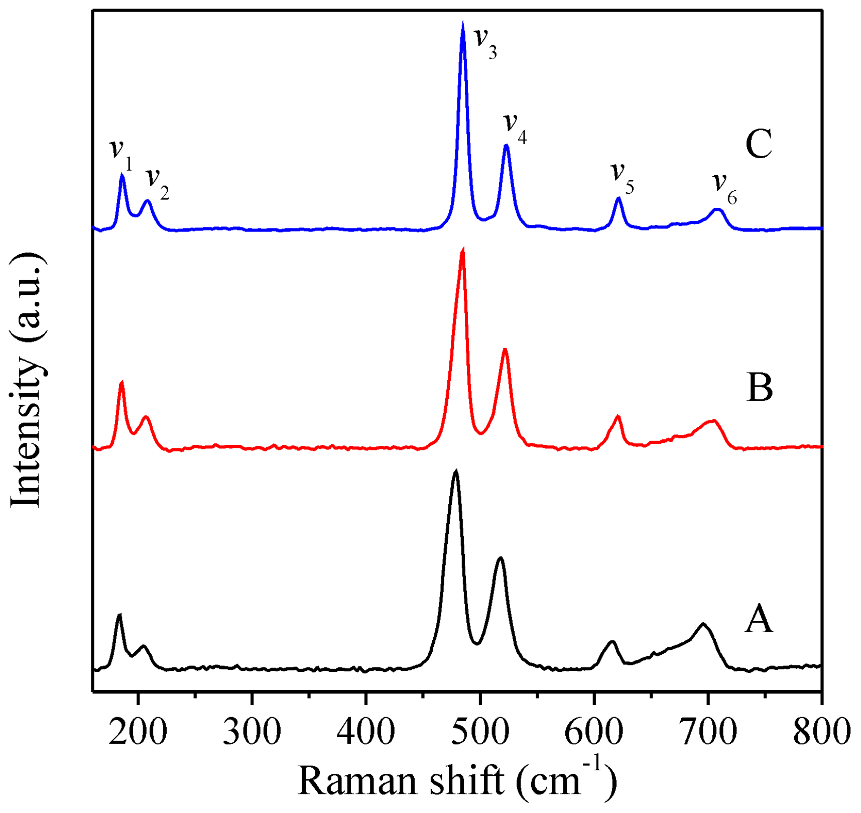
  47. Julien, C.M.; Gendron, F.; Amdouni, A.; Massot, M. Lattice vibrations of materials for lithium rechargeable batteries. VI: Ordered Spinels. Mater. Sci. Eng. B. 2006, 130, 41–48. [Google Scholar] [CrossRef]
  48. Samanta, K.; Bhattacharya, P.; Katiyar, R.S. Raman scattering studies in dilute magnetic semiconductor Zn1-xCoxO. Phys. Rev. B 2006, 73, 245213. [Google Scholar] [CrossRef]
  49. Shirai, H.; Morioka, Y.; Nakagawa, I. Infrared and Raman spectra and lattice vibrations of some oxide spinels. J. Phys. Soc. Jpn. 1982, 51, 592–597. [Google Scholar] [CrossRef]
  50. Chang, S.C. Oxygen chemisorption on tin oxide: Correlation between electrical conductivity and EPR measurements. J. Vac. Sci. Technol. 1979, 17, 366–369. [Google Scholar] [CrossRef]
  51. Gildo-Ortiz, L.; Guillén-Bonilla, H.; Santoyo-Salazar, J.; Olvera, M.L.; Karthik, T.V.K.; Campos-González, E.; Reyes-Gómez, J. Low-temperature synthesis and gas sensitivity of perovskite-type LaCoO3 nanoparticles. J. Nanomater. 2014, 2014, 61. [Google Scholar] [CrossRef]
  52. Kim, H.-J.; Lee, J.-H. Highly sensitive and selective gas sensors using p-type oxide semiconductors: Overview. Sens. Actuators B Chem. 2014, 192, 607–627. [Google Scholar] [CrossRef]
  53. Hübner, M.; Simion, C.E.; Haensch, A.; Barsan, N.; Weimar, U. CO sensing mechanism with WO3 based gas sensors. Sens. Actuators B Chem. 2010, 151, 103–106. [Google Scholar] [CrossRef]
  54. Guillén-Bonilla, H.; Gildo-Ortiz, L.; Olvera, M.L.; Santoyo-Salazar, J.; Rodríguez-Betancourtt, V.-M.; Guillen-Bonilla, A.; Reyes-Gómez, J. Sensitivity of mesoporous CoSb2O6 nanoparticles to gaseous CO and C3H8 at low temperatures. J. Nanomater. 2015, 2015. [Google Scholar] [CrossRef]
  55. Korotcenkov, G. Metal oxides for solid-state gas sensors: What determines our choice? Mater. Sci. Eng. B Chem. 2007, 139, 1–23. [Google Scholar] [CrossRef]
  56. Bochenkov, V.E.; Sergeev, G.B. Preparation and chemiresistive properties of nanostructured materials. Adv. Colloid Interface Sci. 2005, 116, 245–254. [Google Scholar] [CrossRef] [PubMed]
  57. Yamazoe, N. Toward innovations of gas sensor technology. Sens. Actuators B Chem. 2005, 108, 2–14. [Google Scholar] [CrossRef]
  58. Karthik, T.V.K.; Olvera-Amador, M.L.; Maldonado, A.; Gómez-Pozos, H. CO Gas sensing properties of pure and Cu-incorporated SnO2 nanoparticles: A study of Cu-induced modifications. Sensors 2016, 16, 1283. [Google Scholar] [CrossRef] [PubMed]
  59. Jin, Z.; Zhou, H.J.; Jin, Z.L.; Savinell, R.F.; Liu, C.C. Application of nano-crystalline porous tin oxide thin film for CO sensing. Sens. Actuators B Chem. 1998, 52, 188–194. [Google Scholar] [CrossRef]
  60. Neri, G. First fifty years of chemoresistive gas sensors. Chemosensors 2015, 3, 1–20. [Google Scholar] [CrossRef]
  61. Moseley, P.T. Materials selection for semiconductor gas sensors. Sens. Actuators B Chem. 1992, 6, 149–156. [Google Scholar] [CrossRef]
  62. Xu, C.; Tamaki, J.; Miura, N.; Yamazoe, N. Grain size effects on gas sensitivity of porous SnO2-based elements. Sens. Actuators B Chem. 1991, 3, 147–155. [Google Scholar] [CrossRef]
  63. Tan, O.K.; Cao, W.; Hu, Y.; Zhu, W. Nano-structured oxide semiconductor materials for gas-sensing applications. Ceram. Int. 2004, 30, 1127–1133. [Google Scholar] [CrossRef]
  64. Gómez-Pozos, H.; González-Vidal, J.L.; Alberto-Torres, G.; Olvera, M.L.; Castañeda, L. Physical characterization and effect of effective surface area on the sensing properties of tin dioxide thin solid films in a propane atmosphere. Sensors 2014, 14, 403–415. [Google Scholar] [CrossRef]
Figure 1. Experimental setup for the gas sensitivity measurements of ZnCo2O4 nanoparticles.
Sensors 16 02162 g001
Figure 2. SEM images of ZnCo2O4 nanoparticles: (a,d) sample A; (b,e) sample B; and (c,f) sample C.
Sensors 16 02162 g002
Figure 3. Particle size distribution for the ZnCo2O4 samples: (a) A; (b) B; and (c) C.
Sensors 16 02162 g003
Figure 4. XRD patterns of ZnCo2O4 samples synthesized with (A) 5.4; (B) 10.8; and (C) 16.2 mmol of dodecylamine. The peak signals for the ZnCo2O4 structure (JCPDF 23-1390) are labeled with black-filled circles.
Sensors 16 02162 g004
Figure 5. Raman spectra of the samples A, B, and C.
Sensors 16 02162 g005
Figure 6. TEM and HRTEM images of the samples A (a,b); B (c,d); and C (e,f).
Sensors 16 02162 g006
Figure 7. (a) HAADF-STEM image; (b) elemental line scan; and (c) EDS microanalysis of sample A.
Sensors 16 02162 g007
Figure 8. Gas response of ZnCo2O4 sensors vs. CO concentration at different operating temperatures: (a) sample A; (b) sample B; and (c) sample C.
Sensors 16 02162 g008
Figure 9. Response of the ZnCo2O4 sensors as a function of C3H8 concentration at different working temperatures: (a) sample A; (b) sample B; and (c) sample C.
Sensors 16 02162 g009
Table 1. Crystallite size of the ZnCo2O4 samples.
SamplesFWHMCrystallite Size (nm)
A0.33824.75
B0.35123.84
C0.42119.92

