
A New Strategy for Silver Deposition on Au Nanoparticles with the Use of Peroxidase-Mimicking DNAzyme Monitored via a Localized Surface Plasmon Resonance Technique

 and
and
Abstract
: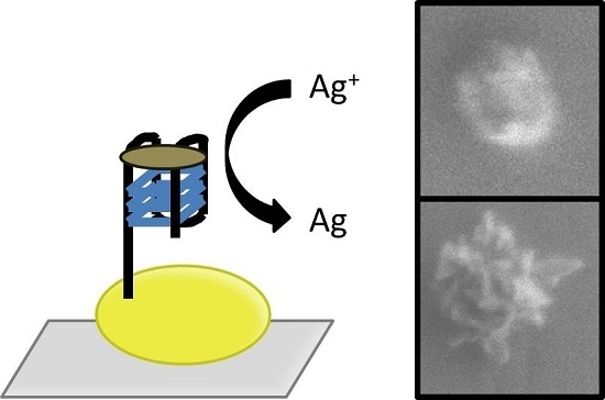
1. Introduction
2. Materials and Methods
2.1. Materials
2.2. Instruments
2.3. Sensor Preparation Procedure
2.4. LSPR Measurement Procedure
2.5. Scanning Electron Microscopy (SEM) Measurement
2.6. Atomic Force Microscopy (AFM) Measurement
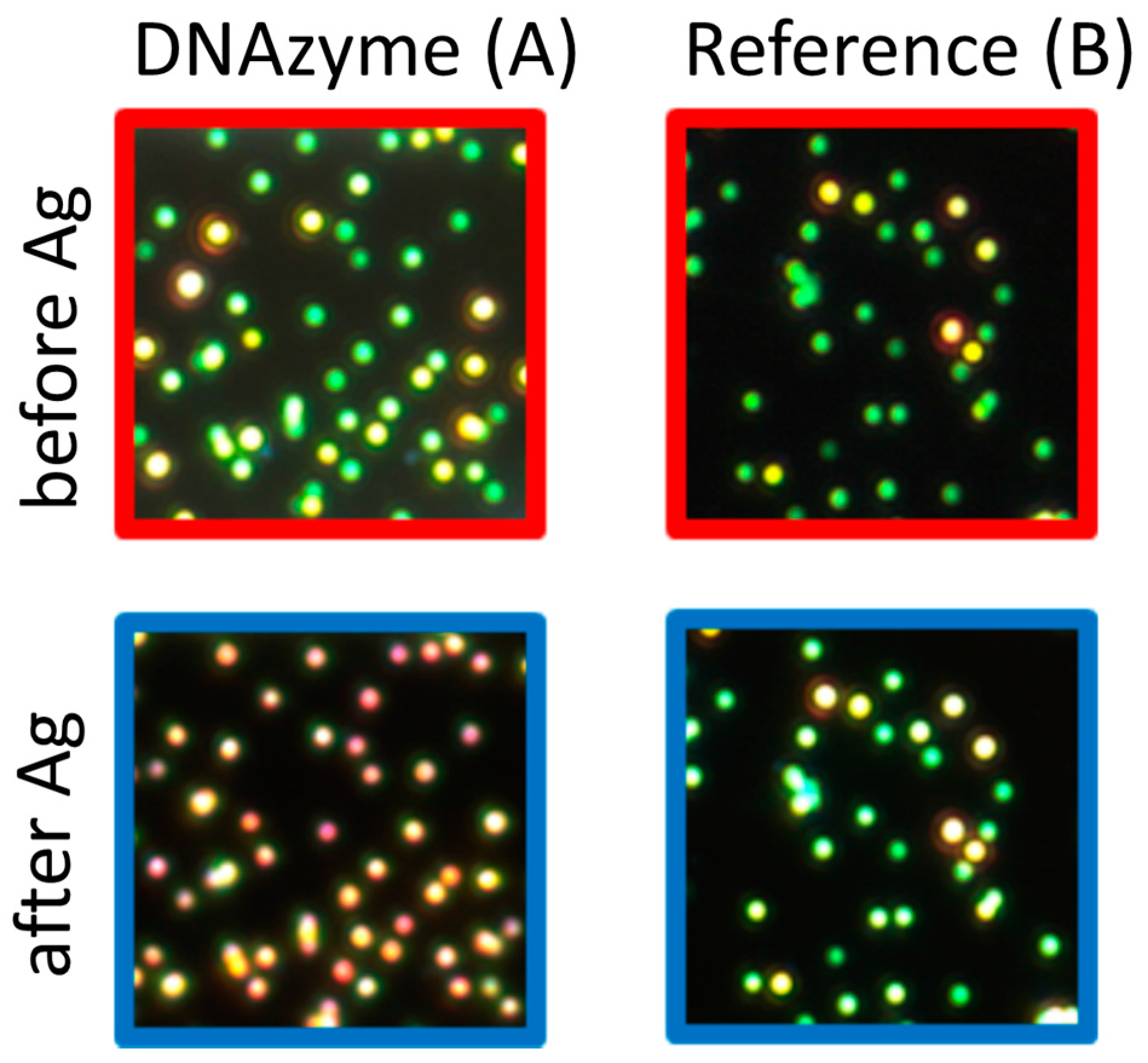
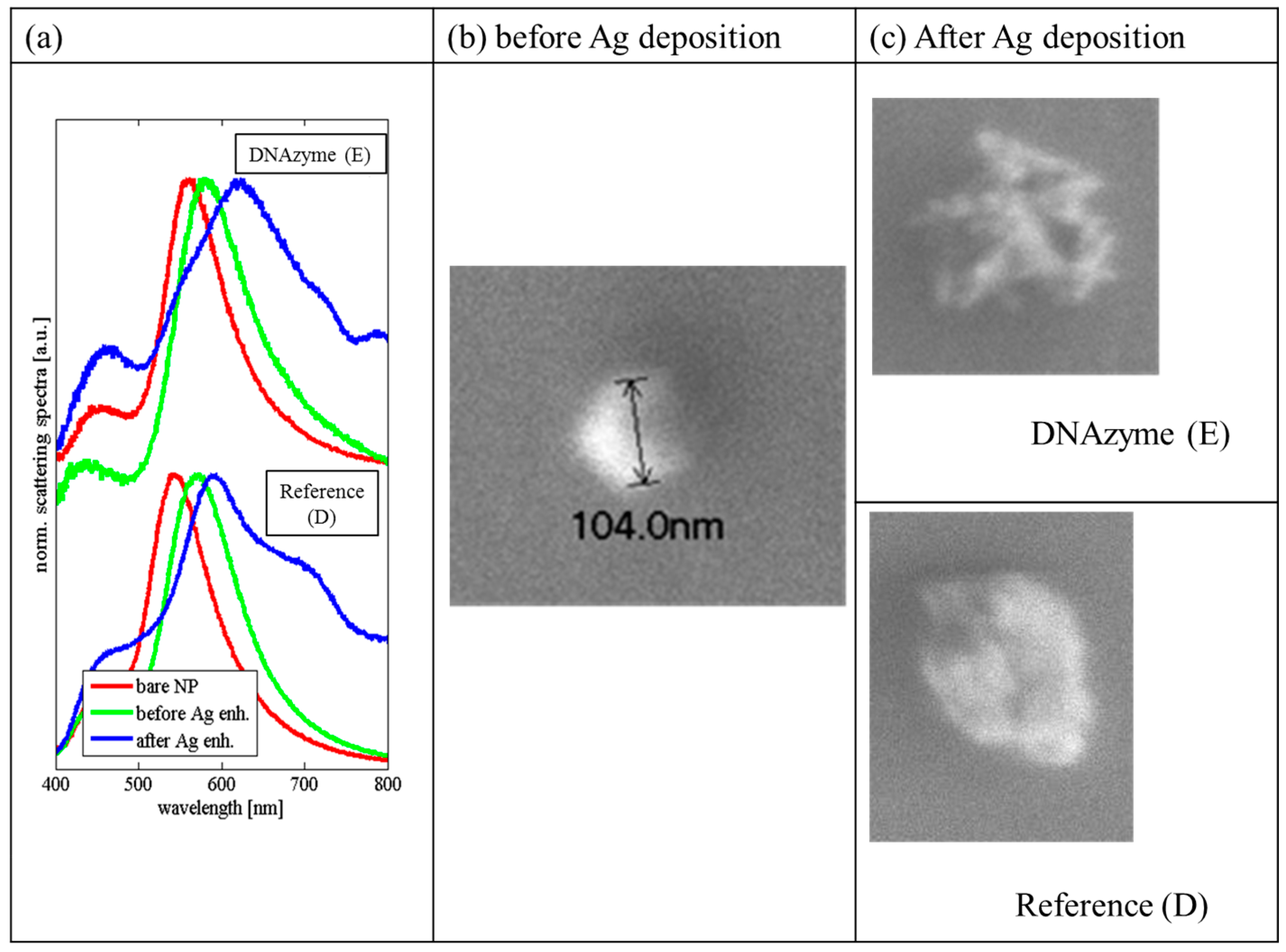
3. Results
4. Conclusions
Supplementary Materials
Acknowledgments
Author Contributions
Conflicts of Interest
References
- Silverman, S.K. Catalytic DNA: Scope, Applications, and Biochemistry of Deoxyribozymes. Trend Biochem. Sci. 2016, 41, 595–609. [Google Scholar] [CrossRef] [PubMed]
- Travascio, P.; Liu, Y.; Sen, D. DNA-enhanced peroxidase activity of a DNA aptamer-hemin complex. Chem. Biol. 1998, 5, 505–517. [Google Scholar] [CrossRef]
- Kosman, J.; Juskowiak, B. Peroxidase-mimicking DNAzymes for biosensing applications: A review. Anal. Chim. Acta 2011, 707, 7–17. [Google Scholar] [CrossRef] [PubMed]
- Rad, A.; Jahanshahi, M.; Ardjmand, M.; Safekordi, A. Hydrogen Peroxide Biosensor Based on Enzymatic Modification of Electrode Using Deposited Silver Nano Layer. Int. J. Electrochem. Sci. 2012, 7, 2623–2632. [Google Scholar]
- Gribas, A.V.; Korolev, S.P.; Zatsepin, T.S.; Gottkich, M.B.; Sakharov, I.Y. Structure-activity relationship study for design of highly active covalent peroxidase-mimicking DNAzyme. RSC Adv. 2015, 5, 51672–51677. [Google Scholar] [CrossRef]
- Li, W.; Li, Y.; Liu, Z.; Lin, B.; Yi, H.; Xu, F.; Nie, Z.; Yao, S. Insight into G-quadruplex-hemin DNAzyme/RNAzyme: Adjacent adenine as the intermolecular species for remarkable enhancement of enzymatic activity. Nucleic Acids Res. 2016, 44, 7373–7384. [Google Scholar] [PubMed]
- Chang, T.; Gong, H.; Ding, P.; Liu, X.; Li, W.; Bing, T.; Cao, Z.; Shangguan, D. Activity Enhancement of G-quadruplex/Hemin DNAzyme by Flanking d(CCC). Chemistry 2016, 22, 4015–4021. [Google Scholar] [CrossRef] [PubMed]
- Xu, J.; Qian, J.; Li, H.; Wu, Z.S.; Shen, W.; Jia, L. Intelligent DNA machine for the ultrasensitive colorimetric detection of nucleic acids. Biosens. Bioelectron. 2016, 75, 41–47. [Google Scholar] [CrossRef] [PubMed]
- Koo, K.M.; Wee, E.J.H.; Rauf, S.; Shiddiky, M.J.A.; Trau, M. Microdevices for detecting locus-specific DNA methylation at CpG resolution. Biosens. Bioelectron. 2014, 56, 278–285. [Google Scholar] [CrossRef] [PubMed]
- Golub, E.; Freeman, R.; Willner, I. A Hemin/G-quadruplex Acts as a NADH Oxidase and NADH Peroxidase Mimicking DNAzyme. Angew. Chem. Int. Ed. 2011, 50, 11710–11714. [Google Scholar] [CrossRef]
- Schuler, T.; Steinbruck, A.; Festag, G.; Moller, R.; Fritzsche, W. Enzyme-induced growth of silver nanoparticles studied on single particle level. J. Nanopart. Res. 2009, 11, 939–946. [Google Scholar] [CrossRef]
- Kreibeg, U.; Vollmer, M. Optical Properties of Metal Clusters; Springer: Berlin, Germany, 1995. [Google Scholar]
- Lu, L.; Burkey, G.; Halaciuga, I.; Goia, D.V. Core-shell gold/silver nanoparticles: Synthesis and optical properties. J. Colloid Interface Sci. 2013, 392, 90–95. [Google Scholar] [CrossRef] [PubMed]
- Maddox, P.H.; Jenkins, D. 3-Aminopropyltriethoxysilane (APES): A new advance in section adhesion. J. Clin. Pathol. 1987, 40, 1256–1257. [Google Scholar] [CrossRef] [PubMed]
- Steinbruck, A.; Stranik, O.; Csaki, A.; Fritzsche, W. Sensoric potential of gold-silver core-shell nanoparticles. Anal. Bioanal. Chem. 2011, 401, 1241–1249. [Google Scholar] [CrossRef] [PubMed]
- Park, S.; Brown, K.A. Hamad-Schifferli K., Changes in Oligonucleotide Conformation on Nanoparticle Surfaces by Modification with Mercaptohexanol. Nano Lett. 2004, 4, 1925–1929. [Google Scholar] [CrossRef]
- Kelly, C.H.; Davies, M.D.; Mantle, D.; Jones, P. Hydroperoxidase activities of ferrihemes: Heme analogues of peroxidase enzyme intermediates. Biochemistry 1997, 8, 3974–3978. [Google Scholar]
- Tom, R.T.; Pradeep, T. Interaction of Azide Ion with Hemin and Cytochrome c Immobilized on Au and Ag Nanoparticles. Langmuir 2005, 21, 11896–11902. [Google Scholar] [CrossRef] [PubMed]
- Choi, H.; Kang, T.; Um, K.; Kim, J.; Lee, K. Reduction of silver ions in gold nanoparticle suspension for detection of dihydroxybenzene isomers. Colloid Surf. A Physicohem. Eng. Asp. 2014, 459, 120–127. [Google Scholar] [CrossRef]


| Name | DNA Sequence |
|---|---|
| ON1 | HS-(CH2)6–5’-TTTT GGGTAGGGCGGGTTGGG3’ |
| OT1 | HS-(CH2)6–3’-TTTT TTA GGC AGC TCG TCT CAA-5’ |
| OT2 | 5’-AAT CCG TCG AGC AGA GTT TTA GGG TTA GGG TTA GGG TTA GGG TTA GGG TTA GGG TTA GGG TTA GGG-3’ |
© 2017 by the authors. Licensee MDPI, Basel, Switzerland. This article is an open access article distributed under the terms and conditions of the Creative Commons Attribution (CC BY) license (http://creativecommons.org/licenses/by/4.0/).
Share and Cite
Kosman, J.; Jatschka, J.; Csaki, A.; Fritzsche, W.; Juskowiak, B.; Stranik, O. A New Strategy for Silver Deposition on Au Nanoparticles with the Use of Peroxidase-Mimicking DNAzyme Monitored via a Localized Surface Plasmon Resonance Technique. Sensors 2017, 17, 849. https://doi.org/10.3390/s17040849
Kosman J, Jatschka J, Csaki A, Fritzsche W, Juskowiak B, Stranik O. A New Strategy for Silver Deposition on Au Nanoparticles with the Use of Peroxidase-Mimicking DNAzyme Monitored via a Localized Surface Plasmon Resonance Technique. Sensors. 2017; 17(4):849. https://doi.org/10.3390/s17040849
Chicago/Turabian StyleKosman, Joanna, Jacqueline Jatschka, Andrea Csaki, Wolfgang Fritzsche, Bernard Juskowiak, and Ondrej Stranik. 2017. "A New Strategy for Silver Deposition on Au Nanoparticles with the Use of Peroxidase-Mimicking DNAzyme Monitored via a Localized Surface Plasmon Resonance Technique" Sensors 17, no. 4: 849. https://doi.org/10.3390/s17040849