Article

A Novel Graphene Oxide-Based Aptasensor for Amplified Fluorescent Detection of Aflatoxin M1 in Milk Powder

1
Key Laboratory of Quality & Safety Control for Milk and Dairy Products of Ministry of Agriculture and Rural Affairs, Institute of Animal Sciences, Chinese Academy of Agricultural Sciences, 100193 Beijing, China
2
Chimie générale et organique, Gembloux Agro-Bio Tech, Université de Liège, Passage des Déportés 2, 5030 Gembloux, Belgium
3
Laboratory of Quality and Safety Risk Assessment for Dairy Products of Ministry of Agriculture and Rural Affairs, Institute of Animal Sciences, Chinese Academy of Agricultural Sciences, 100193 Beijing, China
4
State Key Laboratory of Animal Nutrition, Institute of Animal Science, Chinese Academy of Agricultural Sciences, 100193 Beijing, China
*
Author to whom correspondence should be addressed.
The authors contributed equally.
Sensors 2019, 19(18), 3840; https://doi.org/10.3390/s19183840
Submission received: 25 July 2019 / Revised: 28 August 2019 / Accepted: 29 August 2019 / Published: 5 September 2019
(This article belongs to the Special Issue Aptamers and Applications)

Abstract

:
In this paper, a rapid and sensitive fluorescent aptasensor for the detection of aflatoxin M1 (AFM1) in milk powder was developed. Graphene oxide (GO) was employed to quench the fluorescence of a carboxyfluorescein-labelled aptamer and protect the aptamer from nuclease cleavage. Upon the addition of AFM1, the formation of an AFM1/aptamer complex resulted in the aptamer detaching from the surface of GO, followed by the aptamer cleavage by DNase I and the release of the target AFM1 for a new cycle, which led to great signal amplification and high sensitivity. Under optimized conditions, the GO-based detection of the aptasensor exhibited a linear response to AFM1 levels in a dynamic range from 0.2 to 10 μg/kg, with a limit of detection (LOD) of 0.05 μg/kg. Moreover, the developed aptasensor showed a high specificity towards AFM1 without interference from other mycotoxins. In addition, the technique was successfully applied for the detection of AFM1 in infant milk powder samples. The aptasensor proposed here offers a promising technology for food safety monitoring and can be extended to various targets.

1. Introduction

Aflatoxin M1 (AFM1), one of the most toxic mycotoxins, was moved from group 2B to group 1 carcinogens by the International Agency for Research on Cancer (IARC) of the World Health Organization (WHO) [1,2]. AFM1 can be encountered in dairy products as a hydroxylate metabolite derived from feeding dairy cows aflatoxin B1-contaminated feeds [3,4,5]. Since dairy products are an important nutrient for humans, especially for infants, the presence of AFM1 in dairy products is one of the most serious hazards for food safety [6]. To protect humans from this health threat, many regulatory agencies have defined maximum residue levels (MRLs) for AFM1 in dairy products [7,8]. In Brazil, China, and USA, the maximum level of AFM1 in milk has been fixed to 0.5 μg/kg [9,10]. The European Commission Regulation has set much more restrictive limits, i.e., 0.05 μg/kg in milk products for adults, and this level is lowered to 0.025 μg/kg for baby and infant products [11]. Considering the severe toxicity and low permitted level of AFM1, a simple, rapid and inexpensive diagnostics with high sensitivity and specificity is vitally required for food safety.
Analytical methods such as high-performance liquid chromatography (HPLC) with fluorescent detectors [12,13,14] and high-performance liquid chromatography coupled with mass spectrometric detectors [15,16] have been developed for quantitative detection of AFM1. All these procedures rely on expensive instruments, qualified staff and complicated time-consuming pretreatments. Meanwhile, enzyme-linked immune sorbent assays (ELISA) [17,18,19] have gained great attention for AFM1 analysis owing to the advantages of rapid, low-cost and high-throughput application. However, expensive, time-consuming and laborious antibody production and antibody instability during storage limit the practical applications of these assays. Therefore, the design of a simple, cheap and sensitive method for rapid detection of AFM1 has become a research hotspot.
Aptamers, single-stranded DNA or RNA oligonucleotides, have been widely applied for recognizing targets such as proteins, nucleic, cells, tissues and small molecules with strong affinity and high specificity similar or even superior to those of antibodies [20,21]. Up to now, a series of aptamer-based biosensors for the detection of several mycotoxins including ochratoxin A (OTA), aflatoxin B1 (AFB1) and AFM1 have been developed [22,23,24,25,26,27]. In these homogeneous methods, the recognition reaction between the aptamer and the target is based on single-site binding, which might limit the method sensitivity. Thus, the development of aptasensors coupled with signal amplification strategies for AFM1 detection is an on-going challenge. In our previous study [27], a sensitive aptasensor based on real-time quantitative polymerase chain reaction (RT-qPCR) for AFM1 was developed. However, the preparation of the qPCR-based aptasensor requires a tedious and time-consuming process with long incubation periods in rigorous conditions. Therefore, there is a demand for aptasensors that could be applied for rapid and real-time analysis of AFM1.
Graphene oxide (GO), a two-dimensional nanomaterial, is a very promising tool for the construction of biosensors because of its extraordinary electrical, thermal and mechanical properties [28,29,30,31]. The predominantly distance-dependent fluorescence quenching ability makes GO a highly efficient fluorescence quencher [32,33,34]. In addition, previous research demonstrated that single-stranded DNA can significantly interact with GO through π stacking between DNA bases and hexagonal cells of GO [35]. Importantly, GO could protect DNA aptamers from nuclease cleavage as a result of the hydrophobic stacking interactions between nucleobases and GO [36,37,38]. To the best of our knowledge, graphene oxide-based nuclease signal amplification aptasensors for AFM1 determination have not been reported.
In this study, a new graphene oxide-based aptasensor for the specific detection of AFM1 was developed, which combined the ability of GO to protect the aptamer from nuclease cleavage with that of DNase I to cleave the aptamer for a target cycling signal amplification. The presence of AFM1 induced the release of the aptamer from the surface of GO because of the formation of an AFM1/aptamer complex, which resulted in the cleavage of the aptamer by DNase I and the release of AFM1 for a new cycle. Therefore, a cycling signal amplification was achieved to improve detection sensitivity. A good linear relationship was measured between the change of the fluorescence intensity signal and AFM1 levels.

2. Experimental

2.1. Materials and Reagents

AFM1 was purchased from Sigma-Aldrich (USA). AFB1 was obtained from the National Standard Reference Center (Beijing, China). OTA, zearalenone (ZEA) and α-zearalenin (α-ZOL) were purchased from Pribolab Co. Ltd (Singapore). Graphene oxide was purchased from Sangon Biotechnology Co. Ltd. (Shanghai, China). DNase I (RNase-free) was obtained from Takara Bio Co. Ltd. (Dalian, China). Other chemicals such as anhydrous calcium chloride (CaCl2), sodium chloride (NaCl), potassium chloride (KCl) and 2-Amino-2-(hydroxymethyl)-1,3-propanediol (Tris) were purchased from Shanghai Chemical Reagent Company (Shanghai, China). All other chemicals were analytical-grade and were used as received without further purification. Water was purified with a Milli-Q purification system. DNA oligonucleotides were chemically synthesized by Sangon Biotechnology Co. Ltd. (Shanghai, China) and purified by HPLC. The sequence of the AFM1 aptamer was optimized according to our previous study [27] and was modified by FAM (carboxyfluorescein). The sequence of the FAM-labelled AFM1 aptamer was as follows:
5′5′-FAM–ATCCGTCACACCTGCTCTGACGCTGGGGTCGACCCG-3′

2.2. Fluorescent Response of the Amplified Aptasensor for AFM1

In this amplification strategy, the FAM-labelled AFM1 aptamer was diluted to 200 nM in Tris buffer (10 mM Tris, 120 mM NaCl, 5 mM KCl, 20 mM CaCl2, pH 7.0), and 20 μg mL−1 of GO was added to the working solution for 15 min at room temperature to form the aptamer/GO complex and quench the fluorescence. Subsequently, solutions at different concentrations of AFM1 and DNase I (200 U) were simultaneously added to the aptamer/GO solutions, and the mixtures were and incubated at room temperature for 1 h. Afterwards, the fluorescence intensities of the mixtures were recorded using an F-7000 fluorophotometer (Hitachi, Tokyo, Japan). The emission spectra were measured in the range of 510 to 630 nm with the excitation wavelength at 480 nm, and slit widths for both of the excitation and the emission were set at 10 nm.

2.3. Specificity Analysis

To evaluate the specificity of this aptasensor for AFM1 over other mycotoxins, four different mycotoxins (AFB1, OTA, ZEA and α-ZOL) were measured at the same concentration of 4 ng mL−1. The other experimental procedures were the same as AFM1 determination, and the changes of fluorescence intensity for these mycotoxins were compared.

2.4. Method Validation

The feasibility and practicability of this sensing platform was verified by the quantitative detection of AFM1 in infant milk powder samples. The samples were spiked with AFM1 at 0, 1.5, 2.5 and 5 μg/kg (three replicates per treatment). Each sample was accurately weighed (0.5 g) into 10 mL centrifuge tubes. Then, 2.5 mL of extraction solution (70% methanol in water) was added to extract AFM1 from the samples. The entire mixture was vortexed using Vortex-Genie 2 (Scientific Industries, Bohemia, NY, USA) for 5 min and then centrifuged at 10,000 g for 10 min. The supernatant was obtained and concentrated to 0.5 mL under a nitrogen stream. Subsequently, each residue was re-dissolved in 2 mL of aqueous methanol solution (5% methanol in water). Finally, the extracts were measured by the fluorescence signal amplification experiment.

2.5. Statistical Analysis

Fluorescence-emission spectra curves for AFM1 were plotted using Origin 8.0 software (OriginLab Corporation, Northampton, MA, USA). Linear regression analysis of the fluorescence intensity as a function of the concentration of AFM1 was carried out with Microsoft Excel. Each analysis including AFM1 calibration curve standards and test samples was performed in triplicate. Standard deviations (SDs) and means of fluorescence intensity were determined from three replicates.

3. Results and Discussion

3.1. Design Strategy for AFM1 Detection Based on a Graphene Oxide Sensing Platform

GO has many advantages due to its unique properties, including its great binding ability to single-stranded DNA (such as aptamers) through π stacking interactions between nucleobases and GO nanosheets and its high distance-dependent fluorescence quenching performance [35,39]. A GO-based aptasensor for the detection of AFM1 was developed taking advantage of the above properties. A schematic illustration of the sensing platform is presented in Figure 1. In this sensing method, when the FAM-modified aptamer was incubated with the GO solution, the fluorescence signal quenched dramatically, demonstrating a strong binding between the aptamer and GO, with a high quenching efficiency. Upon the addition of AFM1, an AFM1/aptamer complex formed. Such an interaction can lead to a conformational change in the aptamer, causing a separation of the conjugated aptamer from the surface of GO. Thus, fluorescence is recovered, since GO would not be able to quench the fluorescence efficiently owing to the long distance. In order to confirm that the presence of AFM1 can lead to the formation of an AFM1/aptamer complex and subsequently to fluorescence recovery, 10 ng mL−1 of AFM1 was added to a Tris buffer solution that contained 200 nM of AFM1 aptamer and 20 μg mL−1 of GO. As seen in Figure 2, a significant fluorescence enhancement was observed, demonstrating that the AFM1/aptamer complex was formed. More importantly, the covalently modified FAM had no impact on the recognition ability of the AFM1 aptamer.
DNase I was adopted as a signal amplification strategy to improve the sensitivity of the aptasensor. As shown in Figure 1, upon the addition of AFM1 and DNase I, the formation of the AFM1/aptamer complex caused the dissociation of the aptamer conjugate from GO, and subsequently the aptamer was digested by DNase I. Once AFM1 was released from the AFM1/aptamer complex, it was again available to bind to another aptamer, inducing a cyclic amplification of the fluorescence signal. As a consequence, a strong amplification of the fluorescence signal could be achieved for the quantification of AFM1.

3.2. Optimization of the Experimental Conditions

The concentration of GO would influence the fluorescence quenching efficiency. Therefore, to optimize the sensing platform, the effect of GO concentration on the change of the fluorescence signal was investigated. Various concentrations of GO were added to a solution containing 200 nM of AFM1 aptamer. As seen in Figure S1, the fluorescence intensity decreased with increasing amounts of GO and reached the lowest level at a concentration of GO of 20 μg mL−1. Thus, 20 μg mL−1 of GO solution was used for further sensing experiments.
To improve the signal amplification efficiency, the optimization of the concentration of DNase I was essential. In this experiment, we measured the fluorescence intensity of the complex with 10 ng mL−1 of AFM1. Various amounts of DNase I were added to the GO/aptamer solution containing 200 nM of AFM1 aptamer and 20 μg mL−1 of GO. As seen in Figure S2, the fluorescence intensity increased as the DNase I concentration increased from 0 to 200 U, and the highest level of fluorescence was observed at 200 U of DNase I. In this case, the optimal amount of DNase I was determined as 200 U.

3.3. Analytical Performance of the Aptasensor

Under optimal conditions, the signal responses of the GO-based aptasensor to different concentrations of AFM1 were measured using a DNase I-induced target recycling amplification platform. The fluorescence emission spectrum was measured for AFM1 determination with excitation and emission wavelengths of 480 nm and 520 nm, respectively. As seen in Figure 3, the fluorescence intensity increased as the concentration of AFM1 increased from 0.2 to 10 μg/kg. The calibration curve of fluorescence intensity versus AFM1 concentrations was linear, as F = 65.77 C + 46.334 (R2 = 0.9939), in which F is the fluorescence intensity, and C is AFM1 concentration. The limit of detection of the amplification aptasensor was determined to be 0.05 μg/kg, which was calculated at the signal-to-noise ratio of 3. As shown in Table 1, the aptasensor displayed a sensitivity for AFM1 comparable to those of other instrumental and rapid screening methods reported previously.

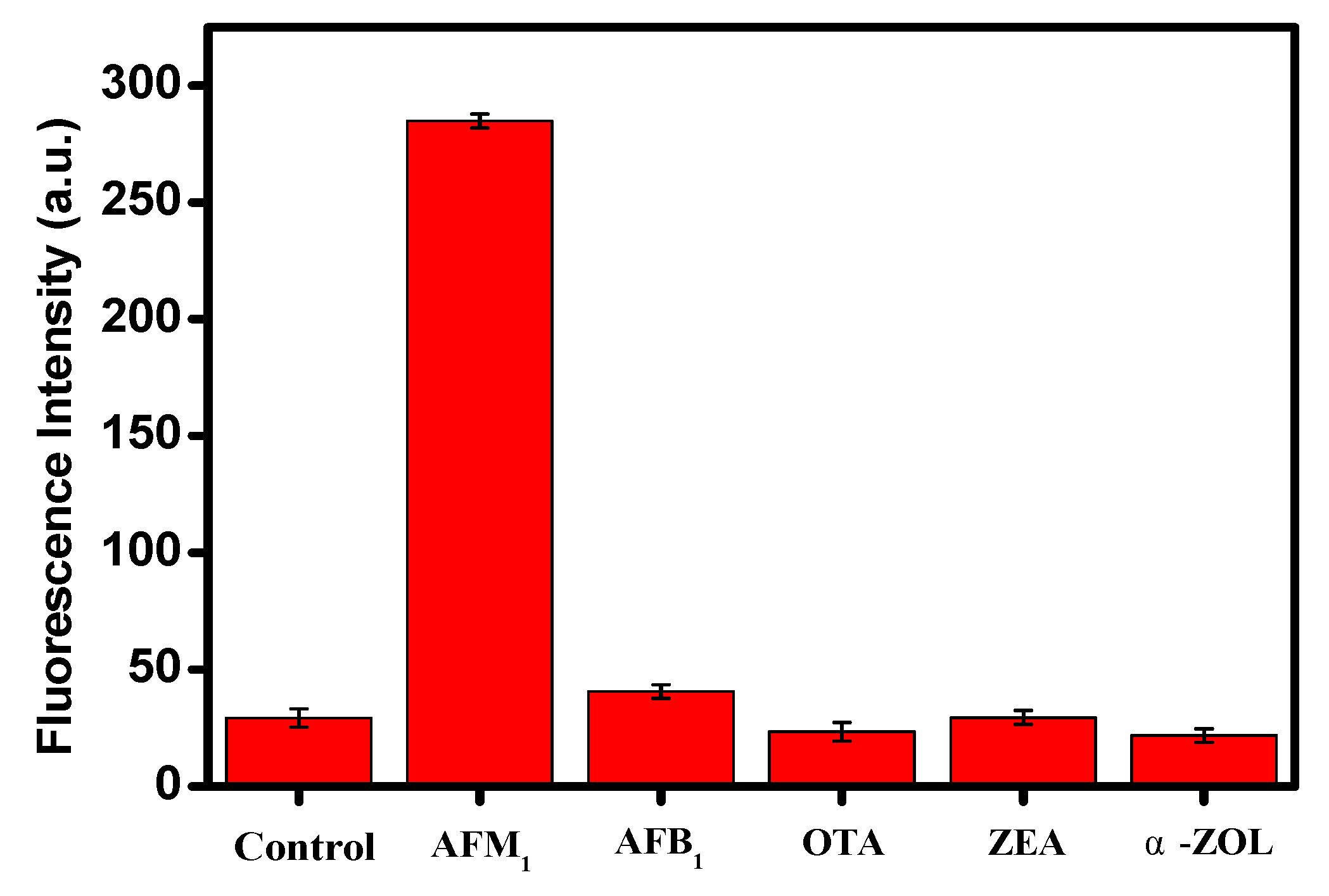
3.4. The Specificity of the Aptsensor

The specificity of the aptasensor was also investigated to assess the effect of other mycotoxins. The change of fluorescence intensity was measured under experiment conditions identical to those used for AFM1 detection in the presence of four other mycotoxins (AFB1, OTA, ZEA and α-ZOL) at a concentration of 4 ng mL−1. It can be seen that significantly higher fluorescence intensity was obtained in the case of AFM1 determination in comparison with other mycotoxins and the control (Figure 4), which indicated that the specificity of this amplifying sensing platform is high for AFM1 determination.

3.5. Method Validation

Ultimately, the applicability and reliability of the aptasensor platform were evaluated by detecting different concentrations of AFM1 in infant milk powder samples. As indicated in Table 2, the recovery of the spiked infant milk powder samples ranged from 92% to 126%, demonstrating that the amplification strategy developed in this work can be useful as a quantitative method for AFM1 analysis in real samples for food safety.

4. Conclusions

A novel graphene oxide-based aptasensor was developed for the detection of AFM1 with high sensitivity and specificity. This technique uses the properties of GO as an aptamer protector against nuclease cleavage, thereby allowing DNase I to cleave the aptamer for a target cycling signal amplification. Under the optimal conditions, a good linear relationship was detected between fluorescence intensity and AFM1 levels in the range of 0.2 to 10 μg/kg, with a detection limit of 0.05 μg/kg. Satisfactory recoveries were measured in infant milk powder samples spiked with different concentrations of AFM1. Furthermore, the aptasensor proposed in this work is rapid, simple and low-cost in comparison with other methods reported previously. This study could thus provide a very promising platform for the analysis of AFM1 in dairy products. More importantly, the aptasensor could be improved by replacing aptamer sequences for the detection of other food safety targets.

Supplementary Materials

The following are available online at https://www.mdpi.com/1424-8220/19/18/3840/s1, Figure S1. Fluorescence emission spectra of the aptasensor in the addition of GO at various concentrations. The experiment conditions are as following: λex = 480nm, 200 nM AFM1 aptamer. Figure S2. Fluorescence intensity with the addition of DNase I at various concentrations. The experiment conditions are as follows: Excitation and emission wavelength are at λex/λem = 480/520 nm, 200 nM AFM1 aptamer, 20 μg mL-1 GO, 10 ng mL-1 AFM1.

Author Contributions

This work proposed in this paper was carried out in collaboration with all the authors. X.G. and F.W. proposed the idea of the paper, wrote the original paper and analyzed the experimental data. Q.Q. and N.Z. supported the structure of the paper. M.S. and M.-L.F. and J.W revised the paper.

Funding

This work was supported by the Special Fund for Agro-scientific Research in the Public Interest (201403071), the National Natural Science Foundation of China (No. 21305158), the Agricultural Science and Technology Innovation Program (ASTIP-IAS12), Modern Agro Industry Technology Research System of the PR China (CARS-36), and Project of risk assessment on raw milk (GJFP2019026).

Acknowledgments

We thank the University of Liège-Gembloux Agro-Bio Tech and more specifically the research platform AgricultureIsLife for the funding of the scientific stay in Belgium that made this paper possible.

Conflicts of Interest

The authors declare that they have no competing interests.

References

  1. International Agency for Research on Cancer. IARC Monographs on the Evaluation of Carcinogenic Risk to Humans; International Agency for Research on Cancer: Lyon, France, 1993; pp. 489–551. [Google Scholar]
  2. International Agency for Research on Cancer (IARC). Some traditional herbal medicines, some mycotoxins, naphthalene and styren. In IARC Monographs on the Evaluation of Carcinogenic Risks to Humans; International Agency for Research on Cancer: Lyon, France, 2002; Volume 82, pp. 171–230. [Google Scholar]
  3. Pei, S.C.; Zhang, Y.Y.; Eremin, S.A.; Lee, W.J. Detection of aflatoxin M1 in milk products from China by ELISA using monoclonal antibodies. Food Control 2009, 20, 1080–1085. [Google Scholar] [CrossRef]
  4. Anfossi, L.; Baggiani, C.; Giovannoli, C.; Biagioli, F.; D’Arco, G.; Giraudi, G. Optimization of a lateral flow immunoassay for the ultrasensitive detection of aflatoxin M1 in milk. Anal. Chim. Acta. 2013, 772, 75–80. [Google Scholar] [CrossRef]
  5. Liu, D.; Huang, Y.; Wang, S.; Liu, K.; Chen, M.; Xiong, Y.; Yang, W.; Lai, W. A modified lateral flow immunoassay for the detection of trace aflatoxin M1 based on immunomagnetic nanobeads with different antibody concentrations. Food Control 2015, 51, 218–224. [Google Scholar] [CrossRef]
  6. Mao, J.; Lei, S.; Liu, Y.; Xiao, D.; Fu, C.; Zhong, L.; Ouyang, H. Quantification of aflatoxin M1 in raw milk by a core-shell column on a conventional HPLC with large volume injection and step gradient elution. Food Control 2015, 51, 156–162. [Google Scholar] [CrossRef]
  7. Van Egmond, H.P.; Jonker, M.A. Worldwide Regulations for Mycotoxins in Food and Feed in 2003. Available online: https://www.rivm.nl/bibliotheek/digitaaldepot/23661ADC.pdf (accessed on 29 August 2019).
  8. Hoyos Ossa, D.E.; Hincapié, D.A.; Peñuela, G.A. Determination of aflatoxin M1 in ice cream samples using immunoaffinity columns and ultra-high performance liquid chromatography coupled to tandem mass spectrometry. Food Control. 2015, 56, 34–40. [Google Scholar] [CrossRef]
  9. Busman, M.; Bobell, J.R.; Maragos, C.M. Determination of the aflatoxin M1 (AFM1) from milk by direct analysis in real time-mass spectrometry (DART-MS). Food Control 2015, 47, 592–598. [Google Scholar] [CrossRef]
  10. Istamboulie, G.; Paniel, N.; Zara, L.; Reguillo Granados, L.; Barthelmebs, L.; Noguer, T. Development of an impedimetric aptasensor for the determination of aflatoxin M1 in milk. Talanta 2016, 146, 464–469. [Google Scholar] [CrossRef]
  11. The Commission of the European Communities. Setting maximum levels for certain contaminants in foodstuffs. Off. J. Eur. Union. 2006, 364, 5–24. [Google Scholar]
  12. Wang, Y.; Liu, X.; Xiao, C.; Wang, Z.; Wang, J.; Xiao, H.; Cui, L.; Xiang, Q.; Yue, T. HPLC determination of aflatoxin M1 in liquid milk and milk powder using solid phase extraction on OASIS HLB. Food Control 2012, 28, 131–134. [Google Scholar] [CrossRef]
  13. Lee, D.; Lee, K.G. Analysis of aflatoxin M1 and M2 in commercial dairy products using high-performance liquid chromatography with a fluorescence detector. Food Control 2015, 50, 467–471. [Google Scholar] [CrossRef]
  14. Pietri, A.; Fortunati, P.; Mulazzi, A.; Bertuzzi, T. Enzyme-assisted extraction for the HPLC determination of aflatoxin M1 in cheese. Food Chem. 2016, 192, 235–241. [Google Scholar] [CrossRef]
  15. Beltrán, E.; Ibáñez, M.; Sancho, J.V.; Cortés, M.Á.; Yusà, V.; Hernández, F. UHPLC–MS/MS highly sensitive determination of aflatoxins, the aflatoxin metabolite M1 and ochratoxin A in baby food and milk. Food Chem. 2011, 126, 737–744. [Google Scholar] [CrossRef]
  16. Wang, X.; Li, P. Rapid screening of mycotoxins in liquid milk and milk powder by automated size-exclusion SPE-UPLC-MS/MS and quantification of matrix effects over the whole chromatographic run. Food Chem. 2015, 173, 897–904. [Google Scholar] [CrossRef]
  17. Li, P.; Zhang, Q.; Zhang, W.; Zhang, J.; Chen, X.; Jiang, J.; Xie, L.; Zhang, D. Development of a class-specific monoclonal antibody-based ELISA for aflatoxins in peanut. Food Chem. 2009, 115, 313–317. [Google Scholar] [CrossRef]
  18. Kav, K.; Col, R.; Kaan Tekinsen, K. Detection of aflatoxin M1 levels by ELISA in white-brined Urfa cheese consumed in Turkey. Food Control 2011, 22, 1883–1886. [Google Scholar] [CrossRef]
  19. Anfossi, L.; Di Nardo, F.; Giovannoli, C.; Passini, C.; Baggiani, C. Enzyme immunoassay for monitoring aflatoxins in eggs. Food Control 2015, 57, 115–121. [Google Scholar] [CrossRef]
  20. Zhang, J.; Li, Z.; Zhao, S.; Lu, Y. Size-dependent modulation of graphene oxide–aptamer interactions for an amplified fluorescence-based detection of aflatoxin B1 with a tunable dynamic range. Analyst 2016, 141, 4029–4034. [Google Scholar] [CrossRef]
  21. Lv, L.; Li, D.; Cui, C.; Zhao, Y.; Guo, Z. Nuclease-aided target recycling signal amplification strategy for ochratoxin A monitoring. Biosens. Bioelectron. 2017, 87, 136–141. [Google Scholar] [CrossRef]
  22. Barthelmebs, L.; Hayat, A.; Limiadi, A.W.; Marty, J.L.; Noguer, T. Electrochemical DNA aptamer-based biosensor for OTA detection, using superparamagnetic nanoparticles. Sens. Actuator B Chem. 2011, 156, 932–937. [Google Scholar] [CrossRef]
  23. Bonel, L.; Vidal, J.C.; Duato, P.; Castillo, J.R. An electrochemical competitive biosensor for ochratoxin A based on a DNA biotinylated aptamer. Biosens. Bioelectron. 2011, 26, 3254–3259. [Google Scholar] [CrossRef]
  24. Guo, X.; Wen, F.; Zheng, N.; Luo, Q.; Wang, H.; Wang, H.; Li, S.; Wang, J. Development of an ultrasensitive aptasensor for the detection of aflatoxin B1. Biosens. Bioelectron. 2014, 56, 340–344. [Google Scholar] [CrossRef]
  25. Shim, W.B.; Kim, M.J.; Mun, H.; Kim, M.G. An aptamer-based dipstick assay for the rapid and simple detection of aflatoxin B1. Biosens. Bioelectron. 2014, 62, 288–294. [Google Scholar] [CrossRef]
  26. Wang, R.; Xiang, Y.; Zhou, X.; Liu, L.H.; Shi, H. A reusable aptamer-based evanescent wave all-fiber biosensor for highly sensitive detection of Ochratoxin A. Biosens. Bioelectron. 2015, 66, 11–18. [Google Scholar] [CrossRef]
  27. Guo, X.; Wen, F.; Zheng, N.; Li, S.; Fauconnier, M.L.; Wang, J. A qPCR aptasensor for sensitive detection of aflatoxin M1. Anal. Bioanal. Chem. 2016, 408, 5577–5584. [Google Scholar] [CrossRef] [Green Version]
  28. Li, D.; Müller, M.B.; Gilje, S.; Kaner, R.B.; Wallace, G.G. Processable aqueous dispersions of graphene nanosheets. Nat. Nanotechnol. 2008, 3, 101–105. [Google Scholar] [CrossRef]
  29. Rao, C.E.E.; Sood, A.E.; Subrahmanyam, K.E.; Govindaraj, A. Graphene: The new two-dimensional nanomaterial. Angew. Chem. Int. Ed. 2009, 48, 7752–7777. [Google Scholar] [CrossRef]
  30. Chen, D.; Feng, H.; Li, J. Graphene oxide: Preparation, functionalization, and electrochemical applications. Chem. Rev. 2012, 112, 6027–6053. [Google Scholar] [CrossRef]
  31. Cheng, F.F.; Zhang, J.J.; He, T.T.; Shi, J.J.; Abdel-Halim, E.; Zhu, J.J. Bimetallic Pd–Pt supported graphene promoted enzymatic redox cycling for ultrasensitive electrochemical quantification of microRNA from cell lysates. Analyst 2014, 139, 3860–3865. [Google Scholar] [CrossRef]
  32. Zhang, H.; Jia, S.; Lv, M.; Shi, J.; Zuo, X.; Su, S.; Wang, L.; Huang, W.; Fan, C.; Huang, Q. Size-dependent programming of the dynamic range of graphene oxide–DNA interaction-based ion sensors. Anal. Chem. 2014, 86, 4047–4051. [Google Scholar] [CrossRef]
  33. Tang, L.; Wang, Y.; Li, J. The graphene/nucleic acid nanobiointerface. Chem. Soc. Rev. 2015, 44, 6954–6980. [Google Scholar] [CrossRef] [Green Version]
  34. Pei, H.; Li, J.; Lv, M.; Wang, J.; Gao, J.; Lu, J.; Li, Y.; Huang, Q.; Hu, J.; Fan, C. A graphene-based sensor array for high-precision and adaptive target identification with ensemble aptamers. J. Am. Chem. Soc. 2012, 134, 13843–13849. [Google Scholar] [CrossRef]
  35. He, S.; Song, B.; Li, D.; Zhu, C.; Qi, W.; Wen, Y.; Wang, L.; Song, S.; Fang, H.; Fan, C. A graphene nanoprobe for rapid, sensitive, and multicolor fluorescent DNA analysis. Adv. Funct. Mater. 2010, 20, 453–459. [Google Scholar] [CrossRef]
  36. Lu, C.H.; Lim, J.; Lin, M.H.; Wang, Y.W.; Yang, H.H.; Chen, X.; Chen, G.N. Amplified Aptamer-Based Assay through Catalytic Recycling of the Analyte. Angew. Chem. Int. Ed. 2010, 122, 8632–8635. [Google Scholar] [CrossRef]
  37. Pu, Y.; Zhu, Z.; Han, D.; Liu, H.; Liu, J.; Liao, J.; Zhang, K.; Tan, W. Insulin-binding aptamer-conjugated graphene oxide for insulin detection. Analyst 2011, 136, 4138–4140. [Google Scholar] [CrossRef] [Green Version]
  38. Tang, D.; Tang, J.; Li, Q.; Su, B.; Chen, G. Ultrasensitive aptamer-based multiplexed electrochemical detection by coupling distinguishable signal tags with catalytic recycling of DNase I. Anal. Chem. 2011, 83, 7255–7259. [Google Scholar] [CrossRef]
  39. Chen, S.; Li, F.; Fan, C.; Song, S. Graphene-based nanoprobes for molecular diagnostics. Analyst 2015, 140, 6439–6451. [Google Scholar] [CrossRef]
  40. Cucci, C.; Mignani, A.G.; Dall’Asta, C.; Pela, R.; Dossena, A. A portable fluorometer for the rapid screening of M1 aflatoxin. Sens. Actuator. B Chem. 2007, 126, 467–472. [Google Scholar] [CrossRef]
  41. Neagu, D.; Perrino, S.; Micheli, L.; Palleschi, G.; Moscone, D. Aflatoxin M1 determination and stability study in milk samples using a screen-printed 96-well electrochemical microplate. Int. Dairy J. 2009, 19, 753–758. [Google Scholar] [CrossRef]
  42. Dinckaya, E.; Kinik, O.; Sezginturk, M.K.; Altug, C.; Akkoca, A. Development of an impedimetric aflatoxin M1 biosensor based on a DNA probe and gold nanoparticles. Biosens. Bioelectron. 2011, 26, 3806–3811. [Google Scholar] [CrossRef]
  43. Larou, E.; Yiakoumettis, I.; Kaltsas, G.; Petropoulos, A.; Skandamis, P.; Kintzios, S. High throughput cellular biosensor for the ultra-sensitive, ultra-rapid detection of aflatoxin M1. Food Control 2013, 29, 208–212. [Google Scholar] [CrossRef]
  44. Vdovenko, M.M.; Lu, C.C.; Yu, F.Y.; Sakharov, I.Y. Development of ultrasensitive direct chemiluminescent enzyme immunoassay for determination of aflatoxin M1 in milk. Food Chem. 2014, 158, 310–314. [Google Scholar] [CrossRef]
Figure 1. Illustration of the aptasensor for the detection of aflatoxin M1 (AFM1). FAM: carboxyfluorescein.
Sensors 19 03840 g001
Figure 2. Fluorescence emission spectra of the aptasensor in the absence (0) of AFM1 and in the presence of 10 ng mL−1 AFM1 or 10 ng mL−1 AFM1 and 200 U DNase I. The excitation wavelength (λex) was set at 480 nm. Conditions: 200 nM AFM1 aptamer, 20 μg mL−1 graphene oxide (GO) in Tris buffer (10 mM Tris, 120 mM NaCl, 5 mM KCl, 20 mM CaCl2, pH 7.0).
Sensors 19 03840 g002
Figure 3. (A) Fluorescence emission spectra of the aptasensor with the addition of AFM1 at various concentrations. (B) Linear relationship between fluorescence intensity and AFM1 concentrations in the range of 0.2 to 10 ng mL−1.
Sensors 19 03840 g003
Figure 4. Fluorescence intensity in the absence (control) and presence of 4 ng mL−1 of the mycotoxins AFM1, AFB1, ochratoxin A (OTA), zearalenone (ZEA) and α-zearalenin (α-ZOL). The experiment conditions were as follows: Excitation wavelength (λex) at 480 nm, 200 nM of AFM1 aptamer, 20 μg mL−1 of GO, 200 U of DNase I. Every data point is the mean of three replicates.
Sensors 19 03840 g004
Table 1. Comparison of the sensitivity of currently available methods for the detection of AFM1.
No.MethodLODReference
1Fluorometric Sensor0.05 µg L−1[40]
2Electrochemical Immunosensors0.001 µg L−1[41]
3Indirect Competitive ELISA0.04 µg L−1[3]
4Impedimetric Biosensor1 µg L−1[42]
5HPLC0.026 µg kg−1[12]
6Cellular Biosensor0.005 µg L−1[43]
7Direct Chemiluminescent ELISA1 ng L−1[44]
8DART-MS0.1 µg kg−1[9]
9SPE–UPLC–MS/MS1.5 ng kg−1[16]
10Impedimetric Aptasensor1.15 ng L−1[10]
11Graphene Oxide-based Aptasensor0.05 µg kg−1This work
Table 2. Determination of AFM1 spiked into infant milk powder samples.
SampleSpiked Concentration (μg/kg)Detected Concentrations Meana ± SD b (μg/kg)Recovery (%)
Infant Milk Powder0ND c-
1.51.48 ± 0.0698
2.52.3 ± 0.4292
5.06.3 ± 0.06126
a The mean of three replicates; b SD = standard deviation; c ND = not detected.

