Article

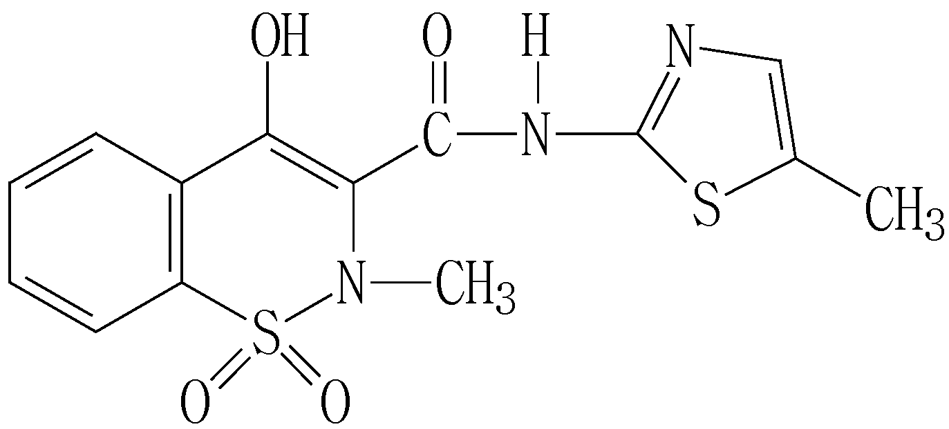
Voltammetric Determination of Meloxicam in Pharmaceutical Formulation and Human Serum at Glassy Carbon Electrode Modified by Cysteic Acid Formed by Electrochemical Oxidation of L-cysteine

1
Department of Chemistry and Chemical Engineering, Yangzhou University, Yangzhou 225002, China
2
Subei Hospital, Yangzhou 225002, China
*
Author to whom correspondence should be addressed.
Sensors 2006, 6(9), 1139-1152; https://doi.org/10.3390/s6091139
Submission received: 5 July 2006 / Accepted: 11 August 2006 / Published: 27 September 2006

Abstract

:
The improvement of electrochemical detection of meloxicam is presented by modification of a glassy carbon electrode with anionic layer of cysteic acid providing electrostatic accumulation of the analyte onto the electrode surface. The modification formed by electrochemical oxidation of L-cysteine was performed by cycling potential in cysteine solution. The anodic peak current obtained at + 1.088 V (vs. Ag/AgCl) by voltammetry was linearly dependent on the meloxicam concentration in the range of 4.3 × 10⁻⁸ ~ 8.5 × 10⁻⁶ M in the B-R buffer solution (0.04 M, pH 1.86) with a correlation coefficient of 0.999. The detection limit (S/N = 3) is 1.5 × 10⁻⁹ M. The low-cost modified electrode shows good sensitivity, selectivity and stability and has been applied to the determination of meloxicam in pharmaceutical formulation and spiked serum with satisfactory results. The electrochemical reaction mechanism of meloxicam was discussed.

1. Introduction

Meloxicam, (Scheme 1) (4-hydroxy-2-methyl-N-(5-methyl-2-thiazoly)-2H-1,2-benzo-thia- zine-3-carbox-amide-1,1dioxide) (C14H13N3O4S2) is a novel nonsteroidal anti-inflammatory drug (NSAID) with a favorable COX-2 (cyclooxgenase-2) [1,2]: COX-1 (cyclooxgenase-1) selectivity has also been shown to have potent anti-inflammatory effects [3,4]. Because of solubility of meloxicam in acidic medium, it may cause local gastrointestinal adverse events [5]. Up to now, there are a limited number of techniques described for the determination of meloxicam in pharmaceuticals formulations only including ultraviolet spectrophotometry [6], flow injection [7] and high-performance liquid chromatography (HPLC) with UV detector [8,9]. The Chinese pharmacopoeia method for conventional assaying meloxicam is based on neutralization titration in the solvent of ethanol [10]. However, There have been a very few methods have been published about the determination of meloxicam in biological media only including capillary zone electrophoresis (CZE) [11] and HPLC [12]. Further more, many of methods mentioned above required time-consuming manipulation steps, sophisticated instruments and special training. For these reasons, the rapid, simple and accurate method for the determination with high sensitivity is expected to be established.
Scheme 1. Structure of meloxicam.
Sensors 06 01139 g009
Electrochemical detection of analytes is a very elegant method in analytical chemistry. The interest in developing electrochemical sensing devices for use in environmental monitoring, clinical assays or process control is growing rapidly. Electrochemical sensors satisfy many of the requirements for such tasks particularly owing to their inherent specificity, speed of response, sensitivity and simplicity of preparation. Up to date, there are only three available electrochemical methods for the determination in the literature including polarography with the mercury electrode [13,14] and voltammetry [15]. However, the polarography would contaminate the environment for the mercury electrode if the mercury is handled with no special care. Radi et al. proposed a voltammetric method with a carbon paste electrode for the determination of meloxicam [15], and the linear range was 5.0 × 10⁻⁷ ~ 5.0 × 10⁻⁵ M with the detection limit of 1.6 × 10⁻⁷ M. These electrochemical methods were only described for the determination of meloxicam in pharmaceutical formulations. There is no literature reported about quantitative determinations of meloxicam in biological media by any electrochemical method.
In the present paper we described a rapid, simple voltammetric method to detect meloxicam in human serum with a glassy carbon electrode modified by cysteic acid formed by electrochemical oxidation of L-cysteine. A sensitive anodic oxidative peak of meloxicam was used for the quantitative determination. A good linear relationship was realized between the anodic peak currents and meloxicam concentrations in the range of 4.3 × 10⁻⁸ ~ 8.5 × 10⁻⁶ M with the detection limit of 1.5 × 10⁻⁹ M. By comparison of the results obtained at bare glassy carbon electrode and carbon paste electrode [15], the modification provided higher signal toward meloxicam in a broad range of experimental conditions. The performance of the modified electrode was illustrated by determination of meloxicam in pharmaceutical preparations and spiked serum. This method has the advantages of lower interference in the serum, rapid and simple operation, and high accuracy. This film is considered to be a promising, low-cost, steady and biocompatible material of electrode modifiers.

2. Experimental Section

2.1. Apparatus

Voltammetry experiments were carried out at a CHI 660B electrochemical workstation (Chenhua Instruments, China). All electrochemical experiments employed a conventional three-electrode system with a glassy carbon electrode or a cysteic acid modified glassy carbon electrode (4.0 mm in diameter) as a working electrode, a platinum wire as an auxiliary electrode and an Ag/AgCl electrode as a reference electrode. All potentials reported in this paper were referenced to the Ag/AgCl electrode. All experiments were carried out at 25 ºC.

2.2. Chemicals and Solutions

L-cysteine (CySH) was obtained from Shanghai Chemical Reagent Co., Ltd. (Shanghai, China). Meloxicam was purchased from Sigma (Milwaukee, Wisconsin, USA). Nafion (5 wt % in alcohols) was purchased from Fluka (Milwaukee, Wisconsin, USA). All the other reagents used were of analytical grade. Doubly distilled water was obtained by purification through a Millipore water system and used throughout. A stock anhydrous ethanol solution of meloxicam (1.0 × 10⁻³ M) was prepared, and kept in the dark under refrigeration (below 4 ºC). Britton-Robinson (B-R) buffer solutions at various pH values were used as base solutions for the determination of meloxicam.

2.3. Fabrication of the Cysteic Acid Modified Glassy Carbon Electrodes

Before modification, the glassy carbon electrode was polished with 1, 0.3 and 0.05 µm α-alumina powder, respectively, rinsed thoroughly with doubly distilled water within each polishing step. And then it was sonicated successively in 1:1 nitric acid, acetone and doubly distilled water. The modified electrode was prepared by cycling the potential between – 1.2 and + 2.6 V at the scan rate of 100 mV·s−1 in 0.04 M HCl solution containing 2.5 × 10⁻³ M L-cysteine. Fig. 1 shows the cyclic voltammograms of L-cysteine at a bare glassy carbon electrode. After 10 consecutive cycles, the surface of the glassy carbon electrode was covered with certain blue substance observed. Then, the modified electrode was rinsed respectively with ethanol and distilled water to remove the physically adsorbed and unreacted species from the electrode surface. Subsequently, the modified electrode was electroactivated by cyclic scanning from ­ 0.2 to + 1.2 V in the B-R buffer solution (0.04 M, pH 1.86), and a steady cyclic voltammogram was obtained. Finally, the electrode was dried with a stream of high purity nitrogen.

2.4. Determination of Meloxicam

The cysteic acid modified glassy carbon electrode, the platinum wire counter electrode and the Ag/AgCl reference electrode were immersed in 20.0 mL the B-R buffer solution (0.04 M, pH 1.86). A certain amount of meloxicam was added into the solution being stirred by a magnetic stirrer. The agitation was stopped after the electrochemical accumulation for 300 s at - 0.2 V. Then the voltammetry was immediately performed to scan from 0.4 to 1.4 V after 30 s of quiet time. The two anodic peaks currents of meloxicam at + 0.695 V (p1) and + 1.088 V (p2) were recorded (Fig. 2). The standard addition method was applied for the quantitative determination of meloxicam at + 1.088 V (p2). After the determination, the renewal of the electrode was accomplished by soaking it in the B-R buffer solution and cycling potentials between - 0.2 and 1.2 V about 6 cycles when there was no peak.
Figure 1. Cyclic voltammograms of 2.5 × 10⁻³ M L-cysteine at a glassy carbon electrode in 0.04 M HCl solution. Scan potential: - 1.2 to 2.6 V; scan rate: 100 mV·s−1.
Sensors 06 01139 g001
Figure 2. Adsorptive voltammograms of 3.5 × 10⁻⁶ M meloxicam (solid line) and blank (dot line) at the modified electrode in the B-R buffer solution (0.04 M, pH 1.86). Accumulation potential under stirring: - 0.2 V; accumulation time: 300 s; quiet time: 30 s; scan rate: 100 mV·s-1.
Sensors 06 01139 g002

2.5. Coulometry Experiments

The coulometry was carried out to determine number of electrons involved in the oxidation process of meloxicam. The meloxicam solution (2.5 × 10⁻⁶ M) was electrolyzed exhaustively at + 1.088 V (p2) and + 0.695 V (p1) respectively until the electrolytic currents were close to zero, and the quantity of electrical charge Q1 was recorded. Likewise, the background solution was electrolyzed under the same conditions, and the quantity of electrical charge Q2 was obtained.

3. Results and Discussion

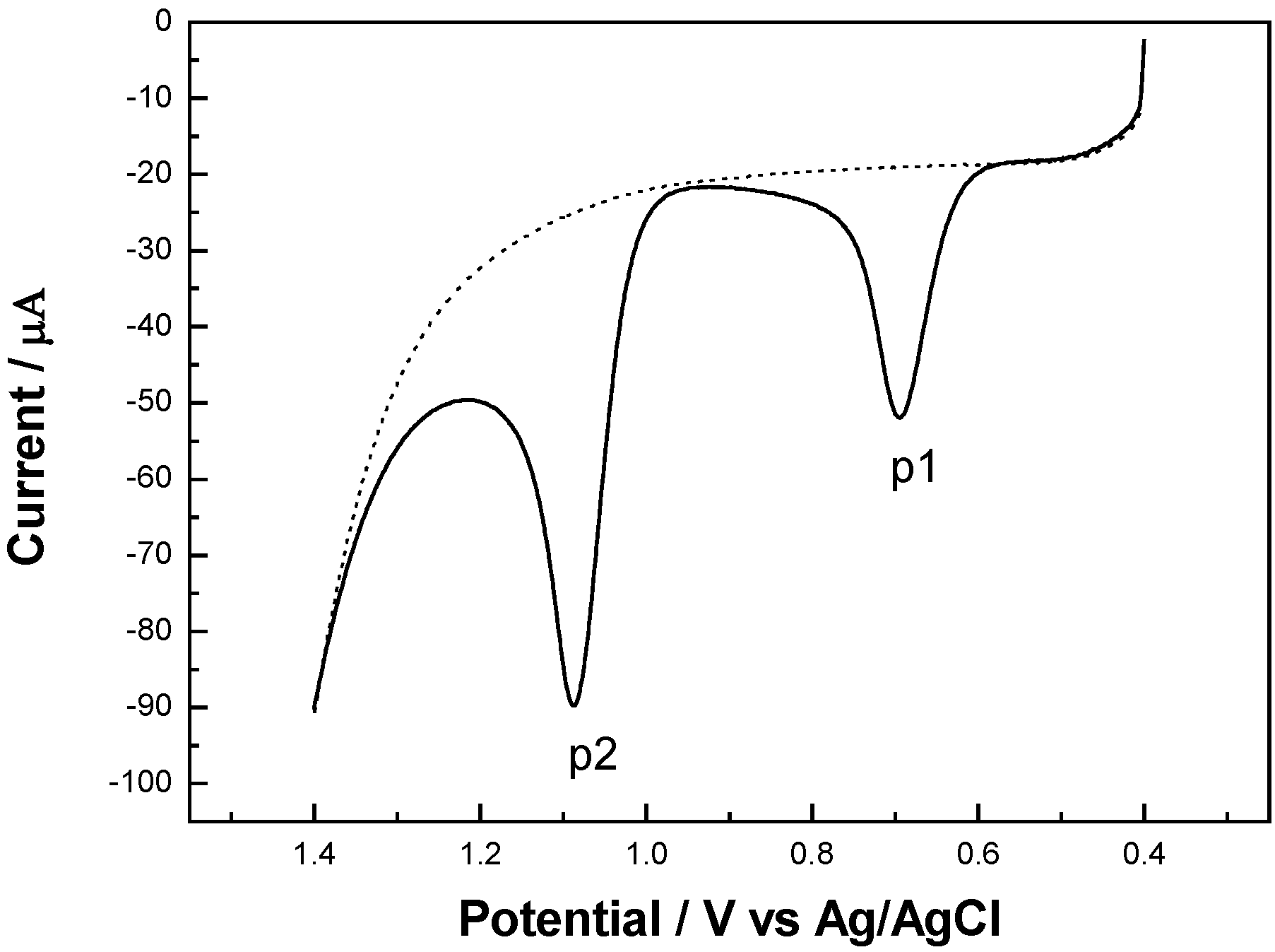
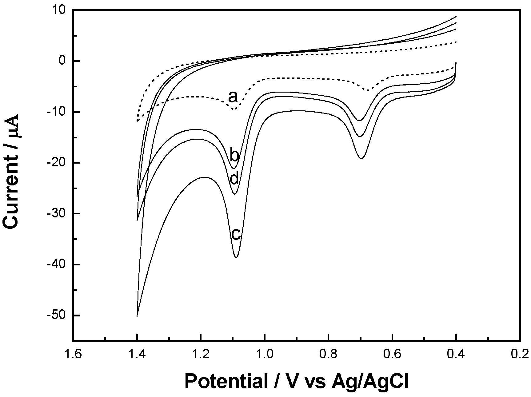
3.1. The Role of Materials Modified on the Glassy Carbon Electrode

Many electrochemical researchers have been devoted to determination of CySH [16,17]. And it is widely accepted that the reaction occurred by the following oxidation reaction mechanism on electrodes [17,18,19,20]:
CySH ↔ CyS- + H+
CyS- → CyS·ads + e-
2CyS·ads → CySSCy
Herein, electrochemical oxidation of CySH at a glassy carbon electrode was also investigated by the cyclic voltammetry in the wide potential range of – 1.2 ~ 2.6 V at a scan rate of 100 mV·s−1 in 0.04 M HCl solution containing 2.5 × 10⁻³ M CySH. Fig. 1 shows the cyclic voltammograms of L-cysteine at a glassy carbon electrode. There was an irreversible oxidation peak at ca. + 0.5 V at the first segment, which implied that CySH was oxidized to L-cystine (CySSCy). And another irreversible oxidation peak was observed at higher potential (ca. + 1.6 V) with further voltammetric cycling. In many cases, cysteic acid (CySO3H) was found as an oxidation product of CySSCy [18,19,20,21]. Fei et al. described that the oxidation product of CySH can be further oxidized to chemisorbing molecules (cysteic acid) under high positive potential [20]. Ralph et al. demonstrated that CySH was adsorbed on the surface of a glassy carbon electrode by using AC voltammetry, and its further oxidation to form cysteic acid was proposed [19]:
RSSR + 3 H2O → RSO3H + 5 H+ + 5 e-
Spataru et al. confirmed that the functional group SO3H of cysteic acid was strongly adsorbed at a glassy carbon electrode by using cyclic voltammetric and polarization measurements [17]. According to its chemical structure, cysteic acid formed by the electrochemical oxidation of CySH is similar to Nafion with sulfonated groups, so it can be used as a novel electrode modifier due to its attractive ion-exchange characteristics. Fig. 6 shows the peak current response of meloxicam at cysteic acid/GCE is enhanced compared with the bare GCE under above identical experimental conditions. It was attributed to the electrostatic attraction of modifier layer for the amine groups of meloxicam molecules carrying positive charges in the acid solution. In order to confirm this property, Nafion film which was characteristic of cation exchange was coated on the surface of the GCE to prepare Nafion/GCE for comparison with the proposed modifier material. 0.1 wt% Nafion solution used in this work was prepared by diluting the 5 wt% Nafion solution in ethanol. A same GCE was coated with 10 µL of the Nafion solution above mentioned. The solvent was left to evaporate at room temperature in air. The two different modified electrodes all improved current responses of meloxicam compared with bare GCE under above identical experimental conditions, implying that cysteic acid and Nafion played the same role in the accumulations of meloxicam. Fig. 3 shows that the oxidation peak potentials at the Nafion/GCE are similar to those at the cysteic acid/GCE. But the current response at the Nafion/GCE is lower than that at cysteic acid/GCE, and it decreases one order of magnitude compared with the cysteic acid/GCE because of poor conductivity of Nafion. Additionally, Nafion is more expensive than CySH. So cysteic acid film has more significant advantages and electroanalytical application than Nafion film. On the other hand, we found that the modified electrode itself had pH sense under different pH environments and exhibited a good pH linear region from 2.0 to 12.0 with a near-Nernstian slope by experiment. The cation exchange property of the modifier was confirmed further. Consequently, a novel solid pH sensor will be fabricated using the thin membrane materials of cysteic acid supported on a glassy carbon electrode in our subsequence research.
Figure 3. Cyclic voltammograms of 5.0 × 10⁻⁷ M meloxicam at different modified GCEs. (A) cysteic acid/GCE; (B) Nafion/GCE. Scan rate: 100 mV·s-1; accumulation potential under stirring: - 0.2 V; accumulation time: 300 s; quiet time: 30 s; electrolyte: B-R buffer solution (0.04 M, pH 1.86).
Sensors 06 01139 g003

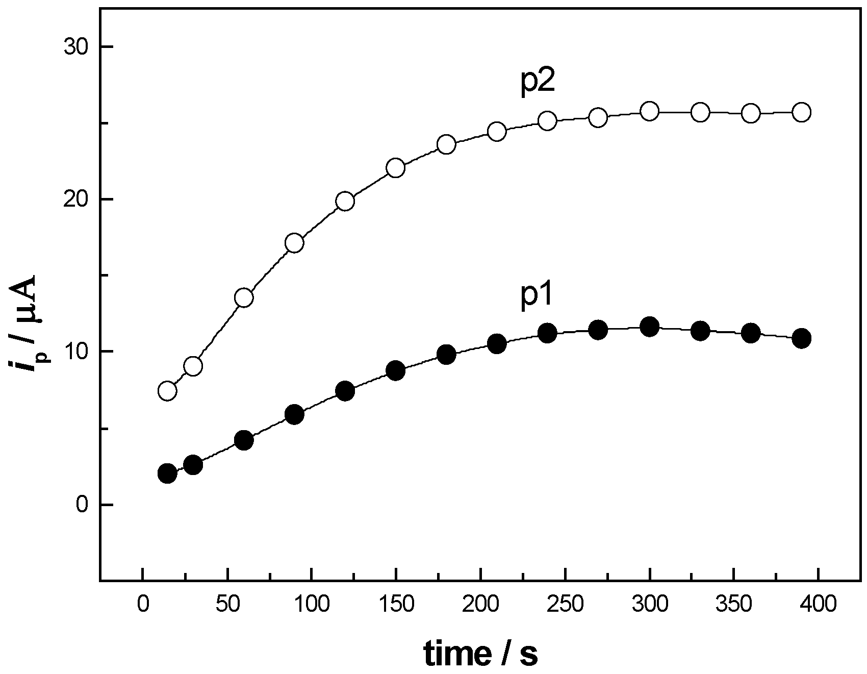
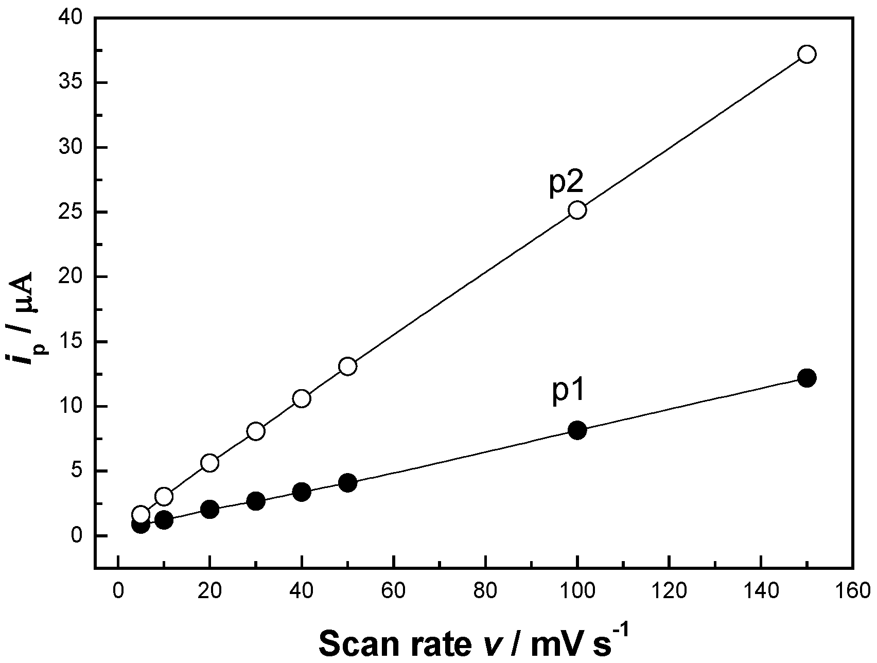
3.2. Optimization of Experimental Conditions

Some conventional supporting electrolytes were tested, such as HCl, H2SO4, HAc-NaAc, NH4Cl-NH3·H2O, KCl, phosphate buffer and B-R buffer solution. But higher sensitive and well-defined peaks of meloxicam in voltammograms were obtained in the B-R buffer solution (0.04 M, pH 1.86). The effects of accumulation potential and accumulation time on the voltammetric current response for meloxicam were studied. The accumulation step proceeded in constantly stirred solution and the voltage-scanning step was performed after 30 s of quiet time. The peak current of meloxicam was the highest at - 0.2 V as the accumulation potential. It is attributed to the existence of meloxicam in the cationic form at pH 1.86, and more favorable accumulation at negative potential. So an accumulation potential of - 0.2 V was chosen in all the subsequent work. The effect of accumulation time on the peak current was investigated, too. Fig. 4 shows the peak currents of meloxicam increased with the increasing the accumulation time within 300 s, which is attributed to the adsorption of meloxicam at the modified electrode surface. Further postponement of the accumulation time did not increase the response of meloxicam after 300 s owing to the surface adsorption saturation. For practical purposes, a 300 s accumulation period was found to be sufficient for the determination of meloxicam.
Figure 4. Effect of the accumulation time on the anodic peak currents of 5.0 × 10⁻⁷ M meloxicam in the B-R buffer solution (0.04 M, pH 1.86). Scan rate: 100 mV·s-1; accumulation potential: -0.2 V; quiet time: 30 s.
Sensors 06 01139 g004
Figure 5. Effect of pH on the anodic peak currents (p1, p2) of 5.0 × 10⁻⁷ M meloxicam at the cysteic acid/GCE. Scan rate: 100 mV·s-1; accumulation potential: - 0.2 V; accumulation time: 300 s; quiet time: 30 s; electrolyte: B-R buffer solution (0.04 M).
Sensors 06 01139 g005
The pH value of the base solution has a significant influence on oxidation current response of meloxicam at the cysteic acid/GCE. Fig. 5 shows the effect of pH value of the base solution in the range of 1.86 ~ 7.43 on the peak currents (p1 and p2). The peak currents increased monotonically with pH value decreasing. But the interference of water electrolysis became serious when the pH value was lower than 1.86. So the B-R buffer solution (0.04 M, pH 1.86) was selected for the determination.
The anodic peak currents of meloxicam were considerably related to the amount of cysteic acid on the surface of the glassy carbon electrode. When the electrode was carried out the sweeps under the cyclic voltammetry, the more cycles of sweep, the more cysteic acid was oxidized on the surface of the electrode. Fig. 6 shows the remarkably effect of cysteic acid contents on the peak currents of meloxicam. The anodic peak currents at the cysteic acid/GCE are obviously higher than that on the bare glassy carbon electrode. It might be ascribed to the accumulation of cysteic acid for meloxicam. The anodic peak currents gradually increased with increasing the amount of cysteic acid on the surface of the electrode. However, the anodic peak currents decreased when excessive cysteic acid was oxidized at the electrode. We owed it to the decline of the electrode conductivity. So a 10-circle-sweep by the cyclic voltammetry was selected in present experiments.
Figure 6. Cyclic voltammograms of 3.5 × 10⁻⁶ M meloxicam at the cysteic acid/GCEs prepared in modified solution with different sweep circles. a, the bare glassy carbon electrode; b, 5 circles; c, 10 circles; d, 15 circles. Scan rate: 100 mV·s-1; electrolyte: B-R buffer solution (0.04 M, pH 1.86).
Sensors 06 01139 g006

3.3. Electrochemical Reaction Mechanism of Meloxicam at the Cysteic Acid Modified Electrode

The coulometry was carried out to determine number of electrons involved in the oxidation of meloxicam. The meloxicam solution (2.5 × 10⁻⁶ M) was electrolyzed exhaustively at + 1.088 V (p2) and + 0.695 V (p1) respectively until the electrolytic currents were close to zero, and the quantity of electrical charge Q1 was recorded. Likewise, the background solution was also electrolyzed under the same conditions, and the quantity of electrical charge Q2 was obtained.
The charge difference Q = (Q1 - Q2) represents the quantities of electrical charge consumed by oxidation of the meloxicam:
Q = nFCV
Here, C and V here are the concentration and volume of the meloxicam solution, respectively. n and F has their conventional meanings. From Eq. (5), the electron numbers both n (p2) and n (p1) of the electrochemical reaction of meloxicam could be approximately calculated to be 1, respectively. The experimental results indicated that there was a good linear relationship between the two peaks’ potentials and log v with the slope of 0.1389 and 0.1014 which equals 0.059/αn according to the Nernst equation. Here α is the coefficient of electrons transfer. Then both αn (p2) and αn (p1) is found to be 0.43 and 0.58, respectively. The pH dependence on the peak potentials was also investigated, and they were observed to shift towards positive direction with decrease of pH value. According to our results from experiments, we found that the modified electrode itself had pH sense under different pH environments and exhibited a good pH linear region from 2.0 to 12.0 with a near-Nernstian slope. But, under different pH environments, we wanted to know if the charge of cysteic acid layer on the electrode surface influenced the peak potential of meloxicam in Faradic process with the transfer of the hydrogen ion. A voltammetry experiment was performed at the modified electrode using KI as a redox probe (I2 +2e = 2I-) without any transfer of the hydrogen ion in its Faradic process. We found the anodic peak of the iodide was constant in variable pH solution ranged from 2 to 8. So the pH dependence on the peak potential of meloxicam reflected only the transfer of the hydrogen ion in its Faradic process. A good linear relationship was obtained between the peak potential (p2) and pH value in the range of pH 1.86 to 7.43 with the slope of - 0.0987 from Nernst equation Ep = K - (0.059m/αn)· pH. Here m was the number of H+ transfer. The m could be calculated to be 0.89. So m was 1 approximately. From above discussion, the oxidation process of the meloxicam involves two electrons and one proton. The result is consistent with the reference [15].
The scan rate effect on the peak current of 5.0 × 10⁻⁷ M meloxicam at the modified electrode was also investigated. It could be seen from Fig.7 that the anodic peak current and cathodic peak current increased with increasing the scan rate. Two good linearity between ip and the scan rate v were obtained from the range of 5 ~ 150 mV·s-1, which demonstrated that the electrode reaction was an adsorption-controlled process [22].
Figure 7. Plots of the anodic peak currents of 5.0 × 10⁻⁷ M meloxicam at the cysteic acid/GCE in the B-R buffer solution (0.04 M, pH 1.86) at different scan rates. p1, the peak current at + 0.695 V; p2, the peak current at +1.088 V. Accumulation potential: -0.2 V; accumulation time: 300 s; quiet time: 30 s.
Sensors 06 01139 g007

3.4. Calibration Curve and Detection Limit
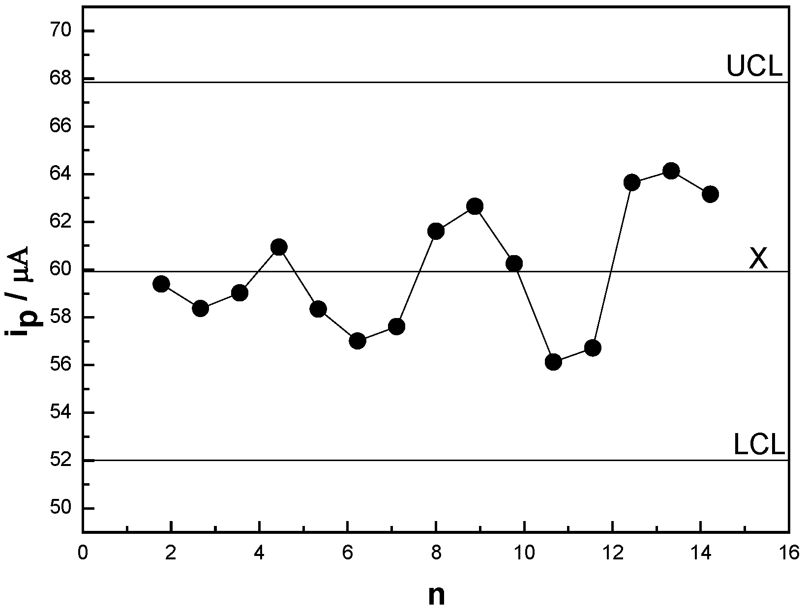
Under above optimum detection conditions, the anodic peak currents at + 1.088 V (p2) recorded were proportional to meloxicam concentration in the range of 4.3 × 10⁻⁸ ~ 8.5 × 10⁻⁶ M. The linear equation is ipa (µA) = 21.5 + 8.4C (10-6 M) at + 1.088 V with the correlation coefficient r = 0.999. The detection limit of meloxicam was 1.5 × 10⁻⁹ M in terms of signal to noise ratio of 3:1 (S/N = 3). When the concentration of meloxicam was more than 8.5 × 10⁻⁶ M, the current response decreased gradually and its plateau was observed, it was attributed to the saturation of meloxicam at the modified electrode.

3.5. Repeatability

The lifetime of the cysteic acid/GCE was examined, and the result demonstrated that the cysteic acid film retained 98.8 % of its initial response after 1 month of storage in B-R buffer solution (0.04 M, pH 1.86). After detecting the meloxicam in every five serum samples, the electrode was washed in the B-R buffer solution under stirring, and then transferred into a standard solution of 4.5 × 10⁻⁶ M meloxicam to record its oxidation peak current (p2). Its average current was 57.7 µA with the relative standard deviation (RSD) of 2.2 % (n = 5). Such stability seemed to be acceptable for most practical applications. Because the modified electrode could adsorb meloxicam, it was necessary to renew the electrode surface. It was found that the renewal of the electrode surface was easily accomplished by immerging the modified electrode in B-R buffer solution by cycling its potential between - 0.2 and + 1.2 V about 6 cycles. A quality control chart (X-chart) has been constructed. Fig. 8 shows a quality control chart (X-chart) of 4.5 × 10⁻⁶ M meloxicam applying the proposed method, where the X-axis represents the number of analyses while the Y-axis represents the anodic current (p2) of meloxicam. There is a series of 15 points over the average score and all of them fall in between the upper control limit (UCL) and the lower control limit (LCL), which indicated that the analysis progress was under the statistical control.
Figure 8. Quality control chart (X - Chart) of 4.5 × 10⁻⁶ M meloxicam.
Sensors 06 01139 g008

3.6. Recovery Test

The recovery tests of meloxicam in the range from 2.8 × 10⁻⁷ to 1.06 × 10⁻⁶ M were performed. The results are listed in Table 1. The recoveries varied in the range from 96.4 % to 102.6 % and the RSD was 2.4 %.
Table 1. Recovery of meloxicam.
Added / MFound / MRecovery (%)
2.8 × 10-72.7 × 10-796.4
3.8 × 10-73.9 × 10-7102.6
7.6 × 10-77.7 × 10-7101.3
8.6 × 10-78.5 × 10-798.8
1.06 × 10-61.05 × 10-699.1

3.7. Interference

For the following solid pharmaceutical formulations analysis, tablet excipient, such as starch, was added in the detection system. It was not found that the results were changed in the presence of the excipient when 5.0 × 10⁻⁷ M meloxicam was detected under the optimum experimental conditions. If the anodic current of the first oxidation potential (p1) was selected for the determination in serum samples referred to the previous literature [15], uric acid (UA) and ascorbic acid (AA) in existence would severely interfere with the determination of the meloxicam because of the overlapping voltammetric response of UA and AA. Whereas the presence of UA and AA in high concentration did not interfere with the determination of meloxicam when the anodic current of the second oxidation potential (+ 1.088 V) was selected for the determination in serum samples. The other conceivable concomitant in serum samples were investigated in this detection system. When the concentration of the meloxicam was 5.0 × 10⁻⁷ M, no interferences were observed in the presence of 5.0 × 10⁻⁴ M of NaCl, KNO3, (NH4)2SO4, Mg(NO3)2, Ca(NO3)2, glucose, oxalic acid, urea, tartaric acid, caffeine, 5.0 × 10⁻⁵ M of cysteine, cystine, DL-tyrosine, citrate, CuSO4, 2.0 × 10⁻⁴ M of albumin fraction and Fe(NO3)3, respectively. The results obtained are listed in Table 2. In addition, some conventional medicaments, such as 1.0 × 10⁻⁴ M of Vitamin B1, Vitamin B2, Vitamin B6, atropine, penicillin, and theine, did not interfere with the determination.
Table 2. Influence of potential interferents on the voltammetric response of 0.50 µM meloxicam.
InterferentConcentration / mMSignal change (imeloxicam, p2 = 100 %)
AA0.05- 0.9 %
AA0.10- 1.8 %
AA0.20- 3.5 %
UA0.50- 2.6 %
albumin fraction0.20+ 1.7 %
urea0.50- 2.2 %
tartaric acid0.50- 3.0 %
cystine0.05- 1.9 %
citrate0.05- 2.5 %
glucose0.50+ 0.5 %
cysteine0.05- 2.9 %
DL-tyrosine0.05- 2.3 %
oxalic acid0.50- 1.9 %
caffeine0.50+ 1.5 %
NaCl0.50- 1.3 %
KNO30.50- 2.2 %
Ca(NO3)20.50- 1.9 %
(NH4)2SO40.50- 2.4 %
Mg(NO3)20.50- 1.0 %
CuSO40.05- 3.2 %
Fe(NO3)30.20- 2.6 %

3.8. Determination of the Pharmaceutical Preparation Samples

The developed voltammetry method for the meloxicam determination was applied to a commercial preparations (meloxicam tablets: Yangzijiang Pharmaceutical Co. Ld., China, Hongqiang®, batch No. 04110402, labeled amount of 7.5 mg per tablet) . A known number of tablets were grounded to fine powers, and an accurate mass of the powers about 200.0 mg was transferred to a 50.0 ml calibrated flasks. It was extracted for 5 min with 50.0 ml cold ethanol. Table 3 shows the results of voltammetry analysis of the commercial preparations. The Chinese pharmacopoeia methods (acid-base titration) [10] was employed as a comparison to evaluate the validity of the developed method (0.4 g of tablets was dissolved in a mixture of 100 ml of ethanol and acid-base titration was carried out with bromophenol blue as an indicator. Each 1 ml of 0.1 M sodium hydroxide is equivalent to 35.14 mg of meloxicam). A good agreement was achieved and there was no significant difference between methods. The results obtained show the developed method could be easily used in quality control laboratory for the analysis of meloxicam in solid pharmaceutical formulations.
Table 3. Determination results of meloxicam in tablets.
Batch No.Pharmacopoeial Method (n=5) (mg/tablet)Present method (n=5) (mg/tablet)R.S.D* (%)
Hongqiang® 041104027.37.52.2
* Here R.S.D is relative standard deviation.

3.9. Detection of Meloxicam in Human Serum Samples

The developed voltammetry method for the meloxicam determination was applied to human serum samples. The serum samples were obtained from volunteers. The recovery from human serum was measured by spiking drug-free human serum with known amounts. The serum samples without further pretreatments were diluted 100 times with the 0.04 M B-R buffer solution (pH 1.86) before analysis. The peak current of meloxicam at + 1.088 V (p2) were recorded. Standard addition method was employed under above optimal experimental conditions. The detection results obtained are listed in Table 4. The recovery determined was ranged from 96.8 to 103.2 %.
Table 4. Determination of meloxicam in spiked serum samples at the cysteic acid/GCE.
SerumSpiked / μgDetected* / μgRecovery / %
Sample 16.05.8 ± 0.296.8
Sample 27.57.7 ± 0.3102.3
Sample 310.610.9 ± 0.4103.2
Sample 413.513.6 ± 0.4100.7
Sample 515.014.8 ± 0.598.7
Sample 616.516.0 ± 0.697.0
* Number of sample assayed was five.

4. Conclusions

A novel modified electrode was fabricated using cysteic acid formed by electrochemical oxidation of L-cysteine at a glassy carbon electrode. Cysteic acid with cation exchange property drastically increased the current response of meloxicam into two well-defined peaks. The peak current at + 1.088 V obtained by voltammetry was linearly dependent on the meloxicam concentration in the range of 4.3 × 10⁻⁸ ~ 8.5 × 10⁻⁶ M in B-R (0.04 M, pH 1.86) with a correlation coefficient of 0.999 and the detection limit was 1.5 × 10⁻⁹ M. The electrochemical reaction mechanism of meloxicam could be concluded that the oxidation process of a meloxicam molecule include one H+ and two single electrons. The modified electrode has also been demonstrated to be applicable in the detection of meloxicam in pharmaceutical formulation and spiked serum samples with excellent sensitivity and selectivity. This novel modifier is considered to be a promising, low-cost, and steady material for the modification of electrodes. The cysteic acid modified electrode will hopefully be of a good application for further sensor development.

Acknowledgements

This work was supported by the National Natural Science Foundation of China (No. 20375034) and the Novel Foundation of Jiangsu Provincial Education Department for Doctors (2005).

References

  1. Noble, S. J.; Balfour, A. Meloxicam. Drugs 1996, 51, 424–430. [Google Scholar] [CrossRef] [PubMed]
  2. Turck, D.; Roth, W.; Busch, U. A review of the clinical pharmacokinetics of meloxicam. British Journal of Rheumatology 1996, 35, 13–16. [Google Scholar] [CrossRef] [PubMed]
  3. Engelhardt, G.; Homma, D.; Schlegel, K.; Utzmann, R.; Schnitzler, C. Anti-inflammatory, analgesic, antipyretic and related properties of meloxicam, a new non-steroidal anti-inflammatory agent with favorable gastrointestinal tolerance. Inflammation Research 1995, 44, 423–433. [Google Scholar] [CrossRef] [PubMed]
  4. Davies, N. M.; Skjodt, N. M. Clinical pharmacokinetics of meloxicam. A cyclo-oxygenase-2 preferential nonsteroidal anti-inflammatory drug. Clinical Pharmacokinetics 1999, 36, 115–126. [Google Scholar] [CrossRef] [PubMed]
  5. Martindale. In The Extra Pharmacopoeia, Thirty second edition; The Pharmaceutical Press: London, England, 1999.
  6. Hassan, E. M. Spectrophotometric and fluorimetric methods for the determination of meloxicam in dosage forms. Journal of Pharmaceutical and Biomedical Analysis 2002, 27, 771–777. [Google Scholar] [CrossRef]
  7. Garcia, Ma. S.; Sanchez-Pedreno, C. Ma.; Albero, I.; Marti, J. Spectrophotometric methods for determining meloxicam in pharmaceuticals using batch and flow-injection procedures. European Journal of Pharmaceutical Sciences 2000, 9, 311–316. [Google Scholar] [CrossRef]
  8. Joseph-Charles, J.; Bertucal, M. Determination of meloxicam in tablet formulations by ultraviolet spectrophotometry and high-performance liquid chromatography. Analytical Letters 1999, 32, 2051–2059. [Google Scholar] [CrossRef]
  9. Joseph-Charles, J.; Bertucal, M. Simultaneous high-performance liquid chromatography analysis of non-steroidal anti-inflammatory oxicams in pharmaceutical preparations. Journal of Liquid Chromatography Related Technologies 1999, 22, 2009–2021. [Google Scholar] [CrossRef]
  10. Meloxicam. In The Chinese Pharmacopoeia; Chemical Industry Press: Beijing, China, 2005.
  11. Emirhan, N.; Sedef, K. Method development and validation for the analysis of meloxicam in tablets by CZE. Joural of Pharmaceutical and Biomedical Analysis 2003, 31, 393–400. [Google Scholar]
  12. Baeyens, W. R. G.; Vander Weken, G.; D’haeninck, E.; Garcı´a-Campan, A. M.; Vankeirsbilck, T.; Vercauteren, A.; Deprez, P. Application of an alkyl-diol silica precolumn in a columns witching system for the determination of meloxicam in plasma. Journal of Pharmaceutical and Biomedical Analysis 2003, 32, 839–846. [Google Scholar] [CrossRef]
  13. Beltagi, A. M.; Ghoneim, M. M.; Radi, A. Electrochemical reduction of meloxicam at mercury electrode and its determination in tablets. Journal of Pharmaceutical and Biomedical Analysis 2000, 28, 1501–1503. [Google Scholar] [CrossRef]
  14. Huang, H.; Gao, H. Y.; Zeng, Y. H. Single sweep oscillopolarography of meloxicam. Chinese Journal of Analytical Chemistry 2000, 17, 135–136. [Google Scholar]
  15. Radi, A.; El Ries, M. A.; El-Anwar, F.; El-Sherif, Z. Electrochemical oxidation of meloxicam and its determination in tablet dosage form. Analytical Letters 2001, 34, 739–748. [Google Scholar] [CrossRef]
  16. Li, H.; Li, T.; Wang, E. Electrocatalytic oxidation and flow detection of cysteine at an aquocobalamin absorbed glassy carbon electrode. Talanta 1995, 42, 885–890. [Google Scholar] [CrossRef]
  17. Spataru, N.; Sarada, B. V.; Popa, E.; Tryk, D. A.; Fujishima, A. Voltammetric determination of L-cysteine at conductive diamond electrodes. Analytical Chemistry 2001, 73, 514–519. [Google Scholar] [CrossRef] [PubMed]
  18. Zagal, J.; Fierro, C.; Rozas, R. Electrocatalytic effects of adsorbed cobalt phthalocyanine tetrasulfonate in the anodic oxidation of cysteine. Journal of Electroanalytical Chemistry 1981, 119, 403–408. [Google Scholar] [CrossRef]
  19. Ralph, T. R.; Hitchman, M. L.; Millington, J. P.; Walsh, F. C. The electrochemistry of L-cystine and L-cysteine Part 1: Thermodynamic and kinetic studies. Journal of Electroanalytical Chemistry 1994, 375, 1–15. [Google Scholar] [CrossRef]
  20. Fei, S. D.; Cheng, J. H.; Yao, S. Z.; Deng, G. H.; He, D. L.; Kuang, Y. F. Electrochemical behavior of L-cysteine and its detection at carbon nanotube electrode modified with platinum. Analytical Biochemistry 2005, 339, 29–35. [Google Scholar] [CrossRef] [PubMed]
  21. Davis, D. G.; Bianco, E. An electrochemical study of the oxidation of L-cysteine. Journal of Electroanalytical Chemistry 1966, 12, 254–260. [Google Scholar] [CrossRef]
  22. Laviron, E. General expression of the linear potential sweep voltammogram in the case of diffusionless electrochemical systems. Journal of Electroanalytical Chemistry 1979, 101, 19–28. [Google Scholar] [CrossRef]

