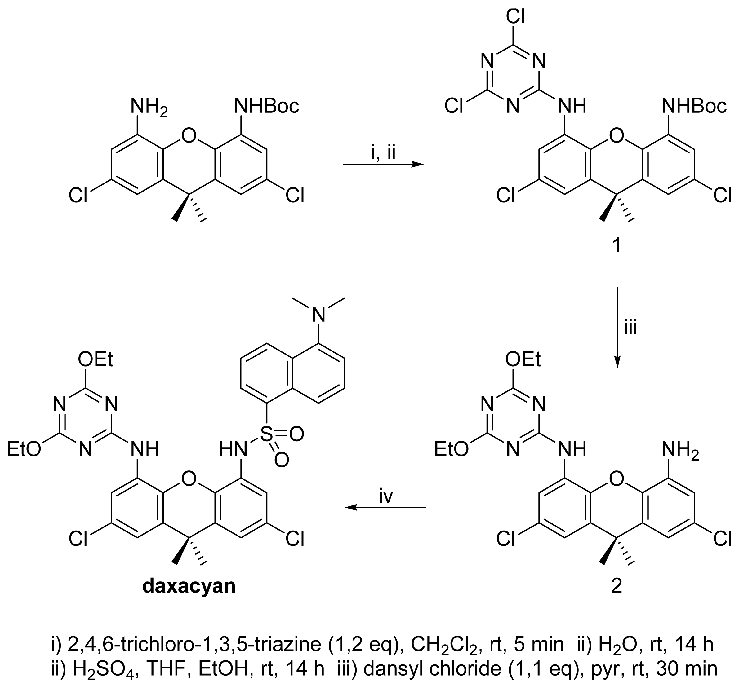
A Fluorescent Sensor for Dinitrobenzoic Acid Based on a Cyanuric Acid and Xanthene Skeleton

Abstract
:1. Introduction
2. Results and Discussion
Supplementary Material
Acknowledgments
References
- Curran, D. P.; Kuo, L. H. Acceleration of a dipolar Claisen rearrangement by hydrogen bonding to a soluble diaryl urea. Tetrahedron Lett. 1995, 36, 6647–6650. [Google Scholar]
- Kelly, T. R.; Meghani, P.; Ekkundi, V. S. Diels-Alder reactions: Rate acceleration promoted by a biphenylenediol. Tetrahedron Lett. 1990, 31, 3381–3384. [Google Scholar]
- Hunter, C. A. Molecular recognition of p-benzoquinone by a macrocyclic host. J. Chem. Soc. Chem. Commun. 1991, 749–751. [Google Scholar]
- Nishizawa, S.; Bühlmann, P.; Iwao, M.; Umezawa, Y. Anion recognition by urea and thiourea groups remarkably simple neutral receptor for dihydrogenphosphate. Tetrahedron Lett. 1995, 36, 6483–6486. [Google Scholar]
- Jubian, V.; Veronese, A.; Dixon, R. P.; Hamilton, A. D. Acceleration of a phosphate diester transesterification reaction by bis(alkylguanidinium) receptors containing an appended general base. Angew. Chem. Int. Ed. Eng. 1995, 34, 1237–1239. [Google Scholar]
- Raposo, C.; Almaraz, M.; Crego, M.; Mussons, M. L.; Pérez, N.; Caballero, M. C.; Morán, J. R. Lactone receptors with catalytic activity. Tetrahedron Lett. 1994, 35, 7065–7068. [Google Scholar]
- García-Garrido, S. E.; Caltagirone, C.; Light, M. E.; Gale, P. A. Acridinone-based anion receptors and sensors. Chem. Commun. 2007, 1450–1452. [Google Scholar]
- Blázquez, M. T.; Muñiz, F. M.; Sáez, S.; Simón, L. M.; Alonso, A.; Raposo, C.; Lithgow, A.; Alcázar, V.; Morán, J. R. Acridone heterocycles as fluorescent sensors for anions. Heterocycles 2006, 69, 73–81. [Google Scholar]
- Crego, M.; Raposo, C.; Mussons, M. L.; Berrocal, A.; Caballero, M. C.; Morán, J. R. Active receptors in the nucleophilic addition of pyrrolidine to acrylamide. Heterocycles 1995, 40, 139–140. [Google Scholar]
- Simón, L.; Muñiz, F. M.; Sáez, S.; Raposo, C.; Morán, J. R. From theozymes to artificial enzymes: Enzyme-like receptors for Michael additions with oxyanion holes and active amino groups. Eur. J. Org. Chem. 2007, 4821–4830. [Google Scholar]
- Shimizu, K. D.; Dewey, T. M.; Rebek, J., Jr. Convergent functional groups. 15. Synthetic and structural studies of large and rigid molecular clefts. J. Am. Chem. Soc. 1994, 116, 5145–5149. [Google Scholar]
- Nowick, J. S.; Ballester, P.; Ebmeyer, F.; Rebek, J., Jr. Convergent functional groups. 9. Complexation in new molecular clefts. J. Am. Chem. Soc. 1990, 112, 8902–8906. [Google Scholar]
- Hamann, B. C.; Branda, N. R.; Rebek, J., Jr. Multipoint recognition of carboxylates by neutral hosts in non-polar solvents. Tetrahedron Lett. 1993, 34, 6837–6840. [Google Scholar]
- Chang, K. J.; An, Y. J.; Uh, H.; Jeong, K. S. Reversible control of assembly and disassembly of interlocked supermolecules. J. Org. Chem. 2004, 69, 6556–6563. [Google Scholar]
- Muñiz, F. M.; Simón, L.; Sáez, S.; Raposo, C.; Morán, J. R. Selective acylation of 4,5-diamino-9,9’-dimethylxanthene through an aggregation effect. Tetrahedron Lett. 2008, 49, 790–793. [Google Scholar]
- Hallas, L. E.; Alexander, M. Microbial transformation of nitroaromatic compounds in sewage effluent. Appl. Environ. Microbiol. 1983, 1234–1241. [Google Scholar]
- De Santis, G.; Fabbrizzi, L.; Licchelli, M.; Poggi, A.; Taglietti, A. Molecular recognition of carboxylate ions based on the metal-ligand interaction and signalled through fluorescence quenching. Angew. Chem. Int. Ed. Eng. 1996, 35, 202–204. [Google Scholar]
- Gunnlaugsson, T.; Glynn, M.; Tocci, G. M.; Kruger, P. E.; Pfeffer, F. M. Anion recognition and sensing in organic and aqueous media using luminescent and colorimetric sensors. Coord. Chem. Rev. 2006, 250, 3094–3117. [Google Scholar]


© 2008 by MDPI Reproduction is permitted for noncommercial purposes.
Share and Cite
Muñiz, F.M.; Simón, L.; Sáez, S.; Raposo, C.; Alcázar, V.; Morán, J.R. A Fluorescent Sensor for Dinitrobenzoic Acid Based on a Cyanuric Acid and Xanthene Skeleton. Sensors 2008, 8, 1637-1644. https://doi.org/10.3390/s8031637
Muñiz FM, Simón L, Sáez S, Raposo C, Alcázar V, Morán JR. A Fluorescent Sensor for Dinitrobenzoic Acid Based on a Cyanuric Acid and Xanthene Skeleton. Sensors. 2008; 8(3):1637-1644. https://doi.org/10.3390/s8031637
Chicago/Turabian StyleMuñiz, Francisco M., Luis Simón, Silvia Sáez, César Raposo, Victoria Alcázar, and Joaquín R. Morán. 2008. "A Fluorescent Sensor for Dinitrobenzoic Acid Based on a Cyanuric Acid and Xanthene Skeleton" Sensors 8, no. 3: 1637-1644. https://doi.org/10.3390/s8031637