Article

Osteogenic Potential and Bioactive Profiles of Piper sarmentosum Ethanolic Extract-Treated Stem Cells

by
Intan Zarina Zainol Abidin
1,*,
Anis Nabilah Johari
2,
Muhammad Dain Yazid
3,
Zaidah Zainal Ariffin
4,
Herryawan Ryadi Eziwar Dyari
5 and
Shahrul Hisham Zainal Ariffin
2,*
1
Centre for Research and Graduate Studies, University of Cyberjaya, Cyberjaya 63000, Malaysia
2
Department of Biological Science and Biotechnology, Faculty of Science and Technology, Universiti Kebangsaan Malaysia, Bangi 43600, Malaysia
3
Centre for Tissue Engineering and Regenerative Medicine, Universiti Kebangsaan Malaysia Medical Centre, Cheras 56000, Malaysia
4
Faculty of Applied Sciences, Universiti Teknologi MARA, Shah Alam 40450, Malaysia
5
Department of Earth Sciences and Environmental, Faculty of Science and Technology, Universiti Kebangsaan Malaysia, Bangi 43600, Malaysia
*
Authors to whom correspondence should be addressed.
Pharmaceuticals 2023, 16(5), 708; https://doi.org/10.3390/ph16050708
Submission received: 19 March 2023 / Revised: 28 April 2023 / Accepted: 3 May 2023 / Published: 7 May 2023

Abstract

:
Piper sarmentosum is a well-known traditional herbal plant in various diseases treatments. Multiple scientific studies have also reported various biological activities exhibited by the plant’s extract, such as antimicrobial, anticarcinogenic and antihyperglycemic activities, and, in addition, a bone protective effect in ovariectomized rats has been reported. However, no known Piper sarmentosum extract is involved in osteoblast differentiation using stem cells. Our study aims to identify the potential of P. sarmentosum ethanolic extract to induce osteoblast differentiation of human peripheral blood stem cells. Prior to the assay, the proliferation ability of the cells was observed for 14 days and the presence of hematopoietic stem cells in the culture was determined by the expression of SLAMF1 and CD34 genes. During the differentiation assay, the cells were treated with P. sarmentosum ethanolic extract for 14 days. Osteoblast differentiation was examined using an (alkaline phosphatase) ALP assay, by monitoring the expression of osteogenic gene markers and by von Kossa staining. The untreated cells served as the negative control, while cells treated with 50 µg/mL ascorbic acid and 10 mM β-glycerophosphate acted as the positive control. Finally, the determination of the compound profile was performed using a gas chromatography-mass spectrometry (GC-MS) analysis. The isolated cells were able to proliferate for 14 days during the proliferation assay. The expression of hematopoietic stem cell markers was also upregulated during the 14 days assay. Following the differentiation induction, the ALP activity exhibited a significant increase (p < 0.05) from day 3 of the differentiation assay. A molecular analysis also showed that the osteogenic markers ALP, RUNX2, OPN and OCN were upregulated compared to the positive control. The presence of mineralized cells with a brownish-stained morphology was observed, indicating the mineralization process increased in a time-dependent manner regardless of the concentration used. There were 54 compounds observed in the GC-MS analysis, including β-asarones, carvacrol and phytol, which have been shown to possess osteoinductive capacities. Our results demonstrate that the ethanolic extract of P. sarmentosum can induce osteoblast differentiation of peripheral blood stem cells. The extract contains potent compounds which can potentially induce the differentiation of bone cells, i.e., osteoblasts.

1. Introduction

Natural products, specifically herbal medicinal plants, are gaining more interest from consumers for treating diseases. The number of commercialized derivatives from medicinal plants is increasing and they can be easily purchased. These natural medicines are deemed to have more advantages because numerous compounds that exist in a particular plant can work synergistically and exert a multitarget efficacy compared to conventional medicines that target one specific disease [1]. Natural products are also preferred due to their low cost, fewer side effects and suitability for long-term consumption [2]. Bacopa monniera, Glycyrrhiza glabra and Polygonum odoratum are a few examples of medicinal plants studied as treatments for Alzheimer’s disease, psoriasis and breast cancer, respectively [1].
Piper sarmentosum, known as ‘Kaduk’, is one of the most well-known herbs that locals use as an alternative medicine. P. sarmentosum belongs to the family Piperaceae and can be easily found in tropical and subtropical regions of the world [3]. This plant is traditionally used by locals to treat minor wounds and diseases such as diabetes, hypertension, coughs, rheumatism and asthma [4,5]. The high content of compounds in P. sarmentosum enables it to possess numerous properties such as antibacterial, antihypertensive, antihyperglycemic and antimalarial properties and tissue regeneration of embryos and caudal fins [6,7,8]. Previous in vivo studies have shown that P. sarmentosum extracts exhibited the ability to act as a bone protective agent by restoring the microstructure and bone mass in glucocorticoid-induced osteoporosis and in ovariectomized and adrenalectomized rats [9,10]. These health benefits are attributed to the synergistic effect of compounds abundantly found in the raw plant extract. Nevertheless, the involvement of compounds in bone remodeling should be identified to give us a better understanding of the mechanisms involved. However, the effect of plant extracts on a cellular level, specifically on human peripheral blood stem cell (hPBSC) differentiation into osteoblasts, which is the building block for bones, is still unknown.
Bone structures undergo a continuous remodeling process throughout life to provide the optimum function by maintaining the mechanical strength of the bone through calcium homeostasis [11]. Two important cells in bone tissue remodeling are osteoclast and osteoblast cells that degrade bone by dissolving the matrix protein and reform the bone by producing extracellular proteins. The imbalance in this process may result in bone diseases such as osteoporosis, which is a skeletal disease that is characterized by a low bone mass and a deteriorating microarchitecture of the bone tissue, which can lead to fragile bones and fractures [12]. Currently, few drugs are used as a treatment for osteoporosis, they include estrogen, calcitonin and bisphosphonates. Unfortunately, the administration of these drugs might lead to side effects such as an increased risk of uterus cancer, myalgias and osteonecrosis [13,14]. Hence, to reduce the dependency on such drugs, the role and potential of P. sarmentosum in bone remodeling should be determined. Although a few previous in vivo studies have shown the ability of P. sarmentosum to improve bone health, the effect of its extract in the bone remodeling process on the cellular level is still not well studied.
In this study, the osteoblast differentiation potential was observed using human peripheral blood stem cells (hPBSCs). This process enables hPBSCs to differentiate into a variety of matured cells such as osteoclasts, chondrocytes and osteoblasts, making it a good model for differentiation studies [15,16]. These findings serve as a good indicator of the potential of P. sarmentosum to be used as a complementary medicine to treat bone diseases by inducing bone formation.
The objective of this study was to identify the potential of P. sarmantosum ethanolic extract to induce osteoblast differentiation of human peripheral blood stem cells (hPBSC) and determine the compound profile. Prior to a differentiation assay, the proliferation ability of the cells was determined using trypan blue exclusion dye staining. Then, a reverse transcription-quantitative polymerase chain reaction (RT-qPCR) was carried out to observe the expression of hematopoietic stem cell markers (CD34 and SLAMF1). Following this, the differentiation of osteoblasts was observed using three approaches which were biochemical (ALP assay), molecular (RT-qPCR) and morphological (von Kossa staining). The expressions of four osteoblasts gene markers were observed, i.e., ALP, RUNX2, OPN and OCN, during molecular observations. Finally, the compounds present in P. sarmentosum ethanolic extracts were screened using a GC-MS analysis.

2. Results

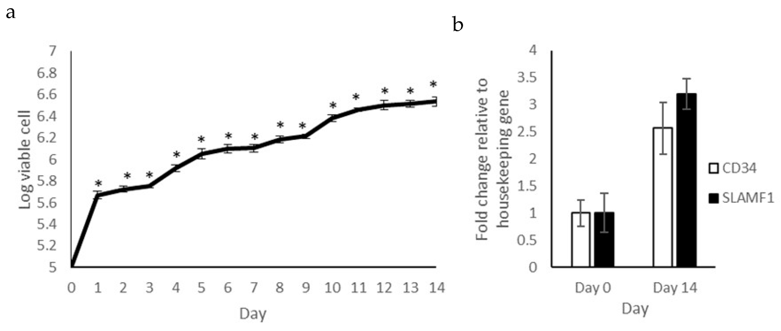
2.1. Proliferation Capacity and Expression of Hematopoietic Stem Cell Markers

The ability of self-renewal is one of the characteristics of a stem cell. The proliferation of isolated cells was observed before conducting the differentiation assay as shown in Figure 1a. The number of viable cells increased significantly (p < 0.05) starting from day 1 to day 14, exhibiting a approximately 34-fold increase on day 14 compared to day 0. This shows that the isolated cells can survive and proliferate well for 14 days in the provided in vitro culture environment.
Other than the ability for self-renewal, the expressions of hematopoietic stem cell markers SLAMF1 and CD34 were observed using the RT-qPCR technique. To observe the regulation of gene expression, RNA was extracted on day 0 and day 14, as shown in Figure 1b. The expressions of both markers were significantly upregulated (p < 0.05) on day 14 compared to day 0. SLAMF1 exhibited a 3.2-fold increase while CD34 exhibited a 2.6-fold increase. The high proliferation capacity of the in vitro culture and the increases in stemness markers indicate the presence of stem cells in the isolated cell population.

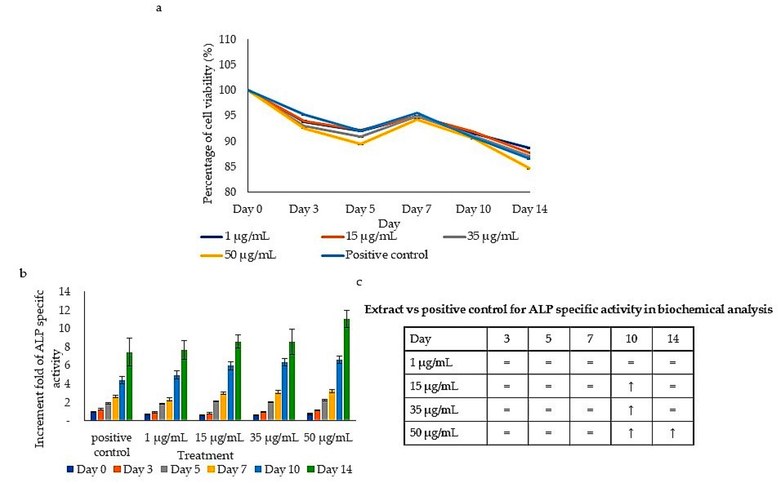
2.2. Viability and ALP-Specific Activity during the Differentiation Assay

The viability of the treated cells was observed during differentiation. Figure 2a shows the percentage of cell viability of human PBSCs on days 0, 3, 5, 7, 10 and 14 of the differentiation assay. Overall, cells treated with 50 µg/mL exhibited the lowest cell increase starting from day 3 to day 14 onwards when compared to the negative control. At day 14, the lowest cell viability was 84.6% (50 µg/mL), followed by 35 µg/mL (87.1%), 15 µg/mL (87.8%) and 1 µg/mL (88.6%) (Figure 2a).
ALP-specific activity is often used as an osteogenesis marker [17,18,19,20]. A gradual increase in ALP-specific activity was shown after treatment with all concentrations (1, 15, 35 and 50 µg/mL) starting from day 3 in a time-dependent manner (Figure 2b). Treatment with 50 µg/mL exhibited the highest ALP-specific activity after 14 days with a 9.69-fold increase, followed by 35 µg/mL (8.86-fold increase), 15 µg/mL (8.68-fold increase) and 1 µg/mL (8.27-fold increase) when compared to the negative control. In addition, the ALP-specific activities of cells treated with all concentrations of extract on day 14 were 8 to 10 times higher than on day 0. Our study showed that the increases in ALP-specific activity at high treatment concentrations (15, 35 and 50 µg/mL) were significantly higher (p < 0.05) on day 10 and on day 14 when compared to the positive control on the respective days (Figure 2c). Meanwhile, the ALP-specific activity throughout treatment with a lower concentration (1 µg/mL) was similar to the positive control (p ≥ 0.05). This indicates that P. sarmentosum extract induced an increase in the ALP activity when compared to the negative control and significantly induced ALP activity at a later stage (days 10 and 14) at higher concentrations when compared to the positive control.

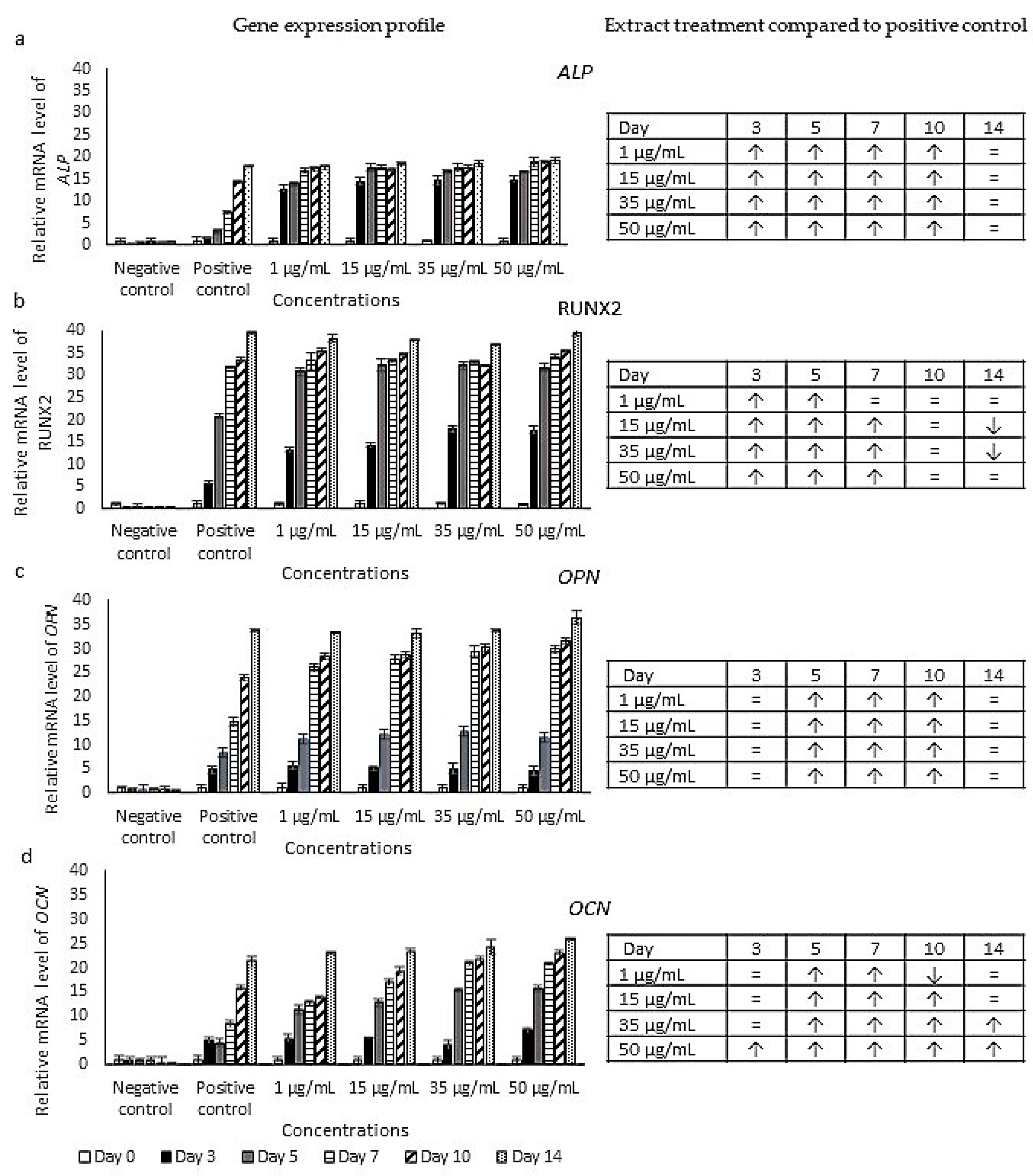
2.3. Expression Profiles of Osteoblast Markers

A molecular analysis of the osteoblast marker, i.e., ALP, RUNX2, OPN and OCN, expressions was carried out using the RT-qPCR technique (Figure 3). The expression of the ALP gene in the extract-treated cells shows an increase in a time-dependent manner. A significant increase was observed on days 3, 5, 7 and 10 for all treatment concentrations when compared to the positive control. The gene was expressed 12 to 14 times higher on day 3 of the treatment (1–50 µg/mL) while only 1.8 times higher in the positive control culture (Figure 3a). The expression of the RUNX2 gene in the extract-treated cells was significantly upregulated on days 3, 5 and 7 compared to the positive control. On the other hand, significant decreases were exhibited after treatment with 15 and 35 µg/mL on day 14 of the differentiation assay (Figure 3b). The OPN and OCN genes were also upregulated throughout the 14-day treatment, with the highest expression on day 14 compared to the negative control. The highest expressions of the OPN gene exhibited a 33–37-fold increase, while OCN gene expressions exhibited a 23–25-fold increase. Figure 3c shows a significant increase (p < 0.05) in OPN gene expression in treatments at all concentrations on days 5, 7 and 10 when compared to the positive control. Meanwhile, cells treated with the highest concentration (50 µg/mL) exhibited a significant increase in OCN gene expression (p < 0.05) in all periods of treatment (days 3, 5, 7, 10 and 14) as compared to the positive control (Figure 3d). Treatment with 1–35 µg/mL exhibited similar expressions during early treatment (day 3) and increased significantly from day 5 to day 10. In addition, an increase was still noted on day 14 after treatment with 35 µg/mL, while similar expressions were exhibited on day 14 after treatments with 1 and 15 µg/mL when compared to the positive control. A significant decrease was only shown after 1 µg/mL treatment on day 10 of the differentiation assay. These results show that the expressions of ALP and RUNX2 increased significantly in early treatment, starting on day 3, and became similar or decreased significantly on day 14. Meanwhile, significantly higher expressions of OPN and OCN were recorded in the later stages of treatment, starting on day 5, with the exception of treatment with 50 µg/mL of P. sarmentosum extract, which was able to significantly increase the expression of OCN from day 3.

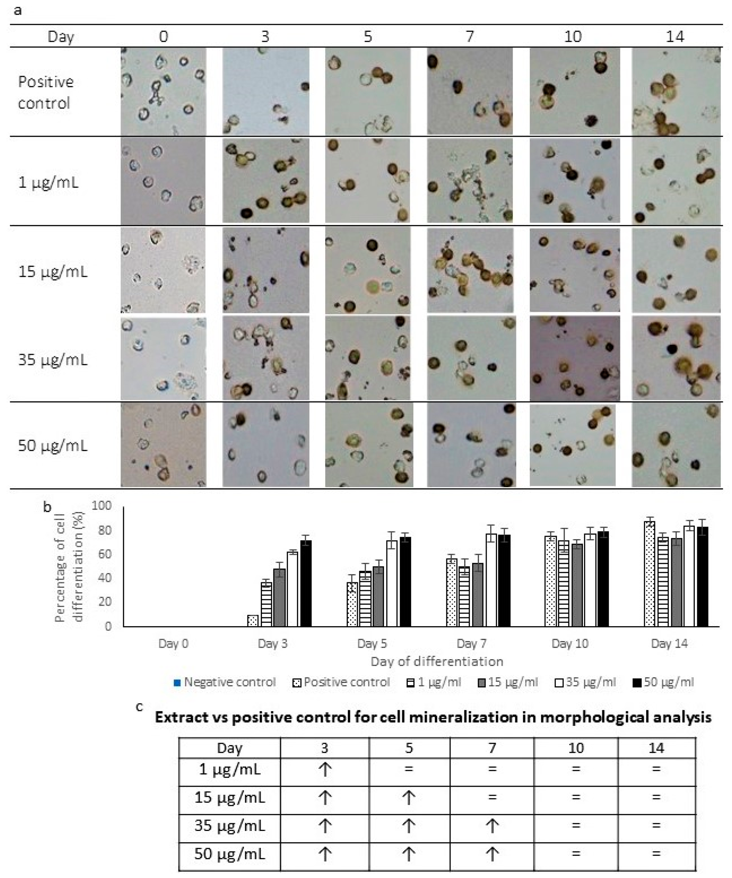
2.4. Morphology of Mineralized Cells

The mineralization rate of osteoblasts was observed using von Kossa staining. Figure 4a shows the morphology of mineralized cells after being treated with P. sarmentosum ethanolic extracts for 14 days. On day 0 of treatment, all von Kossa stained cells appeared unstained and translucent when observed under a microscope. A positive mineralization morphology was shown after day 3 of treatment onwards. Cells that proliferated were stained and appeared darker (brown or black), as observed in Figure 4a. The differentiated cells could be observed to increase proportionally to the period of treatment at all four concentrations.
The numbers of both differentiated and undifferentiated cells were counted to determine the percentage of differentiation that occurred (Figure 4b). On day 0, there was no cell differentiation in all treatments. Then, a significant increase (p < 0.05) in the cell differentiation percentage was observed in 1 µg/mL (day 3), 15 µg/mL (days 3 and 5), 35 µg/mL and 50 µg/mL (days 3, 5 and 7) treatments when compared to the positive control (Figure 4c). The highest percentage of differentiation was recorded on day 14 of the positive control culture (87%), followed by the treatment using 35 µg/mL (84%), 50 µg/mL (83%) and 1 µg/mL (81%). On the other hand, there were no stained cells present in the negative control culture. This shows that the extract induced higher cell mineralization at the early phase (day 3) and still induced mineralization until day 7 at higher extract concentrations compared to the positive control. The mineralization exhibited by the extract-treated cells increased proportionally to the concentrations of the extract.

2.5. Compound Content in P. sarmentosum Ethanolic Extracts

GC-MS analysis is a technique to identify volatile compounds present in a plant extract. Hence, this analysis was carried out to reveal the compounds in P. sarmentosum ethanolic leaf extracts. The GC-MS analysis revealed 54 volatile compounds in the ethanolic extracts of P. sarmentosum leaves. 1-dodecane took the shortest time to be eluted (9.972 min), while 3,4-dimethoxycinnamic acid was the last compound to be analyzed at 33.736 min. The most prevalent compounds were 2,4-di-tert-butylphenol (32.24%), phenol, 2,5-bis (1, 1-dimethylethyl) (32.24%), γ asarone (10.97%), benzene, 1,2,3-trimethoxy-5-(2-propenyl) (10.97%) and asarone (10.31%).
Out of these 54 volatiles compounds, 21, or approximately 39%, have been reported to exhibit biological activities, including antimicrobial, antioxidant, anticancer and antifungal properties and cell differentiation induction (Table 1). In addition, 10 out of 21 comp ounds have been reported to possess more than one biological activity. The most reported biological activities associated with the compounds screened are antimicrobial and antioxidant properties (Figure 5). Furthermore, the GC-MS analysis identified the presence of three volatile compounds that have been reported to be involved in cell differentiation induction activity, which are phytol, β-asarone and carvacrol. Another important biological activity that is potentially involved in bone health is antioxidant activity, which also has been reported with the second highest frequency (Figure 5).

3. Discussion

3.1. Stem Cell Characterization and Osteoblast Differentiation Induction

The ability to renew itself for an indefinite period is a characteristic of a stem cell. The proliferation ability of the isolated cells exhibited that human PBSCs were present in the culture before the differentiation process. Other than proliferation activity, a molecular approach was also taken by analyzing the expression of two hematopoietic stem cell markers, i.e., SLAMF1 and CD34. SLAMF1 is categorized under the SLAM subfamily, which are involved in the regulation of the proliferation and activation of lymphocytes. SLAMF1 is also important for maintaining the undifferentiated state of hematopoietic stem cells by stimulating the proliferation process [42,43], explaining the upregulation during the proliferation assay (Figure 1a). CD34 is well known to be expressed in human hematopoietic stem cells. The expression of this gene is involved in cell cycle entry, mobilization and metabolic activation [44,45].
Cell differentiation consists of four stages: proliferation, extracellular matrix (ECM) production, ECM maturation and apoptosis [46]. The reduction in the cell proliferation percentage after treatment with the extract, Figure 2a, indicates that the cells have entered the differentiation phase. This occurs because of the inability of the cells to carry out both proliferation and differentiation at one time [17,47]. ALP, which is involved in the release of phosphate for mineralization, has been used as a marker to indicate osteoblast differentiation. The upregulation of ALP activity is one of the main events that occur during the early phase of osteogenesis, thus it is commonly used by researchers to determine osteoblast formation [17,48]. Hydrolyzation by ALP provides the chemical conditions that eventually lead to mineral deposition, which can be visualized using von Kossa staining [49]. The increase in the ALP profile from day 3 to day 14 observed in the present study signifies the regulatory potential of P. sarmentosum extracts on ALP-specific activity that leads to the osteoblast differentiation process.
In this study, the increases in ALP-specific activity and differentiated cell percentage were also supported by the expression of osteoblasts-specific markers using RT-qPCR, i.e., ALP, RUNX2, OCN and OPN. These markers have commonly been used in other osteoblast differentiation studies to represent the molecular observation of the mineralization process [50]. ALP is a known marker for osteogenic differentiation and is secreted to promote the mineralization of the extracellular matrix. The upregulation in ALP expression and the increase in ALP activity indicate the potential of P. sarmentosum ethanolic extract to induce calcification during osteoblast development. Then, the expression of RUNX2 results in higher differentiation and mineralization of osteoblasts because of its role in the early stage of the differentiation process [50]. RUNX2 is also involved in inducing the expression of OCN, which is a mature osteoblast marker that is expressed during the late phase of osteoblast formation [51]. The significantly higher expression of these early genes, ALP and RUNX2, on days 3, 5, 7 and 10 shows that the extract is highly capable of initiating the differentiation process. OCN is the most synthesized protein by osteoblasts, making it one of the most important markers during bone mineralization [52]. Another osteoblast gene marker observed was OPN, which is a non-collagenous bone matrix protein that is expressed during the late phase of the mineralization process. During in vitro differentiation, the secretion of OCN and OPN will lead to the final osteoblast phenotypic marker, which is the mineralization of bone nodules as visualized during von Kossa staining [53]. Meanwhile, OPN is involved in the bone formation process via osteoblasts which enable the attachment of osteoclasts [54]. The expressions of OPN and OCN were significantly higher than the positive control starting from day 5, indicating the maturation phase experienced by the extract-treated cells. However, a high concentration of P. sarmentosum extract (50 µg/mL) was observed to increase the expression of OCN throughout the experiment, while the concentration of the extract did not affect the expression of OPN. Our study shows that P. sarmentosum ethanolic extract promotes the expression of these osteoblast markers, showing the ability of the extract to induce the formation of mineralized nodules in the treated cells similar to the positive control (50 µg/mL ascorbic acid + 10 mM β-glycerophosphate).
von Kossa staining is a common method to visualize the free inorganic phosphate present, wherein the silver ions in the stain react with phosphates and carbonates in calcium deposits, producing black precipitates [55], as observed in Figure 4a. This staining method has been carried out to observe the osteoblast differentiation of various cells such as dental pulp stem cells, human exfoliated deciduous teeth and MC3T3-E1 cell lines [20,48,56]. Our study also reported similar results to these studies by exhibiting an increased number of brownish to black cells as the mineralization process occurs continuously following the differentiation induction by P. sarmentosum extract. The effects of P. sarmentosum ethanolic extract on ALP-specific activity and the visualized mineralization process were comparable to the use of ascorbic acid and β-glycerophosphate, suggesting the ethanolic extract of P. sarmentosum could serve as a good inducer of osteoblasts differentiation.

3.2. Compound and Biological Activity of Piper sarmentosum Ethanolic Extract

The results of the GC-MS analysis unveiled the presence of 54 compounds in the ethanolic extracts of P. sarmentosum leaves. Among these 54 compounds, 21 of them have been reported to exhibit biological activities. The most prevailing compounds identified with the highest peak areas were 2,4-di-tert-butylphenol, phenol, 2,5-bis (1, 1-dimethylethyl), γ asarone, benzene, 1,2,3-trimethoxy-5-(2-propenyl) and asarone. Most of these compounds have been reported to exhibit biological activities. 2,4-di-tert-butylphenol is a lipophilic phenol that can be found in numerous organisms such as bacteria, fungi and plants [55,56,57]. This phenol exhibits multiple bioactivities such as antioxidant, anti-inflammatory, cytotoxicity and antifungal activities [24,57,58]. These compounds might be important in osteoblast differentiation induction shown in our study as phenolic compounds have exhibited a positive effect on bone homeostasis [59]. On the other hand, multiple studies also have reported a wide range of bioactivities exhibited by β-asarone and γ-asarone, such as antioxidant, anticancer, anti-ischemia and insecticidal properties [26,34,60,61]. Following these major compounds, 1-tetradecene and 1-pentadecene (peaks of 2.77% and 4.65%) have been reported to exhibit antimicrobial and antioxidant activities, respectively [23,62]. On top of that, three compounds have been reported to be involved in cell differentiation induction activity. The compounds mentioned are β-asarone, phytol and carvacrol, which were involved in the differentiation of neuron cells, osteoblast and endothelial cells, respectively [33,39,40]. Although there are only three compounds that are associated with cell differentiation activity, the presence of the other compounds in the extract is worth taking note of, as there are studies that have reported that the biological activities of plant-derived products are contributed to by the synergistic effect between compounds [63]. Hence, the list of compounds in this study can be used as a reference for further studies to identify the potential components that might also be present in different osteoinductive extracts. Our GC-MS analysis also showed that P. sarmentosum ethanolic extract contains several potential highly volatile compounds or specifically antioxidants that are beneficial for the bone remodeling process. The presence of antioxidant compounds is crucial in maintaining the bone remodeling process as the reactive oxygen species (ROS) can induce apoptosis of osteoblasts that will favor osteoclastogenesis. Imbalanced and increased osteoclastogenesis causes a high turnover of bone remodeling and bone loss [64]. Hence, antioxidants present in P. sarmentosum could prevent over-formation of osteoclasts and promote bone formation.

4. Material and Methods

4.1. Preparation of Plant Material

The leaves of P. sarmentosum were collected from the Forest Research Institute of Malaysia (FRIM), Kuala Lumpur, Malaysia (GPS coordinate: 3°14′7.80″ N 101°38′9.59″ E), and identified by a plant taxonomist with a voucher specimen FRI 45870. The collected P. sarmentosum leaves were cleaned and dried in an oven at 50 °C. The dried leaves were ground to a fine powder for extraction. Approximately 20 g of dry powdered leaves was extracted using 200 mL of ethanol in Soxhlet apparatus for six hours, followed by condensation of the extract in a rotary evaporator by evaporating the ethanol. Finally, the condensed extract was left in the fume hood for seven days to ensure a complete removal of the solvent. The fully dried extract was kept at −20 °C in a sticky form until further use. Before further analyses, the extract was diluted with 1% dimethyl sulfoxide (DMSO) to generate a range of stock concentrations, i.e., 10 µg/mL to 500 µg/mL.

4.2. Collection, Isolation and Culture of Human Peripheral Blood Stem Cells

The osteoinductive effect of P. sarmentosum was tested on human peripheral blood stem cells. Prior to sample collection, donor consent and ethics approval (reference number: UKM PPI/111/8/JEP-2019-612) were obtained from the Research Ethics Committee Universiti Kebangsaan Malaysia (RECUKM). Three peripheral blood samples were collected from healthy adults aged between 18 and 25 years old. Then, the collected blood was diluted with Hanks Balanced salt solution in a ratio of 1:3 before being layered onto Ficoll-PaqueTM PLUS (1:1.5) for density gradient centrifugation. The blood was then centrifuged for 20 min at 400× g at 27 °C. The second layer containing mononucleated cells was removed and washed three times using phosphate-buffered saline (PBS). The pelleted cells were resuspended and cultured in a 24-well plate in a complete medium consisting of α-medium essential (AMEM), 2% (v/v) penicillin-streptomycin and 10% (v/v) new-born calf serum (NBCS). These cells were cultured in an incubator at a temperature of 37 °C in the presence of 5% CO2 for 7 days before further experiments [16,17,18].

4.3. Proliferation Ability of the Isolated Cells

Cells were seeded at a density of 1 × 10⁵ cells/mL and cultured in the proliferation medium for 14 days. The viability and proliferation capacity of the isolated cells were observed every day for 14 days using trypan blue exclusion dye. The unstained and stained cells were observed under a microscope and viable cells were determined using a hemacytometer [17].

4.4. Molecular Characteristics of Hematopoietic Stem Cells

Total RNA was extracted from the treated cells using Trisure reagent (Bioline, Meridian Bioscience, Memphis, TN, USA) according to the manufacturer’s instructions. One milligram of total RNA was used in reverse transcription to produce complementary DNA using a Sensifast cDNA Synthesis Kit (Bioline, Meridian Bioscience, Memphis, TN, USA). A real-time polymerase chain reaction was later performed using a Thunderbird SYBR qPCR Mix kit (Toyobo, Osaka, Japan) with the following cycling conditions: 95 °C for 30 s at the holding stage, then followed by a two–step amplification (40 cycles of denaturation at 95 °C for 5 s and extension at the respective temperatures as listed in Table 2 for 10 s). The primer sequences for hematopoietic stem cell markers (SLAMF1 and CD34) and extension temperatures are listed in Table 2.

4.5. Induction of Osteoblast Differentiation Using P. sarmentosum Ethanolic Extract

The cells were seeded at 1 × 10⁵ cells/mL for an osteoblast differentiation assay [18]. The prepared P. sarmentosum ethanolic extracts were added into the culture at final concentrations from 1 µg/mL to 50 µg/mL. Cells treated with a cocktail consisting of 50 µg/mL ascorbic acid and 10 mM/L β-glycerophosphate served as the positive control, while untreated cells in the complete medium were the negative control. The differentiation of the cells was observed on days 0, 3, 5, 7, 10 and 14.

4.6. Cell Viability of Differentiated Cells

The viability and proliferation ability of the treated cells were observed for 14 days using the trypan blue exclusion dye approach [17]. On days 0, 3, 5, 7, 10 and 14, the cells were harvested and stained with trypan blue in a ratio of 1:1 and the cells were counted using a hemacytometer under the microscope. The cell viability was normalized to the untreated cells on the respective days and presented as a percentage.

4.7. Alkaline Phosphatase (ALP) Assay

PBS was used to wash 1 × 10⁵ cells/mL and the cells were lysed using 0.1% Triton. The total protein content was determined using Bradford reagent for 5 min at room temperature and measured at 595 nm. For the ALP assay, the cells were incubated in 0.1 mol/L sodium bicarbonate–sodium carbonate buffer, 2 mmol/L magnesium sulfate and 6 mmol/L p-nitrophenyl inorganic phosphate for 30 min at 37 °C. The reaction was stopped by adding 1 mol/L sodium hydroxide and the absorbance was measured at 405 nm. The ALP activity was represented as specific activity, which was determined by the unit activity per total protein (mg). One unit of ALP activity represents the hydrolysis of 1 µM p-nitrophenol per min at 37 °C. The ALP-specific activity was presented as a percentage, which was normalized to the negative control [17].

4.8. Molecular Characteristics of Osteoblast Cells

Total RNA was extracted from the treated cells using Trisure reagent (Bioline) and used in reverse transcription to produce complementary DNA using a Sensifast cDNA Synthesis Kit. A real-time polymerase chain reaction using a Thunderbird SYBR qPCR Mix kit was later performed. The primer sequences and extension temperatures for osteoblast markers (ALP, RUNX2, OPN and OCN) are listed in Table 1.

4.9. von Kossa Staining

Approximately 1 × 10⁵ cells/mL were washed using PBS and fixed onto a glass slide using 10% (v/v) formalin in PBS for 30 min. Then, the slide was rinsed using deionized water and the cells were stained with a 5% (v/v) silver nitrate solution. After 30 min, the slide was rinsed and 5% (v/v) sodium carbonate in 25% (v/v) formalin was added for 5 min followed by 5% (v/v) sodium thiosulfate for 2 min. Finally, the slide was rinsed and left to air dry before being observed under the microscope. A mineralized cell was defined as a cell stained dark brown or black [17].

4.10. Gas Chromatography-Mass Spectrometry (GC-MS) and Compound Identification

For GC-MS analyses, the sample was placed in a 30 m × 0.25 mm ID × 0.25 μm capillary column. The injection port temperature was set at 290 °C in splitless mode and helium (99.999%) was used with a flow rate of 36.3 cm/s. The sample was diluted in DMSO and injected using an AOC-20i + s autoinjector. The temperature was programmed as follows: 5 min at 50 °C, heated at 2 °C/min to 300 °C and held for 10 min. The mass spectral scan range was set at 30–700 (m/z). The compounds present were compared to the database of The National Institute of Standards and Technology (NIST).

4.11. Statistical Analysis

Our data were analyzed using a two-way ANOVA followed by Dunnet’s post hoc test. A result was considered to be statistically significant at a p value of <0.05.

5. Conclusions

The potential for osteogenesis induction was determined by the increase in specific activity of the ALP enzyme, the expression profiles of osteoblast markers and the mineralization of differentiated cells. A concentration of 50 µg/mL of P. sarmentosum ethanolic extract is suggested to possess the highest osteoblast-differentiation-inducing properties. Following the differentiation assay, screening of volatile compounds using GC-MS also indicated that P. sarmentosum contains 54 compounds, with three (β-asarone, carvacrol and phytol) compounds already reported to have cell differentiation induction potentials, 19 with other biological activities and 33 that are still not well studied. This array of compounds may contribute individually or synergistically to the ability to induce the differentiation of osteoblasts. Hence, further studies are required to investigate the presence of the other compounds that are responsible for the osteoinductive capacity exhibited in this study. In conclusion, P. sarmentosum ethanolic extracts could potentially replace the available osteoblast differentiation factor for bone formation.

Author Contributions

Conceptualization, I.Z.Z.A. and S.H.Z.A.; methodology, I.Z.Z.A., A.N.J. and S.H.Z.A.; validation, I.Z.Z.A., A.N.J., Z.Z.A. and S.H.Z.A.; formal analysis, A.N.J.; resources, I.Z.Z.A., Z.Z.A., H.R.E.D. and M.D.Y.; writing—original draft preparation, A.N.J. writing—review and editing, I.Z.Z.A., A.N.J. and S.H.Z.A.; funding acquisition, I.Z.Z.A. and S.H.Z.A. All authors have read and agreed to the published version of the manuscript.

Funding

This research was funded by the Ministry of Higher Education, Malaysia (FRGS/1/2018/STG05/CUCMS/02/1), University of Cyberjaya (CRG/01/01/2018) and Universiti Kebangsaan Malaysia (GP-2020-K007744).

Institutional Review Board Statement

The study was conducted in accordance with the Declaration of Helsinki, and approved by Research Ethics Committee Universiti Kebangsaan Malaysia (UKM PPI/111/8/JEP-2019-612 on September 2019).

Informed Consent Statement

Informed consent was obtained from the subjects involved in this study.

Data Availability Statement

Data is contained within the article.

Conflicts of Interest

The authors declare no conflict of interest.

References

  1. Gibbons, S. An overview of plant extracts as potential therapeutics. Expert. Opin. Ther. Pat. 2003, 13, 489–497. [Google Scholar] [CrossRef]
  2. Yuan, H.; Ma, Q.; Ye, L.; Piao, G. The Traditional Medicine and Modern Medicine from Natural Products. Molecules 2016, 21, 559. [Google Scholar] [CrossRef] [PubMed]
  3. Hussain, K.; Hashmi, F.K.; Latif, A.; Ismail, Z.; Sadikun, A. A review of the literature and latest advances in research of Piper sarmentosum. Pharm. Biol. 2012, 50, 1045–1052. [Google Scholar] [CrossRef] [PubMed]
  4. Subramaniam, V.; Ilham Adenan, M.; Rashih Ahmad, A.; Sahdan, R. Natural antioxidants: Piper sarmentosum (Kadok) and Morinda elliptica (Mengkudu). Mal. J. Nutr. 2003, 9, 41–51. [Google Scholar]
  5. Rukachaisirikul, T.; Siriwattanakit, P.; Sukcharoenphol, K.; Wongvein, C.; Ruttanaweang, P.; Wongwattanavuch, P.; Suksamrarn, A. Chemical constituents and bioactivity of Piper sarmentosum. J. Ethnopharmacol. 2004, 93, 173–176. [Google Scholar] [CrossRef]
  6. Nguyen, N.H.; Nhi, T.T.Y.; Van Nhi, N.T.; Cuc, T.T.T.; Tuan, P.M.; Nguyen, D.H. Comparative Study of the Silver Nanoparticle Synthesis Ability and Antibacterial Activity of the Piper Betle L. And Piper Sarmentosum Roxb. Extracts. J. Nanomater. 2021, 2021, 5518389. [Google Scholar] [CrossRef]
  7. Zainol Abidin, I.Z.; Fazry, S.; Jamar, N.H.; Ediwar Dyari, H.R.; Zainal Ariffin, Z.; Johari, A.N.; Ashaari, N.S.; Johari, N.A.; Megat Abdul Wahab, R.; Zainal Ariffin, S.H. The effects of Piper sarmentosum aqueous extracts on zebrafish (Danio rerio) embryos and caudal fin tissue regeneration. Sci. Rep. 2020, 10, 14165. [Google Scholar] [CrossRef]
  8. Zin, N.N.I.N.M.; Khatap Munawwarah Omar Sul’ain, M.D.; Abu Bakar, N. Evaluation of antimalarial and toxicological activities of methanol and water leaves extracts of Piper sarmentosum. Asian J. Med. Biomed. 2019, 3, 19–24. [Google Scholar]
  9. Mohamad Asri, S.F.; Soelaiman, I.N.; Mohd Moklas, M.A.; Mohd Nor, N.H.; Mohd Ramli, E.S. The Role of Piper sarmentosum Aqueous Extract as a Bone Protective Agent, a Histomorphometric Study. Int. J. Mol. Sci. 2020, 21, 7715. [Google Scholar] [CrossRef]
  10. Estai, M.A.; Suhaimi, F.H.; Das, S.; Fadzilah, F.M.; Alhabshi, S.M.I.; Shuid, A.N.; Soelaiman, I.N. Piper sarmentosum enhances fracture healing in ovariectomized osteoporotic rats: A radiological study. Clinics 2011, 66, 865–872. [Google Scholar] [CrossRef]
  11. Feng, X.; Mcdonald, J.M. Disorders of Bone Remodeling. Annu. Rev. Pathol. Mech. Dis. 2011, 6, 121–145. [Google Scholar] [CrossRef]
  12. Chapurlat, R.D. Osteoporosis. In Endocrinology: Adult and Pediatric, 2-Volume Set, 7th ed.; Elsevier Inc.: Amsterdam, The Netherlands, 2016; pp. 1184–1213.e6. [Google Scholar] [CrossRef]
  13. Rodan, G.A.; Martin, T.J. Therapeutic approaches to bone diseases. Science 2000, 289, 1508–1514. [Google Scholar] [CrossRef]
  14. Ilyas, Z.; Camacho, P.M. Rare adverse effects of bisphosphonate therapy. Curr. Opin. Endocrinol. Diabetes Obes. 2019, 26, 335–338. [Google Scholar] [CrossRef]
  15. Chong, P.; Selvaratnem, L.; Abbas, A.; Kamarul, T. Human peripheral blood derived mesenchymal stem cells demonstrate similar characteristics and chondrogenic differentiation potential to bone marrow derived mesenchymal stem cells. J. Orthop. Res. 2012, 30, 634–642. [Google Scholar] [CrossRef]
  16. Muhammad Dain, Y.; Shahrul Hisham, Z.A.; Senafi, S.; Zaidah, Z.A.; Rohaya, M.A.W. Stem Cell Heterogeneity of Mononucleated Cells from Murine Peripheral Blood: Molecular Analysis. Sci. World J. 2011, 11, 2150–2159. [Google Scholar]
  17. Hadzir, S.N.; Ibrahim, S.N.; Abdul Wahab, R.M.; Zainol Abidin, I.Z.; Senafi, S.; Ariffin, Z.Z.; Razak, M.A.; Ariffin, S.H.Z. Ascorbic acid induces osteoblast differentiation of human suspension mononuclear cells. Cytotherapy 2014, 16, 674–682. [Google Scholar] [CrossRef]
  18. Ruzanna, A.K.; Ariffin, S.H.Z.; Wahab, R.M.A.; Senafi, S.; Huyop, F.Z. Differentiation potential of human suspension mononucleated peripheral blood cells. African J. Biotechnol. 2011, 10, 10756–10764. [Google Scholar] [CrossRef]
  19. Ruzanna, A.K.; Shahrul Hisham, Z.A.; Rohaya, M.A.W.; Kermani, S.; Sahidan, S. Characterization of mononucleated human peripheral blood cells. Sci. World J. 2012, 2012, 843843. [Google Scholar]
  20. Intan Zarina, Z.A.; Manogaran, T.; Rohaya, M.A.W.; Farinawati, Y.; Shahrul Hisham, Z.A. A Comparative Analysis of Ascorbic Acid-induced Cytotoxicity and Differentiation betwen SHED and DPSC. Curr. Stem Cell Res. Ther. 2022, 17, 576–588. [Google Scholar]
  21. Saravanakumar, K.; Ramkumar, B.; Muthuraj, V. In Vitro Antimicrobial Potential Efficiency of Clathria Frondifera Marine Sponge. Int. J. Res. Pharm. Chem. 2016, 6, 458–464. [Google Scholar]
  22. Togashi, N.; Shiraishi, A.; Nishizaka, M.; Matsuoka, K.; Endo, K.; Hamashima, H.; Inoue, Y. Antibacterial activity of long-chain fatty alcohols against Staphylococcus aureus. Molecules 2007, 12, 139–148. [Google Scholar] [CrossRef] [PubMed]
  23. Attia, H.; Harrathi, J.; Alamer, K.H.; Alsalmi, F.A.; Magné, C.; Khalil, M. Effects of nacl on antioxidant, antifungal, and antibacterial activities in safflower essential oils. Plants 2021, 10, 2809. [Google Scholar] [CrossRef] [PubMed]
  24. Choi, S.J.; Kim, J.K.; Kim, H.K.; Harris, K.; Kim, C.J.; Park, G.G.; Park, C.S.; Shin, D.H. 2,4-Di-tert-butylphenol from sweet potato protects against oxidative stress in PC12 cells and in mice. J. Med. Food. 2013, 16, 977–983. [Google Scholar] [CrossRef] [PubMed]
  25. Melappa, G.; Prakash, B. In vitro antimitotic, antiproliferative and GC-MS studies on the methanolic extract of endophytic fungi, Penicillium species of Tabebuia Argentea bur & K. Sch. Farm. 2017, 5, 301–309. [Google Scholar]
  26. Hematpoor, A.; Paydar, M.; Liew, S.Y.; Sivasothy, Y.; Mohebali, N.; Looi, C.Y.; Wong, W.F.; Azirun, M.S.; Awang, K. Phenylpropanoids isolated from Piper sarmentosum Roxb. induce apoptosis in breast cancer cells through reactive oxygen species and mitochondrial-dependent pathways. Chem. Biol. Interact. 2018, 279, 210–218. [Google Scholar] [CrossRef]
  27. Nikhila, G.; Sangeetha, G.; Preetha, T.; Swapna, T. GC-MS analysis of phytochemical compounds present in the rhizome of Gloriosa superba L. J. Pharmacogn. Phytochem. 2016, 5, 17–20. [Google Scholar]
  28. Nasir, N.A.H.A.; Roslly, N.A.L.; Rosli, N.M.; Razali, Z. Phytochemical Screening and Potential DPPH Radical Scavenging Activity of Banana Peel Extract. In Charting the Sustainable Future of ASEAN in Science and Technology 2; Springer: Singapore, 2020; pp. 249–258. [Google Scholar]
  29. Mou, Y.; Meng, J.; Fu, X.; Wang, X.; Tian, J.; Wang, M.; Peng, Y.; Zhou, L. Antimicrobial and Antioxidant Activities and Effect of 1-Hexadecene Addition on Palmarumycin C2 and C3 Yields in Liquid Culture of Endophytic Fungus Berkleasmium sp. Dzf12. Molecules 2013, 18, 15587–15599. [Google Scholar] [CrossRef]
  30. Turkez, H.; Celik, K.; Toghar, B. Effects of copaene, a tricyclic sesquiterpene, on human lymphocytes cells in vitro. Cytotechnology 2014, 66, 597–603. [Google Scholar] [CrossRef]
  31. Ferreira, R.; Monteiro, M.; Silva, J.; Maia, J. Antifungal Action of the Dillapiole-rich Oil of Piper aduncum against Dermatomycoses Caused by Filamentous Fungi. Br. J. Med. Med. Res. 2016, 15, 1–10. [Google Scholar] [CrossRef]
  32. Kumar, H.; Kim, B.W.; Song, S.Y.; Kim, J.S.; Kim, I.S.; Kwon, Y.S.; Koppula, S.; Choi, D.K. Cognitive enhancing effects of alpha asarone in amnesic mice by influencing cholinergic and antioxidant defense mechanisms. Biosci. Biotechnol. Biochem. 2012, 76, 1518–1522. [Google Scholar] [CrossRef]
  33. Lam, K.Y.; Chen, J.; Lam, C.T.; Wu, Q.; Yao, P.; Dong, T.T.; Lin, H.; Tsim, K.W. Asarone from Acori Tatarinowii Rhizoma Potentiates the Nerve Growth Factor-Induced Neuronal Differentiation in Cultured PC12 Cells: A Signaling Mediated by Protein Kinase A. PLoS ONE 2016, 11, e0163337. [Google Scholar] [CrossRef]
  34. Yang, Y.X.; Chen, Y.T.; Zhou, X.J.; Hong, C.L.; Li, C.Y.; Guo, J.Y. Beta-asarone, a major component of Acorus tatarinowii Schott, attenuates focal cerebral ischemia induced by middle cerebral artery occlusion in rats. BMC Complement. Altern. Med. 2013, 13, 236. [Google Scholar] [CrossRef]
  35. Faridha Begum, I.; Mohankumar, R.; Jeevan, M.; Ramani, K. GC–MS Analysis of Bio-active Molecules Derived from Paracoccus pantotrophus FMR19 and the Antimicrobial Activity Against Bacterial Pathogens and MDROs. Indian J. Microbiol. 2016, 56, 426–432. [Google Scholar] [CrossRef]
  36. Lee, Y.S.; Kang, M.H.; Cho, S.Y.; Jeong, C.S. Effects of Constituents of Amomum xanthioides on Gastritis in Rats and on Growth of Gastric Cancer Cells. Arch. Pharm. Res. 2007, 30, 436–443. [Google Scholar] [CrossRef]
  37. Naragani, K.; Mangamuri, V.; Muvva, S.; Munaganti, R.K. Antimicrobial potential of Streptomyces cheonanensis VUK-A from mangrove origin. Int. J. Pharm. Sci. 2016, 8, 53–57. [Google Scholar]
  38. Ceyhan-Guvensen, N.; Keskin, D. Chemical content and antimicrobial properties of three different extracts of Mentha pulegium leaves from Mugla Region, Turkey. J. Environ. Biol. 2016, 37, 1341–1346. [Google Scholar]
  39. Sanjeev, G.; Sidharthan, D.S.; Pranavkrishna, S.; Pranavadithya, S.; Abhinandan, R.; Akshaya, R.L.; Balagangadharan, K.; Siddabathuni, N.; Srinivasan, S.; Selvamurugan, N. An osteoinductive effect of phytol on mouse mesenchymal stem cells (C3H10T1/2) towards osteoblasts. Bioorg. Med. Chem. Lett. 2020, 30, 127137. [Google Scholar] [CrossRef]
  40. Matluobi, D.; Araghi, A.; Maragheh, A.; Rezabakhsh, A.; Soltani, S.; Khaksar, M.; Siavashi, V.; Feyzi, A.; Bagheri, H.S.; Rahbarghazi, R.; et al. Carvacrol promotes angiogenic paracrine potential and endothelial differentiation of human mesenchymal stem cells at low concentrations. Microvasc. Res. 2018, 115, 20–27. [Google Scholar] [CrossRef]
  41. Xuan, H.; Wang, Y.; Li, A.; Fu, C.; Wang, Y.; Peng, W. Bioactive Components of Chinese Propolis Water Extract on Antitumor Activity and Quality Control. Evidence-Based Complement. Altern. Med. 2016, 2016, 9641965. [Google Scholar] [CrossRef]
  42. Kiel, M.J.; Yilmaz, Ö.H.; Iwashita, T.; Yilmaz, O.H.; Terhorst, C.; Morrison, S.J. SLAM family receptors distinguish hematopoietic stem and progenitor cells and reveal endothelial niches for stem cells. Cell 2005, 121, 1109–1121. [Google Scholar] [CrossRef]
  43. Intan Zarina, Z.A.; Anis Nabilah, J.; Zaidah, A.Z.; Shahrul Hisham, Z.A. Cytotoxicity Of Hexane And Ethanol Piper Sarmentosum Extracts On Human Hematopoietic. Eur. Chem. Bull. 2022, 11, 7–11. [Google Scholar]
  44. Manfredini, R.; Zini, R.; Salati, S.; Siena, M.; Tenedini, E.; Tagliafico, E.; Montanari, M.; Zanocco-Marani, T.; Gemelli, C.; Vignudelli, T.; et al. The kinetic status of hematopoietic stem cell subpopulations underlies a differential expression of genes involved in self-renewal, commitment, and engraftment. Stem Cells 2005, 23, 496–506. [Google Scholar] [CrossRef] [PubMed]
  45. Han, Y.; Gong, Z.; Takakura, N. Murine hematopoietic stem cell dormancy controlled by induction of a novel short form of PSF1 by histone deacetylase inhibitors. Exp. Cell Res. 2015, 334, 183–193. [Google Scholar] [CrossRef] [PubMed]
  46. Stein, G.S.; Lian, J.B.; Wijnen AJVan Stein, J.L.; Montecino, M.; Javed, A.; Zaidi, S.K.; Young, D.W.; Choi, J.Y.; Pockwinse, S.M. Runx2 control of organization, assembly and activity of the regulatory machinery for skeletal gene expression. Oncogene 2004, 23, 4315–4329. [Google Scholar] [CrossRef] [PubMed]
  47. Quarles, L.D.; Yohay, D.A.; Lever, L.W.; Caton, R.; Wenstrup, R.J. Distinct proliferative and differentiated stages of murine MC3T3-E1 cells in culture: An in vitro model of osteoblast development. J. Bone Miner. Res. 1992, 7, 683–692. [Google Scholar] [CrossRef]
  48. Intan Zarina, Z.A.; Shaipul Islam Shazwana Ling, J.P.; Zainal Ariffin, Z.; Shahrul Hisham, Z.A. The Effects Of Piper Sarmentosum Aqueous Extract On MC3T3-E1 Differentiation.pdf. NVEO-Natural Volatiles Essent Oils J. 2021, 8, 4063–4079. [Google Scholar]
  49. Coelho, M.J.; Fernandes, M.H. Human bone cell cultures in biocompatibility testing. Part II: Effect of ascorbic acid, b-glycerophosphate and dexamethasone on osteoblastic differentiation. Biomaterials 2000, 21, 1095–1102. [Google Scholar] [CrossRef]
  50. Thu, H.E.; Mohamed, I.N.; Husain, Z.; Shuid, A.N. Exploring molecular mechanism of bone-forming capacity of Eurycoma longifolia: Evidence of enhanced expression of bone-related biomarkers. J. Ayurveda Integr. Med. 2018, 9, 272–280. [Google Scholar] [CrossRef]
  51. Lehti, M.S.; Henriksso, H.; Rummukainen, P.; Wang, F.; Uusitalo-Kylmala, L.; Kiviranta, R.; Heino, T.J.; Kotaja, N.; Sironen, A. Cilia-related protein SPEF2 regulates osteoblast differentiation. Sci. Rep. 2018, 8, 859. [Google Scholar] [CrossRef]
  52. Liu, J.; Si, S.; Qin, Y.; Zhang, B.; Song, S.; Guo, Y. The effect of different molecular weight collagen peptides on MC3T3-E1 cells differentiation. Biomed. Mater. Eng. 2015, 26, 2041–2047. [Google Scholar] [CrossRef]
  53. Lo, Y.-C.; Chang, Y.-H.; Wei, B.-L.; Huang, Y.-L.; Chiou, W.-F. Betulinic Acid Stimulates the Differentiation and Mineralization of Osteoblastic MC3T3-E1 Cells: Involvement of BMP / Runx2 and β -Catenin Signals. J. Agric. Food Chem. 2010, 58, 6643–6649. [Google Scholar] [CrossRef]
  54. Huang, W.; Carlsen, B.; Rudkin, G.; Berry, M.; Ishida, K.; Yamaguchi, D.T.; Miller, T.A. Osteopontin is a negative regulator of proliferation and differentiation in MC3T3-E1 pre-osteoblastic cells. Bone 2004, 34, 799–808. [Google Scholar] [CrossRef]
  55. Boyan, B. The importance of mineral in bone and mineral research. Bone 2000, 27, 341–342. [Google Scholar] [CrossRef]
  56. Gorman, D.M.O.; Tierney, C.M.; Brennan, O.; Brien, F.J.O. The Marine-derived, Multi-mineral formula, Aquamin, Enhances Mineralisation of Osteoblast Cells In Vitro. Phyther. Res. 2012, 26, 375–380. [Google Scholar] [CrossRef]
  57. Varsha, K.K.; Devendra, L.; Shilpa, G.; Priya, S.; Pandey, A.; Nampoothiri, K.M. 2,4-Di-tert-butyl phenol as the antifungal, antioxidant bioactive purified from a newly isolated Lactococcus sp. Int. J. Food Microbiol. 2015, 211, 44–50. [Google Scholar] [CrossRef]
  58. Nair, R.V.R.; Jayasree, D.V.; Biju, P.G.; Baby, S. Anti-inflammatory and anticancer activities of erythrodiol-3-acetate and 2,4-di-tert-butylphenol isolated from Humboldtia unijuga. Nat. Prod. Res. 2020, 34, 2319–2322. [Google Scholar] [CrossRef]
  59. Surget, G.; Roberto, V.P.; Le Lann, K.; Mira, S.; Guérard, F.; Laizé, V.; Poupart, N.; Cancela, M.L.; Stiger-Pouvreau, V. Marine green macroalgae: A source of natural compounds with mineralogenic and antioxidant activities. J. Appl. Phycol. 2017, 29, 575–584. [Google Scholar] [CrossRef]
  60. Liu, L.; Wang, J.; Shi, L.; Zhang, W.; Du, X.; Wang, Z.; Zhang, Y. β-Asarone induces senescence in colorectal cancer cells by inducing lamin B1 expression. Phytomedicine 2013, 20, 512–520. [Google Scholar] [CrossRef]
  61. Hematpoor, A.; Liew, S.Y.; Azirun, M.S.; Awang, K. Insecticidal activity and the mechanism of action of three phenylpropanoids isolated from the roots of Piper sarmentosum Roxb. Sci. Rep. 2017, 7, 12576. [Google Scholar] [CrossRef]
  62. Seow, L.J.; Beh, H.K.; Ibrahim, P.; Sadikun, A.; Asmawi, M.Z. Antimicrobial activity of Gynura segetum’ s leaf extracts and its active fractions. Cellmed 2012, 2, 20–21. [Google Scholar]
  63. Park, E.; Kim, J.; Yeo, S.; Lim, E.; Choi, C.W.; Choi, S.; Li, W.Y.; Lee, J.W.; Park, J.H.; Huh, D.; et al. Anti-osteoporotic effects of combined extract of Lycii Radicis Cortex and Achyranthes japonica in osteoblast and osteoclast cells and ovariectomized mice. Nutrients 2019, 11, 2716. [Google Scholar] [CrossRef] [PubMed]
  64. Domazetovic, V.; Marcucci, G.; Iantomasi, T.; Brandi, M.L.; Vincenzini, M.T. Oxidative stress in bone remodeling: Role of antioxidants. Clin. Cases Miner. Bone Metab. 2017, 14, 209–216. [Google Scholar] [CrossRef] [PubMed]
Figure 1. Hematopoietic stem cell proliferation potential and molecular stemness marker. (a) The proliferation of isolated mononucleated cells was measured every day for 14 days. (b) The expressions of SLAMF1 and CD34 were analyzed using RT-qPCR on days 0 and 14. The expressions of both genes were normalized to the housekeeping gene (GAPDH). The means ± standard error of the mean are given for three independent experiments (n = 3). * denotes significant differences (p < 0.05) compared to day 0.
Pharmaceuticals 16 00708 g001
Figure 2. Viability and ALP profiles during differentiation. (a) The percentage of cell viability of the treated cells on days 0, 3, 5, 7, 10 and 14 normalized to the untreated cells (untreated cells act as 100%). (b) The increase in ALP-specific activity after treatment with a range of concentrations of extract for 14 days. (c) A comparison of ALP-specific activity in extract-treated cells to the positive control. Means ± standard error of the mean are given for three separate experiments (n = 3). ↑: significantly higher expression (p < 0.05) and =: similar expression (p ≥ 0.05) compared to the positive control.
Pharmaceuticals 16 00708 g002
Figure 3. Osteoblast gene profiles during treatment, i.e., (a) ALP, (b) RUNX2, (c) OPN and (d) OCN. The expression of osteoblast gene markers corresponds to the comparisons of the expressions in the extract-treated cells to the positive control. Means ± standard error of the mean are given for three separate experiments (n = 3). ↑: significantly higher expression compared to the positive control (p < 0.05), ↓: Significantly lower expression compared to the positive control (p < 0.05), =: similar expression compared to the positive control (p ≥ 0.05).
Pharmaceuticals 16 00708 g003
Figure 4. Mineralized hematopoietic stem cells during treatment. (a) Representative images of mineralized cells following von Kossa staining (magnification ×400) (b) The percentage of mineralized cells on days 0, 3, 5, 7, 10 and 14. (c) A comparison of cell mineralization in extract-treated cells with the positive control. Means ± standard error of the mean are given for three separate experiments (n = 3). ↑: significantly higher expression compared to positive control (p < 0.05), =: similar expression compared to positive control (p ≥ 0.05).
Pharmaceuticals 16 00708 g004
Figure 5. Frequencies of biological activities of volatile compounds in P. sarmentosum.
Pharmaceuticals 16 00708 g005
Table 1. Screening of compounds using a GC-MS analysis.
NoRetention Time (RT)Peak Area (%)Name of the Volatile CompoundBiological Activity
19.9722.631-dodecaneAntimicrobial, antioxidant and antifungal [21]
210.1010.31-dodecanol Antibacterial [22]
313.8852.771-tetradeceneAntioxidants [23]
415.9132.242,4-di-tert-butylphenol Antioxidant, antimicrobial and anticancer [24]
515.9132.24Phenol, 2,5-bis (1,1-dimethylethyl) Anticancer [25]
616.88510.97γ-asarone Anticancer, antimicrobial [26]
716.88510.97Benzene, 1,2,3-trimethoxy-5-(2-propenyl) Antimicrobial [27]
817.0996.232-tetradecene, (E) Antioxidant [28]
917.0996.23CeteneAntioxidant [29]
1017.234.651-pentadeceneAntioxidant, antimicrobial [23]
1118.0260.55Copaene Antioxidant [30]
1218.1591.661,3-benzodioxole, 4,5-dimethoxy-6-(2-propenyl) Antifungal [31]
1318.5310.31AsaroneAntioxidant [32]
1418.715.16β-asaroneCell differentiation inducing [33], antioxidant, antimicrobial, anticancer and antifungal [34]
1519.9181.99E-15-heptadecenal Antioxidant, antimicrobial [35]
1619.9181.991-octadecene Anticancer, antimicrobial and antioxidant [36]
1719.9181.995-octadecene, (E)Antimicrobial [37]
1820.550.96Neophytadiene Antimicrobial [38]
1926.1460.16PhytolCell differentiation inducing, antimicrobial [39]
2030.9950.5CarvacrolCell differentiation inducing, antimicrobial [40]
2133.7361.313,4-dimethoxycinnamic acid Antitumor, antimicrobial [41]
Table 2. Primer sequences used for hematopoietic stem cell characterization and osteoblast detection.
GeneSequences (5′–3′)Extension Temperature (°C)
GAPDHForward- GACCACTTTGTCAAGCTCATTTC60
Reverse- CTCTCTTCCTCTTGTGCTCTTG
SLAMF1Forward- GGAAAGCAGGAAGGAGGA60
Reverse- GCAGCCCAGTATCAAGGT
CD34Forward- TAGCCAAGTCTGCCAACTATTC55
Reverse- CCAACATACCACCCTCCATTT
ALPForward-GGAGTATGAGAGTGACGAGAAAG54
Reverse- GAAGTGGGAGTGCTTGTATCT
RUNX2Forward- CGGAATGCCTCTGCTGTTAT55
Reverse- TGTGAAGACGGTTATGGTCAAG
OPNForward- GCTAAACCCTGACCCATCTC56
Reverse- ATAACTGTCCTTCCCACGGC
OCNForward- CCTGAAAGCCGATGTGGT57
Reverse- GGCAGCGAGGTAGTGAAGA
Disclaimer/Publisher’s Note: The statements, opinions and data contained in all publications are solely those of the individual author(s) and contributor(s) and not of MDPI and/or the editor(s). MDPI and/or the editor(s) disclaim responsibility for any injury to people or property resulting from any ideas, methods, instructions or products referred to in the content.

