
Metformin and Breast Cancer: Current Findings and Future Perspectives from Preclinical and Clinical Studies

1
Department of Nutrition, Texas A&M University, College Station, TX 77843, USA
2
School of Kinesiology and Rogel Cancer Center, University of Michigan, Ann Arbor, MI 48109, USA
*
Author to whom correspondence should be addressed.
Pharmaceuticals 2024, 17(3), 396; https://doi.org/10.3390/ph17030396
Submission received: 8 January 2024
/
Revised: 11 March 2024
/
Accepted: 12 March 2024
/
Published: 19 March 2024
(This article belongs to the Special Issue Metformin: Mechanism and Application 2023)
Abstract
:Over the last several decades, a growing body of research has investigated the potential to repurpose the anti-diabetic drug metformin for breast cancer prevention and/or treatment. Observational studies in the early 2000s demonstrated that patients with diabetes taking metformin had decreased cancer risk, providing the first evidence supporting the potential role of metformin as an anti-cancer agent. Despite substantial efforts, two decades later, the exact mechanisms and clinical efficacy of metformin for breast cancer remain ambiguous. Here, we have summarized key findings from studies examining the effect of metformin on breast cancer across the translational spectrum including in vitro, in vivo, and human studies. Importantly, we discuss critical factors that may help explain the significant heterogeneity in study outcomes, highlighting how metformin dose, underlying metabolic health, menopausal status, tumor subtype, membrane transporter expression, diet, and other factors may play a role in modulating metformin’s anti-cancer effects. We hope that these insights will help with interpreting data from completed studies, improve the design of future studies, and aid in the identification of patient subsets with breast cancer or at high risk for the disease who are most likely to benefit from metformin treatment.
1. Introduction
Metformin (1,1-dimethyl biguanide) has a long history of use, primarily for the treatment of type 2 diabetes. Over the last two decades, numerous studies have reported that metformin use is associated with a reduction in cancer incidence and mortality [1,2], leading to strong interest in repurposing this drug as an anticancer agent.
Metformin’s origins stem from medieval Europe, where the plant Galega officinalis, which contains a toxic glucose-lowering guanidine-like alkaloid called galegine, was used as a medicinal agent [3,4,5]. In 1922, Werner and Bell synthesized metformin (Figure 1), a structurally similar biguanide compound [6]. In the following years, the glucose-lowering potential of metformin and other related biguanides was demonstrated in animals. Still, it was not until 1957 that metformin (originally trademarked as Glucophage) was successfully used to treat diabetes in humans [4]. Currently, metformin is one of the most prescribed antidiabetic drugs, and it is on the World Health Organization’s list of essential medicines [4,7].
The potential anti-cancer effects of metformin were first identified in 2005, with a case–control study demonstrating reduced risk of cancer in patients with type 2 diabetes who were prescribed metformin compared to controls, with a potential dose–response [8]. A population-based cohort study the following year found reduced cancer-related mortality in patients with type 2 diabetes taking metformin compared to those on sulfonylureas [9]. Subsequently, other studies in the early 2000s found similar results [10,11,12,13]. Since those groundbreaking findings, the anticancer properties of metformin have been studied in various cancer types, including glioma, lymphoma, lung, colorectal, esophageal, kidney, liver, bladder, ovarian, pancreatic, prostate, uterine, and breast cancer, among others [14,15,16,17].
There is a large body of work investigating the potential role of metformin in the prevention and treatment of breast cancer. A PubMed search (29 December 2023) of metformin AND “breast cancer” shows 837 publications, with most papers published within the last decade. While cell culture and animal studies have shown promising results in repurposing metformin as a part of breast cancer treatment, not all clinical trials of metformin in breast cancer patients have shown the same benefits. Thus, after years of studying metformin in the context of cancer, many questions remain unanswered; the anticancer mechanisms of metformin are not fully understood, and its clinical efficacy is unclear. Here, we evaluate the current state of the knowledge of the anticancer potential of metformin in the context of breast cancer by summarizing mechanisms of action, key findings from basic and translational studies, and the findings from studies of metformin in patients with breast cancer. We also attempt to clarify the factors that may influence outcomes that could be used to identify patients most likely to benefit from this drug, and improve its clinical efficacy.
2. Preclinical Evidence: Findings from In Vitro and In Vivo Models
2.1. Mechanisms of Action
Metformin is an antidiabetic drug that regulates circulating glucose levels by decreasing gluconeogenesis in the liver and increasing insulin sensitivity [18,19]. Numerous mechanisms have been proposed to explain the anticancer activity of metformin. These mechanisms can broadly be categorized as either (1) direct effects on tumors or (2) indirect (systemic) effects, which include the same ability to improve whole-body insulin sensitivity and glucose control that underlie its benefits as an antidiabetic agent.
Direct, tumor-specific effects of metformin include the ability to inhibit cancer cell proliferation, induce apoptosis, and decrease the number of stem-like cells within tumors, which impairs the tumor self-renew [18,20]. Specifically, metformin can reduce cellular energy consumption by inhibiting respiratory Complex I of the electron transport chain in mitochondria [18,19] and it can inhibit protein synthesis and cell growth through activation of LKB1 and AMPK, resulting in the inhibition of the mammalian target of rapamycin (mTOR) [20]. Additional studies have also suggested that metformin may exert functions upstream of AMPK activation, specifically by interfering with hexokinase I and II (the enzyme that catalyzes the first step in glucose metabolism) [21]. In tumor cells, this metformin-induced effect on cell metabolism has been shown to result in apoptosis and subsequent cell death [22]. Metformin may also act independently of AMPK through the human epidermal growth factor receptor-2 (HER2) [23]. Metformin has shown the ability to block HER2 tyrosine kinase activity [24], and breast cancer cells with overexpression of HER2 have improved metformin-induced inhibition of cell growth [25]. Finally, studies also suggest that metformin can inhibit several STAT3-related signaling pathways known to be involved in breast cancer, including IL-6/JAK2/STAT3 signaling [26]. In addition, at least in triple-negative breast cancer cells in vitro, metformin can reduce the activation (phosphorylation) of STAT3 but does not alter STAT3 expression levels. Further, combining metformin with a STAT3 inhibitor had synergistic effects [27]. While the exact mechanisms underlying these effects are unclear, they appear independent of metformin’s actions on mTOR.
Indirect mechanisms are generally related to improvements in insulin sensitivity and the modulation of inflammation [18,20]. Metformin improves metabolic health and decreases circulating insulin and insulin-like growth factor 1 (IGF-1) [28,29], both of which are known to promote cancer risk and progression [30]. Therefore, whole-body improvements in insulin signaling by metformin may contribute to cancer prevention mechanisms and mitigate cancer progression. Figure 2 summarizes the main indirect and direct effects of metformin on breast cancer.
2.2. Metformin: Impact on Breast Cancer Outcomes in Preclinical Models
In preclinical models of breast cancer, metformin has shown varying effects; some studies demonstrate a beneficial impact on tumor outcomes, while others show no discernible benefits. Outcomes from key studies have been summarized in Table 1. In 2005, Anisimov and colleagues demonstrated that metformin treatment (1200 mg/L, equivalent to 240 mg/kg body weight) from 2 months of age until natural death in transgenic FVB/N female mice carrying the HER-2/neu mammary cancer gene improved lifespan by approximately 8% [31]. While metformin did not alter tumor incidence or multiplicity (100% of mice developed tumors), it significantly increased tumor latency and decreased tumor size [31]. Similarly, our group has shown that in both ovary-intact and ovariectomized (OVX’d) female Wistar rats on a high-fat diet with MNU-induced mammary tumors, 8 weeks of metformin (2 mg/mL) treatment significantly reduced tumor volumes [32,33]. We also found that in OVX’d rats, metformin reduced the number of aromatase-positive macrophages in the tumor border [33]. Aromatase is a key enzyme that converts androgens to estrogens [34]; therefore, this suggests that metformin decreases estrogen production in the mammary tumor microenvironment, which could contribute to the reduced growth of ER+ tumor cells. The effects of metformin do not appear to be limited to hormone-dependent tumors as metformin also decreased the growth of triple-negative tumors (injection of MDA-MB-231 into BALB/c-nu nude mice) [35]. Other studies investigating the effects of metformin in non-diabetic rats and mice show less efficacy in improving tumor outcomes [36,37,38].
2.3. Metformin: Anticancer Effects Are Influenced by Glycemic Status/Metabolic Status
Conflicting findings in the literature may be attributed to the influence of glucose levels and/or overall metabolic status on the effects of metformin on tumor development and/or progression. We have summarized results from in vitro studies in Table 2; many of these studies support that local glucose levels influence the anti-cancer effects of metformin. For instance, Wahdan-Alaswad and colleagues assessed the efficacy of metformin (5 mM and 10 mM) across glucose conditions (5 mM, 10 mM, and 17 mM) in breast cancer cell lines representing luminal A and B, HER2, and triple-negative subtypes [39]. In triple-negative cells, metformin (5 mM and 10 mM) decreased proliferation, regardless of glucose concentration. However, among all other cell lines investigated, the anti-proliferative effects of metformin were more pronounced at physiological glucose levels (5 mM) [39]. These results suggest high glucose levels may dampen the efficacy of metformin. Intriguingly, another study showed that in the triple-negative cell lines MDA-MB-231 and MDA-MD-468, low concentrations of metformin (50 µM–500 µM and 100 µM, respectively, for each cell line) combined with 25 mM glucose conditions led to increased cell proliferation compared to controls. When glucose remained high and the metformin concentration was increased, MDA-MB-468 cells exhibited greater sensitivity to decreased proliferation, in contrast to MDA-MB-231 where proliferation remained unchanged compared to controls [40]. However, at physiological glucose (5.5 mM), both cell lines showed decreased proliferation with 500 µM–10 mM metformin concentrations [40]. Furthermore, in both cell lines, the effect of 2 mM metformin on mTOR pathway proteins was examined in glucose-starved cells (0 mM), 5.5 mM, and 25 mM glucose-exposed cells. Notably, a reduction in mTOR pathway proteins was observed when comparing glucose-starved cells to the other glucose conditions [40]. This data suggests that glucose deprivation makes cells more sensitive to metformin inhibition of the mTOR pathway. Furthermore, in MDA-MB-231 cells, metformin treatment combined with glucose deprivation has been shown to activate genes related to the unfolded protein response of the endoplasmic reticulum, decrease proliferation, and increase cell apoptosis [41].
In vivo, diet manipulations to lower or normalize plasma glucose levels have similarly been shown to modulate the effects of metformin. For example, in rats, combining metformin with caloric restriction significantly reduced the development of carcinogen-induced mammary tumors compared to control rats, and tended to be more effective than calorie restriction alone [42]. Further, when metformin and calorie restriction were stopped, the improvements in cancer incidence and burden persisted during a two-week follow-up period [42]. Similarly, in the 4T1 breast cancer mouse model, metformin combined with a ketogenic diet more effectively reduced mammary tumor volume compared to metformin or a ketogenic diet alone, or untreated controls [43]. In the same model, combining metformin with short-term starvation also improved metformin response by tempering tumor growth compared to control and metformin-treated animals [44], further supporting the idea that combining metformin with energy or glucose restriction may increase its anti-cancer potential.
In summary, current literature suggests that metformin is more cytotoxic to cancer cells both in vitro and in vivo when combined with glucose deprivation. In cell culture models, high glucose concentrations blunt the antiproliferative effects of metformin compared to normal physiological glucose concentrations or a glucose-deprived state. The strength of this effect does, however, vary by cell line. In animal models, interventions that decreased glucose levels, such as caloric restriction, enhanced metformin’s anti-cancer effects. How these findings translate clinically is unclear as most patients taking metformin do so to improve impaired glucose levels.
2.4. Metformin Dose Modulates Cancer Prevention and Treatment Efficacy
Translating the dose of metformin that has shown benefits in preclinical studies to those achievable in humans is possibly one of the most important considerations for evaluating the potential use of metformin as an anti-cancer agent. Human plasma levels of metformin achieved therapeutically typically range from 0.465–2.5 mg/L (~3.6–19.4 µM) [45]. In contrast, most in vitro studies have used much higher doses and are, thus, unlikely to be clinically relevant. Zhu et al. evaluated the effects of 8 metformin doses (ranging from 0.02 to 20.0 mM) on several breast cancer cell lines [42]. While responses were cell line dependent, the concentrations required to inhibit cell growth (>1 mM) were much higher than levels achieved clinically (~20 µM) [42]. These cell culture experiments were followed up with in vivo work that investigated two dosing regimens. Rodents received metformin at a higher loading dose for 5 days (0.5% or 1.0% w/w) and then remained on a lower dose until the end of the study (0.05 or 0.25 w/w) [42]. Only the rodents on the higher dose combination 1.0/0.25% showed improvements in tumor outcomes [42]. Another preclinical study comparing low and high metformin doses (50 vs. 500 µg/mL in the water) found that neither dose impacted tumor growth; however, the higher metformin dose delayed tumor onset [38]. In non-diabetic rats, a clinically relevant dose of metformin did not improve tumor outcomes [36]; however, in this study, metformin was provided in the food or by oral gavage, and plasma levels measured over 24 h showed that, despite achieving high levels at 2 h, metformin levels dropped quickly and did not accumulate throughout the day, which could underlie this lack of tumor effect. Additional studies in non-diabetic, metabolically healthy rodents using clinically relevant doses much lower than what is commonly used in culture have also demonstrated little to no anti-cancer benefits [36,37,42]. In contrast, studies in metabolically impaired rodents that use clinically achievable metformin levels have seen significant improvements in tumor outcomes [32,33]. Together, these data suggest that under the conditions of metabolic impairment, the anti-cancer effects of metformin may involve both direct and indirect effects, with both the improvements in whole-body metabolic health and direct effects on the tumor contributing to decreased tumor development and growth. In a metabolically healthy context, indirect effects may not be as prominent as in metabolic impairment, and the doses used may not be high enough for metformin to affect tumors directly; under these conditions, strategies such as caloric restriction or ketogenic diets may be used to improve efficacy.
2.5. Membrane Transporters Determine Metformin Efficacy
Metformin is a hydrophilic molecule; thus, membrane transporters are required for the drug to cross membranes. The organic cation transporters (OCTs), multidrug and toxin extrusion transporters (MATEs), and the plasma membrane monoamine transporter (PMAT)—including OCT1, OCT2, OCT3, MATE1, MATE2, PMAT, and OCTN1—are among the transporters that enable metformin to enter cells [46]. OCTs facilitate metformin uptake in the intestines, liver, and excretion by the kidneys [47]. Genetic polymorphisms in OCTs impact the uptake and excretion of drugs, such as metformin, that use these transporters [48]. Thus, both tumor expression of OCT and associated polymorphisms have been investigated to understand their possible role underlying the effectiveness of metformin as an anticancer agent.
Microarray analysis of epithelial cells from rat mammary glands and mammary carcinomas indicates that OCT-1 and OCT-2 expression is low in both normal and tumor tissue, while OCT-3 is downregulated in mammary carcinomas compared to normal epithelial tissue [42]. Our previous work has demonstrated that OCT2 plays a role in the response of rat mammary tumors to metformin treatment [32]. While, overall, metformin-treated rats had lower tumor volume and Ki-67 proliferative index compared to untreated controls, we found variability in response within the metformin-treated group. Further investigation revealed that tumors that responded to metformin treatment had significantly higher tumor expression of OCT2 by IHC compared to non-responding tumors [32]. Further, OCT2 levels positively correlated with metformin accumulation within the tumor, and negatively correlated with change in tumor volume [32]. Cia and colleagues also investigated the expression of metformin transporters in breast cancer cell lines and human breast tissues [49]. The luminal human breast cancer cell lines MCF-7, SKBR-3, ZR-75–1, and BT-474 and the basal cell lines BT-20 and MDA-MB-435S had minimal gene expression of OCTs and MATEs. However, transporter gene expression was high in the basal cell lines MDA-MB-231, MDA-MB-468, and BT-549, and MATE1 was identified to have the highest expression of all transporters measured. Cell lines with higher expression of these transporters also had higher metformin uptake, whereas metformin uptake was low in cells, such as BT-20, that had low/undetectable expression of transporter genes and associated transporter protein levels [49]. When OCT3 was subsequently overexpressed in BT-20, metformin uptake increased >13-fold, leading to an increase in the antiproliferative effect of metformin [49]. Similarly, in a xenograft mouse model of breast cancer, overexpressing OCT3 in MCF-7 cells increased tumor sensitivity to metformin (50 mg/kg, i.p.), with a 3-fold reduction in tumor size compared to parental MCF-7 cells [49]. In human breast normal and tumor tissues, OCT3 and PMAT were determined to be the primary transporter genes expressed [49]. Overall, current preclinical data indicates that the presence of transporters increases the ability of metformin to enter cancer cells and have antiproliferative effects.

Table 1.
Outcomes of studies examining metformin treatment in animal models of breast cancer.
| Study | Model | Metformin Treatment | Impact of Metformin |
|---|---|---|---|
| Anisimov et al., 2005 [31] | Female Transgenic FVB/N mice carrying HER-2/neu mammary cancer gene | Dose: 1200 mg/L in drinking water; 5 d/wk Duration: from 2 months of age to natural death | ↑ life span by 8% vs. control ↑ tumor latency vs. control ↓ mean tumor diameter vs. control |
| Checkley et al., 2017 [32] | Female Wistar rats MNU-induced ER+ mammary tumors Diet: high-fat (45% kcal fat) | Dose: 2 mg/mL in drinking water Duration: 8 wks | 2/3 of tumors had ↓ size ↓ tumor proliferative index (Ki67) |
| Giles et al., 2018 [33] | Female Wistar rats; Ovariectomized MNU-induced ER+ mammary tumors Diet: high-fat (45% kcal fat) | Dose: 2 mg/mL in the drinking water Duration: 8 wks | ↓ tumor size vs. control ↓ new tumor formation ↓ aromatase+, CD68+ MΦ in tumor microenvironment vs. control + D4:D6 |
| Song et al., 2023 [35] | BALB/c-nu nude mice with MDA-MB-231 cells to form tumors | Dose: 22 mM Duration: 7 days | ↓ tumor size & weight vs. control |
| Thompson et al., 2015 [36] | Model 1: Female Sprague Dawley rats MNU-induced ER+ mammary tumors Diet: low fat (8% kcal fat) | Doses (2): 50 or 150 mg/kg BW/d (gavage) Duration: 121 days | No significant effect on tumor outcomes vs. controls |
| Model 2: MMTV-Neu+/−/p53 KO+/− mouse model (ER- mammary tumors) Diet: low fat (8% kcal fat) | Dose: 1500 mg/kg diet Duration: 60 days of age until ~11 months of age | No significant effect on tumor outcomes vs. controls | |
| Model 3: ER- orthotopic mammary tumors (cells from C3(1)Tag tumor–bearing mice implanted in female SCID mice) | Doses (2): 100 or 150 mg/kg BW/d (i.p. injection) Duration: once tumors reached 125 mg, daily for 14 d | Neither dose significantly inhibited the tumor growth | |
| Zhu et al., 2015 [37] | Female Sprague Dawley rats MNU-induced ER+ mammary tumors Diet: AIN-93G | Dose: 9.3 mmol/kg diet Duration: ~6 wks (started MET 1 wk after carcinogen) | No significant effect on tumor outcomes vs. controls |
| Bojkova et al., 2009 [38] | Female Sprague Dawley rats MNU-induced ER+ mammary tumors | Dose1: 50 μg/mL Dose2: 500 μg/mL Duration: ~20 wks | No significant effect on tumor outcomes vs. controls |
| Zhu et al., 2011 [42] | Female Sprague Dawley rats MNU-induced ER+ mammary tumors Diet: AIN-93G ad libitum or 40% calorie restriction (Expt 3 only) | Experiment 1 Dose1: loading 0.5%, then 0.05% Dose2: loading 1.0%, then 0.25% (w/w) in the diet Duration: 5 d loading + 28 d maintenance dose | Dose1: No significant effect on tumor outcomes vs. controls Dose2: ↓ tumor weight & multiplicity ↑ tumor latency vs. control |
| Experiment 2 Dose: 0.3% (w/w) in the diet Duration: started 7 d post carcinogen, for 9 wks | No significant effect on tumor outcomes vs. controls, suggesting that early events in the carcinogenic process are more susceptible to high dose metformin | ||
| Experiment 3 Dose: 0.25% (w/w) + 40% calorie restriction Duration1: 10 wks Duration2: 8 wks treatment, last 2 weeks of 10 wks removed from 0.25% (w/w) + 40% calorie restriction | 40% caloric restriction (CR) alone or combined with MET were equally effective in inhibiting mammary carcinogenesis ↓ tumor weight & multiplicity vs. controls ↑ tumor latency vs. control Stopping both CR & MET: - retained benefits on preventing new tumors - lost benefit on suppressing growth of existing tumors | ||
| Zhuang et al., 2014 [43] | Female Balb/C mice injected with 4T1 cells Diet: control (24% kcal fat) vs. ketogenic (4.6% pr, 93.4% fat, 2% cho—also calorically restricted) | Dose: 2 mg/d (i.p) Duration: ~2 wks | Control diet: MET had no effect on tumor volume Ketogenic diet: ↓ tumor volume vs. control MET + ketogenic diet: ↓ tumor volume vs. control & vs. ketogenic diet alone |
| Marini et al., 2016 [44] | Female BALB/c mice + syngeneic 4T1 cells (TNBC) Diet: chow (12% fat) +/− short-term starvation (2 × 48 h periods on days 5–7 & 12–14) | Dose: 3 mg/mL in the drinking water Duration: 14 days | MET ↓ tumor volume vs. chow control Short-term starvation + MET further ↓ tumor growth |
| Zhu et al., 2014 [50] | Female FVB/N-Tg MMTV-ErbB2 transgenic mice Diet: estrogen-free AIN-93G diet | Premalignant stage experiment Dose: 250 mg/kg injected i.p. Duration: 10 weeks | ↓ lateral branching & alveolar structures ↓ CD61high/CD49fhigh tumor-initiating cells |
| Syngeneic grafting of MET pretreated 78617 tumor cells (derived from MMTV-ErbB2 tumors) into MMTV-ErbB2 mice | In vitro pretreatment + tumor grafts experiment Dose: 1 mmol/L in the media (in vitro) Duration: 72 h Tumors monitored for 14 d post injection | MET pretreatment ↓ tumor volume | |
| Barbieri et al., 2015 [51] | Female NOD-SCID mice (non-obese diabetic severe combined immunodeficient) injected with cancer stem cell like cells from mammary canine tumors. | Dose: 360 mg/Kg BW/day in the drinking water Duration: 6 months | ↓ tumor weight ↓ tumor Ki-67 & mitotic index |
↑ indicates an increase; ↓ indicates a decrease.
Table 2.
Effect of glucose and metformin concentration on the proliferation of breast cancer cell lines.
Table 2.
Effect of glucose and metformin concentration on the proliferation of breast cancer cell lines.
| Study | Glucose | Metformin | Outcomes |
|---|---|---|---|
| Wahdan-Alaswad et al., 2013 [39] | 5 mM 10 mM 17 mM | 5 mM | Most cell lines: ↓ proliferation |
| 5 mM 10 mM 17 mM | 10 mM | ↓ proliferation in TNBC but no other cell lines | |
| Varghese et al., 2019 [40] | 5.5 mM | 25–100 µM | no change: MDA-MB-231 no change: MDA-MB-468 |
| 500 µM–10 mM | ↓ proliferation: MDA-MB-231: ↓ proliferation: MDA-MB-468: | ||
| 25 mM | 25 µM | no change: MDA-MB-231 no change: MDA-MB-468 | |
| 100 µM | ↑ proliferation: MDA-MB-231 ↑ proliferation: MDA-MB-468 | ||
| 250, 500 uM | ↑ proliferation: MDA-MB-231 ↓ proliferation: MDA-MB-468 | ||
| 1–10 mM | no change: MDA-MB-231 ↓ proliferation: MDA-MB-468 | ||
| Zhu et al., 2011 [42] | Not stated | 0.02–0.2 mM | no change: MDA-MB-468 |
| 0.02–1 mM | no change: MCF7 | ||
| 0.02–2 mM | no change: BT-20 no change: MDA-MB-453 | ||
| 0.02–5 mM | no change: BT-549 no change: MDA-MB-231 | ||
| 0.02–10 mM | No change: SK-BR-3 | ||
| 1–20 mM | ↓ proliferation: MDA-MB-468 | ||
| 2–20 mM | ↓ proliferation: MCF7 | ||
| 5–20 mM | ↓ proliferation: BT-20 ↓ proliferation: MDA-MB-453 | ||
| 10–20 mM | ↓ proliferation: BT-549 ↓ proliferation: MDA-MB-231 | ||
| 20 mM | ↓ proliferation: SK-BR-3 |
↑ indicates an increase; ↓ indicates a decrease.
2.6. Metformin and Breast Cancer Stem Cells
Cancer stem cells (CSCs) are a self-renewing cell population pivotal in tumor initiation, development, and recurrence. Due to their crucial role across the cancer continuum, several studies have focused on using metformin to target CSCs. Using MMTV-ErbB2 transgenic mice (a model of HER2+ breast cancer), Zhu et al. reported that metformin inhibits cancer stem cells [50]. In this model, metformin (250 mg/kg/day i.p.), for ten weeks at the premalignant stage, decreased lateral branching and alveolar structures compared to controls. Further, mammary epithelial stem/progenitor cells isolated from metformin-treated animals had decreased mammosphere-forming efficiency compared to matched controls, likely due to a reduction in the CD61high/CD49fhigh subpopulation of mammary stem/progenitor cells [50]. Further supporting their findings, they showed that pretreatment with metformin inhibited development of ErbB2 overexpressing tumors in a syngeneic graft mouse model, and inhibited phosphorylation of ErbB2 and EGFR, downstream AKT signaling and ERK1/2 signaling [50]. Barbieri et al. similarly investigated the effect of metformin treatment on CSCs derived from primary canine mammary carcinomas [51]. CSCs were treated with metformin at concentrations ranging from 0.1 mM to 100 mM, and, after 48 h, cell viability significantly decreased in a dose-dependent manner starting at the 1 mM dose [51]. To follow up on the cell culture studies, NOD-SCID mice were xenografted with the CSCs and treated with metformin (360 mg/kg/day). Compared to controls, treated mice had significantly lower tumor weight, Ki-67 labeling index, and mitotic index [51].
The ability for metformin to decrease CSCs is mediated, at least in part, via modulation of Rab27A and Krüppel-like factor 5 (KLF5). Rab27A is a member of the RAS oncogene family that facilitates the growth of mammospheres and has been identified as a mediator of breast cancer stem cells [52]. Rab27A expression is upregulated in MDA-MB-231 grown as mammospheres compared with those grown as adherent cells [52], and reducing Rab27A expression decreases mammosphere formation by lowering the proportion of cancer-initiating CD44+/CD24−/low cells. Metformin treatment (1 and 5 mM) suppressed mammosphere growth in a dose-dependent manner by reducing the expression of Rab27A [52]. KLF5, a transcription factor associated with basal-type breast cancer stem cells, has also been shown to be a target of metformin [53]. Using the triple-negative cell lines HCC1806 and HCC1937, metformin treatment at 20 mM and 50 mM decreased the expression of KLF5 and downstream target genes, including FGF-BP1 and Nanog, and decreased the mammosphere formation in both tested cell lines. Similarly, metformin decreased the tumor formation efficiency of HCC1806 xenografts in nude mice [53]. A similar study demonstrated that 30 µM metformin reduced the survival of MCF-7 breast cancer cells, preferentially targeting CD44high/CD24low CSCs. This study further found that hyperthermia (42 °C) improved the cytotoxic impact of metformin on cancer cells; again, mainly targeting cancer stem cells [54].
2.7. Metformin: Modulation of microRNAs and Long Non-Coding RNAs in Breast Cancer
MicroRNAs (miRNAs) and long noncoding RNAs (lncRNAs) are major families of non-coding RNAs that play known roles in cancers. As such, the potential modulation of miRNAs and lncRNAs has become an area of active investigation. miRNAs are short non-coding RNAs that are 17–25 nucleotides that are evolutionarily conserved, while lncRNAs have a length of >200 nucleotides [55]. Dysregulation of miRNAs in cancer has been associated with EMT, invasion, migration, proliferation, and other negative consequences [56]. Similarly, research indicates that lncRNAs are integral players in various phases of cancer initiation and progression [55,57,58]. In the context of breast cancer, they can act as promoters or inhibitors of invasion and metastasis [58]. The role of miRNAs and lncRNAs in breast cancer have been reviewed elsewhere [55,56,58,59,60]; here, we summarize the main findings of how metformin modulation of RNAs can impact breast cancer outcomes.
2.7.1. MicroRNAs (miRNAs)
Metformin regulates metabolic miRNAs such as miR-00c, miR-26a, and miR-21-5p, and upregulation of miRNAs via metformin has demonstrated antineoplastic properties. For example, metformin-induced upregulation of the tumor suppressor miR-26a results in decreased cancer cell proliferation [61]. Similarly, an miRNA targeting AKT2, miR-200c, reduced tumor cell proliferation in both ER+ and ER– cell lines [62]. In contrast, the downregulation of miR-21-5p, a key regulator of AMPK, decreases tumor cell proliferation [63]. Metformin modulation of miRNAs occurs through numerous mechanisms, including endoribonuclease Dicer (DICER) targeting. Specifically, metformin targets the promoter of DICER, an RNase III-type endonuclease. Metformin modulation of miRNAs via DICER decreased tumor proliferation in a xenograft model using CD1 nude mice with subcutaneously transplanted triple-negative breast cancer SUM159PT cells [64]. Metformin can also upregulate the expression of miR-324-3p, inducing ferroptosis in MDA-MB-231 cells and xenografts, decreasing proliferation and tumor size compared to controls [65]. Additional miRNAs such as miR-193a-3p and miR-193B were found to be upregulated by metformin [66]. miR-193b induces apoptosis in triple-negative breast cancer cells via increased poly-ADP ribose polymerase (PARP), an enzyme that repairs DNA. Interestingly, the same result was not found in a mammary epithelial cell line [66]. An additional miRNA, miR-483-3p, was found to act as a tumor suppressor by targeting various breast cancer genes [67]. Metformin repressed breast tumor growth via the inhibition of METTL3, a methyltransferase known for inducing tumor cell proliferation via the miR-483-3p pathway [67].
2.7.2. Long Non-Coding RNAs
In addition to targeting miRNAs, studies have also demonstrated metformin-mediation of long non-coding RNAs. For example, in MDA-MB-231 cells, metformin induced a dose and time-dependent suppression of the lncRNA HOTAIR by methylating CpG islands, leading to the dysregulation of the epithelial-mesenchymal transition and a decrease in cell migration and invasion [68]. Studies in MCF-7R breast cancer cells have shown that upregulation of the lncRNA GAS5 may also contribute to the anti-proliferative effects of metformin [69]. GAS5 modifies the activation of mTOR, a target protein for rapamycin [70]; thus, the reduction of mTOR activity via upregulation of GAS5 results in cell apoptosis [69]. Finally, additional studies suggest that metformin-induced downregulation of the lncRNAs H19 and MALAT1 could also contribute to anti-proliferative effects. Downregulation of lncRNA-H19 with metformin has been shown to decrease autophagy, which induces ferroptosis [71], and MALAT1, a known driver of cell migration and metastasis, is also downregulated with metformin [72].
2.8. Metformin and Immune Modulation
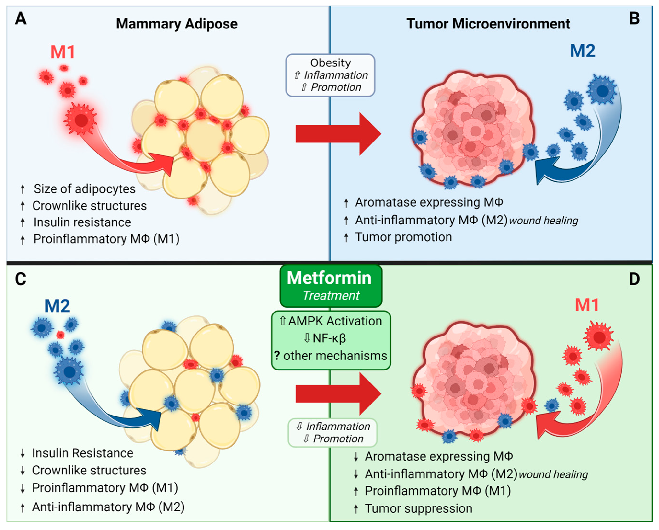
Metformin may help prevent or treat breast cancer by modulating immune cells in mammary adipose tissue and/or the tumor microenvironment. Macrophages are immune cells that play a significant role in breast cancer growth and progression [73,74,75]. Macrophages are traditionally classified as polarized to an M1 phenotype with proinflammatory properties and the ability to recognize and kill tumor cells, or the M2 phenotype that is anti-inflammatory and has wound-healing functions. In reality, however, it should be noted that polarization exists along a spectrum, with M1 and M2 phenotypes representing the two extremes. Tumor-associated macrophages (TAMs) are of an M2-like phenotype, utilizing their tissue remodeling properties to promote tumor growth and progression, leading to poorer prognosis [73,74,75].
Metformin has been shown to affect macrophage polarization, and, interestingly, a vast majority of data suggests that it polarizes cells to an M2 phenotype. Chen et al. found that RAW264.7 macrophages treated with metformin led to an M2 phenotype, both alone and when combined with LPS, which traditionally induces an M1 phenotype [76]. Another study demonstrated increased macrophage apoptosis in response to metformin treatment, and, in this study, metformin predominantly targeted the apoptosis of M1 macrophages [77]. These anti-inflammatory effects of metformin are generally thought to occur through AMPK-dependent mechanisms and the subsequent inhibition of NF-ĸβ signaling [71,72,73,74]. In a skin defect model, metformin accelerated wound healing and increased M2 macrophage polarization via the AMPK/mTOR singling pathway inhibiting NLRP3 inflammasome activation [78]. This poses a unique paradox since TAMs are also M2-like, utilizing their tissue remodeling properties to promote tumorigenesis [74]. Yet, metformin treatment generally improves tumor outcomes and decreases growth, which does not reconcile with the evidence supporting an increase in M2-like macrophages in response to metformin.
It is possible that the underlying host physiology could be important in helping explain this discrepancy. For example, in the context of obesity, the ability for metformin to reduce inflammation could be particularly beneficial, especially for postmenopausal breast cancer. Obesity is a risk factor for postmenopausal breast cancer and is widely considered a state of chronic, low-grade inflammation characterized by an increase in adipose tissue macrophages, which contribute to the development and/or progression of insulin resistance [79]. Additionally, adipose inflammation, including inflammation of the breast, is linked to increased breast cancer risk [80]. In most adipose tissue depots, metformin reduces proinflammatory M1-like macrophages and increases M2-like macrophages which help maintain insulin sensitivity [81]. Work by Jing and colleagues revealed that metformin reduces pro-inflammatory markers IL-6 and TNF-α in vivo in the serum of high-fat fed mice and in vitro in palmitate-stimulated RAW264.7 macrophages. In vivo, they also saw a decrease in adipose M1-markers, supporting the ability for metformin to decrease obesity-related inflammation [82]. Together, these data suggest that a reduction in adipose tissue inflammation could underlie the beneficial effects of metformin in breast cancer, particularly in those with underlying obesity and associated adipose tissue inflammation, which are known to be tumor-promotional [81,82,83].
To further fuel the paradox of the effect of metformin on macrophages and how it may affect tumors, studies examining metformin’s effects on the tumor microenvironment show a decrease in M2-like macrophages across several cancer types, including breast cancer [33,83,84,85,86]. In a mouse model of colon cancer, metformin improved tumor outcomes and this was linked to a decrease in M2 macrophages in the tumor microenvironment [86]. Similarly, our group has shown a reduction in M2-like macrophages that express aromatase in the tumor border in a rat model of postmenopausal breast cancer [33]. Chiang et al. showed that breast cancer cells treated with metformin had decreased secretion of cytokines such as IL-4 and IL-13 that induce M2 polarization [84]. They further showed that conditioned media from breast cancer cells treated with metformin diminished the macrophage expression of M2-related cytokines (IL-8, IL-10, and TGF-β) and increased the macrophage expression of M1-related cytokines (IL-12 and TNF-α), and this translated to fewer M2-like and more M1-like macrophages in the tumors of the mice treated with metformin [84].
In summary, while many of these studies seem contradictory, it is likely that the effects of metformin on macrophages are tissue- and context-specific, and also modulated by the presence of a tumor, as represented in Figure 3. The ability of metformin to increase M2 macrophages may only occur in the absence of a tumor. These intriguing findings show that more research is needed to fully understand how metformin may modulate inflammatory responses in distant mammary adipose tissue vs. the tumor microenvironment.
3. Translating Metformin to the Clinic
3.1. Epidemiological Evidence
Epidemiological studies on metformin and cancer risk and/or mortality have shown mixed results. Many studies have found a decreased risk of cancer and/or cancer-related mortality in individuals with diabetes on metformin, compared to other antidiabetic medications [9,10,13,87]; however, other studies have found no impact on cancer outcomes [88,89,90]. Among studies that show that metformin is beneficial for decreasing cancer risk, there is evidence for time- and dose-dependent responses, with patients who received more metformin doses or who have been on the drug for a longer duration having the most benefits [8,87]. One study in patients with type 2 diabetes that evaluated different cancers did not find that effect of metformin on overall cancer risk; however, when they evaluated different cancers separately, they did observe a modest reduction in breast cancer with an incidence rate ratio of 0.77 (95% CI:0.43; 1.40) [91]. The use of metformin after a cancer diagnosis has also been shown to decrease the risk of breast cancer-specific death [92]. An early meta-analysis of observational studies found that metformin decreased breast cancer risk in postmenopausal women with diabetes compared to those on other antidiabetic therapies [11]. A subsequent meta-analysis also showed a decrease in all-cause mortality in breast cancer patients with diabetes taking metformin, but it did not impact breast cancer incidence [93]. Conversely, several studies do not support that metformin decreases cancer risk in women with diabetes [88,89,90]. Several meta-analyses have also reported no difference or non-significant decreases in breast cancer risk, with the duration of the metformin potentially influencing results [16,94,95]. Certain groups caution that the observational trials may be impacted by time-related biases, such as the immortal time bias, that could overestimate the anti-cancer properties of metformin [93,96,97].
Aside from potential time biases, several other factors could contribute to the variations in outcomes observed across metformin studies. For example, the inclusion of both pre- and postmenopausal women in many studies could introduce variations in results due to differences caused by menopausal status. Molecular features such as tumor subtypes and other risk factors can be affected by menopausal status; for example, excess adiposity is a risk factor for postmenopausal breast cancer with less clear impacts before menopause [98]. In studies that specifically examined postmenopausal women with diabetes, metformin appears to reduce breast cancer risk [10,99,100]. In the postmenopausal population, specific cancer subtypes may be more sensitive to metformin treatment. Studies have found that metformin reduced the risk of ER+, PR+, and HER2− breast cancers [10,100], suggesting that these subtypes may be more susceptible to the anticancer effect of metformin in postmenopausal women.
It should also be noted that when interpreting the results of observation studies, many studies evaluated patients with type 2 diabetes who were taking metformin for diabetes management. Therefore, it is unclear if these findings can be applied to patients who do not have diabetes. In addition, for many studies, reduced risk was seen in comparison to other diabetic drugs, which may complicate interpretation of the effect of metformin.
3.2. Clinical Findings: Window of Opportunity Trials
Building on evidence from preclinical and epidemiologic studies, there have been several clinical trials addressing metformin treatment for breast cancers. As of 29 December 2023, there are 57 studies registered under “breast cancer” and “metformin” at clinicaltrials.gov. Some studies have been completed with results and published, while others have publications pending, and some are still in the recruitment phase (Table 3).
Several studies have been “window of opportunity” trials investigating the effect of metformin on mammary tumors in patients without diabetes. In these pre-surgical, window-of-opportunity studies, women diagnosed with breast cancer are treated with metformin prior to treatment-related surgery. The duration and dose of metformin varies by study but is generally several weeks, with post-treatment tissue samples collected at the time of surgery for analysis. Many studies have evaluated proliferation markers or metabolic targets as primary and secondary endpoints. Several studies have noted decreased Ki67 staining [101,102,103,104,105,106]. Interestingly, there is evidence that underlying insulin sensitivity in part modulates the response of metformin on proliferation, with more beneficial effects observed in patients with homeostatic model assessment for insulin resistance (HOMA-IR) > 2.8, indicating that tumors from patients with insulin resistance were more receptive to metformin [105,106], likely because metformin has been shown to reduce several markers of metabolic health, including body weight, HOMA-IR, cholesterol, and leptin [101,107]. Hadad et al. found that metformin treatment decreased genes associated with p53, BRCA1, and cell cycle pathways in non-diabetic women with operable invasive breast cancer [102]. A follow-up study from the same group also showed that tumor samples had a significant up-regulation of pAMPK, down-regulation of pAkt, decreased Ki67, and cleaved caspase-3 [103]. Another study showed that after 13–21 days of metformin treatment, primary breast cancer tumors had changes in fatty acid oxidation, suggesting treatment targeted lipid metabolism [108]. Other studies have found that metformin treatment before surgery had no effects on cell proliferation overall [107,109]. The window of opportunity trials demonstrates that metformin may improve metabolic health and decrease cancer cell proliferation in women with operable breast cancers, but effects may be modulated by patient metabolic status.
Table 3.
Clinical studies registered at ClinicalTrials.gov that include metformin and breast cancer.
Table 3.
Clinical studies registered at ClinicalTrials.gov that include metformin and breast cancer.
| Study Type | ID ClinicalTrials.gov | Population/Cancer Details | Intervention(s) | Primary Outcome(s) | Related Publications | Key Findings |
|---|---|---|---|---|---|---|
| Completed Studies | ||||||
| RCT | NCT01302379 | Postmenopausal breast cancer survivors w/BMI > 25 kg/m2 | 6-month intervention (4 groups) (1) placebo + lifestyle intervention (2) MET + lifestyle intervention (3) placebo + standard printed dietary guidelines (4) MET + standard printed dietary guidelines MET dosing: wk 1: 500 mg/day (PM) wks 2–4: 1000 mg/day (PM) wks 5+: 1500 mg/day (500 mg AM + 1000 mg PM) | Change (baseline to 6 mos) for: - insulin, - glucose, - C-reactive protein, - bioavailable testosterone, - sex hormone binding globulin | Nwanaji-Enwerem et al., 2021 [110], Bellerba et al., 2022 [111] | MET + Lifestyle Intervention ↓ insulin −21.8% (CI −29.7 to −13.0) ↓ C-reactive protein −21.4% (CI −38.9 to 1.0) MET + Standard Dietary Guidelines ↓ insulin −13.2% (CI −21.7 to −3.7) ↓ C-reactive protein −9.2% (CI −29.0 to 16.1) |
| Phase 2 | NCT00930579 | Newly diagnosed early invasive BC | Pre-surgical intervention MET 1500 mg/day for a minimum of 2 wks prior to surgery | Kalinsky et al., 2014 [107], Kalinsky et al., 2017 [112] | No change in tumor proliferation (Ki-67) ↓ BMI, cholesterol, and leptin MET modulated proteins involved in apoptosis/cell cycle, cell signaling, & invasion/motility, including: - ↑ tumor Raptor, C-Raf, Cyclin B1, Cyclin D1, TRFC, and Syk - ↓ tumor pMAPKpT202, Y204, JNKpT183, pT185, BadpS112, PKC.alphapS657, and SrcpY416 | |
| NCT02028221 | Premenopausal w/BMI > 25 kg/m2 and metabolic syndrome | BC prevention 12-mo intervention (1) Placebo (2) MET wks 1–4 850 mg/day, wks 5 + 1700 mg/day | Change in breast density at 6 and 12 mos | Martinez et al., 2016 [113], Tapia et al., 2021 [114] | ↓ waist circumference and waist-to-hip ratio No change in % breast density or dense breast volume Non-significant (p = 0.07).↓ in non-dense breast volume | |
| NCT01310231 | Metastatic or unresectable locally advanced BC on 1st-4th line chemotherapy w/anthracycline, taxane, platinum, capecitabine or vinorelbine based regimens w/o diabetes | Intervention continued until disease progression (1) placebo + standard chemotherapy (2) MET 850 mg/2× day + standard chemotherapy | Progression-free survival | Pimentel et al., 2019 [115] | No effect on progression-free survival, overall survival, and response rate | |
| NCT02431676 | Individuals that have survived solid malignant tumors including breast, prostate, lung, colon, skin melanoma, endometrial, liver, pancreatic, rectal, kidney, other solid malignant tumors. | Secondary prevention study evaluating the effect of the interventions on IGF-1 (1) Self-directed weight loss (2) Coach-directed behavioral weight loss (3) MET up to 2000 mg/day | IGF-1 at 6 mos IGF1/IGFBP3 molar ratio levels at 6 and 12 mos | Juraschek et al., 2018 [116], Mueller et al., 2021 [117], Hu et al., 2021 [118], Tilves et al., 2022a [119], Tilves et al., 2022b [120] | ↓ BMI ↑ butyrate, acetate, and valerate at 6 months Altered microbiota composition: ↑ Escherichia coli and Ruminococcus torques, ↓ Intestinibacter bartlettii, R. faecis and R. intestinalis | |
| Phase 3 | NCT01101438 | Women w/o diabetes w/high-risk nonmetastatic BC | 5 Year Intervention (1) Placebo + standard treatment (2) MET + standard treatment MET dosing: wks 1–4 850 mg/day, wks 5+ 850 mg/2× day | Invasive disease-free survival | Goodwin et al., 2022 [121], Goodwin et al., 2023 [122] | No change in invasive disease-free survival or the risk of developing new cancers. |
| Window of opportunity | NCT00897884 | w/o diabetes w/newly diagnosed untreated BC | Window-of-opportunity neoadjuvant study, intervention 2–3 wks prior to surgical removal of tumor MET 500 mg/3× day | Tumor proliferation rate (pre vs. post treatment) | Dowling et al., 2015 [123] | ↓ insulin receptor expression in tumors ↓ phosphorylation status of protein kinase B (PKB)/Akt (S473), extracellular signal-regulated kinase 1/2 (ERK1/2, T202/Y204), AMPK (T172) and acetyl coenzyme A carboxylase (S79) in tumors |
| Phase 2 | NCT01340300 | Stage I-III CRC & BC survivors, >2 mo from completing standard therapy (excluding hormone rx or trastuzumab) | Randomized to 12 wks of: (1) Control educational materials (2) MET (3) Exercise (4) MET + Exercise MET dosing: wks 1–2 1/day, wks 3+ 2/day Exercise: Training w/exercise physiologist 2× wk | Change in fasting insulin (baseline to 3 mos) | Brown et al., 2020 [124], Meyerhardt et al., 2020 [125] | MET ↓ insulin from baseline −1.16 mU/L ± 1.18 ↓ leptin from baseline −2.56 ng/mL ± 1.33 ↓ IGF1 from baseline −2.66 ng/mL ± 3.28 MET + Exercise ↓ insulin from baseline −2.47 mU/L ± 1.07 ↓ leptin from baseline −5.09 ng/mL ± 1.21 ↓ IGF1 from baseline −1.29 ng/mL ± 2.98 |
| Early Phase 1 | NCT01980823 | Newly diagnosed operable invasive BC or DCIS No prior treatment | Window-of-opportunity intervention ~2 wks prior to surgical removal of tumor MET 1500 mg/day + Atorvastatin 80 mg/day | Change in tumor Ki-67 (proliferation; baseline to 2 wks) | N/A | No results reported. |
| NCT01793948 | Postmenopausal w/elevated risk for breast cancer w/BMI ≥ 25 w/o diabetes | BC prevention study MET (850 mg/ 2× day) for 12 cycles vs. placebo | Changes in protein phosphorylation after MET exposure from baseline to 12 mos | N/A | No results reported. | |
| Phase 1 | NCT02882581 | Adults >50 years w/BC | Radiation: 11C-metformin | MET uptake in BC | N/A | No results reported. |
| NCT01650506 | TNBC w/o diabetes | Single arm phase 1 study Intervention: Erlotinib + Metformin MET dosing: Dose titrated from 850 mg/2× day to 850 mg/3× day Erlotinib dosing: 150 mg/day | Max tolerated dose of MET in combination with a 150 mg erlotinib/day | N/A | No results reported. | |
| NCT02278965 | Pre- and postmenopausal w/history of early stage BC w/BMI ≥ 25 w/o diabetes | Single group assignment, intervention for 12 mos MET 850 mg/2× day + Omega-3 fatty acids 560 mg/2× day | # of participants completing the 1-year intervention | N/A | No results reported. | |
| NCT00933309 | Postmenopausal w/history HR+ BC and evidence metastasis w/BMI ≥ 25 | Duration: as long as the disease is stable and/or responding (1) Exemestane 25 mg/day (2) Exemestane 25 mg/day + Avandamet (MET 500 mg + Rosiglitazone 2 mg)/day | Dose-limiting toxicity | N/A | No results reported. | |
| NCT00659568 | Metastatic or unresectable solid tumor (breast, endometrial, kidney, lung, unspecified) or lymphoma | Determine max tolerated dose of MET when administered with temsirolimus Intervention: MET + temsirolimus | Max tolerated and recommended dose of MET when administered w/temsirolimus | N/A | No results reported. | |
| NCT02145559 | Adults w/solid tumors that is metastatic or unresectable and standard or palliative measures are not an option (breast, lung, liver, lymphoma, kidney) | Evaluate the pharmacodynamic markers sirolimus + metformin therapy (1) MET XR up to 1000 mg/day + Sirolimus (2) Delayed MET (no MET for 2 wks then titrated up to 1000 mg/day) + Sirolimus | Pharmacodynamic biomarker p70S6K | N/A | No results reported. | |
| Phase 2 | NCT01266486 | Early stage BC w/o diabetes | Single group assignment Intervention (14–21 days): Extended-release MET 1500 mg/day | Phosphorylation of S6K, 4E-BP-1 and AMPK | Lord et al., 2018 [126] Lord et al., 2020 [108], Ralli et al., 2022 [127] | ↑ in genes that regulate fatty acid oxidation ↓ mitochondrial metabolites, activates mitochondrial metabolic pathways, and ↑ 18-FDG flux in tumors Tumor heterogeneity: - Mitochondrial response to MET dictates response - Identified OXPHOS transcriptional response (OTR) signature in tumors that were resistant to MET - Tumors that ↑ OXPHOS genes had ↑ proliferation score |
| NCT04143282 | MBC w/o diabetes | Chemotherapy alone vs MET + chemotherapy | Radiologic response rate at 3 mos Overall & progression-free survival at 6 mos | Rabea et al., 2021 [128] | Improved radiologic response, ↓ mortality & ↓ disease progression but overall survival & progression-free survival not significantly affected | |
| NCT04170465 | BC w/o metastasis and w/o diabetes | RCT, intervention 6 mos (1) MET 850 mg/2× day + AC-T neoadjuvant chemotherapy (2) AC-T neoadjuvant chemotherapy alone | Tumor apoptosis and chemotherapy toxicities at 6 mos | Serageldin et al., 2023 [129] | MET + AC-T: ↓ peripheral neuropathy incidence & severity ↓ oral mucositis ↓ fatigue ↓ fatty liver incidence preserved cardiac function | |
| NCT05053841 | Postmenopausal women w/BC w/o diabetes | Parallel assignment, 6-mo intervention, randomized: (1) Control: women w/obesity (n = 15) letrozole alone (2) Women w/obesity (n = 15) letrozole + MET 2000 ± 500 mg/day (3) Women w/o obesity (n = 15) letrozole alone | Change in serum biomarkers from baseline to 6 mos | El-Attar et al., 2023 [130] | ↓ estradiol, leptin, fasting glucose, insulin, osteocalcin serum levels, and HOMA-IR | |
| NCT02488564 | Patients w/operable BC or locally advanced BC that is HER2+ w/o diabetes | Single group assignment, trial duration 36 mos Intervention: Liposomal doxorubicin + Docetaxel + Trastuzumab + MET 1000 mg/2× day | Pathologic complete response rate | N/A | No results reported. | |
| NCT01589367 | Postmenopausal w/ER+ BC w/o diabetes | 1:1 randomized clinical (1) Letrozole 2.5 mg/day + MET (2) Letrozole 2.5 mg/day + placebo MET dosing 1 wk 500 mg/2× day, followed by 1 wk (1000 mg AM|500 mg PM)/day, followed by 22 wks 1000 mg/2× day | Clinical response rate from baseline to 24 wks | Kim et al., 2014 [131] | No results reported. | |
| NCT05351021 | Early stage BC w/o diabetes receiving adjuvant paclitaxel | Randomized, parallel assignment study to test MET as a preventive for paclitaxel-induced peripheral neuropathy (1) MET 850 mg/2× day + adjuvant paclitaxel (2) placebo + adjuvant paclitaxel | Incidence of grade II or > peripheral neuropathy at end of paclitaxel treatment | Bakry et al., 2023 [132] | ↓ paclitaxel-induced peripheral neuropathy ↑ quality of life | |
| NCT00909506 | Women w/operable BC w/overweight or pre-DM | Randomized, parallel assignment, 24 wk intervention (1) placebo (2) MET 500 mg/day. | Weight loss at 6 mos | N/A | No results reported. | |
| NCT01885013 | HER2− MBC w/o diabetes | Randomized, parallel assignment, study 24 mos + 12-mo follow-up (1) MET 1000 mg/2× day + Myocet + Cyclophosphamide (2) Myocet + Cyclophosphamide | Progression-free survival | Gennari et al., 2020 [133] | no effect on IGF-1R expression or circulating tumor cell count | |
| Phase 4 | (breast, lung, liver, lymphoma, kidney) NCT05840068 | MBC w/o diabetes | Randomized, parallel assignment, intervention 6 mos (1) MET 500 mg/2× day + chemotherapy (2) Control chemotherapy alone | IGF-1 levels at 6 mos | N/A | No results reported. |
| Active—Not Recruiting | ||||||
| Phase 2 | NCT04248998 | Stage I-III TNBC w/fasting glucose ≤ 250 mg/dL | Randomized, parallel assignment (1) Chemotherapy + fasting-mimicking diet (2) Chemotherapy + fasting-mimicking diet + MET 1700 mg/day | Rate of pathologic complete responses | N/A | Pathologic complete response is higher in diabetic patients who utilize neoadjuvant chemotherapy and take metformin. |
| NCT04300790 | ER+ and/or PgR+, HER2− advanced BC w/centrally confirmed PI3KCAMut on an aromatase inhibitor | Single Group Assignment (1) Normoglycemic patients: Alpelisib + MET (up to 100 mg/2× day) + endocrine therapy (2) Pre-diabetic patients: Alpelisib + MET (up to 100 mg/2× day) + endocrine therapy (3) Insulin naïve type 2 diabetic patients: Alpelisib + MET (up to 100 mg/2× day) + endocrine therapy | Development of treatment-induced hyperglycemia (Alpelisib + Endocrine Therapy) | N/A | No results reported. | |
| NCT02874430 | Localized breast, uterine, or cervical cancer | Single Group Assignment, intervention 6 wks Doxycycline + MET | Change in % of stromal cells expressing Caveolin-1 at an intensity of 1+ or greater | N/A | No results reported. | |
| Phase 3 | NCT01905046 | Women w/high risk for BC (such as w/atypical hyperplasia, LCIS, DCIS, family history, etc.) | BC prevention, randomized, crossover assignment, (1) MET 850 mg/2× day for 24 mos (2) Placebo 12 mos, may crossover to MET mos 13–24 | Presence or absence of cytological atypia in unilateral or bilateral RPFNA aspirates after 12 and 24 mos | N/A | No results reported. |
| Active—Recruiting | ||||||
| Observational | NCT02695121 | Adults w/type 2 diabetes exposed to dapagliflozin and other antidiabetic treatments (including MET) | Observational cohort study | Incidence of bladder and breast cancer | N/A | No results reported. |
| Phase 1 | NCT03006172 | Locally advanced or metastatic PIK3CA-mutant solid tumors including BC | Non-randomized, sequential assignment (1) Inavolisib single agent (2) Inavolisib + palbociclib + letrozole (3) Inavolisib + letrozole (4) Inavolisib + fulvestrant (5) Inavolisib + palbociclib + fulvestrant (6) Inavolisib + palbociclib + fulvestrant + MET (7) Inavolisib + trastuzumab + pertuzumab | % of participants w/dose limiting toxicities Recommended phase II dose of inavolisib % of participants w/adverse and serious adverse events | N/A | No results reported. |
| Phase 2 | NCT05023967 | Early BC w/o diabetes | Randomized, parallel assignment, intervention 4–6 wks (1) Fasting (≥16 h every night) + continuous glucose monitoring + nutritional counseling + MET extended-release (2) Usual dietary pattern + continuous glucose monitoring | Frequency of dose-limiting toxicity Change cell proliferation pre-post treatment (Ki67) | N/A | No results reported. |
| NCT05660083 | HER2− metaplastic BC and/or TNBC | Single group assignment Combination of an iNOS inhibitor + nab-paclitaxel + alpelisib To prevent deep venous thrombosis & hypertension: aspirin + amlodipine To reduce risk of severe hyperglycemia: MET up to 1000 mg/2× day | Recommended phase II dose of Alpelisib + standard + nab-paclitaxel + L-NMMA Objective response rate | N/A | No results reported. | |
| NCT01042379 | Adults w/BC | I-SPY Trial: Randomized to one of 36 experimental agents (compared to standard therapy) One group contained MET: MET + Ganitumab (AMG 479; an anti-IGF-1R antibody) | Does adding experimental agents to standard neoadjuvant medications ↑ the probability of pathologic complete response over standard neoadjuvant chemotherapy | Wang and Yee 2019 [134], Yee et al., 2021 [135] | Numerous publications, only 1 metformin related Metformin + ganitumab + paclitaxel (PGM) - metformin not sufficient to control drug-induced hyperglycemia | |
| Active—Pre-Recruitment | ||||||
| Early Phase 1 | NCT05680662 | Early BC, MBC, TNBC | Randomized, parallel assignment (1) Adjuvant quadruple therapy (quercetin 500 mg/day + zinc sulfate 50 mg/day + EGCG 300 mg/day + MET 850 mg/day) + standard chemotherapy (2) Control only standard chemotherapy | Invasive disease-free survival | N/A | No results reported. |
| Phase 4 | NCT05507398 | Non-MBC | Randomized, parallel assignment (1) Placebo (2) MET 1000 mg/day (3) Atorvastatin 20 mg/day | Improvement in overall response rate and pathological response | N/A | No results reported. |
| Terminated | ||||||
| Early Phase 1 | NCT01302002 | Early BC w/o diabetes | Metformin pre-surgery, non-randomized, single-group assignment MET 500 mg/2× day for 3 wks | Cell proliferation (Ki67), apoptosis (TUNEL), fosforilate, AKT, CD1a CD83, CD68, F40/80, arginase, iNOS, T cells [CD4(+),CD45RA(+), CD 45RO, CD4, CD8 and FOXP3(+)] | Withdrawn—no enrollment | No results reported. |
| Phase 2 | NCT01477060 | HER2−, ER+ and/or PgR+, MBC w/o diabetes | Randomized, parallel assignment, intervention until disease progression or other circumstance that mitigates termination (1) hormonal therapy + lapatinib 1250 mg/day (2) hormonal therapy + MET 1500 mg/day (3) hormonal therapy + lapatinib 1250 mg/day + MET 1500 mg/day | Progression-free survival | Terminated; low accrual | No results reported. |
| NCT01627067 | Postmenopausal w/BMI ≥ 25 w/HR+ BC and metastatic disease | Single group assignment Everolimus 10 mg/day + Exemestane 25 mg/day + MET 1000 mg/2× day | Progression-free survival (compare between overweight and obese patients) | Terminated; funding issues Yam et al., 2019 [136] | MET + everolimus + exemestane - was safe - moderate clinical benefit in patients with both overweight and obesity - 5/22 = partial response; 7/22 = stable disease for ≥24 weeks --> clinical benefit rate of 54.5% | |
| NCT02360059 | Adults w/invasive BC w/o diabetes to undergo paclitaxel chemotherapy | MET for paclitaxel neurotoxicity, randomized, parallel assignment (1) MET 1000 mg 2× day for 12 wks during paclitaxel treatment (2) Placebo for 12 wks during paclitaxel treatment | Mean change in neuropathy | Terminated; low accrual | No results reported. | |
| NCT02472353 | Adults w/BC w/o diabetes needing neoadjuvant or adjuvant therapy w/doxorubicin | MET to reduce cardiac toxicity in BC, randomized, parallel assignment (1) Standard care w/doxorubicin (2) Standard care w/doxorubicin + MET | # of patients w/≤ 5% ↓ in left ventricle ejection fraction on echocardiogram | Terminated; low accrual | No results reported. | |
| NCT04899349 | Adults w/HR+, HER2− advanced BC | Randomized, parallel assignment (1) Alpelisib + fulvestrant + dapagliflozin + MET XR (500–2000 mg/day) (2) Alpelisib + fulvestrant + MET XR (500–2000 mg/day) | # of patients w/hyperglycemia grade ≥ 3 over the 1st 8 wks of alpelisib + fulvestrant | Terminated; low accrual | No results reported. | |
| Phase 3 | NCT02201381 | Adults w/cancer | Single group assignment Atorvastatin up to 80 mg/day + MET up to 1000 mg/2× day + doxycycline 100 mg/day + mebendazole 100 mg/day | Overall survival | Withdrawn Agrawal et al., 2023 [137] | No MET BC results reported. |
| Phase 4 | NCT04741204 | Non-Hispanic white or black females w/BMI ≥ 25 w/newly diagnosed BC | Non-randomized, single-group assignment, compare outcomes between black & white women MET XR release 750 mg/2× day | Tumor progression | Withdrawn—Staffing issues | No results reported. |
| NA | NCT00984490 | Stage I or II BC that can be surgically removed w/o diabetes | Single group assignment, intervention 7–21 days prior to surgery MET 850 mg/2× day | Change cell proliferation (Ki67) from baseline to post-treatment | Terminated; low accrual | No results reported. |
| Status Unknown | ||||||
| Phase 2 | NCT04559308 | w/o diabetes w/BC receiving neoadjuvant chemotherapy | Randomized, parallel assignment (1) 4 cycles (doxorubicin+cyclophosphamide), then 12 cycles paclitaxel+ MET 1000 mg/2× day, then surgery (2) 4 cycles (doxorubicin + cyclophosphamide), then 12 cycles paclitaxel, then surgery | Clinical benefit rate (tumor size) at 8 mos | N/A | No results reported. |
| NCT03238495 | HER2+ BC | Randomized, parallel assignment (1) Chemotherapy only (TCH + P) (2) Chemotherapy (TCH + P) + MET 850 mg/2× day | Pathologic complete response | N/A | No results reported. | |
| NCT02506777 | Locally advanced BC | Randomized, parallel assignment (1) Conventional chemotherapy—fluoruracil, doxorubicin, cyclophosphamide (FDC) × 6 cycles + MET 850 mg/2× day (2) Conventional chemotherapy FDC × 6 cycles + melatonin 3 mg/day (3) Conventional chemotherapy FDC × 6 cycles | Response rate and pathomorphological response after 6 mos | N/A | No results reported. | |
| NCT03192293 | HR+ HER2− MBC | Single group assignment MET 850 mg/2× day + Simvastatin + Fulvestrant | Clinical benefit rate after 24 wks | N/A | No results reported. | |
| NCT02506790 | ER+ locally advanced BC | Randomized, parallel assignment (1) Toremifene 60 mg/day + MET 850 mg/2× day (2) Toremifene 60 mg/day + melatonin 3 mg/day (3) Toremifene 60 mg/day | Response rate and pathomorphological response after 4 mos | N/A | No results reported. | |
| NCT01566799 | HR+, HER2− BC w/o diabetes | Single group assignment Standard chemotherapy + MET 500 mg/day for 24 wks | Pathologic complete response after 24 wks | N/A | No results reported. | |
| NCT04001725 | Melanoma, lung or BC w/brain metastasis requiring high-dose dexamethasone treatment | Randomized, parallel assignment (1) Dexamethasone min dose of 8 mg/day (2) Dexamethasone min dose of 8 mg/day + MET up to max 2550 mg/day | MET preventing precocious dexamethasone-induced diabetes after 14 days | Green et al., 2022 [138] | No MET BC results reported. | |
| NCT01929811 | BC | Randomized, parallel assignment (1) Standard chemotherapy + MET 500 mg/3× day (2) Standard chemotherapy | Pathologic complete response rate after 5 mos | Huang et al., 2023 [139] | No change in pathological complete response or disease outcome w/MET No difference in proliferation (Ki67) w/MET MET prevented the ↑ in total cholesterol and LDL-C after standard treatment | |
| Phase 2/3 | NCT04387630 | Early BC w/o diabetes | Randomized, parallel assignment (1) MET max 2550 mg/day + standard treatment (2) Placebo + standard treatment | Clinical response rate after 3 mos of therapy | N/A | No results reported. |
| Unknown | NCT01666171 | BC patients concurrently enrolling or previously enrolled in MA.32 study w/breast density ≥ 25% | (1) MET 850 mg/2× day for 5 years (2) placebo | % change mammographic breast density in contralateral (unaffected) breast from baseline to 1 year | N/A | No results reported. |
| NCT01286233 | BC patients eligible for randomization to MA.32 study | (1) MET 850 mg/2× day for 5 years (2) placebo | Patient-reported fatigue, stress, sleep, depression, general quality of life, comorbid conditions, and behavioral risks Biological correlates of fatigue DNA polymorphisms Changes in RNA gene expression | N/A | No results reported. | |
Abbreviations: weeks (wks), with (w/), without (w/o), breast cancer (BC), metformin (MET), metastatic breast cancer (MBC), triple negative breast cancer (TNBC), hormone receptor (HR), estrogen receptor (ER), human epidermal growth factor receptor 2 (HER2), month/s (mo/mos), number (#), and body mass index (BMI). ↑ indicate an increase; ↓ indicate a decrease.
3.3. Has the Translation of Metformin to the Clinic as an Anticancer Agent Failed?
Other clinical trials have evaluated the addition of metformin to standard treatment; most of these studies suggest that while metformin has a good safety profile when added to usual care, it does not appear to improve outcomes. In patients with metabolic syndrome, combining neoadjuvant systemic anticancer therapy with metformin reduced disease progression and improved the incidence of clinically complete tumor regression; however, these changes did not result in improved progression-free or overall survival [140]. Similarly, two separate studies investigated the impact of metformin in women without diabetes with metastatic breast cancer (primarily ER/PR+, HER2− disease), and both concluded that combining metformin with standard chemotherapy did not improve survival compared to chemotherapy alone [115,141]. Similarly, the addition of metformin to neoadjuvant chemotherapy in locally advanced breast cancer did not improve clinical and pathological tumor responses [142]. Additionally, in postmenopausal women with HR+ locally advanced or metastatic breast cancer, adding metformin to aromatase inhibitors also failed to improve progression-free or overall survival [143]. Finally, meta-analyses of clinical trials of metformin treatment in breast cancer patients without diabetes do not show improved survival [144,145].
One of the most extensive clinical trials of metformin to date is the MA.32 trial completed by Goodwin and colleagues. This was a phase III, placebo-controlled double-blind study that enrolled 3649 patients with nonmetastatic breast cancer without diabetes [121,122]. Patients randomized to the treatment group received 850 mg of metformin once daily for four weeks, followed by twice daily for five years [121,122]. Preliminary results of this trial were promising; after six months of treatment, patients on metformin had significant improvements in body weight, glucose, insulin, leptin, and C-reactive protein [146] and reduced circulating levels of the cancer antigen 15-3 [147]. In a subgroup of postmenopausal patients with HER2− breast cancer, there was also a reduction in estradiol [148]. However, at the trial’s completion, metformin had no impact on invasive disease-free survival (HR = 1.01; 95% CI, 0.84–1.21) [121], or risk of new cancer development [122]. Thus, while metformin initially improved overall metabolic health, as seen in other studies with patients without diabetes [149], it did not improve cancer outcomes, questioning if metformin has a role as an anticancer agent in the clinical care of breast cancer patients.
Despite many clinical trials failing to demonstrate improvements when metformin is added to standard cancer care, some studies indicate that metformin may still have anti-cancer benefits. In women with newly diagnosed breast cancer without diabetes, adding metformin to adjuvant therapy has been shown to decrease IGF-1, ratio of IGF-1 to IGFBP-3, insulin, fasting blood glucose, and HOMA-IR [150]. These improvements in metabolic health could have a beneficial impact on tumor outcomes since insulin and IFG-1 can contribute to breast cancer promotion and metastasis [30,151]. In fact, this same study also noted that after 6 months of metformin combined hormone therapy, the patients in the metformin group had a decreased number of metastatic cases [150]. In a subset of patients with diabetes taking metformin from the ALTTO trial, a phase III adjuvant trial for patients with HER2+ breast cancer receiving trastuzumab, lapatinib, or the combination, a beneficial effect of metformin was observed. Interestingly, in women with HR+ cancer and diabetes, those taking metformin had improved disease-free survival, distant disease-free survival, and overall survival compared to patients not on metformin [152]. Therefore, clinical use of metformin may benefit subpopulations of breast cancer patients. However, it does not appear to be a suitable one-size-fits-all approach for cancer care.
4. Future Perspectives and Conclusions
Over the years, the efficacy of metformin in cancer treatment has been widely debated. While many preclinical studies have demonstrated benefits of metformin treatment with plausible mechanistic backing, clinical trials have often failed to see similar improvements in critical clinical outcomes such as disease-free and overall survival. These inconsistent findings may be due to the heterogeneity of study design and subject characteristics in both the preclinical and clinical work. Preclinically, glucose levels, tumor subtype, the presence of membrane transporters, and dose all modulate the effects of metformin on cancer cells in culture and tumors in vivo. Human studies have also seen that underlying insulin resistance and tumor subtype impact the response to metformin. In clinical studies, 2000 mg/day is generally the highest dose of metformin prescribed. At this level, metformin may reach sufficient plasma concentrations to improve tumor outcomes through indirect effects linked to improvements in metabolic health, but it is unclear if such doses are high enough to have a direct effect on tumors and/or cancer stem cells. Most direct effects of metformin in cell culture experiments have been observed at very high concentrations that may not be clinically feasible due to concerns that supratherapeutic doses could lead to lactic acidosis [153].
Regarding cancer prevention, several observational trials in diabetic patients have seen a decrease in breast cancer risk; however, these results should be interpreted with caution and within context. Time-related bias may affect these studies [96], and since the populations observed had diabetes, they may not be directly applicable to non-diabetic patients. Finally, based on the review of the literature, there is evidence that metformin treatment in the context of breast cancer may be better suited for patients with metabolic impairment and HR+/HER2− tumors [105,106,150,152].
It should be noted that we have purposely limited our review to only include preclinical studies where metformin was used as a single agent, or combined with dietary interventions (caloric restriction or a ketogenic diet). There are numerous other studies that have examined metformin in combination with chemotherapy, endocrine therapy, and other standard cancer treatments. For example, there is in vitro evidence suggesting that metformin may help resensitize breast cancer cells that have become resistant to chemotherapy [154,155]; however, it is not yet clear if such findings will translate to humans. While reviewing all the studies of metformin combined with other agents is beyond the scope of the current review, readers interested in this topic can find other excellent papers that address these topics [156,157]. We have, however, included all clinical trials of metformin registered at clinicaltrials.gov, which includes those where metformin is combined with both standard cancer treatments, novel therapeutics and/or lifestyle interventions (Table 3).
We also propose that future preclinical and clinical studies should consider the effect of diet and dietary components on the efficacy of metformin treatment. Current data demonstrate that glucose levels likely modulate the efficacy of metformin in preclinical models, and that strategies such as caloric restriction, short-term starvation, and ketogenic diets enhance the benefits of metformin for improving tumor outcomes [41,42,43]. Another study has shown that metformin was more effective at reducing tumor volume in rodents when combined with a diet depleted of the amino acid asparagine [158]. Further, a recent study in prediabetic diet-induced obese male mice demonstrated that a nonnutritive sweetener decreased the efficacy of metformin as an antidiabetic drug [159]. It remains to be seen if similar effects are observed in humans and, if so, what possible implications this may have on repurposing metformin for cancer treatment. The interaction of metformin and diet remains an area that requires further research.
In summary, the preclinical and clinical data reviewed here suggests that the clinical future of metformin for treating breast cancers is not a one-size-fits-all prescription; metformin is likely to have therapeutic benefits for specific patient subpopulations, including those with underlying metabolic impairment, and only for a subset of breast cancer subtypes. For researchers wishing to continue the quest of repurposing metformin, rigorous study design and patient selection are critical. Metformin dose, menopausal status, metabolic health, tumor subtype, and membrane transporter expression should all be carefully considered, and, hopefully, future studies will be able to discern additional features of tumors and patients most likely to benefit from this drug.
Author Contributions
Writing—original draft preparation, K.A.C. and J.L.S.; writing—review and editing, K.A.C., J.L.S. and E.D.G.; supervision, E.D.G. All authors have read and agreed to the published version of the manuscript.
Funding
This work was supported in part by the National Institutes of Health (#CA169430) and the Cancer Prevention Research Institute of Texas (#RP180801).
Acknowledgments
We appreciate the assistance provided by Samantha Foster and Nanci Lammoglia. Figures were created with BioRender.com.
Conflicts of Interest
The authors declare no conflicts of interest.
References
- Soranna, D.; Scotti, L.; Zambon, A.; Bosetti, C.; Grassi, G.; Catapano, A.; La Vecchia, C.; Mancia, G.; Corrao, G. Cancer risk associated with use of metformin and sulfonylurea in type 2 diabetes: A meta-analysis. Oncologist 2012, 17, 813–822. [Google Scholar] [CrossRef]
- Gong, Z.; Aragaki, A.K.; Chlebowski, R.T.; Manson, J.E.; Rohan, T.E.; Chen, C.; Vitolins, M.Z.; Tinker, L.F.; LeBlanc, E.S.; Kuller, L.H.; et al. Diabetes, metformin and incidence of and death from invasive cancer in postmenopausal women: Results from the women’s health initiative. Int. J. Cancer 2016, 138, 1915–1927. [Google Scholar] [CrossRef]
- Bailey, C.J.; Campbell, I.W.; Chan, J.C.N.; Davidson, J.A.; Howlett, H.C.S.; Ritz, P. Metformin—The Gold Standard: A Scientific Handbook; Wiley: Hoboken, NJ, USA, 2007. [Google Scholar]
- Bailey, C.J. Metformin: Historical overview. Diabetologia 2017, 60, 1566–1576. [Google Scholar] [CrossRef]
- Samuel, S.M.; Varghese, E.; Kubatka, P.; Triggle, C.R.; Büsselberg, D. Metformin: The Answer to Cancer in a Flower? Current Knowledge and Future Prospects of Metformin as an Anti-Cancer Agent in Breast Cancer. Biomolecules 2019, 9, 846. [Google Scholar] [CrossRef]
- Werner, E.A.; Bell, J. CCXIV.—The preparation of methylguanidine, and of ββ-dimethylguanidine by the interaction of dicyanodiamide, and methylammonium and dimethylammonium chlorides respectively. J. Chem. Soc. Trans. 1922, 121, 1790–1794. [Google Scholar] [CrossRef]
- Cejuela, M.; Martin-Castillo, B.; Menendez, J.A.; Pernas, S. Metformin and Breast Cancer: Where Are We Now? Int. J. Mol. Sci. 2022, 23, 2705. [Google Scholar] [CrossRef]
- Evans, J.M.; Donnelly, L.A.; Emslie-Smith, A.M.; Alessi, D.R.; Morris, A.D. Metformin and reduced risk of cancer in diabetic patients. BMJ 2005, 330, 1304–1305. [Google Scholar] [CrossRef] [PubMed]
- Bowker, S.L.; Majumdar, S.R.; Veugelers, P.; Johnson, J.A. Increased cancer-related mortality for patients with type 2 diabetes who use sulfonylureas or insulin. Diabetes Care 2006, 29, 254–258. [Google Scholar] [CrossRef]
- Chlebowski, R.T.; McTiernan, A.; Wactawski-Wende, J.; Manson, J.E.; Aragaki, A.K.; Rohan, T.; Ipp, E.; Kaklamani, V.G.; Vitolins, M.; Wallace, R.; et al. Diabetes, metformin, and breast cancer in postmenopausal women. J. Clin. Oncol. 2012, 30, 2844–2852. [Google Scholar] [CrossRef] [PubMed]
- Col, N.F.; Ochs, L.; Springmann, V.; Aragaki, A.K.; Chlebowski, R.T. Metformin and breast cancer risk: A meta-analysis and critical literature review. Breast Cancer Res. Treat. 2012, 135, 639–646. [Google Scholar] [CrossRef]
- He, X.; Esteva, F.J.; Ensor, J.; Hortobagyi, G.N.; Lee, M.H.; Yeung, S.C. Metformin and thiazolidinediones are associated with improved breast cancer-specific survival of diabetic women with HER2+ breast cancer. Ann. Oncol. 2012, 23, 1771–1780. [Google Scholar] [CrossRef] [PubMed]
- Libby, G.; Donnelly, L.A.; Donnan, P.T.; Alessi, D.R.; Morris, A.D.; Evans, J.M.M. New Users of Metformin Are at Low Risk of Incident Cancer: A cohort study among people with type 2 diabetes. Diabetes Care 2009, 32, 1620–1625. [Google Scholar] [CrossRef]
- Skuli, S.J.; Alomari, S.; Gaitsch, H.; Bakayoko, A.; Skuli, N.; Tyler, B.M. Metformin and Cancer, an Ambiguanidous Relationship. Pharmaceuticals 2022, 15, 626. [Google Scholar] [CrossRef] [PubMed]
- Naseri, A.; Sanaie, S.; Hamzehzadeh, S.; Seyedi-Sahebari, S.; Hosseini, M.S.; Gholipour-Khalili, E.; Rezazadeh-Gavgani, E.; Majidazar, R.; Seraji, P.; Daneshvar, S.; et al. Metformin: New applications for an old drug. J. Basic Clin. Physiol. Pharmacol. 2023, 34, 151–160. [Google Scholar] [CrossRef] [PubMed]
- Najafi, F.; Rajati, F.; Sarokhani, D.; Bavandpour, M.; Moradinazar, M. The Relationship between Metformin Consumption and Cancer Risk: An Updated Umbrella Review of Systematic Reviews and Meta-Analyses. Int. J. Prev. Med. 2023, 14, 90. [Google Scholar] [CrossRef] [PubMed]
- Lv, Z.; Guo, Y. Metformin and Its Benefits for Various Diseases. Front. Endocrinol. 2020, 11, 191. [Google Scholar] [CrossRef]
- Heckman-Stoddard, B.M.; De Censi, A.; Sahasrabuddhe, V.V.; Ford, L.G. Repurposing metformin for the prevention of cancer and cancer recurrence. Diabetologia 2017, 60, 1639–1647. [Google Scholar] [CrossRef]
- Rena, G.; Hardie, D.G.; Pearson, E.R. The mechanisms of action of metformin. Diabetologia 2017, 60, 1577–1585. [Google Scholar] [CrossRef]
- Saraei, P.; Asadi, I.; Kakar, M.A.; Moradi-Kor, N. The beneficial effects of metformin on cancer prevention and therapy: A comprehensive review of recent advances. Cancer Manag. Res. 2019, 11, 3295–3313. [Google Scholar] [CrossRef]
- Salani, B.; Del Rio, A.; Marini, C.; Sambuceti, G.; Cordera, R.; Maggi, D. Metformin, cancer and glucose metabolism. Endocr. Relat. Cancer 2014, 21, R461–R471. [Google Scholar] [CrossRef]
- Marini, C.; Salani, B.; Massollo, M.; Amaro, A.; Esposito, A.I.; Orengo, A.M.; Capitanio, S.; Emionite, L.; Riondato, M.; Bottoni, G.; et al. Direct inhibition of hexokinase activity by metformin at least partially impairs glucose metabolism and tumor growth in experimental breast cancer. Cell Cycle 2013, 12, 3490–3499. [Google Scholar] [CrossRef]
- Lei, Y.; Yi, Y.; Liu, Y.; Liu, X.; Keller, E.T.; Qian, C.-N.; Zhang, J.; Lu, Y. Metformin targets multiple signaling pathways in cancer. Chin. J. Cancer 2017, 36, 17. [Google Scholar] [CrossRef]
- Alimova, I.N.; Liu, B.; Fan, Z.; Edgerton, S.M.; Dillon, T.; Lind, S.E.; Thor, A.D. Metformin inhibits breast cancer cell growth, colony formation and induces cell cycle arrest in vitro. Cell Cycle 2009, 8, 909–915. [Google Scholar] [CrossRef]
- Vazquez-Martin, A.; Oliveras-Ferraros, C.; Menendez, J.A. The antidiabetic drug metformin suppresses HER2 (erbB-2) oncoprotein overexpression via inhibition of the mTOR effector p70S6K1 in human breast carcinoma cells. Cell Cycle 2009, 8, 88–96. [Google Scholar] [CrossRef] [PubMed]
- Zhang, W.; Li, D.; Li, B.; Chu, X.; Kong, B. STAT3 as a therapeutic target in the metformin-related treatment. Int. Immunopharmacol. 2023, 116, 109770. [Google Scholar] [CrossRef]
- Deng, X.S.; Wang, S.; Deng, A.; Liu, B.; Edgerton, S.M.; Lind, S.E.; Wahdan-Alaswad, R.; Thor, A.D. Metformin targets Stat3 to inhibit cell growth and induce apoptosis in triple-negative breast cancers. Cell Cycle 2012, 11, 367–376. [Google Scholar] [CrossRef] [PubMed]
- Kasznicki, J.; Sliwinska, A.; Drzewoski, J. Metformin in cancer prevention and therapy. Ann. Transl. Med. 2014, 2, 57. [Google Scholar] [CrossRef] [PubMed]
- Yeh, H.C.; Maruthur, N.M.; Wang, N.Y.; Jerome, G.J.; Dalcin, A.T.; Tseng, E.; White, K.; Miller, E.R.; Juraschek, S.P.; Mueller, N.T.; et al. Effects of Behavioral Weight Loss and Metformin on IGFs in Cancer Survivors: A Randomized Trial. J. Clin. Endocrinol. Metab. 2021, 106, e4179–e4191. [Google Scholar] [CrossRef]
- Lero, M.W.; Shaw, L.M. Diversity of insulin and IGF signaling in breast cancer: Implications for therapy. Mol. Cell. Endocrinol. 2021, 527, 111213. [Google Scholar] [CrossRef] [PubMed]
- Anisimov, V.; Egormin, P.; Bershtein, L.; Zabezhinskii, M.; Piskunova, T.; Popovich, I.; Semenchenko, A. Metformin decelerates aging and development of mammary tumors in HER-2/neu transgenic mice. Bull. Exp. Biol. Med. 2005, 139, 721–723. [Google Scholar] [CrossRef]
- Checkley, L.A.; Rudolph, M.C.; Wellberg, E.A.; Giles, E.D.; Wahdan-Alaswad, R.S.; Houck, J.A.; Edgerton, S.M.; Thor, A.D.; Schedin, P.; Anderson, S.M. Metformin accumulation correlates with organic cation transporter 2 protein expression and predicts mammary tumor regression in vivo. Cancer Prev. Res. 2017, 10, 198–207. [Google Scholar] [CrossRef]
- Giles, E.D.; Jindal, S.; Wellberg, E.A.; Schedin, T.; Anderson, S.M.; Thor, A.D.; Edwards, D.P.; MacLean, P.S.; Schedin, P. Metformin inhibits stromal aromatase expression and tumor progression in a rodent model of postmenopausal breast cancer. Breast Cancer Res. 2018, 20, 50. [Google Scholar] [CrossRef] [PubMed]
- Caciolla, J.; Bisi, A.; Belluti, F.; Rampa, A.; Gobbi, S. Reconsidering aromatase for breast cancer treatment: New roles for an old target. Molecules 2020, 25, 5351. [Google Scholar] [CrossRef] [PubMed]
- Song, J.; Du, J.; Han, L.; Lin, X.; Fan, C.; Chen, G. The Effect of Metformin on Triple-Negative Breast Cancer Cells and Nude Mice. Altern. Ther. Health Med. 2023, 29, 389–395. [Google Scholar] [PubMed]
- Thompson, M.D.; Grubbs, C.J.; Bode, A.M.; Reid, J.M.; McGovern, R.; Bernard, P.S.; Stijleman, I.J.; Green, J.E.; Bennett, C.; Juliana, M.M.; et al. Lack of effect of metformin on mammary carcinogenesis in nondiabetic rat and mouse models. Cancer Prev. Res. 2015, 8, 231–239. [Google Scholar] [CrossRef] [PubMed]
- Zhu, Z.; Jiang, W.; Thompson, M.D.; Echeverria, D.; McGinley, J.N.; Thompson, H.J. Effects of metformin, buformin, and phenformin on the post-initiation stage of chemically induced mammary carcinogenesis in the rat. Cancer Prev. Res. 2015, 8, 518–527. [Google Scholar] [CrossRef]
- Bojkova, B.; Orendas, P.; Garajova, M.; Kassayova, M.; Kutna, V.; Ahlersova, E.; Ahlers, I. Metformin in chemically-induced mammary carcinogenesis in rats. Neoplasma 2009, 56, 269. [Google Scholar] [CrossRef] [PubMed]
- Wahdan-Alaswad, R.; Fan, Z.; Edgerton, S.M.; Liu, B.; Deng, X.S.; Arnadottir, S.S.; Richer, J.K.; Anderson, S.M.; Thor, A.D. Glucose promotes breast cancer aggression and reduces metformin efficacy. Cell Cycle 2013, 12, 3759–3769. [Google Scholar] [CrossRef]
- Varghese, S.; Samuel, S.M.; Varghese, E.; Kubatka, P.; Büsselberg, D. High Glucose Represses the Anti-Proliferative and Pro-Apoptotic Effect of Metformin in Triple Negative Breast Cancer Cells. Biomolecules 2019, 9, 16. [Google Scholar] [CrossRef]
- Li, Y.; Zhang, Q.; Yang, J.; He, W.; Jiang, Y.; Chen, Y.; Wang, Y. Metformin combined with glucose starvation synergistically suppress triple-negative breast cancer by enhanced unfolded protein response. Biochem. Biophys. Res. Commun. 2023, 675, 146–154. [Google Scholar] [CrossRef]
- Zhu, Z.; Jiang, W.; Thompson, M.D.; McGinley, J.N.; Thompson, H.J. Metformin as an energy restriction mimetic agent for breast cancer prevention. J. Carcinog. 2011, 10, 17. [Google Scholar] [PubMed]
- Zhuang, Y.; Chan, D.K.; Haugrud, A.B.; Miskimins, W.K. Mechanisms by which low glucose enhances the cytotoxicity of metformin to cancer cells both in vitro and in vivo. PLoS ONE 2014, 9, e108444. [Google Scholar] [CrossRef] [PubMed]
- Marini, C.; Bianchi, G.; Buschiazzo, A.; Ravera, S.; Martella, R.; Bottoni, G.; Petretto, A.; Emionite, L.; Monteverde, E.; Capitanio, S.; et al. Divergent targets of glycolysis and oxidative phosphorylation result in additive effects of metformin and starvation in colon and breast cancer. Sci. Rep. 2016, 6, 19569. [Google Scholar] [CrossRef] [PubMed]
- Stambolic, V.; Woodgett, J.R.; Fantus, I.G.; Pritchard, K.I.; Goodwin, P.J. Utility of metformin in breast cancer treatment, is neoangiogenesis a risk factor? Breast Cancer Res. Treat. 2009, 114, 387–389. [Google Scholar] [CrossRef]
- Liang, X.; Giacomini, K.M. Transporters Involved in Metformin Pharmacokinetics and Treatment Response. J. Pharm. Sci. 2017, 106, 2245–2250. [Google Scholar] [CrossRef]
- Chen, S.; Zhou, J.; Xi, M.; Jia, Y.; Wong, Y.; Zhao, J.; Ding, L.; Zhang, J.; Wen, A. Pharmacogenetic variation and metformin response. Curr. Drug. Metab. 2013, 14, 1070–1082. [Google Scholar] [CrossRef]
- Samodelov, S.L.; Kullak-Ublick, G.A.; Gai, Z.; Visentin, M. Organic Cation Transporters in Human Physiology, Pharmacology, and Toxicology. Int. J. Mol. Sci. 2020, 21, 7890. [Google Scholar] [CrossRef]
- Cai, H.; Zhang, Y.; Han, T.K.; Everett, R.S.; Thakker, D.R. Cation-selective transporters are critical to the AMPK-mediated antiproliferative effects of metformin in human breast cancer cells. Int. J. Cancer 2016, 138, 2281–2292. [Google Scholar] [CrossRef]
- Zhu, P.; Davis, M.; Blackwelder, A.J.; Bachman, N.; Liu, B.; Edgerton, S.; Williams, L.L.; Thor, A.D.; Yang, X. Metformin selectively targets tumor-initiating cells in ErbB2-overexpressing breast cancer models. Cancer Prev. Res. 2014, 7, 199–210. [Google Scholar] [CrossRef]
- Barbieri, F.; Thellung, S.; Ratto, A.; Carra, E.; Marini, V.; Fucile, C.; Bajetto, A.; Pattarozzi, A.; Würth, R.; Gatti, M.; et al. In vitro and in vivo antiproliferative activity of metformin on stem-like cells isolated from spontaneous canine mammary carcinomas: Translational implications for human tumors. BMC Cancer 2015, 15, 228. [Google Scholar] [CrossRef]
- Feng, F.; Zhang, J.; Fan, X.; Yuan, F.; Jiang, Y.; Lv, R.; Ma, Y. Downregulation of Rab27A contributes to metformin-induced suppression of breast cancer stem cells. Oncol. Lett. 2017, 14, 2947–2953. [Google Scholar] [CrossRef]
- Shi, P.; Liu, W.; Tala; Wang, H.; Li, F.; Zhang, H.; Wu, Y.; Kong, Y.; Zhou, Z.; Wang, C.; et al. Metformin suppresses triple-negative breast cancer stem cells by targeting KLF5 for degradation. Cell Discov. 2017, 3, 17010. [Google Scholar] [CrossRef]
- Lee, H.; Park, H.J.; Park, C.S.; Oh, E.T.; Choi, B.H.; Williams, B.; Lee, C.K.; Song, C.W. Response of breast cancer cells and cancer stem cells to metformin and hyperthermia alone or combined. PLoS ONE 2014, 9, e87979. [Google Scholar] [CrossRef]
- Venkatesh, J.; Wasson, M.D.; Brown, J.M.; Fernando, W.; Marcato, P. LncRNA-miRNA axes in breast cancer: Novel points of interaction for strategic attack. Cancer Lett. 2021, 509, 81–88. [Google Scholar] [CrossRef]
- Wu, D.; Thompson, L.U.; Comelli, E.M. MicroRNAs: A Link between Mammary Gland Development and Breast Cancer. Int. J. Mol. Sci. 2022, 23, 15978. [Google Scholar] [CrossRef]
- Gholami, M.; Klashami, Z.N.; Ebrahimi, P.; Mahboobipour, A.A.; Farid, A.S.; Vahidi, A.; Zoughi, M.; Asadi, M.; Amoli, M.M. Metformin and long non-coding RNAs in breast cancer. J. Transl. Med. 2023, 21, 155. [Google Scholar] [CrossRef] [PubMed]
- Liu, L.; Zhang, Y.; Lu, J. The roles of long noncoding RNAs in breast cancer metastasis. Cell Death Dis. 2020, 11, 749. [Google Scholar] [CrossRef]
- Abdalla, F.; Singh, B.; Bhat, H.K. MicroRNAs and gene regulation in breast cancer. J. Biochem. Mol. Toxicol. 2020, 34, e22567. [Google Scholar] [CrossRef] [PubMed]
- Fridrichova, I.; Zmetakova, I. MicroRNAs Contribute to Breast Cancer Invasiveness. Cells 2019, 8, 1361. [Google Scholar] [CrossRef] [PubMed]
- Cabello, P.; Pineda, B.; Tormo, E.; Lluch, A.; Eroles, P. The Antitumor Effect of Metformin Is Mediated by miR-26a in Breast Cancer. Int. J. Mol. Sci. 2016, 17, 1298. [Google Scholar] [CrossRef] [PubMed]
- Zhang, J.; Li, G.; Chen, Y.; Fang, L.; Guan, C.; Bai, F.; Ma, M.; Lyu, J.; Meng, Q.H. Metformin Inhibits Tumorigenesis and Tumor Growth of Breast Cancer Cells by Upregulating miR-200c but Downregulating AKT2 Expression. J. Cancer 2017, 8, 1849–1864. [Google Scholar] [CrossRef]
- Pulito, C.; Mori, F.; Sacconi, A.; Goeman, F.; Ferraiuolo, M.; Pasanisi, P.; Campagnoli, C.; Berrino, F.; Fanciulli, M.; Ford, R.J.; et al. Metformin-induced ablation of microRNA 21-5p releases Sestrin-1 and CAB39L antitumoral activities. Cell Discov. 2017, 3, 17022. [Google Scholar] [CrossRef] [PubMed]
- Blandino, G.; Valerio, M.; Cioce, M.; Mori, F.; Casadei, L.; Pulito, C.; Sacconi, A.; Biagioni, F.; Cortese, G.; Galanti, S.; et al. Metformin elicits anticancer effects through the sequential modulation of DICER and c-MYC. Nat. Commun. 2012, 3, 865. [Google Scholar] [CrossRef] [PubMed]
- Hou, Y.; Cai, S.; Yu, S.; Lin, H. Metformin induces ferroptosis by targeting miR-324-3p/GPX4 axis in breast cancer. Acta Biochim. Biophys. Sin. 2021, 53, 333–341. [Google Scholar] [CrossRef] [PubMed]
- Wahdan-Alaswad, R.S.; Cochrane, D.R.; Spoelstra, N.S.; Howe, E.N.; Edgerton, S.M.; Anderson, S.M.; Thor, A.D.; Richer, J.K. Metformin-induced killing of triple-negative breast cancer cells is mediated by reduction in fatty acid synthase via miRNA-193b. Horm. Cancer 2014, 5, 374–389. [Google Scholar] [CrossRef]
- Cheng, L.; Zhang, X.; Huang, Y.Z.; Zhu, Y.L.; Xu, L.Y.; Li, Z.; Dai, X.Y.; Shi, L.; Zhou, X.J.; Wei, J.F.; et al. Metformin exhibits antiproliferation activity in breast cancer via miR-483-3p/METTL3/m(6)A/p21 pathway. Oncogenesis 2021, 10, 7. [Google Scholar] [CrossRef]
- Golshan, M.; Khaleghi, S.; Shafiee, S.M.; Valaee, S.; Ghanei, Z.; Jamshidizad, A.; Dashtizad, M.; Shamsara, M. Metformin modulates oncogenic expression of HOTAIR gene via promoter methylation and reverses epithelial-mesenchymal transition in MDA-MB-231 cells. J. Cell. Biochem. 2021, 122, 385–393. [Google Scholar] [CrossRef]
- Jiang, Y.; Qian, T.; Li, S.; Xie, Y.; Tao, M. Metformin reverses tamoxifen resistance through the lncRNA GAS5-medicated mTOR pathway in breast cancer. Ann. Transl. Med. 2022, 10, 366. [Google Scholar] [CrossRef]
- Li, J.; Kim, S.G.; Blenis, J. Rapamycin: One drug, many effects. Cell Metab. 2014, 19, 373–379. [Google Scholar] [CrossRef]
- Chen, J.; Qin, C.; Zhou, Y.; Chen, Y.; Mao, M.; Yang, J. Metformin may induce ferroptosis by inhibiting autophagy via lncRNA H19 in breast cancer. FEBS Open Bio 2022, 12, 146–153. [Google Scholar] [CrossRef]
- Huang, Y.; Zhou, Z.; Zhang, J.; Hao, Z.; He, Y.; Wu, Z.; Song, Y.; Yuan, K.; Zheng, S.; Zhao, Q. lncRNA MALAT1 participates in metformin inhibiting the proliferation of breast cancer cell. J. Cell. Mol. Med. 2021, 25, 7135–7145. [Google Scholar] [CrossRef]
- Tao, S.; Zhao, Z.; Zhang, X.; Guan, X.; Wei, J.; Yuan, B.; He, S.; Zhao, D.; Zhang, J.; Liu, Q.; et al. The role of macrophages during breast cancer development and response to chemotherapy. Clin. Transl. Oncol. 2020, 22, 1938–1951. [Google Scholar] [CrossRef]
- Qiu, S.-Q.; Waaijer, S.J.H.; Zwager, M.C.; de Vries, E.G.E.; van der Vegt, B.; Schröder, C.P. Tumor-associated macrophages in breast cancer: Innocent bystander or important player? Cancer Treat. Rev. 2018, 70, 178–189. [Google Scholar] [CrossRef]
- Huang, X.; Cao, J.; Zu, X. Tumor-associated macrophages: An important player in breast cancer progression. Thorac. Cancer 2022, 13, 269–276. [Google Scholar] [CrossRef] [PubMed]
- Chen, M.; Zhang, J.; Liu, S.; Zhou, Z. Effects of metformin on the polarization and Notch 1 expression of RAW264.7 macrophages. Zhonghua Yi Xue Za Zhi 2015, 95, 1258–1261. [Google Scholar] [PubMed]
- Hu, X.; Luo, H.; Dou, C.; Chen, X.; Huang, Y.; Wang, L.; Xue, S.; Sun, Z.; Chen, S.; Xu, Q.; et al. Metformin Triggers Apoptosis and Induction of the G0/G1 Switch 2 Gene in Macrophages. Genes 2021, 12, 1437. [Google Scholar] [CrossRef] [PubMed]
- Qing, L.; Fu, J.; Wu, P.; Zhou, Z.; Yu, F.; Tang, J. Metformin induces the M2 macrophage polarization to accelerate the wound healing via regulating AMPK/mTOR/NLRP3 inflammasome singling pathway. Am. J. Transl. Res. 2019, 11, 655–668. [Google Scholar]
- Keuper, M. On the role of macrophages in the control of adipocyte energy metabolism. Endocr. Connect. 2019, 8, R105–R121. [Google Scholar] [CrossRef] [PubMed]
- Iyengar, N.M.; Brown, K.A.; Zhou, X.K.; Gucalp, A.; Subbaramaiah, K.; Giri, D.D.; Zahid, H.; Bhardwaj, P.; Wendel, N.K.; Falcone, D.J.; et al. Metabolic Obesity, Adipose Inflammation and Elevated Breast Aromatase in Women with Normal Body Mass Index. Cancer Prev. Res. 2017, 10, 235–243. [Google Scholar] [CrossRef]
- Deng, T.; Lyon, C.J.; Bergin, S.; Caligiuri, M.A.; Hsueh, W.A. Obesity, Inflammation, and Cancer. Annu. Rev. Pathol. 2016, 11, 421–449. [Google Scholar] [CrossRef]
- Jing, Y.; Wu, F.; Li, D.; Yang, L.; Li, Q.; Li, R. Metformin improves obesity-associated inflammation by altering macrophages polarization. Mol. Cell. Endocrinol. 2018, 461, 256–264. [Google Scholar] [CrossRef] [PubMed]
- Kurelac, I.; Umesh Ganesh, N.; Iorio, M.; Porcelli, A.M.; Gasparre, G. The multifaceted effects of metformin on tumor microenvironment. Semin. Cell Dev. Biol. 2020, 98, 90–97. [Google Scholar] [CrossRef]
- Chiang, C.F.; Chao, T.T.; Su, Y.F.; Hsu, C.C.; Chien, C.Y.; Chiu, K.C.; Shiah, S.G.; Lee, C.H.; Liu, S.Y.; Shieh, Y.S. Metformin-treated cancer cells modulate macrophage polarization through AMPK-NF-κB signaling. Oncotarget 2017, 8, 20706–20718. [Google Scholar] [CrossRef] [PubMed]
- Ding, L.; Liang, G.; Yao, Z.; Zhang, J.; Liu, R.; Chen, H.; Zhou, Y.; Wu, H.; Yang, B.; He, Q. Metformin prevents cancer metastasis by inhibiting M2-like polarization of tumor associated macrophages. Oncotarget 2015, 6, 36441–36455. [Google Scholar] [CrossRef] [PubMed]
- Kang, J.; Lee, D.; Lee, K.J.; Yoon, J.E.; Kwon, J.H.; Seo, Y.; Kim, J.; Chang, S.Y.; Park, J.; Kang, E.A.; et al. Tumor-Suppressive Effect of Metformin via the Regulation of M2 Macrophages and Myeloid-Derived Suppressor Cells in the Tumor Microenvironment of Colorectal Cancer. Cancers 2022, 14, 2881. [Google Scholar] [CrossRef]
- Bodmer, M.; Meier, C.; Krähenbühl, S.; Jick, S.S.; Meier, C.R. Long-term metformin use is associated with decreased risk of breast cancer. Diabetes Care 2010, 33, 1304–1308. [Google Scholar] [CrossRef] [PubMed]
- Calip, G.S.; Yu, O.; Elmore, J.G.; Boudreau, D.M. Comparative safety of diabetes medications and risk of incident invasive breast cancer: A population-based cohort study. Cancer Causes Control 2016, 27, 709–720. [Google Scholar] [CrossRef]
- Hosio, M.; Urpilainen, E.; Marttila, M.; Hautakoski, A.; Arffman, M.; Sund, R.; Puistola, U.; Läärä, E.; Jukkola, A.; Karihtala, P. Association of antidiabetic medication and statins with breast cancer incidence in women with type 2 diabetes. Breast Cancer Res. Treat. 2019, 175, 741–748. [Google Scholar] [CrossRef]
- Dankner, R.; Agay, N.; Olmer, L.; Murad, H.; Keinan Boker, L.; Balicer, R.D.; Freedman, L.S. Metformin Treatment and Cancer Risk: Cox Regression Analysis, With Time-Dependent Covariates, of 320,000 Persons With Incident Diabetes Mellitus. Am. J. Epidemiol. 2019, 188, 1794–1800. [Google Scholar] [CrossRef]
- Vicentini, M.; Ballotari, P.; Giorgi Rossi, P.; Venturelli, F.; Sacchettini, C.; Greci, M.; Mangone, L.; Pezzarossi, A.; Manicardi, V. Effect of different glucose-lowering therapies on cancer incidence in type 2 diabetes: An observational population-based study. Diabetes Res. Clin. Pract. 2018, 143, 398–408. [Google Scholar] [CrossRef]
- Löfling, L.L.; Støer, N.C.; Andreassen, B.K.; Ursin, G.; Botteri, E. Low-dose aspirin, statins, and metformin and survival in patients with breast cancers: A Norwegian population-based cohort study. Breast Cancer Res. 2023, 25, 101. [Google Scholar] [CrossRef]
- Tang, G.H.; Satkunam, M.; Pond, G.R.; Steinberg, G.R.; Blandino, G.; Schünemann, H.J.; Muti, P. Association of Metformin with Breast Cancer Incidence and Mortality in Patients with Type II Diabetes: A GRADE-Assessed Systematic Review and Meta-analysis. Cancer Epidemiol. Biomark. Prev. 2018, 27, 627–635. [Google Scholar] [CrossRef] [PubMed]
- Lu, Y.; Hajjar, A.; Cryns, V.L.; Trentham-Dietz, A.; Gangnon, R.E.; Heckman-Stoddard, B.M.; Alagoz, O. Breast cancer risk for women with diabetes and the impact of metformin: A meta-analysis. Cancer Med. 2023, 12, 11703–11718. [Google Scholar] [CrossRef] [PubMed]
- Chen, Y.; Mushashi, F.; Son, S.; Bhatti, P.; Dummer, T.; Murphy, R.A. Diabetes medications and cancer risk associations: A systematic review and meta-analysis of evidence over the past 10 years. Sci. Rep. 2023, 13, 11844. [Google Scholar] [CrossRef] [PubMed]
- Yu, O.H.Y.; Suissa, S. Metformin and Cancer: Solutions to a Real-World Evidence Failure. Diabetes Care 2023, 46, 904–912. [Google Scholar] [CrossRef] [PubMed]
- Yang, X.; Chan, J.C. Comment on: Suissa and Azoulay. Metformin and the risk of cancer: Time-related biases in observational studies. Diabetes Care 2012, 35, 2665–2673. [Google Scholar] [CrossRef]
- Heer, E.; Harper, A.; Escandor, N.; Sung, H.; McCormack, V.; Fidler-Benaoudia, M.M. Global burden and trends in premenopausal and postmenopausal breast cancer: A population-based study. Lancet Glob. Health 2020, 8, e1027–e1037. [Google Scholar] [CrossRef] [PubMed]
- Bosco, J.L.; Antonsen, S.; Sørensen, H.T.; Pedersen, L.; Lash, T.L. Metformin and incident breast cancer among diabetic women: A population-based case-control study in Denmark. Cancer Epidemiol. Biomarkers Prev. 2011, 20, 101–111. [Google Scholar] [CrossRef]
- García-Esquinas, E.; Guinó, E.; Castaño-Vinyals, G.; Pérez-Gómez, B.; Llorca, J.; Altzibar, J.M.; Peiró-Pérez, R.; Martín, V.; Moreno-Iribas, C.; Tardón, A.; et al. Association of diabetes and diabetes treatment with incidence of breast cancer. Acta Diabetol. 2016, 53, 99–107. [Google Scholar] [CrossRef]
- Niraula, S.; Dowling, R.J.O.; Ennis, M.; Chang, M.C.; Done, S.J.; Hood, N.; Escallon, J.; Leong, W.L.; McCready, D.R.; Reedijk, M.; et al. Metformin in early breast cancer: A prospective window of opportunity neoadjuvant study. Breast Cancer Res. Treat. 2012, 135, 821–830. [Google Scholar] [CrossRef]
- Hadad, S.; Iwamoto, T.; Jordan, L.; Purdie, C.; Bray, S.; Baker, L.; Jellema, G.; Deharo, S.; Hardie, D.G.; Pusztai, L.; et al. Evidence for biological effects of metformin in operable breast cancer: A pre-operative, window-of-opportunity, randomized trial. Breast Cancer Res. Treat. 2011, 128, 783–794. [Google Scholar] [CrossRef]
- Hadad, S.M.; Coates, P.; Jordan, L.B.; Dowling, R.J.O.; Chang, M.C.; Done, S.J.; Purdie, C.A.; Goodwin, P.J.; Stambolic, V.; Moulder-Thompson, S.; et al. Evidence for biological effects of metformin in operable breast cancer: Biomarker analysis in a pre-operative window of opportunity randomized trial. Breast Cancer Res. Treat. 2015, 150, 149–155. [Google Scholar] [CrossRef]
- DeCensi, A.; Puntoni, M.; Guerrieri-Gonzaga, A.; Cazzaniga, M.; Serrano, D.; Lazzeroni, M.; Vingiani, A.; Gentilini, O.; Petrera, M.; Viale, G.; et al. Effect of Metformin on Breast Ductal Carcinoma In Situ Proliferation in a Randomized Presurgical Trial. Cancer Prev. Res. 2015, 8, 888–894. [Google Scholar] [CrossRef] [PubMed]
- DeCensi, A.; Puntoni, M.; Gandini, S.; Guerrieri-Gonzaga, A.; Johansson, H.A.; Cazzaniga, M.; Pruneri, G.; Serrano, D.; Schwab, M.; Hofmann, U.; et al. Differential effects of metformin on breast cancer proliferation according to markers of insulin resistance and tumor subtype in a randomized presurgical trial. Breast Cancer Res. Treat. 2014, 148, 81–90. [Google Scholar] [CrossRef]
- Bonanni, B.; Puntoni, M.; Cazzaniga, M.; Pruneri, G.; Serrano, D.; Guerrieri-Gonzaga, A.; Gennari, A.; Trabacca, M.S.; Galimberti, V.; Veronesi, P.; et al. Dual effect of metformin on breast cancer proliferation in a randomized presurgical trial. J. Clin. Oncol. 2012, 30, 2593–2600. [Google Scholar] [CrossRef] [PubMed]
- Kalinsky, K.; Crew, K.D.; Refice, S.; Xiao, T.; Wang, A.; Feldman, S.M.; Taback, B.; Ahmad, A.; Cremers, S.; Hibshoosh, H.; et al. Presurgical trial of metformin in overweight and obese patients with newly diagnosed breast cancer. Cancer Investig. 2014, 32, 150–157. [Google Scholar] [CrossRef] [PubMed]
- Lord, S.R.; Collins, J.M.; Cheng, W.C.; Haider, S.; Wigfield, S.; Gaude, E.; Fielding, B.A.; Pinnick, K.E.; Harjes, U.; Segaran, A.; et al. Transcriptomic analysis of human primary breast cancer identifies fatty acid oxidation as a target for metformin. Br. J. Cancer 2020, 122, 258–265. [Google Scholar] [CrossRef]
- Cazzaniga, M.; DeCensi, A.; Pruneri, G.; Puntoni, M.; Bottiglieri, L.; Varricchio, C.; Guerrieri-Gonzaga, A.; Gentilini, O.D.; Pagani, G.; Dell’Orto, P.; et al. The effect of metformin on apoptosis in a breast cancer presurgical trial. Br. J. Cancer 2013, 109, 2792–2797. [Google Scholar] [CrossRef]
- Nwanaji-Enwerem, J.C.; Chung, F.F.; Van der Laan, L.; Novoloaca, A.; Cuenin, C.; Johansson, H.; Bonanni, B.; Hubbard, A.E.; Smith, M.T.; Hartman, S.J.; et al. An epigenetic aging analysis of randomized metformin and weight loss interventions in overweight postmenopausal breast cancer survivors. Clin. Epigenetics 2021, 13, 224. [Google Scholar] [CrossRef]
- Bellerba, F.; Chatziioannou, A.C.; Jasbi, P.; Robinot, N.; Keski-Rahkonen, P.; Trolat, A.; Vozar, B.; Hartman, S.J.; Scalbert, A.; Bonanni, B.; et al. Metabolomic profiles of metformin in breast cancer survivors: A pooled analysis of plasmas from two randomized placebo-controlled trials. J. Transl. Med. 2022, 20, 629. [Google Scholar] [CrossRef]
- Kalinsky, K.; Zheng, T.; Hibshoosh, H.; Du, X.; Mundi, P.; Yang, J.; Refice, S.; Feldman, S.M.; Taback, B.; Connolly, E.; et al. Proteomic modulation in breast tumors after metformin exposure: Results from a “window of opportunity” trial. Clin. Transl. Oncol. 2017, 19, 180–188. [Google Scholar] [CrossRef]
- Martinez, J.A.; Chalasani, P.; Thomson, C.A.; Roe, D.; Altbach, M.; Galons, J.P.; Stopeck, A.; Thompson, P.A.; Villa-Guillen, D.E.; Chow, H.H. Phase II study of metformin for reduction of obesity-associated breast cancer risk: A randomized controlled trial protocol. BMC Cancer 2016, 16, 500. [Google Scholar] [CrossRef] [PubMed]
- Tapia, E.; Villa-Guillen, D.E.; Chalasani, P.; Centuori, S.; Roe, D.J.; Guillen-Rodriguez, J.; Huang, C.; Galons, J.P.; Thomson, C.A.; Altbach, M.; et al. A randomized controlled trial of metformin in women with components of metabolic syndrome: Intervention feasibility and effects on adiposity and breast density. Breast Cancer Res. Treat. 2021, 190, 69–78. [Google Scholar] [CrossRef] [PubMed]
- Pimentel, I.; Lohmann, A.E.; Ennis, M.; Dowling, R.J.O.; Cescon, D.; Elser, C.; Potvin, K.R.; Haq, R.; Hamm, C.; Chang, M.C.; et al. A phase II randomized clinical trial of the effect of metformin versus placebo on progression-free survival in women with metastatic breast cancer receiving standard chemotherapy. Breast 2019, 48, 17–23. [Google Scholar] [CrossRef]
- Juraschek, S.P.; Plante, T.B.; Charleston, J.; Miller, E.R.; Yeh, H.C.; Appel, L.J.; Jerome, G.J.; Gayles, D.; Durkin, N.; White, K.; et al. Use of online recruitment strategies in a randomized trial of cancer survivors. Clin. Trials 2018, 15, 130–138. [Google Scholar] [CrossRef]
- Mueller, N.T.; Differding, M.K.; Zhang, M.; Maruthur, N.M.; Juraschek, S.P.; Miller, E.R., 3rd; Appel, L.J.; Yeh, H.C. Metformin Affects Gut Microbiome Composition and Function and Circulating Short-Chain Fatty Acids: A Randomized Trial. Diabetes Care 2021, 44, 1462–1471. [Google Scholar] [CrossRef]
- Hu, J.R.; Yeh, H.C.; Mueller, N.T.; Appel, L.J.; Miller, E.R., 3rd; Maruthur, N.M.; Jerome, G.J.; Chang, A.R.; Gelber, A.C.; Juraschek, S.P. Effects of a Behavioral Weight Loss Intervention and Metformin Treatment on Serum Urate: Results from a Randomized Clinical Trial. Nutrients 2021, 13, 2673. [Google Scholar] [CrossRef]
- Tilves, C.; Yeh, H.C.; Maruthur, N.; Juraschek, S.P.; Miller, E.R.; Appel, L.J.; Mueller, N.T. A behavioral weight-loss intervention, but not metformin, decreases a marker of gut barrier permeability: Results from the SPIRIT randomized trial. Int. J. Obes. 2022, 46, 655–660. [Google Scholar] [CrossRef] [PubMed]
- Tilves, C.; Yeh, H.C.; Maruthur, N.; Juraschek, S.P.; Miller, E.; White, K.; Appel, L.J.; Mueller, N.T. Increases in Circulating and Fecal Butyrate are Associated With Reduced Blood Pressure and Hypertension: Results From the SPIRIT Trial. J. Am. Heart Assoc. 2022, 11, e024763. [Google Scholar] [CrossRef] [PubMed]
- Goodwin, P.J.; Chen, B.E.; Gelmon, K.A.; Whelan, T.J.; Ennis, M.; Lemieux, J.; Ligibel, J.A.; Hershman, D.L.; Mayer, I.A.; Hobday, T.J.; et al. Effect of Metformin vs Placebo on Invasive Disease-Free Survival in Patients With Breast Cancer: The MA.32 Randomized Clinical Trial. JAMA 2022, 327, 1963–1973. [Google Scholar] [CrossRef]
- Goodwin, P.J.; Chen, B.E.; Gelmon, K.A.; Whelan, T.J.; Ennis, M.; Lemieux, J.; Ligibel, J.A.; Hershman, D.L.; Mayer, I.A.; Hobday, T.J.; et al. Effect of Metformin Versus Placebo on New Primary Cancers in Canadian Cancer Trials Group MA.32: A Secondary Analysis of a Phase III Randomized Double-Blind Trial in Early Breast Cancer. J. Clin. Oncol. 2023, 41, 5356–5362. [Google Scholar] [CrossRef]
- Dowling, R.J.; Niraula, S.; Chang, M.C.; Done, S.J.; Ennis, M.; McCready, D.R.; Leong, W.L.; Escallon, J.M.; Reedijk, M.; Goodwin, P.J.; et al. Changes in insulin receptor signaling underlie neoadjuvant metformin administration in breast cancer: A prospective window of opportunity neoadjuvant study. Breast Cancer Res. 2015, 17, 32. [Google Scholar] [CrossRef]
- Brown, J.C.; Zhang, S.; Ligibel, J.A.; Irwin, M.L.; Jones, L.W.; Campbell, N.; Pollak, M.N.; Sorrentino, A.; Cartmel, B.; Harrigan, M.; et al. Effect of Exercise or Metformin on Biomarkers of Inflammation in Breast and Colorectal Cancer: A Randomized Trial. Cancer Prev. Res. 2020, 13, 1055–1062. [Google Scholar] [CrossRef]
- Meyerhardt, J.A.; Irwin, M.L.; Jones, L.W.; Zhang, S.; Campbell, N.; Brown, J.C.; Pollak, M.; Sorrentino, A.; Cartmel, B.; Harrigan, M.; et al. Randomized Phase II Trial of Exercise, Metformin, or Both on Metabolic Biomarkers in Colorectal and Breast Cancer Survivors. JNCI Cancer Spectr. 2020, 4, pkz096. [Google Scholar] [CrossRef]
- Lord, S.R.; Cheng, W.C.; Liu, D.; Gaude, E.; Haider, S.; Metcalf, T.; Patel, N.; Teoh, E.J.; Gleeson, F.; Bradley, K.; et al. Integrated Pharmacodynamic Analysis Identifies Two Metabolic Adaption Pathways to Metformin in Breast Cancer. Cell Metab. 2018, 28, 679–688.e674. [Google Scholar] [CrossRef]
- Ralli, G.P.; Carter, R.D.; McGowan, D.R.; Cheng, W.C.; Liu, D.; Teoh, E.J.; Patel, N.; Gleeson, F.; Harris, A.L.; Lord, S.R.; et al. Radiogenomic analysis of primary breast cancer reveals [18F]-fluorodeoxglucose dynamic flux-constants are positively associated with immune pathways and outperform static uptake measures in associating with glucose metabolism. Breast Cancer Res. 2022, 24, 34. [Google Scholar] [CrossRef]
- Rabea, H.; Hassan, A.; Elberry, A.A. Metformin as an Adjuvant Treatment in Non-Diabetic Metastatic Breast Cancer. Bahrain Med. Bull. 2021, 43, 477–481. [Google Scholar]
- Serageldin, M.A.; Kassem, A.B.; El-Kerm, Y.; Helmy, M.W.; El-Mas, M.M.; El-Bassiouny, N.A. The Effect of Metformin on Chemotherapy-Induced Toxicities in Non-diabetic Breast Cancer Patients: A Randomised Controlled Study. Drug Saf. 2023, 46, 587–599. [Google Scholar] [CrossRef] [PubMed]
- El-Attar, A.A.; Ibrahim, O.M.; Alhassanin, S.A.; Essa, E.S.; Mostafa, T.M. Effect of metformin as an adjuvant therapy to letrozole on estradiol and other biomarkers involved in the pathogenesis of breast cancer in overweight and obese postmenopausal women: A pilot study. Eur. J. Clin. Pharmacol. 2023, 79, 299–309. [Google Scholar] [CrossRef] [PubMed]
- Kim, J.; Lim, W.; Kim, E.K.; Kim, M.K.; Paik, N.S.; Jeong, S.S.; Yoon, J.H.; Park, C.H.; Ahn, S.H.; Kim, L.S.; et al. Phase II randomized trial of neoadjuvant metformin plus letrozole versus placebo plus letrozole for estrogen receptor positive postmenopausal breast cancer (METEOR). BMC Cancer 2014, 14, 170. [Google Scholar] [CrossRef] [PubMed]
- Bakry, H.M.; Mansour, N.O.; ElKhodary, T.R.; Soliman, M.M. Efficacy of metformin in prevention of paclitaxel-induced peripheral neuropathy in breast cancer patients: A randomized controlled trial. Front. Pharmacol. 2023, 14, 1181312. [Google Scholar] [CrossRef]
- Gennari, A.; Foca, F.; Zamarchi, R.; Rocca, A.; Amadori, D.; De Censi, A.; Bologna, A.; Cavanna, L.; Gianni, L.; Scaltriti, L.; et al. Insulin-like growth factor-1 receptor (IGF-1R) expression on circulating tumor cells (CTCs) and metastatic breast cancer outcome: Results from the TransMYME trial. Breast Cancer Res. Treat. 2020, 181, 61–68. [Google Scholar] [CrossRef]
- Wang, H.; Yee, D. I-SPY 2: A Neoadjuvant Adaptive Clinical Trial Designed to Improve Outcomes in High-Risk Breast Cancer. Curr. Breast Cancer Rep. 2019, 11, 303–310. [Google Scholar] [CrossRef]
- Yee, D.; Isaacs, C.; Wolf, D.M.; Yau, C.; Haluska, P.; Giridhar, K.V.; Forero-Torres, A.; Jo Chien, A.; Wallace, A.M.; Pusztai, L.; et al. Ganitumab and metformin plus standard neoadjuvant therapy in stage 2/3 breast cancer. NPJ Breast Cancer 2021, 7, 131. [Google Scholar] [CrossRef]
- Yam, C.; Esteva, F.J.; Patel, M.M.; Raghavendra, A.S.; Ueno, N.T.; Moulder, S.L.; Hess, K.R.; Shroff, G.S.; Hodge, S.; Koenig, K.H.; et al. Efficacy and safety of the combination of metformin, everolimus and exemestane in overweight and obese postmenopausal patients with metastatic, hormone receptor-positive, HER2-negative breast cancer: A phase II study. Investig. New Drugs 2019, 37, 345–351. [Google Scholar] [CrossRef] [PubMed]
- Agrawal, S.; Vamadevan, P.; Mazibuko, N.; Bannister, R.; Swery, R.; Wilson, S.; Edwards, S. A New Method for Ethical and Efficient Evidence Generation for Off-Label Medication Use in Oncology (A Case Study in Glioblastoma). Front. Pharmacol. 2019, 10, 681. [Google Scholar] [CrossRef] [PubMed]
- Green, B.J.; Marazzini, M.; Hershey, B.; Fardin, A.; Li, Q.; Wang, Z.; Giangreco, G.; Pisati, F.; Marchesi, S.; Disanza, A.; et al. PillarX: A Microfluidic Device to Profile Circulating Tumor Cell Clusters Based on Geometry, Deformability, and Epithelial State. Small 2022, 18, e2106097. [Google Scholar] [CrossRef]
- Huang, J.; Tong, Y.; Hong, J.; Huang, O.; Wu, J.; He, J.; Chen, W.; Li, Y.; Chen, X.; Shen, K. Neoadjuvant docetaxel, epirubicin, and cyclophosphamide with or without metformin in breast cancer patients with metabolic abnormality: Results from the randomized Phase II NeoMET trial. Breast Cancer Res. Treat. 2023, 197, 525–533. [Google Scholar] [CrossRef]
- Liubota, R.; Cheshuk, V.; Zotov, O.; Vereshchako, R.; Anikusko, M.; Liubota, I.; Gur’yanov, V. Metformin in neoadjuvant systemic therapy of breast cancer patients with metabolic syndrome. Arch. Oncol. 2018, 24, 1–5. [Google Scholar] [CrossRef]
- Nanni, O.; Amadori, D.; De Censi, A.; Rocca, A.; Freschi, A.; Bologna, A.; Gianni, L.; Rosetti, F.; Amaducci, L.; Cavanna, L.; et al. Metformin plus chemotherapy versus chemotherapy alone in the first-line treatment of HER2-negative metastatic breast cancer. The MYME randomized, phase 2 clinical trial. Breast Cancer Res. Treat. 2019, 174, 433–442. [Google Scholar] [CrossRef] [PubMed]
- Barakat, H.E.; Hussein, R.R.S.; Elberry, A.A.; Zaki, M.A.; Ramadan, M.E. The impact of metformin use on the outcomes of locally advanced breast cancer patients receiving neoadjuvant chemotherapy: An open-labelled randomized controlled trial. Sci. Rep. 2022, 12, 7656. [Google Scholar] [CrossRef]
- Zhao, Y.; Gong, C.; Wang, Z.; Zhang, J.; Wang, L.; Zhang, S.; Cao, J.; Tao, Z.; Li, T.; Wang, B.; et al. A randomized phase II study of aromatase inhibitors plus metformin in pre-treated postmenopausal patients with hormone receptor positive metastatic breast cancer. Oncotarget 2017, 8, 84224. [Google Scholar] [CrossRef] [PubMed]
- Wang, Q.; Ma, X.; Long, J.; Du, X.; Pan, B.; Mao, H. Metformin and survival of women with breast cancer: A meta-analysis of randomized controlled trials. J. Clin. Pharm. Ther. 2022, 47, 263–269. [Google Scholar] [CrossRef] [PubMed]
- Lusica, P.M.M.; Eugenio, K.P.Y.; Sacdalan, D.B.L.; Jimeno, C.A. A systematic review and meta-analysis on the efficacy and safety of metformin as adjunctive therapy among women with metastatic breast cancer. Cancer Treat. Res. Commun. 2021, 29, 100457. [Google Scholar] [CrossRef]
- Goodwin, P.J.; Parulekar, W.R.; Gelmon, K.A.; Shepherd, L.E.; Ligibel, J.A.; Hershman, D.L.; Rastogi, P.; Mayer, I.A.; Hobday, T.J.; Lemieux, J.; et al. Effect of metformin vs placebo on and metabolic factors in NCIC CTG MA.32. J. Natl. Cancer Inst. 2015, 107, djv006. [Google Scholar] [CrossRef]
- Goodwin, P.J.; Dowling, R.J.O.; Ennis, M.; Chen, B.E.; Parulekar, W.R.; Shepherd, L.E.; Gelmon, K.A.; Whelan, T.J.; Ligibel, J.A.; Hershman, D.L.; et al. Cancer Antigen 15-3/Mucin 1 Levels in CCTG MA.32: A Breast Cancer Randomized Trial of Metformin vs Placebo. JNCI Cancer Spectr. 2021, 5, pkab066. [Google Scholar] [CrossRef] [PubMed]
- Pimentel, I.; Chen, B.E.; Lohmann, A.E.; Ennis, M.; Ligibel, J.; Shepherd, L.; Hershman, D.L.; Whelan, T.; Stambolic, V.; Mayer, I.; et al. The Effect of Metformin vs Placebo on Sex Hormones in Canadian Cancer Trials Group MA.32. J. Natl. Cancer Inst. 2021, 113, 192–198. [Google Scholar] [CrossRef]
- Ko, K.P.; Ma, S.H.; Yang, J.J.; Hwang, Y.; Ahn, C.; Cho, Y.M.; Noh, D.Y.; Park, B.J.; Han, W.; Park, S.K. Metformin intervention in obese non-diabetic patients with breast cancer: Phase II randomized, double-blind, placebo-controlled trial. Breast Cancer Res. Treat. 2015, 153, 361–370. [Google Scholar] [CrossRef]
- El-Haggar, S.M.; El-Shitany, N.A.; Mostafa, M.F.; El-Bassiouny, N.A. Metformin may protect nondiabetic breast cancer women from metastasis. Clin. Exp. Metastasis 2016, 33, 339–357. [Google Scholar] [CrossRef]
- Giles, E.D.; Singh, G. Role of insulin-like growth factor binding proteins (IGFBPs) in breast cancer proliferation and metastasis. Clin. Exp. Metastasis 2003, 20, 481–487. [Google Scholar] [CrossRef]
- Sonnenblick, A.; Agbor-Tarh, D.; Bradbury, I.; Di Cosimo, S.; Azim, H.A., Jr.; Fumagalli, D.; Sarp, S.; Wolff, A.C.; Andersson, M.; Kroep, J.; et al. Impact of Diabetes, Insulin, and Metformin Use on the Outcome of Patients With Human Epidermal Growth Factor Receptor 2-Positive Primary Breast Cancer: Analysis From the ALTTO Phase III Randomized Trial. J. Clin. Oncol. 2017, 35, 1421–1429. [Google Scholar] [CrossRef]
- Dyatlova, N.; Tobarran, N.V.; Kannan, L.; North, R.; Wills, B.K. Metformin-Associated Lactic Acidosis (MALA). In StatPearls; StatPearls Publishing LLC.: Treasure Island, FL, USA, 2023. [Google Scholar]
- Davies, G.; Lobanova, L.; Dawicki, W.; Groot, G.; Gordon, J.R.; Bowen, M.; Harkness, T.; Arnason, T. Metformin inhibits the development, and promotes the resensitization, of treatment-resistant breast cancer. PLoS ONE 2017, 12, e0187191. [Google Scholar] [CrossRef]
- Lee, J.O.; Kang, M.J.; Byun, W.S.; Kim, S.A.; Seo, I.H.; Han, J.A.; Moon, J.W.; Kim, J.H.; Kim, S.J.; Lee, E.J.; et al. Metformin overcomes resistance to cisplatin in triple-negative breast cancer (TNBC) cells by targeting RAD51. Breast Cancer Res. 2019, 21, 115. [Google Scholar] [CrossRef]
- Zhang, H.H.; Guo, X.L. Combinational strategies of metformin and chemotherapy in cancers. Cancer Chemother. Pharmacol. 2016, 78, 13–26. [Google Scholar] [CrossRef]
- Morio, K.; Kurata, Y.; Kawaguchi-Sakita, N.; Shiroshita, A.; Kataoka, Y. Efficacy of Metformin in Patients With Breast Cancer Receiving Chemotherapy or Endocrine Therapy: Systematic Review and Meta-analysis. Ann. Pharmacother. 2022, 56, 245–255. [Google Scholar] [CrossRef] [PubMed]
- Krall, A.S.; Mullen, P.J.; Surjono, F.; Momcilovic, M.; Schmid, E.W.; Halbrook, C.J.; Thambundit, A.; Mittelman, S.D.; Lyssiotis, C.A.; Shackelford, D.B.; et al. Asparagine couples mitochondrial respiration to ATF4 activity and tumor growth. Cell Metab. 2021, 33, 1013–1026.e1016. [Google Scholar] [CrossRef] [PubMed]
- Singh, A.; Rourk, K.; Bernier, A.; de Lartigue, G. Non-Nutritive Sweetened Beverages Impair Therapeutic Benefits of Metformin in Prediabetic Diet-Induced Obese Mice. Nutrients 2023, 15, 2472. [Google Scholar] [CrossRef] [PubMed]
Figure 1.
Timeline depicting the identification of galegine and subsequent development of metformin as an antidiabetic drug.
Figure 1.
Timeline depicting the identification of galegine and subsequent development of metformin as an antidiabetic drug.
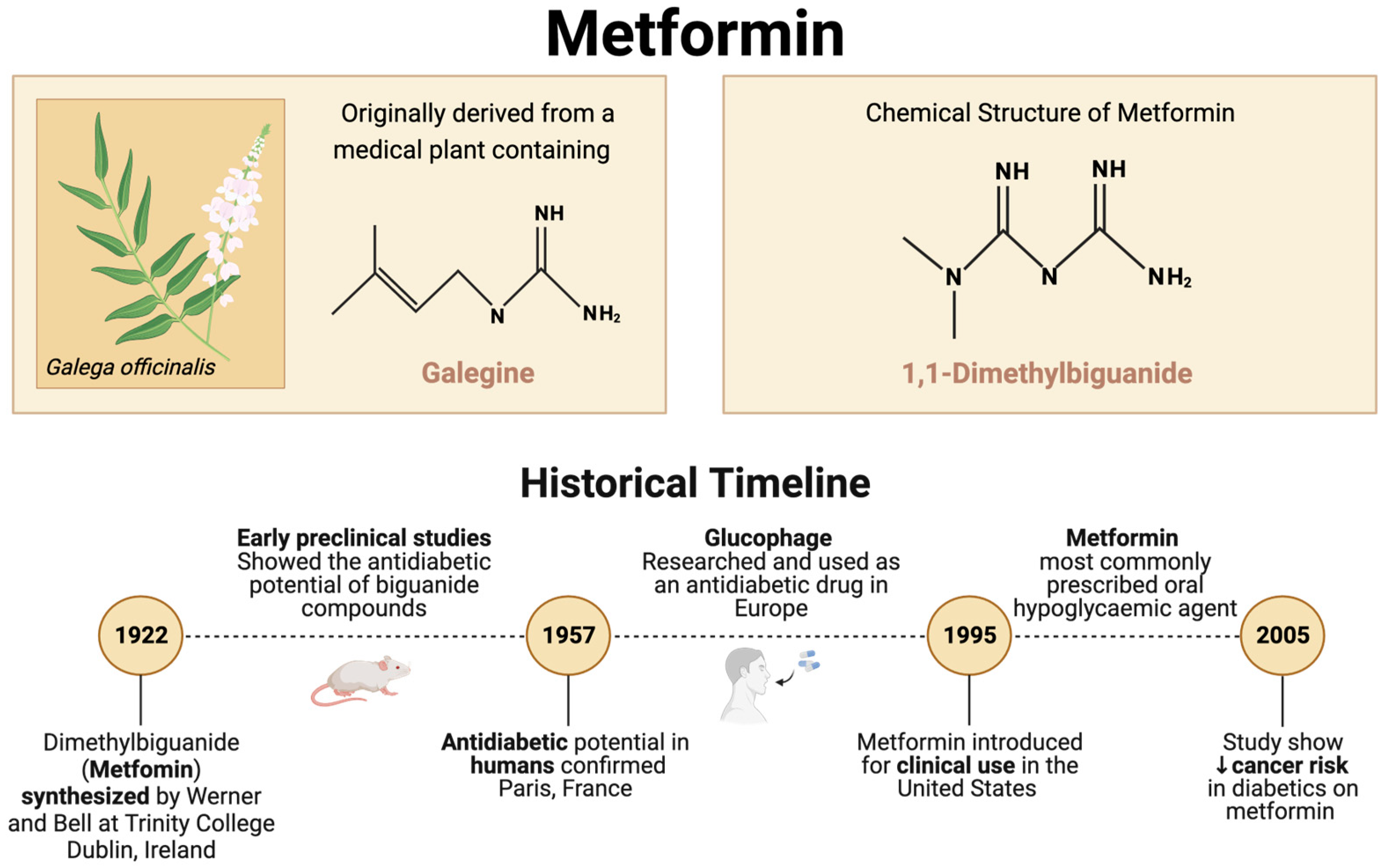
Figure 2.
Indirect and direct effects of metformin on breast cancer. ↑ indicates an increase; ↓ indicate a decrease.
Figure 2.
Indirect and direct effects of metformin on breast cancer. ↑ indicates an increase; ↓ indicate a decrease.
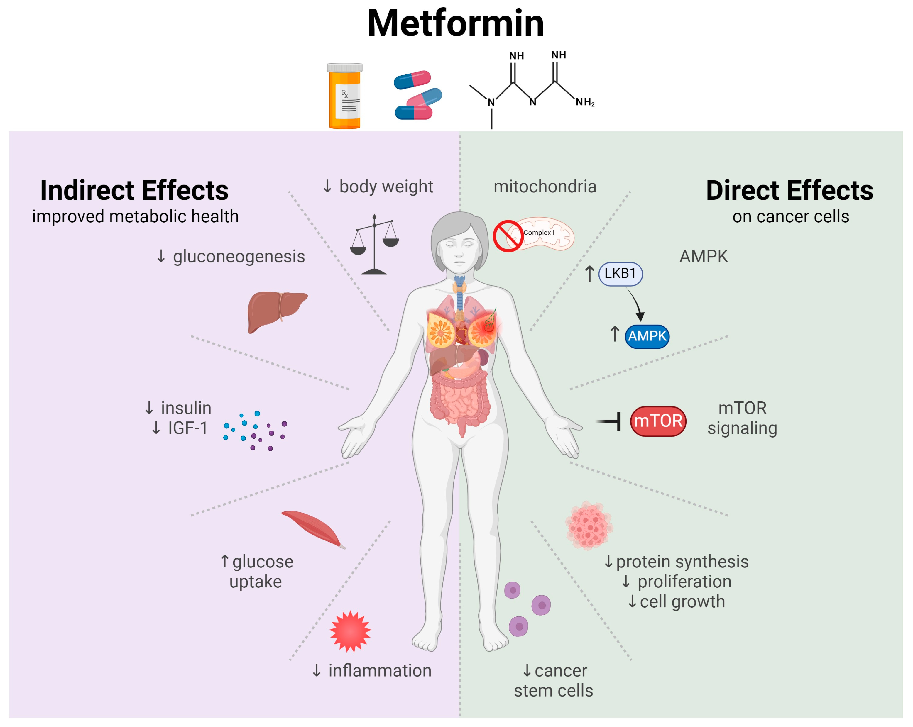
Figure 3.
Proposed modulation of inflammation by metformin, based on the current knowledge of the field. The role of macrophages (MΦ) in inflammation and tumor promotion in (A) mammary adipose and (B) tumor microenvironment. (C) Metformin is expected to reduce inflammation in mammary adipose and (D) reduce M2 polarization in the tumor microenvironments. ↑ indicates an increase; ↓ indicates a decrease.

Disclaimer/Publisher’s Note: The statements, opinions and data contained in all publications are solely those of the individual author(s) and contributor(s) and not of MDPI and/or the editor(s). MDPI and/or the editor(s) disclaim responsibility for any injury to people or property resulting from any ideas, methods, instructions or products referred to in the content. |
© 2024 by the authors. Licensee MDPI, Basel, Switzerland. This article is an open access article distributed under the terms and conditions of the Creative Commons Attribution (CC BY) license (https://creativecommons.org/licenses/by/4.0/).
Share and Cite
MDPI and ACS Style
Corleto, K.A.; Strandmo, J.L.; Giles, E.D. Metformin and Breast Cancer: Current Findings and Future Perspectives from Preclinical and Clinical Studies. Pharmaceuticals 2024, 17, 396. https://doi.org/10.3390/ph17030396
AMA Style
Corleto KA, Strandmo JL, Giles ED. Metformin and Breast Cancer: Current Findings and Future Perspectives from Preclinical and Clinical Studies. Pharmaceuticals. 2024; 17(3):396. https://doi.org/10.3390/ph17030396
Chicago/Turabian StyleCorleto, Karen A., Jenna L. Strandmo, and Erin D. Giles. 2024. "Metformin and Breast Cancer: Current Findings and Future Perspectives from Preclinical and Clinical Studies" Pharmaceuticals 17, no. 3: 396. https://doi.org/10.3390/ph17030396
Note that from the first issue of 2016, this journal uses article numbers instead of page numbers. See further details here.