Review

Mechanism of Allosteric Modulation of the Cys-loop Receptors

1
Division of Neurobiology, Barrow Neurological Institute, St. Joseph’s Hospital and Medical Center, Phoenix, AZ 85013, USA
2
Department of Obstetrics and Gynecology, St. Joseph’s Hospital and Medical Center, Phoenix, USA
*
Author to whom correspondence should be addressed.
Pharmaceuticals 2010, 3(8), 2592-2609; https://doi.org/10.3390/ph3082592
Submission received: 24 June 2010 / Revised: 30 July 2010 / Accepted: 9 August 2010 / Published: 12 August 2010
(This article belongs to the Special Issue Allosteric Modulation)

Abstract

:
The cys-loop receptor family is a major family of neurotransmitter-operated ion channels. They play important roles in fast synaptic transmission, controlling neuronal excitability, and brain function. These receptors are allosteric proteins, in that binding of a neurotransmitter to its binding site remotely controls the channel function. The cys-loop receptors also are subject to allosteric modulation by many pharmaceutical agents and endogenous modulators. By binding to a site of the receptor distinct from the neurotransmitter binding site, allosteric modulators alter the response of the receptors to their agonists. The mechanism of allosteric modulation is traditionally believed to be that allosteric modulators directly change the binding affinity of receptors for their agonists. More recent studies support the notion that these allosteric modulators are very weak agonists or antagonists by themselves. They directly alter channel gating, and thus change the distribution of the receptor across multiple different affinity states, indirectly influencing receptors’ sensitivity to agonists. There are two major locations of allosteric modulator binding sites. One is in subunit interfaces of the amino-terminal domain. The other is in the transmembrane domain close to the channel gating machinery. In this review, we also give some examples of well characterized allosteric binding pockets.

Graphical Abstract

1. Introduction

The cys-loop receptor family is a major family of ligand-gated ion channels (LGICs). All members of this receptor family have a signature cysteine loop, two cysteine residues separated by 13 other residues, in the amino-terminal domain of each subunit. This family includes subfamilies of nicotinic receptors (nAChRs) [1], serotonin receptor type 3 (5-HT3R) [2], γ-aminobutyric acid receptors type A and C [3,4,5] (GABAA/CR), glycine receptors [6], zinc-activated cation channel [7], and invertebrate glutamate/serotonin-activated anionic channels or GABA-gated cation channels [8,9,10]. Recently, prokaryotic proton-gated ion channels have also been considered to be in the same family, although they do not have signature cysteines in the corresponding loop region [11,12,13]. In eukaryotic ligand-gated ion-channels, each receptor subunit has a large amino-terminal extracellular domain, four transmembrane domains (M1–M4) with the second transmembrane domain (M2) lining the pore, and a large intracellular loop between M3 and M4 [14,15]. Prokaryotic channels have similar subunit topology, but with a short M3–M4 intracellular loop. The receptors in this family have a pentameric structure. That is, five subunits form a pseudo-symmetrical receptor with a central vestibule (Figure 1A). Different combinations of subunits, and their isoforms, create structural diversity and thus different subtypes within each cys-loop receptor subfamily. Since each subtype has specialized function, the structural diversity also provides opportunities for subtype-selective drug development. The neurotransmitter binding sites (orthosteric sites) are located in the amino-terminal domain at subunit interfaces [15,16]. Figure 1B shows the crystal structure of nicotine in the binding site of the homologous protein, acetylcholine binding protein (AChBP) [17]. AChBP is homologous to the amino-terminal domain of cys-loop receptor’s subunits [18]. It also has a pentameric structure [18] and can bind nicotinic receptor agonists and antagonists [17,19,20]. Thus, AChBP crystal structures provide the best structural models for the amino-terminal domain and orthosteric binding sites. In the center of the transmembrane region of all cys-loop receptors is the ion conducting pore (Figure 1C) [15], which is controlled by neurotransmitter binding to orthosteric sites in the distal amino-terminal domain. Thus, the cys-loop receptors are allosteric proteins; binding of a neurotransmitter to the extracellular amino-terminal domain of its receptor allosterically controls the distantly located transmembrane ion channel pore, a process termed allosteric activation [21,22]. This allosteric activation occurs through an evolutionarily conserved allosteric network that connects orthosteric binding sites to the channel gate [23]. The detailed activation mechanism of cys-loop receptors has been extensively reviewed recently [21,24,25,26,27,28]. Figure 1D shows the neurotransmitter binding sites located at subunit interfaces of major subtypes of nicotinic receptors and GABAA/C receptors. A typical heteromeric receptor (containing at least two different subunits) has two orthosteric binding sites. The remaining subunit interfaces of the amino-terminal domain of heteromeric receptors can potentially act as allosteric modulator binding sites. In fact, the well-characterized benzodiazepine binding site in a typical αβγ GABAA receptor is located in the amino-terminal domain αγ subunit interface, homologous to the orthosteric GABA binding sites at the βα interfaces (Figure 1D). A typical homomeric receptor (with identical subunits), such as α7 or α9 nAChR, ρ1 GABA receptor, 5-HT3A receptor, or glycine α1 receptor, has five potential orthosteric binding sites.
The function of the cys-loop receptors can be modified by allosteric modulators. By binding to the receptor at an allosteric site, a site distinct from the orthosteric neurotransmitter binding sites, allosteric modulators alter the agonist sensitivity (shift of concentration-response curve), agonist efficacy (maximum response), and channel gating kinetics for activation and desensitization. Note the difference between allosteric activation and allosteric modulation. For allosteric activation, the binding of neurotransmitter to the “orthosteric” binding site (allosterically) controls the distantly located gating machinery. However, for allosteric modulation, modulator binding to additional “allosteric” sites modifies the action of agonists binding to the orthosteric sites. That is, the modulator allosterically alters the orthosteric agonist’s action We will discuss kinetic models and mechanisms of allosteric modulation in section 3, and well-characterized allosteric sites in section 3.
Figure 1. Structures of prevalent cys-loop receptors and locations of agonist binding sites. (A) Top view of the EM structure of the Torpedo nicotinic receptor, generated from 2BG9 PDB file [16], showing pentameric structure; (B) Side view of crystal structure of AChBP with nicotine bound in the subunit interface, generated from 1UW6 PDB file [17]. The nicotine is surrounded by residues that form the agonist binding pocket (within blue dashed circle); (C) Side view of the EM structure of the Torpedo nicotinic receptor with β subunit removed, revealing central vestibule and channel pore (red arrow), generated from 2BG9 PDB file [16]; (D) Location of agonist binding sites in different subunit interfaces in major subtypes of nicotinic and GABAA/C receptors. Note that most heteromeric receptors have two orthosteric agonist binding pockets. In some cases (heteromeric α1β glycine receptor [29], α4β2 nicotinic receptor [30]), functional studies suggest a possible third neurotransmitter binding site. In addition, the αβγ GABAA receptor has one allosteric benzodiazepine binding site (BZ).
Pharmaceuticals 03 02592 g001

2. Kinetic Models of Allosteric Activation

The activation mechanism of the nicotinic receptors can be understood in terms of the Monod-Wyman-Changeux allosteric activation (MWC) model [8,22,31]. This model can explain the spontaneous openings observed in the wild type nicotinic receptor [32]. Similarly, activation of the wild type, and spontaneously-opening gating mutant, GABAA receptors can be well fitted by the MWC model (Figure 2A).
Figure 2. Kinetic schemes for allosteric activation of a typical heteromeric cys-loop receptor. (A) Monod-Wyman-Changeux allosteric activation (MWC) model for allosteric activation [22,33], R, AR, A2R are receptors in the resting states with 0, 1 or 2 agonist bound. They have low binding affinity (KD). R*, AR*, A2R* are receptors in open states with high binding affinity (KD*) for agonist; (B) Refined MWC model with intermediate high affinity flip states F, AF, and A2F [34]. Flip states are pre-open states with increased affinity for agonists, suggesting a conformational changes in the binding site. Thus, flip and open states in each oval may be considered “activated” states.
Pharmaceuticals 03 02592 g002
An important feature of this model is that the receptor has low affinity (KD) resting closed states and high affinity (KD*) open states [22,33]. Binding of the receptor to its agonist facilitates transitions from low affinity states to high affinity states. This model can explain how mutations in the channel gate make the receptor more sensitive to agonist. That is, loosening the channel gate would increase spontaneous openings, resulting in more receptors in the high affinity states, and thus increasing sensitivity of the receptor to agonists. This mutational effect is similar to the effects of positive allosteric modulators (see below). The MWC model was further refined by the finding that partial and full agonist-induced openings have similar opening and closing rates, but with a major difference in equilibrium distribution between resting state and “flipped” state, a pre-opening intermediate state with higher affinity. For a full agonist, the equilibrium constant for flipping is large, so that most fully liganded receptors will shift to the flip state. For a partial agonist, the equilibrium constant is small, so that a smaller fraction of fully-liganded receptors will transit to the flip state. However, once the receptor transitions to a flipped state, full and partial agonists will gate the channel similarly. Thus, there is a high affinity “flip” state between low affinity resting state and high affinity open state (Figure 2B;[34]). The pre-opening flip state is an agonist-induced conformational state preceding channel opening. In this sense, the flip state and open state of the receptor could be collectively considered as “activated states” (as enclosed in the dashed ovals in Figure 2B). Note that in the kinetic scheme of Figure 2B, there is no connection between open states. This is because association to, and dissociation from, the open states are rare events, and adding connections between open states does not produce any detectable improvement in fitting [29]. Thus, agonist association and dissociation rates for the open states are much smaller than those for binding in the flip state or resting state. The molecular basis for the scarcity of transitions between open states is still not clear. One possible explanation is that the open states could have even higher affinity than the flip states, so that further binding pocket closure would limit association and dissociation. Detailed descriptions of the evolving kinetic and activation mechanism models can be found in several recent review papers [21,24,25,26,27].
In summary, kinetic studies revealed that binding and gating are coupled, with mutual influence. The receptor’s affinity for agonists is state dependent. Alteration of the receptor distribution across different conformational states, either by mutation or by allosteric modulator binding, alters the distribution of receptors across different agonist affinity states. These alterations change the apparent affinity of the receptor for its agonists. This concept forms the foundation of allosteric modulation.

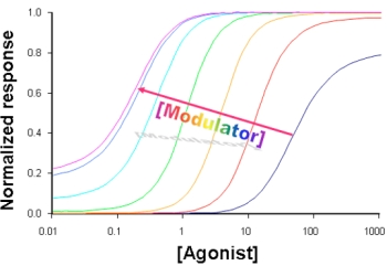
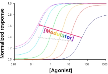
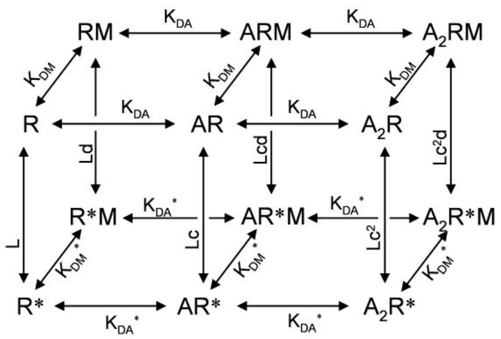
3. Kinetic Models and Mechanisms of Allosteric Modulation

For the mechanism of allosteric modulator action, it has long been believed that binding of allosteric modulators to the receptor directly changes receptor affinity for its agonist. In this model, the allosteric modulator does not have an intrinsic ability to open the channel. However, this view has been changed by the observation that the benzodiazepine allosteric agonist, diazepam, can directly activate the wild type GABAA receptor (with very low efficacy). This effect is only visible when the receptor is highly expressed in Xenopus oocytes [35]. The super-high expression of the wild type GABAA receptor also makes it possible to observe spontaneous opening, allowing for testing of the direct action of GABAA receptor negative allosteric modulators. In fact, the benzodiazepine inverse agonist methyl-6,7-dimethoxy-4-ethyl-β-carboline-3-carboxylate (DMCM) can inhibit the measurable current formed by spontaneous opening of highly expressed wild type GABAA receptors. These small fractional changes in channel opening induced by positive or negative allosteric modulators can account for the alteration of the receptor sensitivity to agonist according to the MWC allosteric activation mechanism [35]. A similar direct gating mechanism, termed allosteric coagonism, was also proposed for the mechanism of benzodiazepine action. This view is supported by the observation of direct activation and inhibition of a mutant GABAA receptor by benzodiazepine agonists and inverse agonists [36]. By making concatenated subunits and selectively destroying one orthosteric binding site, Baur and Sigel found that the benzodiazepine allosteric potentiator diazepam can facilitate channel opening when the receptor has only one orthosteric binding site [37]. This finding also supports the view that allosteric modulators facilitate channel gating, and that apparent agonist affinity changes are due to this gating effect. Normally, in the resting unbound state, GABAA receptors have very low open probabilities (10−4–10−5) [33,36]. The receptor with a single agonist bound will have an open probability increase of ~700 fold [33]. However, the open probability of a singly bound receptor is still very low. Binding of a positive allosteric modulator to the receptor will further increase open probability as observed by Baur and Sigel. The kinetic model in Figure 3 represents an expanded MWC model that includes direct gating effects of GABAA receptors by a modulator [35]. In this model, direct gating by a positive allosteric modulator also increases the agonist binding affinity. In fact, the benzodiazepine agonist flurazepam can induce structural changes in the orthosteric GABA binding pocket, further supporting a direct gating mechanism for allosteric modulators [38].
Figure 3. Kinetic scheme for allosteric activation and modulation mechanisms (adapted from [35]). Front is the conventional MWC model. The only difference is that KDA and KDA* are used to represent agonist binding affinity. Allosteric modulator (M) bound states are shown at the back of the figure, with KDM and KDM* describing two different affinities for the modulator in resting or activated states. L is the ratio between R and R*. c is the ratio between KDA* and KDA. d is the ratio between KDM and KDM*.
Pharmaceuticals 03 02592 g003
This co-activation mechanism of GABAARs by benzodiazepines seems to be applicable to other allosteric modulators acting on different sites. For example, the general anesthetic etomidate potentiates GABAA receptor agonist responses at low concentrations, but directly activates the GABAA receptor at high concentrations. At the same time, high concentration etomidate also further shifts the GABA concentration-response curve to the left. Both potentiation and activation effects can be well described by a single MWC kinetic model [39], similar to that for benzodiazepines. Affinity labeling has identified the binding site of etomidate in the transmembrane domain (Met236 in the M1 domain of α1 subunit and Met286 in M3 domain of β2 subunit) [40]. The allosteric effect of etomidate can be mimicked by two substitutions at these sites with the bulky residue tryptophan [41]. In contrast, the benzodiazepine binding pocket is located in the amino-terminal domain (see section 4.1). Thus, regardless the location of the binding site, in the amino-terminal domain distant from the channel gate or in the transmembrane domain close to the channel gate, the binding of allosteric modulators to their sites alters the channel gating. Positive allosteric modulators reduce the gating energy barrier to facilitate channel opening, serving as weak co-agonists (binding to a site that is distinct from the orthosteric neurotransmitter binding site). Conversely, negative allosteric modulators increase the gating energy barrier to inhibit channel opening. For example, ρ1 GABAC receptor function can be inhibited by several neurosteroids [42]. These neurosteroids inhibit the GABAC receptor allosterically as suggested by monitoring conformational changes with site-specific fluorescence techniques [43]. However, the mechanisms of GABAC receptor inhibition by individual neurosteroids are not the same. Functional inhibition by β-estradiol is correlated to the inhibition of the GABA-induced fluorescence changes in multiple locations of the amino-terminal domain, including at K217C in loop F, L166C in loop E, and L66C away from the binding loops, [43]. This suggests that β-estradiol can antagonize GABA-induced conformational changes. In contrast, pregnanolone, pregnanolone sulfate, allo-pregnanolone sulfate, and tetrahydrodeoxycorticosterone influence GABA-induced fluorescence changes differently, suggesting distinct detailed structural mechanisms [43]. Note that some competitive antagonists could be very weak agonists or inverse agonists. However, since they compete with agonist for the same binding site, their modulatory effect on the agonist response could only be seen at very low concentrations when the agonist concentration is also very low, with low probability of mutual competition. In any case, since these competitive antagonists compete with agonists for the same orthosteric binding site, they are not allosteric modulators.
Since nicotinic receptors have an allosteric activation mechanism similar to that for GABAA receptor [22], they likely have a similar allosteric modulation mechanism. In fact, the allosteric modulator PNU-120596 for α7 nicotinic receptor has its binding site in the transmembrane domain [44] (see Section 4.2 for details), but it can induce a similar, though not identical, conformational change along the activation pathway as acetylcholine-induced conformational changes during channel activation [45]. Similarly, allosteric modulation by divalent cations of nicotinic receptors involves conformational changes in the amino-terminal domain, partially shared with agonist-induced conformational changes [46]. These experimental observations further support a co-agonist action for positive allosteric modulators in nicotinic receptors.
In glycine and GABA receptors, alcohol size cut-off (see Section 4.2 for a fuller explanation) is related to the size of the sidechain of two residues in the transmembrane domains (M2 and M3, see Section 4.2 for details), suggesting a transmembrane location of their binding site. However, the structure of loop 2 from the amino-terminal domain, which plays an important role in coupling agonist binding and channel gating, is also critical for ethanol’s effect [47,48]. Whether loop 2 and its surrounding residues form a second binding site for ethanol is still not clear. Ethanol’s modulatory effect on glycine receptor can also be influenced by a third site within the intracellular loop between M3 and M4, which modifies ethanol’s effect by interacting with G protein subunits [49]. Thus, it is likely that the allosteric modulation of receptor function by ethanol is through co-agonism with ethanol binding to one site in the M2 and M3 domains, and that this co-agonist effect can itself be allosterically modulated by other allosteric modulators, such as G protein interacting with the M3–M4 site, or by mutations of important residues along the activation pathway, such as in loop 2.
In summary, several lines of evidence suggest that allosteric modulation of cys-loop receptor function is mediated mainly through a weak direct gating effect by allosteric modulators.

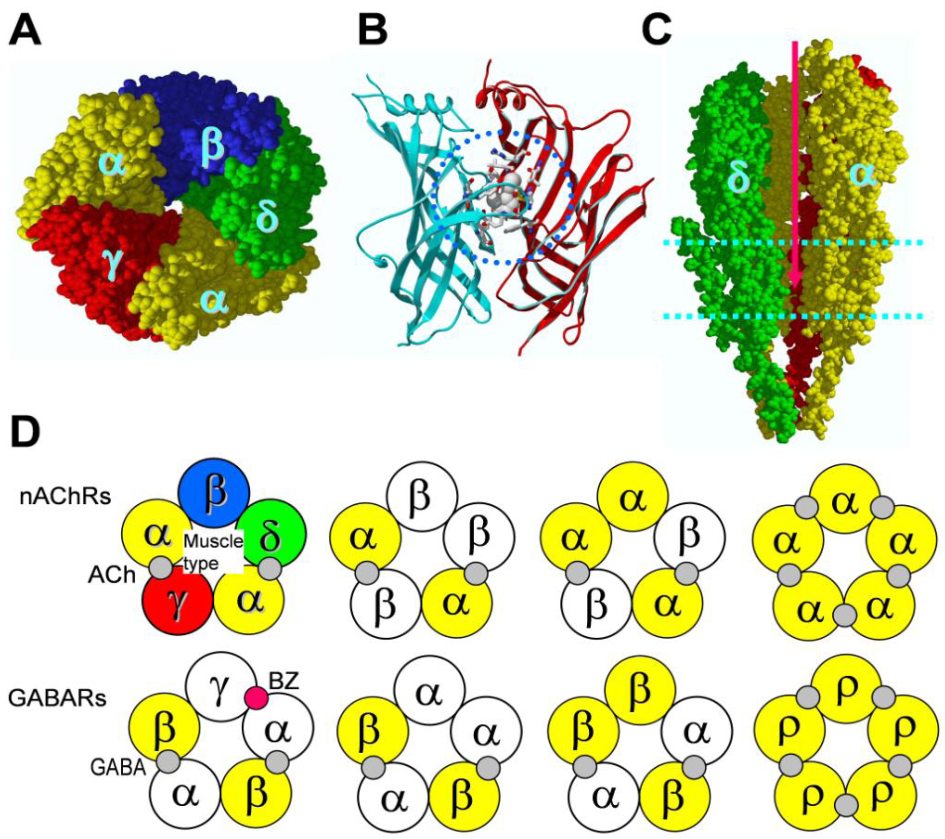
4. Binding Sites for Allosteric Modulators

As just described, allosteric modulators of cys-loop receptors can bind in two major locations. The first location is at subunit interfaces of the amino-terminal domain, homologous to the orthosteric agonist binding sites, but at a different subunit interface. Benzodiazepine agonists, partial agonists, antagonists, partial inverse agonists, and inverse agonists bind to sites in this category. We will describe the amino-terminal binding sites for allosteric modulators in Section 4.1. The second major location of allosteric modulator binding sites is in the transmembrane domain near the channel gate. Barbiturates, neurosteroids, alcohols and general anesthetics bind to sites of this type. We will describe the transmembrane binding sites with several examples in Section 4.2. Noncompetitive antagonists also bind to the transmembrane domain. Some of them have binding sites within the pore. Thus, binding of these antagonists will occlude the channel pore. The effects of these channel blockers are usually use-dependent. That is, the binding site is only accessible in the open state. Other noncompetitive antagonists allosterically inhibit channel activity. It is also possible that a single antagonist can block the pore, and simultaneously allosterically inhibit channel function. This may explain a small shift in concentration-response curve sometimes observed in the presence of noncompetitive antagonist. Picrotoxin is an example of a pore blocker, although the mechanism of its non-competitive antagonism is still not fully understood. Three studies suggest that it can actively antagonize conformational changes induced by GABA in the amino-terminal domain; suggesting that, in addition to blocking the pore, picrotoxin also negatively modulates channel gating and facilitates channel closing [43,50,51].
In addition to these two major binding sites, residues in the coupling region between the agonist binding site and the channel domains may also be involved in mediating allosteric modulation. For example, mutations of the Gly219 residue in the pre-M1 region of the GABAA receptor β2 subunit can abolish the effect of several allosteric modulators, such as barbiturates, alphaxalone, etomidate and propofol [52]. The pre-M1 residue Arg246 of the 5-HT3A receptor is also an important determinant for allosteric modulation [53]. Another example is that the zinc modulation of GABAA receptors involves two charged residues located in the cys-loop region [54] that plays an important role in coupling the amino-terminal domain to the channel gate [55,56].

4.1. Amino terminal binding sites for allosteric modulators

Since most heteromeric cys-loop receptors have only two orthosteric binding sites for agonists, there is a lot of potential for future drug development in the remaining subunit interfaces that do not bind conventional agonists. The best-characterized example of an allosteric site in amino-terminal domain is the benzodiazepine binding pocket of GABAA receptors. It is located in the α−γ subunit interface, homologous to the GABA binding site in the β−α subunit interface (Figure 1D). The benzodiazepine binding pocket is formed by residues in six binding loops, designated as loops A through F, and homologous to those that form the orthosteric binding pockets. Residues (with rat α1 subunit numbering) identified from Loops A (His101 [57,58], B (Tyr159 [59]), and C (Gly200 [60], Thr206, and Tyr209 [59,61]) are contributed by the α subunit and form the principal face of the binding pocket. Residues in loops D (Phe77 [62,63], Ala79, Thr81 [64,65]), E (Met130 [63,66]), and F (Glu189, Thr193, and Arg194 [67]) are contributed by γ2 subunit and form the complementary face of the binding pocket (Figure 4).
For nicotinic receptors, a recent study shows that there is an amino-terminal noncanonical allosteric site for the positive allosteric modulator morantel in the α3β2 subtype [68]. This binding site for the allosteric modulator is located in the β−α subunit interface (Figure 4), in contrast to the α−β interface for the ACh binding site (Figure 1D). Note that binding residues identified for morantel are located what is equivalent to the upper half of the homologous orthosteric binding pocket.
Figure 4. Allosteric modulator binding sites (shown as residues with side-chains displayed) mapped on to the homologous sites of the EM structure of Torpedo nicotinic receptor α (right) and β (left) subunits (made from 2BG9 PDB file [16]). A wide range of binding sites for different allosteric modulators are color-coded: Yellow: benzodiazepine sites; Pink: morantel sites; Red: endogenous neurosteroid sites; Green: etomidate sites; Dark green: volatile anesthetic site; Blue: PNU-120596 site for α7 nicotinic receptor; Cyan: overlapped sites in M2 or M3 domains shared by multiple modulators; Grey: divalent cation sites; Burgundy: pre-M1 residues that influence anesthetic modulation in GABAA receptor or 5-HT3 receptor; Note that if a binding site residue is located within a subunit, then the residue is mapped to the α subunit (right). If a binding pocket is in subunit interface (involving both subunits), then the residues are mapped to the positions in α or β subunits, so that they are facing to the same subunit interface. The G protein binding site in the GlyR intracellular loop is not shown, since much of the intracellular loop structure is undefined in the EM structure used to create this illustration.
Pharmaceuticals 03 02592 g004

4.2. Transmembrane domain binding sites for allosteric modulators

The transmembrane domain is the channel forming domain, which includes gating machinery to control channel opening and closing. Several classes of allosteric modulators have their binding pockets in the transmembrane domain. These include ethanol and other short-chain alcohols, neurosteroids, barbiturates, general anesthetics, and some α7 nAChR positive allosteric modulators (described more-fully later). A general feature of this type of modulation is that the modulators bind to a cavity near the channel pore-lining domain, M2, and more directly influence channel gating. The following are some examples of allosteric modulator binding sites in this category.
Binding of alcohol and volatile anesthetics to the receptor has a cutoff phenomenon. That is, the allosteric effect is dependent on the size of these compounds. When the size of the compounds increases above a certain level, the allosteric effect either decreases or is abolished. This cutoff phenomenon suggests that the receptor has a cavity with a defined volume to accommodate this type of allosteric modulator. For example, using three volatile anesthetics combined with site-directed mutagenesis at the 232, 270 and 279 positions of human GABAA receptor α2 subunit to residues with different sizes, Jenkins et al. estimated the size of the binding cavity for these anesthetics as between 250 and 370 Å3 in the wild type GABAA receptor [69]. Thus, the residues Leu232 (M1), Ser270 (M2, 15’ position), and Ala291 (M3, homologous to β2Met286) in the α2 subunit play an important role in defining the binding cavity for these small molecules. For the alcohol cutoff, the effect of an alcohol is dependent on the length of its carbon chain. When the carbon chain of an alcohol exceeds a given length, its effect will decrease. The cutoff value for the carbon chain length of alcohols varies with different receptors. For example, the homomeric α1 glycine receptor has an alcohol cutoff value of 10, whereas the homomeric ρ1 GABA receptor has a cutoff value of 7. Using chimeras of glycine and GABA receptors, Wick et al. identified two critical residues in M2 and M3 domains as the main determinants of the observed difference in cutoff [70]. These two residues are Ser267 (M2, 15’ position) and Ala288 (M3) in the glycine receptor α1 subunit. The homologous residues in the ρ1 GABA receptor are Ile307 (M2) and Trp328 (M3, homologous to β2Met286). Mutation at Ser267 of the glycine receptor α1 subunit to a larger sized residue decreases the cutoff, whereas mutations of Ile307 and Trp328 in the ρ1 GABA receptor subunit to smaller residues increases the cutoff, supporting the concept of a defined cavity between M2 and M3 domains for alcohol binding.
Non-volatile general anesthetics, such as etomidate, propofol and barbiturates are mainly positive allosteric modulators of GABAA receptors, although they can inhibit nicotinic receptors and influence other channel function [71]. The binding sites of these allosteric modulators are also located in the transmembrane domain. In GABAA receptors, the etomidate binding site was identified by photoaffinity labeling [α1Met236 (M1), β2Met286 (M3)] [40] and by a mutagenesis study [β2/β3Asn265 (M2, 15’ position)] [72]. The M3 binding site Met286 is shared with propofol [73], whereas M2 residue Asn265 is homologous to Leu270 of the 5-HT3 receptor, which is also responsible for volatile anesthetic action [74]. ([H3]azi)Etomidate photoaffinity labeling of α1Met236 (M1) &β2Met286 (M3) can be directly competed by volatile anesthetic isoflurane [75], suggesting they share the binding site. However, ([H3]azi) etomidate photoaffinity labeling is allosterically inhibited by barbiturates and propofol [75], and allosterically enhanced by neurosteroids [76], suggesting these allosteric modulators can directly induce conformational changes in the transmembrane domains of the receptor and thus alter the binding of other allosteric modulators.
Neuroactive steroids, termed neurosteroids, include steroid hormones, their metabolites, and synthetic steroid anesthetics. In addition to genomic effects (regulating gene expression), many steroid hormones and their metabolites have non-genomic effects, including allosteric modulation of ligand-gated ion channel function. Naturally occurring neurosteroids are endogenous regulators of GABAA receptors [77]. A good example of non-genomic neurosteroid effects is positive allosteric modulation of GABAA receptors by the endogenous neurosteroids allopregnanolone and tetrahydrodeoxycorticosterone (THDOC). At low concentrations, they potentiate GABA effects, whereas at high concentrations, they directly activate GABAA receptors. These two different effects are mediated by binding of the neurosteroids to two different binding sites in the transmembrane domain [78]. The potentiation site is located within the transmembrane domain of the α1 subunit, formed by residues Gln241 (M1), Asn407 (M4), and Tyr410 (M4). In contrast, the activation site is located at the subunit interface of the transmembrane domains between β2 and α1 subunits, formed by β2Tyr284 (M3) and α1Thr236 (M1). Neurosteroids also modulate nicotinic receptors. However, they are not well characterized in this context.
Compared to GABA receptors, development of nicotinic receptor allosteric modulators is more recent. Positive allosteric modulators for the α7 nAChR are the best-developed examples of nicotinic receptor modulators. α7 nAChR has fast desensitization kinetics. Positive allosteric modulators (PAMs) for α7 nAChR can be classified into two types: type I and type II, according to their influence on current kinetics. Type I PAMs mainly potentiate current without significantly influencing receptor desensitization. Ivermectin [79], 5-hydroxyindole (5-HI) [80], genistein [81], compound 6 [82], and NS-1738 [83] are examples of type I PAMs. 5-hydroxyindole is also an allosteric modulator for serotonin receptor type 3. The binding site of 5-HI in 5-HT3A receptor is located at Leu293 of the M2 domain [84]. In contrast, type II PAMs can dramatically reduce desensitization or even re-activate desensitized receptors. PNU-120596 is the best-characterized representative of this type [85]. Its binding pocket is located in the transmembrane domain [44], and is formed by five residues: Ser222 (M1), Ala225 (M1), Met253 (M2, 15’ position), Phe455 (M4), and Cys459 (M4). Each α7 subunit harbors one such site.
Other allosteric modulators: In addition to small drug compounds, cys-loop receptors can be allosterically modulated by divalent cations, such as zinc and calcium, and some small peptides. For example, zinc can inhibit GABAA [86] and GABAC receptors [87]. Calcium can modulate nicotinic receptor desensitization kinetics [88]. Secreted mammalian Ly-6/uPAR-related protein 1 (SLURP-1) is also a potentiator of α7 nicotinic receptors [89]. Small peptide allosteric modulators of glycine receptors can be selected from a peptide phage-display library [90]. The zinc binding sites for zinc modulation of GABAA receptors have been identified as five charged residues in three locations of the receptor. They are α1Glu137 (pre-cys-loop), α1His141 (cys-loop), β3Glu182 (loop F), β3His267 (M2, 17’ position), β3Glu270 (M2, 20’ position) [54]. Similar sites in the β2 subunit are responsible for the high sensitivity of the α1β2 GABAA receptor to zinc potentiation [91]. The calcium binding sites in α7 nAChR are at Glu172 of loop F, homologous to the zinc binding site in the β3 GABA receptor subunit (β3Glu182), and Glu44 of loop 2 [92,93].
In summary, the mechanism of action for all allosteric modulators of cys-loop proteins is likely to involve altering channel gating. The alteration of channel gating will influence the distribution of receptors across different affinity states. The net observable effects are changes in the receptor maximal current, gating kinetics, and sensitivity to agonist. All these changes can be explained by a single MWC-type allosteric model. There are two major binding site locations for allosteric modulators: at amino-terminal domain subunit interfaces and the transmembrane domain. However, other locations along the activation pathway, especially the coupling region between amino-terminal domain and transmembrane domain, or even the intracellular loop, could be potential sites for novel allosteric modulator development. In fact, G protein βγ can modify glycine receptor function by interacting with two basic motifs in the beginning and the end of the M3−M4 intracellular loop [94]. The positively charged residues in these two motifs, along with the Gβγ protein, are also critical for the action of ethanol on the glycine receptor [49]. Figure 4 summarizes the binding sites discussed above, mapped onto the 3-D EM structure of Torpedo nicotinic receptor α and β subunits [16].
GABAA receptor allosteric modulators are relatively well characterized and developed. A recent research trend is to develop novel allosteric modulators with subtype specificity in other members of the cys-loop family, especially nicotinic receptors. Another important development is negative allosteric modulators of GABAA receptors. Development of GABAA receptor negative allosteric modulators was hampered by their non-selectivity between different receptor subtypes, resulting in increased seizure susceptibility. However, recent development of more subtype selective negative allosteric modulators shows promising results. For example, α5 subunit containing GABAA receptors are mainly distributed in the hippocampus, and α5 selective inverse agonists (negative allosteric modulators) can improve cognitive function [95,96]. We expect more subtype specific allosteric modulators for cys-loop receptors will be developed in the near future.

Acknowledgements

This work is supported by the Barrow Neurological Foundation and NIH R01GM085237 grant to YC.

References

  1. Albuquerque, E.; Pereira, E.; Alkondon, M.; Rogers, S. Mammalian nicotinic acetylcholine receptors: from structure to function. Physiol. Rev. 2009, 89, 73–120. [Google Scholar]
  2. Thompson, A.; Lummis, S. The 5-HT3 receptor as a therapeutic target. Expert Opin. Ther. Targets 2007, 11, 527–540. [Google Scholar]
  3. Olsen, R.; Sieghart, W. International Union of Pharmacology. LXX. Subtypes of γ-aminobutyric acid (A) receptors: classification on the basis of subunit composition, pharmacology, and function. Update. Pharmacol. Rev. 2008, 60, 243–260. [Google Scholar]
  4. Olsen, R.; Sieghart, W. GABAA receptors: subtypes provide diversity of function and pharmacology. Neuropharmacology 2009, 56, 273–284. [Google Scholar]
  5. Johnston, G.; Chebib, M.; Hanrahan, J.; Mewett, K. GABAC receptors as drug targets. Curr. Drug Targets CNS Neurol. Disord. 2003, 2, 260. [Google Scholar]
  6. Lynch, J. Native glycine receptor subtypes and their physiological roles. Neuropharmacology 2009, 56, 303–309. [Google Scholar]
  7. Davies, P.; Wang, W.; Hales, T.; Kirkness, E. A novel class of ligand-gated ion channel is activated by Zn2+. J. Biol. Chem. 2003, 278, 712–717. [Google Scholar]
  8. Changeux, J.; Edelstein, S. Allosteric receptors after 30 years. Neuron 1998, 21, 959–980. [Google Scholar]
  9. Lester, H.; Dibas, M.; Dahan, D.; Leite, J.; Dougherty, D. Cys-loop receptors: new twists and turns. Trends. Neurosci. 2004, 27, 329–336. [Google Scholar]
  10. Ortells, M.; Lunt, G. Evolutionary history of the ligand-gated ion-channel superfamily of receptors. Trends. Neurosci. 1995, 18, 121–127. [Google Scholar]
  11. Bocquet, N.; Nury, H.; Baaden, M.; Poupon, C.; Changeux, J.; Delarue, M.; Corringer, P. X-ray structure of a pentameric ligand-gated ion channel in an apparently open conformation. Nature 2009, 457, 111–114. [Google Scholar]
  12. Tasneem, A.; Iyer, L.M.; Jakobsson, E.; Aravind, L. Identification of the prokaryotic ligand-gated ion channels and their implications for the mechanisms and origins of animal Cys-loop ion channels. Genome. Biol. 2004, 6, R4. [Google Scholar]
  13. Hilf, R.; Dutzler, R. X-ray structure of a prokaryotic pentameric ligand-gated ion channel. Nature 2008, 452, 375–379. [Google Scholar]
  14. Unwin, N. Nicotinic acetylcholine receptor at 9 Å resolution. J. Mol. Biol. 1993, 229, 1101–1124. [Google Scholar]
  15. Miyazawa, A.; Fujiyoshi, Y.; Unwin, N. Structure and gating mechanism of the acetylcholine receptor pore. Nature 2003, 423, 949–955. [Google Scholar]
  16. Unwin, N. Refined structure of the nicotinic acetylcholine receptor at 4 Å resolution. J. Mol. Biol. 2005, 346, 967–989. [Google Scholar]
  17. Celie, P.; Rossum-Fikkert, S.; Dijk, W.; Brejc, K.; Smit, A.; Sixma, T. Nicotine and carbamylcholine binding to nicotinic acetylcholine receptors as studied in AChBP crystal structures. Neuron 2004, 41, 907–914. [Google Scholar]
  18. Brejc, K.; Dijk, W.; Klaassen, R.; Schuurmans, M.; Oost, J.; Smit, A.; Sixma, T. Crystal structure of an ACh-binding protein reveals the ligand-binding domain of nicotinic receptors. Nature 2001, 411, 269–276. [Google Scholar]
  19. Ulens, C.; Hogg, R.; Celie, P.; Bertrand, D.; Tsetlin, V.; Smit, A.; Sixma, T. Structural determinants of selective α-conotoxin binding to a nicotinic acetylcholine receptor homolog AChBP. Proc. Natl. Acad. Sci. USA 2006, 103, 3615–3620. [Google Scholar]
  20. Hansen, S.; Sulzenbacher, G.; Huxford, T.; Marchot, P.; Taylor, P.; Bourne, Y. Structures of Aplysia AChBP complexes with nicotinic agonists and antagonists reveal distinctive binding interfaces and conformations. EMBO J. 2005, 24, 3635–3646. [Google Scholar]
  21. Chang, Y.; Wu, W.; Zhang, J.; Huang, Y. Allosteric activation mechanism of the cys-loop receptors. Acta Pharmacol. Sin. 2009, 30, 663–672. [Google Scholar]
  22. Edelstein, S.; Changeux, J. Allosteric proteins after thirty years: the binding and state functions of the neuronal α7 nicotinic acetylcholine receptors. Experientia 1996, 52, 1083–1090. [Google Scholar]
  23. Chen, Y.; Reilly, K.; Chang, Y. Evolutionarily conserved allosteric network in the cys-loop family of ligand-gated ion channels revealed by statistical covariance analyses. J. Biol. Chem. 2006, 281, 18184–18192. [Google Scholar]
  24. Millar, P.; Smart, T. Binding; activation and modulation of Cys-loop receptors. Trends. Pharmacol. Sci. 2010, 31, 161–174. [Google Scholar]
  25. Bartos, M.; Corradi, J.; Bouzat, C. Structural basis of activation of cys-loop receptors: the extracellular-transmembrane interface as a coupling region. Mol. Neurobiol. 2009, 40, 236–252. [Google Scholar]
  26. Yakel, J. Gating of nicotinic ACh receptors: latest insights into ligand binding and function. J. Physiol. 2010, 588, 597–602. [Google Scholar]
  27. Cederholm, J.; Schofield, P.; Lewis, T. Gating mechanisms in Cys-loop receptors. Eur. Biophys. J. 2009, 39, 37–49. [Google Scholar]
  28. Taly, A.; Corringer, P.; Guedin, D.; Lestage, P.; Changeux, J. Nicotinic receptors: allosteric transitions and therapeutic targets in the nervous system. Nature Rev. Drug Dicovery 2009, 8, 733–750. [Google Scholar]
  29. Burzomato, V.; Beato, M.; Groot-Kormelink, P.; Colquhoun, D.; Sivilotti, L. Single-channel behavior of heteromeric α1β glycine receptors: an attempt to detect a conformational change before the channel opens. J. Neurosci. 2004, 24, 10924–10940. [Google Scholar]
  30. Moroni, M.; Zwart, R.; Sher, E.; Cassels, B.; Bermudez, I. α4β2 nicotinic receptors with high and low acetylcholine sensitivity: pharmacology, stoichiometry, and sensitivity to long-term exposure to nicotine. Mol. Pharmacol. 2006, 70, 755–768. [Google Scholar]
  31. Monod, J.; Wyman, J.; Changeux, J. On the nature of allosteric proteins: a plausible model. J. Mol. Biol. 1965, 12, 88–118. [Google Scholar]
  32. Jackson, M. Spontaneous openings of the acetylcholine receptor channel. Proc. Natl. Acad. Sci. USA 1984, 81, 3901–3904. [Google Scholar]
  33. Chang, Y.; Weiss, D. Allosteric activation mechanism of the α1β2γ2 γ-aminobutyric acid type A receptor revealed by mutation of the conserved M2 leucine. Biophys. J. 1999, 77, 2542–2551. [Google Scholar]
  34. Lape, R.; Colquhoun, D.; Sivilotti, L. On the nature of partial agonism in the nicotinic receptor superfamily. Nature 2008, 454, 722–727. [Google Scholar]
  35. Campo-Soria, C.; Chang, Y.; Weiss, D. Mechanism of action of benzodiazepines on GABAA receptors. Br. J. Pharmacol. 2006, 148, 984–990. [Google Scholar]
  36. Rüsch, D.; Forman, S. Classic benzodiazepines modulate the open–close equilibrium in α1β2γ2L γ-aminobutyric acid type A receptors. Anesthesiology 2005, 102, 783–792. [Google Scholar]
  37. Bau, R.; Sigel, E. Benzodiazepines affect channel opening of GABAA receptors induced by either agonist binding site. Mol. Pharmacol. 2005, 67, 1005–1008. [Google Scholar]
  38. Kloda, H.; Czajkowski, C. Agonist-, antagonist-, and benzodiazepine-Induced structural changes in the a1Met113-Leu132 region of the GABAA receptor. Mol. Pharmacol. 2007, 71, 483–493. [Google Scholar]
  39. Rüsch, D.; Zhong, H.; Forman, S. Gating allosterism at a single class of etomidate sites on α1β2γ2L GABAA receptors accounts for both direct activation and agonist modulation. J. Biol. Chem. 2004, 279, 20982–20992. [Google Scholar]
  40. Li, G.; Chiara, D.; Sawyer, G.; Husain, S.; Olsen, R.; Cohen, J. Identification of a GABAA receptor anesthetic binding site at subunit interfaces by photolabeling with an etomidate analog. J. Neurosci. 2006, 26, 11599–11605. [Google Scholar]
  41. Stewart, D.; Desai, R.; Cheng, Q.; Liu, A.; Forman, S. Tryptophan mutations at Azi-Etomidate photo-incorporation sites on α1 or β2 subunits enhance GABAA receptor gating and reduce etomidate modulation. Mol. Pharmacol. 2008, 74, 1687–1695. [Google Scholar]
  42. Li, W.; Jin, X.; Covey, D.; Steinbach, J. Neuroactive steroids and human recombinant ρ1 GABAC receptors. J. Pharmacol. Exp. Ther. 2007, 323, 236–247. [Google Scholar]
  43. Li, P.; Khatri, A.; Bracamontes, J.; Weiss, D.; Steinbach, J.; Akk, G. Site-specific fluorescence reveals distinct structural changes induced in the human ρ1 GABA receptor by inhibitory neurosteroids. Mol. Pharmacol. 2010, 77, 539–546. [Google Scholar]
  44. Young, G.; Zwart, R.; Walker, A.; Sher, E.; Millar, N. Potentiation of α7 nicotinic acetylcholine receptors via an allosteric transmembrane site. Proc. Natl. Acad. Sci. USA 2008, 105, 14686–14691. [Google Scholar]
  45. Barron, S.; McLaughlin, J.; See, J.; Richards, V.; Rosenberg, R. An allosteric modulator of α7 nicotinic receptors, N- (5-Chloro-2,4-dimethoxyphenyl)-N'- (5-methyl-3-isoxazolyl)-urea (PNU-120596), causes conformational changes in the extracellular ligand binding domain similar to those caused by acetylcholine. Mol. Pharmacol. 2009, 76, 253–263. [Google Scholar]
  46. McLaughlin, J.; Barron, S.; See, J.; Rosenberg, R. Conformational changes in α7 acetylcholine receptors underlying allosteric modulation by divalent cations. BMC Pharmacol. 2009, 9, 1–13. [Google Scholar]
  47. Perkins, D.; Trudell, J.; Crawford, D.; Asatryan, L.; Alkana, R.; Davies, D. Loop 2 structure in glycine and GABAA receptors plays a key role in determining ethanol sensitivity. J. Biol. Chem. 2009, 284, 27304–27314. [Google Scholar]
  48. Perkins, D.; JR, T.; Crawford, D.; Alkana, R.; Davies, D. Targets for ethanol action and antagonism in loop 2 of the extracellular domain of glycine receptors. J. Neurochem. 2008, 106, 1337–1349. [Google Scholar]
  49. Yevenes, G.; Moraga-Cid, G.; Peoples, R.; Schmalzing, G.; Aguayo, L. A selective Gβγ-linked intracellular mechanism for modulation of a ligand-gated ion channel by ethanol. Proc. Natl. Acad. Sci. USA 2008, 51, 20523–20528. [Google Scholar]
  50. Chang, Y.; Weiss, D. Site-specific fluorescence reveals distinct structural changes with GABA receptor activation and antagonism. Nat. Neurosci. 2002, 5, 1163–1168. [Google Scholar]
  51. Zhang, J.; Xue, F.; Chang, Y. Agonist- and antagonist-induced conformational changes of loop F and their contributions to the ρ1 GABA receptor function. J. Physiol. 2009, 587, 139–153. [Google Scholar]
  52. Chang, C.; Olcese, R.; Olsen, R. A single M1 residue in the β2 subunit alters channel gating of GABAA receptor in anesthetic modulation and direct activation. J. Biol. Chem. 2003, 278, 42821–42828. [Google Scholar]
  53. Hu, X.; Peoples, R. Arginine 246 of the pretransmembrane domain 1 region alters 2,2,2-richloroethanol action in the 5-hydroxytryptamine3A receptor. J. Pharmacol. Exp. Ther. 2008, 324, 1011–1018. [Google Scholar]
  54. Hosie, A.; Dunne, E.; Harvey, R.; Smart, T. Zinc-mediated inhibition of GABA (A) receptors: discrete binding sites underlie subtype specificity. Nat. Neurosci. 2003, 6, 362–369. [Google Scholar]
  55. Kash, T.; Jenkins, A.; Kelley, J.; Trudell, J.; Harrison, N. Coupling of agonist binding to channel gating in the GABAA receptor. Nature 2003, 421, 272–275. [Google Scholar]
  56. Kash, T.; Dizon, M.; Trudell, J.; Harrison, N. Charged residues in the β2 subunit involved in GABAA receptor activation. J. Biol. Chem. 2004, 279, 4887–4893. [Google Scholar]
  57. Duncalfe, L.; Carpenter, M.; Smillie, L.; Martin, I.; Dunn, S. The major site of photoaffinity labeling of the GABAA receptor by [3H] flunitrazepam is histidine 102 of the a subunit. J. Biol. Chem. 1996, 271, 9209–9214. [Google Scholar]
  58. Wieland, H.; Luddens, H.; Seeburg, P. A single histidine in GABAA receptors is essential for benzodiazepine agonist binding. J. Biol. Chem. 1992, 267, 1426–1429. [Google Scholar]
  59. Amin, J.; Brooks-Kayal, A.; Weiss, D. Two tyrosine residues on the a subunit are crucial for benzodiazepine binding and allosteric modulation of γ-aminobutyric acid A receptors. Mol. Pharmacol. 1997, 51, 833–841. [Google Scholar]
  60. Schaerer, M.; Buhr, A.; Baur, R.; Sigel, E. Amino acid residue 200 on the α1 subunit of GABA (A) receptors affects the interaction with selected benzodiazepine binding site ligands. Eur. J. Pharmacol. 1998, 354, 283–288. [Google Scholar]
  61. Buhr, A.; Schaerer, M.; Baur, R.; Sigel, E. Residues at positions 206 and 209 of the α1 subunit of γ-aminobutyric acid A receptors influence affinities for benzodiazepine binding site ligands. Mol. Pharmacol. 1997, 52, 676–682. [Google Scholar]
  62. Buhr, A.; Baur, R.; Sigel, E. Subtle changes in residue 77 of the γ subunit of α1β2γ2 GABAA receptors drastically alter the affinity for ligands of the benzodiazepine binding site and suggest the presence of two sites. J. Biol. Chem. 1997, 272, 11799–11804. [Google Scholar]
  63. Wingrove, P.; Thompson, S.; Wafford, K.; Whiting, P. Key amino acids in the gamma subunit of the γ-aminobutyric acid A receptor that determine ligand binding and modulation at the benzodiazepine site. Mol. Pharmacol. 1997, 52, 874–881. [Google Scholar]
  64. Kucken, A.M.; Teissere, J.A.; Seffinga-Clark, J.; Wagner, D.A.; Czajkowski, C. Structural requirements for imidazobenzodiazepine binding to GABA (A) receptors. Mol. Pharmacol. 2003, 63, 289–296. [Google Scholar]
  65. Teissere, J.; Czajkowski, C. A β-strand in the γ2 subunit lines the benzodiazepine binding site of the GABAA receptor: structural rearrangements detected during channel gating. J. Neurosci. 2001, 21, 4977–4986. [Google Scholar]
  66. Buhr, A.; Sigel, E. A point mutation in the γ2 subunit of γ-aminobutyric acid type A receptors results in altered benzodiazepine site specificity. Proc. Natl. Acad. Sci. USA 1997, 94, 8824–8829. [Google Scholar]
  67. Sancar, F.; Ericksen, S.; Kucken, A.; Teissére, J.; Czajkowski, C. Structural determinants for high-affinity zolpidem binding to GABA-A receptors. Mol. Pharmacol. 2007, 71, 38–46. [Google Scholar]
  68. Seo, S.; Henry, J.; Lewis, A.; Wang, N.; Levandoski, M. The positive allosteric modulator morantel binds at noncanonical subunit interfaces of neuronal nicotinic acetylcholine receptors. J. Neurosci. 2009, 29, 8734–8742. [Google Scholar]
  69. Jenkins, A.; Greenblatt, E.; Faulkner, H.; Bertaccini, E.; Light, A.; Lin, A.; Andreasen, A.; Viner, A.; Trudell, J.; Harrison, N. Evidence for a common binding cavity for three general anesthetics within the GABAA receptor. J. Neurosci. 2001, 21, RC136. [Google Scholar]
  70. Wick, M.; Mihic, S.; Ueno, S.; Mascia, M.; Trudell, J.; Brozowski, S.; Ye, Q.; Harrison, N.; Harris, R. Mutations of gamma-aminobutyric acid and glycine receptors change alcohol cutoff: evidence for an alcohol receptor? Proc. Natl. Acad. Sci. USA 1998, 95, 6504–6509. [Google Scholar]
  71. Frank, N. General anaesthesia: from molecular targets to neuronal pathways of sleep and arousal. Nat. Rev. Neurosci. 2008, 9, 370–386. [Google Scholar]
  72. Belelli, D.; Lambert, J.; Peters, J.; Wafford, K.; Whiting, P. The interaction of the general anesthetic etomidate with the γ-aminobutyric acid type A receptor is influenced by a single amino acid. Proc. Natl. Acad. Sci. USA 1997, 94, 11031–11036. [Google Scholar]
  73. Bali, M.; Akabas, M. Defining the propofol binding site location on the GABAA receptor. Mol. Pharmacol. 2004, 65, 68–76. [Google Scholar]
  74. Lopreato, G.; Banerjee, P.; Mihic, S. Amino acids in transmembrane domain two influence anesthetic enhancement of serotonin-3A receptor function. Brain Res. Mol. Brain Res. 2003, 118, 45–51. [Google Scholar]
  75. Li, G.; Chiara, D.; Cohen, J.; Olsen, R. Numerous classes of general anesthetics inhibit etomidate binding to γ-aminobutyric acid type A (GABAA) receptors. J. Biol. Chem. 2010, 285, 8615–8620. [Google Scholar]
  76. Li, G.; Chiara, D.; Cohen, J.; RW, O. Neurosteroids allosterically modulate binding of the anesthetic etomidate to γ-aminobutyric acid type A receptors. J. Biol. Chem. 2009, 284, 11771–11775. [Google Scholar]
  77. Belelli, D.; Lambert, J. Neurosteroids: endogenous regulators of the GABAA receptor. Nat. Rev. Neurosci. 2005, 6, 565–575. [Google Scholar]
  78. Hosie, A.; Wilkins, M.; da Silva, H.; Smart, T. Endogenous neurosteroids regulate GABAA receptors through two discrete transmembrane sites. Nature 2006, 444, 486–489. [Google Scholar]
  79. Krause, R.; Buisson, B.; Bertrand, S.; Corringer, P.; Galzi, J.; Changeux, J.; Bertrand, D. Ivermectin: a positive allosteric effector of the α7 neuronal nicotinic acetylcholine receptor. Mol. Pharmacol. 1998, 53, 283–294. [Google Scholar]
  80. Zwart, R.; De Filippi, G.; Broad, L.; McPhie, G.; Pearson, K.; Baldwinson, T.; Sher, E. 5-Hydroxyindole potentiates human α7 nicotinic receptor-mediated responses and enhances acetylcholine-induced glutamate release in cerebellar slices. Neuropharmacology 2002, 43, 374–384. [Google Scholar]
  81. Charpantier, E.; Wiesner, A.; Huh, K.; Ogier, R.; Hoda, J.; Allaman, G.; Raggenbass, M.; Feuerbach, D.; Bertrand, D.; Fuhrer, C. α7 neuronal nicotinic acetylcholine receptors are negatively regulated by tyrosine phosphorylation and Src-family kinases. J. Neurosci. 2005, 25, 9836–9849. [Google Scholar]
  82. Ng, H.; Whittemore, E.; Tran, M.; Hogenkamp, D.; Broide, R.; Johnstone, T.; Zheng, L.; Stevens, K.; Gee, K. Nootropic α7 nicotinic receptor allosteric modulator derived from GABAA receptor modulators. Proc. Natl. Acad. Sci. USA 2007, 104, 8059–8064. [Google Scholar]
  83. Timmermann, D.; Grønlien, J.; Kohlhaas, K.; Nielsen, E.; Dam, E.; Jørgensen, T.; Ahring, P.; Peters, D.; Holst, D.; Christensen, J.; Malysz, J.; Briggs, C.; Gopalakrishnan, M.; Olsen, G. An allosteric modulator of the α7 nicotinic acetylcholine receptor possessing cognition-enhancing properties in vivo. J. Pharmacol. Exp. Ther. 2007, 323, 294–307. [Google Scholar]
  84. Hu, X.; Lovinger, D. The L293 residue in transmembrane domain 2 of the 5-HT3A receptor is a molecular determinant of allosteric modulation by 5-hydroxyindole. Neuropharmacology 2008, 54, 1153–1165. [Google Scholar]
  85. Hurst, R.; Hajós, M.; Raggenbass, M.; Wall, T.; Higdon, N.; Lawson, J.; Rutherford-Root, K.; Berkenpas, M.; Hoffmann, W.; Piotrowski, D.; Groppi, V.; Allaman, G.; Ogier, R.; Bertrand, S.; Bertrand, D.; Arneric, S. A novel positive allosteric modulator of the α7 neuronal nicotinic acetylcholine receptor: in vitro and in vivo characterization. J. Neurosci. 2005, 25, 4396–4405. [Google Scholar]
  86. Smart, T.; Moss, S.; Xie, X.; Huganir, R. GABAA receptors are differentially sensitive to zinc: dependence on subunit composition. Br. J. Pharmacol. 1991, 103, 1837–1839. [Google Scholar]
  87. Chang, Y.; Amin, J.; Weiss, D. Zinc is a mixed antagonist of homomeric ρ1 γ-aminobutyric acid-activated channels. Mol. Pharmacol. 1995, 47, 595–602. [Google Scholar]
  88. Guo, X.; Lester, R. Regulation of nicotinic acetylcholine receptor desensitization by Ca2+. J. Neurophysiol. 2007, 97, 93–101. [Google Scholar]
  89. Chimienti, F.; Hogg, R.; Plantard, L.; Lehmann, C.; Brakch, N.; Fischer, J.; Huber, M.; Bertrand, D.; Hohl, D. Identification of SLURP-1 as an epidermal neuromodulator explains the clinical phenotype of Mal de Meleda. Hum. Mol. Genet. 2003, 12, 3017–3024. [Google Scholar]
  90. Tipps, M.; Lawshe, J.; Ellington, A.; Mihic, S. Identification of novel specific allosteric modulators of the glycine receptor using phage display. J. Biol. Chem. 2010. Epub ahead of print.. [Google Scholar]
  91. Trudell, J.; Yue, M.; Bertaccini, E.; Jenkins, A.; Harrison, N. Molecular modeling and mutagenesis reveals a tetradentate binding site for Zn2+ in GABA (A) αβ receptors and provides a structural basis for the modulating effect of the gamma subunit. J. Chem. Inf. Mod. 2008, 48, 344–349. [Google Scholar]
  92. Le Novère, N.; Grutter, T.; Changeux, J. Models of the extracellular domain of the nicotinic receptors and of agonist- and Ca2+-binding sites. Proc Natl Acad Sci USA 2002, 99, 3210–3215. [Google Scholar]
  93. Galzi, J.; Bertrand, S.; Corringer, P.; Changeux, J.; Bertrand, D. Identification of calcium binding sites that regulate potentiation of a neuronal nicotinic acetylcholine receptor. EMBO J 1996, 15, 5824–5832. [Google Scholar]
  94. Yevenes, G.; Moraga-Cid, G.; Guzma´n, L.; Haeger, S.; Oliveira, L.; Olate, J.; Schmalzing, G.; Aguayo, L. Molecular determinants for G protein βγ modulation of ionotropic glycine receptors. J. Biol. Chem. 2006, 281, 39300–39307. [Google Scholar]
  95. Atack, J. Preclinical and clinical pharmacology of the GABAA receptor α5 subtype-selective inverse agonist alpha5IA. Pharmacol. Ther. 2010, 125, 11–26. [Google Scholar]
  96. Ballard, T.; Knoflach, F.; Prinssen, E.; Borroni, E.; Vivian, J.; Basile, J.; Gasser, R.; Moreau, J.; Wettstein, J.; Buettelmann, B.; Knust, H.; Thomas, A.; Trube, G.; Hernandez, M. RO4938581, a novel cognitive enhancer acting at GABAA α5 subunit-containing receptors. Psychopharmocology (Berlin) 2009, 202, 207–223. [Google Scholar]

