

Clinical Trial to Assess Physiology and Activity of Masticatory Muscles of Complete Denture Wearer Following Vitamin D Intervention
 , , ,
, , ,  and
and
Abstract
:1. Introduction
2. Materials and Methods
2.1. Study Design
2.2. Study Subjects and Sample Size
2.3. Methodology
Recording of S Vit D, USG, and sEMG
- Measurement of S Vit D
- sEMG of masseter muscle
- USG of Masseter muscle
2.4. Statistical Methods
3. Results
4. Discussion
5. Conclusions
Author Contributions
Funding
Institutional Review Board Statement
Informed Consent Statement
Data Availability Statement
Conflicts of Interest
References
- Zarb, G.A.; Hobkirk, J.; Eckert, S.; Jacob, R. Prosthodontics Treatment for Edentulous Patients, 13th ed.; Elsevier: St. Louis, MO, USA, 2012; pp. 128–141. [Google Scholar]
- Ettinger, R.L. Diet, nutrition, and masticatory ability in a group of elderly edentulous patients. Aust. Dent. J. 1973, 18, 12–19. [Google Scholar] [CrossRef]
- Moynihan, P.; Petersen, P.E. Diet, nutrition and the prevention of dental diseases. Public Health Nutr. 2004, 7, 201–226. [Google Scholar] [CrossRef] [PubMed] [Green Version]
- Jacobson, T.; Krol, A. A contemporary review of the factors involved in complete denture retention, stability, and support. Part I: Retention. J. Prosthet. Dent. 1983, 49, 5–15. [Google Scholar] [CrossRef]
- Crescente, G.; Minervini, G.; Spagnuolo, C.; Moccia, S. Cannabis Bioactive Compound-Based Formulations: New Perspectives for the Management of Orofacial Pain. Molecules. 2022, 28, 106. [Google Scholar] [CrossRef] [PubMed]
- Guntona, J.E.; Girgisa, C.M. Vitamin D and Muscle. Bone Rep. 2018, 18, 163–167. [Google Scholar] [CrossRef] [PubMed]
- Atwood, D.A. Postextraction changes in the adult mandible as illustrated by microradiographs of midsagittal sections and serial cephalometric roentgenograms. J. Prosthet. Dent. 1963, 13, 810–824. [Google Scholar] [CrossRef]
- Chaturvedi, S.; Elmahdi, A.E.; Abdelmonem, A.M.; Haralur, S.B.; Alqahtani, N.M.; Suleman, G.; Sharif, R.A.; Gurumurthy, V.; A Alfarsi, M. Predoctoral dental implant education techniques—students’ perception and attitude. J. Dent. Educ. 2020, 85, 392–400. [Google Scholar] [CrossRef] [PubMed]
- Romagnoli, E.; Mascia, M.L.; Cipriani, C.; Fassino, V.; Mazzei, F.; D’Erasmo, E.; Carnevale, V.; Scillitani, A.; Minisola, S. Short and Long-Term Variations in Serum Calciotropic Hormones after a Single Very Large Dose of Ergocalciferol (Vitamin D2) or Cholecalciferol (Vitamin D3) in the Elderly. J. Clin. Endocrinol. Metab. 2008, 93, 3015–3020. [Google Scholar] [CrossRef]
- Hewison, M.; Zehnder, D.; Bland, R.; Stewart, P. 1alpha-Hydroxylase and the action of vitamin D. J. Mol. Endocrinol. 2000, 25, 141–148. [Google Scholar] [CrossRef] [Green Version]
- Scientific Advisory Committee on Nutrition (SACN). Vitamin D and Health. 2016. Available online: https://www.gov.uk/government/groups/scientific-advisory-committee-on-nutrition (accessed on 21 April 2022).
- Nair, U.P.; Shivamurthy, R.; Nagate, R.R.; Chaturvedi, S.; Al-Qahtani, S.M.; Al Magbol, M.; Gokhale, S.T.; Tikare, S.; Chaturvedi, M. Effect of Injectable Platelet-Rich Fibrin with a Nano-Hydroxyapatite Bone Graft on the Treatment of a Grade II Furcation Defect. Bioengineering 2022, 9, 602. [Google Scholar] [CrossRef]
- Butera, A.; Pascadopoli, M.; Gallo, S.; Alovisi, M.; Lovati, E.; Mutti, E.; Scribante, A. Domiciliary Management of Periodontal Indexes and Glycosylated Hemoglobin (HbA1c) in Type 1 Diabetic Patients with Paraprobiotic-Based Toothpaste and Mousse: Randomized Clinical Trial. Appl. Sci. 2022, 12, 8610. [Google Scholar] [CrossRef]
- Rajput, G.; Ahmed, S.; Chaturvedi, S.; Addas, M.K.; Bhagat, T.V.; Gurumurthy, V.; Alqahtani, S.M.; Alobaid, M.A.; Alsubaiy, E.F.; Gupta, K. Comparison of Microleakage in Nanocomposite and Amalgam as a Crown Foundation Material Luted with Different Luting Cements under CAD-CAM Milled Metal Crowns: An In Vitro Microscopic Study. Polymers 2022, 14, 2609. [Google Scholar]
- Minervini, G.; Cervino, G.; Chaturvedi, S.; Franco, R.; di Francesco, F.; Fiorillo, L.; Cicciù, M. Advanced method of rehabilitating edentulous Jaws: A review on telescopic denture. Technol. Health Care 2023, 1–17. [Google Scholar] [CrossRef] [PubMed]
- Mittal, P.; Gokhale, S.T.; Manjunath, S.; Al-Qahtani, S.M.; Magbol, M.A.; Nagate, R.R.; Tikare, S.; Chaturvedi, S.; Agarwal, A.; Venkataram, V. Comparative Evaluation of Locally Administered 2% Gel Fabricated from Lemongrass Polymer and 10% Doxycycline Hyclate Gel as an Adjunct to Scaling and Root Planing in the Treatment of Chronic Periodontitis—A Randomized Controlled Trial. Polymers 2022, 14, 2766. [Google Scholar] [CrossRef] [PubMed]
- Cervino, G.; Montanari, M.; Santonocito, D.; Nicita, F.; Baldari, R.; De Angelis, C.; Storni, G.; Fiorillo, L. Comparison of Two Low-Profile Prosthetic Retention System Interfaces: Preliminary Data of an In Vitro Study. Prosthesis 2019, 1, 54–60. [Google Scholar] [CrossRef] [Green Version]
- de Sire, A.; Ferrillo, M.; Gennari, A.; Cisari, C.; Pasqua, S.; Foglio Bonda, P.L.; Migliario, M. Bone health, vitamin d status and oral hygiene screening in breast cancer women before starting osteoporosis treatment: A cross-sectional study. J. Biol. Regul. Homeost. Agents 2021, 35, 287–292. [Google Scholar]
- Marenzi, G.; Spagnuolo, G.; Sammartino, J.C.; Gasparro, R.; Rebaudi, A.; Salerno, M. Micro-Scale Surface Patterning of Titanium Dental Implants by Anodization in the Presence of Modifying Salts. Materials 2019, 12, 1753. [Google Scholar] [CrossRef] [Green Version]
- Naddeo, P.; Laino, L.; la Noce, M.; Piattelli, A.; de Rosa, A.; Iezzi, G.; Tirino, V. Surface biocompatibility of differently textured titanium implants with mesenchymal stem cells. Dent. Mater. 2015, 31, 235–243. [Google Scholar] [CrossRef]
- Abouzeid, H.L.; Chaturvedi, S.; Abdelaziz, K.M.; Alzahrani, F.A.; AlQarni, A.A.S.; Alqahtani, N.M. Role of Robotics and Artificial Intelligence in Oral Health and Preventive Dentistry—Knowledge, Perception and Attitude of Dentists. Oral Health Prev. Dent. 2021, 19, 353–363. [Google Scholar]
- Reddy, L.K.V.; Madithati, P.; Narapureddy, B.R.; Ravula, S.R.; Vaddamanu, S.K.; Alhamoudi, F.H.; Minervini, G.; Chaturvedi, S. Perception about Health Applications (Apps) in Smartphones towards Telemedicine during COVID-19: A Cross-Sectional Study. J. Pers. Med. 2022, 12, 1920. [Google Scholar] [CrossRef]
- Shrestha, B.; Basnet, B.B.; Adhikari, G. A questionnaire study on the impact on oral health-related quality of life by conventional rehabilitation of edentulous patient. BDJ Open 2020, 6, 3. [Google Scholar] [CrossRef] [Green Version]
- Chaturvedi, S.; Addas, M.K.; Alqahtani, N.M.; Al Ahmari, N.M.; Alfarsi, M.A. Clinical analysis of CAD-CAM milled and printed complete dentures using computerized occlusal force analyser. Technol. Health Care 2021, 29, 797–811. [Google Scholar] [CrossRef] [PubMed]
- Cicciù, M.; Fiorillo, L.; D’Amico, C.; Gambino, D.; Amantia, E.M.; Laino, L.; Cervino, G. 3D digital impression systems compared with traditional techniques in dentistry: A recent data systematic review. Materials 2020, 13, 1982. [Google Scholar] [CrossRef] [PubMed]
- Chaturvedi, S.; Addas, M.K.; Alqahtani, N.M.; Al Ahmari, N.M.; Alfarsi, M.A. Computerized occlusal forces analysis in complete dentures fabricated by additive and subtractive techniques. Technol. Health Care 2021, 29, 781–795. [Google Scholar] [CrossRef]
- Francesco, D.; di Francesco, F.; Lanza, A.; di Blasio, M.; Vaienti, B.; Cafferata, E.A.; Cervino, G. Application of Botulinum Toxin in Temporomandibular Disorders: A Systematic Review of Randomized Controlled Trials (RCTs). Appl. Sci. 2022, 12, 12409. [Google Scholar] [CrossRef]
- Minervini, G.; del Mondo, D.; Russo, D.; Cervino, G.; D’Amico, C.; Fiorillo, L. Stem Cells in Temporomandibular Joint Engineering: State of Art and Future Persectives. J. Craniofacial Surg. 2022, 33, 2181–2187. [Google Scholar] [CrossRef]
- Iorio-Siciliano, V.; Blasi, A.; Stratul, S.-I.; Ramaglia, L.; Octavia, V.; Salvi, G.E.; Sculean, A. Healing of periodontal suprabony defects following treatment with open flap debridement with or without an enamel matrix derivative: A randomized controlled clinical study. Clin. Oral Investig. 2021, 25, 1019–1027. [Google Scholar] [CrossRef]
- Winkler, S. Essentials of Complete Denture Prosthodontics, 3rd ed.; AITBS Publishers: Delhi, India, 2015; pp. 112–142. [Google Scholar]
- Minervini, G.; Mariani, P.; Fiorillo, L.; Cervino, G.; Cicciù, M.; Laino, L. Prevalence of temporomandibular disorders in people with multiple sclerosis: A systematic review and meta-analysis. CRANIO® 2022, 31, 1–9. [Google Scholar] [CrossRef]
- Holick, M.H. Vitamin D Status: Measurement, Interpretation and Clinical Application. Ann. Epidemiol. 2009, 19, 73–78. [Google Scholar] [CrossRef] [Green Version]
- Byrd, K. Loci and Characteristics of EMG Silent Periods During Masticatory Mandibular Movements in Rats. J. Dent. Res. 1988, 67, 1284–1288. [Google Scholar] [CrossRef]
- Santos, A.C.; Silva, C.A. Surface electromyography of masseter and temporal muscles with use percentage while chewing on candidates for gastroplasty. Arq. Bras. Cir. Dig. 2016, 29 (Suppl. S1), 48–52. [Google Scholar] [CrossRef] [PubMed] [Green Version]
- Ksiazek, A.; Zagrodna, A.; Słowińska-Lisowska, M. Vitamin D, Skeletal Muscle Function and Athletic Performance in Athletes—A Narrative Review. Nutrients 2019, 4, 18–22. [Google Scholar]
- Durão, A.P.R.; Morosolli, A.; Brown, J.; Jacobs, R. Masseter muscle measurement performed by ultrasound: A systematic review. Dentomaxillofacial Radiol. 2017, 46, 20170052. [Google Scholar] [CrossRef] [PubMed]
- Schiffman, E.; Ohrbach, R.; Truelove, E.; Truelove, E.; Look, J.; Anderson, G.; Ceusters, W.; Smith, B. Diagnostic criteria for temporomandibular disorders (DC/TMD) for clinical and research applications. J. Oral Facial Pain Headache 2014, 28, 6–27. [Google Scholar] [CrossRef]
- Kimball, M.; Holick, M.F. Official recommendations for vitamin D through the life stages in developed countries. Eur. J. Clin. Nutr. 2020, 74, 1514–1518. [Google Scholar] [CrossRef]
- Grant, W.B.; Anouti, F.A.; Moukayed, M. Targeted 25-hydroxyvitamin D concentration measurements and vitamin D3 supplementation can have important patient and public health benefits. Eur. J. Clin. Nutr. 2020, 74, 366–376. [Google Scholar] [CrossRef]
- Vieth, R. Vitamin D supplementation: Cholecalciferol, calcifediol, and calcitriol. Eur. J. Clin. Nutr. 2020, 74, 1493–1497. [Google Scholar] [CrossRef]
- Limpuangthip, N.; Somkotra, T.; Arksornnukit, M. Impacts of Denture Retention and Stability on Oral Health-Related Quality of Life, General Health, and Happiness in Elderly Thais. Curr. Gerontol. Geriatr. Res. 2019, 2019, 3830267. [Google Scholar] [CrossRef] [Green Version]
- Institute of Medicine, Food and Nutrition Board. Dietary Reference Intakes for Calcium and Vitamin D; National Academy Press: Washington, DC, USA, 2010; Volume 5, pp. 54–57. [Google Scholar]
- Gupta, A. Vitamin D Deficiency in India: Prevalence, Causalities and Interventions. Nutrients 2014, 6, 729–775. [Google Scholar]
- Díaz-López, A.; Jardí, C.; Villalobos, M.; Serrat, N.; Basora, J.; Arija, V. Prevalence and risk factors of hypovitaminosis D in pregnant Spanish women. Sci. Rep. 2020, 25, 10–15. [Google Scholar] [CrossRef]
- Żebrowska, A.; Sadowska-Krępa, E.; Stanula, A.; Waśkiewicz, Z.; Łakomy, O.; Bezuglov, E.; Nikolaidis, P.T.; Rosemann, T.; Knechtle, B. The effect of vitamin D supplementation on serum total 25(OH) levels and biochemical markers of skeletal muscles in runners. J. Int. Soc. Sport. Nutr. 2020, 9, 17–18. [Google Scholar] [CrossRef] [Green Version]
- Latham, N.K.; Anderson, C.S.; Reid, I.R. Effects of vitamin D supplementation on strength, physical performance, and falls in older persons: A systematic review. J. Am. Geriatr. Soc. 2003, 51, 1219–1226. [Google Scholar] [CrossRef] [PubMed]
- Ceglia, L. Vitamin D and its role in skeletal muscle. Curr. Opin. Clin. Nutr. Metab. Care 2009, 12, 628–633. [Google Scholar] [CrossRef] [PubMed] [Green Version]
- Abrams, G.D.; Feldman, D.; Safran, M.R. Effects of Vitamin D on Skeletal Muscle and Athletic Performance. J. Am. Acad. Orthop. Surg. 2018, 26, 278–285. [Google Scholar] [CrossRef] [PubMed]
- Glerup, H.; Eriksen, E.F. Acroparaesthesia—A typical finding in vitamin D deficiency. Rheumatology 1999, 38, 482. [Google Scholar] [CrossRef] [Green Version]
- Bischoff-Ferrari, H.A.; Borchers, M.; Gudat, F.; Dürmüller, U.; Stähelin, H.B.; Dick, W. Vitamin D Receptor Expression in Human Muscle Tissue Decreases With Age. J. Bone Miner. Res. 2004, 19, 265–269. [Google Scholar] [CrossRef]
- Jackson, C.; Gaugris, S.; Sen, S.S.; Hosking, D. The effect of cholecalciferol (vitamin D3) on the risk of fall and fracture: A meta-analysis. QJM 2007, 100, 185–192. [Google Scholar] [CrossRef] [Green Version]
- Kuchuk, N.O.; Pluijm, S.M.F.; Van Schoor, N.M.; Looman, C.W.N.; Smit, J.H.; Lips, P. Relationships of Serum 25-Hydroxyvitamin D to Bone Mineral Density and Serum Parathyroid Hormone and Markers of Bone Turnover in Older Persons. J. Clin. Endocrinol. Metab. 2009, 94, 1244–1250. [Google Scholar] [CrossRef] [Green Version]
- Stewart, J.W.; Alekel, D.L.; Ritland, L.M.; Van Loan, M.; Gertz, E.; Genschel, U. Serum 25-hydroxyvitamin D is related to indicators of overall physical fitness in healthy postmenopausal women. Menopause 2009, 16, 1093–1101. [Google Scholar] [CrossRef] [Green Version]
- Ward, K.A.; Das, G.; Berry, J.L.; Roberts, S.A.; Rawer, R.; Adams, J.E.; Mughal, Z. Vitamin D Status and Muscle Function in Post-Menarchal Adolescent Girls. J. Clin. Endocrinol. Metab. 2009, 94, 559–563. [Google Scholar] [CrossRef] [Green Version]
- Bischoff, H.A.; Stahelin, H.B.; Urscheler, N.; Ehrsam, R.; Vonthein, R.; Perrig-Chiello, P.; Tyndall, A.; Theiler, R. Muscle strength in the elderly: Its relation to vitamin d metabolites. Arch. Phys. Med. Rehabil. 1999, 80, 54–58. [Google Scholar] [CrossRef] [PubMed]
- Hamilton, B. Vitamin D and Human Skeletal Muscle. Scand. J. Med. Sci. Sports 2009, 20, 182–190. [Google Scholar] [CrossRef] [PubMed] [Green Version]
- Ferrillo, M.; Nucci, L.; Giudice, A.; Calafiore, D.; Marotta, N.; Minervini, G.; de Sire, A. Efficacy of conservative approaches on pain relief in patients with temporomandibular joint disorders: A systematic review with network meta-analysis. CRANIO® 2022, 1–17. [Google Scholar] [CrossRef] [PubMed]


| Variables | T0 | T3 | T6 |
|---|---|---|---|
| Age | 60.62 ± 6.94 | ||
| rp = 0.115 | rp = −0.052 | rp = −0.037 | |
| Gender Male Female | 93 (71.5%) 37 (28.5%) | ||
| rs = −0.263 | rs = −0.116 | rs = −0.126 | |
| Occupation Farmer/Labourer/field Skilled worker/priest Shopkeeper/Service/business Professional Housewife/household | 47 (36.2%) 5 (3.8%) 49 (37.7%) 2 (1.5%) 27 (20.8%) | ||
| rs = −0.388 | rs = −0.085 | rs = −0.096 | |
| Sun Exposure No Yes | 62 (47.7%) 68 (52.3%) | ||
| rs = 0.223 | rs = 0.037 | rs = 0.045 | |
| Eating Habit Vegetarian Non Vegetarian | 90 (69.2%) 40 (30.8%) | ||
| rs = −0.012 | rs = −0.053 | rs = −0.037 | |
| Address Rural Urban | 71 (54.6%) 59 (45.4%) | ||
| rs = −0.179 | rs = 0.013 | rs = 0.003 | |
| Old Denture No Yes | 77 (59.2%) 53 (40.8%) | ||
| rs = 0.244 | rs = 0.053 | rs = 0.105 | |
| Serum D (ng/mL) | 16.09 ± 5.62 | 33.47 ± 10.83 | 31.31 ± 9.12 |
| USG (mm) | 6.53 ± 1.29 | 7.39 ± 1.21 | 7.59 ± 1.05 |
| rp = 0.611 | rp = 0.313 | rp = 0.419 | |
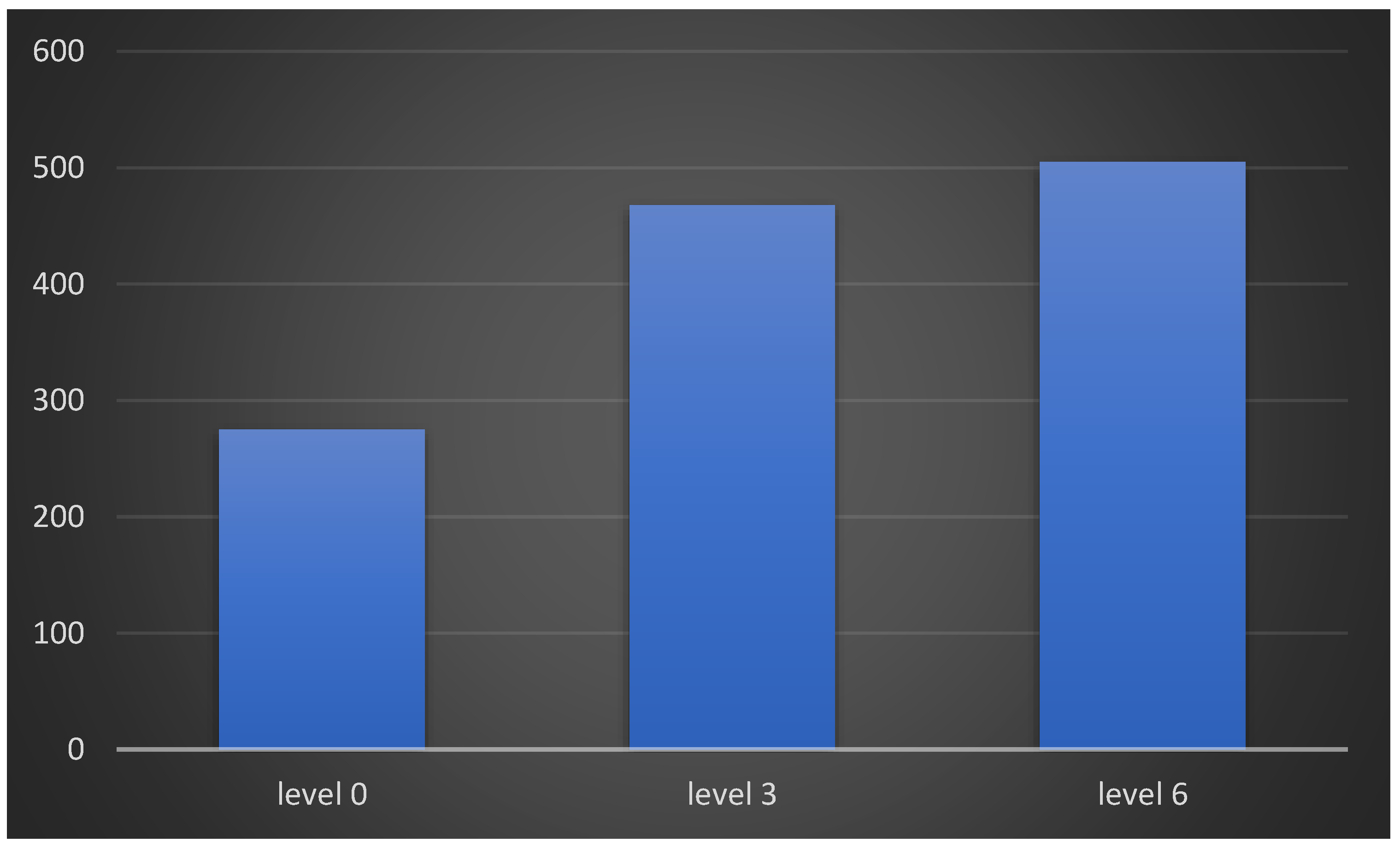
| sEMG (μV) | 151.21 ± 51.65 | 223.15 ± 52.81 | 248.91 ± 49.39 |
| rp = 0.778 | rp = 0.424 | rp = 0.583 | |
| Vitamin D Status Severely Deficient Insufficient Deficient Sufficient | 12 (9.2%) 94 (72.3%) 21 (16.2%) 3 (2.3%) | 0 (0.0%) 11 (8.5%) 38 (29.2%) 81 (62.3%) | 0 (0.0%) 11 (8.5%) 42 (32.3%) 77 (59.2%) |
| rs = 0.786 | rs = 0.856 | rs = 0.871 | |
| Vitamin D | USG | sEMG | ||||
|---|---|---|---|---|---|---|
| Level 0 | Level 3 | Level 6 | Level 0 | Level 3 | Level 6 | |
| Level 0 | 0.611 * | 0.490 * | 0.501 * | 0.778 * | 0.517 * | 0.491 * |
| Level 3 | 0.160 | 0.313 * | 0.355 * | 0.155 | 0.424 * | 0.558 * |
| Level 6 | 0.193 * | 0.352 * | 0.419 * | 0.180 * | 0.471 * | 0.583 * |
| Vitamin D | ||||||
|---|---|---|---|---|---|---|
| Variables | Level 0 | Level 3 | Level 6 | p Value (Level 0) | p Value (Level 3) | p Value (Level 6) |
| Gender Male Female | 16.91 ± 5.58 14.09 ± 5.23 | 33.94 ± 10.09 32.31 ± 12.58 | 31.87 ± 8.87 29.95 ± 9.74 | 0.003 | 0.187 | 0.281 |
| Occupation Farmer/Labourer/field Skilled worker/priest Shopkeeper/Service/business Professional Housewife/household | 18.89 ± 6.14 16.94 ± 3.65 14.59 ± 4.12 17.25 ± 2.05 13.75 ± 5.61 | 33.74 ± 7.87 32.20 ± 4.28 33.79 ± 11.63 44.35 ± 1.63 31.87 ± 14.41 | 31.81 ± 6.87 31.48 ± 3.41 31.54 ± 10.11 43.01 ± 0.42 29.19 ± 11.11 | <0.001 | 0.219 | 0.301 |
| Farmer vs. Housewife (0.001) & Farmer vs. Shopkeeper (0.002) | ||||||
| Sun Exposure No Yes | 14.75 ± 4.63 17.34 ± 6.14 | 33.59 ± 12.51 33.37 ± 14.51 | 31.11 ± 12.01 31.51 ± 13.51 | 0.011 | 0.673 | 0.807 |
| Eating Habit Vegetarian Non-Vegetarian | 15.96 ± 4.86 16.43 ± 7.07 | 33.57 ± 9.62 33.26 ± 13.29 | 31.42 ± 8.43 31.12 ± 10.65 | 0.890 | 0.545 | 0.866 |
| Address Rural Urban | 17.07 ± 6.28 14.94 ± 4.45 | 33.62 ± 11.83 33.29 ± 9.59 | 31.43 ± 9.77 31.19 ± 8.38 | 0.042 | 0.879 | 0.888 |
| Old Denture No Yes | 14.95 ± 4.61 17.79 ± 6.49 | 33.25 ± 11.24 33.79 ± 10.31 | 30.81 ± 9.28 32.08 ± 8.94 | 0.005 | 0.547 | 0.435 |
| Vitamin D Status (Level 0) Severely Deficient Insufficient Deficient Sufficient | 8.11 ± 1.44 14.98 ± 2.81 22.72 ± 2.59 37.07 ± 4.67 | 20.38 ± 5.21 35.44 ± 9.81 31.51 ± 12.82 38.01 ± 3.77 | 20.51 ± 6.09 32.96 ± 8.41 29.33 ± 9.75 37.41 ± 3.35 | <0.001 | <0.001 | <0.001 |
| Vitamin D Status (Level 3) Severely Deficient Insufficient Deficient Sufficient | - 9.71 ± 2.71 16.17 ± 6.17 16.94 ± 5.08 | - 16.06 ± 2.37 24.97 ± 3.04 39.82 ± 8.21 | - 15.27 ± 2.82 24.47 ± 3.52 36.72 ± 6.33 | <0.001 | <0.001 | <0.001 |
| Vitamin D Status (Level 6) Severely Deficient Insufficient Deficient Sufficient | - 10.62 ± 3.25 16.42 ± 6.24 16.71 ± 5.13 | - 16.55 ± 3.29 25.85 ± 4.24 40.05 ± 8.39 | - 15.23 ± 2.72 24.74 ± 3.37 37.21 ± 6.14 | 0.001 | <0.001 | <0.001 |
| Vitamin D Status | Vitamin D | Severely Deficient vs. Insufficient | Severely Deficient vs. Deficient | Severely Deficient vs. Sufficient | Insufficient vs. Deficient | Insufficient vs. Sufficient | Deficient vs. Sufficient |
|---|---|---|---|---|---|---|---|
| Level 0 Status | Level 0 | <0.001 | <0.001 | <0.001 | <0.001 | 0.010 | 1.000 |
| Level 3 | 0.070 | <0.001 | 0.019 | 0.087 | 0.654 | 1.000 | |
| Level 6 | <0.001 | 0.026 | 0.014 | 0.456 | 1.000 | 0.733 | |
| Level 3 Status | Level 0 | - | - | - | <0.001 | <0.001 | 1.000 |
| Level 3 | - | - | - | 0.172 | <0.001 | <0.001 | |
| Level 6 | - | - | - | <0.001 | <0.001 | <0.001 | |
| Level 6 Status | Level 0 | - | - | - | 0.002 | <0.001 | 1.000 |
| Level 3 | - | - | - | 0.120 | <0.001 | <0.001 | |
| Level 6 | - | - | - | <0.001 | <0.001 | <0.001 |
| Vitamin D | Friedman Test | ||||
|---|---|---|---|---|---|
| Variables | Shapiro Wilk (p Value) | p Value (Level 0) | p Value (Level 3) | p Value (Level 6) | |
| Age | 0.008 | <0.001 | <0.001 | <0.001 | |
| Serum D (Level 0) | 0.000 | - | <0.001 | <0.001 | <0.001 |
| Serum D (Level 3) | 0.001 | <0.001 | - | 0.115 | |
| Serum D (Level 6) | 0.112 * | <0.001 | 0.115 | - | |
| USG (Level 0) | 0.001 | <0.001 | <0.001 | <0.001 | <0.001 |
| USG (Level 3) | 0.010 | <0.001 | <0.001 | <0.001 | |
| USG(Level 6) | 0.155 * | <0.001 | <0.001 | <0.001 | |
| sEMG (Level 0) | 0.000 | <0.001 | <0.001 | <0.001 | <0.001 |
| sEMG (Level 3) | 0.335 * | <0.001 | <0.001 | <0.001 | |
| sEMG (Level 6) | 0.007 | <0.001 | <0.001 | <0.001 | |
| p Values and Correlation Coefficients | |||
|---|---|---|---|
| Variables | Level 0 vs. Level 3 | Level 0 vs. Level 6 | Level 3 vs. Level 6 |
| Vitamin D | <0.001 | <0.001 | <0.001 |
| rp = 0.269 * | rp = 0.292 * | rp = 0.950 * | |
| USG | <0.001 | <0.001 | 0.001 |
| rp = 0.926 * | rp = 0.860 * | rp = 0.945 * | |
| sEMG | <0.001 | <0.001 | <0.001 |
| rp = 0.645 * | rp = 0.544 * | rp = 0.889 * | |
Disclaimer/Publisher’s Note: The statements, opinions and data contained in all publications are solely those of the individual author(s) and contributor(s) and not of MDPI and/or the editor(s). MDPI and/or the editor(s) disclaim responsibility for any injury to people or property resulting from any ideas, methods, instructions or products referred to in the content. |
© 2023 by the authors. Licensee MDPI, Basel, Switzerland. This article is an open access article distributed under the terms and conditions of the Creative Commons Attribution (CC BY) license (https://creativecommons.org/licenses/by/4.0/).
Share and Cite
Rathi, S.; Chaturvedi, S.; Abdullah, S.; Rajput, G.; Alqahtani, N.M.; Chaturvedi, M.; Gurumurthy, V.; Saini, R.; Bavabeedu, S.S.; Minervini, G. Clinical Trial to Assess Physiology and Activity of Masticatory Muscles of Complete Denture Wearer Following Vitamin D Intervention. Medicina 2023, 59, 410. https://doi.org/10.3390/medicina59020410
Rathi S, Chaturvedi S, Abdullah S, Rajput G, Alqahtani NM, Chaturvedi M, Gurumurthy V, Saini R, Bavabeedu SS, Minervini G. Clinical Trial to Assess Physiology and Activity of Masticatory Muscles of Complete Denture Wearer Following Vitamin D Intervention. Medicina. 2023; 59(2):410. https://doi.org/10.3390/medicina59020410
Chicago/Turabian StyleRathi, Shraddha, Saurabh Chaturvedi, Sabzar Abdullah, Geeta Rajput, Nasser M. Alqahtani, Mudita Chaturvedi, Vishwanath Gurumurthy, Ravinder Saini, Shashit Shetty Bavabeedu, and Giuseppe Minervini. 2023. "Clinical Trial to Assess Physiology and Activity of Masticatory Muscles of Complete Denture Wearer Following Vitamin D Intervention" Medicina 59, no. 2: 410. https://doi.org/10.3390/medicina59020410