Article

Cellular and Transcriptional Responses of Crassostrea gigas Hemocytes Exposed in Vitro to Brevetoxin (PbTx-2)

by
Danielle F. Mello
1,2,
Eliza S. De Oliveira
1,
Renato C. Vieira
1,
Erik Simoes
1,
Rafael Trevisan
2,
Alcir Luiz Dafre
2 and
Margherita Anna Barracco
1,*
1
Laboratory of Immunology Applied to Aquaculture, Department of Cell Biology, Embryology and Genetics, Biological Sciences Center, Federal University of Santa Catarina, 88040-900, Florianópolis, SC, Brazil
2
Laboratory of Cell Defense, Department of Biochemistry, Biological Sciences Center, Federal University of Santa Catarina, 88040-900, Florianópolis, SC, Brazil
*
Author to whom correspondence should be addressed.
Mar. Drugs 2012, 10(3), 583-597; https://doi.org/10.3390/md10030583
Submission received: 31 January 2012 / Revised: 20 February 2012 / Accepted: 24 February 2012 / Published: 5 March 2012

Abstract

:
Hemocytes mediate a series of immune reactions essential for bivalve survival in the environment, however, the impact of harmful algal species and their associated phycotoxins upon bivalve immune system is under debate. To better understand the possible toxic effects of these toxins, Crassostrea gigas hemocytes were exposed to brevetoxin (PbTx-2). Hemocyte viability, monitored through the neutral red retention and MTT reduction assays, and apoptosis (Hoechst staining) remained unchanged during 12 h of exposure to PbTx-2 in concentrations up to 1000 µg/L. Despite cell viability and apoptosis remained stable, hemocytes incubated for 4 h with 1000 µg/L of PbTx-2 revealed higher expression levels of Hsp70 (p < 0.01) and CYP356A1 (p < 0.05) transcripts and a tendency to increase FABP expression, as evaluated by Real-Time quantitative PCR. The expression of other studied genes (BPI, IL-17, GSTO, EcSOD, Prx6, SOD and GPx) remained unchanged. The results suggest that the absence of cytotoxic effects of PbTx-2 in Crassostrea gigas hemocytes, even at high concentrations, allow early defense responses to be produced by activating protective mechanisms associated to detoxification (CYP356A1 and possibly FABP) and stress (Hsp70), but not to immune or to antioxidant (BPI, IL-17, EcSOD, Prx6, GPx and SOD) related genes.

1. Introduction

Brevetoxins represent a group of polyether compounds known to cause neurotoxic shellfish poisoning (NSP) through the consumption of brevetoxin-containing shellfish. They are produced mainly by the marine dinoflagellate Karena brevis, but recently brevetoxin-like compounds were also found in other microalgae species such as Chatonella marina, Chatonella antiqua, Fibrocapsa japonica and Heterosigma akashiwo [1]. Besides the threats to humans, blooms of these neurotoxin-producing species occur worldwide, causing massive fish kills along with bird and marine mammal mortalities through the direct exposure to these blooms, or indirectly by the food web [2]. K. brevis, mainly produces two brevetoxins types, with PbTx-1 being the most potent and PbTx-2 being the most abundant [3]. Bivalves accumulate and metabolize these toxins, producing oxidized, reduced, hydrolized and conjugated metabolites that vary in their toxic potency [4,5].
Although shellfish appear to be only toxin vectors unaffected by harmful algal blooms (HABs), some behavioral, physiological and cellular responses of bivalves to harmful algae have already been described, along with mortality events [2,6]. Responses of bivalves associated to brevetoxin or brevetoxin-producing algae include reduced clearance rates [7], impairment of larval survival and development [8,9] and increases in lysosomal destabilization in oyster hepatopancreas [10,11]. Most recently, the HAB species H. akashiwo, described to produce brevetoxin-like compounds [12], affected significantly bivalve immune cells viability and phagocytosis in vitro [13].
The study of the effects of harmful algae upon bivalve immune system has recently become an area of interest of researchers. Various publications have demonstrated that hemocytes and immune parameters can be activated or modulated by several toxic microalgae species [13,14,15,16,17,18,19,20,21,22,23,24]. However, to our knowledge, analysis of the effects of a purified brevetoxin upon the immune system of bivalves has never been assessed, especially at the gene expression level.
Bivalve innate immune responses are mediated by hemocytes. They are involved in cellular immune reactions, such as phagocytosis of invading microorganisms, formation of nodules and capsules surrounding pathogens and their subsequent degradation through the release of host defense molecules [25]. Moreover, circulating hemocytes are able to migrate from the hemolymph to connective tissues and promote localized responses following injury or microorganism invasion.
The Pacific oyster Crassostrea gigas (Thunberg, 1793) is a bivalve mollusc belonging to the Ostreidae family (Mollusca, Bivalvia), with a global distribution and probably accounts for the highest annual production of any freshwater or marine organism [26]. This species has a reasonable number of cDNA sequences deposited in GenBank, which permits the study of several environmental stressors at the gene expression level.
The objective of this study was to evaluate the in vitro effects of the purified brevetoxin PbTx-2 over C. gigas hemocyte viability, apoptosis and gene expression.

2. Results

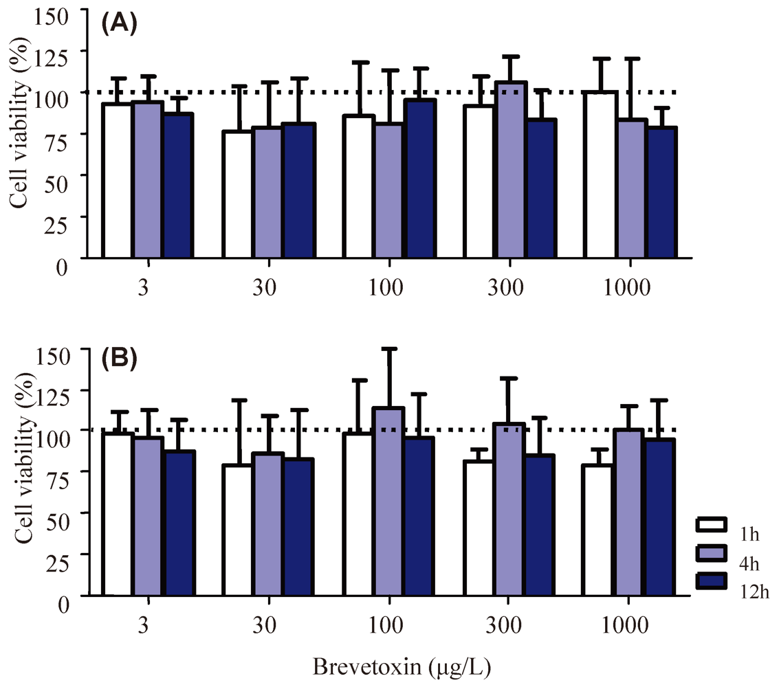
2.1. Cellular Parameters

In order to investigate whether PbTx-2 could cause hemocyte toxicity, cell viability assays were performed using the MTT and NR methods. No significant differences were observed in the viability of C. gigas hemocytes after incubation with PbTx-2 concentrations ranging from 3 to 1000 µg/L for 1, 4 and 12 h, according to either viability assay (Figure 1).
Apoptotic hemocytes (Supplementary Figure S1) were also quantified using Hoechst staining after cell exposure to the highest concentrations of PbTx-2 for 12 h. The percentage of hemocytes presenting characteristic altered nuclei remained unchanged after incubation with 300 (4.7 ± 3.4%) or 1000 (3.2 ± 1.8%) µg/L of PbTx-2 when compared to the control group (3.1 ± 1.6%).
Figure 1. Viability of Crassostrea gigas hemocytes evaluated through the neutral red (NR) retention (A) and 3-(4,5-dimethylthiazol-2-yl)-2,5-diphenyltetrazolium bromide (MTT) reduction (B) assays after exposure to different brevetoxin (PbTx-2) concentrations. Bars represent mean + standard deviation. The sample size was 3–4, performed in independent experiments. The range of individuals used varied from 5 to 30 oysters per pool, depending on the number of treatment groups analyzed in each experiment. No significant differences were obtained in the analysis of variance.
Marinedrugs 10 00583 g001

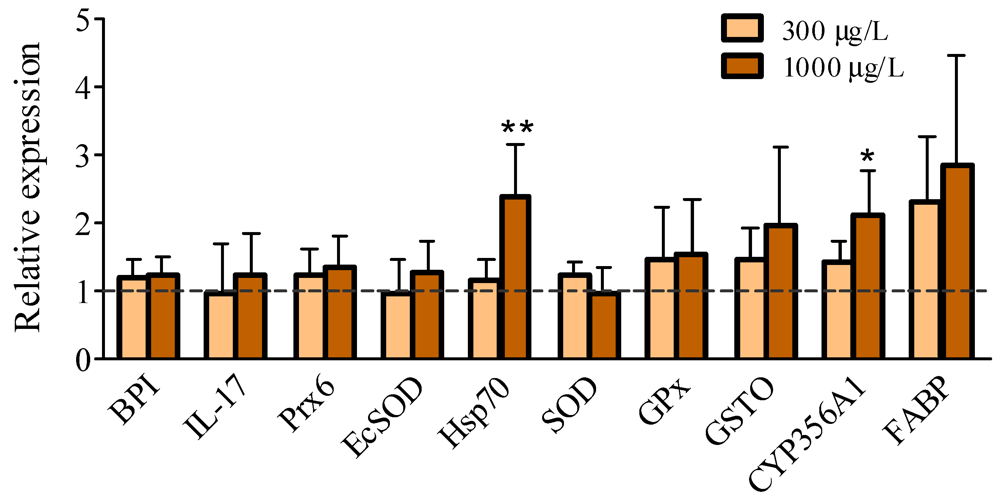
2.2. Gene Expression Levels

The gene expression analysis was evaluated after hemocytes were incubated during 4 h with the highest concentrations of PbTx-2 analyzed in the viability assays, in view of the fact that no cytotoxic effects were observed. Previous studies have demonstrated that a 4 h incubation period is sufficient to induce gene expression and obtain early molecular responses in hemocytes [27,28]. The immune related genes selected are involved in pathogen recognition (EcSOD) and elimination (BPI) and cell signaling (IL-17 and possibly Prx6). Also other genes associated to the antioxidant system, stress and detoxification were selected (GPx, SOD, Hsp70, GSTO, CYP356A1 and FABP), to span various branches of oyster defense systems. In previous studies the genes GSTO, CYP356A1 and FABP were significantly induced following C. gigas exposure to sewage [29]. Prx6 protein levels decreased after in vivo exposure to zinc in gills of the brown mussel Perna perna [30] and also its gene expression was modulated in oysters selected for disease resistance [31] and in oysters sampled from contaminated estuaries [32]. GPx and SOD are major antioxidant players that can be modulated by different xenobiotics [33] and their gene expression analysis could demonstrate any up or downregulation of the antioxidant system.
In the present study, C. gigas hemocytes presented a more than 2-fold increase (p < 0.05) in the transcript levels of genes encoding Hsp70 and CYP356A1, but only when incubated with the highest concentration of PbTx-2 (1000 µg/L) (Figure 2). All of the other transcripts analyzed, however, showed similar expression profiles between the control and treated groups, which included BPI, IL-17, FABP, EcSOD, Prx6, GPx, SOD and GSTO (Figure 2).
Figure 2. Relative expression of the 10 gene transcripts (normalized to GAPDH) in Crassostrea gigas hemocytes exposed for 4 h to 300 or 1000 µg/L of PbTx-2. The dashed line represents the mean value of the control groups (vehicle) for each gene. Bars represent mean + standard deviation. The sample size was comprised of 4 pools of 5–9 animals. * (p < 0.05) and ** (p < 0.01) represent significant differences between each gene compared to the corresponding control group.
Marinedrugs 10 00583 g002

3. Discussion

Although many studies have assessed the effects of harmful algae and their toxins upon the immune system of bivalves, the biological interactions between them are still very poorly understood, especially at the molecular level. Therefore, in this study we evaluated C. gigas hemocyte responses to purified PbTx-2 in vitro. The objective of this work was an initial screening of cellular and transcriptional effects of a purified ficotoxin upon bivalve immune cells. Although other brevetoxin metabolites were already detected in oysters tissues [4,5], PbTx-2 was chosen since it is the most abundant brevetoxin produced by Karenia brevis during natural blooms [3].
Hemocytes may have multiple opportunities to become in direct contact with toxins during HABs events. Firstly, in the pallial cavity where hemocytes can be found protecting host from potential pathogens even before invasion [34,35]. Secondly, after filter-feeding and digestion of the harmful algae, when intracellular toxins can be released and end up in animal tissues and semi-open vascular system where hemocytes are plentiful [36]. Ford et al. [36] discussed that hemocytes can migrate through the intestine epithelium, phagocyte partially digested algae and return to circulation in the hemolymph. Moreover, Franchini et al. [37] showed a great quantity of another phycotoxin, the yessotoxin, in the cytoplasm of mussel hemocytes through immunolocalization. Therefore, there are multiple opportunities for in vivo toxins-hemocyte encounters.
The absence of effects of a purified brevetoxin upon hemocyte viability, even in the highest concentration and time exposure tested, was unexpected. Also, no changes in the percentage of apoptotic hemocytes were observed, which supports the idea of an absence of gross toxic effects of brevetoxin upon hemocytes. On the other hand, these results may not be so surprising, since the toxic mechanism of this toxin is through the blocking of voltage-gated sodium channels of excitable cells [1]. Furthermore, these results are consistent with other studies, which observed no toxic effects in bivalve hemocytes exposed in vitro and in vivo to microalgal species that produce saxitoxin (STX), a neurotoxin which has a mechanism of action related to brevetoxin [36,38]. It is important to note that the use of PbTx-2 in vitro does not take in account the metabolization that could take place in vivo, producing metabolites that could be toxic to oyster immune cells.
The lack of effects of PbTx-2 in the cellular parameters tested, could probably also be explained by the induction of genes related to protective mechanisms. In this study, the results of the gene expression analysis showed an increase in the transcriptional levels of Hsp70 and CYP356A1.
The induction of chaperones, such as Hsp70, in response to thermal stress has already been demonstrated in many organisms, including oysters [39,40,41,42,43]. Additionally, an increase in the expression of heat shock proteins was also reported in a variety of bivalves species exposed to toxic compounds or contaminated sites [44,45,46,47,48,49,50]. In bivalves, increases in the expression of Hsp70 were also associated with infections [46,51,52,53]. These studies, together with the result obtained in the present study, support the role of Hsp70 as a response to stress in general and not only to thermal shock [54,55]. Furthermore, the upregulation of Hsp70 could recurrently be associated to increases in animal resistance to stressors, including toxic compounds [46,55,56].
The cytochrome P450 (CYP) family is characterized as one of the major phase I-type classes of detoxification enzymes found in living organisms, including the aquatic organisms. These enzymes metabolize a wide variety of substrates, such as fatty acids, hormones and xenobiotics [57]. According to Toledo-Silva et al. [58], the cytochrome isoform CYP356A1, has great similarity to the CYP17 family, which is associated with the metabolism of steroids, but also a lower similarity to the CYP1 family, which comprises CYP1A, commonly associated with the metabolism of polycyclic aromatic hydrocarbons. Despite the debate on the existence o CYP-dependent phase I of biotransformation, CYP356A1 has the potential to participate in the brevetoxins-metabolism. Bivalves accumulate and extensively metabolize these toxins, producing oxidized, reduced, hydrolized and cysteine conjugated metabolites [4,5]. To our knowledge, the metabolizing pathways of brevetoxins have not been described yet in these organisms. On the other hand, PbTx-2 metabolization associated to cytochrome P450 activity was confirmed in human, rat and fish CYPs [59,60,61]. Within metabolites described in these studies, some corresponded to metabolites identified in oyster tissues, e.g., PbTx-3 and PbTx-9 [62,63]. Therefore, the increase in the transcriptional level of a CYP isoform found in the present study opens the opportunity for testing this enzyme in the biotransformation of PbTx-2.
GST is key enzyme of phase II biotransformation pathway, indicated to be similar between aquatic invertebrates and other vertebrates, is responsible for the conjugation of glutathione (GSH) to electrophilic compounds, increasing its hydrophilicity and allowing the elimination of these conjugates into the extracellular environment [64]. Previous studies have suggested that rat and fish GSTs could be associated to the metabolization of PbTx-2 [60,61]. Moreover, brevetoxin conjugated to GSH and its corresponding dipeptides were found in oyster tissues [62]. Unexpectedly, the transcript levels of the gene encoding GST omega (GSTO) did not show significant differences between hemocytes exposed to the highest concentration of PbTx-2. It is important to note that the gene expression of only one GST isoform was evaluated, therefore it is possible that other isoforms could be involved in metabolizing PbTx-2 and be up-regulated. In addition, according to Livingstone et al. [64], Phase II enzymes in aquatic invertebrates, such as GST, are generally much less responsive to organic xenobiotics exposure than CYPs.
Another protein, possibly involved in detoxification pathways is FABP, a small cytosolic molecule with affinity for hydrophobic compounds. Although its functions are not yet fully understood, it is believed that FABP is involved in the uptake and transport of fatty acids [65]. However, this molecule can also be associated with xenobiotic metabolism since Velkov et al. [66] reported that the FABP has high affinity for lipophilic drugs and Medeiros et al. [29] observed an induction of its gene in oysters exposed to domestic sewage. In the present study, the level of FABP transcripts were increased ~2.5 in hemocytes exposed to both concentrations of PbTx-2, when compared to the control group, but significance was not reached. This result suggests that FABP may be involved in the metabolism of PbTx-2 in hemocytes, which remains to be confirmed in future studies.
None of the studied genes encoding proteins associated to the immune system were modulated. Among some immune effector molecules already described in C. gigas, we can cite (1) BPI, which increases membrane permeability leading to bacterial cell extravasation and can be induced in hemocytes of oysters by challenge with bacteria [67]; (2) IL-17, a cytokine similar to the IL-17 of vertebrates, which its first report in an invertebrate was in C. gigas and can also be induced in hemocytes after bacterial challenge [68]; (3) EcSOD, a protein with superoxide dismutase activity, that also has the property to bind to lipopolysaccharide (LPS) present on the surface of Gram (−) and is believed to act as an opsonin by binding to integrins of the membranes of hemocytes [69], thus enhancing recognition and elimination of bacteria through phagocytosis; and (4) Prx6, an enzyme implicated in the antioxidant defenses, with only one cysteine residue in its active site, involved in the metabolism of peroxides [32]. Recent studies regarding vertebrates and invertebrates (helminths) suggest that the peroxiredoxins can assume an important role in the immune system, mediating the activation of defense cells [70]. Along with Prx6, other proteins associated to antioxidant defenses did not show any significant modulations in their transcript levels, such as SOD and GPx. SOD enzyme is a metalloprotein responsible for the dismutation of superoxide (O2•−) to hydrogen peroxide (H2O2) and was identified in C. gigas by Boutet et al. [45], whereas GPx is a selenium protein that has peroxidase activity against H2O2 and organic peroxides, described in the Pacific oyster by Jo et al. [71].

4. Experimental Section

4.1. Animals

Adult oysters of C. gigas (Thunberg, 1793) species were obtained from a mariculture farm located in the southern bay of Florianópolis, Santa Catarina, southern Brazil (27°38′56″S and 48°32′31″W). Oysters were acclimated for a minimum period of 10 days and kept in filtered seawater (FSW-1 µm) at 18–20 °C, before their use in experiments. During this period, the oysters were fed with the microalgae Tetraselmis sp. The experiments here presented are in accordance to the Federal University of Santa Catarina ethic committee on the animal use (CEUA).

4.2. Hemolymph Extraction and Hemocyte Incubation with Brevetoxin (PbTx-2)

The oyster hemolymph (4–5 pools of 5–30 animals) was extracted from the adductor muscle by inserting a 0.8 × 30 mm needle attached to a 1 mL syringe, after making a small hole in the valves of the animals close to the muscle. The hemolymph was maintained on ice until use. Immediately after hemolymph collection, the total number of hemocytes was determined using a Neubauer hemocyte chamber. In order to collect hemocytes, hemolymph was centrifuged (600 g, 10 min, 4 °C) and the hemocyte pellet was resuspended in FSW (0.22 µm) to obtain a concentration of 1 × 10⁶ cells/mL.
For the viability assays, hemocyte suspensions (1 × 10⁶ cells/mL) were incubated with 3, 30, 100, 300 and 1000 µg/L of purified PbTx-2 (World Ocean Solutions, Wilmington, EUA), previously solubilized in dimethyl-sulfoxide (DMSO - at a maximal final concentration of 0.05%) for 1, 4 and 12 h at 18 °C, in the dark. For the control groups, hemocytes were incubated with FSW containing the corresponding DMSO concentration (vehicle). For the quantification of apoptotic hemocytes, cells were incubated for 12 h with 300 or 1000 µg/L of PbTx-2 or the corresponding vehicle, while for the gene expression analysis the incubation period was only for 4 h.

4.3. Cellular Viability Assays and Apoptosis

After treatments, oyster hemocytes were used to determine their viability, using the 3-(4,5-dimethylthiazol-2-yl)-2,5-diphenyltetrazolium bromide (MTT) or neutral red (NR) assays following established methods as previously employed by Trevisan et al. [72]. A total of 6 × 10⁵ (MTT) and 3 × 10⁵ (NR) cells were used for each assay and conducted in triplicate. The hemolymph was pooled to obtain the number of cells necessary for the analysis, with a total of 3–4 pools (N = 3–4). The range of individuals used varied from 5 to 30 oysters per pool, depending on the number of treatment groups analyzed in each experiment.
The percentage of potentially apoptotic hemocytes was determined by using Hoechst 33258 staining (Sigma-Aldrich, São Paulo, Brazil). Fixed hemocyte monolayers (N = 3 pools of 5–7 animals) were immersed in McIlvane buffer (0.1 M citric acid, 0.4 M disodium hydrogen phosphate, pH 5.5) for 5 min and then treated with a Hoechst solution in McIlvane buffer (1:1500) for 5 min. The slides were then mounted with cover slips and observed under a fluorescence microscope (excitation of 365 nm). The percentage of apoptotic cells was estimated by examining 300 cells per group and counting morphologically altered nuclei characteristic of apoptotic cells (Supplementary Figure S1). All cell types were represented in the samples (both hyaline and granular hemocytes were observed) although it should be taken in account that hyaline hemocytes could be less represented since they may be less adherent to glass slides.

4.4. Extraction of Total RNA and cDNA Synthesis

At the end of the incubation, samples (N = 4 pools of 5–9 animals) were centrifuged (1000 g, 10 min, 4 °C) and cells resuspended in RNALater® (Invitrogen, São Paulo, Brazil) and stored until use. The RNA extraction from hemocytes was performed using TRIzol® reagent (Invitrogen, São Paulo, Brazil). Samples were then treated with DNase I (Fermentas, São Paulo, Brazil) and RNA concentration and purity were assessed spectrophotometrically (NanoVue, GE Healthcare, Uppsala, Sweden). For cDNA synthesis, only RNA samples with absorbance ratio (A260/A280) between 1.8 and 2.1 were used. The cDNA synthesis was performed on 250 ng of total RNA using 0.5 µg of oligo (dT)18 primer (Fermentas, São Paulo, Brazil), 1 U of RevertAid® Reverse Transcriptase (Fermentas, São Paulo, Brazil) and 20 U of RiboLockTM RNase Inhibitor (Fermentas, São Paulo, Brazil).

4.5. Real-Time Quantitative PCR (qPCR)

cDNAs were used for qPCR analysis in order to determine the relative expression of mRNA coding ten proteins: bactericidal/permeability increasing protein (BPI), interleukin-17 (IL-17), extracellular superoxide dismutase (EcSOD), peroxiredoxin 6 (Prx6), heat shock protein (Hsp70), glutathione peroxidase (GPx), superoxide dismutase (SOD), glutathione S-transferase omega (GSTO), fatty acid binding protein (FABP) and a member of the cytochrome P450 family (CYP356A1). Primers (Table 1) that weren’t used in previous works were designed from specific sequences of C. gigas deposited in GenBank, with the following characteristics: size from 18–20 bp, Tm between 59–61 °C, GC content between 40–60% and amplicon size between 90–200 bp. The sequences and the GenBank accession numbers [73] are listed in Table 1. Primers efficiency was tested using the standard curve method. For this purpose, a serial dilution (1:3, 1:7, 1:15, 1:31, 1:63) was made from a single cDNA sample consisted of a pool of all cDNAs from the different treatments. Only primers that showed efficiencies between 1.8 and 2.2 were used. The qPCR analysis was performed in 96-well microplates, in duplicate, using the ABI 7900HT thermocycler (Applied Biosystems, São Paulo, Brazil), with a total reaction volume of 10 µL. Each reaction had 5 µL of Maxima SYBR® Green/ROX qPCR Master Mix 2X (Fermentas, São Paulo, Brazil), 0.3 mM of each primer and 1 µL of each diluted cDNA (1:4). Amplification conditions were 95 °C for 10 min followed by 40 cycles of 95 °C for 15 s and 60 °C for 60 s. The specificity of the qPCR product was analyzed by a dissociation curve performed after amplification, observing a single peak at the expected Tm. The results were expressed as relative expression of transcripts normalized by the reference gene glyceraldehyde 3-phosphate dehydrogenase (GAPDH), using the 2−(ΔΔct) method [74].
Table 1. Gene names and primer sequences used for Real-Time Quantitative PCR (qPCR) and GenBank accession numbers.
Gene NamePrimersSequence 5'-3'GenBank
Glyceraldehyde 3-phosphate dehydrogenaseCgGAPDH-Fw GCTGTGACACCATTGGAGAAAJ544886.1
CgGAPDH-RvACCAATGACGCAACAAGCGA
Peroxiredoxin 6CgPrx6-Fw GAGCCAGAGTTCAAGAAGAGAM265552.1
CgPrx6-RvTGCATTGTCCTTTTCGGCTG
Interleukin-17CgIL17-FwACTGAGGCTCGATGCAAGTGEF190193.1
CgIL17-RvAGCCTTCTTGCTTCATGTGG
Bactericidal/permeabilityCgBPI-FwGATAGAAATAGGAATGGACGGHM992925.1
increasing proteinCgBPI-RvGTTATAGATCCACGCTGCTCC
Heat shock protein 70CgHsp70-FwTCATCAAGTGGATGGACCAGAB122063.1
CgHsp70-RvCATTCCTCCAGGCATGCCA
Extracellular superoxide dismutaseCgEcSOD-FwGCTGTGACACCATTGGAGAADQ010420.1
CgEcSOD-RvACCAATGACGCAACAAGCGA
Glutathione S-transferase omegaCgGSTO-FwTGATGAGTTCACCACCGCAAAJ557141.1
CgGSTO-RvTTCAAACCATGGCCACAGCA
Cytochrome P450 isoform 356A1CgCYP3561A-FwATGAAACCCGCGAAACCAGAEF645271.1
CgCYP3561A-RvTAAATTCGGCTTCACGCCCT
Superoxide dismutaseCgSOD-FwTCAACAAAGAGCATGGCGTCAJ496219.1
CgSOD-RvTTTCCGGTCGTCTTACTGAG
Glutathione peroxidaseCgGPx-FwTCAAGATCCGAGATGTCGTCEF692639.1
CgGPx-RvACTCGGTTTCCAGACATGAG
Fatty acid binding proteinCgFABP-FwGTTTGAGGGAAACTGGGAATGCEU069496
CgFABP-RvTCCGTCGGAATATGTCAGTTTAGC

4.6. Statistical Analysis

The results of the viability assays were compared by two-way ANOVA, while qPCR results and quantification of apoptotic hemocytes were compared by one-way ANOVA, both followed by Tukey’s post hoc test for comparison of means. For the quantification of apoptotic hemocytes (percentages), the data were arcsin transformed. The results were considered significant at p < 0.05. Statistical analysis was performed using Statistica [75].

5. Conclusions

In vitro studies have many advantages, such as feasibility, control of variables, rapid results for screening experiments, lower number of animals and reproducibility. Our experiments showed that Crassostrea gigas hemocytes fully retained viability and did not undergo apoptosis during the first 12 h in the presence of PbTx-2, and that, apparently, PbTx-2 triggered defense mechanisms including activation of stress (Hsp70) and detoxification related genes (CYP356A1 and possibly FABP), suggesting a specific response. However, further studies both in vitro and in vivo are necessary to confirm the real effects of brevetoxins upon the immune system of cultivated bivalves or wild stocks, particularly at the gene expression level.

Acknowledgments

The authors thank Afonso C. D. Bainy (Dept. Biochemistry, UFSC) who kindly provided the GSTO, FABP and CYP primers for qPCR. This study was supported by CNPq (research project No. 474539/2008-3) and FAPESC. The provided scholarships are sincerely appreciated: Danielle F. Mello (REUNI/UFSC), Eliza S. de Oliveira (PIBIC/CNPq/UFSC), Renato C. Vieira (PIBIC/CNPq/UFSC), Erik Simões (CNPq), Rafael Trevisan (Capes). Alcir Luiz Dafre and Marguerita Anna Barracco are research fellows of CNPq.

References

  1. Wang, D.Z. Neurotoxins from marine dinoflagellates: A brief review. Mar Drugs 2008, 6, 349–371. [Google Scholar]
  2. Landsberg, J.H. The effects of harmful algal blooms on aquatic organisms. Rev. Fish. Sci. 2002, 10, 113–390. [Google Scholar]
  3. Brand, L.E.; Campbell, L.; Bresnan, E. KARENIA: The biology and ecology of a toxic genus. Harmful Algae 2011, 14, 156–178. [Google Scholar]
  4. Plakas, S.M.; Jester, E.L.E.; El Said, K.R.; Granade, H.R.; Abraham, A.; Dickey, R.W.; Scott, P.S.; Flewelling, L.J.; Henry, M.; Blum, P.; et al. Monitoring of brevetoxins in the Karenia brevis bloom-exposed Eastern oyster (Crassostrea virginica). Toxicon 2008, 52, 32–38. [Google Scholar] [CrossRef]
  5. Plakas, S.M.; Dickey, R.W. Advances in monitoring and toxicity assessment of brevetoxins in molluscan shellfish. Toxicon 2010, 56, 137–149. [Google Scholar]
  6. Shumway, S.E. A review of the effects of algal blooms on shellfish and aquaculture. J. World Aquac. Soc. 1990, 21, 65–104. [Google Scholar]
  7. Leverone, J.R.; Shumway, S.E.; Blake, N.J. Comparative effects of the toxic dinoflagellate Karenia brevis on clearance rates in juveniles of four bivalve molluscs from Florida, USA. Toxicon 2007, 49, 634–645. [Google Scholar]
  8. Leverone, J.R.; Blake, N.J.; Pierce, R.H.; Shumway, S.E. Effects of the dinoflagellate Karenia brevis on larval development in three species of bivalve mollusc from Florida. Toxicon 2006, 48, 75–84. [Google Scholar] [CrossRef]
  9. Wang, L.P.; Yan, T.; Zhou, M.J. Impacts of HAB species Heterosigma akashiwo on early development of the scallop Argopecten irradians Lamarck. Aquaculture 2006, 255, 374–383. [Google Scholar] [CrossRef]
  10. Keppler, C.J.; Lewitus, A.J.; Ringwood, A.H.; Hoguet, J.; Staton, T. Sublethal cellular effects of short-term raphidophyte and brevetoxin exposures on the eastern oyster Crassostrea virginica. Mar. Ecol. Prog. Ser. 2006, 312, 141–147. [Google Scholar] [CrossRef]
  11. Keppler, C.J.; Hoguet, J.; Smith, K.; Ringwood, A.H.; Lewitus, A.J. Sublethal effects of the toxic alga Heterosigma akashiwo on the southeastern oyster (Crassostrea virginica). Harmful Algae 2005, 4, 275–285. [Google Scholar] [CrossRef]
  12. Khan, S.; Arakawa, O.; Onoue, Y. Neurotoxins in a toxic red tide of Heterosigma akashiwo (Raphidophyceae) in Kagoshima Bay, Japan. Aquac. Res. 1997, 28, 9–14. [Google Scholar] [CrossRef]
  13. Hégaret, H.; da Silva, P.M.; Wikfors, G.H.; Haberkorn, H.; Shumway, S.E.; Soudant, P. In vitro interactions between several species of harmful algae and haemocytes of bivalve molluscs. Cell Biol. Toxicol. 2011, 27, 249–266. [Google Scholar] [CrossRef]
  14. Hégaret, H.; Smolowitz, R.M.; Sunila, I.; Shumway, S.E.; Alix, J.; Dixon, M.; Wikfors, G.H. Combined effects of a parasite, QPX, and the harmful-alga, Prorocentrum minimum on northern quahogs, Mercenaria mercena. Mar. Environ. Res. 2010, 69, 337–344. [Google Scholar] [CrossRef]
  15. da Silva, P.M.; Hégaret, H.; Lambert, C.; Wikfors, G.H.; Le Goic, N.; Shumway, S.E.; Soudant, P. Immunological responses of the Manila clam (Ruditapes philippinarum) with varying parasite (Perkinsus olseni) burden, during a long-term exposure to the harmful alga, Karenia selliformis, and possible interactions. Toxicon 2008, 51, 563–573. [Google Scholar] [CrossRef] [Green Version]
  16. Galimany, E.; Sunila, I.; Hégaret, H.; Ramon, M.; Wikfors, G.H. Pathology and immune response of the blue mussel (Mytilus edulis L.) after an exposure to the harmful dinoflagellate Prorocentrum minimum. Harmful Algae 2008, 7, 630–638. [Google Scholar] [CrossRef]
  17. Galimany, E.; Sunila, I.; Hégaret, H.; Ramon, M.; Wikfors, G.H. Experimental exposure of the blue mussel (Mytilus edulis, L.) to the toxic dinoflagellate Alexandrium fundyense: Histopathology, immune responses, and recovery. Harmful Algae 2008, 7, 702–711. [Google Scholar] [CrossRef]
  18. Hégaret, H.; da Silva, P.M.; Wikfors, G.H.; Lambert, C.; De Bettignies, T.; Shumway, S.E.; Soudant, P. Hemocyte responses of Manila clams, Ruditapes philippinarum, with varying parasite, Perkinsus olseni, severity to toxic-algal exposures. Aquat. Toxicol. 2007, 84, 469–479. [Google Scholar] [CrossRef]
  19. Hégaret, H.; Shumway, S.E.; Wikfors, G.H. Effects of harmful algae on physiology and hemocyte parameters of the Northern Bay scallop, Argopecten irradians irradians. J. Shellfish Res. 2007, 26, 1316–1316. [Google Scholar]
  20. Hégaret, H.; Wikfors, G.H. Time-dependent changes in hemocytes of eastern oysters, Crassostrea virginica, and northern bay scallops, Argopecten irradians irradians, exposed to a cultured strain of Prorocentrum minimum. Harmful Algae 2005, 4, 187–199. [Google Scholar] [CrossRef]
  21. Hégaret, H.; Wikfors, G.H. Effects of natural and field-simulated blooms of the dinoflagellate Prorocentrum minimum upon hemocytes of eastern oysters, Crassostrea virginica, from two different populations. Harmful Algae 2005, 4, 201–209. [Google Scholar]
  22. Mello, D.F.; Proença, L.A.O.; Barracco, M.A. Comparative study of various immune parameters in three bivalve species during a natural bloom of Dinophysis acuminata in Santa Catarina Island, Brazil. Toxins 2010, 2, 1166–1178. [Google Scholar] [CrossRef]
  23. Dizer, H.; Fischer, B.; Harabawy, A.S.A.; Hennion, M.C.; Hansen, P.D. Toxicity of domoic acid in the marine mussel Mytilus edulis. Aquat. Toxicol. 2001, 55, 149–156. [Google Scholar] [CrossRef]
  24. Jones, T.O.; Whyte, J.N.C.; Ginther, N.G.; Townsend, L.D.; Iwama, G.K. Hemocyte changes in the pacific oyster, Crassostrea gigas, caused by exposure to domoic acid in the diatom Pseudonitzschia pungens f. multiseries. Toxicon 1995, 33, 347–353. [Google Scholar] [CrossRef]
  25. Song, L.; Wang, L.; Qiu, L.; Zhang, H. Bivalve Immunity. In Invertebrate Immunity; Soderhall, K., Ed.; Springer: New York, NY, USA, 2010; Volume 708, p. 316. [Google Scholar]
  26. Hedgecock, D.; Gaffney, P.; Goulletquer, P.; Guo, X.; Reece, K.; Warr, G. The case for sequencing the Pacific oyster genome. J. Shellfish Res. 2005, 24, 429–441. [Google Scholar]
  27. Jose, S.; Mohandas, A.; Philip, R.; Singh, I.S.B. Primary hemocyte culture of Penaeus monodon as an in vitro model for white spot syndrome virus titration, viral and immune related gene expression and cytotoxicity assays. J. Invertebr. Pathol. 2010, 105, 312–321. [Google Scholar] [CrossRef]
  28. Araya, M.T.; Markham, F.; Mateo, D.R.; McKenna, P.; Johnson, G.R.; Berthe, F.C.J.; Siah, A. Identification and expression of immune-related genes in hemocytes of soft-shell clams, Mya arenaria, challenged with Vibrio splendidus. Fish Shellfish Immunol. 2010, 29, 557–564. [Google Scholar] [CrossRef]
  29. Medeiros, I.D.; Siebert, M.N.; Toledo-Silva, G.; Rodrigues, T.B.; Marques, M.R.F.; Bainy, A.C.D. Induced gene expression in oyster Crassostrea gigas exposed to sewage. Environ. Toxicol. Pharmacol. 2008, 26, 362–365. [Google Scholar] [CrossRef]
  30. Trevisan, R.; Flesch, S.; Mattos, J.J.; Milani, M.R.; Bainy, A.C.D.; Dafre, A.L. Zinc exposure targets peroxiredoxin 6, thioredoxin reductase and glutathione metabolism in brown mussel Perna perna gills. Comp. Biochem. Physiol. to be submitted..
  31. Green, T.J.; Dixon, T.J.; Devic, E.; Adlard, R.D.; Barnes, A.C. Differential expression of genes encoding anti-oxidant enzymes in Sydney rock oysters, Saccostrea glomerata (Gould) selected for disease resistance. Fish Shellfish Immunol. 2009, 26, 799–810. [Google Scholar] [CrossRef]
  32. David, E.; Tanguy, A.; Moraga, D. Peroxiredoxin 6 gene: A new physiological and genetic indicator of multiple environmental stress response in Pacific oyster Crassostrea gigas. Aquat. Toxicol. 2007, 84, 389–398. [Google Scholar] [CrossRef]
  33. Valavanidis, A.; Vlahogianni, T.; Dassenakis, M.; Scoullos, M. Molecular biomarkers of oxidative stress in aquatic organisms in relation to toxic environmental pollutants. Ecotoxicol. Environ. Saf. 2006, 64, 178–189. [Google Scholar]
  34. Paillard, C.; AshtonAlcox, K.A.; Ford, S.E. Changes in bacterial densities and hemocyte parameters in eastern oysters, Crassostrea virginica, affected by juvenile oyster disease. Aquat. Living Resour. 1996, 9, 145–158. [Google Scholar]
  35. Allam, B.; Paillard, C. Defense factors in clam extrapallial fluids. Dis. Aquat. Org. 1998, 33, 123–128. [Google Scholar]
  36. Ford, S.E.; Bricelj, V.M.; Lambert, C.; Paillard, C. Deleterious effects of a nonPST bioactive compound(s) from Alexandrium tamarense on bivalve hemocytes. Mar. Biol. 2008, 154, 241–253. [Google Scholar] [CrossRef] [Green Version]
  37. Franchini, A.; Milandri, A.; Poletti, R.; Ottaviani, E. Immunolocalization of yessotoxins in the mussel Mytilus galloprovincialis. Toxicon 2003, 41, 967–970. [Google Scholar] [CrossRef]
  38. Hégaret, H.; Wikfors, G.H.; Soudant, P.; Lambert, C.; Shumway, S.E.; Berard, J.B.; Lassus, P. Toxic dinoflagellates (Alexandrium fundyense and A-catenella) have minimal apparent effects on oyster hemocytes. Mar. Biol. 2007, 152, 441–447. [Google Scholar] [CrossRef]
  39. Jackson, S.A.; Uhlinger, K.R.; Clegg, J.S. Duration of induced thermal tolerance and tissue-specific expression of hsp/hsc70 in the eastern oyster, Crassostrea virginica and the pacific oyster, Crassostrea gigas. Aquaculture 2011, 317, 168–174. [Google Scholar] [CrossRef]
  40. Clegg, J.S.; Uhlinger, K.R.; Jackson, S.A.; Cherr, G.N.; Rifkin, E.; Friedman, C.S. Induced thermotolerance and the heat shock protein-70 family in the Pacific oyster Crassostrea gigas. Mol. Mar. Biol. Biotechnol. 1998, 7, 21–30. [Google Scholar]
  41. Shamseldin, A.A.; Clegg, J.S.; Friedman, C.S.; Cherr, G.N.; Pillai, M.C. Induced thermotolerance in the Pacific oyster, Crassostrea gigas. J. Shellfish Res. 1997, 16, 487–491. [Google Scholar]
  42. Farcy, E.; Voiseux, C.; Lebel, J.M.; Fievet, B. Transcriptional expression levels of cell stress marker genes in the Pacific oyster Crassostrea gigas exposed to acute thermal stress. Cell Stress Chaperones 2009, 14, 371–380. [Google Scholar] [CrossRef]
  43. Hamdoun, A.M.; Cheney, D.P.; Cherr, G.N. Phenotypic plasticity of hsp70 and hsp70 gene expression in the Pacific oyster (Crassostrea gigas): Implications for thermal limits and induction of thermal tolerance. Biol. Bull. 2003, 205, 160–169. [Google Scholar] [CrossRef]
  44. Clayton, M.E.; Steinmann, R.; Fent, K. Different expression patterns of heat shock proteins hsp 60 and hsp 70 in zebra mussels (Dreissena polymorpha) exposed to copper and tributyltin. Aquat. Toxicol. 2000, 47, 213–226. [Google Scholar] [CrossRef]
  45. Boutet, I.; Tanguy, A.; Moraga, D. Response of the Pacific oyster Crassostrea gigas to hydrocarbon contamination under experimental conditions. Gene 2004, 329, 147–157. [Google Scholar] [CrossRef]
  46. Song, L.; Wu, L.; Ni, D.; Chang, Y.; Xu, W.; Xing, K. The cDNA cloning and mRNA expression of heat shock protein 70 gene in the haemocytes of bay scallop (Argopecten irradians, Lamarck 1819) responding to bacteria challenge and naphthalin stress. Fish Shellfish Immunol. 2006, 21, 335–345. [Google Scholar] [CrossRef]
  47. Piano, A.; Valbonesi, P.; Fabbri, E. Expression of cytoprotective proteins, heat shock protein 70 and metallothioneins, in tissues of Ostrea edulis exposed to heat and heavy metals. Cell Stress Chaperones 2004, 9, 134–142. [Google Scholar] [CrossRef]
  48. La Porte, P. Mytilus trossulus hsp70 as a biomarker for arsenic exposure in the marine environment: Laboratory and real-world results. Biomarkers 2005, 10, 417–428. [Google Scholar] [CrossRef]
  49. Mićović, V.; Bulog, A.; Kučić, N.; Jakovac, H.; Radošević-Stašić, B. Metallothioneins and heat shock proteins 70 in marine mussels as sensors of environmental pollution in Northern Adriatic Sea. Environ. Toxicol. Pharmacol. 2009, 28, 439–447. [Google Scholar]
  50. Hamer, B.; Hamer, D.P.; Müller, W.E.G.; Batel, R. Stress-70 proteins in marine mussel Mytilus galloprovincialis as biomarkers of environmental pollution: A field study. Environ. Int. 2004, 30, 873–882. [Google Scholar] [CrossRef]
  51. Tirard, C.T.; Grossfeld, R.M.; Volety, A.K.; Chu, F.L.E. Heat-shock proteins of the oyster parasite perkinsus-marinus. Dis. Aquat. Org. 1995, 22, 147–151. [Google Scholar]
  52. Encomio, V.G.; Chu, F.L.E. Seasonal variation of heat shock protein 70 in eastern oysters (Crassostrea virginica) infected with Perkinsus marinus (Dermo). J. Shellfish Res. 2005, 24, 167–175. [Google Scholar]
  53. Cellura, C.; Toubiana, M.; Parrinello, N.; Roch, P. Specific expression of antimicrobial peptide and HSP70 genes in response to heat-shock and several bacterial challenges in mussels. Fish Shellfish Immunol. 2007, 22, 340–350. [Google Scholar]
  54. Fabbri, E.; Valbonesi, P.; Franzellitti, S. HSP expression in bivalves. Invertebr. Surviv. J. 2008, 5, 135–161. [Google Scholar]
  55. Feder, M.E.; Hofmann, G.E. Heat-shock proteins, molecular chaperones, and the stress response: Evolutionary and ecological physiology. Annu. Rev. Physiol. 1999, 61, 243–282. [Google Scholar] [CrossRef]
  56. Tedengren, M.; Olsson, B.; Reimer, O.; Brown, D.C.; Bradley, B.P. Heat pretreatment increases cadmium resistance and HSP 70 levels in Baltic Sea mussels. Aquat. Toxicol. 2000, 48, 1–12. [Google Scholar]
  57. Snyder, M.J. Cytochrome P450 enzymes in aquatic invertebrates: Recent advances and future directions. Aquat. Toxicol. 2000, 48, 529–547. [Google Scholar]
  58. de Toledo-Silva, G.; Siebert, M.N.; Medeiros, I.D.; Sincero, T.C.M.; Moraes, M.O.; Goldstone, J.V.; Bainy, A.C.D. Cloning a new cytochrome P450 isoform (CYP356A1) from oyster Crassostrea gigas. Mar. Environ. Res. 2008, 66, 15–18. [Google Scholar] [CrossRef]
  59. Guo, F.; An, T.; Rein, K.S. Human metabolites of brevetoxin PbTx-2: Identification and confirmation of structure. Toxicon 2010, 56, 648–651. [Google Scholar]
  60. Radwan, F.F.Y.; Ramsdell, J.S. Characterization of in vitro oxidative and conjugative metabolic pathways for brevetoxin (PbTx-2). Toxicol. Sci. 2006, 89, 57–65. [Google Scholar]
  61. Washburn, B.S.; Baden, D.G.; Gassman, N.J.; Walsh, P.J. Brevetoxin: Tissue distribution and effect on cytochrome P450 enzymes in fish. Toxicon 1994, 32, 799–805. [Google Scholar]
  62. Plakas, S.M.; Wang, Z.H.; El Said, K.R.; Jester, E.L.E.; Granade, H.R.; Flewelling, L.; Scott, P.; Dickey, R.W. Brevetoxin metabolism and elimination in the Eastern oyster (Crassostrea virginica) after controlled exposures to Karenia brevis. Toxicon 2004, 44, 677–685. [Google Scholar] [CrossRef]
  63. Plakas, S.M.; El Said, K.R.; Jester, E.L.E.; Granade, H.R.; Musser, S.M.; Dickey, R.W. Confirmation of brevetoxin metabolism in the Eastern oyster (Crassostrea virginica) by controlled exposures to pure toxins and to Karenia brevis cultures. Toxicon 2002, 40, 721–729. [Google Scholar] [CrossRef]
  64. Livingstone, D.R. The fate of organic xenobiotics in aquatic ecosystems: Quantitative and qualitative differences in biotransformation by invertebrates and fish. Comp. Biochem. Physiol A 1998, 120, 43–49. [Google Scholar]
  65. Esteves, A.; Ehrlich, R. Invertebrate intracellular fatty acid binding proteins. Comp. Biochem. Physiol. C 2006, 142, 262–274. [Google Scholar]
  66. Velkov, T.; Chuang, S.; Wielens, J.; Sakellaris, H.; Charman, W.N.; Porter, C.J.H.; Scanlon, M.J. The interaction of lipophilic drugs with intestinal fatty acid-binding protein. J. Biol. Chem. 2005, 280, 17769–17776. [Google Scholar]
  67. Gonzalez, M.; Gueguen, Y.; Destoumieux-Garzon, D.; Romestand, B.; Fievet, J.; Pugniere, M.; Roquet, F.; Escoubas, J.M.; Vandenbulcke, F.; Levy, O.; et al. Evidence of a bactericidal permeability increasing protein in an invertebrate, the Crassostrea gigas Cg-BPI. Proc. Natl. Acad. Sci. USA 2007, 104, 17759–17764. [Google Scholar]
  68. Roberts, S.; Gueguen, Y.; de Lorgeril, J.; Goetz, F. Rapid accumulation of an interleukin 17 homolog transcript in Crassostrea gigas hemocytes following bacterial exposure. Dev. Comp. Immunol. 2008, 32, 1099–1104. [Google Scholar] [CrossRef]
  69. Gonzalez, M.; Romestand, B.; Fievet, J.; Huvet, A.; Lebart, M.-C.; Gueguen, Y.; Bachère, E. Evidence in oyster of a plasma extracellular superoxide dismutase which binds LPS. Biochem. Biophys. Res. Commun. 2005, 338, 1089–1097. [Google Scholar]
  70. Robinson, M.W.; Hutchinson, A.T.; Dalton, J.P.; Donnelly, S. Peroxiredoxin: A central player in immune modulation. Parasite Immunol. 2010, 32, 305–313. [Google Scholar]
  71. Jo, P.G.; Choi, Y.K.; Choi, C.Y. Cloning and mRNA expression of antioxidant enzymes in the Pacific oyster, Crassostrea gigas in response to cadmium exposure. Comp. Biochem. Physiol. C 2008, 147, 460–469. [Google Scholar]
  72. Trevisan, R.; Arl, M.; Sacchet, C.L.; Engel, C.S.; Danielli, N.M.; Mello, D.F.; Brocardo, C.; Maris, A.F.; Dafre, A.L. Antioxidant deficit in gills of Pacific oyster (Crassostrea gigas) exposed to chlorodinitrobenzene increases menadione toxicity. Aquat. Toxicol. 2012, 108, 85–93. [Google Scholar] [CrossRef]
  73. National Center for Biotechnology Information. Available online: http://www.ncbi.nlm.nih.gov/nuccore (accessed on 12 August 2011).
  74. Pfaffl, M.W. A new mathematical model for relative quantification in real-time RT-PCR. Nucleic Acids Res. 2001, 29. [Google Scholar]
  75. StatSoft, version 7.0, StatSoft Inc.: Tulsa, OK, USA, 2004.
  • Sample Availability: Available from the authors.

Supplementary Files

  • Supplementary File 1::

    PDF-Document (PDF, 44 KB)

  • Share and Cite

    MDPI and ACS Style

    Mello, D.F.; De Oliveira, E.S.; Vieira, R.C.; Simoes, E.; Trevisan, R.; Dafre, A.L.; Barracco, M.A. Cellular and Transcriptional Responses of Crassostrea gigas Hemocytes Exposed in Vitro to Brevetoxin (PbTx-2). Mar. Drugs 2012, 10, 583-597. https://doi.org/10.3390/md10030583

    AMA Style

    Mello DF, De Oliveira ES, Vieira RC, Simoes E, Trevisan R, Dafre AL, Barracco MA. Cellular and Transcriptional Responses of Crassostrea gigas Hemocytes Exposed in Vitro to Brevetoxin (PbTx-2). Marine Drugs. 2012; 10(3):583-597. https://doi.org/10.3390/md10030583

    Chicago/Turabian Style

    Mello, Danielle F., Eliza S. De Oliveira, Renato C. Vieira, Erik Simoes, Rafael Trevisan, Alcir Luiz Dafre, and Margherita Anna Barracco. 2012. "Cellular and Transcriptional Responses of Crassostrea gigas Hemocytes Exposed in Vitro to Brevetoxin (PbTx-2)" Marine Drugs 10, no. 3: 583-597. https://doi.org/10.3390/md10030583

    Article Metrics

    Back to TopTop