Numerosol A–D, New Cembranoid Diterpenes from the Soft Coral Sinularia numerosa

Abstract
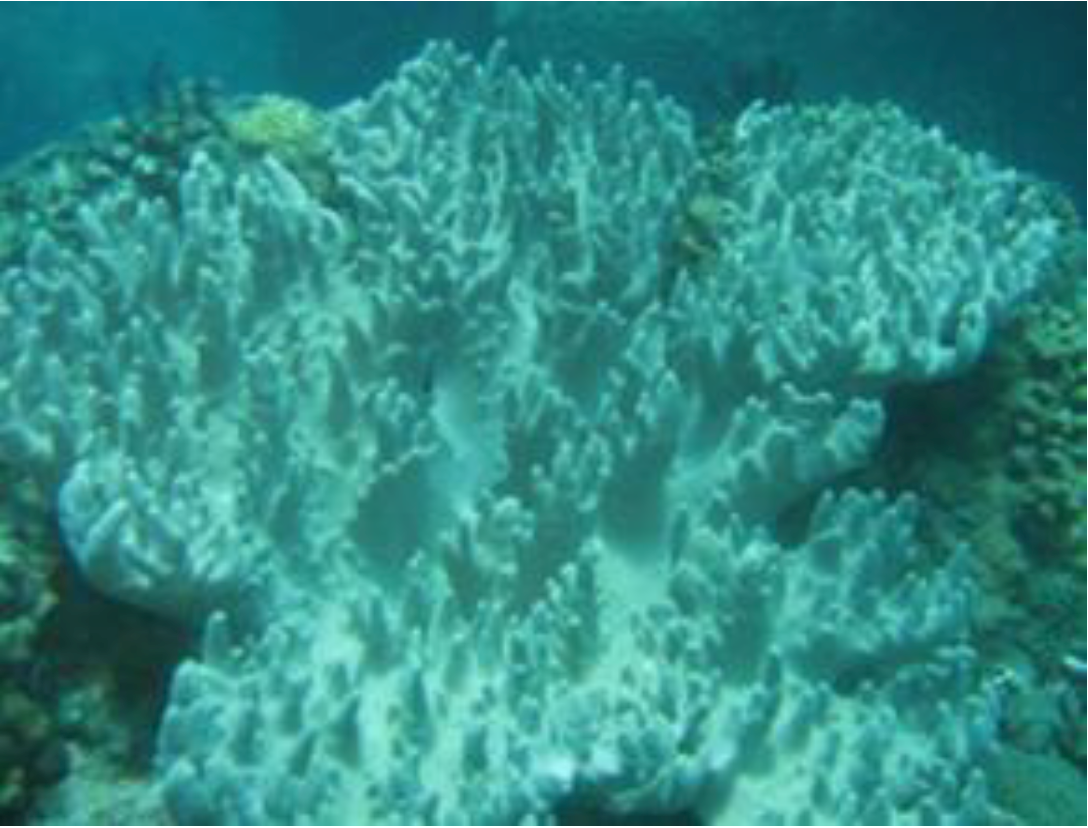
:1. Introduction


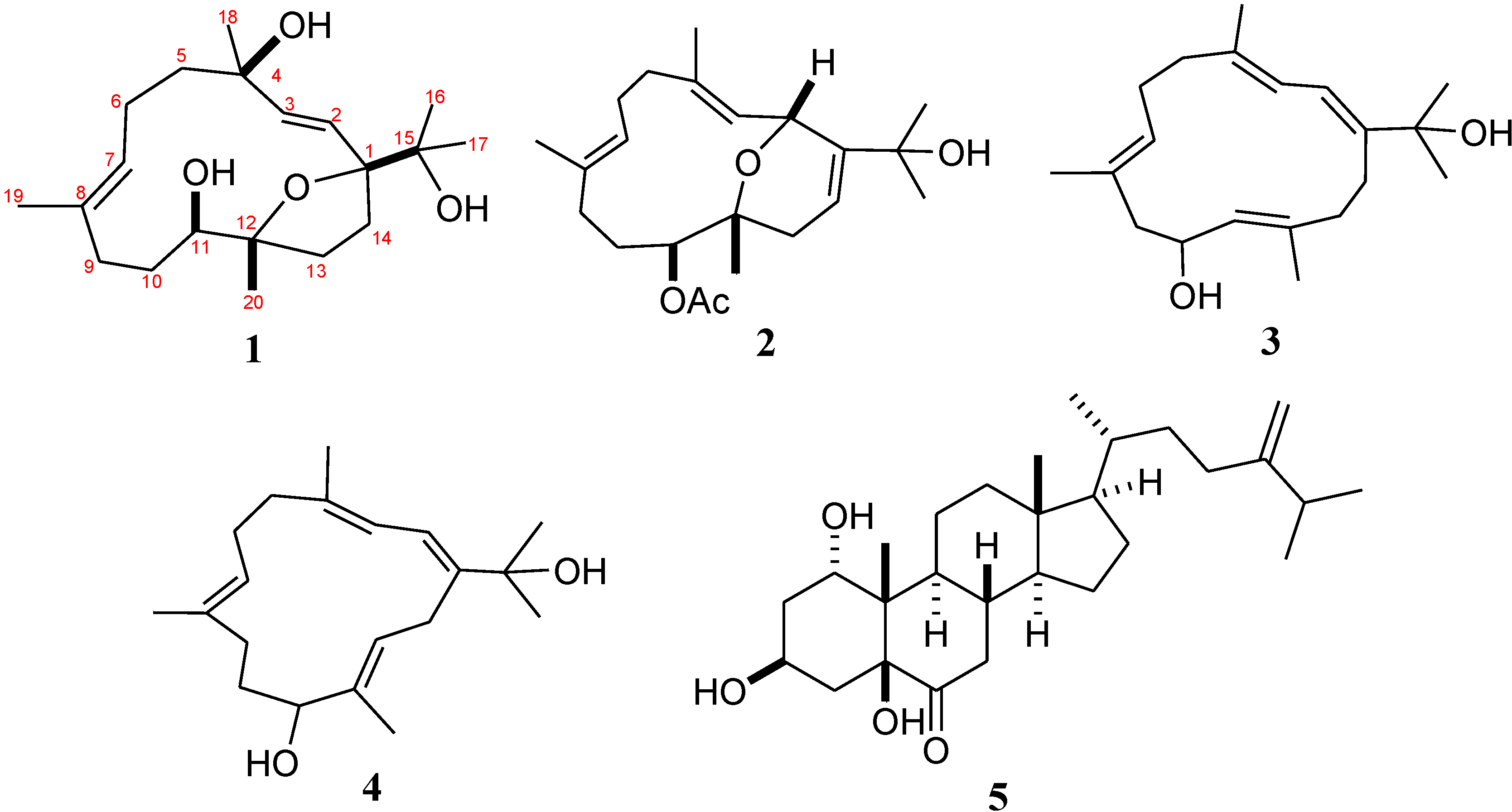
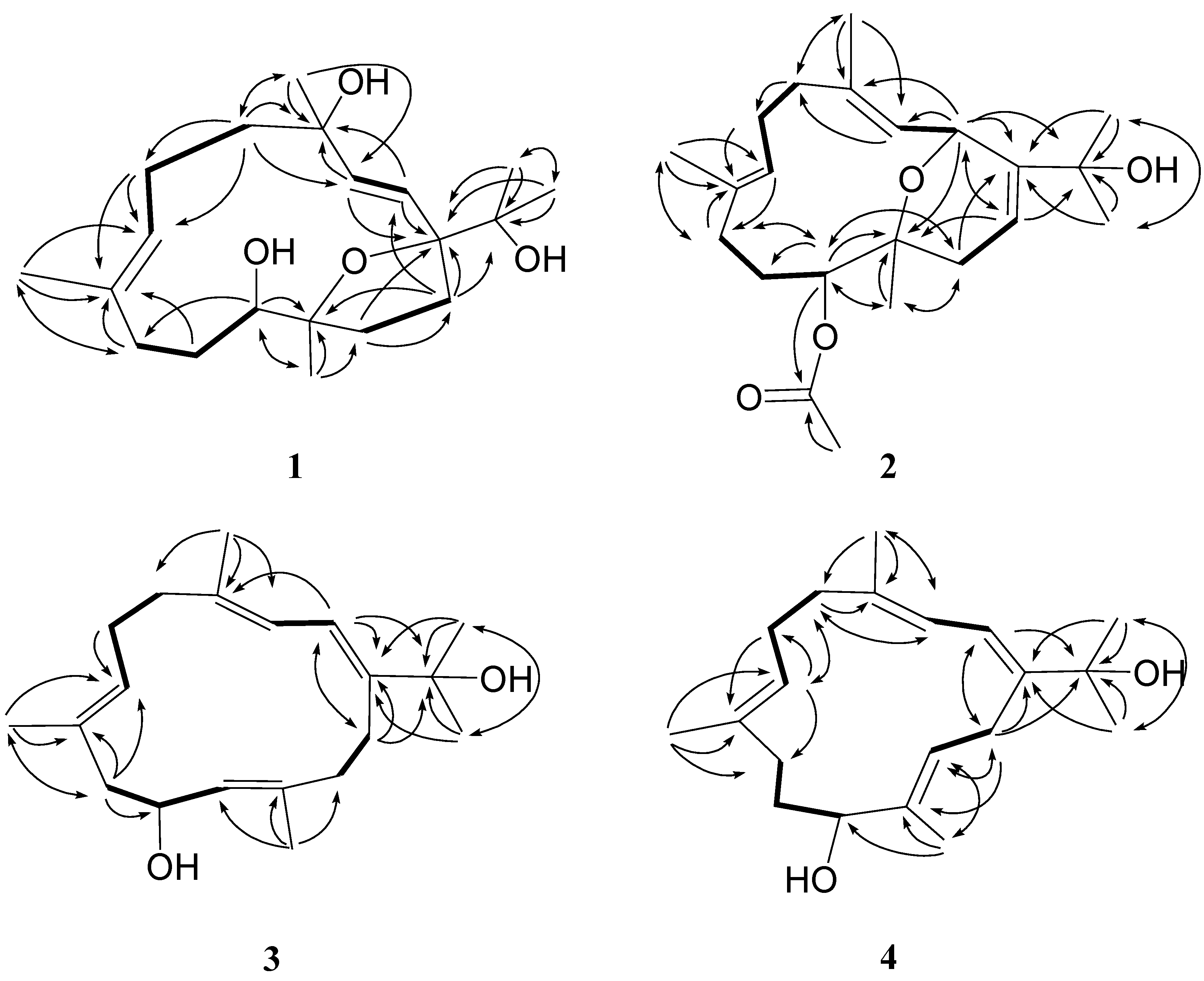
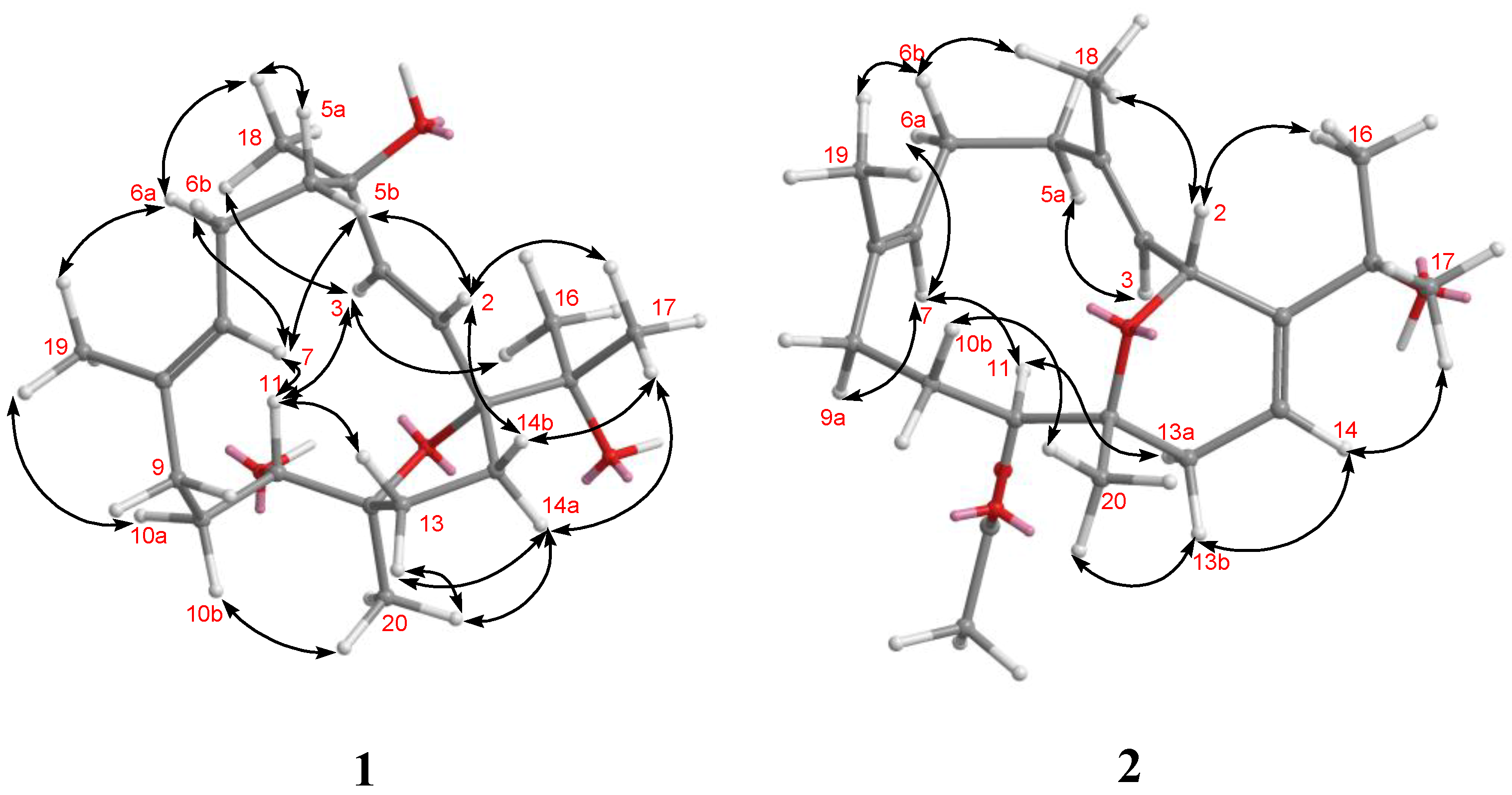
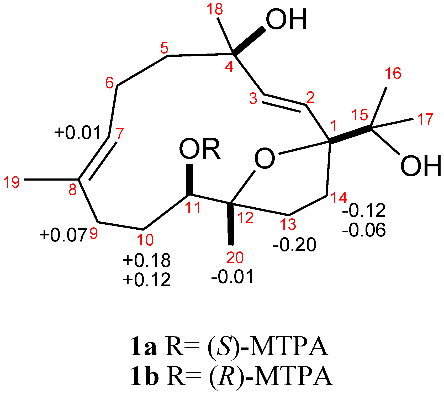
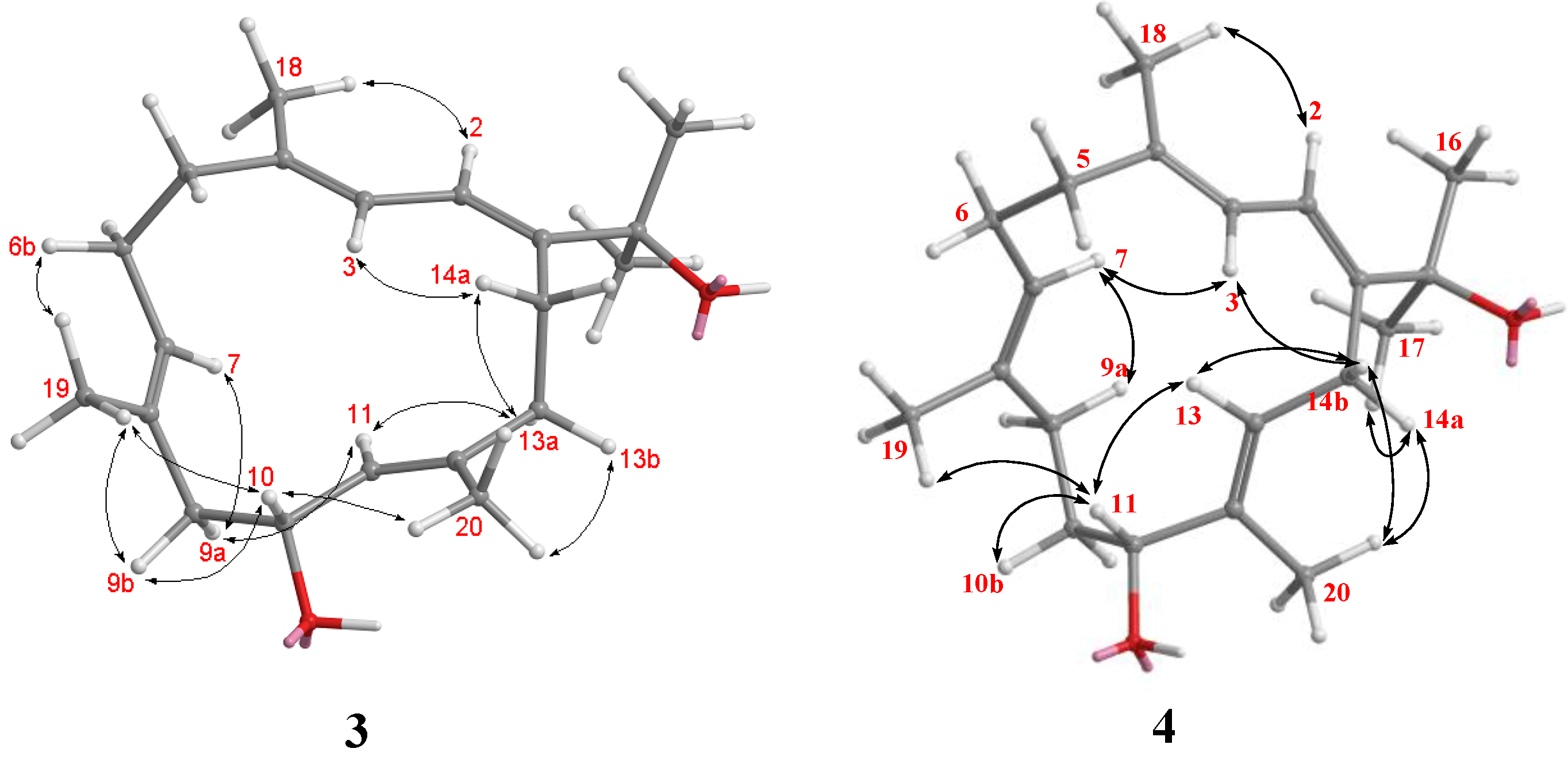
2. Results and Discussion


| Position | 1 | 2 | |||
|---|---|---|---|---|---|
| δH a | δC b | δH c | δC d | ||
| 1 | 91.3, qC f | 150.3, qC | |||
| 2 | 5.63 d (15.6) e | 128.2, CH | 5.01 d (10.5) | 69.2, CH | |
| 3 | 5.94 d (15.6) | 138.4, CH | 5.35 d (10.5) | 126.2, CH | |
| 4 | 74.5, qC | 138.8, qC | |||
| 5 | a:1.85 m; b:1.59 m | 43.9, CH2 | a:2.08 m; b:2.20 m | 39.7, CH2 | |
| 6 | a:2.27 m; b:2.17 m | 24.35, CH2 | a:2.08 m; b:2.43 dd (11.0, 3.0); | 26.1, CH2 | |
| 7 | 5.20 d (9.6) | 129.2, CH | 5.24 dd (11.0, 3.0) | 125.7, CH | |
| 8 | 133.5, qC | 134.7, qC | |||
| 9 | 2.10 m | 35.4, CH2 | a:1.82 t (3.0); b:1.94 m | 35.4, CH2 | |
| 10 | a:1.91 dd (14.7, 9.6); b:1.36 d (14.7) | 29.4, CH2 | a:1.82 m; b:1.44 m | 26.5, CH2 | |
| 11 | 3.56 d (9.6) | 76.4, CH | 5.39 d (9.0) | 78.8, CH | |
| 12 | 85.4, qC | 73.6, qC | |||
| 13 | 1.79 m | 36.6, CH2 | a:2.22 m; b:2.14 m | 32.4, CH2 | |
| 14 | a:2.43 m; b:1.69 m | 31.0, CH2 | 5.83 dd (7.5, 2.5) | 115.3, CH | |
| 15 | 72.4, qC | 71.8, qC | |||
| 16 | 1.07 s | 25.9, CH3 | 1.31 s | 28.8, CH3 | |
| 17 | 1.14 s | 24.41, CH3 | 1.31 s | 28.9, CH3 | |
| 18 | 1.28 s | 28.4, CH3 | 1.74 s | 15.2, CH3 | |
| 19 | 1.69 s | 16.2, CH3 | 1.59 s | 16.4, CH3 | |
| 20 | 1.12 s | 19.4, CH3 | 1.05 s | 23.3, CH3 | |
| OAc-11 | 2.10 s | 21.3, CH3 | |||
| 171.7, qC | |||||
| Position | 3 | 4 | ||
|---|---|---|---|---|
| δH a | δC b | δH a | δC b | |
| 1 | 147.1, qC d | 145.5, qC | ||
| 2 | 6.39 d (11.6) c | 118.7, CH | 6.37 d (10.4) | 118.7, CH |
| 3 | 5.78 d (11.6) | 120.0, CH | 5.71 d (10.4) | 121.3, CH |
| 4 | 138.3, qC | 137.4, qC | ||
| 5 | 2.18 m | 38.4, CH2 | 2.15 m | 38.0, CH2 |
| 6 | a:2.24 m; b:2.18 m; | 24.5, CH2 | 2.16 m | 24.5, CH2 |
| 7 | 5.02 d (5.2) | 127.8, CH | 4.95 brs | 126.0, CH |
| 8 | 131.2, qC | 133.4, qC | ||
| 9 | a:2.10 dd (12.4, 10.8); b:2.46 m | 48.0, CH2 | a:2.13 m; b:2.00 m | 35.9, CH2 |
| 10 | 4.55 ddd (14.8, 9.6, 4.0) | 66.2, CH | a:1.85 m; b:1.74 m | 29.4, CH2 |
| 11 | 5.13 d (9.6) | 128.4, CH | 3.93 dd (10.4, 3.6) | 79.1, CH |
| 12 | 140.2, qC | 135.5, qC | ||
| 13 | a:1.92 m; b:2.42 m | 42.4, CH2 | 5.20 t (5.2) | 130.4, CH |
| 14 | a:2.22 m b:2.40 m | 26.0, CH2 | a:2.83 dd (16.8, 5.2) b:3.00 dd (16.8, 5.2) | 26.7, CH2 |
| 15 | 74.1, qC | 73.9, qC | ||
| 16 | 1.38 s | 29.9, CH3 | 1.37 s | 28.8, CH3 |
| 17 | 1.38 s | 29.8, CH3 | 1.37 s | 28.9, CH3 |
| 18 | 1.79 s | 18.0, CH3 | 1.76 s | 17.6, CH3 |
| 19 | 1.60 s | 16.7, CH3 | 1.56 s | 15.2, CH3 |
| 20 | 1.74 s | 15.9, CH3 | 1.65 s | 11.0, CH3 |

3. Experimental Section
3.1. General Experimental Procedures
3.2. Animal Material
3.3. Extraction and Separation
3.4. Cytotoxicity Assay
3.5. Anti-HCMV Assay
4. Conclusions
Supplementary Files
Acknowledgments
Author Contributions
Conflicts of Interest
References
- Lakshmi, V.; Kumar, R. Metabolites from Sinularia species. Nat. Prod. Res. 2009, 23, 801–850. [Google Scholar] [CrossRef]
- Blunt, J.W.; Copp, B.R.; Keyzers, R.A.; Munro, M.H.G.; Prinsep, M.R. Marine natural products. Nat. Prod. Rep. 2013, 29, 144–222. [Google Scholar]
- Cheng, S.-Y.; Huang, K.-J.; Wang, S.-K.; Duh, C.-Y. Capilloquinol: A novel farnesyl quinol from the Dongsha Atoll soft coral Sinularia capillosa. Mar. Drugs 2011, 9, 1469–1476. [Google Scholar] [CrossRef]
- Cheng, S.-Y.; Huang, K.-J.; Wang, S.-K.; Wen, Z.-H.; Hsu, C.-H.; Dai, C.-F.; Duh, C.-Y. New terpenoids from the soft corals Sinularia capillosa and Nephthea chabroli. Org. Lett. 2009, 11, 4830–4833. [Google Scholar] [CrossRef]
- Cheng, S.-Y.; Chuang, C.-T.; Wen, Z.-H.; Wang, S.-K.; Chiou, S.-F.; Hsu, C.-H.; Dai, C.-F.; Duh, C.-Y. Bioactive norditerpenoids from the soft coral Sinularia gyrosa. Bioorg. Med. Chem. 2010, 18, 3379–3386. [Google Scholar] [CrossRef]
- Cheng, S.-Y.; Chuang, C.-T.; Wang, S.-K.; Wen, Z.-H.; Chiou, S.-F.; Hsu, C.-H.; Dai, C.-F.; Duh, C.-Y. Antiviral and anti-inflammatory diterpenoids from the soft coral Sinularia gyrosa. J. Nat. Prod. 2010, 73, 1184–1187. [Google Scholar] [CrossRef]
- Duh, C.-Y.; Wang, S.-K.; Tseng, H.-K.; Sheu, J.-H.; Chiang, M.Y. Novel cytotoxic cembranoids from the soft coral Sinularia flexibilis. J. Nat. Prod. 1998, 61, 844–847. [Google Scholar] [CrossRef]
- Duh, C.-Y.; Wang, S.-K.; Tseng, H.-K.; Sheu, J.-H. A novel cytotoxic biscembranoid from the Formosan soft coral Sinularia flexibilis. Tetrahedron Lett. 1998, 39, 7121–7122. [Google Scholar]
- Cheng, S.-Y.; Huang, K.-J.; Wang, S.-K.; Wen, Z.-H.; Chen, P.-W.; Duh, C.-Y. Antiviral and anti-inflammatory metabolites from the soft coral Sinularia capillosa. J. Nat. Prod. 2010, 73, 771–775. [Google Scholar] [CrossRef]
- Ahmed, A.F.; Dai, C.-F.; Kuo, Y.-H.; Sheu, J.-H. 1α,3β,5β-Trihydroxy-24-methylenecholestan-6-one: A novel steroid from a soft coral Sinularia gibberosa. Steroids 2003, 68, 377–381. [Google Scholar] [CrossRef]
- Bhemmasankra Rao, C.; Satyanarayana, C.; Srinivasa Rao, D.; Venkata Rao, D. Metabolites of the soft coral Sinularia ovispiculata from the Indian ocean. J. Nat. Prod. 1993, 56, 2003–2007. [Google Scholar] [CrossRef]
- Ahmed, A.F.; Wen, Z.-H.; Su, J.-H.; Hsieh, Y.-T.; Wu, Y.-C.; Hu, W.-P.; Sheu, J.-H. Oxygenated cembranoids from a Formosan soft coral Sinularia gibberosa. J. Nat. Prod. 2008, 71, 179–185. [Google Scholar] [CrossRef]
- Hou, R.-S.; Duh, C.-Y.; Chiang, M.Y.; Lin, C.-N. Sinugibberol, a new cytotoxic cembranoid diterpene from the soft coral Sinularia gibberosa. J. Nat. Prod. 1995, 58, 1126–1130. [Google Scholar] [CrossRef]
- Geran, R.I.; Greenberg, N.H.; MacDonald, M.M.; Schumacher, A.M.; Abbott, B.J. Protocols forscreening chemical agents and natural products against animal tumors and other biologicalsyatems. Cancer Chemother. Rep. 1972, 3, 51–61. [Google Scholar]
- Stevens, M.; Balzarini, J.; Tabarrini, O.; Andrei, G.; Snoeck, R.; Cecchetti, V.; Fravolini, A.; de Clercq, E.; Pannecouque, C. Cell-dependent interference of a series of new 6-aminoquinolone derivatives with viral (HIV/CMV) transactivation. J. Antimicrob. Chemother. 2005, 56, 847–855. [Google Scholar] [CrossRef]
© 2014 by the authors; licensee MDPI, Basel, Switzerland. This article is an open access article distributed under the terms and conditions of the Creative Commons Attribution license (http://creativecommons.org/licenses/by/3.0/).
Share and Cite
Tseng, Y.-J.; Yang, Y.-C.; Wang, S.-K.; Duh, C.-Y. Numerosol A–D, New Cembranoid Diterpenes from the Soft Coral Sinularia numerosa. Mar. Drugs 2014, 12, 3371-3380. https://doi.org/10.3390/md12063371
Tseng Y-J, Yang Y-C, Wang S-K, Duh C-Y. Numerosol A–D, New Cembranoid Diterpenes from the Soft Coral Sinularia numerosa. Marine Drugs. 2014; 12(6):3371-3380. https://doi.org/10.3390/md12063371
Chicago/Turabian StyleTseng, Yen-Ju, Yuan-Chien Yang, Shang-Kwei Wang, and Chang-Yih Duh. 2014. "Numerosol A–D, New Cembranoid Diterpenes from the Soft Coral Sinularia numerosa" Marine Drugs 12, no. 6: 3371-3380. https://doi.org/10.3390/md12063371