Article

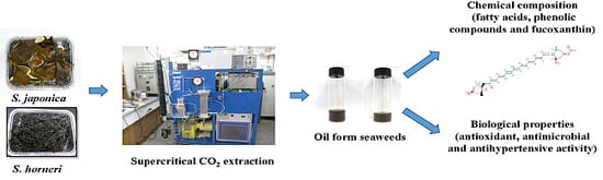
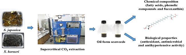
Biological Properties of Fucoxanthin in Oil Recovered from Two Brown Seaweeds Using Supercritical CO2 Extraction

1
Department of Food Science and Technology, Pukyong National University, 45 Yongso-ro, Nam-gu, Busan 608-737, Korea
2
Department of Chemical Engineering, Pukyong National University, 365 Sinseon-ro, 599-1 Daeyeon-3dong, Nam-gu, Busan 608-737, Korea
*
Author to whom correspondence should be addressed.
Mar. Drugs 2015, 13(6), 3422-3442; https://doi.org/10.3390/md13063422
Submission received: 13 March 2015 / Revised: 19 May 2015 / Accepted: 21 May 2015 / Published: 29 May 2015
(This article belongs to the Special Issue Marine Functional Food)

Abstract

:
The bioactive materials in brown seaweeds hold great interest for developing new drugs and healthy foods. The oil content in brown seaweeds (Saccharina japonica and Sargassum horneri) was extracted by using environmentally friendly supercritical CO2 (SC-CO2) with ethanol as a co-solvent in a semi-batch flow extraction process and compared the results with a conventional extraction process using hexane, ethanol, and acetone mixed with methanol (1:1, v/v). The SC-CO2 method was used at a temperature of 45 °C and pressure of 250 bar. The flow rate of CO2 (27 g/min) was constant for the entire extraction period of 2 h. The obtained oil from the brown seaweeds was analyzed to determine their valuable compounds such as fatty acids, phenolic compounds, fucoxanthin and biological properties including antioxidant, antimicrobial, and antihypertension effects. The amounts of fucoxanthin extracted from the SC-CO2 oils of S. japonica and S. horneri were 0.41 ± 0.05 and 0.77 ± 0.07 mg/g, respectively. High antihypertensive activity was detected when using mixed acetone and methanol, whereas the phenolic content and antioxidant property were higher in the oil extracted by SC-CO2. The acetone–methanol mix extracts exhibited better antimicrobial activities than those obtained by other means. Thus, the SC-CO2 extraction process appears to be a good method for obtaining valuable compounds from both brown seaweeds, and showed stronger biological activity than that obtained by the conventional extraction process.

Graphical Abstract

1. Introduction

Algae can be divided into two groups including macro-algae, or seaweeds and micro-algae. Similar to true plants, seaweeds are photosynthetic and form basic biomasses in intertidal zones. Approximately, 9000 seaweed species have been identified. On the basis of their pigmentation, these species have been broadly classified into three main groups of brown (Phaeophyta), red (Rhodophyta), and green (Chlorophyta) seaweeds [1]. Seaweeds contain several physiologically bioactive compounds with important economical relevance including polysaccharides, iodine organic products, macro- and micro-elements, vitamins, and unsaturated fatty acids (FAs) [2]. Brown seaweeds, the second-most abundant group of marine algae, include approximately 2000 species. Among them, Sargassum spp., Laminaria spp., Ascophyllum spp., Fucus spp., and Turbinaria spp. are most commonly used at the industrial level [3]. Because seaweeds lack many of the distinct organs found in terrestrial plants, the entire plant is available as a biomass source. A significant amount of attention has been paid recently to seaweeds as effective biomass sources because of their high CO2 absorption rate relative to that of terrestrial plants [1]. Seaweeds are of emerging interest in biomedical areas mainly due to their content of bioactive substances that show high potential as antioxidants, antimicrobials, anticoagulants, antithrombotics, and anti-inflammatories for the treatment of several diseases, in addition to their anti-tumor and anti-viral properties [4,5,6]. Therefore, seaweeds have been explored as sources of food, medicine, cosmetics, fertilizer, feed, and bio-energy [7].
The antioxidant sources of seaweeds are related mainly to their polyphenol content, particularly phlorotannins, which are the largest group of polyphenols in brown seaweed [7]. Seaweed lipids have drawn increased interest due to their health benefits. Brown seaweed lipids contain many types of bioactive compounds such as omega-3 polyunsaturated (PUFAs), omega-6, arachidonic acid, fucoxanthin, fucosterol, and some polyphenols. Among these compounds, fucoxanthin, a major carotenoid in brown seaweed, is regarded as a nutraceutical compound specific to brown seaweed lipids because it shows several physiological effects based on unique molecular mechanisms [8]. Therefore, brown seaweed represents a highly functional lipid source. However, because of the high level of omega-3 PUFAs such as eicosapentaenoic acid (20:5n-3) and stearidonic acid (18:4n-3), brown seaweed lipids may be susceptible to oxidation [1].
Fucoxanthin, a member of the carotenoid family, is abundant in edible brown algae and contributes to more than 10% of the estimated total production of carotenoids in nature. This compound plays a role in light harvesting and energy transfer and thus helps marine brown alga survive in shallow coastal waters by offering efficient photosynthesis for acclimatization to their environment [9]. Although less attention has been paid to the physiological effects of carotenoids in seaweeds, fucoxanthin has recently attracted high attention due to its strong antioxidant properties against the effects of cancer, hypertension, obesity, and inflammation [10].
Hypertension is considered to be the most common chronic disease and a major risk factor for cardiovascular disease, a main cause of death worldwide [11]. Angiotensin-I converting enzyme (ACE) regulates blood pressure function by converting angiotensin-I into a potent vasoconstrictor, angiotensin-II, and catalyzing the degradation of a potent vasodilator, bradykinin [12]. ACE inhibitors (ACE-I), a new class of antiangiotensive drugs, are effective in inhibiting the formation of angiotensin-II [13]. Synthetic ACE-I has been developed for antihypertension drugs including captopril, enalapril, alacepril, and lisinopril, which are widely used in the treatment of hypertension and heart failure in humans. However, some side effects caused by these synthetic drugs have been reported [12]. Many ACE-inhibitory peptides have been discovered in the enzymatic hydrolysates of various protein-rich foods such as soy sauce, fish sauce, sake, soybeans, and milk. Among various sources, marine organisms have been widely used in the search for ACE-I peptides [13]. Hence, scientists have focused special attention on marine algae to identify and evaluate natural antihypertension compounds. The amino acid heterogeneity of proteins is very high among seaweeds even in those containing low amounts of protein. Therefore, it is believed that the evaluation of algal species is a promising way to find new anti-hypertensive compounds [11].
The aim of this study is to extract the oil from two brown seaweeds, S. japonica and S. horneri, by using environmental friendly SC-CO2 extraction and in addition to that by conventional solvents. The obtained oil was investigated to determine its composition of FAs, fucoxanthin, and total phenolics in addition to antioxidant, antimicrobial, and antihypertension activities.

2. Results and Discussion

2.1. Conventional Solvent Extraction

The efficiencies of oil extracted by using three different solvents including hexane, ethanol, and acetone mixed with methanol (1:1, v/v) was checked by examining their different polarities. As shown in Table 1, various oil yields were produced by the various solvents. The extracted oil contents in S. japonica and S. horneri were 1.19 ± 0.21 and 1.29 ± 0.05 g/100 g dry weight (DW) when acetone mixed with methanol was used as the solvent, 1.24 ± 0.06 and 1.42 ± 0.08 g/100 g DW when hexane was used, and 1.22 ± 0.12 and 1.36 ± 0.14 g/100 g DW when ethanol was used, respectively. These results indicate that acetone mixed with methanol was the most efficient extraction solvent. Furthermore, the recovery of acetone–methanol was the highest of the solvents in both the seaweeds, and S. horneri had the higher oil yield.

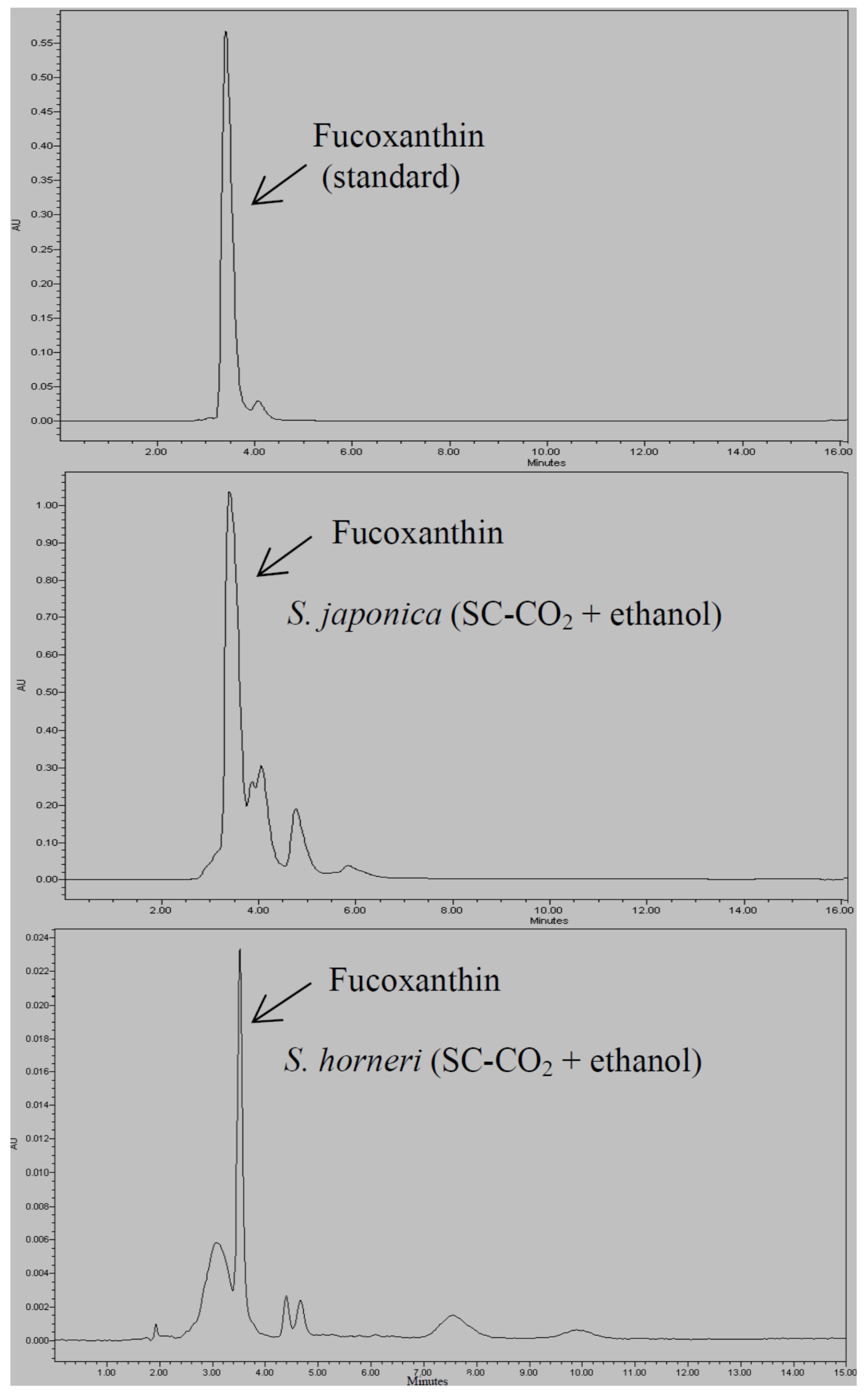
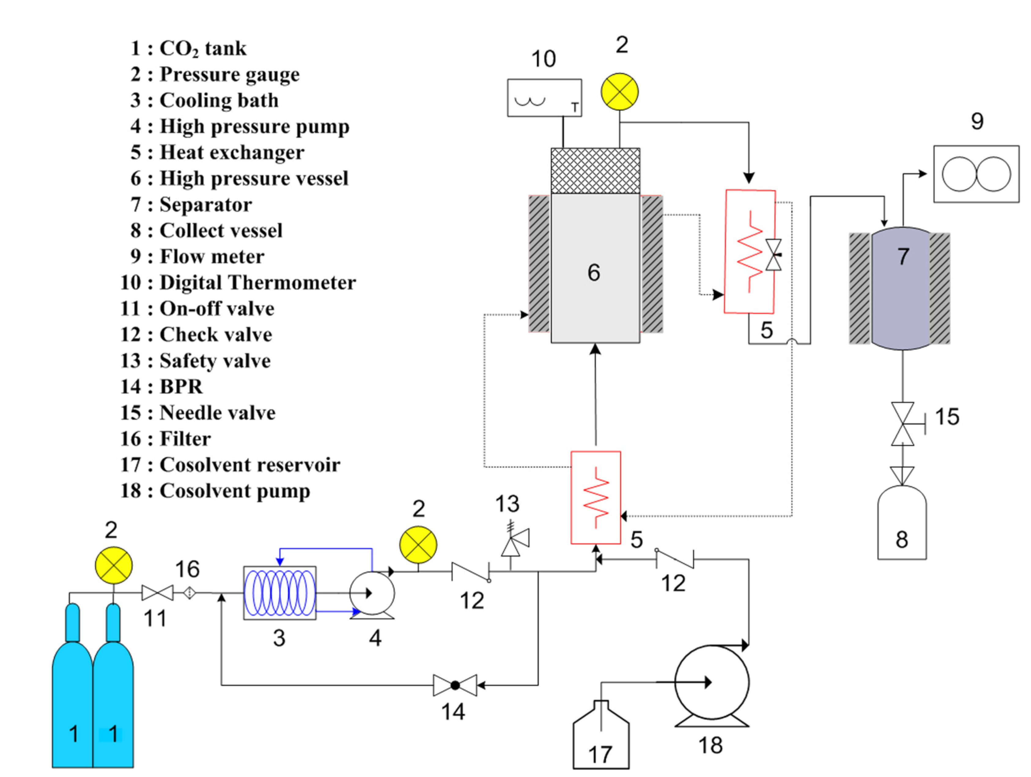
2.2. Extraction of Oil Using SC-CO2 with Ethanol as Co-Solvent

In a previous study, the extraction of lipids and fucoxanthin was reported to be very low from brown seaweed when pure SC-CO2 was used; however, the yield was increased when using a co-solvent [14,15]. In the present study, we evaluated the effects of SC-CO2 with ethanol as a co-solvent in the extraction of oil from brown seaweeds at operational condition of 250 bar and 45 °C. The oil contents obtained from S. japonica and S. horneri through this process were 1.09 ± 0.56 and 1.41 ± 0.15 g/100 g DW, respectively (Table 1). Therefore, extraction experiments using SC-CO2 with ethanol as a co-solvent can yield similar oil content through an environmental friendly extraction process.
Conde et al. [15] reported that under the influence of SC-CO2 modified with 10% ethanol, the extract yield from S. muticum was significantly improved; the yields of total oil and fucoxanthin were up to three and 90 times higher, respectively.
Table 1. Extraction yield and fucoxanthin content of brown seaweeds from various extracts.
SolventExtraction Yield (g/100 g DW)Fucoxanthin Content (mg/g)
S. japonicaS. horneriS. japonicaS. horneri
SC-CO2 + Ethanol1.09 ± 0.56 a1.41 ± 0.15 b0.41 ± 0.05 a0.77 ± 0.07 a
Acetone mix methanol1.19 ± 0.21 a1.29 ± 0.05 a0.48 ± 0.10 b0.71 ± 0.05 b
Hexane1.24 ± 0.06 a1.42 ± 0.08 c0.16 ± 0.01 a0.05 ± 0.03 c
Ethanol1.22 ± 0.12 a1.36 ± 0.14 c0.12 ± 0.02 a,b0.08 ± 0.05 c
Values are expressed as mean ± SD. Different letters indicate significant differences (p < 0.05) according to Tukey’s multiple range test.

2.3. Measurement of FA Composition and Fucoxanthin Content

The FA compositions of the obtained oil from different extraction systems including hexane, ethanol, acetone–methanol extract, and SC-CO2 with ethanol from brown seaweeds determined by gas chromatography (GC) are shown in Table 2. The percentages of the total saturated FAs (SFAs) were high in S. horneri extracts, whereas those of monounsaturated FAs (MUFAs) were higher in the S. japonica extracts. In S. horneri extracts, palmitic acid (C16:0) was found in high amounts ranging from 180.70 ± 1.80 to 233.20 ± 2.90 mg/g in extracted oil, whereas more elaidic acid was found in S. japonica extracts, from 168.90 ± 0.20 to 340.10 ± 0.10 mg/g in extracted oil. Important PUFAs such as eicosapentaenoic acid (EPA; C20:5n3) were present in high amounts. In S. horneri, 80.80 ± 0.90 mg/g in extracted oil of EPA was found in the SC-CO2 extract, and 70.60 ± 0.10 mg/g in extracted oil was in the acetone–methanol extract of S. japonica. No docosahexaenoic acid (DHA; C22:6n3) was found in the oil of either brown seaweed with the various solvents used in this experiment. Among the solvent systems, the total FA compositions were high in the SC-CO2 extract and in acetone–methanol extract. Terasaki et al. [16] reported that 9.70% EPA was found in S. horneri,and Dawczynski et al. [17] reported levels of 16.2% ± 8.90% in Laminaria sp. The lower amounts obtained from our experiments could be attributed to seasonal variation and continental location, which can dramatically alter the composition.
Seaweed products represent an important source of long-chain polyunsaturated FAs (LC-PUFA) that are fundamental for the formation of important structural lipids and elements of cell membranes. In addition, these LC-PUFA are precursors of eicosanoids, which influence inflammation processes and immune reactions [18]. Although these two classes of PUFA have opposing physiological functions, their balance is important for normal growth and development. These FAs are beneficial for the prevention of cardiovascular diseases and other chronic diseases such as diabetes, hypertension, and autoimmune diseases. The European Nutritional Societies have recommended that human diet include an n-6:n-3 ratio of 5:1 for promoting good health [19]. Thus, consumption of seaweed products can contribute to improvement in the dietary supply of n-3 FA. Intake of foods rich in n-3 LC-PUFA can have a positive influence on the composition of blood lipids and can therefore be used for the prevention of arteriosclerosis [20].
Table 2. Fatty acid composition of S. japonica and S. horneri from various extracts.
Fatty acid compositionsS. japonica (mg/g in Extracted Oil)S. horneri (mg/g in Extracted Oil)
SC-CO2 + EthanolAcetone mix methanolHexaneEthanolSC-CO2 + EthanolAcetone mix methanolHexaneEthanol
Saturated fatty acid (SFA)C10:0N.DN.DN.DN.D49.1 ± 0.7051.4 ± 0.50211.7 ± 3.1N.D
C11:0N.DN.DN.DN.DN.D8.5 ± 0.30N.D250.10 ± 3.50
C12:0N.DN.DN.DN.D17.4 ± 0.1020.1 ± 0.2027.9 ± 0.30N.D
C13:014.10 ± 1.0N.DN.DN.D34.3 ± 0.5029.90 ± 0.2023.00 ± 0.50N.D
C14:0141.40 ± 8.00123.8 ± 1.00130.50 ± 1.40114.80 ± 0.2075.50 ± 0.8065.80 ± 0.4013.00 ± 0.3038.60 ± 0.40
C15:04.60 ± 0.1010.20 ± 0.1014.50 ± 0.204.30 ± 0.1041.60 ± 0.5052.40 ± 0.4021.20 ± 0.4016.30 ± 0.30
C16:0N.DN.DN.DN.D214.80 ± 3.40185.40 ± 2.40180.70 ± 1.80233.2 ± 2.90
C17:0N.DN.DN.D18.40 ± 0.10N.D20.10 ± 0.1022.50 ± 0.60N.D
C18:0N.DN.DN.DN.D17.10 ± 0.2019.50 ± 0.2026.00 ± 0.40N.D
C20:0N.DN.D48.10 ± 0.1051.70 ± 0.208.50 ± 0.1012.10 ± 0.208.60 ± 0.1029.80 ± 0.40
C21:029.80 ± 0.10107.10 ± 0.1091.90 ± 1.00102.70 ± 0.5062.30 ± 0.8061.10 ± 0.6044.30 ± 0.4039.30 ± 0.50
C23:0N.DN.D7.20 ± 0.105.80 ± 0.10N.D7.60 ± 0.20N.D11.30 ± 0.40
Total SFA189.90 ± 9.20241.10 ± 1.20292.20 ± 2.80297.70 ± 1.20520.60 ± 7.10533.90 ± 5.70578.90 ± 7.90618.60 ± 8.40
Monounsaturated fatty acid (MUFA)C15:1252.70 ± 0.10179.80 ± 0.10178.00 ± 1.00200.90 ± 0.1011.30 ± 0.2015.90 ± 0.3028.40 ± 0.50N.D
C14:15.00 ± 0.1029.90 ± 0.008.60 ± 0.5021.30 ± 0.4016.60 ± 0.2021.40 ± 0.3021.10 ± 0.2056.40 ± 0.50
C16:118.70 ± 0.1045.30 ± 0.1045.80 ± 0.1040.10 ± 0.7026.90 ± 0.4023.10 ± 0.2013.10 ± 0.3056.00 ± 0.80
C17:126.90 ± 0.1029.60 ± 0.4010.20 ± 0.1019.60 ± 0.106.20 ± 0.1029.60 ± 0.4037.90 ± 0.70N.D
C18:1n9cN.DN.DN.DN.D122.90 ± 0.50112.40 ± 0.7089.80 ± 0.7083.70 ± 1.10
C18:1n9t340.10 ± 0.10182.90 ± 0.50177.50 ± 1.00168.90 ± 0.2016.90 ± 0.2018.50 ± 0.5023.40 ± 0.30N.D
C22:1n9N.DN.DN.DN.DN.D8.30 ± 0.20N.DN.D
Total MUFA644.30 ± 0.50467.50 ± 1.10420.10 ± 2.70450.80 ± 1.50200.80 ± 1.60229.20 ± 2.60213.70 ± 2.70196.10 ± 2.40
Polyunsaturated fatty acid (PUFA)C18:2n6c10.40 ± 0.1053.80 ± 0.2078.70 ± 0.1083.50 ± 0.207.30 ± 0.1012.50 ± 0.1011.70 ± 0.2038.60 ± 0.40
C18:2n6t4.10 ± 0.10N.DN.DN.D64.30 ± 0.5070.10 ± 0.4020.60 ± 0.40N.D
C18:3n623.20 ± 0.1047.80 ± 0.1045.00 ± 0.1045.90 ± 0.5024.60 ± 0.3016.90 ± 0.509.50 ± 0.10N.D
C20:223.50 ± 0.1056.40 ± 1.00N.DN.D10.50 ± 0.1018.70 ± 0.3010.60 ± 0.2022.0 ± 0.40
C18:3n3N.DN.DN.DN.DN.D4.50 ± 0.105.10 ± 0.2030.20 ± 0.60
C20:3n627.80 ± 0.2062.80 ± 0.50107.00 ± 1.40104.80 ± 0.8065.80 ± 0.9064.50 ± 0.5066.70 ± 0.7060.10 ± 0.80
C22:2N.DN.DN.DN.D13.90 ± 0.309.50 ± 0.408.00 ± 0.10N.D
C20:5n357.70 ± 0.4070.60 ± 0.1057.00 ± 0.2017.30 ± 0.2080.80 ± 0.9054.70 ± 1.2075.20 ± 0.8034.40 ± 0.50
C22:6n3N.DN.DN.DN.DN.DN.DN.DN.D
Total PUFA146.70 ± 1.00291.40 ± 1.90287.70 ± 1.80251.50 ± 1.70267.20 ± 3.10251.40 ± 3.50202.30 ± 2.70185.30 ± 2.70
Values are mean ± SD of three determinations. N.D means not detected.
Figure 1. HPLC chromatogram of fucoxanthin content in S. japonica (SC-CO2 + ethanol) and S. horneri (SC-CO2 + ethanol) extracts.
Marinedrugs 13 03422 g001
The fucoxanthin contents in the brown seaweeds extracted by the various methods are shown in Table 1. The extracted oil from S. horneri by using SC-CO2 showed the highest content 0.77 ± 0.07 mg/g, followed by 0.71 ± 0.05 mg/g in the acetone–methanol extract. Moreover, the fucoxanthin in the S. japonica oil extracted by acetone–methanol, 0.48 ± 0.10 mg/g, was higher than that by SC-CO2 at 0.41 ± 0.05 mg/g. These results show that the SC-CO2 process can extract a similar content of fucoxanthin as when a solvent is used (Figure 1 and Supplementary Figure S1). A previous report on the fucoxanthin content in S. horneri noted 11.24 mg/g in methanol extract [21], and another reported 3.7 ± 1.6 mg/g DW in seaweed by using the same process [16]. In a third report, the fucoxanthin content in S. japonica by using ethanol was reported as 18.7 mg/100 g fresh weight [22]. Conde et al. [15] reported that 12 mg/100 g in S. muticum (SC-CO2 with 10% ethanol). The content of fucoxanthin was reported to vary significantly with season and life cycle of the algae, peaking between the winter and spring (mature phase of sporophyte) and lowest during summer (senescence phase) [16].
Carotenoids are widely present in marine biota, and their occurrence is species-dependent. Fucoxanthin is a dominant carotenoid characteristic of brown algae, particularly diatoms [22], whereas β-carotene, zeaxanthin and fucoxanthin are major pigments of red seaweed [16]. Fucoxanthin is the main carotenoid in various edible brown seaweed types such as Undaria pinnatifida and Hijikia fusiforme [23]. However, information on the content of fucoxanthin in the other brown seaweeds, edible or otherwise, is highly limited. The results of this study are important because several previous studies have established fucoxanthin as an anticancerous, antidiabetic, and antiobesity agent [16]. The results of our study indicate the potential of several brown types as sources of these multifunctional biomolecules.

2.4. Antioxidant Properties and Total Phenolic Content

2.4.1. Hydrogen Peroxide Scavenging (H2O2) Assay

The yield of extracts from seaweed through SC-CO2 with ethanol was significantly higher than that when other extracts were used. The obtained results of all the extracts are shown in Table 3. The half maximal inhibitory concentration (IC50) value of SC-CO2 with ethanol extract from S. horneri was 686.31 ± 0.20 μg/mL, and that from S. japonica was 600.73 ± 0.15 μg/mL. The activities were found to be proportionately decreased when solvents such as hexane and ethanol were used. It is noted that acetone–methanol for both brown seaweeds was significantly effective in scavenging H2O2, with yields of 450.49 ± 1.42 μg/mL and 546.10 ± 1.05 μg/mL for S. japonica and S. horneri, respectively. The IC50 value of standard ascorbic acid was 448.19 ± 0.78 μg/mL.
Athiperumalsami et al. [24] reported that when using the H2O2 scavenging method, the methanoland water extracts of the seaweeds Padina tetrastromatica, Ulva lactuca, and Acanthophora spicifera showed the highest antioxidant activity, although the IC50 values were lower than that of standard tocopherol. The methanol extract of the seaweed Gracilaria foliifera had antioxidant activities higher than the standard tocopherol when tested by the H2O2 scavenging method. In the present study, similar results were obtained when ascorbic acid was used as a standard, and the SC-CO2 with ethanol extracts of seaweed showed higher values than the standard.
Table 3. Antioxidant activity, hydrogen peroxide scavenging assay, β-carotene linoleic acid method, total antioxidant activity and total phenolic content of brown seaweeds from various extracts.
H2O2 IC50 (μg/mL)β-carotene Bleaching Assay (%)Total Antioxidant Capacity (mg/g α-tocopherol DW)Total Phenolic Content (mg/g GAE DW)
SolventS. japonicaS. horneriS. japonicaS. horneriS. japonicaS. horneriS. japonicaS. horneri
SC-CO2 + ethanol600.73 ± 0.15 a686.31 ± 0.20 a72.00 ± 0.74 a75.03 ± 1.56 a30.74 ± 0.13 a38.91 ± 3.79 a0.60 ± 0.05 a0.64 ± 0.01 a
Acetone mix methanol450.49 ± 1.42 b546.10 ± 1.05 b65.10 ± 3.03 b69.51 ± 0.35 b28.74 ± 1.38 b28.05 ± 0.72 b0.56 ± 0.04 b0.60 ± 0.01 b
Hexane100.15 ± 2.17 c125.18 ± 0.35 c31.81 ± 0.76 c36.58 ± 0.65 c16.67 ± 2.41 c19.77 ± 0.86 c0.28 ± 0.01 c0.42 ± 0.02 c
Ethanol200.02 ± 4.21 d220.62 ± 1.57 d42.66 ± 0.85 d46.71 ± 2.62 d22.5 ± 0.10 d25.63 ± 0.93 d0.34 ± 0.05 d0.43 ± 0.01 c
Ascorbic acid448.19 ± 0.78
Trolox84.56 ± 0.34
Values are expressed as mean ± SD. Different letters indicate significant differences (p < 0.05) according to Tukey’s multiple range test.
Figure 2. (a) Reducing power of S. japonica extracts with a standard antioxidant; (b) S. horneri extracts with a standard antioxidant. Error bars represent standard deviation with three replicates. Different letters indicate significant differences (p < 0.05) according to Tukey’s multiple range test.
Marinedrugs 13 03422 g002

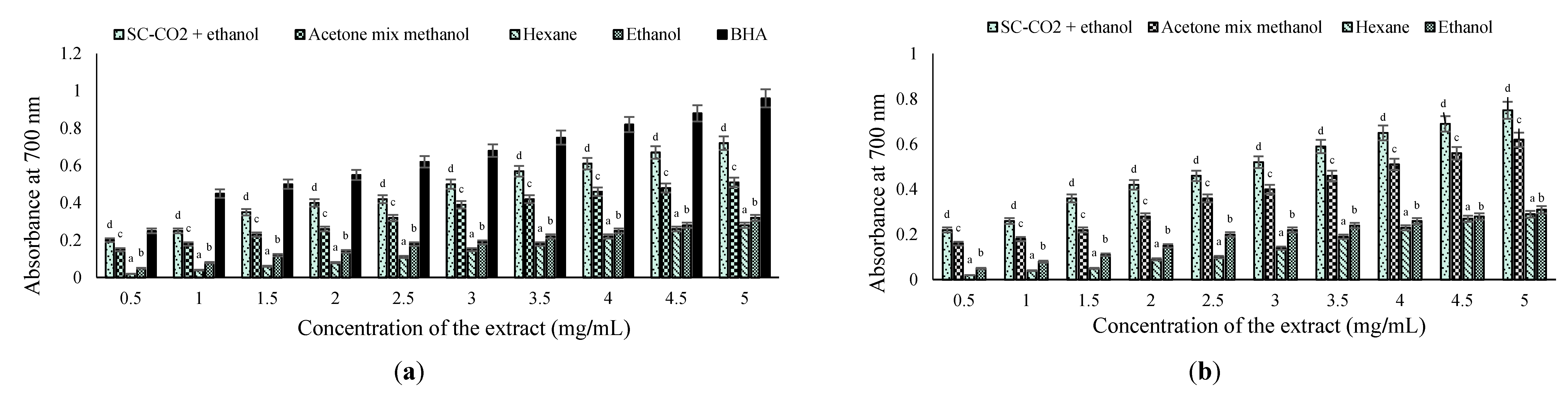
2.4.2. Reducing Power (RP) Method

The reducing powers of all of the extracts were increased at higher concentrations. In particular, the SC-CO2 with ethanol extract of S. horneri exhibited a reducing power effect as high as ascorbic acid at 5 mg/mL, transforming radical to non-radical by giving away hydrogen or an electron. The reducing powers of brown seaweed extracts were concentration-dependent (Figure 2a,b). As the concentration increased from 0.5 to 5.0 mg/mL, an increase in absorbance was noted for all of the solvents except hexane. However, the reducing powers of the samples were in the following order: butylated hydroxyanisole (BHA) (0.25–0.96) > SC-CO2 with ethanol extract of S. horneri (0.22–0.74) > SC-CO2 with ethanol extract of S. japonica (0.20–0.70) > acetone–methanol extract of S. horneri (0.16–0.62) > acetone–methanol extract of S. japonica (0.15–0.53) > ethanol extract of S. horneri (0.05–0.31) > ethanol extract of S. japonica (0.05–0.30) > hexane extract of S. horneri (0.02–0.29) > hexane extract of S. japonica (0.02–0.28). The reducing power increased with an increase in concentration in all of the samples. The same trend was also reported by Kumaran et al. [25] in the methanolic extracts of higher plants. All concentrations exhibited an optical density (OD) value of <1.0. This property is associated with the presence of reductones that are reported to be terminators of the free radical chain reaction [26].

2.4.3. β-Carotene Linoleic Acid Method

The antioxidant activities of the seaweed extracts through β-carotene bleaching assay are shown in Table 3. Among all tested extracts, only SC-CO2 with ethanol showed good activity against this system, with S. japonica at 72.00% ± 0.74% and S. horneri at 75.03% ± 1.56%. The standard (Trolox) showed 84.56% ± 0.34%. Many tests have been developed to evaluate the antioxidant activity of various samples to account for their chemical diversity. Some determine the ability of antioxidants in scavenging free radicals generated in the reaction medium, such as DPPH (2,2-diphenyl-1-picrylhydrazyl); others measure the inhibition of lipid oxidation through the antioxidant system to be tested, such as β-carotene. Therefore, it is necessary to evaluate the effect of the methodology used.
The mechanism of bleaching of β-carotene is a free radical-mediated phenomenon resulting from the H2O2 formed from linoleic acid. The β-carotene in this model system undergoes rapid discoloration in the absence of an antioxidant. The linoleic acid free radical attacks the highly unsaturated β-carotene molecules. As β-carotene molecules lose their double bonds by oxidation, the compound loses its chromophore and characteristic orange color, which can be monitored spectrophotometrically; that is, the absorbance values decrease with time as indicated by the rate of peroxidation [27]. The presence of an antioxidant can hinder the extent of β-carotene bleaching by neutralizing the linoleate free radical and other free radicals formed in this system; consequently, the absorbance values show different decreasing patterns. Generally, stronger antioxidant activity relate to slower decreases in absorbance values with time [28].
In the β-carotene assay, the oxidation of linoleic acid generates peroxyl free radicals due to the abstraction of hydrogen atoms from diallylic methylene groups of linoleic acid [29]. The free radical then will oxidize the highly unsaturated β-carotene. Consequently, the orange-colored chromophore of β-carotene would be degraded, and the result could be monitored spectrophotometrically. However, the presence of antioxidant constituents could neutralize the linoleate free radical and hence prevent the bleaching of β-carotene [28].

2.4.4. Total Antioxidant Activity

The global antioxidant activities of extracts were expressed as the number of α-tocopherol equivalents. The phosphomolybdenum method was based on the reduction of Mo (VI) to Mo (V) by the antioxidant compound and the formation of a green phosphate/Mo (V) complex. The antioxidant activity of S. horneri was higher than that of S. japonica extracts (Table 3). Higher activity of 38.91 ± 3.79 and 30.74 ± 0.13 mg/g α-tocopherol DW was observed in SC-CO2 with ethanol extract of S. horneri and S. japonica, respectively. The acetone–methanol extract also showed 28.05 ± 0.72 and 28.74 ± 1.38 mg/g α-tocopherol DW of S. horneri and S. japonica, respectively.
Chandini et al. [25] have reported total antioxidant activity in the range of 245 to 376 mg ascorbic acid equivalents/g in the case of brown seaweed extracts. Higher activity was observed in fractions as compared to total methanolic extract. Higher activity in fractions may be due to the interferences of other compounds present in crude (methanolic) extract; and, it has also been reported that solvents used for extraction have dramatic effect on the chemical species [30]. It has been earlier reported that some major active compounds from brown seaweed that have antioxidative properties are phlorotannins and fucoxanthin. Also, antioxidative activities of brown seaweeds cannot be attributed to their characteristic pigment (fucoxanthin) or any other carotenoids alone [25].

2.4.5. Total Phenolic Content

Phenolic compounds are widely distributed in the plant kingdom and have been reported to have several biological activities including antioxidant properties. Earlier reports revealed that marine seaweed extracts, particular their polyphenols, have antioxidant activity. Phlorotannins and fucoxanthin have been reported as major active compounds in various seaweed extracts [31]. Previous studies have shown that phenolic compounds are the main contributors to the antioxidant activity of various seaweeds [32]. The total phenolic content (TPC) of the seaweed extracts was calculated by using a modified Folin-Ciocalteu method (Table 3). The oil of S. japonica extracted with SC-CO2 and ethanol and that of S. horneri , at 0.60 ± 0.05 mg/g and 0.64 ± 0.01 mg/g GAE DW, respectively, demonstrated higher TPC than that in all other seaweed-extracted oils. The acetone–methanol-extracted oil demonstrated high levels of phenolic in the lipid extraction from S. japonica and S. horneri at 0.56 ± 0.02 and 0.60 ± 0.01 mg/g GAE DW, respectively.
The concentration of polyphenols in seaweed depends on many variables such as habitat, season of harvesting and environmental conditions including light, temperature, and salinity. In addition, the distribution of phenols varies among species [33]. Brown seaweeds showed higher total polyphenol contents than those in red seaweeds. Among the species studied, two Fucales, Bifurcaria bifurcata and Himantalia elongata, displayed the highest polyphenol contents. This feature is in agreement with previous studies of some Phaeophyceae from Brittany coasts [34] that report Fucales as those among brown seaweeds with the highest polyphenol content. In addition, higher polyphenol contents have been reported in brown seaweeds than those in red [35]. The organic solvent was more efficient than aqueous extraction for polyphenolic compounds in all species tested. This result is in agreement with those in previous studies reporting the aqueous mixtures of methanol, ethanol, or acetone as more effective extractants of polyphenol compounds [36,37,38].

2.5. Antimicrobial Studies

To examine the antimicrobial activity of seaweed extracts obtained from various solvent extracts, we tested three gram-positive bacteria, Listeria monocytogenes, Bacillus cereus, and Staphylococcus aureus; three gram-negative bacteria, Escherichia coli, Pseudomonas aeruginosa, and Salmonella typhimurium; and two fungi, Candida albicans and Aspergillus brasiliensis (Table 4). Among the different seaweeds extracts, the exhibited antimicrobial activity against E. coli and S. aureus, at 28 ± 1.50 and 24 ± 0.10 mm, respectively, was the highest in the extract of acetone–methanol of S. horneri, followed by the acetone–methanol extract of S. japonica against E. coli and S. aureus, at 21 ± 0.50 and 20 ± 0.50 mm, respectively. No activity was found against P. aeruginosa and S. typhimurium. The SC-CO2 with ethanol extract also showed good activity similar to the solvent extractions. The SC-CO2 with ethanol extract of S. japonica showed good antimicrobial activity against E. coli at 18 ± 0.10 mm, L. monocytogenes at 12 ± 0.05 mm, B. cereus at 10 ± 0.01 mm, S. aureus at 18 ± 0.50 mm, C. albicans at 12 ± 0.20 mm, and A. brasiliensis at 16 ± 0.00 mm. The SC-CO2 with ethanol extract of S. horneri showed good antimicrobial activity against E. coli at 21 ± 1.10 mm, L. monocytogenes at 14 ± 0.20 mm, B. cereus at 12 ± 0.60 mm, S. aureus at 20 ± 0.45 mm, C. albicans at 16 ± 0.80 mm, and A. brasiliensis at 18 ± 0.50 mm). These results clearly show that the S. horneri extract had a higher amount of activity than that of S. japonica. The acetone–methanol showed more activity than the SC-CO2–ethanol extracts.
Table 4. Antimicrobial activity of S. japonica and S. horneri from various extracts by the diameters of the inhibition zones (mm) using the disc diffusion method.
Inhibition Zone (mm)
MicroorganismsS. japonica extractsS. horneri extracts
SC-CO2 + EthanolAcetone mix MethanolHexaneEthanolSC-CO2 + EthanolAcetone Mix MethanolHexaneEthanol
E. coli18 ± 0.10 d21 ± 0.50 e15 ± 0.10 b16 ± 0.70 c21 ± 1.10 f28 ± 1.50 c18 ± 0.90 c19 ± 0.90 c
P. aeruginosaN.DN.DN.DN.DN.DN.DN.DN.D
S. typhimuriumN.DN.DN.DN.DN.DN.DN.DN.D
L. monocytogenes12 ± 0.05 b15 ± 0.20 a8 ± 0.25 a9 ± 0.08 b14 ± 0.20 b24 ± 0.10 b8 ± 0.02 a10 ± 0.02 b
B. cereus10 ± 0.01 a16 ± 0.60 bN.DN.D12 ± 0.60 a17 ± 0.30 aN.DN.D
S. aureus18 ± 0.50 d20 ± 0.50 d8 ± 0.40 a6 ± 0.05 a20 ± 0.45 e24 ± 0.10 b10 ± 0.15 b7 ± 0.04 a
C. albicans12 ± 0.20 b18 ± 0.10 cN.DN.D16 ± 0.80 c24 ± 0.55 bN.DN.D
A. brasiliensis16 ± 0.00 c20 ± 0.10 dN.DN.D18 ± 0.50 d28 ± 0.85 cN.DN.D
Values are expressed as mean ± SD. N.D means not detected. Different letters indicate significant differences (p < 0.05) according to Tukey’s multiple range test.
Gaurav et al. [39] report the antimicrobial activity of various methanolic extracts of H. elongate by using the disc diffusion method. Various food spoilage bacteria such as E. faecalis and P. aeruginosa and pathogenic bacteria such as L. monocytogenes and S. abony were used to determine the antibacterial activities of brown seaweed aqueous methanolic extracts and synthetic compounds by disc diffusion assay. The zone of inhibition of seaweed extracts was measured by using the reference of the inhibition exhibited by synthetic food antimicrobials. The antimicrobial activity in our studied seaweed was significantly higher than that reported in the previous study [39].

2.6. Antihypertensive Activity

In this study, the extracts of S. japonica and S. horneri were screened for their potential ACE-I activities, as shown in Table 5. The acetone mix methanol extract of S. horneri had the maximum IC50 value at 1.28 ± 0.50 μg/mL, whereas that of the SC-CO2–ethanol extract was 0.97 ± 0.11 μg/mL. S. japonica also exhibited significant activity in acetone–methanol extract, with an IC50 value of 1.05 ± 0.14 μg/mL. That in the SC-CO2–ethanol extract was 0.89 ± 0.07 μg/mL, respectively, whereas hexane and ethanol exhibited weak activities. S. horneri extracts showed good ACE-I activity compared with those of S. japonica extracts. Therefore, our two brown seaweeds extracts exhibited moderate ACE-I activity. Katsumi et al. [40] reported that fucoxanthin isolated from Undaria pinnatifida has been shown to lessen the development of hypertension and its related diseases in stroke-prone spontaneously hypertensive rats. So, the above ACE-I activity could be attributed to the fucoxanthin content in the crude extracts of both brown seaweed.
Jung et al. [41] evaluated the ACE-I activity of some brown seaweeds using ethanol extracts. In his report, except Hizikia fusiforme, all of the brown algal crude extracts showed favorable ACE-I activity, particularly Ecklonia stolonifera Okamura and Ecklonia cava Kjellman at 64.86% and 166.67%, respectively, at a concentration of 163.93 mg/mL. Cha et al. [42] performed a study to screen in vitro ACE-I activities of separate methanol and aqueous prepared from 26 macro algae obtained from the Coast of Jeju Island in Korea. The macroalgal extracts with the most impressive ACE-I activities were the aqueous extracts of Lithophyllum okamurae Foslie and Lomentaria catenata with IC50 values of 13.78 and 12.21 mg/mL, respectively, and the methanol extract from Ahnfeltiopsis flabelliformis with an IC50 value of 13.84 mg/mL [42].
Table 5. Antihypertensive activity of S. japonica and S. horneri from various extracts.
IC50 (μg/mL)
SolventS. japonica ExtractsS. horneri Extracts
SC-CO2 + ethanol0.89 ± 0.07 b0.97 ± 0.11 b
Acetone mix methanol1.05 ± 0.14 a1.28 ± 0.50 a
Hexane0.03 ± 0.01 c0.01 ± 0.00 c
Ethanol0.05 ± 0.01 c0.07 ± 0.01 c
Captopril0.08 ± 0.02 μg/mL
Values are expressed as mean ± SD. Different letters indicate significant differences (p < 0.05) according to Tukey’s multiple range test.

3. Experimental Section

3.1. Materials

During June 2013, S. horneri (Turner), C.Agardh, 1820, was collected from the seacoast in the southern part of Korea, and S. japonica, J.E. Areschoug, 1851, Lane, Mayes, Druehl, and Saunders was collected from Guemil-eup, Wando-gun, Jeollanam-do, Korea. High-purity CO2 gas (99%) was supplied by KOSEM (Yangsan, Korea). Fucoxanthin, Captopril, O-phthalaldehyde (OPA), hippuryl histidyl leucine (HHL), BHA, gallic acid, Trolox, H2O2, α-tocopherol, ascorbic acid, β-carotene, Folin-Ciocalteu reagent, Mueller-Hinton agar, and dimethyl sulphoxide (DMSO) were purchased from Sigma-Aldrich Chemical Co. (St. Louis, MI, USA). All of the reagents used in this study are analytical or high-performance liquid chromatography (HPLC) grade.

3.2. Sample Preparation

The fresh S. horneri and S. japonica samples were washed by using distilled water, and the samples were then chopped into small pieces. They were next dried by using a freeze drier (Eyela FDU-2100, Tokyo Rikakikai Co., Ltd., Tokyo, Japan) equipped with a square-type drying chamber (Eyela DRC-1000, Tokyo Rikakikai Co., Ltd., Tokyo, Japan) at a temperature of −80 °C for 72 h. The freeze-dried seaweed samples were then collected, finely ground by using a mechanical blender (PN SMKA-4000 mixer, PN Co., Ltd., Ansan-si, Korea), and sieved by using a 710 μm stainless steel sieving mesh. Samples that passed though the sieving mesh were stored at −20 °C for one day prior to use.

3.3. Conventional Solvent Extraction

Seaweed powder were contacted with hexane, ethanol, acetone mixed methanol (1:1, v/v) at a liquid of solid ratio of 20:1 (v/w) at 25 °C for 300 rpm using magnetic stirrer for 20 h protected from light to limit the possibility of oxidation. Solids were separated by filtration through Whatman No.1 filter paper and liquid solutions were evaporated in a rotary vacuum evaporator (EYELA N-1100, Tokyo, Japan) at 40 °C. The oils were stored at 4 °C until further use and analysis.

3.4. Supercritical Fluid Extraction with Carbon Dioxide

A laboratory scale of the SC-CO2 process is shown in Figure 3. Exactly 100 g of freeze dried seaweed powder was filled into a 200 mL stainless steel extraction vessel. A thin layer of cotton was placed at the bottom of the extraction vessel and on the top of the sample, and the extraction vessel was then plugged with a cap. A high-pressure pump (Milroyal; Milton Roy, PA, USA) was applied to pump liquid CO2 into the extraction vessel to reach the desired pressure. The CO2 pressure was controlled by a back pressure regulator. A water bath connected to the extraction vessel was used to maintain the temperature. A gas flow meter (Shinagawa, DC-1, Tokyo, Japan) was used to measure the CO2 consumed during the extraction period. The seaweeds were studied at a temperature of 45 °C and pressure of 250 bar for oil extraction. The CO2 flow rate was constant at 27 g/min during the entire extraction period of 2 h. Ethanol (96%) was used as a co-solvent with a flow rate of 1 mL/min. Finally, the extracted oil was collected from the separating vessel, and the ethanol was removed in a rotary vacuum evaporator (EYELA N-1100, Tokyo, Japan) at 40 °C before being stored at 4 °C until further use and analysis.
Figure 3. Schematic diagram of supercritical fluid extraction with ethanol as co-solvent.
Marinedrugs 13 03422 g003

3.5. Fucoxanthin Analysis by HPLC

The HPLC analysis was conducted by using a Waters 600 E HPLC system (Waters, Milford, MA, USA) equipped with a tunable absorbance detector (Waters, Milford, MA, USA). The fucoxanthin was analyzed following the method of Roh et al. [43] with minor modification. The fucoxanthin content in the seaweed extracts was determined by reversed-phase HPLC (RP-HPLC) with methanol–acetonitrile (7:3, v/v) as the mobile phase at a flow rate of 1.0 mL/min. All RP-HPLC analysis was conducted at ambient temperature by using a RP column (XTerra® MS C18) with a 5.0 μm particle size and 250 mm × 4.6 mm inner diameter (id; Waters, Milford, MA, USA) protected by a guard column of 10 mm × 4.6 mm id with the same stationary phase. Briefly, an aliquot of seaweed extract was dissolved in a mobile phase and was then filtered with a 0.22 μm membrane filter. The fucoxanthin detection wavelength was set at 450 nm. A standard curve prepared by using the authentic standard was used for quantification of the fucoxanthin content in the seaweed samples. The fucoxanthin content in the seaweed sample was expressed as milligrams per gram. The amount of fucoxanthin was quantified from the peak area by using a standard curve with commercial fucoxanthin (Sigma-Aldrich, St. Louis, MI, USA).

3.6. FA Composition Analysis

FA compositions of seaweed obtained after SC-CO2 and organic solvent extraction were analyzed by GC following the method of Lee et al. [44]. A 6890 Agilent Technologies (Wilmington, DE, USA) gas chromatograph was used with a fused silica capillary column of 100 m in length, 0.25 mm in i.d., and film length of 0.2 μm (Supelco, Bellefonte, PA, USA). Before running the sample in the GC, the sample was prepared by using methyl ester according to the official method and recommended practices of the American Oil Chemists’ Society (AOCS) [45]. The flow rate of Helium as a carrier gas was 1.0 mL/min. The oven temperature was programmed to begin at 130 °C constant temperature for 3 min, increase to 240 °C at a rate of 4 °C min−1, and hold at 240 °C for 10 min. The injector and detector temperatures were both 250 °C. FA methyl esters were identified by comparison of retention time with standard FA methyl esters mixture (Supelco, Bellefonte, PA, USA).

3.7. Antioxidant Activity Assessment

3.7.1. H2O2 Scavenging Assay

Hydroxyl radical scavenging activity was analyzed according to Xia et al. [46]. A 40 mM solution of H2O2 was prepared in 50 mM of a phosphate buffer at pH 7.4. The concentration of H2O2 was determined by absorption at 230 nm using a spectrophotometer (UVmini-1240, Shimadzu Co., Kyoto, Japan). 20–60 μg/mL of seaweed extract was added to H2O2, and absorbance at 230 nm was determined after 10 min against a blank solution of methanol containing a phosphate buffer without H2O2. 0.1 M of ascorbic acid was used as standard. The percentage of H2O2 scavenging was calculated as
% Scavenged (H2O2) = [(AiAt)/Ai] × 100
where Ai is the absorbance of the control, and At is the absorbance of the test or standard. The IC50 value (mg/mL) is the concentration of the sample or standard required to scavenge 50% of H2O2 free radicals.

3.7.2. RP Method

The reducing power of the seaweed extracts was determined following Deng et al. [47] with minor modifications. Briefly, 1 mL of seaweed extract solution with various concentrations of 0.5–5 mg/mL was mixed with 0.2 mL of 2 M sodium phosphate buffer (pH 6.6) and 0.5 mL 1% (w/v) aqueous potassium ferricyanide. The mixture was incubated at 50 °C for 20 min in a water bath. Then, 2.5 mL of 10% (w/v) of trichloroacetic acid was added to the mixture. The resultant mixture was centrifuged at 3500 rpm for 10 min. Two milliliters of the supernatant was diluted with 3 mL distilled water and was then mixed with 0.5 mL of 0.3% (w/v) ferric chloride. The absorbance was measured at 700 nm against distilled water. The increase in absorbance indicated an increase in reducing power. The sample blank (methanol) and control (0.5 mg/mL of standard BHA) sample analysis was also performed according to the method of Deng et al. [47].

3.7.3. β-Carotene Linoleic Acid Method/Conjugated Diene Assay

The β-carotene linoleic acid method was performed following the method of Wang et al. [48] with some modifications. Briefly, oxygenated water was mixed with β-carotene, Tween™ 40, and linoleic acid. Two hundred microliters of the mixture was incubated with 10 mg/mL of seaweed extracts, and 50 μL was incubated without seaweed extracts for 120 min at 50 °C. The absorbance was read at 450 nm (UVmini 1240, Shimadzu Co., Kyoto, Japan) at time 0 and every 30 min thereafter. The results are expressed as percentage of β-carotene bleaching inhibition. The sample blank (methanol) and Trolox (0.5 mg/mL) were used as standards, and antioxidant activity was calculated as a percentage of inhibition (I%) relative to the control by using the following equation:
I% = [1 − (AsAs120)/AcAc120)]
where As is initial absorbance, As120 is the absorbance of the sample at 120 min, Ac is the initial absorbance of the control, and Ac120 is the absorbance of the control at 120 min.

3.7.4. Total Antioxidant Activity

The total antioxidant activity assay was based on the reduction of Mo (VI) to Mo (V) by the sample analyses and subsequent formation of a green phosphate Mo (V) complex at acidic pH. The total antioxidant capacity can be calculated by the method described by Pan et al. [49]. Then, 0.1 mL of 100 μg/mL seaweed extract solution was combined with 1 mL of reagent including 0.6 M sulfuric acid, 28 mM sodium phosphate, and 4 mM ammonium molybdate. The tube was capped and incubated in a boiling water bath at 95 °C for 90 min. After cooling the sample to room temperature, the absorbance of the aqueous solution was measured at 695 nm against the blank by using the UV spectrophotometer. A typical blank solution contained 1 mL of the reagent solution and the appropriate volume of the same solvent used for the sample. The solution was incubated under the same conditions as those for rest of the sample. For samples of unknown composition, the antioxidant capacity was expressed as equivalents of α-tocopherol.

3.8. TPC Assay

The TPC of the seaweed extracts was determined by using the Folin-Ciocalteu colorimetric method as described by Li et al. [50] with minor modification. 1 mL of 1 g/10 mL seaweed extract was mixed 1 mL of 1:10 (v/v, in deionized water) diluted Folin–Ciocalteu reagent (FCR). After 4 min, 800 μL of 7.5% sodium carbonate solution (w/v) was added into the mixture. Then, the mixture was vortexed for 5 s and stored at room temperature in a dark environment for 2 h. The blank was also prepared by replacing 1 mL of the deionized water. The absorbance of the mixture was measured at 765 nm against the blank by using the UV-spectrophotometer. The measurements were performed in triplicate. Gallic acid was used for calibration of the standard curve (y = 0.000653x + 0.072). The results are expressed in Gallic acid equivalents (GAE).

3.9. Antimicrobial Activity

Six bacterial strains of food-borne pathogens were used in this study including three gram-negative bacteria, E. coli ATCC 25922, P. aeruginosa ATCC 9027, and S. typhimurium KCCM 11862; three gram-positive bacteria, S. aureus ATCC 6538p, L. monocytogenes ATCC 7644, and B. cereus ATCC 13061 in addition two fungal strains, C. albicans ATCC 10231 and A. brasiliensis ATCC 16404. The microbial strains were obtained from the Korean Culture Center of Microorganisms, Republic of Korea. The antimicrobial activities were determined by using the agar diffusion method following Meillisa et al. [51] with modifications. McFarland standard No. 0.5 was used in the preparation of microorganism suspension. The turbidity of the bacterial suspension was adjusted according to the McFarland standard. Accurate turbidity of the bacterial suspension was confirmed by spectrophotometry at 625 nm, with the approximate cell density of each bacterial strain at 107 CFU/mL. Mueller-Hinton agar (Sigma-Aldrich, Bengaluru, India), was used as a growth medium and was sterilized at 121 °C for 15 min. The agar was poured into sterile glass Petri dishes and was allowed to set, and the bacterial suspension was spread on the agar surface with sterile cotton. An Advantec paper disk containing 10 µL (100 μg /mL) seaweed extract was diluted by using DMSO and 10 μL of DMSO to serve as a negative control, which was placed on the surface of the agar. The plates were incubated at 37 °C for 24 to 48 h, and the antimicrobial activity was identified by measuring the diameter of the clear zone.

3.10. Determination of Antihypertensive Activity

Antihypertensive activity was detected by using Angiotensin I-Converting Enzyme (ACE) inhibitory assay following Ko et al. with slight modifications [52]. Buffer A, pH 8.3 with HCl, contained 20 mM sodium borate and 0.3 M NaCl. Buffer B, pH 12.0 with NaOH, was composed of 0.1 M sodium borate and 0.2 M NaOH. The OPA reagent, composed of Fluoraldehyde Reagent Solution, was prepared at least 1 h prior to performing the experiment by mixing 1.5 mL of 10 mg/mL OPA solution in ethanol and 1.5 mL of 5 µL/mL of 2-mercaptoethanol solution in ethanol in 100 mL of Buffer B. The final concentrations of OPA and 2-mercaptoethanol in the OPA reagent were both approximately 1 mM. The ACE and HHL solutions were freshly prepared by dissolving 40 mU/mL ACE and 15 mM HHL with Buffer A, respectively.
Seaweed extracts were diluted to various extents of 0.1–5 mg/mL with 0.1% DMSO. The ACE catalyzed reactions (37 °C × 2 h) were performed in test tubes containing 100 µL of seaweed extract, 100 µL of ACE solution, and 100 µL of HHL solution (Mixture 1; M1). A second mixture containing 100 µL of sample solution and 200 µL of Buffer A (Mixture 2; M2) was used to obtain the background absorbance of the sample solutions for the colorimetric method. A third mixture containing 100 µL of Buffer A, 100 µL of ACE solution, and 100 µL of HHL solution (Mixture 3; M3) was used to obtain the data for 100% reaction. A fourth mixture containing 300 µL of Buffer A (Mixture 4; M4) was used to obtain the background absorbance of the OPA reagent. The enzymatic reactions at working pH levels of 5–10 were terminated by adding 3 mL of the alkaline (pH 12.0). All the mixtures were measured at 390 nm after 20 min of 25 °C incubation. The inhibitory ratios were calculated by the following equation:
I (%) = [1 − (M1 − M2)/(M3 − M4)] × 100
The IC50 value is defined as the concentration of inhibitor required for 50% of ACE-I activity. Captopril was used as the standard.

3.11. Statistical Analysis

All mean values were analyzed by one-way analysis of variance (ANOVA). Values are expressed as mean ± standard deviation. (SD) with three replicates (SPSS software, Version 20 for Windows, IBM, Chicago, IL, USA). Statistical analysis was performed by using the Tukey method; p < 0.05 was considered to be significant. The IC50 concentration was calculated by using BioToolKit 320 trial version.

4. Conclusions

In this study, SC-CO2 extraction proved successful for extracting high yields of oil, FAs, phenolic compounds, and fucoxanthin content from brown seaweeds S. horneri and S. japonica when compared with other solvent extraction processes. The extracted oil from SC-CO2 also showed strong antioxidants, phenolics, antimicrobial and antihypertensive activities. The extracted oil from S. horneri by SC-CO2 showed high contents of fucoxanthin and also had better biological activities than that of S. japonica. Therefore, the obtained oil, with a rich content of bioactive materials, can be used in foods, pharmaceuticals, and cosmetics. Further research is underway for the purification of fucoxanthin from the brown seaweeds by using environmentally friendly SC-CO2 extraction.

Supplementary Files

Supplementary File 1

Acknowledgments

This work was supported by Ministry of Oceans and Fisheries, Korea.

Author Contributions

All the experimental work was conducted by P.S. Saravana, Y. Shipeng, J.H. Choi, Y.B. Park, and H.C. Woo. Manuscript was prepared by P.S. Saravana. B.S. Chun supervised this work and provided all experimental and analytical equipment. All authors read and approved the final manuscript.

Conflicts of Interest

The authors declare no conflict of interest.

References

  1. Miyashita, K.; Mikami, N.; Hosokawa, M. Chemical and nutritional characteristics of brown seaweed lipids: A review. J. Funct. Foods. 2013, 5, 1507–1517. [Google Scholar] [CrossRef]
  2. Rodriguez-Jasso, R.M.; Mussatto, S.I.; Pastrana, L.; Aguilar, C.N.; Teixeira, J.A. Microwave-assisted extraction of sulfated polysaccharides (fucoidan) from brown seaweed. Carbohydr. Polym. 2011, 86, 1137–1144. [Google Scholar] [CrossRef] [Green Version]
  3. Hong, D.; Hien, H.; Son, P. Seaweeds from Vietnam used for functional food, medicine and biofertilizer. J. Appl. Phycol. 2007, 19, 817–826. [Google Scholar] [CrossRef]
  4. Synytsya, A.; Kim, W.-J.; Kim, S.-M.; Pohl, R.; Synytsya, A.; Kvasnička, F.; Čopíková, J.; Park, Y.I. Structure and antitumour activity of fucoidan isolated from sporophyll of Korean brown seaweed Undaria pinnatifida. Carbohydr. Polym. 2010, 81, 41–48. [Google Scholar] [CrossRef]
  5. Wang, J.; Guo, H.; Zhang, J.; Wang, X.; Zhao, B.; Yao, J.; Wang, Y. Sulfated modification, characterization and structure antioxidant relationships of Artemisia sphaerocephala polysaccharides. Carbohydr. Polym. 2010, 81, 897–905. [Google Scholar] [CrossRef]
  6. Jang, S.-S.; Shirai, Y.; Uchida, M.; Wakisaka, M. Production of mono sugar from acid hydrolysis of seaweed. Afr. J. Biotechnol. 2014, 11, 1953–1963. [Google Scholar]
  7. Eom, S.-H.; Kim, Y.-M.; Kim, S.-K. Antimicrobial effect of phlorotannins from marine brown algae. Food Chem. Toxicol. 2012, 50, 3251–3255. [Google Scholar] [PubMed]
  8. Miyashita, K.; Nishikawa, S.; Beppu, F.; Tsukui, T.; Abe, M.; Hosokawa, M. The allenic carotenoid fucoxanthin, a novel marine nutraceutical from brown seaweeds. J. Sci. Food Agric. 2011, 91, 1166–1174. [Google Scholar] [CrossRef] [PubMed]
  9. Hosokawa, M.; Okada, T.; Mikami, N.; Konishi, I.; Miyashita, K. Bio-functions of marine carotenoids. Food Sci. Biotechnol. 2009, 18, 1–11. [Google Scholar]
  10. Quitain, A.T.; Kai, T.; Sasaki, M.; Goto, M. Supercritical carbon dioxide extraction of fucoxanthin from Undaria pinnatifida. J. Agric. Food Chem. 2013, 61, 5792–5797. [Google Scholar] [CrossRef] [PubMed]
  11. Athukorala, Y.; Jeon, Y.-J. Screening for angiotensin 1-converting enzyme inhibitory activity of Ecklonia cava. J. Food Sci. Nutr. 2005, 10, 134–139. [Google Scholar]
  12. Yoshiki, M.; Tsuge, K.; Tsuruta, Y.; Yoshimura, T.; Koganemaru, K.; Sumi, T.; Matsui, T.; Matsumoto, K. Production of new antioxidant compound from mycosporine-like amino acid, porphyra-334 by heat treatment. Food Chem. 2009, 113, 1127–1132. [Google Scholar] [CrossRef]
  13. Tierney, M.S.; Croft, A.K.; Hayes, M. A review of antihypertensive and antioxidant activities in macroalgae. Bot. Mar. 2010, 53, 387–408. [Google Scholar] [CrossRef]
  14. Lee, J.-H.; Chun, B.-S. Effect of antioxidant activity of mixture obtained from brown seaweed and wheat germ oils using different extraction methods. Food. Sci. Biotechnol. 2013, 22, 9–17. [Google Scholar] [CrossRef]
  15. Conde, E.; Moure, A.; Domínguez, H. Supercritical CO2 extraction of fatty acids, phenolics and fucoxanthin from freeze-dried Sargassum muticum. J. Appl. Phycol. 2014, 27, 957–964. [Google Scholar] [CrossRef]
  16. Terasaki, M.; Hirose, A.; Narayan, B.; Baba, Y.; Kawagoe, C.; Yasui, H.; Saga, N.; Hosokawa, M.; Miyashita, K. Evaluation of recoverable functional lipid components of several brown seaweeds (Phaeophyta) from Japan with special reference to fucoxanthin and fucosterol contents. J. Phycol. 2009, 45, 974–980. [Google Scholar] [CrossRef]
  17. Dawczynski, C.; Schubert, R.; Jahreis, G. Amino acids, fatty acids, and dietary fibre in edible seaweed products. Food Chem. 2007, 103, 891–899. [Google Scholar] [CrossRef]
  18. Calder, P.C. Polyunsaturated fatty acids, inflammation, and immunity. Lipids 2001, 36, 1007–1024. [Google Scholar] [CrossRef] [PubMed]
  19. Alles, M.S.; Eussen, S.R.B.M.; van der Beek, E.M. Nutritional challenges and opportunities during the weaning period and in young childhood. Ann. Nutr. Metab. 2014, 64, 284–293. [Google Scholar] [CrossRef] [PubMed]
  20. Erkkilä, A.T.; Lehto, S.; Pyörälä, K.; Uusitupa, M.I. n-3 Fatty acids and 5-y risks of death and cardiovascular disease events in patients with coronary artery disease. Am. J. Clin. Nutr. 2003, 78, 65–71. [Google Scholar] [PubMed]
  21. Airanthi, M.; Hosokawa, M.; Miyashita, K. Comparative antioxidant activity of edible Japanese brown seaweeds. J. Food Sci. 2011, 76, 104–111. [Google Scholar] [CrossRef] [PubMed]
  22. Kanazawa, K.; Ozaki, Y.; Hashimoto, T.; Das, S.K.; Matsushita, S.; Hirano, M.; Okada, T.; Komoto, A.; Mori, N.; Nakatsuka, M. Commercial-scale preparation of biofunctional fucoxanthin from waste parts of brown sea algae Laminalia japonica. Food. Sci. Technol. Res. 2008, 14, 573–582. [Google Scholar] [CrossRef]
  23. Gupta, S.; Abu-Ghannam, N. Bioactive potential and possible health effects of edible brown seaweeds. Trends Food. Sci. Technol. 2011, 22, 315–326. [Google Scholar] [CrossRef]
  24. Athiperumalsami, T.; Rajeswari, V.D.; Poorna, S.H.; Kumar, V.; Jesudass, L.L. Antioxidant activity of seagrasses and seaweeds. Bot. Mar. 2010, 53, 251–257. [Google Scholar] [CrossRef]
  25. Kumaran, A.; Karunakaran, R.J. In vitro antioxidant activities of methanol extracts of five Phyllanthus species from India. LWT-Food. Sci. Technol. 2007, 40, 344–352. [Google Scholar] [CrossRef]
  26. Chandini, S.K.; Ganesan, P.; Bhaskar, N. In vitro antioxidant activities of three selected brown seaweeds of India. Food Chem. 2008, 107, 707–713. [Google Scholar] [CrossRef]
  27. Martins, C.D.L.; Ramlov, F.; Carneiro, N.P.N.; Gestinari, L.M.; dos Santos, B.F.; Bento, L.M.; Lhullier, C.; Gouvea, L.; Bastos, E.; Horta, P.A. Antioxidant properties and total phenolic contents of some tropical seaweeds of the Brazilian coast. J. Appl. Phycol. 2013, 25, 1179–1187. [Google Scholar] [CrossRef]
  28. Kelman, D.; Posner, E.K.; McDermid, K.J.; Tabandera, N.K.; Wright, P.R.; Wright, A.D. Antioxidant activity of Hawaiian marine algae. Mar. Drugs 2012, 10, 403–416. [Google Scholar] [CrossRef] [PubMed]
  29. Chew, Y.L.; Lim, Y.Y.; Omar, M.; Khoo, K.S. Antioxidant activity of three edible seaweeds from two areas in South East Asia. LWT Food. Sci. Technol. 2008, 41, 1067–1072. [Google Scholar] [CrossRef]
  30. Yuan, Y.V.; Bone, D.E.; Carrington, M.F. Antioxidant activity of dulse (Palmaria palmata) extract evaluated in vitro. Food. Chem. 2005, 91, 485–494. [Google Scholar] [CrossRef]
  31. Luo, H.-Y.; Wang, B.; Yu, C.-G.; Qu, Y.; Su, C. Evaluation of antioxidant activities of five selected brown seaweeds from China. Med. Plants. Res. 2010, 4, 2557–2565. [Google Scholar]
  32. Zhang, W.-W.; Duan, X.-J.; Huang, H.-L.; Zhang, Y.; Wang, B.-G. Evaluation of 28 marine algae from the Qingdao coast for antioxidative capacity and determination of antioxidant efficiency and total phenolic content of fractions and subfractions derived from Symphyocladia latiuscula (Rhodomelaceae). J. Appl. Phycol. 2007, 19, 97–108. [Google Scholar] [CrossRef]
  33. De Quirós, A.R.-B.; Lage-Yusty, M.; López-Hernández, J. Determination of phenolic compounds in macroalgae for human consumption. Food. Chem. 2010, 121, 634–638. [Google Scholar]
  34. Zubia, M.; Fabre, M.S.; Kerjean, V.; Le Lann, K.; Stiger-Pouvreau, V.; Fauchon, M.; Deslandes, E. Antioxidant and antitumoural activities of some Phaeophyta from Brittany coasts. Food. Chem. 2009, 116, 693–701. [Google Scholar] [CrossRef]
  35. Jiménez-Escrig, A.; Gómez-Ordóñez, E.; Rupérez, P. Brown and red seaweeds as potential sources of antioxidant nutraceuticals. J. Appl. Phycol. 2012, 24, 1123–1132. [Google Scholar] [CrossRef]
  36. Wang, T.; Jonsdottir, R.; Ólafsdóttir, G. Total phenolic compounds, radical scavenging and metal chelation of extracts from Icelandic seaweeds. Food Chem. 2009, 116, 240–248. [Google Scholar] [CrossRef]
  37. Jo, J.-H.; Kim, D.; Lee, S.; Lee, T.K. Total phenolic contents and biological activities of Korean seaweed extracts. Food Sci. Biotechnol. 2005, 14, 798–802. [Google Scholar]
  38. Val, A.; Platas, G.; Basilio, A.; Cabello, A.; Gorrochategui, J.; Suay, I.; Vicente, F.; Portillo, E.; Río, M.; Reina, G. Screening of antimicrobial activities in red, green and brown macroalgae from Gran Canaria (Canary Islands, Spain). Int. Microbiol. 2001, 4, 35–40. [Google Scholar]
  39. Rajauria, G.; Jaiswal, A.K.; Abu-Gannam, N.; Gupta, S. Antimicrobial, antioxidant and free radical-scavenging capacity of brown seaweed Himanthalia elongata from western coast of Ireland. J. Food. Biochem. 2013, 37, 322–335. [Google Scholar] [CrossRef]
  40. Katsumi, I.; Akiko, K.; Hiroko, M.; Miyuki, W.; Hiroko, N.; Junko, H.; Takahisa, N. Effect of Undaria pinnatifida (Wakame) on the development of cardiovascular disease in strokeprone spontaneously hypertensive rats. Clin. Exp. Pharmacol. Physiol. 2003, 30, 44–48. [Google Scholar]
  41. Jung, H.A.; Hyun, S.K.; Kim, H.R.; Choi, J.S. Angiotensin-converting enzyme I inhibitory activity of phlorotannins from Ecklonia stolonifera. Fish. Sci. 2006, 72, 1292–1299. [Google Scholar] [CrossRef]
  42. Cha, S.; Lee, K.; Jeon, Y. Screening of extracts from red algae in Jeju for potentials marine angiotensin-I converting enzyme (ACE) inhibitory activity. Algae 2006, 21, 343. [Google Scholar] [CrossRef]
  43. Roh, M.-K.; Uddin, M.S.; Chun, B.-S. Extraction of fucoxanthin and polyphenol from Undaria pinnatifida using supercritical carbon dioxide with co-solvent. Biotechnol. Bioprocess Eng. 2008, 13, 724–729. [Google Scholar] [CrossRef]
  44. Lee, S.M.; Asaduzzaman, A.; Chun, B.S. Characterization of lecithin isolated from anchovy (Engraulis japonica) residues deoiled by supercritical carbon dioxide and organic solvent extraction. J. Food Sci. 2012, 77, 773–778. [Google Scholar] [CrossRef] [PubMed]
  45. Firestone, D. Official Methods and Recommended Practices of the AOCS; AOCS: Urbana, IL, USA, 2009. [Google Scholar]
  46. Xia, S.; Gao, B.; Li, A.; Xiong, J.; Ao, Z.; Zhang, C. Preliminary characterization, antioxidant properties and production of chrysolaminarin from marine diatom Odontella aurita. Mar. Drugs 2014, 12, 4883–4897. [Google Scholar] [CrossRef] [PubMed]
  47. Deng, C.; Hu, Z.; Fu, H.; Hu, M.; Xu, X.; Chen, J. Chemical analysis and antioxidant activity in vitro of a β-d-glucan isolated from Dictyophora indusiata. Int. J. Biol. Macromol. 2012, 51, 70–75. [Google Scholar] [CrossRef] [PubMed]
  48. Wang, B.-G.; Zhang, W.-W.; Duan, X.-J.; Li, X.-M. In vitro antioxidative activities of extract and semi-purified fractions of the marine red alga, Rhodomela confervoides (Rhodomelaceae). Food. Chem. 2009, 113, 1101–1105. [Google Scholar] [CrossRef]
  49. Pan, Y.; Wang, K.; Huang, S.; Wang, H.; Mu, X.; He, C.; Ji, X.; Zhang, J.; Huang, F. Antioxidant activity of microwave-assisted extract of longan (Dimocarpus longan Lour.) peel. Food. Chem. 2008, 106, 1264–1270. [Google Scholar] [CrossRef]
  50. Li, H.-B.; Wong, C.-C.; Cheng, K.-W.; Chen, F. Antioxidant properties in vitro and total phenolic contents in methanol extracts from medicinal plants. LWT Food. Sci. Technol. 2008, 41, 385–390. [Google Scholar] [CrossRef]
  51. Meillisa, A.; Siahaan, E.A.; Park, J.-N.; Woo, H.-C.; Chun, B.-S. Effect of subcritical water hydrolysate in the brown seaweed Saccharina japonica as a potential antibacterial agent on food-borne pathogens. J. Appl. Phycol. 2013, 25, 763–769. [Google Scholar] [CrossRef]
  52. Ko, S.-C.; Kang, N.; Kim, E.-A.; Kang, M.C.; Lee, S.-H.; Kang, S.-M.; Lee, J.-B.; Jeon, B.-T.; Kim, S.-K.; Park, S.-J. A novel angiotensin I-converting enzyme (ACE) inhibitory peptide from a marine Chlorella ellipsoidea and its antihypertensive effect in spontaneously hypertensive rats. Process. Biochem. 2012, 47, 2005–2011. [Google Scholar] [CrossRef]

