New Furanocembranoids from Briareum violaceum
by
 , , , and
Pin-Chang Huang
1,2,†, 
Wen-Sou Lin
3,4,†,
Bo-Rong Peng
2,
Yu-Chia Chang
5,
Lee-Shing Fang
6,7,
Guo-Qiang Li
8,9,
Tsong-Long Hwang
5,10,11,12,
Zhi-Hong Wen
4,* and
Ping-Jyun Sung
1,2,4,13,14,15,* 1
Graduate Institute of Marine Biology, National Dong Hwa University, Pingtung 94450, Taiwan
2
Department of Planning and Research, National Museum of Marine Biology and Aquarium, Pingtung 94450, Taiwan
3
Department of Neurology, Kaohsiung Armed Forces General Hospital, Kaohsiung 80284, Taiwan
4
Department of Marine Biotechnology and Resources, National Sun Yat-sen University, Kaohsiung 80424, Taiwan
5
Research Center for Chinese Herbal Medicine, Research Center for Food and Cosmetic Safety, Graduate Institute of Healthy Industry Technology, College of Human Ecology, Chang Gung University of Science and Technology, Taoyuan 33303, Taiwan
6
Center for Environmental Toxin and Emerging-Contaminant Research, Cheng Shiu University, Kaohsiung 83347, Taiwan
7
Super Micro Mass Research and Technology Center, Cheng Shiu University, Kaohsiung 83347, Taiwan
8
Key Laboratory of Marine Drugs, Chinese Ministry of Education, School of Medicine and Pharmacy, Ocean University of China, Qingdao 266033, China
9
Laboratory of Marine Drugs and Biological Products, National Laboratory for Marine Science and Technology, Qingdao 266235, China
10
Graduate Institute of Natural Products, College of Medicine, Chang Gung University, Taoyuan 33302, Taiwan
11
Chinese Herbal Medicine Research Team, Healthy Aging Research Center, Chang Gung University, Taoyuan 33302, Taiwan
12
Department of Anaesthesiology, Chang Gung Memorial Hospital, Taoyuan 33305, Taiwan
13
Graduate Institute of Natural Products, Kaohsiung Medical University, Kaohsiung 80708, Taiwan
14
Research Center for Natural Products and Drug Development, Kaohsiung Medical University, Kaohsiung 80708, Taiwan
15
Chinese Medicine Research and Development Center, China Medical University Hospital, Taichung 40447, Taiwan
*
Authors to whom correspondence should be addressed.
†
These authors contributed equally to this work.
Mar. Drugs 2019, 17(4), 214; https://doi.org/10.3390/md17040214
Submission received: 17 February 2019
/
Revised: 16 March 2019
/
Accepted: 3 April 2019
/
Published: 5 April 2019
(This article belongs to the Special Issue Terpenoids from Marine Organisms)
Abstract
:Three new furanocembranoids—briaviodiol F (1) and briaviotriols A (2) and B (3)—along with a known analogue, briaviodiol A (4), were obtained from a cultured-type octocoral Briareum violaceum. The structures of cembranoids 1–3 were elucidated by using spectroscopic methods. In vitro study demonstrated that compounds 2 and 4 exerted inhibition effects on inducible nitric oxide synthase (iNOS) release from RAW 264.7, a macrophage cell line that originated from a mouse monocyte macrophage, stimulated with lipopolysaccharides.
1. Introduction
Briareum violaceum (Quoy and Gaimard, 1883) is a soft coral of the family Briareidae [1,2], which has been found to contain cembrane-type diterpenoids in abundance [3,4,5,6,7,8,9,10]. Diterpenoids of this type have been reported to have complicated structures and possess a variety of bioactivities [3,4,5,6,7,8,9,10]. Recently, in our research into the chemical constituents and properties of a cultured octocoral B. violaceum, we have isolated three previously unreported furanocembranoids— briaviodiol F (1), and briaviotriols A (2) and B (3)—along with a known analogue, briaviodiol A (4) [9] (Figure 1). A pro-inflammatory suppression assay was employed to assess the activities of these isolated compounds against the release of inducible nitric oxide synthase (iNOS) from macrophage cells.
2. Results and Discussion
Briaviodiol F (1) was isolated as a colorless oil. Compound 1 displayed a pseudomolecular ion at m/z 403.20886 in the (+)-HRESIMS, which indicated its molecular formula was C21H32O6 (calcd. for C21H32O6 + Na, 403.20911), suggesting six degrees of unsaturation. Additionally, IR absorptions at 3497 and 1754 cm–1 indicated that 1 contained hydroxy and ester groups. As shown in Table 1, DEPT and 13C NMR spectra indicated that a suite of 13C resonances at δC 172.1 (C-17), 154.3 (C-1), 127.2 (C-15), 109.5 (C-2), and 9.2 (CH3-16) were due to an α-methyl-γ-butenolide moiety by comparison with the data of known cembranoids briaviodiol A (4) [9] and pachyclavulariolide F [6]. Moreover, resonances at δC 127.1 (C-4) and 135.1 (CH-5), and the olefinic proton at δH 5.30 (1H, dd, J = 8.0, 5.6 Hz, H-5) (Table 1), indicated an additional unsaturated functionality, suggesting the presence of a trisubstituted olefin. In the HSQC spectrum, an sp2 carbon (δC 135.1) correlated with the methine proton (δH 5.30). This proton had 3J-correlations with H2-6 (δH 1.93–1.99, 2H, m) in the 1H–1H COSY spectrum, and had 3J-correlations with C-3 and C-18 in the HMBC spectrum (Table 1), further confirming the existence of a trisubstituted olefin. In light of the 1H and 13C NMR data, together with the degrees of unsaturation, 1 was determined as a tricyclic cembrane diterpene.
The 1H NMR coupling information in the COSY spectrum of 1 enabled the determination of the proton sequences between H-5/H2-6/H2-7/H-8/H-9/H2-10/H2-11 and H-8/H3-19 (Table 1). The carbon skeleton of 1 was elucidated based on the key HMBC from H-3β, H-13, H-14, H3-16 to C-1; H2-3, H-14 to C-2; H2-3, H3-18 to C-4; H-10β, H-11α, H-13, H-14, H3-20 to C-12; H-14, H3-16 to C-15; and H3-16 to C-17. The presence of a vinyl methyl group on C-4 was supported by HMBC from H3-18 to C-3, C-4, C-5; H-3α (δH 2.78) to C-18 and H-5 to C-18. Furthermore, HMBC from OH-13 to C-13, C-14 and OH-14 to C-1, C-13, C-14 suggested the existence of hydroxy groups at C-13 and C-14, respectively. Therefore, the methoxy group was on C-2, since the HMBC spectrum exhibited a correlation between the singlet at δH 3.39 (OMe) and C-2 (δC 109.5). Taking into account the molecular formula, the remaining oxygen atom must be part of the tetrahydrofuran ring located between C-9 and C-12.
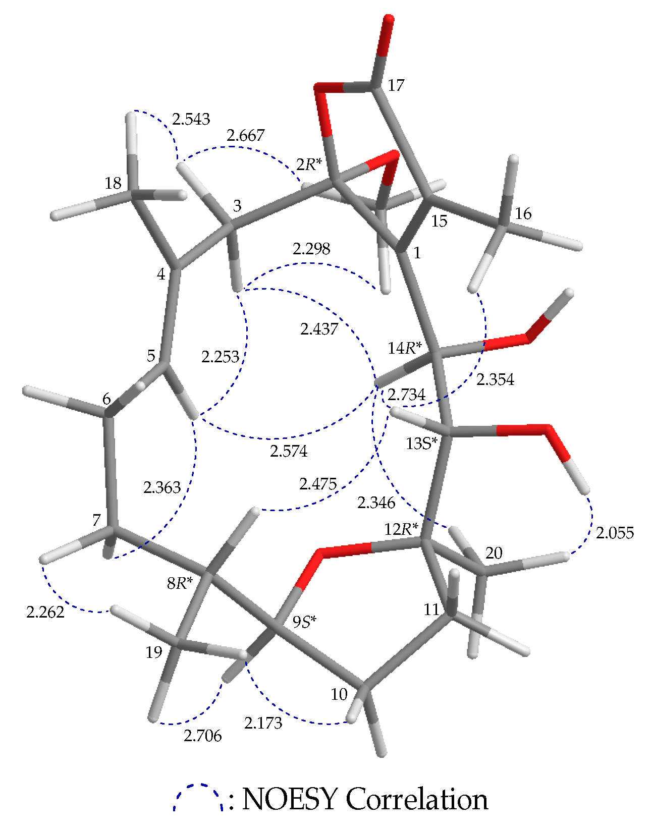
Based on NOESY correlations and further information provided by MM2 forcefield calculations [11], the relative stereochemistry of 1 with the stable conformation is shown in Figure 2 (Supplementary Figures S1–10). When H-9 was α-oriented in 1, a correlation between H-9 and H3-19 was observed, suggesting that these protons were on the α-face, and H-8 was β-oriented. H-8 correlated with H-13, and the hydroxy proton OH-13 correlated with H3-20, suggesting that the hydroxy group at C-13 and the Me-20 at C-12 were α-oriented. H-14 exhibited a NOESY correlation with H-13, and no coupling constant was detected between H-13 and H-14 in the 1H NMR spectrum, implying that the dihedral angle located between H-13 and H-14 was about 90°, and the 14-hydroxy group was β-oriented. Correlations between H-5 and H-3, and H-14 and H-3 (δH 2.78) suggested that this proton is α, and the proton at δH 3.04 is 3β. Additionally, the proton signal of a methoxy group displayed NOESY correlations with both H-3α/β, which indicated that the methoxy group at C-2 was α-oriented. H3-18 was found to show a NOESY correlation with H-3β, but not with H-5, and H-5 was shown to be correlated with H-3α and H-14, which suggested an E-configuration of the C-4/5 double bond. The aforementioned results enabled establishment of the relative configuration of 1, and therefore its stereogenic carbons were assigned as 2R*,8R*,9S*,12R*,13S*, 14R*.
Briaviotriol A (2) was found to have the molecular formula C21H32O7, as established by (+)-HRESIMS at m/z 419.20377 (calcd. for C21H32O7 + Na, 419.20402). The 1H and 13C NMR spectra of 2 were very similar to those of 1. Comparison between the 1H and 13C NMR data of 2 (Table 2) and those of 1 suggested that the double bond is located between C-4 and C-18 in 2 instead of C-4 and C-15 in 1. HMBC from H2-18 to C-3, C-4, C-5; and from H2-3 and H-5 to C-18, corroborated the existence of an exocyclic double bond at C-4. In the HSQC spectrum, an oxymethine carbon (δC 69.1) correlated with the methine proton (δH 4.57), and this proton had 3J-correlations with H2-6 (δH 1.81, 1H, m and 1.93, 1H, m) in the 1H–1H COSY spectrum, demonstrating that a hydroxy group was attached to C-5.
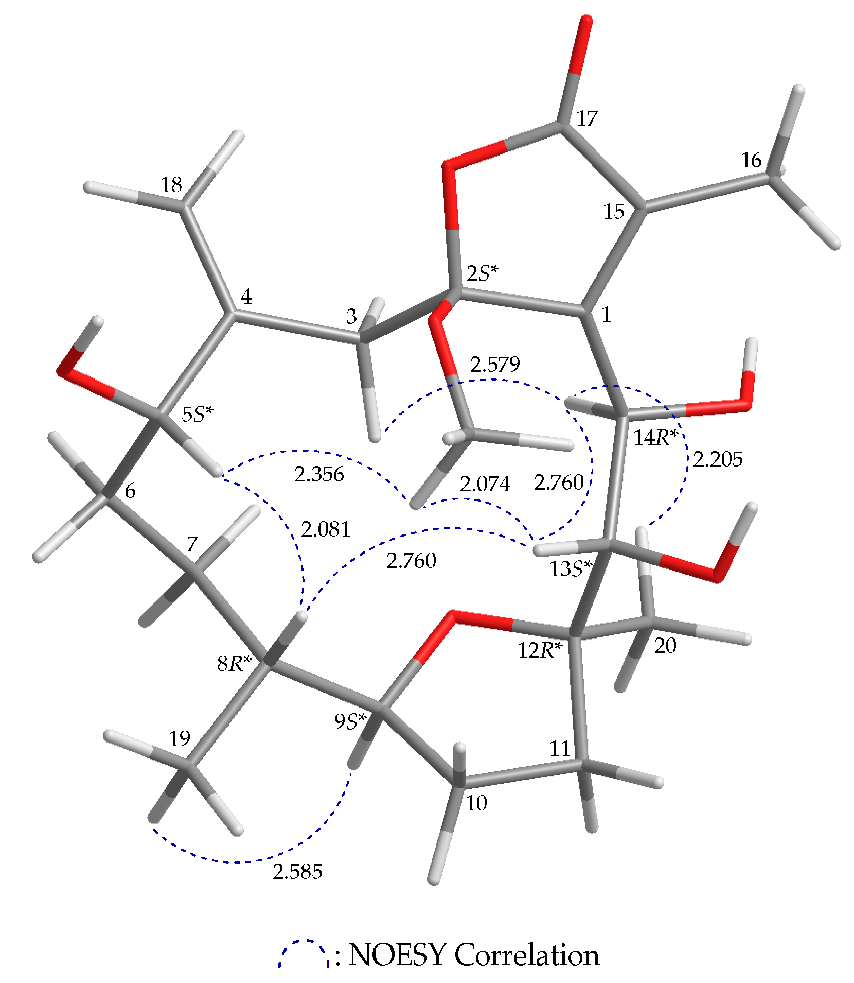
The stereochemistry of 2 was established from the correlations observed in the NOESY spectrum (Figure 3 and Supplementary Figures S2–10). In addition, in the NOESY spectrum H-9 was correlated with H3-19, which suggested that these protons were positioned on the same face and were assigned as α protons, as H-8 was β-oriented. H-13 correlated with H-8 and H-14, but no coupling between H-13 and H-14 was observed, demonstrating that the hydroxy groups at C-13 and C-14 were α- and β-oriented, respectively. Correlations between an oxygen-bearing methyl (δH 3.26) and H-13 suggested that the C-2 methoxy group was situated on the β face. Additionally, correlation between H-5 and H-8 supported a β-orientation of H-5. Based on the aforementioned results, the relative configurations of the stereogenic carbons of 2 were determined as 2S*,5S*,8R*, 9S*,12R*,13S*,14R*.
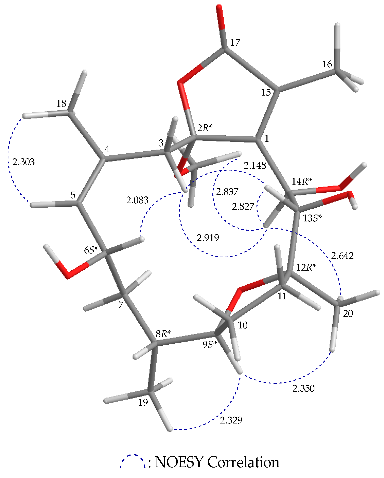
Compound 3 has a molecular formula C21H32O7 according to its (+)-HRESIMS m/z 419.20399 (calcd. for C21H32O8 + Na, 419.20402). The 1H and 13C NMR features of 3 resemble those of 1; comparison of the 1H and 13C NMR chemical shifts of the sp2 methine proton and its respective carbon (δH 5.29, 1H, d, J = 8.4 Hz; δC 133.5, CH-5), and the sp2 quaternary carbon (δC 131.0, C-4) of 3 (Table 3) with those of 1 (δH 5.30, 1H, dd, J = 8.0, 5.6 Hz; δC 135.1, CH-5; δC 127.1, C-4) (Table 1), as well as a NOESY correlation between H-5 and H3-18, indicated the Z-configuration of the C-4/5 double bond (Figure 4 and Supplementary Figure S3–10). Furthermore, the HSQC spectrum showed that an oxymethine carbon (δC 68.7) was correlated with a methine proton (δH 4.86; H-6), and this proton exhibited 3J-correlations with the olefinic proton H-5 (δH 5.29) and H2-7 (δH 1.34, 1H, m; 1.80, 1H, m) in the COSY spectrum, which confirmed a hydroxy group at C-6. As H-6 showed a NOESY correlation with H-3β, this suggested that the C-6 hydroxy group was α-oriented. Based on a NOESY experiment (Figure 4 and Supplementary Figures S3–10), 3 was identified to have the stereogenic centers 2R*,6S*,8R*,9S*,12R*,13S*,14R*. Since 3 has never been previously reported, it was named briaviotriol B.
Compound 4 was identified as briaviodiol A (Figure 1), by comparison of its 1H and 13C NMR data with those in the literature [9].
Using an in vitro pro-inflammatory suppression assay, the effects of 1–4 on the release of iNOS protein from lipopolysaccharide (LPS)-stimulated RAW 264.7 macrophage cells were assessed. First, alamar blue cell viability assessment revealed that 1–4 did not have significant cytotoxic effects in RAW 264.7 cells. The results of the in vitro pro-inflammatory suppression assay showed that 2 and 4 at 10 μM suppressed the release of iNOS to 67.7 and 61.9%, respectively, when compared with results of the cells stimulated with only LPS (Table 4). Compound 1 showed no suppression effect on iNOS release.
3. Experimental Section
3.1. General Experimental Procedures
The JEOL NMR spectrometer (model ECZ400S, Tokyo, Japan) was used to record the spectra with the solvent peak of CHCl3 (δH 7.26 ppm) and CDCl3 (δC 77.1 ppm) as internal references for 1H NMR and 13C NMR, respectively. ESIMS and HRESIMS were obtained from the Bruker mass spectrometer with 7 Tesla magnets (model: SolariX FTMS system) (Bremen, Germany). Column chromatography, IR spectra and optical rotation were performed according to our earlier research [10].
3.2. Animal Material
Specimens of B. violaceum used for this study were collected in December 2016 from the cultivation tank (capacity = 270 tons) at the National Museum of Marine Biology and Aquarium (NMMBA) in Southern Taiwan. For its identification, this coral species was compared to reliable sources published earlier [1,2]. A voucher specimen was deposited in the NMMBA (voucher no.: NMMBA-CSC-005).
3.3. Extraction and Isolation
Sliced bodies (wet/dry weight = 358.7/144.5 g) of the coral specimen were prepared and extracted with a 1:1 mixture of MeOH and CH2Cl2 to give 17.2 g of crude extract which was partitioned between EtOAc and H2O to obtain 6.3 g of the EtOAc extract. The EtOAc extract was then applied onto a silica gel column and eluted with gradients of n-hexane/EtOAc (100% n-hexane−100% EtOAc, stepwise), to furnish 14 fractions (fractions: A−N). Fraction G was further chromatographed on a silica gel column and eluted with gradients of n-hexane/Me2CO (20:1−100% Me2CO, stepwise) to afford 11 subfractions (fractions: G1−G11). Fraction G4 was applied onto a silica gel column and eluted with gradients of n-hexane and Me2CO (20:1−100% Me2CO, stepwise) to give 12 subfractions (fractions: G4A−G4L). Afterwards, fraction G4E was then separated by normal-phase HPLC (NP-HPLC) using a mixture of n-hexane and Me2CO (5:1) as solvent to obtain 5 subfractions (fractions: G4E1−G4E5). Then, fraction G4E1 was separated by NP-HPLC using a mixture of CH2Cl2 and Me2CO (with volume: volume = 80:1; at a flow rate = 3.0 mL/min) to afford 1 (62.7 mg). Fraction G4H was separated by NP-HPLC using a mixture of n-hexane and Me2CO (with volume: volume = 4:1; at a flow rate = 2.0 mL/min) to afford 4 (17.0 mg). Fraction G4J was repurified by NP-HPLC using a mixture of n-hexane and Me2CO (with volume: volume = 3:1; at a flow rate = 2.0 mL/min) to afford 3 (0.9 mg). Fraction G8 was separated by NP-HPLC using a mixture of n-hexane and Me2CO (3:1) to obtain 6 subfractions G8A−G8F. Fraction G8F was repurified by reverse-phase HPLC (RP-HPLC) using a mixture of MeCN and H2O (with volume: volume = 1:1; at a flow rate = 1.0 mL/min) to yield 2 (1.2 mg).
Briaviodiol F (1): Colorless oil; +223 (c 1.48, CHCl3); IR (neat) νmax 3497, 1754 cm−1; 1H and 13C NMR data (see Table 1); ESIMS: m/z 403 [M + Na]+; HRESIMS: m/z 403.20886 (calcd. for C21H32O6 + Na, 403.20911).
Briaviotriol A (2): Colorless oil; −68 (c 0.06, CHCl3); IR (neat) νmax 3424, 1749 cm−1; 1H and 13C NMR data (see Table 2); ESIMS: m/z 419 [M + Na]+; HRESIMS: m/z 419.20377 (calcd. for C21H32O7 + Na, 419.20402).
Briaviotriol B (3): Colorless oil; −39 (c 0.04, CHCl3); IR (neat) νmax 3424, 1749 cm−1; 1H and 13C NMR data (see Table 3); ESIMS: m/z 419 [M + Na]+; HRESIMS: m/z 419.20399 (calcd. for C21H32O7 + Na, 419.20402).
3.4. Molecular Mechanics Calculations
The molecular models were generated by implementing the MM2 force field [11] in ChemBio 3D Ultra software (ver. 12.0) which was created by CambridgeSoft (PerkinElmer, Cambridge, MA, USA).
3.5. In Vitro Anti-Inflammatory Assay
The pro-inflammatory suppression assay was performed using a murine macrophage cell line, RAW 264.7, which was purchased from the American Type Culture Collection (ATCC cell line no. TIB-71; Manassas, VA, USA). Untreated or LPS-induced RAW 264.7 cells were used to determine the anti-inflammatory activities of cembranoids 1–4 by assessing the inhibition of pro-inflammatory iNOS release from macrophage cells. The iNOS protein levels were measured by using western blotting analysis [12,13,14]. Briefly, in the control group, macrophages were incubated in compound-free medium with LPS (10 μM) alone for 16 h; and in the cembranoid-treated groups, the cells were pre-treated with cembranoids 1–4 (10 μM) for 10 min followed by an LPS challenge for 16 h. After the incubation, cell lysates were collected, and equal amounts of the total protein samples were subjected to western blot analysis. The immunoreactivities were caculated based on the optical densities of the corresponding iNOS bands of each group on the membrane, and the cells with LPS treatment alone were set to be 100%. Viability of macrophage cells of different groups was determined after treatment with alamar blue (Invitrogen, Carlsbad, CA, USA), a chemical of tetrazolium dye that is reduced by living cells to a fluorescent substance. The assay has been shown to have accurate measurement in determining the survival of RAW 264.7 cells [15,16], which is based on a mechanism similar to that of an assay using 3-(4,5-dimethyldiazol-2-yl)-2,5- diphenyltetrazolium bromide. Data analyses were firstly performed using one-way analysis of variance (ANOVA), and further analyzed by the Student-Newman-Keuls post hoc test for multiple comparison. All the data with a p-value of < 0.05 were considered as a significant difference.
4. Conclusions
B. violaceum has been demonstrated to have a wide structural diversity of interesting diterpenoids that possess various pharmacological properties [17]. This specimen was encrusted on different species of scleractinian hard corals in the Indo-Pacific coral reef system [18]. In our continued study of B. violaceum, three previously unreported furanocembranoids 1–3 were isolated, together with the previously described briaviodiol A (4). In the present study, the anti- inflammatory activities of 1–4 were assessed using inhibition of pro-inflammatory iNOS release from macrophages. The results indicated that briaviotriol A (2) and briaviodiol A (4) showed the most potent suppressive effects on iNOS release.
Supplementary Materials
The Supplementary Materials are available online at https://www.mdpi.com/1660-3397/17/4/214/s1. ESIMS, HRESIMS, IR, 1D (1H NMR, 13C NMR, and DEPT spectra), and 2D (COSY, HSQC, HMBC, and NOESY) spectra of new compounds 1–3 and 1H and 13C NMR spectra of 4.
Author Contributions
P.-C.H., W.-S.L., Z.-H.W., and P.-J.S. designed the whole experiment and contributed to manuscript preparation. B.-R.P., Y.-C.C., L.-S.F., G.-Q.L., and T.-L.H. analyzed the data and performed data acquisition.
Funding
This research was supported by grants from the National Museum of Marine Biology and Aquarium; the National Dong Hwa University; and the Ministry of Science and Technology, Taiwan (Grant Nos: MOST 104-2320-B-291-001-MY3 and 107-2320-B-291-001-MY3) awarded to Ping-Jyun Sung.
Conflicts of Interest
The authors declare no conflicts of interest.
References
- Samimi-Namin, K.; van Ofwegen, L.P. Overview of the genus Briareum (Cnidaria, Octocorallia, Briareidae) in the Indo-Pacific, with the description of a new species. ZooKeys 2016, 557, 1–44. [Google Scholar] [CrossRef] [PubMed]
- Bayer, F.M. Key to the genera of octocorallia exclusive of Pennatulacea (Coelenterata: Anthozoa), with diagnoses of new taxa. Proc. Biol. Soc. Wash. 1981, 94, 902–947. [Google Scholar]
- Bowden, B.F.; Coll, J.C.; Mitchell, S.J.; Raston, C.L.; Stokie, G.J.; White, A.H. Studies of Australian soft corals XV. The structure of pachyclavulariadiol, a novel furano-diterpene from Pachyclavularia violacea. Aust. J. Chem. 1979, 32, 2265–2274. [Google Scholar] [CrossRef]
- Inman, W.; Crews, P. The structure and conformational properties of a cembranolide diterpene from Clavularia violacea. J. Org. Chem. 1989, 54, 2526–2529. [Google Scholar] [CrossRef]
- Sheu, J.-H.; Wang, G.-H.; Sung, P.-J.; Duh, C.-Y.; Chiang, M.Y. Pachyclavulariolides G–L and seco- pachyclavulariaenone A, seven novel diterpenoids from the soft coral Pachyclavularia violacea. Tetrahedron 2001, 57, 7639–7648. [Google Scholar] [CrossRef]
- Xu, L.; Patrick, B.O.; Roberge, M.; Allen, T.; van Ofwegen, L.; Andersen, R.J. New diterpenoids from the octocoral Pachyclavularia violacea collected in Papua New Guinea. Tetrahedron 2000, 56, 9031–9037. [Google Scholar] [CrossRef]
- Sheu, J.-H.; Wang, G.-H.; Duh, C.-Y.; Soong, K. Pachyclavulariolides M–R, six novel diterpenoids from a Taiwanese soft coral Pachyclavularia violacea. J. Nat. Prod. 2003, 66, 662–666. [Google Scholar] [CrossRef] [PubMed]
- Duh, C.-Y.; El-Gamal, A.A.H.; Chu, C.-J.; Wang, S.-K.; Dai, C.-F. New cytotoxic constituents from the Formosan soft corals Clavularia viridis and Clavularia violacea. J. Nat. Prod. 2002, 65, 1535–1539. [Google Scholar] [CrossRef] [PubMed]
- Chang, Y.-C.; Huang, I.-C.; Chiang, M.Y.-N.; Hwang, T.-L.; Kung, T.-H.; Lin, C.-S.; Sheu, J.-H.; Sung, P.-J. Briaviodiol A, a new cembranoid from a soft coral Briareum violacea. Chem. Pharm. Bull. 2010, 58, 1666–1668. [Google Scholar] [CrossRef] [PubMed]
- Huang, P.-C.; Tseng, C.-C.; Peng, B.-R.; Hu, C.-C.; Lin, N.-C.; Chen, N.-F.; Chen, J.-J.; Wen, Z.-H.; Wu, Y.-C.; Sung, P.-J. Briaviodiols B–E, new anti-inflammatory hydroperoxyfurancembranoids from Briareum violaceum. Tetrahedron 2019, 75, 921–927. [Google Scholar] [CrossRef]
- Allinger, N.L. Conformational analysis. 130. MM2. A hydrocarbon force field utilizing V1 and V2 torsional terms. J. Am. Chem. Soc. 1977, 99, 8127–8134. [Google Scholar] [CrossRef]
- Huang, S.-Y.; Chen, N.-F.; Chen, W.-F.; Hung, H.-C.; Lee, H.-P.; Lin, Y.-Y.; Wang, H.-M.; Sung, P.-J.; Sheu, J.-H.; Wen, Z.-H. Sinularin from indigenous soft coral attenuates nociceptive responses and spinal neuroinflammation in carrageenan-induced inflammatory rat model. Mar. Drugs 2012, 10, 1899–1919. [Google Scholar] [CrossRef] [PubMed]
- Jean, Y.-H.; Chen, W.-F.; Sung, C.-S.; Duh, C.-Y.; Huang, S.-Y.; Lin, C.-S.; Tai, M.-H.; Tzeng, S.-F.; Wen, Z.-H. Capnellene, a natural marine compound derived from soft coral, attenuates chronic constriction injury-induced neuropathic in rats. Br. J. Pharmacol. 2009, 158, 713–725. [Google Scholar] [CrossRef] [PubMed]
- Jean, Y.-H.; Chen, W.-F.; Duh, C.-Y.; Huang, S.-Y.; Hsu, C.-H.; Lin, C.-S.; Sung, C.-S.; Chen, I.-M.; Wen, Z.-H. Inducible nitric oxide synthase and cyclooxygenase-2 participate in anti-inflammatory and analgesic effects of the natural marine compound lemnalol from Formosan soft coral Lemnalia cervicorni. Eur. J. Pharmacol. 2008, 578, 323–331. [Google Scholar] [CrossRef] [PubMed]
- Chen, L.-C.; Lin, Y.-Y.; Jean, Y.-H.; Lu, Y.; Chen, W.-F.; Yang, S.-N.; Wang, H.-M.D.; Jang, I.-Y.; Chen, I.-M.; Su, J.-H.; et al. Anti-inflammatory and analgesic effects of the marine-derived compound comaparvin isolated from the crinoid Comanthus bennetti. Molecules 2014, 19, 14667–14686. [Google Scholar] [CrossRef] [PubMed]
- Oliveira, T.; Figueiredo, C.A.; Brito, C.; Stavroullakis, A.; Prakki, A.; da Silva Velozo, E.; Nogueira-Filho, G. Effect of Allium cepa L. on lipopolysaccharide-stimulated osteoclast precursor cell viability, count, and morphology using 4′,6-diamidino-2-phenylindole-staining. Int. J. Cell Biol. 2014, 2014, 535789. [Google Scholar] [CrossRef] [PubMed]
- Chang, Y.-C.; Sheu, J.-H.; Wu, Y.-C.; Sung, P.-J. Terpenoids from octocorals of the genus Pachyclavularia. Mar. Drugs 2017, 15, 382. [Google Scholar] [CrossRef] [PubMed]
- Geetha, S.; Kumar, J.S.Y.; Raghunathan, C.; Sornaraj, R. Space competition studies between Briareum violaceum (Octocorallia; Alcyonacea) and scleractinian corals in Shark Island, North Andaman, India. Indian J. Geo-Mar. Sci. 2018, 47, 2390–2394. [Google Scholar]
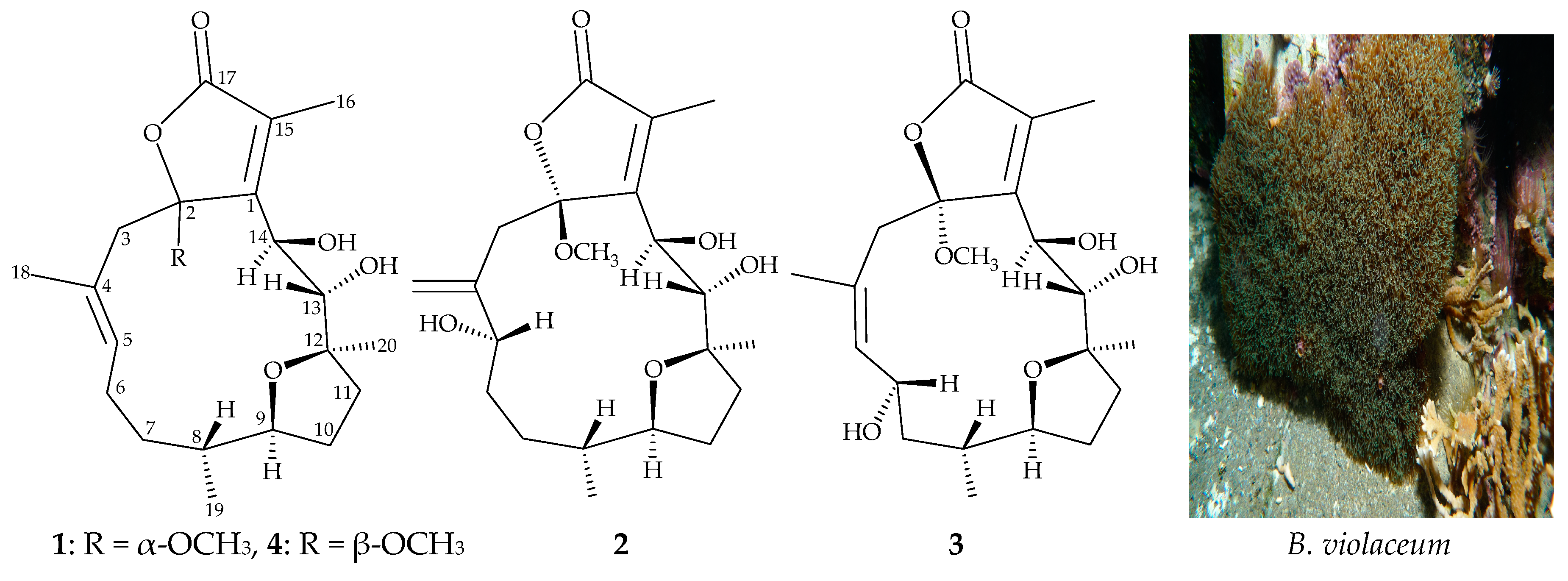
Figure 1.
Structures of briaviodiol F (1), briaviotriols A (2) and B (3), and briaviodiol A (4), and a picture of the octocoral B. violaceum.
Figure 1.
Structures of briaviodiol F (1), briaviotriols A (2) and B (3), and briaviodiol A (4), and a picture of the octocoral B. violaceum.

Figure 2.
Computer-depicted model drawing of 1 and calculated distances (unit = Å) between protons with main NOESY correlations.
Figure 2.
Computer-depicted model drawing of 1 and calculated distances (unit = Å) between protons with main NOESY correlations.

Figure 3.
Computer-depicted model drawing of 2 and calculated distances (unit = Å) between protons with main NOESY correlations.
Figure 3.
Computer-depicted model drawing of 2 and calculated distances (unit = Å) between protons with main NOESY correlations.

Figure 4.
Computer-depicted model drawing of 3 and calculated distances (unit = Å) between protons with main NOESY correlations.
Figure 4.
Computer-depicted model drawing of 3 and calculated distances (unit = Å) between protons with main NOESY correlations.


Table 1.
1H (400 MHz, CDCl3) and 13C (100 MHz, CDCl3) NMR, COSY, HMBC data for 1.
| Position | δH (J in Hz) | δC, type | COSY | HMBC |
|---|---|---|---|---|
| 1 | 154.3, C | |||
| 2 | 109.5, C | |||
| 3α/β | 2.78 d (14.0); 3.04 d (14.0) | 44.8, CH2 | C-1, C-2, C-4, C-5, C-18 | |
| 4 | 127.1, C | |||
| 5 | 5.30 dd (8.0, 5.6) | 135.1, CH | H2-6 | C-3, C-18 |
| 6 | 1.93–1.99 m | 26.3, CH2 | H-5, H2-7 | C-4, C-5, C-7, C-8 |
| 7α/β | 1.80 m; 1.26 m | 34.9, CH2 | H2-6, H-8 | C-5, C-6, C-8, C-9, C-19 |
| 8 | 0.68 m | 41.4, CH | H2-7, H-9, H3-19 | C-9 |
| 9 | 3.68 ddd (9.6, 9.6, 6.0) | 85.2, CH | H-8, H2-10 | C-7 |
| 10α/β | 1.51 m; 1.98 m | 30.3, CH2 | H-9, H2-11 | C-11, C-12 |
| 11α/β | 1.56 m; 2.38 dd (12.0, 6.4) | 36.9, CH2 | H2-10 | C-9, C-10, C-12, C-13, C-20 |
| 12 | 84.1, C | |||
| 13 | 3.60 d (10.0) | 70.7, CH | OH-13 | C-1, C-11, C-12, C-20 |
| 14 | 5.06 s | 63.9, CH | - | C-1, C-2, C-12, C-15 |
| 15 | 127.2, C | |||
| 16 | 2.11 s | 9.2, CH3 | C-1, C-15, C-17 | |
| 17 | 172.1, C | |||
| 18 | 1.44 s | 15.3, CH3 | C-3, C-4, C-5 | |
| 19 | 0.78 d (6.4) | 17.3, CH3 | H-8 | C-7, C-8, C-9 |
| 20 | 1.30 s | 20.9, CH3 | C-11, C-12, C-13 | |
| OMe-2 | 3.39 s | 51.0, CH3 | C-2 | |
| OH-13 | 3.14 d (10.0) | H-13 | C-13, C-14 | |
| OH-14 | 3.34 s | - | C-1, C-13, C-14 |
Table 2.
1H (400 MHz, CDCl3) and 13C (100 MHz, CDCl3) NMR, COSY, HMBC data for 2.
| Position | δH (J in Hz) | δC, type | COSY | HMBC |
|---|---|---|---|---|
| 1 | 157.9, C | |||
| 2 | 107.8, C | |||
| 3α/β | 2.67 d (14.4); 3.15 d (14.4) | 42.2, CH2 | C-1, C-2, C-4, C-5, C-18 | |
| 4 | 146.4, C | |||
| 5 | 4.57 dd (7.2, 6.0) | 69.1, CH | H2-6 | C-4, C-6, C-18 |
| 6/6′ | 1.81m; 1.93 m | 32.2, CH2 | H-5, H2-7 | - |
| 7/7′ | 1.33 m; 1.89 ddd (14.4, 4.8, 4.4) | 30.6, CH2 | H2-6, H-8 | C-5 |
| 8 | 1.53 m | 37.4, CH | H2-7, H-9, H3-19 | - |
| 9 | 3.65 ddd (8.4, 8.4, 6.0) | 85.4, CH | H-8, H2-10 | - |
| 10/10′ | 1.52 m; 2.10 m | 31.5, CH2 | H-9, H2-11 | - |
| 11/11′ | 1.65 m; 2.16 m | 37.2, CH2 | H2-10 | C-9, C-10, C-12, C-13, C-20 |
| 12 | 84.4, C | |||
| 13 | 3.50 d (5.6) | 75.2, CH | OH-13 | - |
| 14 | 5.18 br s | 67.7, CH | OH-14 | - |
| 15 | 130.0, C | |||
| 16 | 2.11 s | 10.1, CH3 | C-1, C-15, C-17 | |
| 17 | 171.1, C | |||
| 18a/b | 5.13 s; 5.30 s | 115.6, CH2 | C-3, C-4, C-5 | |
| 19 | 0.85 d (6.4) | 16.7, CH3 | H-8 | C-7, C-8, C-9 |
| 20 | 1.29 s | 21.8, CH3 | C-11, C-12, C-13 | |
| OMe-2 | 3.26 s | 50.7, CH3 | C-2 | |
| OH-13 | 2.42 br d (5.6) | H-13 | - | |
| OH-14 | 2.29 br d (4.4) | H-14 | - |
Table 3.
1H (400 MHz, CDCl3) and 13C (100 MHz, CDCl3) NMR, and COSY, HMBC data for 3.
| Position | δH (J in Hz) | δC, type | COSY | HMBC |
|---|---|---|---|---|
| 1 | 159.8, C | |||
| 2 | 107.7, C | |||
| 3α/β | 1.76 d (14.4); 3.56 d (14.4) | 39.6, CH2 | C-1, C-2, C-4, C-5, C-18 | |
| 4 | 131.0, C | |||
| 5 | 5.29 d (8.4) | 133.5, CH | H-6 | C-3, C-18 |
| 6 | 4.86 ddd (12.0, 8.4, 3.2) | 68.7, CH | H-5, H2-7 | - |
| 7/7′ | 1.34 m; 1.80 m | 44.9, CH2 | H-6, H-8 | C-5, C-6, C-8, C-9, C-19 |
| 8 | 1.21 m | 35.3, CH | H2-7, H-9, H3-19 | - |
| 9 | 3.65 ddd (10.0, 10.0, 4.4) | 87.3, CH | H-8, H2-10 | - |
| 10/10′ | 1.38 m; 2.05 m | 32.8, CH2 | H-9, H2-11 | C-9, C-11, C-12 |
| 11/11′ | 1.61 m; 2.22 dd (13.2, 8.0) | 35.5, CH2 | H2-10 | C-9, C-12, C-13, C-20 |
| 12 | 85.1, C | |||
| 13 | 3.40 d (8.4) | 76.7, CH | OH-13 | C-1, C-12, C-20 |
| 14 | 4.75 d (5.2) | 63.5, CH | OH-14 | C-1, C-2, C-12, C-13, C-15 |
| 15 | 128.5, C | |||
| 16 | 2.08 s | 9.6, CH3 | C-1, C-15, C-17 | |
| 17 | 171.9, C | |||
| 18 | 1.83 s | 24.0, CH3 | C-3, C-4, C-5 | |
| 19 | 0.84 d (6.8) | 20.1, CH3 | H-8 | C-7, C-8, C-9 |
| 20 | 1.28 s | 21.1, CH3 | C-11, C-12, C-13 | |
| OMe-2 | 3.20 s | 51.1, CH3 | C-2 | |
| OH-13 | 3.05 d (8.4) | H-13 | C-13, C-14 | |
| OH-14 | 2.70 d (5.2) | H-14 | C-13, C-14 |
Table 4.
Effects of 1–4 on LPS-induced pro-inflammatory iNOS release in RAW 264.7 cells at a concentration of 10 μM. The data presented are the relative intensity normalized to the LPS- stimulated group. Compounds 2 and 4 were found to have the higher inhibition effects on LPS- induced iNOS expression in macrophages expression.
| iNOS | |
|---|---|
| Expression (% of LPS) | |
| LPS | 100.0 ± 7.0 |
| 1 | 109.0 ± 19.2 |
| 2 | 67.7 ± 2.4 |
| 3 | 79.5 ± 9.4 |
| 4 | 61.9 ± 7.3 |
© 2019 by the authors. Licensee MDPI, Basel, Switzerland. This article is an open access article distributed under the terms and conditions of the Creative Commons Attribution (CC BY) license (http://creativecommons.org/licenses/by/4.0/).
Share and Cite
MDPI and ACS Style
Huang, P.-C.; Lin, W.-S.; Peng, B.-R.; Chang, Y.-C.; Fang, L.-S.; Li, G.-Q.; Hwang, T.-L.; Wen, Z.-H.; Sung, P.-J. New Furanocembranoids from Briareum violaceum. Mar. Drugs 2019, 17, 214. https://doi.org/10.3390/md17040214
AMA Style
Huang P-C, Lin W-S, Peng B-R, Chang Y-C, Fang L-S, Li G-Q, Hwang T-L, Wen Z-H, Sung P-J. New Furanocembranoids from Briareum violaceum. Marine Drugs. 2019; 17(4):214. https://doi.org/10.3390/md17040214
Chicago/Turabian StyleHuang, Pin-Chang, Wen-Sou Lin, Bo-Rong Peng, Yu-Chia Chang, Lee-Shing Fang, Guo-Qiang Li, Tsong-Long Hwang, Zhi-Hong Wen, and Ping-Jyun Sung. 2019. "New Furanocembranoids from Briareum violaceum" Marine Drugs 17, no. 4: 214. https://doi.org/10.3390/md17040214
Note that from the first issue of 2016, this journal uses article numbers instead of page numbers. See further details here.