Marine Macrolides with Antibacterial and/or Antifungal Activity


Department of Medical Microbiology, Poznań University of Medical Sciences, Wieniawskiego 3, 61-712 Poznań, Poland
Mar. Drugs 2019, 17(4), 241; https://doi.org/10.3390/md17040241
Submission received: 16 March 2019
/
Revised: 19 April 2019
/
Accepted: 19 April 2019
/
Published: 23 April 2019
(This article belongs to the Special Issue Marine Macrolides: Structure, Biosynthetic Aspects and Potential as a New Drugs)
Abstract
:Currently, the increasing resistance of microorganisms to antibiotics is a serious problem. Marine organisms are the source of thousands of substances, which also have antibacterial and antifungal effects. Among them, marine macrolides are significant. In this review, the antibacterial and/or antifungal activities of 34 groups of marine macrolides are presented. Exemplary groups are chalcomycins, curvulides, halichondramides, lobophorins, macrolactins, modiolides, scytophycins, spongistatins, or zearalanones. In the paper, 74 antibiotics or their analog sets, among which 29 with antifungal activity, 25 that are antibacterial, and 20 that are both antifungal and antibacterial are summarized. Also, 36 macrolides or their sets are produced by bacteria, 18 by fungi, ten by sponges, seven by algae, two by porifera, and one by nudibranch. Moreover, the chemical structures of representatives from each of the 34 groups of these antibiotics are presented. To summarize, marine organisms are rich in natural macrolides. Some of these may be used in the future in the treatment of bacterial and fungal infections. Marine macrolides can also be potential drugs applicable against pathogens resistant to currently known antibiotics.
1. Introduction
The marine world is rich in species and very diverse. Thus, marine organisms are a source of many substances with biological activity, including cytotoxic and antimicrobial. According to Burja et al., it was determined that in the marine world, there are over 13,000 unique compounds [1]. Important marine groups containing biologically active substances, including macrolides, are, among others, sponges [2] and cyanobacteria [3]. Swian et al. only described 121 compounds with antimicrobial activity among cyanobacteria, including the following chemical classes: alkaloids, aromatic compounds, pigments, fatty acids, phenols, macrolides, peptides, polyketides, porphinoids, and terpenoids [4]. Liu et al. described 118 marine macrolides, most with cytotoxic activity [5].
Macrolides are a group of polyketides. Currently, few of these substances are used in medicine. Among the antibacterial macrolides, the most important are erythromycin, azithromycin, roxithromycin, clarithromycin, josamycin, and spiramycin; among ketolides, telithromycin [6]. On the other hand, among antifungal polyene macrolides, amphotericin B, nystatin, and natamycin are most often used [7]. In general, antibacterial macrolides are active against Staphylococcus sp., Streptococcus sp., Neisseria gonorrhoea, Haemophilus influenzae, Bordetella pertussis, and Neisseria meningitis. Additionally, they are used in infections caused by intracellular pathogens, Mycoplasma sp. and Chlamydia sp. [8,9]. Clarithromycin is one of the antibiotics used in Helicobacter pylori infections [10]. The action of antibacterial macrolides is bacteriostatic. They reversibly bind to 23S ribosomal RNA of the 50s subunit of the bacterial ribosome inhibiting RNA-dependent protein synthesis [11]. The antifungal macrolides bind to ergosterol and lead to pore formation, leakage of monovalent ions (K+, Na+, H+ and Cl−), and finally to fungal cell death [12].
Recently, the increasing resistance of bacteria to antibiotics has become a serious problem. Globally, about 700,000 deaths every year may be caused by microorganisms resistant to antimicrobials [13]. In epidemiology, the most significant are the multidrug-resistant bacteria, e.g., Escherichia coli, Klebsiella pneumoniae, Acinetobacter baumanii, methicillin-resistant Staphylococcus aureus (MRSA), vancomycin-resistant MRSA, penicillin-resistant Streptococcus pneumoniae (PRSP), vancomycin-resistant Enterococcus (VRE), and extensively drug-resistant (XDR) Mycobacterium tuberculosis [14]. Antimicrobial resistance is increasingly common among both human and animal pathogens [10,15,16,17]. Antimicrobial resistance related to food containing zoonotic and fecal bacteria (Salmonella sp., Campylobacter sp., Escherichia coli and Enterococcus sp.) is also gaining importance [18]. The hope is to find new antibiotics to fight against multidrug-resistant strains. The source of these drugs could be marine macrolides.
In this paper, literature regarding the structures and biological (antibacterial and antifungal) activities of marine macrolides was examined. This literature was found by searching for articles published in PubMed/MEDLINE using combinations of the following keywords: “marine”, “macrolide/s”, “antibacterial”, “antifungal” and “antimicrobial”. Titles and abstracts of the resulting papers were examined to exclude or include articles for review. From the references of the included articles, additional works were selected. Finally, ninety-four papers have been incorporated into this narrative review.
The antibacterial and/or antifungal activities of 34 groups of marine macrolides are presented in this review. Moreover, the chemical structures of representatives from each group of these antibiotics are also represented. The origin and biological target of marine macrolides are presented in Table.
2. Antimicrobial Activity of Marine Macrolides
2.1. Macrolides 10-Membered
2.1.1. Curvulides
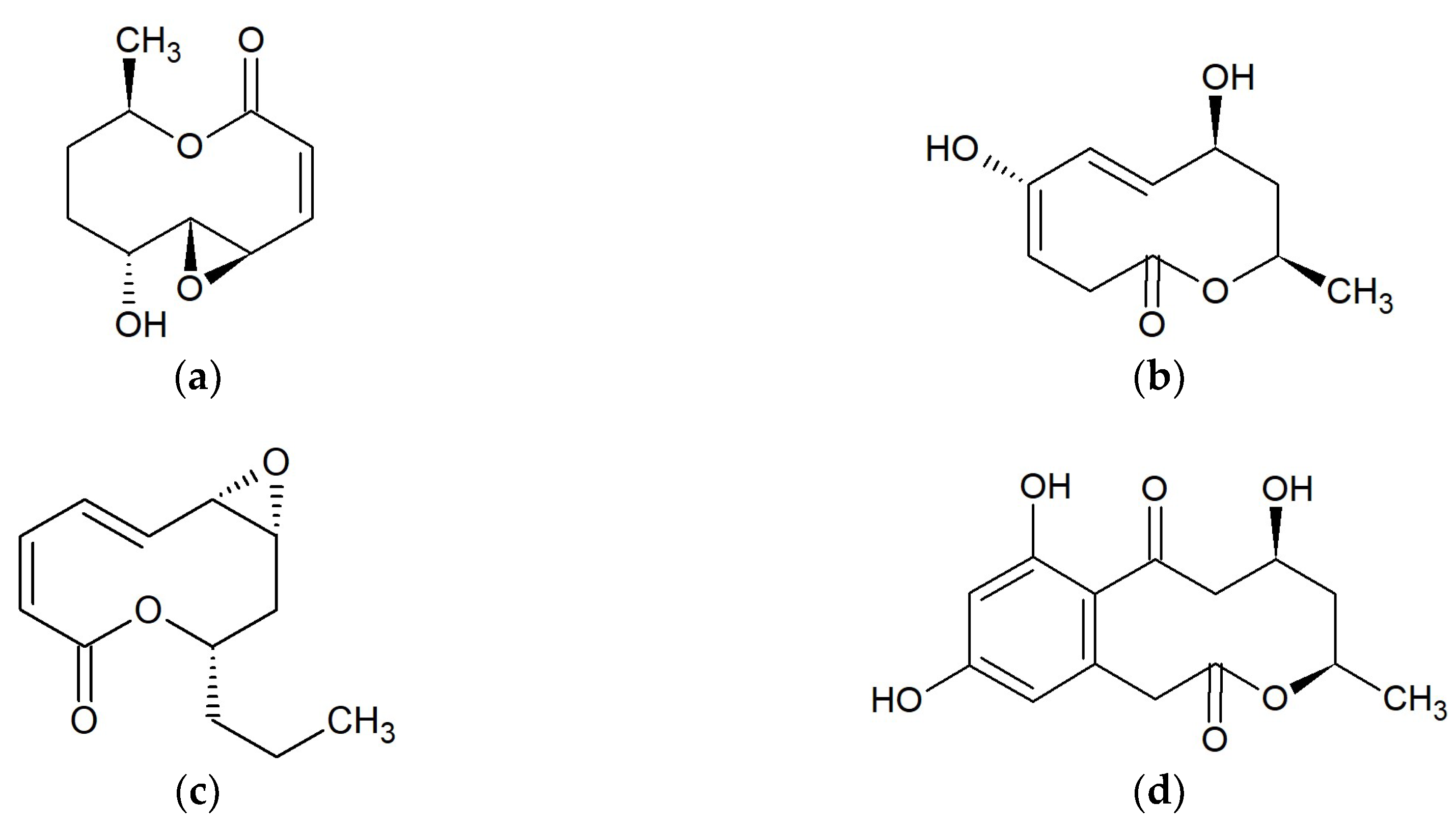
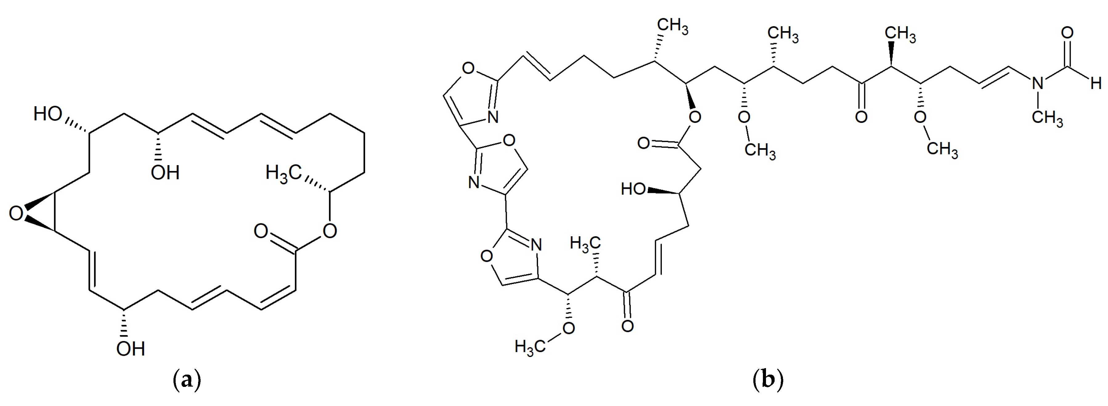
Curvulides are compounds obtained from strains of the fungus Curvularia sp. From one strain associated with the red alga Acanthophora spicifera occurring in Fingers Reef, Apra Harbor and Guam, 10-membered lactones have been isolated: curvulide A (Figure 1a [19]), curvulide B1 and B2 [20]. Curvularin and (S)-dehydrocurvularin obtained from Curvularia sp. strain M12, showed activity against fungus-like Phytophthora capsici exhibited in zoospore motility disorder [21]. Curvularin stereoisomers additionally possess anti-inflammatory activity [22] and are cytotoxic toward human tumor cell lines [23]. The two 11-hydroxycurvularin isomers isolated from the marine actinomycete Pseudonocardia sp. HS7 obtained from the sea cucumber Holothuria moebii, showed antibacterial activity towards Escherichia coli [24]. It was demonstrated that curvularin and αβ-dehydrocurvularin have anti-fungal activity against Saccharomyces cerevisiae (minimum inhibitory concentration (MIC) 375–750 μg/mL) and Sclerotinia sclerotiorum (MIC > 3000 μg/mL). Both substances also inhibited the growth of Bacillus subtilis (MICs of 1500 and > 3000 μg/mL), while αβ-dehydrocurvularin was additionally active against Staphylococcus aureus with an MIC of 375 μg/mL. Presented macrolides were not active against Gram-negative bacteria such as Escherichia coli and Pseudomonas aeruginosa. Both compounds were produced by the fungus Eupenicillium sp. associated with marine sponge Axinella sp. collected in the South China Sea near Sanya, China [25]. Curvulalide, curvulapyrone, and an uncyclized modiolide macrolide, curvulalic acid isolated from a sea fan-derived Curvularia sp. PSUF22 were not active against Staphylococcus aureus ATCC 25923, methicillin-resistant S. aureus SK1, or Microsporum gypseum SHMU-4 [26].
2.1.2. Modiolides
To the 10-membered macrolides, belong modiolides A (Figure 1b) and B. Both are produced by fungus Paraphaeosphaeria sp. strain N-119, which was obtained from a marine horse mussel Modiolus auriculatus occurring in Hedo Cape, Japan. Modiolides A and B showed antibacterial activity against Micrococcus luteus (MIC = 16.7 mg/mL) and antifungal activity against Neurospora crassa (MIC = 33.3 mg/mL) [27]. Modiolide A is also the secondary metabolite of the marine-derived fungus Curvularia sp. Modiolide A and at least four substances resembling 10-membered lactones but featuring modified oxidation patterns around their macrocycles were shown to occur in this species [20]. In other studies, it was demonstrated that modiolide A obtained from Curvularia sp. strain M12, acts against the fungus-like eukaryotic microorganism Phytophthora capsici, leading to the disorder of zoospore motility at high concentrations (IC50: 50–100 μg/mL) [21]. Trisuvan et al. showed a lack of modiolide A activity against strains Staphylococcus aureus ATCC 25923, methicillin-resistant S. aureus, and Microsporum gypseum SH-MU-4 at the initial concentration of 200 μg/mL [26].
2.1.3. Phomolides
Two 9-propyl-substituted 10-membered macrolides, phomolide A (Figure 1c) and B have been isolated from the marine fungus Phomopsis sp. hzla01-1. Both substances had significant activities against bacteria Escherichia coli CMCC44103, and fungi Candida albicans AS2.538 and Saccharomyces cerevisiae ATCC9763 with MIC values of 5–10 mg/mL [28,29]. Another similar chemical constituent, phomolide C, was obtained from the strain Phomopsis sp. B27 [30] and from the fungus Diaporthe sp., however it did not show antifungal activity against Cochliobolus miyabeanus [31].
2.1.4. Xestodecalactones
Xestodecalactones A–C, were obtained from an isolate of the fungus Penicillium cf. montanense from the marine sponge Xestospongia exigua collected from the Bali Sea, Indonesia. Among these metabolites, xestodecalactone B was found to have anti-fungal activity against the yeast Candida albicans at concentrations of 20 µM and higher. Simultaneously, xestodecalactones A–C (Figure 1d) were inactive toward the bacteria Bacillus subtilis, Staphylococcus aureus, and Escherichia coli [32,33]. Xestodecalactones D–F obtained from Corynespora cassiicola, isolated from the Chinese mangrove plant Laguncularia racemosa, neither showed antibacterial nor antifungal activity [34].
2.2. Macrolides 12-Membered
2.2.1. Amphidinolides
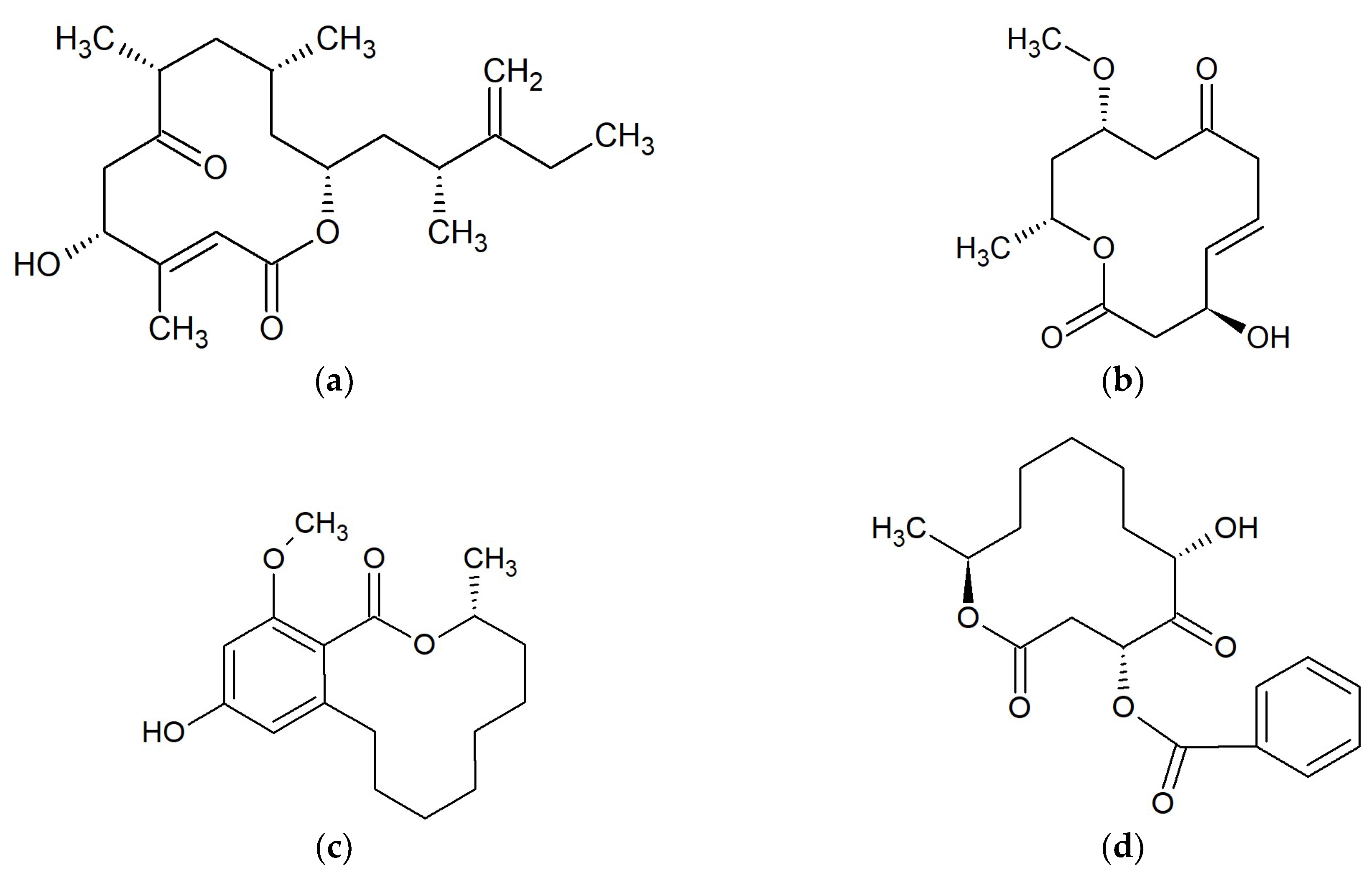
Amphidinolide Q (Figure 2a) and four analogs; amphidinins C–F, were isolated from the symbiotic dinoflagellate Amphidinium sp. The dinoflagellate Amphidinium sp. (2012-7-4A strain) was obtained from the marine acoel flatworm Amphiscolops sp. collected at Ishigaki Island, Okinawa, Japan. All compounds were active against Trichophyton mentagrophytes (MIC 16–32 μg/mL). Moreover, amphidinolide Q was active against S. aureus, B. subtilis, Escherichia coli, and Candida albicans (MICs 16–32 μg/mL) [35].
2.2.2. Dendrodolides
Marine-derived Cladosporium fungi are a source of 12-membered macrolides dendrodolides A (Figure 2b), C, L, M and cladospolide B. Cladosporium sp. were cultivated from the gorgonian Anthogorgia ochracea obtained from the South China Sea. Three dendrodolides (A, C and M) showed antibacterial activity against Bacillus cereus, Tetragenococcus halophilus, Staphylococcus epidermidis, Staphylococcus aureus, Escherichia coli, Pseudomonas putida, Nocardia brasiliensis, and Vibrio parahaemolyticus, with MIC values ranging from 3.13 to 25.0 μM [36].
2.2.3. Lasiodiplodins
Lasiodiplodins (Figure 2c) are resorcinolic macrolides [37] isolated among others from marine endophytic fungus No. ZZF36 connected with brown alga (Sargassum sp.). The fungus was collected from Zhanjiang Sea, China. Compound de-O-methyllasiodiplodin exhibited inhibitory activity against Staphylococcus aureus with an MIC of 6.25 µg/mL, and lower activities against Bacillus subtilis, Salmonella enteritidis, Candida albicans and Fusarium oxysporum f.sp. cubense. Lasiodiplodin inhibited the growth of S. aureus, B. subtilis, and F. oxysporum (MICs 25–100 µg/mL), while 5-hydroxy-de-O-methyllasiodiplodin was shown to be only effective against S. aureus at 100 µg/mL. Compound 6-oxo-de-O-methyllasiodiplodin was not active against all tested pathogens [38].
2.2.4. Sporiolides
Sporiolides are 12-membered macrocyclic lactones. Sporiolides A (Figure 2d) and B were isolated from the fungus Cladosporium sp., which was separated from a marine brown alga Actinotrichia fragilis (Okinawa Island, Japan). Both sporiolides were active against Micrococcus luteus with a MIC value of 16.7 µg/mL. Moreover, sporiolide A showed antifungal activity against Aspergillus niger, Candida albicans, Cryptococcus neoformans, and Neurospora crassa with MICs of 8.4–16.7 µg/mL. Neither of the sporiolides were active against Bacillus subtilis, Escherichia coli, or Paecilomyces variotii [39,40].
2.3. Macrolides 14-Membered
2.3.1. Lobophorins
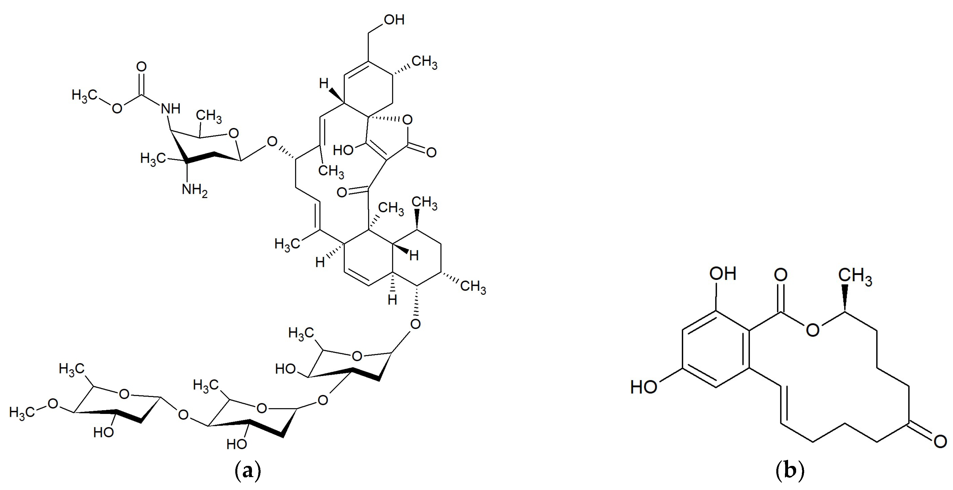
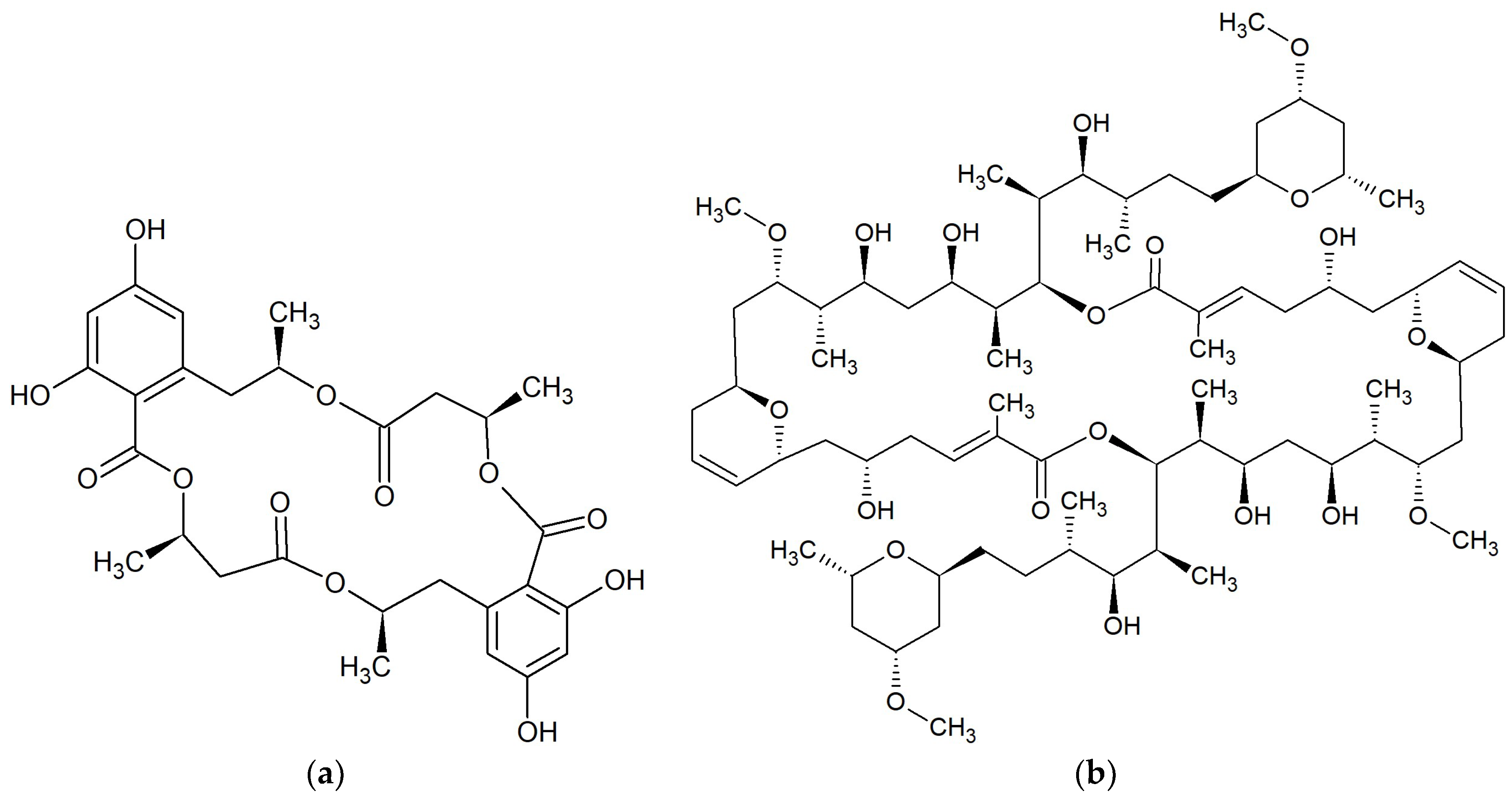
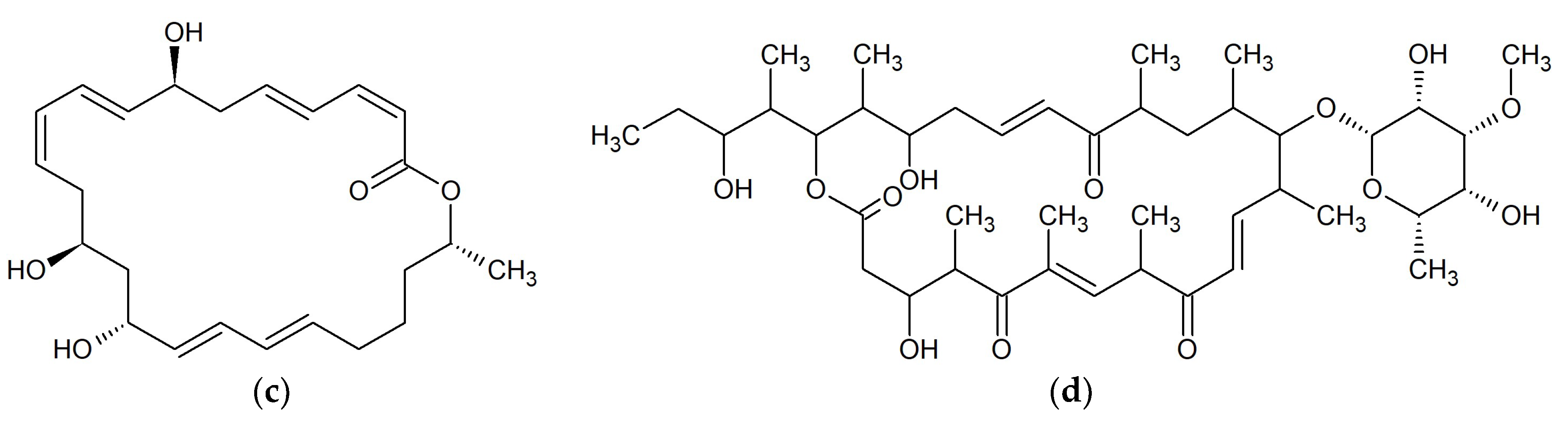
Lobophorins A (Figure 3a) and B were isolated from a marine Actinomycete strain #CNB-837 isolated from the surface of the Caribbean brown alga Lobophora variegata [41,42]. Spirotetronate antibiotics; lobophorins E and F, were isolated from Streptomyces sp. SCSIO 01127 obtained from sediment in the South China Sea [40]. Lobophorins H and I were obtained from Streptomyces sp. strain 12A35, which was isolated from the deep-sea sediment of the South China Sea [43]. Lobophorins A, B, E, and F exhibited activities against Bacillus thuringensis SCSIO BT01 with MIC values of 2–8 µg/mL. Lobophorin F displayed antibacterial activities against Staphylococcus aureus ATCC 29213 and Enterococcus faecalis ATCC 29212 with MIC values of 8 µg/mL [43]. Additionally, lobophorins B, F, I and H exhibited inhibitory activities against Bacillus subtilis CMCC63501. Lobophorins B and H showed strong activities (MICs of 1.57–3.13 μg/mL), while lobophorins F and I possessed moderate activities (MICs 6.25–50 μg/mL). Lobophorins F and H also had moderate activities against Staphylococcus aureus ATCC29213 with MIC values of 6.25–50 μg/mL. However, none of the studied compounds inhibited bacterium Escherichia coli, fungi Candida albicans or Fusarium moniliforme [44].
2.3.2. Zearalanones
β-resorcylic acid lactones were obtained from the culture of a Penicillium sp. derived from cotton clothing drifting off Namhae Island, which were zearalanone analogs: 8’-hydroxyzearalanone, 2’-hydroxyzearalanol, zearalanone, β-zearalanol, zearalenone (Figure 3b), and β-zearalenol [45,46]. Some β-resorcylic macrolides were obtained from the marine Fusarium sp. O5ABR26 isolated from a sponge collected in the Miura Peninsula of Japan. Among these substances, zearalenone displayed the best inhibitory activity against fungus Pyricularia oryzae (MIC 6.25 μg/mL). Simultaneously, 8′-hydroxyzearalenone was less active with a MIC value of 200 µg/mL [47]. It was demonstrated that fungus Fusarium sp. PSU-ES73, isolated from the seagrass Thalassia hemprichii found throughout the shores of the Indian and the Western Pacific Oceans, contains β-resorcylic macrolides 5’-hydroxyzearalenone, zearalenone, 8’-hydroxyzearalenone, 7’-dehydrozearalenone, β-zearalenol, 5’-hydroxyzearalenol, and relgro. Only zearalenone exhibited weak activity against Staphylococcus aureus ATCC25923, methicillin-resistant S. aureus SK1 (MIC 400 μM), and Cryptococcus neoformans ATCC90113 (MIC 50.26 μM). The remaining compounds were inactive [48].
2.4. Macrolides 15- and 16-Membered
Bromophycolides
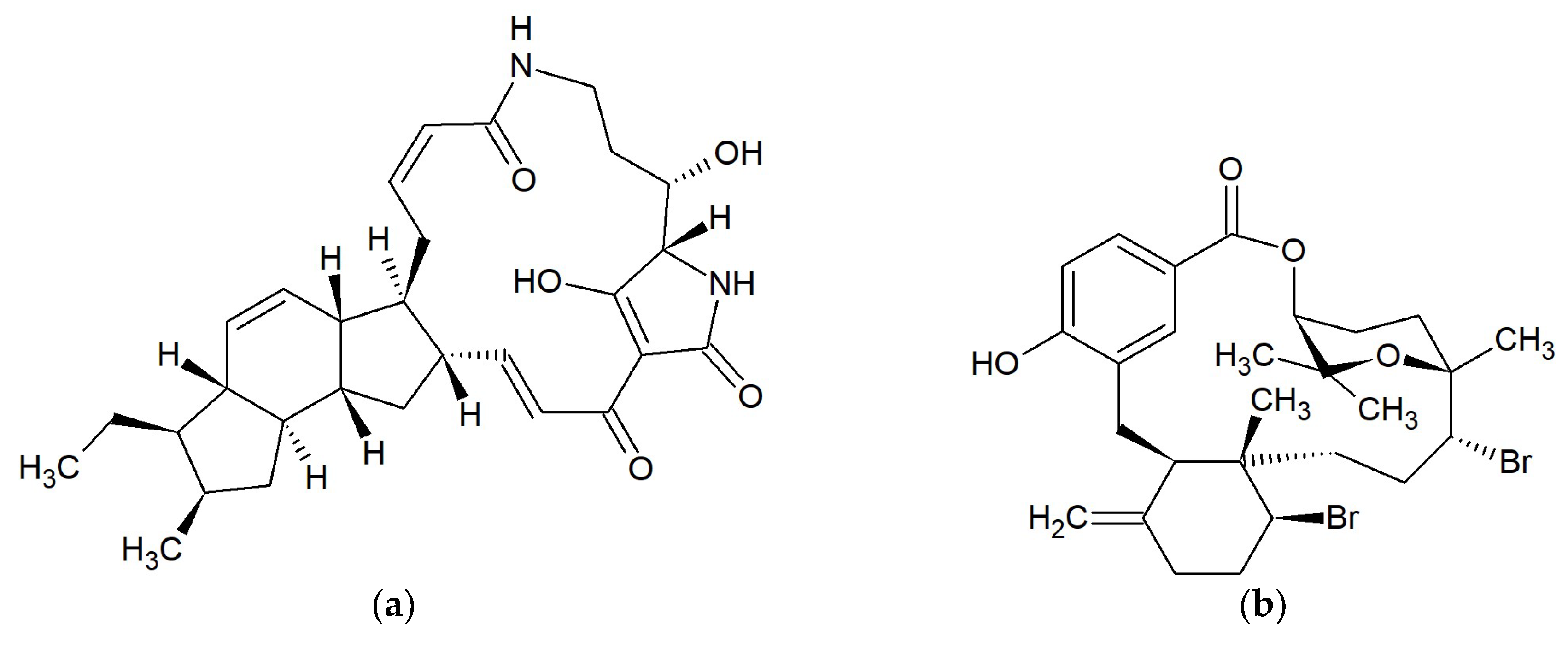
15- and 16-membered bromophycolides J-Q were isolated from extracts of the red alga Callophycus serratus from Yanuca, Fiji. Bromophycolides P (Figure 4b) and Q exhibited antibacterial activity against methicillin-resistant Staphylococcus aureus (MRSA) with an IC50 of 1.4 and 1.8 µM, respectively, and vancomycin-resistant Enterococcus faecium (VRE) with an IC50 of 13 and 5.8 µM, respectively [49].
2.5. Macrolides 16-Membered
2.5.1. Butremycin
Butremycin (Figure 4a) was isolated from Micromonospora sp. K310 obtained from mangrove river sediment in the Western Region of Ghana. Macrolide showed weak activity against Staphylococcus aureus ATCC 25923, Escherichia coli ATCC 25922 with a MIC of 50 µg/mL and some strains of methicillin-resistant S. aureus (MRSA) with a MIC > 50 µg/mL [50].
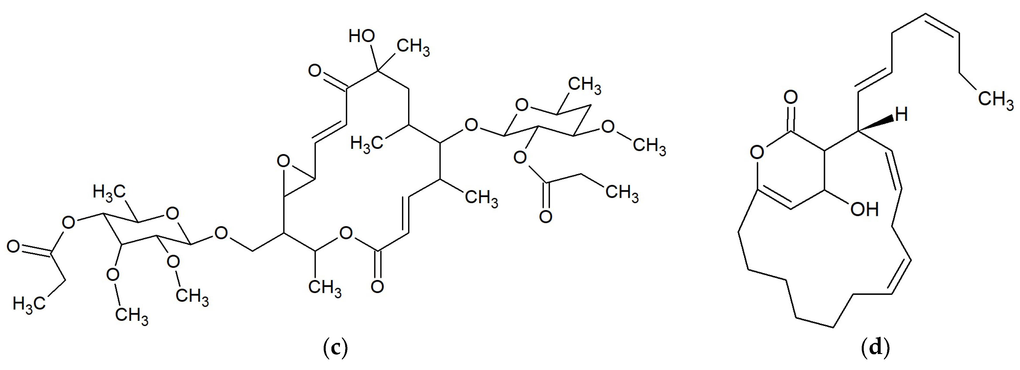
2.5.2. Chalcomycins
Chalcomycin A and chalcomycin B (Figure 4c) were isolated from the marine strain Streptomyces sp. B7064 derived from mangrove sediment near Pohoiki, Hawaii (Pacific Ocean). Both compounds exhibited excellent activities against bacteria Staphylococcus aureus (MIC 0.39 µg/mL) and Bacillus subtilis (MIC 6.25 µg/mL), and low activities against Escherichia coli (MIC >50 µg/mL). Chalcomycins did not show any activity towards fungi Candida albicans or Mucor miehei [51]. Dihydrochalcomycin, chalcomycin and chalcomycin E were isolated from marine-derived Streptomyces sp. HK-2006-1. Two compounds, dihydrochalcomycin and chalcomycin, exhibited activities against Staphylococcus aureus with MICs of 4–32 µg/mL but were not active against Escherichia coli, Candida albicans, or Aspergillus niger [52].
2.5.3. Neurymenolides
Two α-pyrone macrolides, neurymenolides A and B [53,54], were isolated from the red alga Neurymenia fraxinifolia collected from Taveuni, Fiji. Neurymenolide A (Figure 4d) possessed activity against methicillin-resistant Staphylococcus aureus with an IC50 of 2.1 μM and vancomycin-resistant Enterococcus faecium with an IC50 of 4.5 μM [53].
2.6. Macrolides 18-Membered
2.6.1. Borrelidins
Halophilic actinomycete Nocardiopsis sp. strain HYJ128 inhabiting a hypersaline saltern in Jeungdo, Jeollanam-do, Republic of Korea, produced 18-membered macrolides: borrelidin (Figure 5a) and borrelidins C–E. Borrelidin inhibited Enterococcus faecalis ATCC 19433, E. faecium ATCC 19434, Proteus hauseri NRBC 3851, Klebsiella pneumoniae ATCC 10031, and Salmonella enterica ATCC 14028 with MICs of 0.51–65 μM. Borrelidins C and D displayed inhibitory activity against S. enterica with MIC values of 16–63 μM. Borrelidin E did not exhibit any inhibitory activity against the tested bacteria [55].
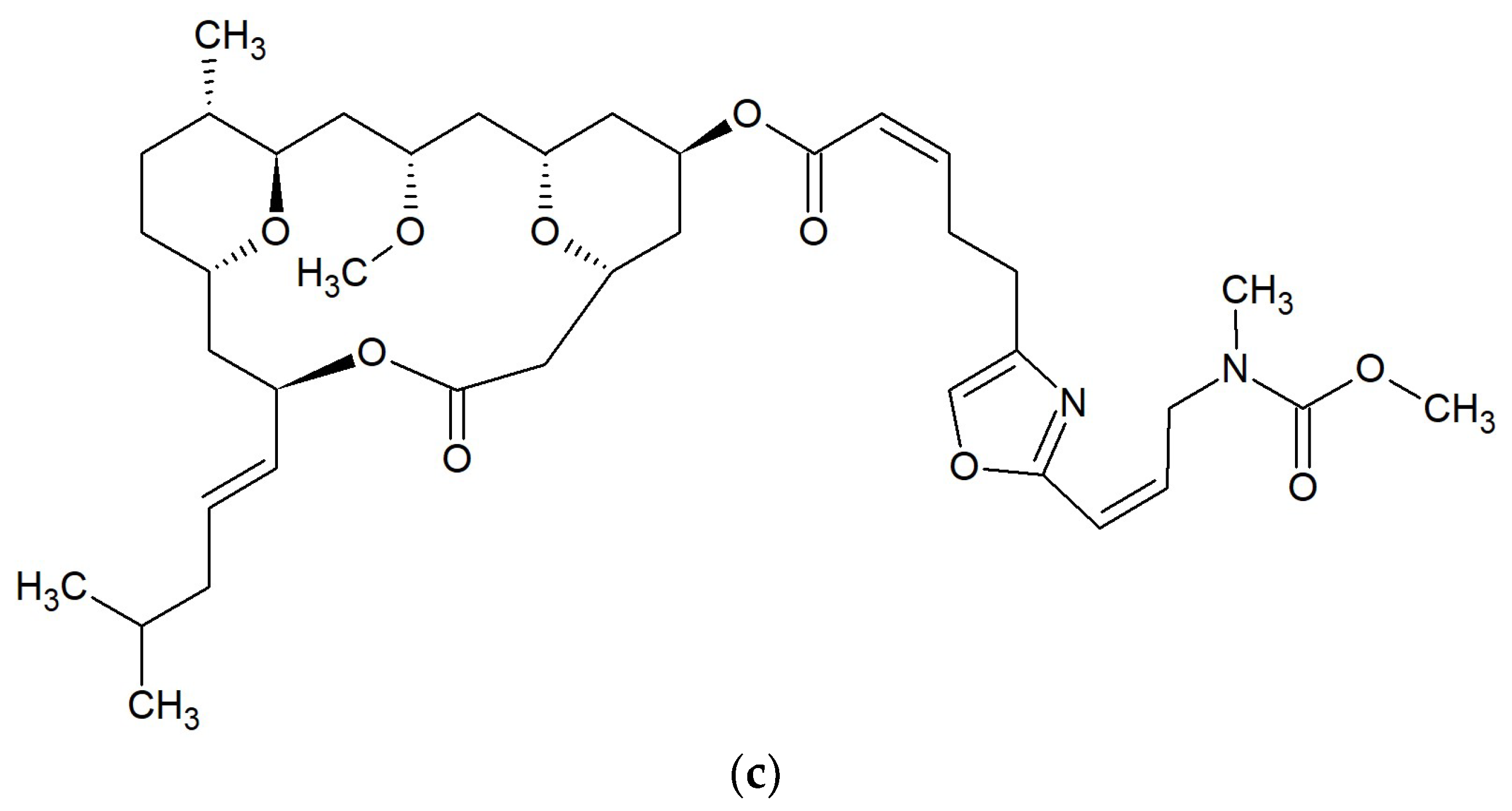
2.6.2. Leucascandrolides
Leucascandrolide A (Figure 5c) [56] was isolated from the calcareous sponge Leucascandra caveolata, collected along the east coast of the Coral Sea, New Caledonia. This compound strongly inhibited fungi Fusarium oxysporum, Helminthosporium sativum, Phytophthora hevea, Botrytis cinerea, Pyricularia oryzae, and yeast Candida albicans [57].
2.6.3. Tedanolides
13-Deoxytedanolide (Figure 5b) is an 18-membered macrolide, which was isolated from the sponge Mycale adhaerens in Japan [58]. This macrolide strongly binds to the 60S large ribosomal subunit, causing inhibition of polypeptide elongation in fungus Saccharomyces cerevisiae, however it does not inhibit the polypeptide synthesis in bacterium Escherichia coli [59].
2.7. Macrolides 20-Membered
2.7.1. Macrocyclic Polyesters
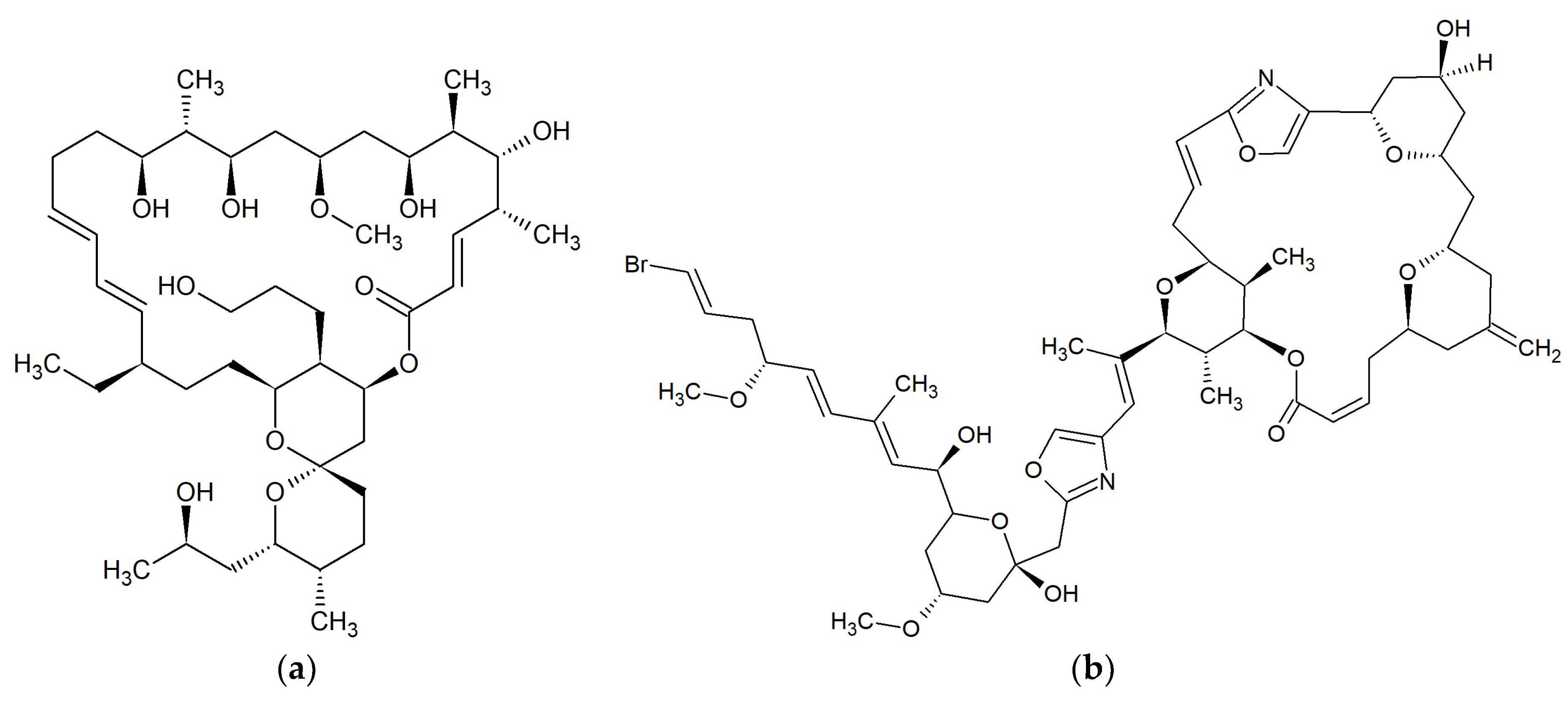
The marine fungus Hypoxylon oceanicum (strain LL-15G256) from mangrove wood collected in Shenzhen, China, produced macrocyclic polyesters [60]. A 20-membered compound 15G256ι (Figure 6a) exhibited low activity against the fungus Neurospora crassa acting as inhibitors of fungal cell wall formation. The 30-membered substance 15G256w had similar activity [61].
2.7.2. Misakinolides
According to Sakai et al. misakinolide A (Figure 6b) is a 20-membered macrolide [62], however this macrolide occurs as 40-membered dimer [63]. Misakinolide A was isolated from the sponge Theonella sp., collected at Maeda-misaki, Okinawa, Japan. This compound possesses antifungal activity against Candida albicans (MIC 5 µg/mL) [62].
2.8. Macrolides 22-Membered
2.8.1. Kabiramides
Kabiramide C (Figure 7a) was isolated from the eggmasses of an unidentified nudibranch collected at Kabira Bay on Ishigaki-jima Island of the Ryukyus Islands, Japan. This 22-membered macrolide showed marked antifungal activity against Candida albicans ATCC 10234, Aspergillus niger ATCC 9642, Penicillium citrium ATCC 9849 and Trichophyton interdigitale [64]. Kabiramides G, J and K were isolated from the sponge Pachastrissa nux collected in the Gulf of Thailand. These macrolides, together with kabiramides B–D, showed anti-parasite activity against Plasmodium falciparum K1 [65].
2.8.2. Scytophycins
Ishibashi et al. demonstrated that scytophycins A–E isolated from terrestrial blue-green alga Scytonema pseudohofmanni collected from Oahu, Hawaii, exhibited cytotoxicity and antifungal activity [66]. Scytophycin B, scytophycin E, 6-hydroxyscytophycin B, and tolytoxin (Figure 7b) (6-hydroxy-7-O-methylscytophycin B) obtained from the terrestrial blue-green alga Cylindrospermum muscicola, isolated on the island of Kauai, Hawaii also had antifungal activity [67]. Tolytoxin and two analogs; 6-hydroxyscytophycin B and 19-O-demethylscytophycin C were also produced by strains of Scytonema mirabile, S. burmanicum, and S. ocellatum. These macrolides had antifungal activity against Aspergillus oryzae, Candida albicans, Penicillium notatum, and Saccharomyces cerevisiae [68]. Tolytoxin isolated from blue-green alga Tolypothrix conglutinata var. colorata found at Fanning Island, Kiribati, exhibited additional inhibitory activity against Alternaria alternata, Bipolaris incurvata, Calonectria critalarae, Colletotrichum coccodes, Phyllosticta capitalensis, Phytophthora nicotianae, Rhizoctonia solani, Sclerotium rofsii, Thielaviopsis paradoxa, and Trichophyton mentagrophytes with MICs of 0.25–8 nM. Tolytoxin did not show any inhibitory activity against bacteria [69]. The presence of scytophycins with activity against Candida albicans and Aspergillus flavus has also been demonstrated in cyanobacteria Anabaena sp. HAN21/1, Anabaena cf. cylindrica PH133, Scytonema sp. HAN3/2, and Nostoc sp. HAN11/1 [70].
2.9. Macrolides 22–25-Membered
2.9.1. Gageomacrolactins
Bacillus subtilis isolated from marine sediment collected from Gageocho, Republic of Korea, produced three gageomacrolactins (Figure 8a), which are 24-membered macrolactin derivatives. Gageomacrolactins displayed strong activity against some bacteria (Staphylococcus aureus, Bacillus subtilis, B. cereus, Escherichia coli, Salmonella typhi, and Pseudomonas aeruginosa) with MIC values of 0.02–0.05 μM. Additionally, isolated gageomacrolactins and macrolactins A, B, F, and W inhibit the growth of Aspergillus niger, Botrytis cinerea, Colletotrichum acutatum, Candida albicans, and Rhizoctonia solani with MIC values of 0.04–0.3 μM [71].
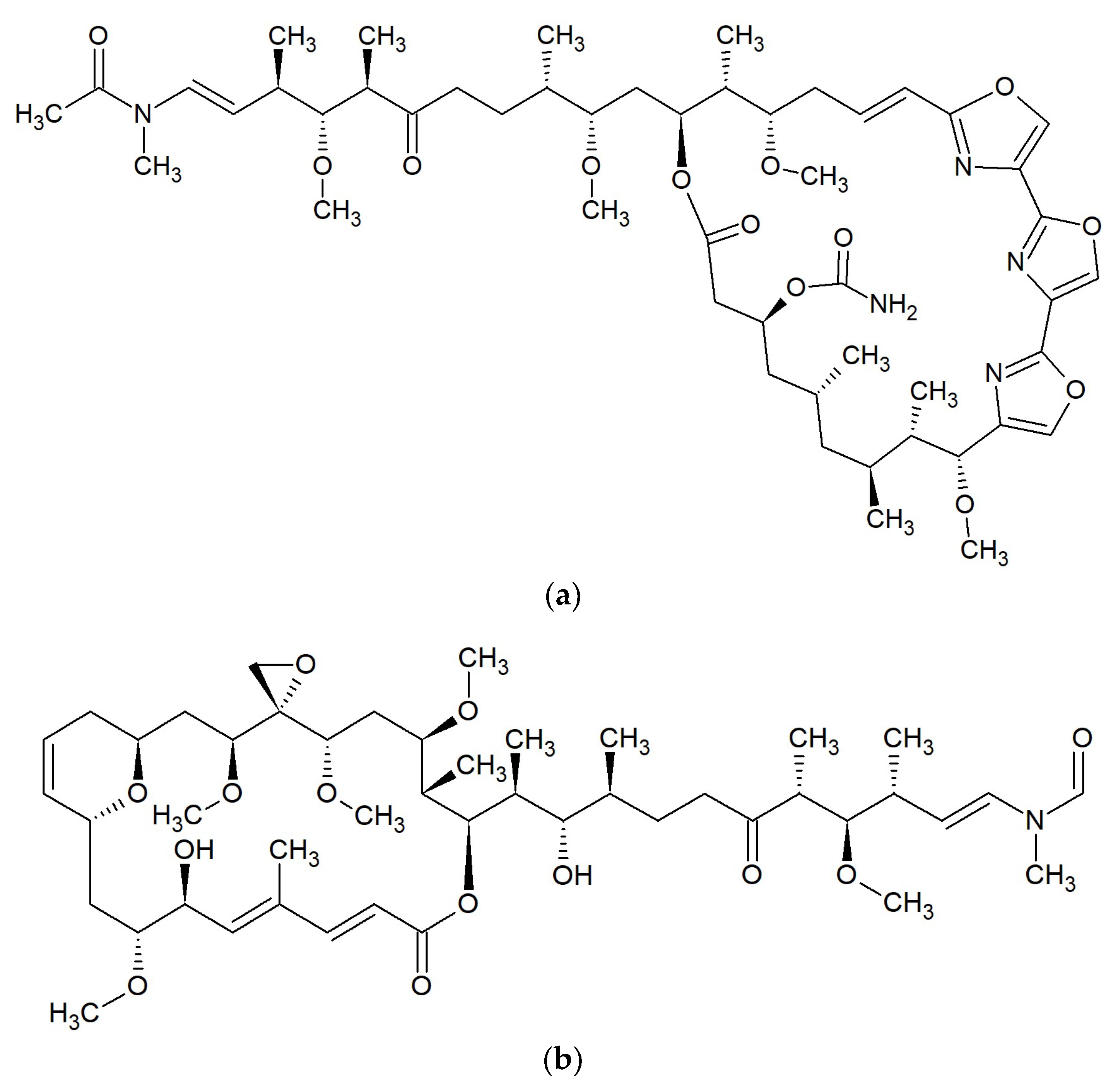
2.9.2. Halichondramides
An oxazole-containing macrolide, halichondramide (Figure 8b), is a 25-membered antibiotic [72]. It was obtained from the sponge Halichondria sp. from Kwajalein Island, Marshall Islands, and showed significant activity against Candida albicans (MIC 0.2 pg/mL) and Trichophyton mentagrophytes (MIC 12.5 pg/mL). Halichondramide did not inhibit bacteria [73]. Further studies revealed that the sponge of the genus Halichondria sp. also contains two other macrolides (dihydrohalichondramide and isohalichondramide) having significant activity against C. albicans. In this same paper, the authors showed that anti-C. albicans activity had the nudibranch Hexabranchus sanguineus, from which dihydrohalichondramide and tetrahydrohalichondramide were isolated [74]. From the marine sponge Chondrosia corticata collected from Guam, more oxazole-containing macrolides were isolated: neohalichondramide, (19Z)-halichondramide, and secohalichondramide. These compounds exhibited antifungal activity toward the Candida albicans and Aspergillus niger [75]. Chung et al. in the sponge C. corticata identified the following macrolides: halichondramide, jaspisamide A, halishigamide D, neohalichondramide, and (19Z)-halichondramide. None of the compounds were active against Gram-positive or Gram-negative bacteria at 100 µg/mL. Halichondramide showed inhibitory activity against Candida albicans, Aspergillus fumigatus, Trichophyton rubrum, and T. mentagrophytes with MIC values of 0.2 to 0.91 µM. Compound (19Z)-halichondramide showed inhibitory activity against all tested fungi with MIC values of 0.78 to 14.55 µM. In the presented study, jaspisamide A, halishigamide D, and neohalichondramide were inactive at 100 µg/mL [76].
2.9.3. Macrolactins
Macrolactins are a big group of 22- to 25-membered lactone macrolides. Some of these were isolated from a culture of Bacillus sp. PP19-H3 obtained from the macroalga Schizymenia dubyi collected on the Omaezaki coast of Shizuoka prefecture in Japan. Macrolactins A (Figure 8c), F, G, I, J, K, and L are 24-membered macrolides, macrolactin H is 22-membered, and macrolactin is M a 25-membered lactone. Macrolactins A, G, H, I, J, L, and M were effective against Staphylococcus aureus (MICs 5–10 ppm), and Bacillus subtilis (MICs 30–60 ppm). The macrolactins F and K had low activity against the above bacteria (MICs 80 and >100). None of the studied macrolides inhibited Escherichia coli or Salinivibrio costicola [77,78]. In other studies, macrolactin A did not have any antimicrobial activity [79,80].
Macrolactins A, B, F, and W isolated from marine Bacillus subtilis from Gageocho, Republic of Korea, inhibit the growth of Aspergillus niger, Botrytis cinerea, Colletotrichum acutatum, Candida albicans, and Rhizoctonia solani with MIC values of 0.04–0.3 μM [71].
7-O-succinylmacrolactin A and 7-O-succinylmacrolactin F, together with macrolactin F, were isolated from the marine Bacillus sp. Sc026 occurring in sediments around Sichang Island, Thailand. These two succinylmacrolactins showed activity against Bacillus subtilis and Staphylococcus aureus [78]. 7-O-malonylmacrolactin A was isolated from soil B. subtilis from Takalar, South Sulawesi in Indonesia. This compound inhibited methicillin-sensitive S. aureus (MSSA), methicillin-resistant S. aureus (MRSA), vancomycin-resistant enterococci (VRE), Burkholderia cepacia, and Candida crusei [79,80].
Macrolactin N was obtained from Bacillus subtilis AT29 and had antibacterial activity against Escherichia coli, Staphylococcus aureus, and Bacillus subtilis. It inhibited the growth of E. coli with a MIC value of 100 µg/mL, while for S. aureus and B. subtilis, the MIC50 is 100 µg/mL. Macrolactin N inhibited S. aureus peptide deformylase with an IC50 value of 7.5 µM [81].
From the marine Bacillus sp. derived from the sea sediment of East China Sea, macrolactin S, a 24-membered ring lactone, was obtained. Macrolactin S, together with macrolactins A and B had antibacterial activity against Escherichia coli, and Staphylococcus aureus [82].
Macrolactins T and U, along with macrolactins A, B, D, O, and S, were isolated from the bacterium Bacillus marinus, which was separated from Suaeda salsa collected in the coastline of the Bohai Sea of China. In the study, authors reported the inhibitory activity of macrolactins B (MIC 4.5–20.1 µg/mL) and D (MIC > 100 µg/mL) against fungi Pyricularia oryzae and Alternaria solani, and bacterium Staphylococcus aureus [83].
From marine bacterium B. amyloliquefaciens SCSIO 00856 isolated from the South China Sea gorgonian Junceella juncea, macrolactin V and S. Macrolactin V were obtained and had strong antibacterial activities against Escherichia coli, Bacillus subtilis, and Staphylococcus aureus with a MIC value of 0.1 μg/mL. Macrolactin S showed potent activity against E. coli and S. aureus (MICs 0.1–0.3 μg/mL), and weak against B. subtilis (MIC 100 μg/mL) [84].
Macrolactin W was isolated from a marine Bacillus sp. 09ID194 collected from Ieodo, a southern reef of South Korea. This macrolide showed antibacterial activities towards Bacillus subtilis, Staphylococcus aureus, Escherichia coli, and Pseudomonas aeruginosa with a MIC of 64 µg/mL [85].
One of the marine Bacillus sp. produces three 24-membered macrolactins, which contain an oxetane, an epoxide, and a tetrahydropyran ring, respectively. All three macrolactins showed antimicrobial activity against Bacillus subtilis and Escherichia coli (MIC 0.16 μM). The macrolactin with an epoxide ring also had excellent activity against Saccharomyces cerevisiae (MIC 0.02–0.16 μM) [86].
From Bacillus subtilis MTCC 10403, isolated from the brown seaweed Anthophycus longifolius collected from the Gulf of Mannar of India, new antimicrobial aryl-crowned polyketide macrolactin was obtained. This substance had bactericidal properties against Escherichia coli, Aeromonas hydrophilla, Pseudomonas aeruginosa, and Vibrio sp. at a low concentration with MIC < 13 µg/mL, and against Klebsiella pneumoniae with MIC ~25 µg/mL. The mode of antimicrobial action of this new acryl-crowned macrolactin was found to be iron chelating similar to siderophores [87].
2.9.4. Maduralide
2.10. Macrolides 26-Membered
2.10.1. Neomaclafungins
Neomaclafungins A–I (Figure 9a) were produced by the bacteria Actinoalloteichus sp. NPS702 isolated from the marine sediment of Usa Bay, Kochi Prefecture, Japan. These oligomycin macrolides exhibited significant antifungal activity in vitro against Trichophyton mentagrophytes ATCC 9533, with MIC values of 1–3 µg/mL [89].
2.10.2. Phorboxazoles
Phorboxazoles A (Figure 9b) and B were isolated from the Indian Ocean marine sponge Phorbas sp. Both antibiotics had antifungal activity against Candida albicans and Saccharomyces carlsbergensis. None of these compounds showed any activity against Escherichia coli, Pseudomonas aeruginosa, or Staphylococcus aureus [90].
2.11. Macrolides 31-Membered
Reedsmycins
Reedsmycins are nonglycosylated polyene-polyol macrolides produced by marine-derived Streptomyces youssoufiensis OUC6819 [91] and by Streptomyces sp. CHQ-64 [92]. Reedsmycin A (Figure 10a) exhibited antifungal activity against Candida albicans (MIC 25–50 μM). Other compounds in this group had lower activity. MICs for reedsmycins C–E were 50–100 μM, for reedsmycin B were 100–200 μM. Reedsmycin F exhibited no inhibitory activity [92].
2.12. Macrolides 34-Membered
Marinisporolides
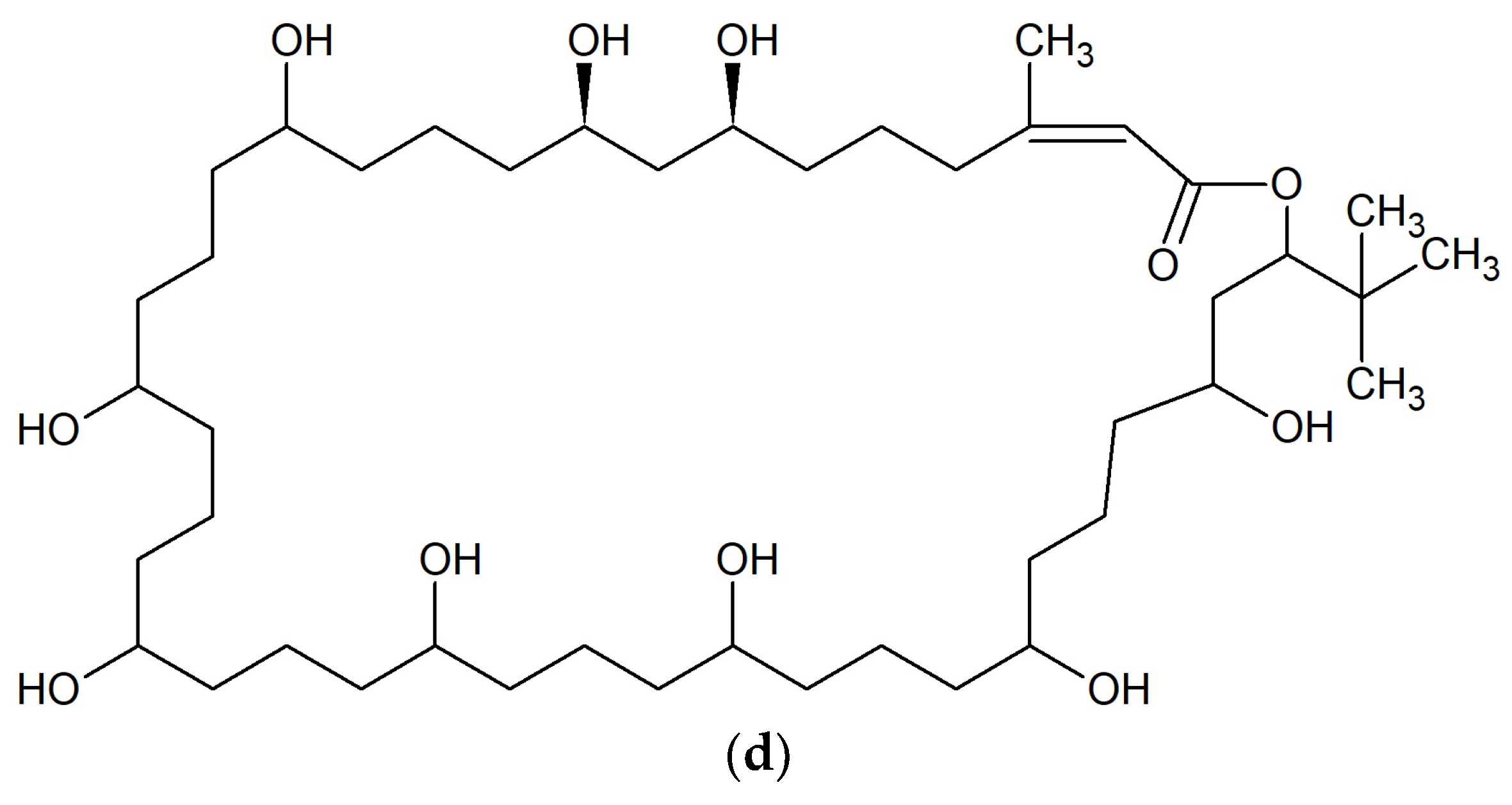
Two polyene-polyol macrolides; marinisporolides A (Figure 10b) and B were isolated from the marine actinomycete Marinispora strain CNQ-140, collected offshore from La Jolla, California, USA. Both marinisporolides showed weak or no antifungal activity against Candida albicans with a MIC value of 22 μM [93].
2.13. Macrolides 36-Membered
2.13.1. Azalomycins
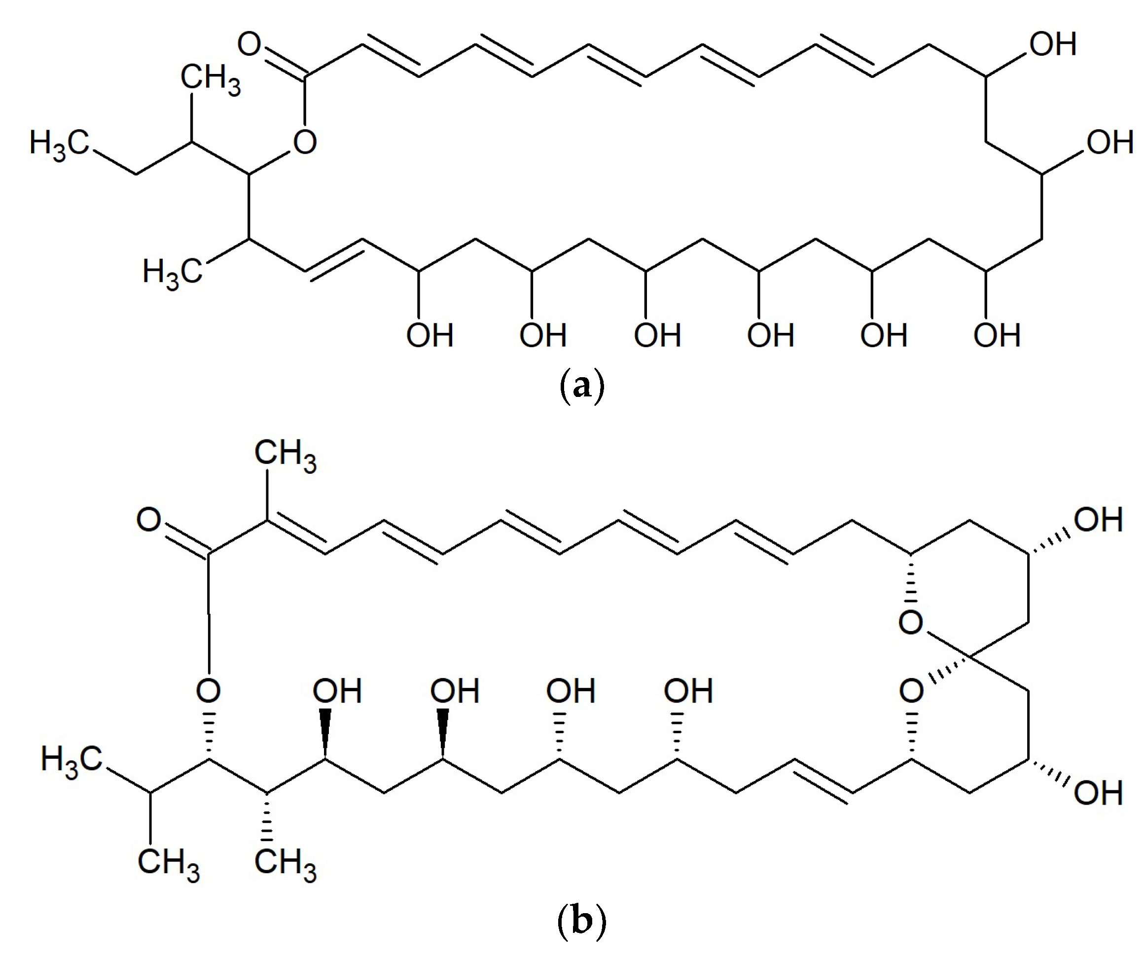
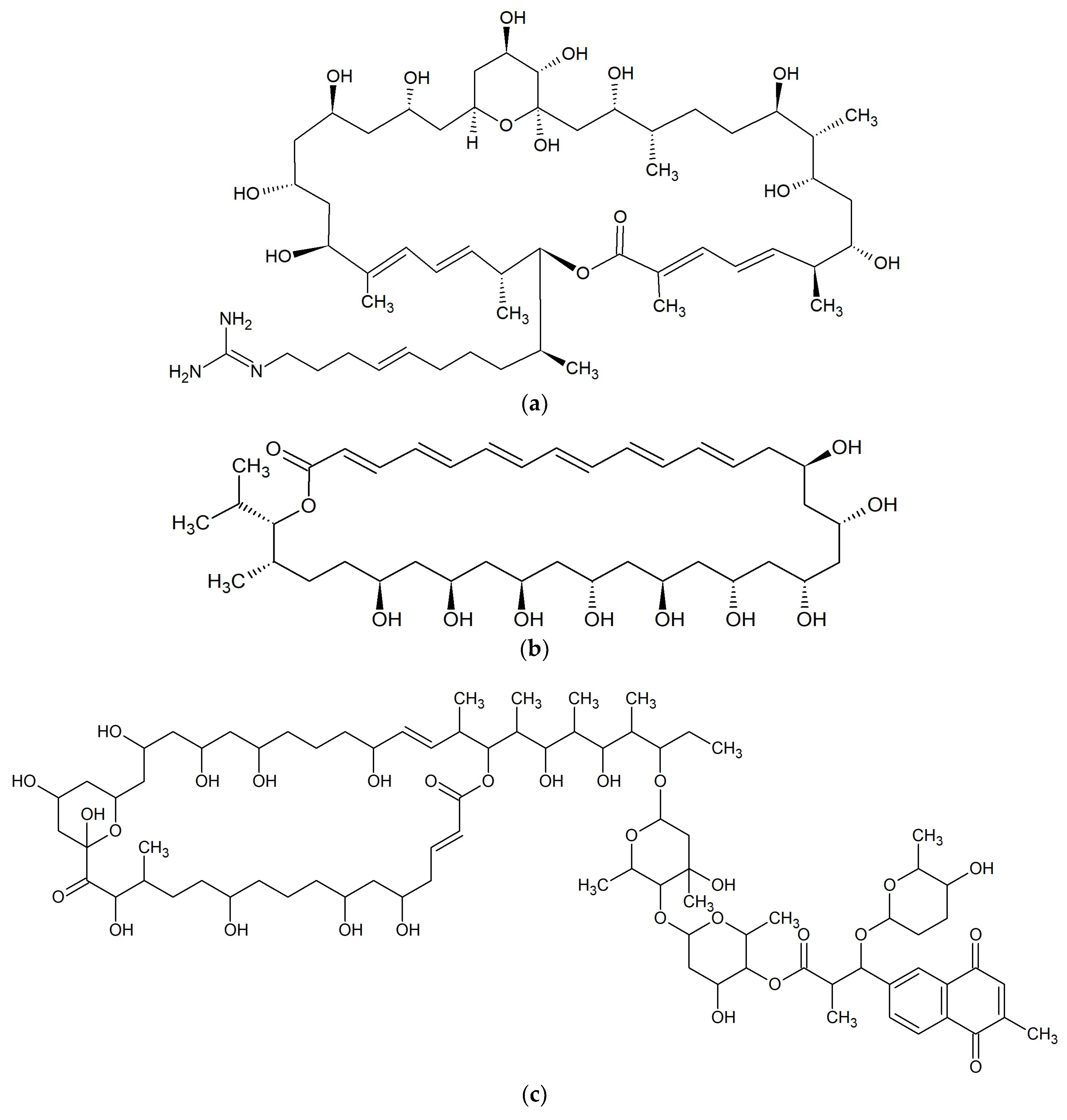
Two macrocyclic lactones, azalomycin F4a 2-ethylpentyl ester and azalomycin F5a 2-ethylpentyl ester, were identified from metabolites of Streptomyces sp. 211726 isolated from mangrove rhizosphere soil of Heritiera globosa collected in Wenchang, China. Both compounds showed moderate activity against Candida albicans ATCC 10231 at the MICs of 2.34 and 12.5 μg/mL [94]. Seven analogs of azalomycin F (Figure 11a) were identified from this strain with the same fermentation condition and showed antimicrobial activity against C. albicans ATCC 10231 (MICs 1.56–6.25 μg/mL), Staphylococcus aureus S014 (MICs 0.39–1.56 μg/mL), Bacillus subtilis S028 (MICs 0.20–0.78 μg/mL), and Escherichia coli S002 (MICs 3.13–25.00 μg/mL) [95].
2.13.2. Bahamaolides
From the marine actinomycete Streptomyces sp. CNQ343 derived from sediment collected at North Cat Cay in the Bahamas, bahamaolides A and B. Bahamaolide A (Figure 11b) displaying significant inhibitory activity against Candida albicans ATCC 10231 with a MIC value of 12.5 μg/mL acting on enzyme isocitrate lyase were isolated. It also possessed antifungal activity against various pathogenic fungi: Aspergillus fumigatus HIC 6094, Trichophyton rubrum IFO 9185, T. mentagrophytes IFO4 0996. Bahamaolide B did not inhibit any tested strain [96].
2.13.3. Polyhydroxyl Macrolides
Two polyhydroxyl macrolide lactones, PM100117 (Figure 11c) and PM100118 were isolated from the marine actinobacteria Streptomyces caniferus GUA-06-05-006A. Both substances possessed antifungal activity against Candida albicans ATCC10231 [97]. PM100117 also showed antibiotic activity against Saccharomyces cerevisiae W303.1A but was not active towards Micrococcus luteus [98].
2.14. Macrolides 40-Membered
Amantelides
Amantelides A (Figure 11d) and B were isolated from gray cyanobacterium belonging to the family Oscillatoriales, collected near Puntan dos Amantes, Tumon Bay, Guam. The antifungal activity of amantelide A was observed against the marine fungi Dendryphiella salina, Lindra thalassiae, and Fusarium sp. at a concentration of 62.5 μg/mL. Moreover, macrolide had weak antibacterial activity against Staphylococcus aureus and Pseudomonas aeruginosa with a MIC of 32 μM. Amantelide B inhibited the growth of Dendryphiella salina at a concentration of 6.25 μg/mL [99].
2.15. Macrolides 42-Membered
Spongistatins
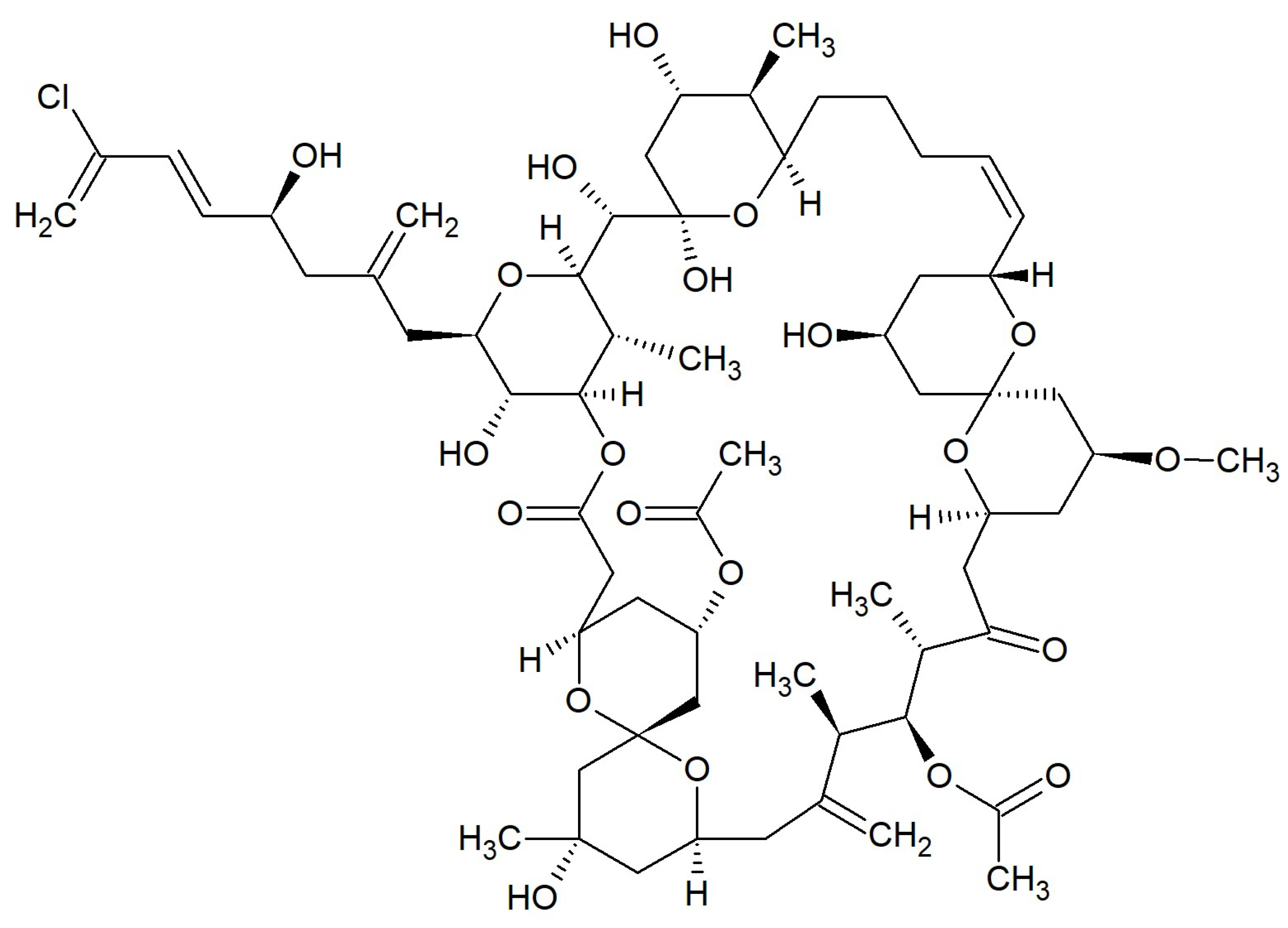
The spongistatins are macrocyclic lactone polyethers isolated from marine porifera. Spongistatin 1 (Figure 12) was discovered in an Indian Ocean Spongia species [100] and Hyrtios erecta together with spongistatins 2 and 3 [101]. Spongistatins 4–7 were obtained from the southeast African Spirastrella spinispirulifera [102,103]. All of these antibiotics inhibited the growth of Candida albicans and Cryptococcus neoformans in disk diffusion assays. Furthermore, Spongistatin 1 acted against Issatchenkia orientalis, Rhodotorula mucilaginosa, Aspergillus fumigatus, and Rhizopus oligosporus with MICs of 0.195–12.5 µg/mL [104].
In Table 1 has been presented the general characteristic of marine macrolides described in this review.
3. Conclusions
Marine organisms produce 34 groups of macrolides with antibacterial and/or antifungal activities. Among seventy-six antibiotics or their analog sets summarized in the Table, 36 are produced by bacteria, 18 by fungi, ten by sponges, seven by algae, two by porifera and one by nudibranch. At the same time, 29 macrolides or their groups have antifungal activity, 25 have antibacterial, and 20 have both antifungal and antibacterial. Summarizing, marine organisms are abundant in natural macrolides, which may be used in the future for the treatment of bacterial and fungal infections. Marine macrolides can also be potential drugs applicable against pathogens resistant to currently known antibiotics, which is also presented in other papers [105,106,107].
Funding
This research received no external funding. The APC was partially funded from the budget of the Prof. Michał Nowicki, MD, PhD - Vice-Rector for Science and University Development, Poznań University of Medical Sciences, Poland.
Conflicts of Interest
The author declares no conflict of interest.
References
- Burja, A.M.; Banaigs, B.; Abou-Mansour, E.; Burgess, J.G.; Wright, P.C. Marine cyanobacteria - a prolific source of natural products. Tetrahedron 2001, 57, 9347–9377. [Google Scholar] [CrossRef]
- El-Demerdash, A.; Tammam, M.A.; Atanasov, A.G.; Hooper, J.N.A.; Al-Mourabit, A.; Kijjoa, A. Chemistry and biological activities of the marine sponges of the genera Mycale (Arenochalina), Biemna and Clathria. Mar. Drugs 2018, 16, 214. [Google Scholar] [CrossRef] [PubMed]
- Wang, M.; Zhang, J.; He, S.; Yan, X. A review study on macrolides isolated from cyanobacteria. Mar. Drugs 2017, 15, 126. [Google Scholar] [CrossRef]
- Swain, S.S.; Paidesetty, S.K.; Padhy, R.N. Antibacterial, antifungal and antimycobacterial compounds from cyanobacteria. Biomed. Pharmacother. 2017, 90, 760–776. [Google Scholar] [CrossRef] [PubMed]
- Liu, Q.-A.; Zheng, J.-J.; Gu, Y.-C.; Wang, C.-Y.; Shao, C.-L. Chapter 7. The chemistry and bioactivity of macrolides from marine microorganisms. Stud. Nat. Prod. Chem. 2015, 44, 353–401. [Google Scholar]
- Jelić, D.; Antolović, R. From erythromycin to azithromycin and new potential ribosome-binding antimicrobials. Antibiotics 2016, 5, 29. [Google Scholar] [CrossRef] [PubMed]
- Campoy, S.; Adrio, J.L. Antifungals. Biochem. Pharmacol. 2017, 133, 86–96. [Google Scholar] [CrossRef] [PubMed]
- Zuckerman, J.M.; Qamar, F.; Bono, B.R. Review of macrolides (azithromycin, clarithromycin), ketolides (telithromycin) and glycylcyclines (tigecycline). Med. Clin. North Am. 2011, 95, 761–791. [Google Scholar] [CrossRef]
- Dinos, G.P. The macrolide antibiotic renaissance. Br. J. Pharmacol. 2017, 174, 2967–2983. [Google Scholar] [CrossRef]
- Karpiński, T.M.; Andrzejewska, E.; Eder, P.; Linke, K.; Szkaradkiewicz, A. Evaluation of antimicrobial resistance of Helicobacter pylori in the last 15 years in West Poland. Acta Microbiol. Immunol. Hung. 2015, 62, 287–293. [Google Scholar] [CrossRef]
- Bolhuis, M.S.; Panday, P.N.; Pranger, A.D.; Kosterink, J.G.; Alffenaar, J.C. Pharmacokinetic drug interactions of antimicrobial drugs: A systematic review on oxazolidinones, rifamycines, macrolides, fluoroquinolones, and beta-lactams. Pharmaceutics. 2011, 3, 865–913. [Google Scholar] [CrossRef] [PubMed]
- Mesa-Arango, A.C.; Scorzoni, L.; Zaragoza, O. It only takes one to do many jobs: Amphotericin B as antifungal and immunomodulatory drug. Front. Microbiol. 2012, 3, 286. [Google Scholar] [CrossRef] [PubMed]
- OECD. Antimicrobial Resistance. Policy Insights. 2016. Available online: https://www.oecd.org/health/health-systems/AMR-Policy-Insights-November2016.pdf (accessed on 24 January 2019).
- Alekshun, M.N.; Levy, S.B. Molecular mechanisms of antibacterial multidrug resistance. Cell 2007, 128, 1037–1050. [Google Scholar] [CrossRef]
- Sanjit Singh, A.; Lekshmi, M.; Prakasan, S.; Nayak, B.B.; Kumar, S. Multiple antibiotic-resistant, extended spectrum-β-lactamase (ESBL)-producing enterobacteria in fresh seafood. Microorganisms 2017, 5, 53. [Google Scholar] [CrossRef] [PubMed]
- Gomaa, F.A.M.; Helal, Z.H.; Khan, M.I. High prevalence of blaNDM-1, blaVIM, qacE, and qacEΔ1 genes and their association with decreased susceptibility to antibiotics and common hospital biocides in clinical isolates of Acinetobacter baumannii. Microorganisms 2017, 5, 18. [Google Scholar] [CrossRef] [PubMed]
- Jayarao, B.; Almeida, R.; Oliver, S.P. Antimicrobial resistance on dairy farms. Foodborne Pathog. Dis. 2019, 16, 1–4. [Google Scholar] [CrossRef] [PubMed]
- Florez-Cuadrado, D.; Moreno, M.A.; Ugarte-Ruíz, M.; Domínguez, L. Antimicrobial resistance in the food chain in the European Union. Adv. Food Nutr. Res. 2018, 86, 115–136. [Google Scholar] [PubMed]
- Bernd, S.; Kunz, O. Bidirectional cross metathesis and ring-closing metathesis/ring opening of a C2-symmetric building block: A strategy for the synthesis of decanolide natural products. Beilstein, J. Org. Chem. 2013, 9, 2544–2555. [Google Scholar]
- Greve, H.; Schupp, P.J.; Eguereva, E.; Kehraus, S.; König, G.M. Ten-membered lactones from the marine-derived fungus Curvularia sp. J. Nat. Prod. 2008, 71, 1651–1653. [Google Scholar] [CrossRef]
- Mondol, M.A.; Farthouse, J.; Islam, M.T.; Schüffler, A.; Laatsch, H. Metabolites from the endophytic fungus Curvularia sp. M12 act as motility inhibitors against Phytophthora capsici zoospores. J. Nat. Prod. 2017, 80, 347–355. [Google Scholar] [CrossRef]
- Ha, T.M.; Ko, W.; Lee, S.J.; Kim, Y.C.; Son, J.Y.; Sohn, J.H.; Yim, J.H.; Oh, H. Anti-inflammatory effects of curvularin-type metabolites from a marine-derived fungal strain Penicillium sp. SF-5859 in lipopolysaccharide-induced RAW264.7 macrophages. Mar. Drugs 2017, 15, 282. [Google Scholar] [CrossRef]
- Greve, H.; Schupp, P.J.; Eguereva, E.; Kehraus, S.; Kelter, G.; Maier, A.; Fiebig, H.H.; König, G.M. Apralactone A and a new stereochemical class of curvularins from the marine-derived fungus Curvularia sp. Eur. J. Org. Chem. 2008, 2008, 5085–5092. [Google Scholar] [CrossRef]
- Ye, X.; Anjum, K.; Song, T.; Wang, W.; Yu, S.; Huang, H.; Lian, X.Y.; Zhang, Z. A new curvularin glycoside and its cytotoxic and antibacterial analogues from marine actinomycete Pseudonocardia sp. HS7. Nat. Prod. Res. 2016, 30, 1156–1161. [Google Scholar] [CrossRef]
- Xie, L.W.; Ouyang, Y.C.; Zou, K.; Wang, G.H.; Chen, M.J.; Sun, H.M.; Dai, S.K.; Li, X. Isolation and difference in anti-Staphylococcus aureus bioactivity of curvularin derivates from fungus Eupenicillium sp. Appl. Biochem. Biotechnol. 2009, 159, 284–293. [Google Scholar] [CrossRef]
- Trisuwan, K.; Rukachaisirikul, V.; Phongpaichit, S.; Preedanon, S.; Sakayaroj, J. Modiolide and pyrone derivatives from the sea fan-derived fungus Curvularia sp. PSU-F22. Arch. Pharm. Res. 2011, 34, 709–714. [Google Scholar] [CrossRef] [PubMed]
- Tsuda, M.; Mugishima, T.; Komatsu, K.; Sone, T.; Tanaka, M.; Mikami, Y.; Kobayashi, J. Modiolides A and B, two new 10-membered macrolides from a marine-derived fungus. J. Nat. Prod. 2003, 66, 412–415. [Google Scholar] [CrossRef]
- Du, X.; Lu, C.; Li, Y.; Zheng, Z.; Su, W.; Shen, Y. Three new antimicrobial metabolites of Phomopsis sp. J. Antibiot. (Tokyo) 2008, 61, 250–253. [Google Scholar] [CrossRef] [PubMed]
- Mohapatra, D.K.; Reddy, D.P.; Dash, U.; Yadav, J.S. Total synthesis of Z-isomer of phomolide B. Tetrahedron Lett. 2011, 52, 151–154. [Google Scholar] [CrossRef]
- Lin, X.; Lu, C.-H.; Shen, Y.-M. One new ten-membered lactone from Phomopsis sp. B27, an endophytic fungus of Annona squamosa. Chin. J. Nat. Med. 2008, 6, 391–394. [Google Scholar] [CrossRef]
- Ito, A.; Maeda, H.; Tonouchi, A.; Hashimoto, M. Relative and absolute structure of phomolide C. Biosci. Biotechnol. Biochem. 2015, 79, 1067–1069. [Google Scholar] [CrossRef] [PubMed]
- Edrada, R.A.; Heubes, M.; Brauers, G.; Wray, V.; Berg, A.; Gräfe, U.; Wohlfarth, M.; Mühlbacher, J.; Schaumann, K.; Bringmann, G.; et al. Online analysis of xestodecalactones A-C, novel bioactive metabolites from the fungus Penicillium cf montanense and their subsequent isolation from the sponge Xestospongia exigua. J. Nat. Prod. 2002, 65, 1598–1604. [Google Scholar] [CrossRef] [PubMed]
- Liang, Q.; Zhang, J.; Quan, W.; Sun, Y.; She, X.; Pan, X. The first asymmetric total syntheses and determination of absolute configurations of xestodecalactones B and C. J. Org. Chem. 2007, 72, 2694–2697. [Google Scholar] [CrossRef]
- Ebrahim, W.; Aly, A.H.; Mándi, A.; Totzke, F.; Kubbutat, M.H.G.; Wray, V.; Lin, W.-H.; Dai, H.; Proksch, P.; Kurtán, T.; et al. Decalactone derivatives from Corynespora cassiicola, an endophytic fungus of the mangrove plant Laguncularia racemosa. Eur. J. Org. Chem. 2012, 18, 3476–3484. [Google Scholar] [CrossRef]
- Kubota, T.; Iwai, T.; Sakai, K.; Gonoi, T.; Kobayashi, J. Amphidinins C-F, amphidinolide Q analogues from marine dinoflagellate Amphidinium sp. Org. Lett. 2014, 16, 5624–5627. [Google Scholar] [CrossRef] [PubMed]
- Cao, F.; Yang, Q.; Shao, C.L.; Kong, C.J.; Zheng, J.J.; Liu, Y.F.; Wang, C.Y. Bioactive 7-oxabicyclic[6.3.0]lactam and 12-membered macrolides from a gorgonian-derived Cladosporium sp. fungus. Mar. Drugs 2015, 13, 4171–4178. [Google Scholar] [CrossRef]
- Xu, J.; Jiang, C.S.; Zhang, Z.L.; Ma, W.Q.; Guo, Y.W. Recent progress regarding the bioactivities, biosynthesis and synthesis of naturally occurring resorcinolic macrolides. Acta Pharmacol. Sin. 2014, 35, 316–330. [Google Scholar] [CrossRef] [PubMed] [Green Version]
- Yang, R.Y.; Li, C.Y.; Lin, Y.C.; Peng, G.T.; She, Z.G.; Zhou, S.N. Lactones from a brown alga endophytic fungus (No. ZZF36) from the South China Sea and their antimicrobial activities. Bioorg. Med. Chem. Lett. 2006, 16, 4205–4208. [Google Scholar] [CrossRef] [PubMed]
- Shigemori, H.; Kasai, Y.; Komatsu, K.; Tsuda, M.; Mikami, Y.; Kobayashi, J. Sporiolides A and B, new cytotoxic twelve-membered macrolides from a marine-derived fungus Cladosporium species. Mar. Drugs 2004, 2, 164–169. [Google Scholar] [CrossRef]
- Du, Y.; Chen, Q.; Linhardt, R.J. The first total synthesis of sporiolide A. J. Org. Chem. 2006, 71, 8446–8451. [Google Scholar] [CrossRef]
- Jiang, Z.D.; Jensen, P.R.; Fenical, W. Lobophorins A and B, new antiinflammatory macrolides produced by a tropical marine bacterium. Bioorg. Med. Chem. Lett. 1999, 9, 2003–2006. [Google Scholar] [CrossRef]
- Li, S.; Xiao, J.; Zhu, Y.; Zhang, G.; Yang, C.; Zhang, H.; Ma, L.; Zhang, C. Dissecting glycosylation steps in lobophorin biosynthesis implies an iterative glycosyltransferase. Org. Lett. 2013, 15, 1374–1377. [Google Scholar] [CrossRef]
- Niu, S.; Li, S.; Chen, Y.; Tian, X.; Zhang, H.; Zhang, G.; Zhang, W.; Yang, X.; Zhang, S.; Ju, J.; et al. Lobophorins E and F, new spirotetronate antibiotics from a South China Sea-derived Streptomyces sp. SCSIO 01127. J. Antibiot. (Tokyo) 2011, 64, 711–716. [Google Scholar] [CrossRef] [PubMed]
- Pan, H.Q.; Zhang, S.Y.; Wang, N.; Li, Z.L.; Hua, H.M.; Hu, J.C.; Wang, S.J. New spirotetronate antibiotics, lobophorins H and I, from a South China Sea-derived Streptomyces sp. 12A35. Mar. Drugs 2013, 11, 3891–3901. [Google Scholar] [CrossRef] [PubMed]
- Yang, X.; Khong, T.T.; Chen, L.; Choi, H.D.; Kang, J.S.; Son, B.W. 8’-Hydroxyzearalanone and 2’-hydroxyzearalanol: Resorcyclic acid lactone derivatives from the marine-derived fungus Penicillium sp. Chem. Pharm. Bull (Tokyo) 2008, 56, 1355–1356. [Google Scholar] [CrossRef] [PubMed]
- Hitchcock, S.A.; Pattenden, G. Synthesis of macrocycles via allylic radical intermediates. A total synthesis of (−)-zearalenone. Tetrahedron Lett. 1990, 31, 3641–3644. [Google Scholar] [CrossRef]
- Zhao, L.L.; Gai, Y.; Kobayashi, H.; Hu, C.Q.; Zhang, H.P. 5′-Hydroxyzearalenol, a new β-resorcylic macrolide from Fusarium sp. 05ABR26. Chin. Chem. Lett. 2008, 19, 1089–1092. [Google Scholar] [CrossRef]
- Arunpanichlert, J.; Rukachaisirikul, V.; Sukpondma, Y.; Phongpaichit, S.; Supaphon, O.; Sakayaroj, J. A β-resorcylic macrolide from the seagrass-derived fungus Fusarium sp. PSU-ES73. Arch. Pharm. Res. 2011, 34, 1633–1637. [Google Scholar] [CrossRef] [PubMed]
- Lane, A.L.; Stout, E.P.; Lin, A.S.; Prudhomme, J.; Le Roch, K.; Fairchild, C.R.; Franzblau, S.G.; Hay, M.E.; Aalbersberg, W.; Kubanek, J. Antimalarial bromophycolides J-Q from the Fijian red alga Callophycus serratus. J. Org. Chem. 2009, 74, 2736–2742. [Google Scholar] [CrossRef] [PubMed]
- Kyeremeh, K.; Acquah, K.S.; Sazak, A.; Houssen, W.; Tabudravu, J.; Deng, H.; Jaspars, M. Butremycin, the 3-hydroxyl derivative of ikarugamycin and a protonated aromatic tautomer of 5’-methylthioinosine from a Ghanaian Micromonospora sp. K310. Mar. Drugs 2014, 12, 999–1012. [Google Scholar] [CrossRef]
- Asolkar, R.N.; Maskey, R.P.; Helmke, E.; Laatsch, H. Chalcomycin B, a new macrolide antibiotic from the marine isolate Streptomyces sp. B7064. J. Antibiot. (Tokyo) 2002, 55, 893–898. [Google Scholar] [CrossRef]
- Jiang, S.; Zhang, L.; Pei, X.; Deng, F.; Hu, D.; Chen, G.; Wang, C.; Hong, K.; Yao, X.; Gao, A.H. Chalcomycins from marine-derived Streptomyces sp. and their antimicrobial activities. Mar. Drugs 2017, 15, 153. [Google Scholar] [CrossRef]
- Stout, E.P.; Hasemeyer, A.P.; Lane, A.L.; Davenport, T.M.; Engel, S.; Hay, M.E.; Fairchild, C.R.; Prudhomme, J.; Le Roch, K.; Aalbersberg, W.; et al. Antibacterial neurymenolides from the Fijian red alga Neurymenia fraxinifolia. Org. Lett. 2009, 11, 225–228. [Google Scholar] [CrossRef]
- Motuhi, S.E.; Feizbakhsh, O.; Foll-Josselin, B.; Baratte, B.; Delehouzé, C.; Cousseau, A.; Fant, X.; Bulinski, J.C.; Payri, C.E.; Ruchaud, S.; et al. Neurymenolide A, a novel mitotic spindle poison from the new caledonian rhodophyta Phacelocarpus neurymenioides. Mar. Drugs 2019, 17, 93. [Google Scholar] [CrossRef]
- Kim, J.; Shin, D.; Kim, S.H.; Park, W.; Shin, Y.; Kim, W.K.; Lee, S.K.; Oh, K.B.; Shin, J.; Oh, D.C. Borrelidins C-E: New antibacterial macrolides from a saltern-derived halophilic Nocardiopsis sp. Mar. Drugs 2017, 15, 166. [Google Scholar] [CrossRef]
- Hunter, T.J.; Zheng, J.; O’Doherty, G.A. Approach to the synthesis of the C1-C11 and C14-C18 portion of leucascandrolide A. Org. Chem. Front. 2016, 3, 1120–1125. [Google Scholar] [CrossRef]
- D’Ambrosio, M.; Guerriero, A.; Pietra, F.; Debitus, C. Leucascandrolide A, a new type of macrolide: The first powerfully bioactive metabolite of calcareous sponges (Leucascandra caveolata, a new genus from the Coral Sea). Helvet. Chim. Acta. 1996, 79, 51–60. [Google Scholar] [CrossRef]
- Fusetani, N.; Sugawara, T.; Matsunaga, S. Cytotoxic metabolites of the marine sponge Mycale adhaerens Lambe. J. Org. Chem. 1991, 56, 4971. [Google Scholar] [CrossRef]
- Nishimura, S.; Matsunaga, S.; Yoshida, S.; Nakao, Y.; Yokoyama, S.; Fusetani, N. 13-Deoxytedanolide, a marine sponge-derived antitumor macrolide, binds to the 60S large ribosomal subunit. Bioorg. Med. Chem. 2005, 13, 449. [Google Scholar] [CrossRef]
- Schlingmann, G.; Milne, L.; Williams, D.R.; Carter, G.T. Cell wall active antifungal compounds produced by the marine fungus Hypoxylon oceanicum LL-15G256. II. Isolation and structure determination. J. Antibiot. (Tokyo) 1998, 51, 303–316. [Google Scholar] [CrossRef]
- Schlingmann, G.; Milne, L.; Carter, G.T. Isolation and identification of antifungal polyesters from the marine fungus Hypoxylon oceanicum LL-15G256. Tetrahedron. 2002, 58, 6825–6835. [Google Scholar] [CrossRef]
- Sakai, R.; Higa, T.; Kashma, Y. Misakinolide-A, an antitumor macrolide from the marine sponge Theonella sp. Chem. Lett. 1986, 1499–1502. [Google Scholar] [CrossRef]
- Kato, Y.; Fusetani, N.; Matsunaga, S.; Hashimoto, K.; Sakai, R.; Higa, T.; Kashman, Y. Antitumor macrodiolides isolated from a marine sponge sp.: Structure revision of misakinolide A. Tetrahedron Lett. 1987, 28, 6225–6228. [Google Scholar] [CrossRef]
- Matsunaga, S.; Fusetani, N.; Hashimoto, K.; Koseki, K.; Noma, M. Kabiramide C, a novel antifungal macrolide from nudibranch eggmasses. J. Am. Chem. Soc. 1986, 108, 847–849. [Google Scholar] [CrossRef]
- Sirirak, T.; Kittiwisut, S.; Janma, C.; Yuenyongsawad, S.; Suwanborirux, K.; Plubrukarn, A. Kabiramides J and K, trisoxazole macrolides from the sponge Pachastrissa nux. J. Nat. Prod. 2011, 74, 1288–1292. [Google Scholar] [CrossRef]
- Ishibashi, M.; Moore, R.E.; Patterson, G.M.L. Scytophycins, cytotoxic and antimycotic agents from the cyanophyte Scytonema pseudohofmanni. J. Org. Chem. 1986, 51, 5300–5306. [Google Scholar] [CrossRef]
- Jung, J.H.; Moore, R.E.; Patterson, G.M.L. Scytophycins from a blue-green alga belonging to the Nostocaceae. Phytochemistry. 1991, 30, 3615–3616. [Google Scholar] [CrossRef]
- Carmeli, S.; Moore, R.E.; Patterson, G.M. Tolytoxin and new scytophycins from three species of Scytonema. J. Nat. Prod. 1990, 53, 1533–1542. [Google Scholar] [CrossRef] [PubMed]
- Patterson, G.M.; Carmeli, S. Biological effects of tolytoxin (6-hydroxy-7-O-methyl-scytophycin b), a potent bioactive metabolite from cyanobacteria. Arch. Microbiol. 1992, 157, 406–410. [Google Scholar] [CrossRef] [PubMed]
- Shishido, T.K.; Humisto, A.; Jokela, J.; Liu, L.; Wahlsten, M.; Tamrakar, A.; Fewer, D.P.; Permi, P.; Andreote, A.P.; Fiore, M.F.; et al. Antifungal compounds from cyanobacteria. Mar. Drugs 2015, 13, 2124–2140. [Google Scholar] [CrossRef]
- Tareq, F.S.; Kim, J.H.; Lee, M.A.; Lee, H.S.; Lee, J.S.; Lee, Y.J.; Shin, H.J. Antimicrobial gageomacrolactins characterized from the fermentation of the marine-derived bacterium Bacillus subtilis under optimum growth conditions. J. Agric. Food Chem. 2013, 61, 3428–3434. [Google Scholar] [CrossRef]
- Ciavatta, M.L.; Lefranc, F.; Carbone, M.; Mollo, E.; Gavagnin, M.; Betancourt, T.; Dasari, R.; Kornienko, A.; Kiss, R. Marine mollusk-derived agents with antiproliferative activity as promising anticancer agents to overcome chemotherapy resistance. Med. Res. Rev. 2017, 37, 702–801. [Google Scholar] [CrossRef]
- Kernan, M.R.; Faulkner, D.J. Halichondramide, an antifungal macrolide from the sponge Halichondria sp. Tetrahedron Lett. 1987, 28, 2809–2812. [Google Scholar] [CrossRef]
- Kernan, M.R.; Molinski, T.F.; Faulkner, D.J. Macrocyclic antifungal metabolites from the Spanish dancer nudibranch Hexabranchus sanguineus and sponges of the genus Halichondria. J. Org. Chem. 1988, 53, 5014–5020. [Google Scholar] [CrossRef]
- Shin, J.; Lee, H.S.; Kim, J.Y.; Shin, H.J.; Ahn, J.W.; Paul, V.J. New macrolides from the sponge Chondrosia corticata. J. Nat. Prod. 2004, 67, 1889–1892. [Google Scholar] [CrossRef]
- Chung, S.C.; Lee, S.H.; Jang, K.H.; Park, W.; Jeon, J.E.; Oh, H.; Shin, J.; Oh, K.B. Actin depolymerizing effect of trisoxazole-containing macrolides. Bioorg. Med. Chem. Lett. 2011, 21, 3198–3201. [Google Scholar] [CrossRef] [PubMed]
- Nagao, T.; Adachi, K.; Sakai, M.; Nishijima, M.; Sano, H. Novel macrolactins as antibiotic lactones from a marine bacterium. J. Antibiot. (Tokyo) 2001, 54, 333–339. [Google Scholar] [CrossRef] [PubMed]
- Jaruchoktaweechai, C.; Suwanborirux, K.; Tanasupawatt, S.; Kittakoop, P.; Menasveta, P. New macrolactins from a marine Bacillus sp. Sc026. J. Nat. Prod. 2000, 63, 984–986. [Google Scholar] [CrossRef] [PubMed]
- Romero-Tabarez, M.; Jansen, R.; Sylla, M.; Lünsdorf, H.; Häussler, S.; Santosa, D.A.; Timmis, K.N.; Molinari, G. 7-O-malonyl macrolactin A, a new macrolactin antibiotic from Bacillus subtilis active against methicillin-resistant Staphylococcus aureus, vancomycin-resistant enterococci, and a small-colony variant of Burkholderia cepacia. Antimicrob. Agents Chemother. 2006, 50, 1701–1709. [Google Scholar] [CrossRef]
- Timmis, K.N.; Molinari, G.; Jansen, R.; Romero-Tabarez, M.; Santosa, D.A. Antibiotic and Method for Production Thereof. EP1791823A1 European Patent Office. Available online: https://patents.google.com/patent/EP1791823A1/en (accessed on 10 February 2019).
- Yoo, J.S.; Zheng, C.J.; Lee, S.; Kwak, J.H.; Kim, W.G. Macrolactin N, a new peptide deformylase inhibitor produced by Bacillus subtilis. Bioorg. Med. Chem. Lett. 2006, 16, 4889–4892. [Google Scholar] [CrossRef] [PubMed]
- Lu, X.L.; Xu, Q.Z.; Shen, Y.H.; Liu, X.Y.; Jiao, B.H.; Zhang, W.D.; Ni, K.Y. Macrolactin S, a novel macrolactin antibiotic from marine Bacillus sp. Nat. Prod. Res. 2008, 22, 342–347. [Google Scholar] [CrossRef]
- Xue, C.; Tian, L.; Xu, M.; Deng, Z.; Lin, W. A new 24-membered lactone and a new polyene delta-lactone from the marine bacterium Bacillus marinus. J. Antibiot. (Tokyo) 2008, 61, 668–674. [Google Scholar] [CrossRef] [PubMed]
- Gao, C.H.; Tian, X.P.; Qi, S.H.; Luo, X.M.; Wang, P.; Zhang, S. Antibacterial and antilarval compounds from marine gorgonian-associated bacterium Bacillus amyloliquefaciens SCSIO 00856. J. Antibiot. (Tokyo) 2010, 63, 191–193. [Google Scholar] [CrossRef] [PubMed]
- Mondol, M.A.; Kim, J.H.; Lee, H.S.; Lee, Y.J.; Shin, H.J. Macrolactin W, a new antibacterial macrolide from a marine Bacillus sp. Bioorg. Med. Chem. Lett. 2011, 21, 3832–3835. [Google Scholar] [CrossRef] [PubMed]
- Mondol, M.A.; Tareq, F.S.; Kim, J.H.; Lee, M.; Lee, H.S.; Lee, Y.J.; Lee, J.S.; Shin, H.J. Cyclic ether-containing macrolactins, antimicrobial 24-membered isomeric macrolactones from a marine Bacillus sp. J. Nat. Prod. 2011, 74, 2582–2587. [Google Scholar] [CrossRef] [PubMed]
- Chakraborty, K.; Thilakan, B.; Kizhakkekalam, V.K. Antibacterial aryl-crowned polyketide from Bacillus subtilis associated with seaweed Anthophycus longifolius. J. Appl. Microbiol. 2018, 124, 108–125. [Google Scholar] [CrossRef]
- Pathirana, C.; Tapiolas, D.; Jensen, P.R.; Dwight, R.; Fenical, W. Structure determination of maduralide: A new 24-membered ring macrolide glycoside produced by a marine bacterium (actinomycetales). Tetrahedron Lett. 1991, 32, 2323–2326. [Google Scholar] [CrossRef]
- Sato, S.; Iwata, F.; Yamada, S.; Katayama, M. Neomaclafungins A-I: Oligomycin-class macrolides from a marine-derived actinomycete. J. Nat. Prod. 2012, 75, 1974–1982. [Google Scholar] [CrossRef]
- Searle, P.A.; Molinski, T.F. Phorboxazoles A and B: Potent cytostatic macrolides from marine sponge Phorbas sp. J. Am. Chem. Soc. 1995, 117, 8126–8131. [Google Scholar] [CrossRef]
- Yao, T.; Liu, Z.; Li, T.; Zhang, H.; Liu, J.; Li, H.; Che, Q.; Zhu, T.; Li, D.; Li, W. Characterization of the biosynthetic gene cluster of the polyene macrolide antibiotic reedsmycins from a marine-derived Streptomyces strain. Microb. Cell Fact. 2018, 17, 98. [Google Scholar] [CrossRef]
- Che, Q.; Li, T.; Liu, X.; Yao, T.; Li, J.; Gu, Q.; Li, D.; Li, W.; Zhu, T. Genome scanning inspired isolation of reedsmycins A-F, polyene-polyol macrolides from Streptomyces sp. CHQ-64. RSC Adv. 2015, 5, 22777–22782. [Google Scholar] [CrossRef]
- Kwon, H.C.; Kauffman, C.A.; Jensen, P.R.; Fenical, W. Marinisporolides, polyene-polyol macrolides from a marine actinomycete of the new genus Marinispora. J. Org. Chem. 2009, 74, 675–684. [Google Scholar] [CrossRef]
- Yuan, G.; Lin, H.; Wang, C.; Hong, K.; Liu, Y.; Li, J. 1H and 13C assignments of two new macrocyclic lactones isolated from Streptomyces sp. 211726 and revised assignments of azalomycins F3a, F4a and F5a. Magn. Reson. Chem. 2011, 49, 30–37. [Google Scholar] [CrossRef] [PubMed]
- Yuan, G.; Hong, K.; Lin, H.; She, Z.; Li, J. New azalomycin F analogs from mangrove Streptomyces sp. 211726 with activity against microbes and cancer cells. Mar. Drugs 2013, 11, 817–829. [Google Scholar] [CrossRef] [PubMed]
- Kim, D.G.; Moon, K.; Kim, S.H.; Park, S.H.; Park, S.; Lee, S.K.; Oh, K.B.; Shin, J.; Oh, D.C. Bahamaolides A and B, antifungal polyene polyol macrolides from the marine actinomycete Streptomyces sp. J. Nat. Prod. 2012, 75, 959–967. [Google Scholar] [CrossRef]
- Pérez, M.; Schleissner, C.; Fernández, R.; Rodríguez, P.; Reyes, F.; Zuñiga, P.; de la Calle, F.; Cuevas, C. PM100117 and PM100118, new antitumor macrolides produced by a marine Streptomyces caniferus GUA-06-05-006A. J. Antibiot. (Tokyo) 2016, 69, 388–394. [Google Scholar] [CrossRef] [PubMed]
- Salcedo, R.G.; Olano, C.; Gómez, C.; Fernández, R.; Braña, A.F.; Méndez, C.; de la Calle, F.; Salas, J.A. Characterization and engineering of the biosynthesis gene cluster for antitumor macrolides PM100117 and PM100118 from a marine actinobacteria: Generation of a novel improved derivative. Microb. Cell Fact. 2016, 15, 44. [Google Scholar] [CrossRef]
- Salvador-Reyes, L.A.; Sneed, J.; Paul, V.J.; Luesch, H. Amantelides A and B, polyhydroxylated macrolides with differential broad-spectrum cytotoxicity from a Guamanian marine cyanobacterium. J. Nat. Prod. 2015, 78, 1957–1962. [Google Scholar] [CrossRef]
- Pettit, G.R.; Cichacz, Z.A.; Herald, C.L.; Boyd, M.R.; Schmidt, J.M.; Hooper, J.N.A. Isolation and structure of spongistatin 1. J. Org. Chem. 1993, 58, 1302–1304. [Google Scholar] [CrossRef]
- Pettit, G.R.; Cichacz, Z.A.; Gao, F.; Herald, C.L.; Boyd, M.R. Isolation and structure of the remarkable human cancer cell growth inhibitors spongistatins 2 and 3 from an Eastern Indian Ocean Spongia sp. J. Chem. Soc. Chem. Commun. 1993, 14, 1166–1168. [Google Scholar] [CrossRef]
- Pettit, G.R.; Herald, C.L.; Cichacz, Z.A.; Gao, F.; Schmidt, J.M.; Boyd, M.R.; Christie, N.D.; Boettner, F.E. Isolation and structure of the powerful human cancer cell growth inhibitors spongistatins 4 and 5 from an African Spirastrella spinispirulifera (Porifera). J. Chem. Soc. Chem. Commun. 1993, 24, 1805–1807. [Google Scholar] [CrossRef]
- Pettit, G.R.; Herald, C.L.; Cichacz, Z.A.; Gao, F.; Schmidt, J.M.; Boyd, M.R.; Christie, N.D.; Boettner, F.E. Antineoplastic agents 293. The exceptional human cancer cell growth inhibitors spongistatins 6 and 7. Nat. Prod. Lett. 1993, 3, 239–244. [Google Scholar] [CrossRef]
- Pettit, R.K.; McAllister, S.C.; Pettit, G.R.; Herald, C.L.; Johnson, J.M.; Cichacz, Z.A. A broad-spectrum antifungal from the marine sponge Hyrtios erecta. Int. J. Antimicrob. Agents. 1997, 9, 147–152. [Google Scholar] [CrossRef]
- Kasanah, N.; Hamann, M.T. Development of antibiotics and the future of marine microorganisms to stem the tide of antibiotic resistance. Curr. Opin. Investig. Drugs. 2004, 5, 827–837. [Google Scholar]
- Fair, R.J.; Tor, Y. Antibiotics and bacterial resistance in the 21st century. Perspect. Medicin. Chem. 2014, 6, 25–64. [Google Scholar] [CrossRef]
- Abdelmohsen, U.R.; Balasubramanian, S.; Oelschlaeger, T.A.; Grkovic, T.; Pham, N.B.; Quinn, R.J.; Hentschel, U. Potential of marine natural products against drug-resistant fungal, viral, and parasitic infections. Lancet Infect. Dis. 2017, 17, e30–e41. [Google Scholar] [CrossRef]
Figure 1.
Chemical structures of 10-membered macrolides: (a) Curvulide A [19]; (b); Modiolide A [27]; (c) Phomolide A [28,29]; (d) Xestodecalactone B [33].

Figure 2.
Chemical structures of 12-membered macrolides: (a) Amphidinolide Q [35]; (b) Dendrodolide A [36]; (c) Lasiodiplodin [37]; (d) Sporiolide A [40].

Figure 3.
Chemical structures of 14-membered macrolides: (a) Lobophorin A [42]; (b) Zearalenone [46].

Figure 4.
Chemical structures of 15- and 16-membered macrolides: (a) Butremycin [49]; (b) Bromophycolide P [50]; (c) Chalcomycin B [51]; (d) Neurymenolide A [53,54].


Figure 5.
Chemical structures of 18-membered macrolides: (a) Borrelidin [55]; (b) 13-Deoxytedanolide [58,59]; (c) Leucascandrolide A [56].


Figure 6.
Chemical structures of 20-membered macrolides: (a) 15G256ι [61]; (b) Misakinolide A [62,63].


Figure 8.
Chemical structures of 22–25-membered macrolides: (a) Gageomacrolactin 1 [71]; (b) Halichondramide [72]; (c) Macrolactin A [77,78]; (d) Maduralide [88].


Figure 9.
Chemical structures of 26-membered macrolides: (a) Neomaclafungin A [89]; (b) Phorboxazole A [90].

Figure 10.
Chemical structures of 31-membered macrolide: (a) Reedsmycin A [92]; and 34-membered macrolide: (b) Marinisporolide A [93].

Figure 11.
Chemical structures of 36-membered macrolides: (a) Azalomycin F [95]; (b) Bahamaolide A [96]; (c) PM100117 [97]; and 40-membered macrolide: (d) Amantelide A [99].


Figure 12.
Chemical structure of 42-membered macrolide: Spongistatin 1 [100].
Figure 12.
Chemical structure of 42-membered macrolide: Spongistatin 1 [100].


Table 1.
The general characteristic of marine macrolides having antimicrobial activity.
| No. | Macrolide | Source | Target | References |
|---|---|---|---|---|
| 1 | (19Z)-halichondramide | sponge Chondrosia corticata | Fungi: Candida albicans, Aspergillus niger, Aspergillus fumigatus, Trichophyton rubrum, T. mentagrophytes | [75,76] |
| 2 | (S)-dehydrocurvularin | fungi Curvularia sp. | Fungi: Phytophthora capsici | [21] |
| 3 | 11-hydroxycurvularin | actinomycete Pseudonocardia sp. | Bacteria: Escherichia coli | [24] |
| 4 | 13-Deoxytedanolide | sponge Mycale adhaerens | Fungi: Saccharomyces cerevisiae | [59] |
| 5 | 15G256w | fungus Hypoxylon oceanicum | Fungi: Neurospora crassa | [60,61] |
| 6 | 15G256ι | fungus Hypoxylon oceanicum | Fungi: Neurospora crassa | [60,61] |
| 7 | 19-O-demethylscytophycin C | algae Scytonema mirabile, S. burmanicum, S. ocellatum | Fungi: Aspergillus oryzae, Candida albicans, Penicillium notatum, Saccharomyces cerevisiae | [68] |
| 8 | 5-hydroxy-de-O-methyllasiodiplodin | fungus No. ZZF36 | Bacteria: Staphylococcus aureus | [38] |
| 9 | 6-hydroxyscytophycin B | algae Cylindrospermum muscicola, Scytonema mirabile, S. burmanicum, S. ocellatum | Fungi: Aspergillus oryzae, Candida albicans, Penicillium notatum, Saccharomyces cerevisiae | [67,68] |
| 10 | 7-O-malonylmacrolactin A | bacteria Bacillus subtilis | Bacteria: Staphylococcus aureus, Enterococcus sp., Burkholderia cepacia; Fungi: Candida crusei | [79,80] |
| 11 | 7-O-succinylmacrolactin A and F | bacteria Bacillus sp. | Bacteria: Bacillus subtilis, Staphylococcus aureus | [78] |
| 12 | 8’-hydroxyzearalanone | fungi Penicillium sp. | Fungi: Pyricularia oryzae | [45,47] |
| 13 | Amantelide A | cyanobacterium from family Oscillatoriales | Bacteria: Staphylococcus aureus, Pseudomonas aeruginosa; Fungi: Dendryphiella salina, Lindra thalassiae, Fusarium sp. | [99] |
| 14 | Amantelide B | cyanobacterium from family Oscillatoriales | Fungi: Dendryphiella salina | [99] |
| 15 | Amphidinolide Q | dinoflagellate Amphidinium sp. | Bacteria: Staphylococcus aureus, Bacillus subtilis, Escherichia coli; Fungi: Candida albicans | [35] |
| 16 | Aryl-crowned polyketide macrolactin | bacterium Bacillus subtilis | Bacteria: Escherichia coli, Aeromonas hydrophilla, Pseudomonas aeruginosa, Klebsiella pneumoniae, Vibrio sp. | [87] |
| 17 | Azalomycin F analogs | bacteria Streptomyces sp. | Bacteria: Staphylococcus aureus, Bacillus subtilis, Escherichia coli; Fungi: Candida albicans | [94,95] |
| 18 | Bahamaolide A | actinomycete Streptomyces sp. | Fungi: Candida albicans, Aspergillus fumigatus, Trichophyton rubrum, T. mentagrophytes | [95] |
| 19 | Borrelidin | actinomycete Nocardiopsis sp. | Bacteria: Enterococcus faecalis, E. faecium, Proteus hauseri, Klebsiella pneumoniae, Salmonella enterica | [53] |
| 20 | Borrelidins C and D | actinomycete Nocardiopsis sp. | Bacteria: Salmonella enterica | [55] |
| 21 | Bromophycolides P and Q | alga Callophycus serratus | Bacteria: Staphylococcus aureus, Enterococcus faecium | [50] |
| 22 | Butremycin | bacteria Micromonospora sp. | Bacteria: Staphylococcus aureus; Escherichia coli | [49] |
| 23 | Chalcomycins A and B | bacteria Streptomyces sp. | Bacteria: Staphylococcus aureus, Bacillus subtilis, Escherichia coli | [51,52] |
| 24 | Curvularin | fungi Curvularia sp., Eupenicillium sp. | Bacteria: Bacillus subtilis; Fungi: Phytophthora capsici, Saccharomyces cerevisiae, Sclerotinia sclerotiorum | [21,25] |
| 25 | Dendrodolides A, C and M | fungi Cladosporium sp. | Bacteria: Bacillus cereus, Tetragenococcus halophilus, Staphylococcus epidermidis, Staphylococcus aureus, Escherichia coli, Pseudomonas putida, Nocardia brasiliensis, Vibrio parahaemolyticus | [36] |
| 26 | de-O-methyllasiodiplodin | fungus No. ZZF36 | Bacteria: Staphylococcus aureus, Bacillus subtilis, Salmonella enteritidis; Fungi: Candida albicans, Fusarium oxysporum f.sp. cubense | [38] |
| 27 | Dihydrochalcomycin | bacteria Streptomyces sp. | Bacteria: Staphylococcus aureus | [52] |
| 28 | Dihydrohalichondramide | sponge Halichondria sp. | Fungi: Candida albicans | [74] |
| 29 | Gageomacrolactins | bacterium Bacillus subtilis | Bacteria: Staphylococcus aureus, Bacillus subtilis, B. cereus, Escherichia coli, Salmonella typhi, Pseudomonas aeruginosa; Fungi: Aspergillus niger, Botrytis cinerea, Colletotrichum acutatum, Candida albicans, Rhizoctonia solani | [71] |
| 30 | Halichondramide | sponge Halichondria sp. | Fungi: Candida albicans, Trichophyton mentagrophytes, Aspergillus fumigatus, Trichophyton rubrum, T. mentagrophytes | [73,76] |
| 31 | Isohalichondramide | sponge Halichondria sp. | Fungi: Candida albicans | [74] |
| 32 | Kabiramide C | unidentified nudibranch | Fungi: Candida albicans, Aspergillus niger, Penicillium citrium, Trichophyton interdigitae | [64] |
| 33 | Lasiodiplodin | fungus No. ZZF36 | Bacteria: Staphylococcus aureus, Bacillus subtilis; Fungi: Fusarium oxysporum | [38] |
| 34 | Leucascandrolide A | sponge Leucascandra caveolata | Fungi: Fusarium oxysporum, Helminthosporium sativum, Phytophtora hevea, Botrytis cinerea, Pyricularia oryzae, Candida albicans | [57] |
| 35 | Lobophorin A | bacteria actinomycete | Bacteria: Bacillus thuringensis | [41,43] |
| 36 | Lobophorin B | bacteria actinomycete | Bacteria: Bacillus thuringensis, Bacillus subtilis | [41,44] |
| 37 | Lobophorin E | bacteria Streptomyces sp. | Bacteria: Bacillus thuringensis, Bacillus subtilis | [43,44] |
| 38 | Lobophorin F | bacteria Streptomyces sp. | Bacteria: Bacillus thuringensis, Bacillus subtilis, Staphylococcus aureus, Enterococcus faecalis | [43,44] |
| 39 | Lobophorin H | bacteria Streptomyces sp. | Bacteria: Bacillus subtilis, Staphylococcus aureus | [44] |
| 40 | Lobophorin I | bacteria Streptomyces sp. | Bacteria: Bacillus subtilis | [44] |
| 41 | Macrolactin A | bacteria Bacillus sp., B. subtilis, B. marinus | Bacteria: Staphylococcus aureus, Bacillus subtilis, Escherichia coli; Fungi: Aspergillus niger, Botrytis cinerea, Colletotrichum acutatum, Candida albicans, Rhizoctonia solani | [71,77,82,83] |
| 42 | Macrolactin B | bacteria Bacillus subtilis, B. marinus | Bacteria: Staphylococcus aureus, Escherichia coli; Fungi: Aspergillus niger, Botrytis cinerea, Colletotrichum acutatum, Candida albicans, Rhizoctonia solani, Pyricularia oryzae, Alternaria solani | [71,82,83] |
| 43 | Macrolactin D | bacterium Bacillus marinus | Bacteria: Staphylococcus aureus; Fungi: Pyricularia oryzae, Alternaria solani | [83] |
| 44 | Macrolactin F | bacteria Bacillus sp., B. subtilis | Bacteria: Staphylococcus aureus, Bacillus subtilis; Fungi: Aspergillus niger, Botrytis cinerea, Colletotrichum acutatum, Candida albicans, and Rhizoctonia solani | [71,77,78] |
| 45 | Macrolactin K | bacteria Bacillus sp. | Bacteria: Staphylococcus aureus, Bacillus subtilis | [77] |
| 46 | Macrolactin N | bacteria Bacillus subtilis | Bacteria: Escherichia coli, Staphylococcus aureus, Bacillus subtilis | [81] |
| 47 | Macrolactin S | bacteria Bacillus sp., B. marinus, B. amyloliquefaciens | Bacteria: Escherichia coli, Bacillus subtilis, Staphylococcus aureus | [82,83,84] |
| 48 | Macrolactin V | bacterium Bacillus amyloliquefaciens | Bacteria: Escherichia coli, Bacillus subtilis, Staphyloccocus aureus | [84] |
| 49 | Macrolactin W | bacteria Bacillus sp., B. subtilis | Bacteria: Bacillus subtilis, Staphylococcus aureus, Escherichia coli, Pseudomonas aeruginosa; Fungi: Aspergillus niger, Botrytis cinerea, Colletotrichum acutatum, Candida albicans, Rhizoctonia solani | [71,85] |
| 50 | Macrolactins G, H, I, J, L and M | bacteria Bacillus sp. | Bacteria: Staphylococcus aureus, Bacillus subtilis | [77] |
| 51 | Maduralide | bacteria actinomycete | Bacteria: Bacillus subtilis | [88] |
| 52 | Marinisporolides A and B | actinomycete Marinispora sp. | Fungi: Candida albicans | [93] |
| 53 | Misakinolide A | sponge Theonella sp. | Fungi: Candida albicans | [62] |
| 54 | Modiolide A | fungi Paraphaeosphaeria sp., Curvularia sp. | Bacteria: Micrococcus luteus, Staphylococcus aureus; Fungi: Neurospora crassa, Phytophthora capsici, Microsporum gypseum | [21,26,27] |
| 55 | Modiolide B | fungi Paraphaeosphaeria sp. | Bacteria: Micrococcus luteus; Fungi: Neurospora crassa | [27] |
| 56 | Neohalichondramide | sponge Chondrosia corticata | Bacteria: Fungi: Candida albicans, Aspergillus niger | [75] |
| 57 | Neomaclafungins A-I | bacteria Actinoalloteichus sp. | Fungi: Trichophyton mentagrophytes | [89] |
| 58 | Neurymenolide A | alga Neurymenia fraxinifolia | Bacteria: Staphylococcus aureus, Enterococcus faecium | [53] |
| 59 | Phomolide A | fungi Phomopsis sp. | Bacteria: Escherichia coli; Fungi: Candida albicans, Saccharomyces cerevisiae | [28] |
| 60 | Phomolide B | fungi Phomopsis sp. | Bacteria: Escherichia coli; Fungi: Candida albicans, Saccharomyces cerevisiae | [28] |
| 61 | Phorboxazoles A and B | sponge Phorbas sp. | Fungi: Candida albicans, Saccharomyces carlsbergensis | [90] |
| 62 | PM100117 | bacterium Streptomyces caniferus | Fungi: Candida albicans, Saccharomyces cerevisiae | [97,98] |
| 63 | PM100118 | bacterium Streptomyces caniferus | Fungi: Candida albicans | [97] |
| 64 | Reedsmycins A-E | bacteria Streptomyces sp., S. youssoufiensis | Fungi: Candida albicans | [91,92] |
| 65 | Scytophycins | algae Scytonema sp., S. pseudohofmanni, Cylindrospermum muscicola, Anabaena sp., Nostoc sp. | Fungi: Candida albicans, Aspergillus flavus | [66,67,71] |
| 66 | Secohalichondramide | sponge Chondrosia corticata | Fungi: Candida albicans, Aspergillus niger | [75] |
| 67 | Spongistatin 1 | porifera Spongia sp., Hyrtios erecta | Fungi: Candida albicans, Cryptococcus neoformans, Issatchenkia orientalis, Rhodotorula mucilaginosa, Aspergillus fumigatus, Rhizopus oligosporus | [100,101,104] |
| 68 | Spongistatins 2-7 | porifera Hyrtios erecta, Spirastrella spinispirulifera | Fungi: Candida albicans, Cryptococcus neoformans | [101,102,103,104] |
| 69 | Sporiolide A | fungi Cladosporium sp. | Bacteria: Micrococcus luteus; Fungi: Aspergillus niger, Candida albicans, Cryptococcus neoformans, Neurospora crassa | [39] |
| 70 | Sporiolide B | fungi Cladosporium sp. | Bacteria: Micrococcus luteus | [39] |
| 71 | Tolytoxin (6-hydroxy-7-O-methylscytophycin B) | algae Cylindrospermum muscicola, Scytonema mirabile, S. burmanicum, S. ocellatum, Tolypothrix conglutinata var. colorata | Fungi: Aspergillus oryzae, Candida albicans, Penicillium notatum, Saccharomyces cerevisiae Alternaria alternata, Bipolaris incurvata, Calonectria critalarae, Colletotrichum coccodes, Phyllosticta capitalensis, Phytophtora nicotianae, Rhizoctonia solani, Sclerotium rofsii, Thielaviopsis paradoxa. Trichophyton mentagrophytes | [67,68,69] |
| 72 | Xestodecalactone B | fungus Penicillium cf. montanense | Fungi: Candida albicans | [32] |
| 73 | Zearalanone | fungi Penicillium sp., Fusarium sp. | Bacteria: Staphylococcus aureus; Fungi: Pyricularia oryzae Cryptococcus neoformans | [45,47,48] |
| 74 | αβ-dehydrocurvularin | fungi Eupenicillium sp. | Bacteria: Bacillus subtilis, Staphylococcus aureus; Fungi: Saccharomyces cerevisiae, Sclerotinia sclerotiorum | [25] |
© 2019 by the author. Licensee MDPI, Basel, Switzerland. This article is an open access article distributed under the terms and conditions of the Creative Commons Attribution (CC BY) license (http://creativecommons.org/licenses/by/4.0/).
Share and Cite
MDPI and ACS Style
Karpiński, T.M. Marine Macrolides with Antibacterial and/or Antifungal Activity. Mar. Drugs 2019, 17, 241. https://doi.org/10.3390/md17040241
AMA Style
Karpiński TM. Marine Macrolides with Antibacterial and/or Antifungal Activity. Marine Drugs. 2019; 17(4):241. https://doi.org/10.3390/md17040241
Chicago/Turabian StyleKarpiński, Tomasz M. 2019. "Marine Macrolides with Antibacterial and/or Antifungal Activity" Marine Drugs 17, no. 4: 241. https://doi.org/10.3390/md17040241
Note that from the first issue of 2016, this journal uses article numbers instead of page numbers. See further details here.