Article

New Alkaloids from the Mediterranean Sponge Hamigera hamigera

by
Wafaa Hassan
1,3,
RuAngelie Edrada
1,
Rainer Ebel
1,
Victor Wray
2 and
Peter Proksch
1,*
1
Institut für Pharmazeutische Biologie, Heinrich-Heine-Universität, Universitätsstr 1, Geb. 26.23, 40225 Düsseldorf, Germany
2
Gesellschaft für Biotechnologische Forschung, Mascheroder Weg 1, 38124 Braunschweig, Germany
3
Department of Pharmacognosy, Faculty of Pharmacy, Zagazig University, Zagazig, Egypt
*
Author to whom correspondence should be addressed.
Mar. Drugs 2004, 2(3), 88-100; https://doi.org/10.3390/md203088
Submission received: 30 April 2004 / Accepted: 27 May 2004 / Published: 25 August 2004

Abstract

:
The Mediterranean sponge Hamigera hamigera (family Anchinoideae) was studied since its total extract showed deterrent activity in a fish feeding assay. Eight compounds were isolated from the biologically active fractions and four of these proved to be new natural products, hamigeroxalamic acid (1), hamigeramine (2), hamigeramide (3) and hamiguanosinol (5). The structures of the new compounds were elucidated by 1D and 2D NMR spectroscopy and mass spectrometry.

Introduction

Marine sponges are known as a prolific source of biologically active and structurally unique metabolites. In the search for biologically active substances from marine sponges, the family Anchinoidae was shown to be a rich source for secondary metabolites which include tripeptide and dimeric peptide alkaloids, sulfur-containing compounds [1, 2, 3] chlorine-containing phenolic compounds [4] and antifungal and cytostatic macrolides [5, 6]. The genus Hamigera belongs to the same family. Biologically active brominated compounds have previously been isolated from Hamigera tarangensis [7, 8].
The Mediterranean sponge Hamigera hamigera was studied because the total extract showed biological activity in a fish-feeding deterrence assay. This study led to the isolation of four new compounds including hamigeroxalamic acid (1), hamigeramine (2), hamigeramide (3) and a sulfated nucleoside, hamiguanosinol (5) together with the known compounds, 1H-indole-3-carbaldehyde (4), and three steroid derivatives 6, 7, and 8.

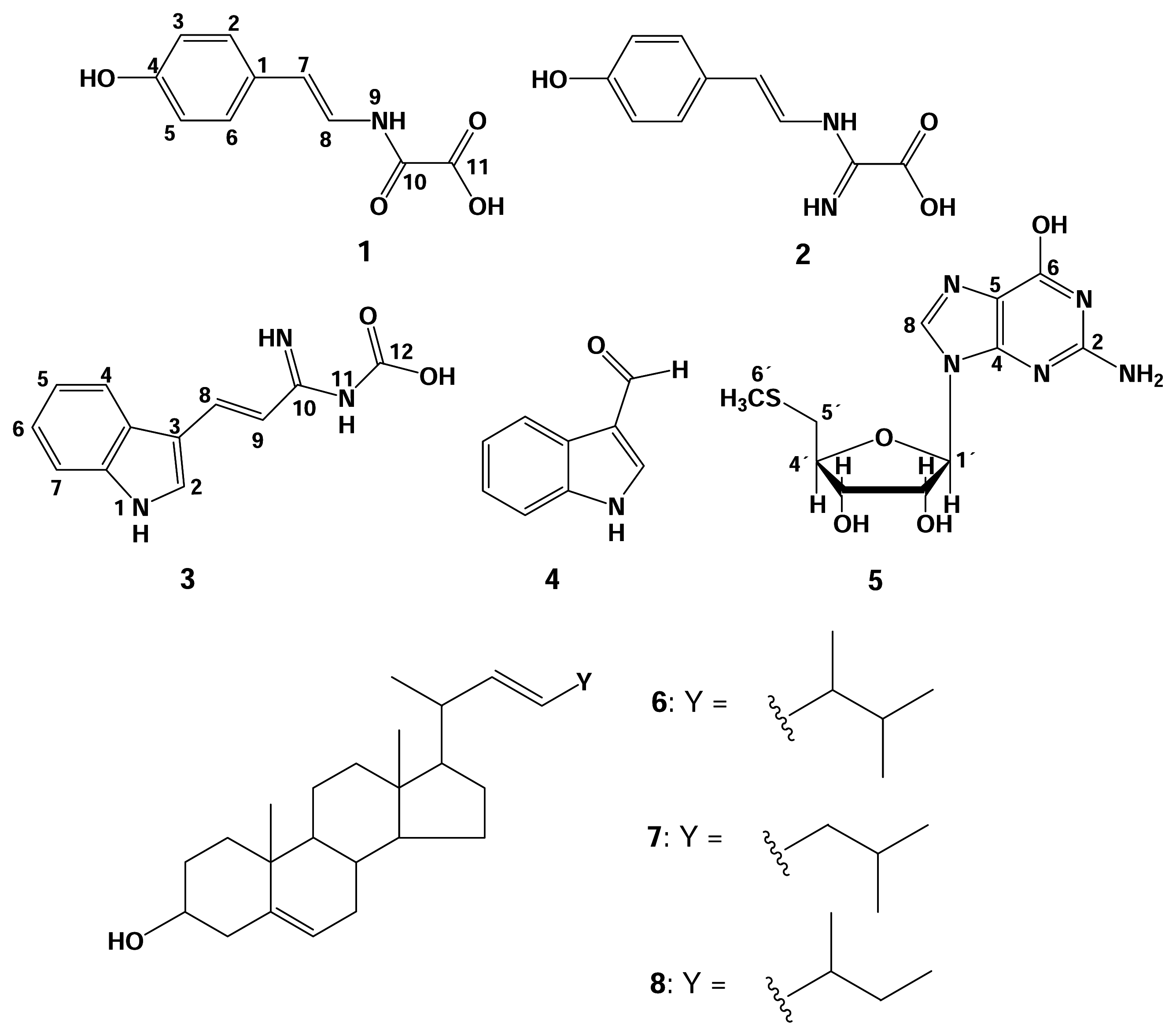
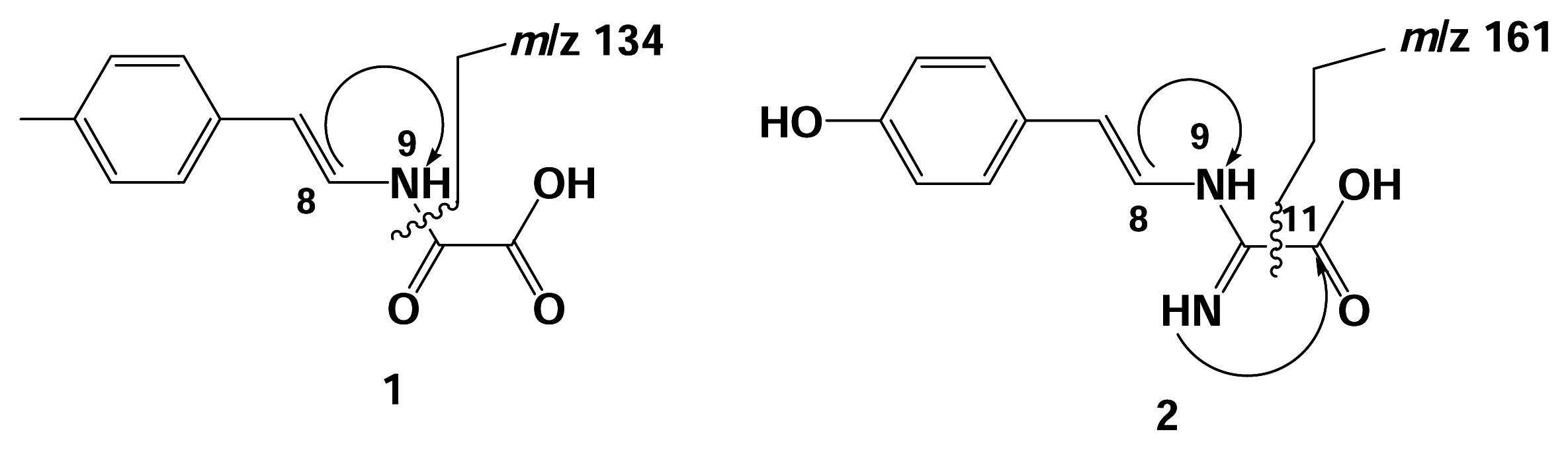
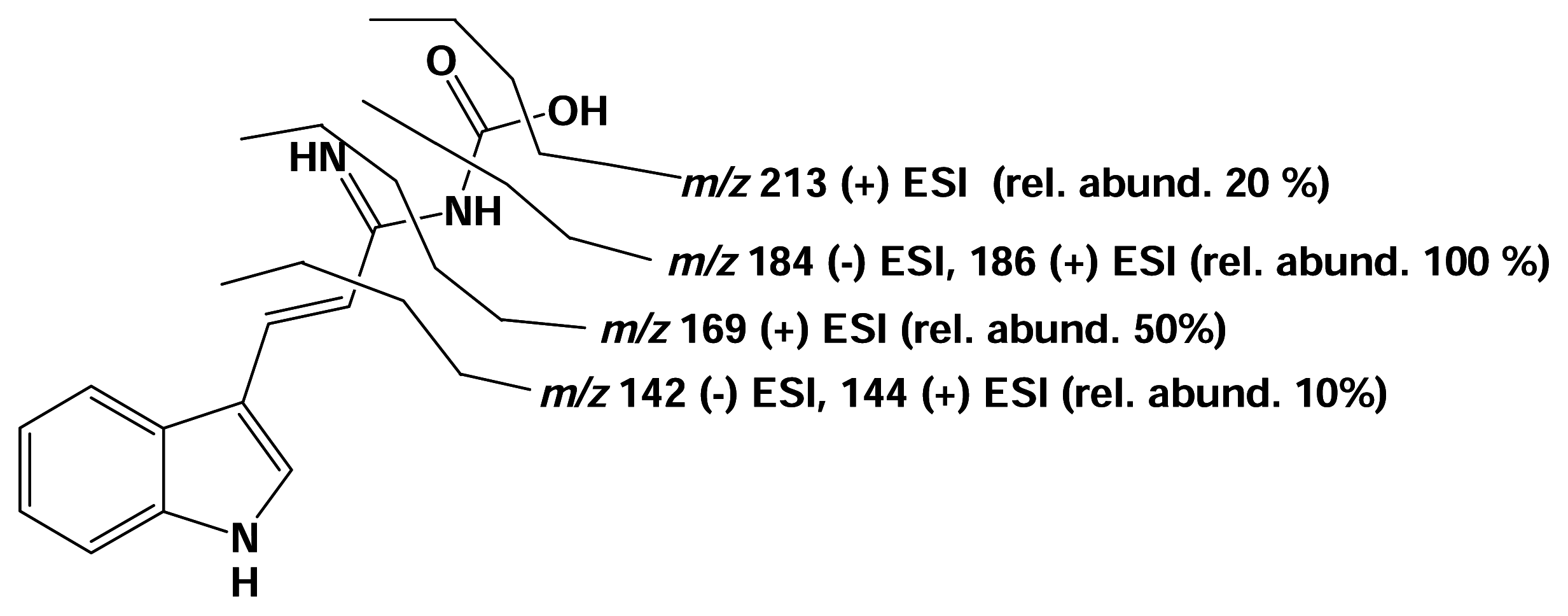
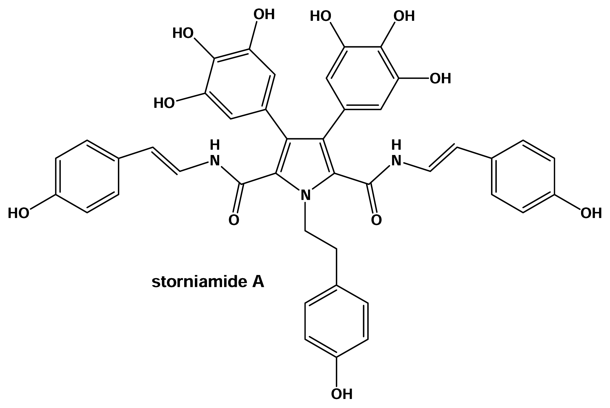
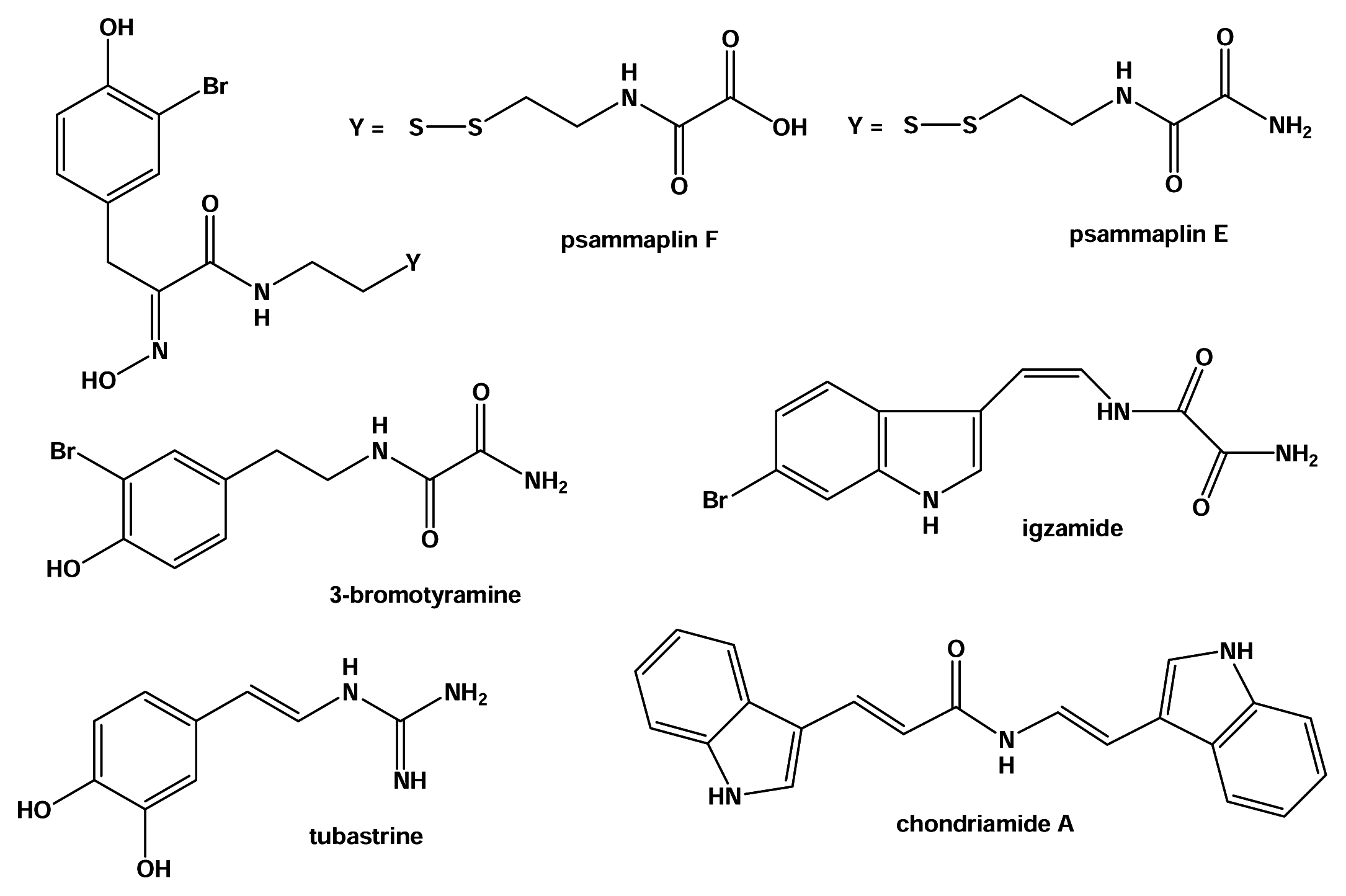
Results and Discussion

The conspicuous dark red colored sponge Hamigera hamigera was collected at a depth of 5 to 7 m off the island of Elba (Mediterranean Sea). The biologically active EtOAc and aqueous extracts were subjected to a series of chromatographic separation steps which yielded four new compounds 1, 2, 3, and 5, while four known compounds (4, 6, 7, and 8) were also isolated. The structures of the known compounds, indole-3-carboxaldehyde (4) [9, 10], 22-dehydrocampasterol (6), 27-nor-22-dehydrocampasterol (7), and 24-methylcholesta-5,22-dien-3β-ol (8) [11, 12, 13] were identified from comparison of their 1H-and 13C-NMR data with those found in literature. The structures of the new compounds were identified unambiguously by 1D and 2D NMR spectroscopy. Through-bond homonuclear (1H-1H COSY) and heteronuclear (long range 13C-1H) correlations were used to establish assignments and atom connectivities. Chemical shifts were compared with literature data for compounds containing similar structural subunits.
Hamigeroxalamic acid (1) was isolated as a pale yellowish white amorphous powder. Positive and negative ion ESIMS established the molecular weight of the compound which is compatible with a molecular formula of C10H9NO4. The 1H-NMR spectra (see Table 1) revealed an AA′BB′ system for 2H doublets at δ 7.09 and 6.67 which indicated a para-disubstituted benzene ring. The broad proton signal at δ 9.31 (OH) suggested 1 was phenolic as it gave a 2J correlation with the 13C NMR signal at δ 156.4 and 3J correlations to the C-3 and C-5 at δ 116.5 of the AA′BB′ system. The presence of a 1,2-disubstituted double bond adjacent to the NH group or trans-enamide substructure were observed as implied by the coupling constants and respective proton signals at δ 6.35 (H-7, d, J = 14.5 Hz) and δ 7.10 (H-8, dd, J = 14.5 and 10.7 Hz) which coupled further with the broad NH doublet proton at δ 10.07 (d, J = 10.7 Hz) [14, 15]. A similar substructure was found in storniamides [14], peptide alkaloids isolated from a Patagonian sponge Cliona sp. The carbon signals of the olefins at δ 114.0 and 122.1 were also compatible to those found in the latter compounds (ca. δ 114 and 120). From the 13C-NMR spectrum of hamigeroxalamic acid, signals at δC 163.0 and 158.3 were observed indicating the presence of two carbonyl carbons which were comparable to those of the terminal oxalamic (NHCOCOOH) functionality in psammaplin F with chemical shifts of 161.9 and 159.2 ppm [17]. The ESI-MS/MS performed on the negative pseudo molecular ion m/z at 206 [M–H] gave a daughter ion at m/z 134.2 indicating the loss of an oxoacetic acid [COCOOH] as a result of fragmentation of the amide linkage of the oxalamic acid functionality. A similar fragmentation pattern was also observed with the storniamides [14]. This confirms that 1 is N-[2-(4-hydroxy-phenyl)-vinyl]-oxalamic acid which was assigned the trivial name hamigeroxalamic acid.
Marinedrugs 02 00088f4
Hamigeramine (2) was isolated as a yellowish white powder. The ESIMS of 2 showed a peak at m/z 207 [M+H]+ in accordance with the molecular formula C10H10N2O3. 1H-and 13C NMR data were comparable to those of hamigeroxalamic acid which also revealed the presence of a 4-hydroxystyryl unit, as found in 1. Its 1H NMR spectrum also showed both the AA′BB′ spin system and trans vinyl amine pattern of resonances. The signals for the NH at δ 10.97 and the corresponding olefinic proton at δ 7.29 appeared very broad such that the expected doublet for a trans vinyl amine coupling was not resolved. However, the COSY spectrum confirmed the presence of the trans vinyl amine function through the correlation observed between δH 10.97 and δH 7.29. The 13C NMR spectrum of hamigeramine (2) was very similar to that of hamigeroxalamic acid (1) except for a significant change in chemical shifts for the carbonyl functionalities (δ 155.7 and δ 155.1) which were to higher field, while the 1H signals of the trans olefinic protons were to lower field. There were also additional broad singlets at δ 9.29 and δ 9.10 that were first thought to indicate the presence of a terminal NH2 as in oxalamide. However, significant discrepancies were observed for the carbonyl signals in the 13C NMR spectrum of 2 when compared to those of psammaplin E [17] (in CD3OD), 3-bromotyramine amide [18] (in CD3OD), and igzamide [19] (in DMSO), all of which possess an oxalamide terminal functional group. The carbonyl shifts of the oxalamide unit for these known compounds were reported at ca. 162 and 165 ppm in CD3OD while in DMSO, the chemical shifts were at ca. 161 and 157 ppm. The carbon chemical shifts at ca. 155 could also suggest the presence of an imine group as found in tubastrine [16, 20, 21]. The 1H and 13C chemical shifts (δH 6.81 and 7.29, δC 121.3 and 119.5) of the olefinic protons of hamigeramine (2) were comparable to those of tubastrine (ca. δH 6.70 and 7.20, δC 120 and 119). The amino imino acetic functionality in hamigeramine (2) was detected through the HMBC correlations of the olefinic H-8 (δ 7.29) with C-10 (δ 155.1) and of the imino NH (δ 9.29) with C-11 (δ 155.7).
ESI-MS/MS fragmentation of the negative pseudo molecular ion [M–H] at m/z 205 gave a daughter ion at m/z 161 instead of at m/z 134 as in hamigeroxalamic acid (1). This indicated the loss of only a COOH unit and absence of the amide linkage found in 1, again implying the presence of a different terminal functionality for hamigeramine (2), and confirmed that hamigeramine 2 was [2-(4-hydroxy-phenyl)-vinylamino]-imino-acetic acid.
Marinedrugs 02 00088f5
Hamigeramide (3) was obtained as a yellowish white powder from the biologically active EtOAc extract. It has the molecular formula of C12H11N3O2 as suggested by EIMS. Its 1H NMR spectrum (Table 2) was very similar to those of 3-indoleacrylamide and its acid congener [22] except for the difference in signals observed for the 1,2-trans-disubstituted olefinic protons. The 13C data for 3 were obtained from a HMBC spectrum and significant differences compared to the known compounds were observed for the olefinic carbons. The 1H signals of the olefinic system of indoleacrylamide and its acid congener were observed at ca. 6.80 and 8.00 ppm while their carbon shifts were detected at ca. 115 and 138 ppm. The same chemical shifts were also observed for the bis-indole alkaloids, chondriamides, isolated from the red alga Chondria atropurpurea [10, 23]. The functionality common to both the chondriamides and indoleacrylamide is the presence of a carbonyl moiety adjacent to the trans olefinic bond. The 1H signals of the olefinic system of hamigeramide (3) were observed at 7.18 and 7.10 ppm while their carbon shifts were detected at 118.2 and 117.3 ppm which suggested that 3 had a unique terminal functionality. Since the 1H and 13C chemical shifts of the olefinic protons were comparable to those of hamigeramine (2), this could suggest the presence of an adjacent imino amino moiety. Due to the very small amount of hamigeramide isolated, it was not possible to obtain a 13C NMR spectrum to prove the presence of the imino group. The HMBC spectra also did not show any discernible correlation between any of the olefinic protons to the expected imino carbon chemical shift at ca. 155 ppm. Ion trap ESI MS/MS showed the presence of the imino amino moiety and its connectivity were confirmed (Figure 3). ESIMS fragmentation positive mode of the [M+H]+ ion at m/z 230.21 gave a major daughter ion peak at m/z 186.1 [M–CO2H]+ which indicated the loss of the terminal acid unit, followed by subsequent loss of an NH3 unit (m/z 169.3) and C=NH (m/z 144.1). This was confirmed by the (−) ESIMS fragmentation of the [M–H] ion at m/z 228.62 which gave a major daughter ion peak at m/z 184.1 [M–CO2H]. The daughter ion peak at m/z 169.3 in the (+) ESIMS fragmentation also indicated the absence of an enamide unit as in hamigeroxalmic acid (1) and hamigeramine (2) which further suggested that the imino unit was adjacent to the 1,2-trans disubstituted double bond. This accounted for the relatively sharp doublet of both the olefinic protons in the 1H NMR spectrum of hamigeramide (3) compared to hamigeramine (2). The fragmentation pattern suggested an imino carbamic acid terminal moiety and thus hamigeramide was elucidated as 1-imino-3-(1H-indol-3-yl)-allyl]-carbamic acid.
Hamiguanosinol (5) was isolated as a pale yellowish white amorphous solid from the biologically active aqueous phase. The ESI-MS showed the molecular ion peak at m/z 314 [M +H]+, corresponding to the molecular formula C11H15N5O4 S, as confirmed by HREIMS. The 1H-and 13C NMR spectra of hamiguanosinol (5) were similar to those of 6-hydroxy guanosine [24] The 1H-NMR signals at δ 5.60, δ 4.50, 3.95, 4.05, 2.80, and 2.70, together with the 13C-NMR signals at δ 87.0, 84.0, 74.0, 71.5 and 35.0, indicated the presence of a β–ribosyl moiety. The remaining 1H-and 13C NMR signals at δH 10.57 (1H, s), 7.90 (1H, s) and 6.40 (2H, br s) and δC at 135.0 (d), 118.0 (d), 157.2 (s) and 153.5 (s), respectively were assigned to the 2-amino purine nucleus. The occurrence of the β–ribosyl moiety was confirmed by the HMBC spectrum which showed correlations between exchangeable hydroxyl protons that resonated at δH 5.40 and 5.20 with δC-3′ 71.5/δC-1′ 87.0 and δC-2′ 74.0/δC-4′ 84.0, respectively. The only difference from known nucleoside, 6-hydroxy guanosine, was the presence of a methyl singlet at δH 2.05 which showed a direct correlation with the quaternary carbon at δC 16.0 suggesting the presence of a SCH3 group. This was confirmed from the ESI-MS/MS experiment which showed daughter ions at m/z 163 for the methylthioribosyl moiety while a subsequent fragment ion at m/z 97 indicated the presence of the thiomethyl (SCH3) moiety. For the guanine nucleus, the presence of a hydroxyl group at C-6 instead of a keto function was confirmed by signals at δH 10.57 and δC 157.2 instead of a 13C signal at ca.170 ppm for a keto amide function. The hydroxyl substituent at C-6 was also evident from the four bond HMBC correlations of δH 7.90 (H-8) with δC 157.2 (C-6). The attachment of the ribosyl moiety with the purine unit at N-9 was confirmed by HMBC correlations of the anomeric proton (H-1′) with C-4 and C-8. Hamiguanosinol (5) was then concluded to be 5′-deoxy-5′-methylthio-6-hydroxy-guanosine. The fish deterrent activity of the aqueous phase was accounted for by the presence of hamiguanosinol. Nucleoside compounds are known for their antiviral activity and the first notable discovery of biologically active marine metabolites was actually unusual nucleosides. They were first isolated from a Caribbean sponge in the 1950s, and later served as lead structures for the development of the important anti-viral drugs such as Ara-A known commercially as acyclovir [25].
The fish deterrent activity of the isolated compounds was not quantified due to the very small amounts isolated. However, the biological activity of related compounds should be mentioned. Hamigeroxalamic acid (1) is among the few examples of natural products to date that carry the oxalamic functionality. Another important natural product having the same oxalamic moiety is psammaplin F which was found to selectively inhibit histone deacetylase, which is an epigenic modifier in the silencing of tumour suppression genes. It is also important to note that among the 10 known psammaplin derivatives, only psammaplin F, the oxalamic congener, was found to inhibit HDAC. Hamigeramide (3) is basically an indole alkaloid. Related compounds are the bis-indole alkaloids chondramides that were reported to exhibit anthelminthic and antiviral activity against HSV II [10].

Material and Methods

General experimental procedures

Optical rotation data were recorded on a Perkin-Elmer Model 341 LC polarimeter. 1D and 2D NMR spectra (chemical shifts in ppm) were recorded on Bruker AMX 300, ARX 400, or DRX 500 NMR spectrometers using standard Bruker software. Mass spectra (ESIMS) were recorded on a ThermoFinnigan Deca LCQ ion trap mass spectrometer. High resolution EIMS were recorded on a Finnigan MAT 8430 and on an Intectra AMD 402. For HPLC analysis, samples were injected into a HPLC system equipped with a photodiode-array detector (Gynkotek, München, Germany). Routine detection was at 254, 280, and 340 nm. The separation column (125 × 4 mm, i.d.) was prefilled with Eurospher 100-C18, 5 μm (Knauer, Berlin, Germany). Separation was achieved by applying a linear gradient from 100% H2O (with 0.2% TFA) to 100% MeOH over 40 min. Compounds were purified by semipreparative HPLC which was conducted on a Merck Hitachi LaChrome L-7100 pump and Merck Hitachi LaChrome L-7400 UV detector. Chromatograms were recorded on a Merck Hitachi D-2000 Chromato-Integrator. Separation columns (300 × 8 mm, i.d.) were prefilled with Eurospher 100-C18, 7 μm (Knauer, Berlin, Germany). TLC was performed on precoated TLC plates with Si gel 60 F254 and Si gel RP-18 F254 (Merck, Darmstadt, Germany). Compounds were detected by UV absorbance or by fluorescence at 254 and 366 nm. Solvents were distilled prior to use and spectral grade solvents were used for spectroscopic measurements.

Animal Material

The sponge Hamigera hamigera was collected in April 2000 from the Mediterranean Sea near Elba, Italy, at a depth of 5 to 7 m. The sponge was frozen immediately at −20º until work up. The taxonomic identification was carried out by Dr. R. Van Soest (Instituut voor Systematiek en populatie Biologie, Zoological Museum, Amsterdam). A voucher specimen (ZMA.POR 14397) was deposited at the Department of Coelenterates and Porifera, Zoologisch Museum, University of Amsterdam.

Extraction and isolation

The freeze-dried sponge was extracted with CH2Cl2 and subsequently with MeOH, the resulting total crude extract obtained with both solvents was evaporated to dryness in vacuo. The total crude extract (5.3 g) was partitioned between H2O and cyclohexane, then H2O and EtOAc and finally between H2O and n-BuOH. The EtOAc and H2O extracts were found to be active in the fish-feeding assay. The biologically active extracts were chromatographed separately over a Sephadex LH-20 column using MeOH as an eluent. Each fraction was collected to a volume of 5 mL and monitored by TLC on a pre-coated silica gel plates (Merck, Darmstadt, Germany) which were developed under the appropriate solvent system. The pooled non-polar fractions from the EtOAc extract were subjected to normal-phased silica gel column chromatography which was eluted with hexane:EtOAc (90:10) resulting in the isolation of steroids 6 (3.1 mg), 7 (2.1 mg), and 8 (1.8 mg) while the polar fraction was chromatographed over a reversed-phase C18 column and eluted with MeOH:H2O (1:1) which yielded the new compound 2. The polar fractions obtained from the EtOAc extract afforded the isolation of new compounds 1 (7.5 mg), 2 (2.5 mg), 3 (3.5 mg), and the known compound indole-3-carbaldehyde (4, 2.0 mg), after the extract was chromatographed over a reversed-phase C18 column which was also eluted with MeOH:H2O (1:1). The compounds were purified by semi-preparative reversed phase HPLC using a gradient of H2O and MeOH. The aqueous extract was subjected to Sephadex LH-20 column chromatography using MeOH as eluent and yielded the new nucleoside (5, 2.3 mg) which was also further purified by semi-preparative reversed phase HPLC using a gradient of H2O and MeOH.

Spectroscopic Data

Hamigeroxalamic acid (N-[2-(4-hydroxy-phenyl)-vinyl]-oxalamic acid) (1): yellowish white powder; UV λmax (MeOH) nm 248.5, 320.1; 1H NMR and 13C NMR (DMSO-d6): Table 1; (+) ESIMS (ion trap) m/z 246.2 [M+K]+; (−) ESIMS m/z 206.2 [M–H], 252.0 [M+HCOOH–H], 413.1 [2M–H]., The results of the MS/MS fragmentation of the (+) and (−) ESIMS (ion trap) are shown in Figure 2.
Hamigeramine ([2-(4-Hydroxy-phenyl)-vinylamino]-imino-acetic acid) (2): yellowish white powder; UV λmax (MeOH) nm 226.7, 324.0; 1H NMR and 13C NMR (DMSO-d6): Table 1; (+) FABMS m/z 207.1 [M+H]+, 229 [M+Na]+; (+) ESIMS (ion trap) m/z 207.1 [M+H]+, 412.9 [2M+H]+; (−) ESIMS (ion trap) 205.4 [M–H], 411.5 [2M–H], 617.1 [3M–H]. The results of the MS/MS fragmentation of the (+) and (−) ESIMS (ion trap) are shown in Figure 2.
Hamigeramide (3): yellowish white powder; UV λmax (MeOH) nm 228.0, 346.0; 1H NMR and 13C NMR (DMSO-d6): Table 2; (+) ESIMS (ion trap) m/z 230.2 [M+H]+, 252.1, 459.0 [2M+H]+; (−) ESIMS (ion trap) 228.6 [M–H], 457.6 [2M–H], 686.3 [3M-H]. The results of the MS/MS fragmentation of the (+) and (−) ESIMS (ion trap) are shown in Figure 3.
Hamiguanosinol (5): pale yellowish white amorphous powder, [α]D20 +15.8° (MeOH, c 0.10); UV λmax (MeOH) nm 254.6, 275.0 (sh); 1H NMR and 13C NMR (DMSO-d6): Table 3; (+)ESIMS m/z 314.5 [M+H]+, 627.3 [2M+H]+; (−)ESIMS m/z 312.1[M-H],625.8 [2M–H], 938.9 [3M–H]; EIMS (70eV) m/z [M]+ 313 (13), 236 (12), 163 (12), 151 (16), 138 (44), 121 (93), 84 (100); HRESIMS m/z 312.0763 [M-H](calcd for C11H14N5O4S, 312.0767).

Feeding experiments with Blennius sphinx

For the feeding experiments, crude sponge extracts and/or fractions were incorporated into the artificial fish diet. The artificial fish diet was treated with physiological concentrations of secondary sponge metabolites as determined in 10 mL ethanol preserved sponge material to match the natural concentrations of the metabolites found in the sponge. All metabolites used for the feeding experiments were dissolved in MeOH and incorporated into 1.053 g of ground commercial fish food granules (TetraMarin Granulat, TetraWerke, Melle, Germany). This mixture was then added to 9 mL of 2% agar at 48° C to give a volume of 10 mL. Control diet was likewise prepared by mixing the food granules in MeOH without the sponge extracts. Freshly prepared treated diet and control diet were then separately poured into rectangular molds (2 × 25 × 250 mm) placed on a meshed screen. The food diet was allowed to set on the meshed screen which were then cut to six equivalent food strips. Each of the food strips contained a treated diet and a control diet, each covering an area of 10 × 15 meshed screen squares.
The feeding experiments were them performed in an aquarium (size: 170 × 80 × 40 cm) using 70 individuals of the polyphagous Mediterranean fish Blennius sphinx. The food strips were placed at the bottom of the aquarium. The food strips were checked at regular time intervals and were removed when approximately one-third of the total food mass was consumed. To determine the amount of control and treated food eaten, the number of empty squares in the meshed screen were counted [25, 27]. The results obtained from several replicate experiments were pooled and analyzed with a paired t-test.
Figure 1. Compounds isolated from the fish feeding deterrent extracts of the Mediterrenean sponge Hamigera hamigera.
Marinedrugs 02 00088f1
Figure 2. ESI-MS/MS fragmentation of hamigeroxalamic acid and hamigeramine in the negative mode. The vinyl amine function in both compounds was confirmed by the COSY correlation between H-8 and NH-9 while the amino imino acetic functionality in hamigeramine was detected through the HMBC correlation between the imino NH and C-11.
Marinedrugs 02 00088f2
Figure 3. MS/MS fragmentation of the pseudomolecular ion peak at m/z 230.21 [M+H]+ and m/z 228.62 [M-H]of hamigeramide (3).
Marinedrugs 02 00088f3
Table 1. 1H, 13C-NMR, and HMBC data of compounds 1 and 2 in DMSO-d6.
12

NoδH (m, J in Hz)δCHMBC (δH to δC)δH (m, J in Hz)δCHMBC (δH to δC)
1128.2 s125.9 s
27.09 (d, 8.5)127.9 dC-1, C-4, C-77.25 (d, 8.4)127.5 dC-1, C-6, C-7
36.67 (d, 8.5)116.5 dC-1, C-4, C-56.75 (d, 8.4)115.6 dC-1, C-4, C-5
49.31 (OH, br s)156.4 sC-3, C-4, C-59.62 (OH, br s)157.3 sC-3, C-4, C-5
56.67 (d, 8.5)116.5 dC-1, C-3, C-46.75 (d, 8.4)115.6 dC-1, C-3, C-4
67.09 (d, 8.5)127.9 dC-1, C-4, C-77.25 (d, 8.4)127.5 dC-1, C-2, C-7
76.35 (d,14.5)114.0 dC-2, C-6, C-86.81 (d, 13.9)121.3 dC-2, C-6, C-8
87.10 (dd, 14.5, 10.7)122.1 dC-1, C-77.29 (br d, 13.9)119.5 dC-7, C-9, C-10
910.07 (NH, d, 10.7)10.97 (NH, br s)
10163.0 s9.29 (NH, br s)a155.1 sC-11
11158.3 s9.10 (OH, br s)a155.7 s
aThese assignments are interchangeable but the signal at 9.29 ppm shows a correlation to C-11.
Table 2. 1H, 13C-NMR, and HMBC data of compound 3 in DMSO-d6.
No.δH (m, J in Hz)δCaHMBC (δH to δC)
111.20 (NH, br s)
27.50 (s)127.3 dC-3, C-3a, C-7a, C-8
3111.2 s
3a124.5 s
47.90 (d, 7.8)120.1 dC-6, C-3a, C-7a
57.08 (t, 7.8)119.0 dC-7, C-3a
67.15 (t, 7.8)121.5 dC-7a
77.40 (d, 7.8)112.3 dC-5, C-3a
7a138.4 s
87.10 (d,14.0)117.3 dC-2, C-3a
97.18 (d, 14.0)118.2 dC-3
10b
118.91 (NH, br s)
12bb
aCarbon assignments were determined indirectly from the HMBC.
bThere are two further very broad exchangeable signals at ca. 9.3 and 10.8 ppm. The carboxylic acid 13C signal was not detected.
Table 3. 1H, 13C-NMR, and HMBC data of compound 5 in DMSO-d6.
No.δH (m, J in Hz)δC1H-1H COSYHMBC (δH to δC)
2(NH2) 6.40 (br s)153.5 s
4153.0 s
5118.0 s
6(OH) 10.57 (br s)157.2 s
87.90 s135.0 dC-5, C-4, C-6
1′5.60 (d, 5.7)87.0 dH-2′C-8, C-4, C-3′
2′4.50 (q, 5.7)74.0 dH-1′, H-3′C-1′, C-4′
2′-OH5.40 (d, 6.3)C-1′, C-3′
3′3.95 (m)71.5 dH-2′, H-4′C-1′
3′-OH5.20 (d, 5.1)C-2′, C-4′
4′4.05 (t, 6.9)84.0 dH-3′, H-5′A, H-5′BC-3′
5′A2.80 (dd, 13.9, 6.9)35.0 tH-5′ B, H-4′C-6′, C-3′, C-4′
5′B2.70 (dd, 13.9, 6.9)
6′2.05 (s)16.0 qH-5′A, H-4′C-5′

Acknowledgments

W.H. would like to thank the government of Egypt for financial support. The authors are grateful to Dr. R. Van Soest for identification of the sponge and to C. Kakoschke and B. Jaschok-Kentner for recording NMR data (GBF, Braunschweig). Financial support by a grant of the BMBF (“BIOTECMARIN”) is gratefully acknowledged.
  • Sample Availability: Samples are available from the authors.

References and Notes

  1. Casapullo, A.; Finamore, E.; Minale, L.; Zollo, F. A dimeric peptide alkaloid of a completely new type, Anchinopeptolide A, from the marine sponge Anchinoe tenacior. Tetrahedron Lett 1993, 34, 6297–6300. [Google Scholar]
  2. Casapullo, A.; Minale, L.; Zollo, F. The unique 6-(p-hydroxyphenyl)-2-H-3,4-dihydro-1,1-dioxo-1,4-thiazine and the new tripeptide L-glu-gly-4-hydroxystirylamine from the marine sponge Anchinoe tenacior. Tetrahedron Lett 1994, 35, 2421–2422. [Google Scholar]
  3. Casapullo, A.; Minale, L.; Zollo, F. Four new dimeric peptide alkaloids, anchinopeptolides B-D, and cycloanchinopeptolide C, congeners of anchinopeptolide A, from the Mediterrenean marine sponge Anchinoe tenacior. J. Nat. Prod 1994, 57, 1227–1233. [Google Scholar]
  4. Rudi, A.; Stein, Z.; Green, S.; Goldberg, I.; Kashman, Y. Phorbazoles A-D, novel chlorinated phenylpyrrolyloxazoles from the marine sponge Phorbas aff. clathrata. Tetrahedron Lett 1994, 35, 2589–2492. [Google Scholar]
  5. Searle, A. S.; Molinski, T. F. Absolute configuration of Phorboxazole A and B from the marine sponge Phorbas sp. 1. Macrolide and hemiketal rings. J. Am. Chem. Soc 1996, 118, 9422–9423. [Google Scholar]
  6. Molinski, T. F. Absolute configuration of Phorboxazole A and B from the marine sponge Phorbas sp. 2. C-43 and complete stereochemistry. Tetrahedron Lett 1996, 37, 7879–7880. [Google Scholar]
  7. Wellington, K. D.; Cambie, R. C.; Rutledge, P. S.; Bergquist, R. Chemistry of sponges. 19. Novel bioactive metabolites from Hamigera tarangensis. J. Nat. Prod 2000, 63, 79–85. [Google Scholar]
  8. Cambie, R. C.; Lai, A. R.; Kernan, M. R.; Bergquist, P. R. Chemistry of sponges. A novel brominated benzocyclooctane derivative from Hamigera hamigera. J. Nat. Prod 1995, 58, 940–942. [Google Scholar]
  9. Guella, G.; Mancini, I.; Zibrowius, H.; Pietra, F. 160. Aplysinopsin-type alkaloids from Dendrophyllia sp., a Scleractinian coral of the family Dendrophylliidae of the Philippines. Facile photochemical (Z/E). Helv. Chim. Acta 1989, 72, 1444–1450. [Google Scholar]
  10. Anjaneyulu, V.; Rao, K. N. R.; Kobayashi, M. (24S)-24-Methylcholesta-4-ene-3β,6β-diol from a gorgonian (Rumphella aggregata) of the Andaman and Nicobar Islands. Ind. J. Chem 1995, 34B, 78–80. [Google Scholar]
  11. Wright, J.; McInnes, A.; Shmizu, S.; Smith, D.; Walter, J. Identification of C-24 alkylepimers of marine sterols by 13C nuclear magnetic resonance spectroscopy. Can. J. Chem 1978, 56, 1898–1903. [Google Scholar]
  12. Itoh, T.; Sica, D.; Djerassi, C. Minor and trace sterols in marine invertebrate. Part 1. 35. Isolation and structure elucidation of seventy four sterols from the sponge Axinella cannabina. J. Chem. Soc. Perkin Trans 1983, 1, 147–153. [Google Scholar]
  13. Palermo, J. A.; Brasco, M. F. R.; Seldes, A. M. Stroniamides A-D: Alkaloids from a Patagonian sponge Cliona sp. Tetrahedron 1996, 52, 2727–2734. [Google Scholar]
  14. Bokesch, H. R.; Pannell, L. K.; McKee, T. C.; Boyd, M. R. Coscinamides A, B, and C, three new indole alkaloids from the marine sponge Coscinoderma sp. Tetrahedron Lett 2000, 41, 6305–6308. [Google Scholar]
  15. Piña, I. C.; Gautschi, J. T.; Wang, G.-Y.-S.; Sanders, M. L.; Schmitz, F. J.; France, D.; Cornell-Kennon, S.; Sambucetti, L. C.; Remiszewski, S. W.; Perez, L. B.; Bair, K. W.; Crews, P. Psammaplins from the sponge Pseudoceratina purpurea: Inhibition of both histone deacetylase and DNA methyltransferase. J. Org. Chem 2003, 68, 3866–3873. [Google Scholar]
  16. Pettit, G. R.; Butler, M. S.; Williams, M. D.; Filiatrault, M. J.; Pettit, R. K. Isolation and structure of hemibastadinols 1–3 from the Papua New Guinea marine sponge Ianthella basta. J. Nat. Prod 1996, 59, 327–934. [Google Scholar]
  17. Dumdei, E.; Andersen, R. J. Igzamide, a metabolite of the marine sponge Plocamissma igzo. J. Nat. Prod 1993, 56, 792–794. [Google Scholar]
  18. Sakai, R.; Higa, T. Tubastrine, a new guanidostyrene from the coral Tubastrea aurea. Chem. Lett 1987, 122–128. [Google Scholar]
  19. Sperry, S.; Crews, P. Dihydrotubastrines: Phenethylguanidine analogues from the Indo-Pacific marine sponge Petrosia cf. contignata. J. Nat. Prod 1998, 61, 859–861. [Google Scholar]
  20. Davyt, D.; Entz, W.; Fernandez, R.; Mariezcurrena, R.; Mombrú, A. W.; Saldaña, J.; Domínguez, L.; Coll, J.; Manta, E. A new indole derivative from the red alga Chondria atropurpurea. Isolation, structure determination, and anthelmintic activity. J. Nat. Prod 1998, 61, 1560–1563. [Google Scholar]
  21. Pretsch, E.; Seibl, J.; Simon, W.; Clerc, T. Tabellen zur Strukturaufklärung organischer Verbindungen mit spektroskopischen Methoden; 1990; Heidelberg; Springer Verlag; p. C212. [Google Scholar]
  22. Balzarini, J.; Haller-Meier, F.; De Clercq, E.; Meier, C. Antiviral activity of cyclosaligenyl prodrugs of acyclovir, carbovir and abacavir. Antivir. Chem. Chemother 2001, 12, 301–306. [Google Scholar]
  23. Hay, M. E.; Stachowicz, J. J.; Cruz-Rivera, E.; Bullard, S.; Deal, M. S.; Linquist, N. Bioassays with marine and freshwater macroorganisms. Haynes, KF, Millar, J. G., Eds.; In Methods in Chemical Ecology; 1998; Volume 2, Bioassay methods; Chapman and Hall: New York; pp. 39–141. [Google Scholar]

