
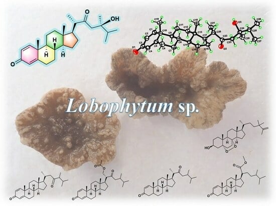
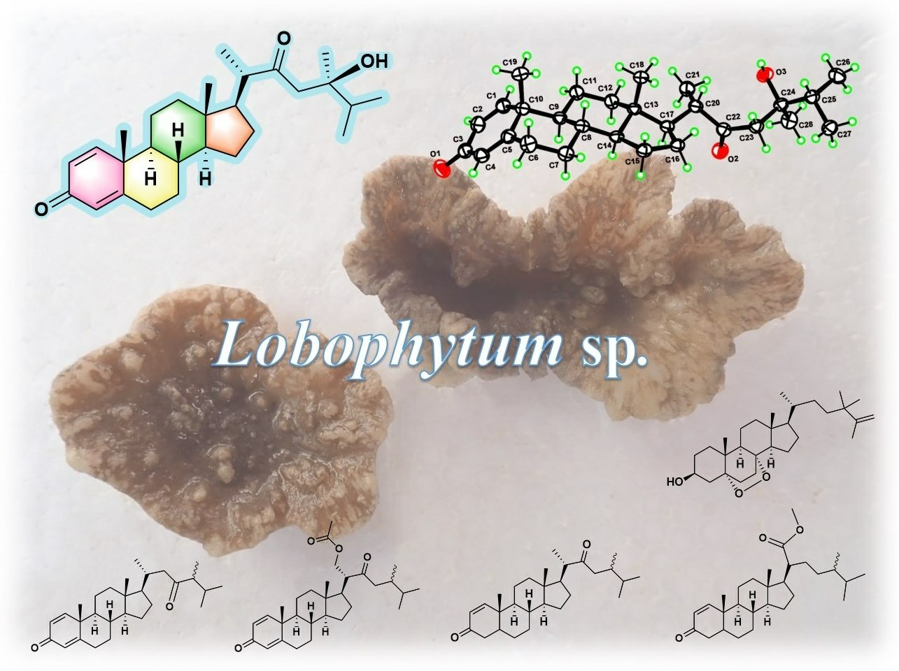
Lobosteroids A–F: Six New Highly Oxidized Steroids from the Chinese Soft Coral Lobophytum sp.
by
 , , and
Zi-Yi Xia
1,
Man-Man Sun
2,
Yang Jin
2,
Li-Gong Yao
2,
Ming-Zhi Su
2,
Lin-Fu Liang
4,*,
Hong Wang
1,* and
Yue-Wei Guo
1,2,3,* 1
Collaborative Innovation Center of Yangtze River Delta Region Green Pharmaceuticals and College of Pharmaceutical Science, Zhejiang University of Technology, Hangzhou 310014, China
2
Shandong Laboratory of Yantai Drug Discovery, Bohai Rim Advanced Research Institute for Drug Discovery, 198 Binhai East Road, High-Tech Zone, Yantai 264117, China
3
School of Medicine, Shanghai University, 99 Shangda Road, Bao Shan District, Shanghai 200444, China
4
College of Materials Science and Engineering, Central South University of Forestry and Technology, 498 South Shaoshan Road, Changsha 410004, China
*
Authors to whom correspondence should be addressed.
Mar. Drugs 2023, 21(8), 457; https://doi.org/10.3390/md21080457
Submission received: 26 July 2023
/
Revised: 17 August 2023
/
Accepted: 18 August 2023
/
Published: 19 August 2023
(This article belongs to the Section Structural Studies on Marine Natural Products)
Abstract
:To explore the steroidal constituents of the soft coral Lobophytum sp. at the coast of Xuwen County, Guangdong Province, China, a chemical investigation of the above-mentioned soft coral was carried out. After repeated column chromatography over silica gel, Sephadex LH-20, and reversed-phase HPLC, six new steroids, namely lobosteroids A–F (1–6), along with four known compounds 7–10, were obtained. Their structures were determined by extensive spectroscopic analysis and comparison with the spectral data reported in the literature. Among them, the absolute configuration of 1 was determined by X-ray diffraction analysis using Cu Kα radiation. These steroids were characterized by either the presence of an α,β-α′,β′-unsaturated carbonyl, or an α,β-unsaturated carbonyl moiety in ring A, or the existence of a 5α,8α-epidioxy system in ring B, as well as diverse oxidation of side chains. The antibacterial bioassays showed that all isolated steroids exhibited significant inhibitory activities against the fish pathogenic bacteria Streptococcus parauberis FP KSP28, Phoyobacterium damselae FP2244, and Streptococcus parauberis SPOF3K, with IC90 values ranging from 0.1 to 11.0 µM. Meanwhile, compounds 2 and 6–10 displayed potent inhibitory effects against the vancomycin-resistant Enterococcus faecium bacterium G7 with IC90 values ranging from 4.4 to 18.3 µM. Therefore, ten highly oxidized steroids with strong antibacterial activities were isolated from the Chinese soft coral Lobophytum sp., which could be developed as new chemotypes of antibacterial drug leads.
1. Introduction
Unlike terrestrial, the unique and complex marine environment creates rich chemodiversities and biodiversities of secondary metabolites in soft corals [1], such as the bioactive steroids with diverse structural features [2]. Structurally, these soft coral-derived steroids display an array of carbon frameworks, ranging from the usual cholestane [3], ergostane [4], and pregnane-type [5] sterols to the rare secosteroids [6] and highly degraded steroids [7]. Biologically, this group of secondary metabolites exhibit a wide spectrum of bioactivities, including antibacterial [8], anti-inflammatory [9], cytotoxic [10], immunosuppressive [11], and PTP1B inhibitory [6] activities. These properties make steroids attract the continuous attention of chemists and pharmacologists [12].
It is well known that soft corals of the genus Lobophytum are one group of the most important marine invertebrates widely distributed in waters. They produce a wealthy biochemical repository of secondary metabolites [13] ranging from terpenoids [14], steroids [15], prostaglandins [16], and amides [17] to quinones [18]. Among these chemical constituents, steroids are one major group of metabolites that were found in many species of the genus Lobophytum, including Lobophytum sarcophytoides [9,17], Lobophytum pauciflorum [15], Lobophytum michaelae [19], Lobophytum crassum [20,21], Lobophytum lobophytum [22], Lobophytum compactum [23], Lobophytum patulum [24], and Lobophytum spp. [10,25,26,27]. Notably, these metabolites display various biological activities, such as anticancer [10,15,25], anti-inflammatory [9,17,19], and 5α-reductase inhibitory [28] activities.
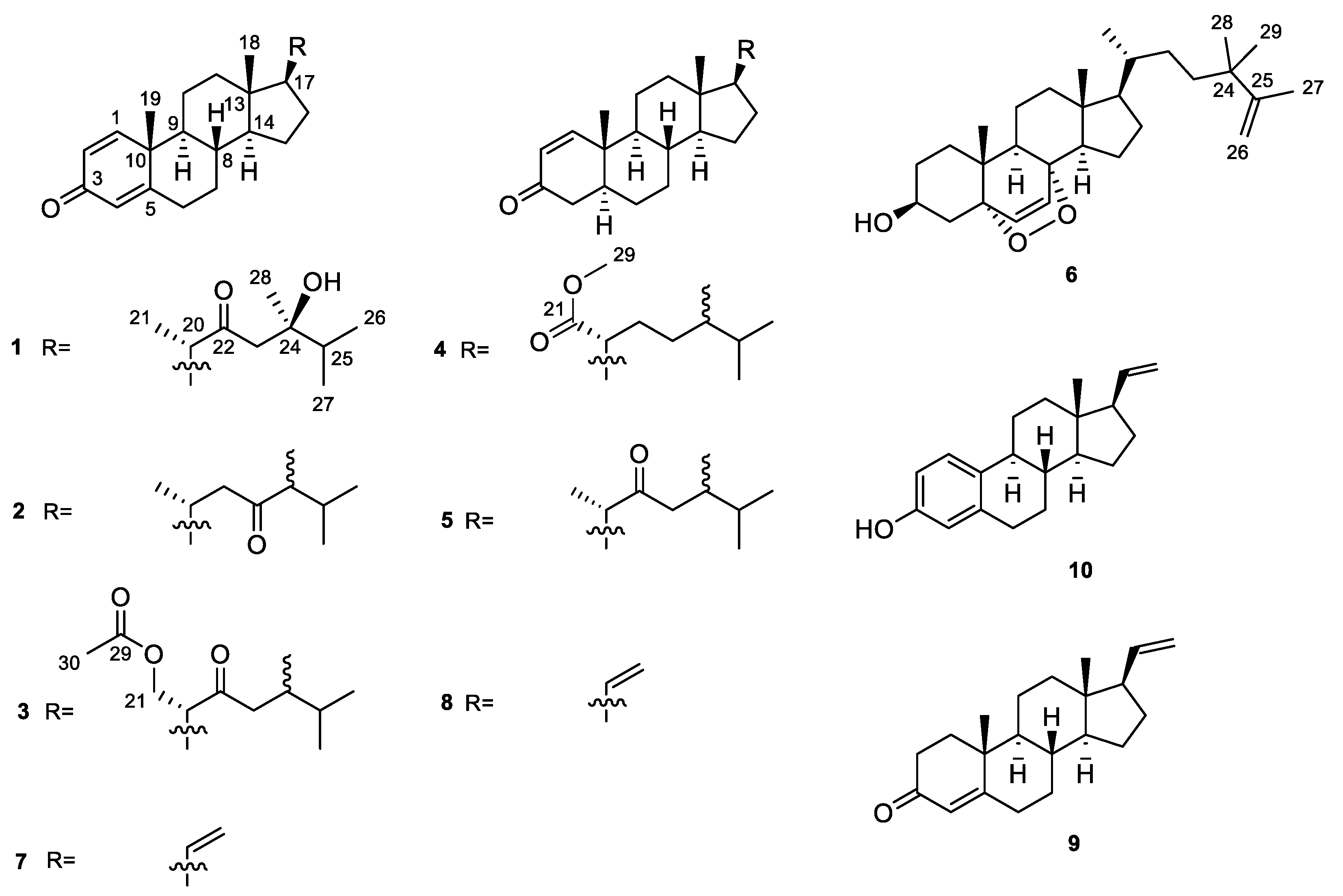
As part of our ongoing research aimed at discovering bioactive substances from marine invertebrates in China [29], we recently collected Lobophytum sp. at the coast of Xuwen County, Guangdong Province, China. In our recent study, we have reported the isolation and structural elucidation of anti-tumor cembrane diterpenoids from the Hainan specimens of Lobophytum sp. [30]. While our current investigation on the Guangdong collection of Lobophytum sp. has now resulted in the isolation of six new steroids, lobosteroids A–F (1–6), together with four known analogs 7–10 (Figure 1). The structural difference of six new steroids 1–6 is mainly attributed to the different degrees of oxidation in rings A and B of the steroidal nucleus and the variations of functional groups on the side chains. This paper describes the isolation, structural elucidation, and bioactivity of these compounds.
2. Results and Discussion
The frozen animals were cut into pieces and extracted with acetone exhaustively. The Et2O-soluble portion of the acetone extract was repeatedly column chromatographed over silica gel, Sephadex LH-20, and reversed-phase HPLC to yield ten pure steroids 1–10 (Figure 1). Four known steroids were readily identified as pregna-1,4,20-trien-3-one (7) [31], pregna-1,20-dien-3-one (8) [32], pregna-4,20-dien-3-one (9) [33], and 19-norpregna-1,3,5(10),20-tetraen-3-ol (10) [34], respectively, by comparison of their NMR data and optical rotation [α]D values with those reported in the literature.
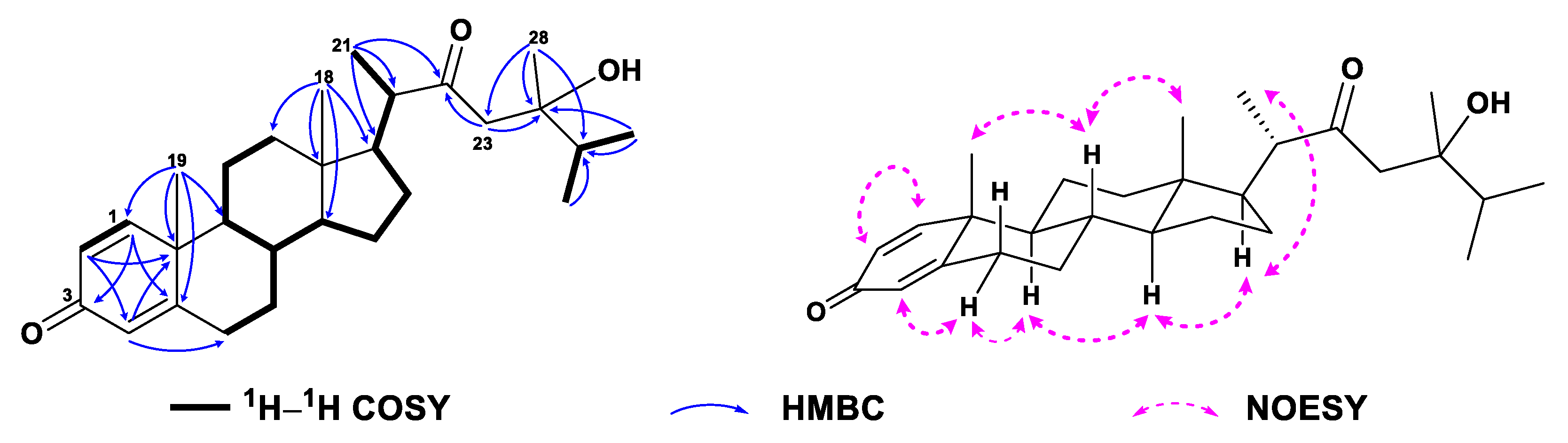
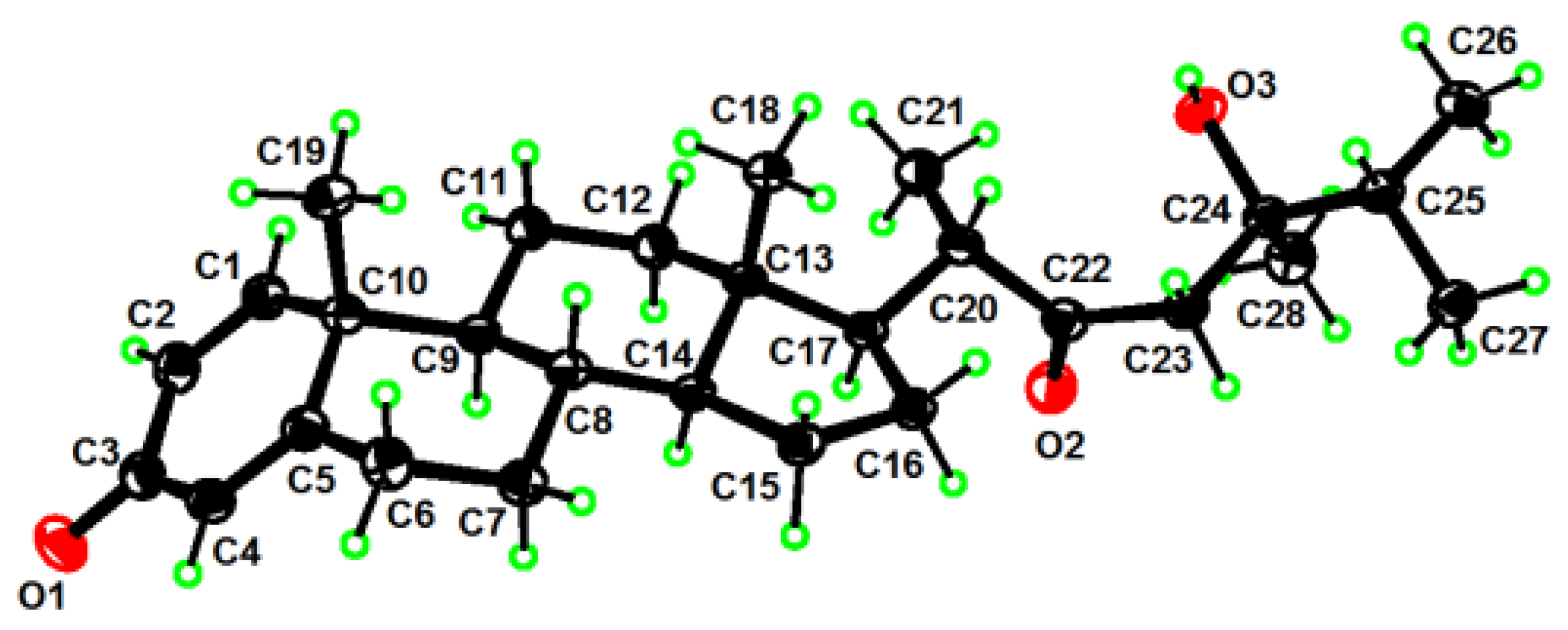
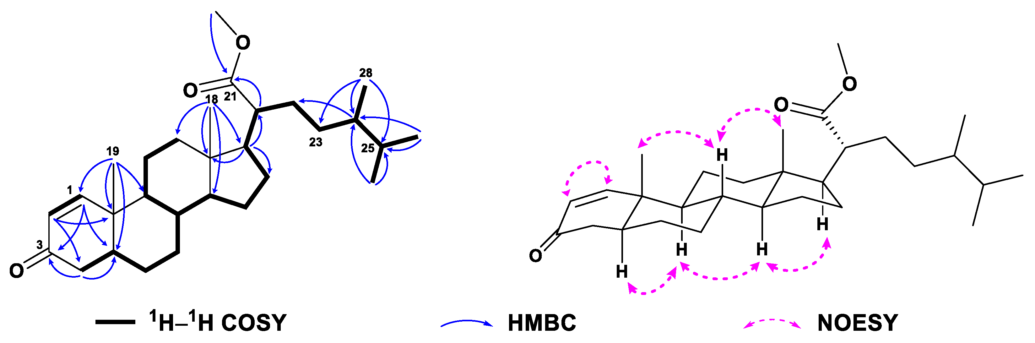
Compound 1, colorless crystals, had the molecular formula of C28H42O3 as established by HRESIMS (Figure S7) from the protonated molecular ion peak observed at m/z 427.3210 [M + H]+ (calcd. 427.3207), implying eight degrees of unsaturation. Extensive analysis of 13C NMR and DEPT spectra of 1 (Table 1, Figure S2) disclosed the presence of 28 carbons, consisting of six methyls, seven sp3 methylenes, six sp3 methines, three sp3 quaternary carbons (including one oxygenated at δC 74.0), three sp2 methines (δC 124.1, 127.7, and 155.8), and three sp2 quaternary carbons (including one olefinic at δC 169.1 and two carbonylic at δC 186.5 and 217.6). Thus, compound 1 still required a four-ring system to satisfy the remaining four degrees of unsaturation. Considering the co-isolated known metabolites, compound 1 was likely a steroid whose basic nucleus was a fused four-ring carbon framework. Overall, the gross 1H and 13C spectral data of 1 (Table 1 and Table 2, Figures S1 and S2) were reminiscent of 24-methylenecholesta-1,4,24(28)-trien-3-one, a sterol previously reported from the soft coral Dendronephthya studeri [35]. Careful comparison of their NMR data revealed they possessed the same steroidal nucleus possessing an α,β-α′,β′-unsaturated carbonyl moiety, which was straightforward from NMR signals at δH 7.05 (d, J = 10.2 Hz, H-1), 6.23 (dd, J = 10.2, 2.0 Hz, H-2), 6.07 (br s, H-4), and δC 155.8 (d, C-1), 127.7 (d, C-2), 186.5 (s, C-3), 124.1 (d, C-4), 169.1 (s, C-5) (Figure S3). The establishment of ring A was further confirmed by the key HMBC correlations from H3-19 (δH 1.10) to C-1 (δC 155.8), C-5 (δC 169.1), C-9 (δC 52.3), and C-10 (δC 43.6), from H-1 (δH 7.05) to C-3 (δC 186.5) and C-5, from H-2 (δH 6.23) to C-4 (δC 124.1) and C-10, and from H-4 (δH 6.07) to C-6 (δC 33.0) and C-10 (Figure 2 and Figure S4). However, they differed in the structures of their side chains. First, the methylene C-22 in 24-methylenecholesta-1,4,24(28)-trien-3-one was oxidized into a ketone in 1, which was characterized by the remarkably down-field chemical shift of δC 217.6 (s, C-22). Secondly, the terminal double bond Δ24(28) in 24-methylenecholesta-1,4,24(28)-trien-3-one was reduced in 1, accompanied with the hydroxylation at C-24, which was indicated by the NMR signals at δH 1.12 (s, H3-28) and δC 23.0 (q, C-28), 74.0 (s, C-24). Furthermore, the structure of the side chain in 1 was verified clearly by the HMBC correlations from H3-21 (δH 1.10) to C-17 (δC 52.0), C-20 (δC 50.9) and C-22 (δC 217.6), from H2-23 (δH 2.53, 2.66) to C-22 and C-24 (δC 74.0), and from H3-28 to C-23 (δC 48.2), C-24 and C-25 (δC 37.4) (Figure 2 and Figure S4). Herein, the planar structure of 1 was determined as depicted in Figure 1. The observed NOE correlations regarding the chiral centers C-8, C-9, C-10, C-13, C-14, C-17, and C-20, and the double bonds Δ1 and Δ4 of 1 (Figure 2 and Figure S6) were similar to those of 24-methylenecholesta-1,4,24(28)-trien-3-one, suggesting they shared the same relative configurations for these stereocenters and double bonds. However, there were insufficient NOE correlations to assign the relative configuration of C-24. Luckily, suitable single crystals of 1 in MeOH were obtained. The X-ray crystallographic analysis using Cu Kα radiation (λ = 1.54178 Å) firmly disclosed the absolute configuration of 1 was 8S,9S,10R,13S,14S,17R,20S,24R (Flack parameter: 0.09 (8), Figure 3).
Compound 2 was obtained as a white amorphous powder and it displayed a protonated molecular ion peak at m/z 411.3255 ([M + H]+; calcd. 411.3258) in the HRESIMS spectrum (Figure S15), consistent with a molecular formula of C28H42O2. Inspection of the NMR data of compound 2 (Table 1 and Table 2, Figures S9 and S10) revealed its spectroscopic features were closely similar to those of 1, suggesting that they possessed the same steroidal nucleus with an α,β-α′,β′-unsaturated carbonyl moiety [δH 7.05 (d, J = 10.1 Hz, H-1), 6.22 (dd, J = 10.1, 1.9 Hz, H-2), 6.06 (br s, H-4), and δC 156.1 (d, C-1), 127.6 (d, C-2), 186.6 (s, C-3), 123.9 (d, C-4), 169.5 (s, C-5)]. In fact, the differences between 2 and 1 were in the structures of their side chains. The carbonyl group shifted from C-22 in 1 to C-23 (δC 215.0) in 2, and the hydroxyl group attached to C-24 (δC 74.0 vs. δC 53.0) in 1 was lost in 2, which was consistent with their 16 mass units difference. The characteristic 1H–1H COSY correlations from H-17 (δH 1.13) through H-20 (δH 2.04) to H2-22 (δH 2.20, 2.44) and from H3-28 (δH 0.98) through H-24 (δH 2.29) and H-25 (δH 1.92) to H3-26 (δH 0.84)/H3-27 (δH 0.90), together with the diagnostic HMBC correlations from H2-22 to C-20 (δC 32.0) and C-23, from H3-28 to C-23, C-24, and C-25 (δC 30.2) (Figure 4, Figures S12 and S13), supported the above-mentioned structure of the side chain. The literature surveys revealed that the NMR data of the side chain of 2 were almost identical to those of the synthetic steroid 3β-hydroxyergost-5,7-diene-23-one [36], further confirming the established structure of the side chain. Due to the isomerization of a single isolated chiral center C-24 in a linear chain would not result in significant shifts of 1H or 13C NMR data, the configuration of C-24 was undetermined. Similar NOE correlations as those of 1 were observed in the NOESY spectrum of 2 (Figure 4 and Figure S14), suggesting they had the same relative configurations for the chiral centers of the parent nucleus. Thus, the structure of 2 was depicted as shown in Figure 1.
Compound 3, a white amorphous powder, had the molecular formula of C30H44O4 as established by HRESIMS (Figure S23) from the protonated molecular ion peak observed at m/z 469.3318 [M + H]+ (calcd. 469.3312). Detailed analysis of NMR data of 3 (Table 1 and Table 2, Figures S17 and S18) disclosed that 3 and 1 possessed the same steroidal nucleus but differed in the side chain. The presence of an acetyl group in 3 was recognized by the characteristic NMR signals at δH 2.00 (s, H3-30) and δC 170.7 (s, C-29), and 21.0 (q, C-30) (Figure S19). The location of the acetyl group at C-21 was straightforward from the significant down-field shifted NMR signals at δH 4.48 (dd, J = 10.7, 4.4 Hz, Ha-21), 3.96 (t, J = 10.7 Hz, Hb-21), and δC 64.6 (t, C-21), which was further established by the diagnostic HMBC correlations from H2-21 to C-17 (δC 49.5), C-20 (δC 53.6), C-22 (δC 211.8), and C-29 (δC 170.7) (Figure 5 and Figure S20). Moreover, the chemical shift of C-24 (δC 38.7) shifted significantly upfield, which indicated that the hydroxyl group attached to C-24 in 1 was lost in 3. Based on the analysis of the NOE correlations, as depicted in Figure 5 and Figure S22, the structure of 3 was determined, as shown in Figure 1. However, the configuration of C-24 could not be assigned herein.
Compound 4 was obtained as a white amorphous powder. Its molecular formula, C29H46O3, was deduced from its protonated molecular ion peak observed at m/z 443.3520 ([M + H]+; calcd. 443.3520) in the HRESIMS spectrum (Figure S31). Careful analysis of its 1H and 13C NMR data (Table 1 and Table 2, Figures S25 and S26) revealed the presence of an α,β-unsaturated carbonyl group [δH 7.11 (d, J = 10.2 Hz, H-1), 5.84 (dd, J = 10.2, 1.0 Hz, H-2), and δC 158.7 (d, C-1), 127.5 (d, C-2), 200.4 (s, C-3)] and a methyl ester functionality [δH 3.65 (s, H3-29) and δC 176.9 (s, C-21), 51.2 (s, C-29)] in the molecule. Searching in our compound library, it was found that the 13C NMR data of C-1–C-21 were nearly identical to those of methyl spongoate, a steroid previously reported from the soft coral Spongodes sp. by our group [37], suggesting they had the same steroidal nucleus and a methoxycarbonyl group at C-21 of the side chain. The only difference between them was at the methyl at C-24 in 4, which was deduced from the 1H–1H COSY correlations from H3-28 (δH 0.76) through H-24 (δH 1.24) and H-25 (δH 1.55) to H3-26 (δH 0.75)/H3-27 (δH 0.84) as well as the HMBC correlations from H3-28 to C-23 (δC 21.2), C-24 (δC 38.7), and C-25 (δC 31.4) (Figure 6, Figures S28 and S29). The established structure of the side chain was further verified in agreement with the 13C NMR data of those of (24S)-3β-acetoxyergost-5-en-21-oic acid, a secondary metabolite previously reported from the soft coral Cladiella australis [38]. Similar NOE correlations as those of methyl spongoate were observed in the ROESY spectrum of 4 (Figure 6 and Figure S30), suggesting they had the same relative configurations for the chiral centers of the parent nucleus. Therefore, compound 4 was established as a 24-methyl derivative of methyl spongoate, as shown in Figure 1, with the configuration of C-24 remaining unknown.
Compound 5 was obtained as a white amorphous powder, and its molecular formula was established as C28H44O2 according to the protonated molecular ion at m/z 413.3411 ([M + H]+; calcd. 413.3414) in the HRESIMS spectrum (Figure S39). A comparison of overall 1H and 13C NMR data (Table 1 and Table 2, Figures S33 and S34) revealed that 5 shared the identical steroidal nucleus with 4 but differed at the side chain, where the presence of a ketone at C-22 and the disappearance of a methoxycarbonyl group at C-21 were observed. These differences were evident by the NMR signals at δH 1.09 (d, J = 6.9 Hz, H3-21)/δC 16.7 (q, C-21) and δC 214.8 (s, C-22) (Figure S35). The 1H–1H COSY correlations from H-17 (δH 1.63) through H-20 (δH 2.50) to H3-21 (δH 1.08), together with the HMBC correlations from H3-21 to C-17 (δC 52.4), C-20 (δC 49.9), and C-22 (δC 214.8) and from H-23 (δH 2.17) to C-22 and C-24 (δC 38.7) (Figure 6, Figures S36 and S37) supported the speculation. Furthermore, the coincident 13C NMR data from C-20 to C-25 and C-28 for 5 and the synthetic steroid 3β-hydroxyergost-5,7-diene-22-one [36] confirmed they shared the same side chain. Based on the analysis of the ROESY correlations, as depicted in Figure 7 and Figure S38, the structure of 5 was determined with the unknown configuration of C-24, as shown in Figure 1.
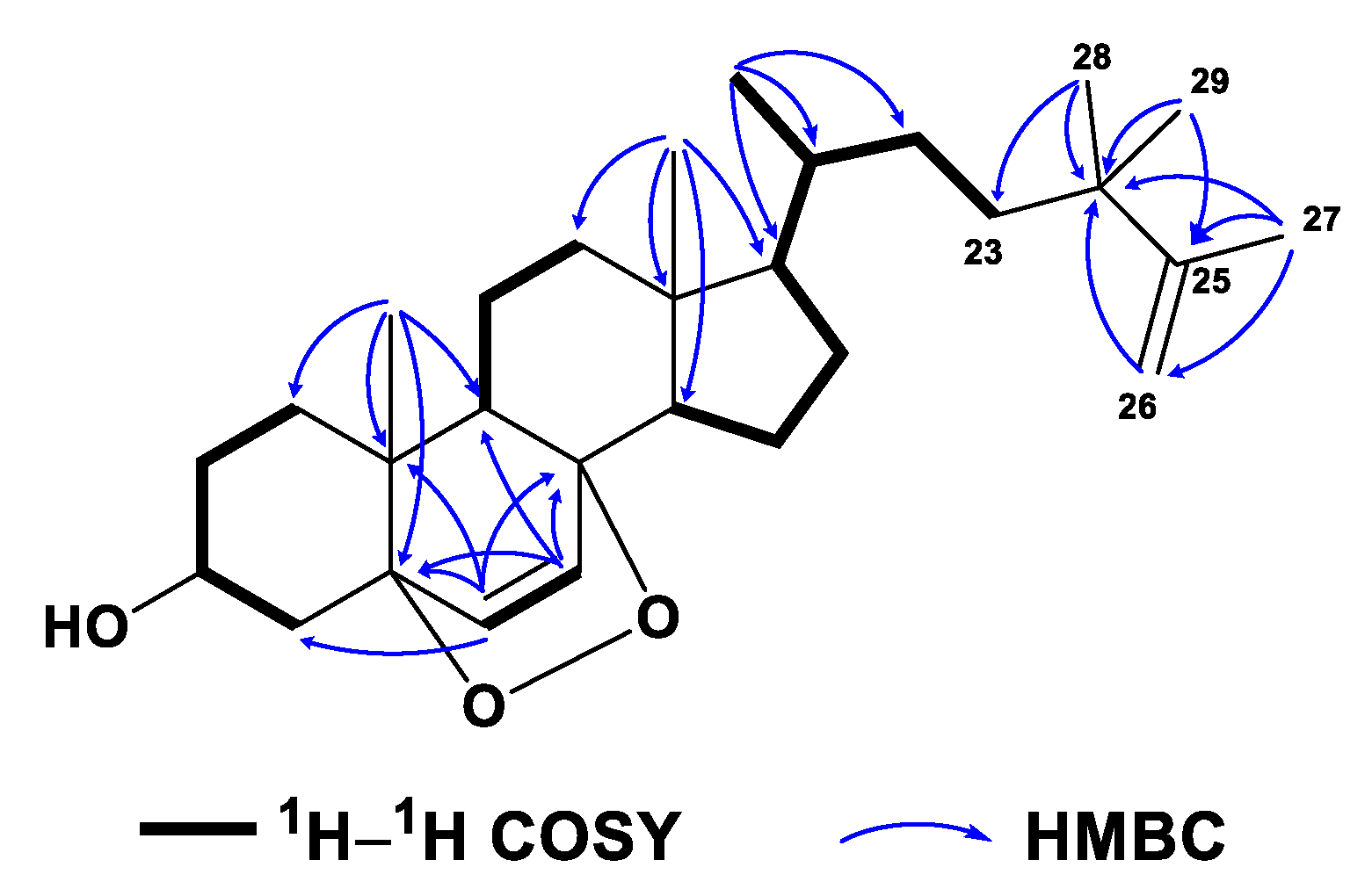
Compound 6 was obtained as a white amorphous powder. Its molecular formula C29H46O3 was determined by the HREIMS ion peak at m/z 424.3325 [M − H2O]+ (calcd. 424.3336, Figure S47), corresponding to seven degrees of unsaturation. Two vicinal coupled olefinic protons at δH 6.24 (d, J = 8.5 Hz, H-6) and 6.50 (d, J = 8.6 Hz, H-7) and an oxygenated methine at δH 3.97 (tt, J = 11.2, 5.1 Hz, H-3) were characteristic of a 3β-hydroxy-6-en-5α,8α-epidioxysterol nucleus, which was also recognized by the 13C NMR signals at δC 66.6 (d, C-3), 82.3 (s, C-5), 135.6 (d, C-6), 130.9 (d, C-7), and 79.6 (s, C-8) (Figure S43). These spectral data of 6 (Table 1 and Table 2, Figures S41 and S42) were reminiscent of yalongsterol A, a sterol previously reported from the soft coral Sinularia sp. by our group [11]. Detailed comparison of the full 1H and 13C NMR data of 6 with yalongsterol A, showing great similarity between them, clearly allowed the assignment of 3β-hydroxy-6-en-5α,8α-epidioxy-cholesta nucleus to 6, which was further justified by the extensive analyses of 2D NMR spectra involving 1H–1H COSY, HSQC, and HMBC (Figure 8 and Figures S43–S45). However, they differed at the side chain. The NMR signals at δH 4.72 (br s, Ha-26), 4.65 (br s, Hb-26), 1.67 (s, H3-27) and δC 152.3 (d, C-25), 109.6 (d, C-26), 19.5 (q, C-27) indicated the presence of a terminal double bound with an allylic methyl in the terminal of the side chain of 6, which was supported by the HMBC correlations from H2-26 (δH 4.65, 4.72) to C-24 (δC 38.8), C-25 (δC 152.3) and C-27 (δC 19.5), H3-27 (δH 1.67) to C-24, C-25 and C-26 (δC 109.6) (Figure 8 and Figure S44). Additional HMBC correlations from H3-28 (δH 1.00) to C-23 (δC 37.1), C-24, C-25, and C-29 (δC 27.7), from H3-29 (δH 1.00) to C-23, C-24, C-25, and C-28 (δC 27.3) (Figure 8 and Figure S44) implied the location of germinal methyls at C-24 of the side chain of 6. With the established structure of the side chain in hand, the structure of 6 was depicted as shown in Figure 1.
In in vitro bioassays, all the isolates were tested for antibacterial, neuroprotective, and anti-inflammatory effects. In the antibacterial bioassays (Table 3), all the steroids exhibited significant antibacterial activities against the fish pathogenic bacteria Streptococcus parauberis FP KSP28, Phoyobacterium damselae FP2244, and Streptococcus parauberis SPOF3K with IC90 values ranging from 0.1 to 11.0 µM. As observed in Table 3, the steroids possessing the unsaturated carbonyl moiety in ring A were favored for the inhibition against Streptococcus parauberis FP KSP28. Moreover, it seemed that a vinyl-type side chain in the steroid could lead to a small increase in the antibacterial activities against Phoyobacterium damselae FP2244 and Streptococcus parauberis SPOF3K, as indicated in Table 3. Only compound 7 displayed potent inhibitory activity against the fish pathogenic bacterium Aeromonas salmonicida AS42 with an IC90 value of 8.8 µM. This might imply that the combination of an α,β-α′,β′-unsaturated carbonyl moiety and a vinyl-type side chain played a key role in the antibacterial activity against Aeromonas salmonicida AS42. The above-mentioned results indicated that these isolated steroids could be used as antibacterial agents in fish farming.
Meanwhile, compounds 2 and 6–10 also displayed potent inhibitory effects against the vancomycin-resistant Enterococcus faecium bacterium G7 with IC90 values ranging from 4.4 to 18.3 µM (Table 3). Among them, steroid 10 also displayed antibacterial effects against the vancomycin-resistant Enterococcus faecium bacteria G1, G4, and G8 with IC90 values of 8.0, 4.0, and 8.0 µM, respectively. The preliminary analysis of the structure-activity relationship for compounds 6–10 revealed that the higher degrees of unsaturation of ring A in the steroids could keep efficacy against more individuals of the vancomycin-resistant Enterococcus faecium bacteria. The above-mentioned results implied that these isolates could be developed as new chemotypes of antibacterial leads against drug-resistant bacteria.
Moreover, all the isolated steroids except 5, 6, and 9 showed strong inhibitory activities against Streptococcus agalactiae WR10 with IC90 values ranging from 4.0 to 22.0 µM (Table 3). However, in the neuroprotective bioassays, none of these steroids displayed significant neuroprotective effects against the corticosterone-induced cellular injuries in human neuroblastoma SH-SY5Y cells at the concentration of 10 μM. In the evaluations of the anti-inflammatory effect in lipopolysaccharide (LPS)-stimulated BV-2 microglial cells, all the isolates were judged as inactive at 10 μM, neither.
3. Materials and Methods
3.1. General Experimental Procedures
Optical rotations were measured on a Perkin–Elmer 241 MC polarimeter. The X-ray measurement was made on a Bruker D8 Venture X-ray diffractometer with Cu Kα radiation (Bruker Biospin AG, Fällanden, Germany). IR spectra were recorded on a Nicolet 6700 spectrometer (Thermo Scientific, Waltham, MA, USA). NMR spectra were measured in CDCl3 with a Bruker DRX-400, Bruker DRX-600, or Bruker DRX-800 spectrometer (Bruker Biospin AG, Fällanden, Germany) with the residual CDCl3 (δH 7.26 ppm, δC 77.16 ppm). Chemical shifts (δ) were reported in ppm with reference to the solvent signals, and coupling constants (J) were expressed in Hz. Structural assignments were supported by 1H–1H COSY, HSQC, HMBC, and NOESY experiments. HREIMS data were recorded on a Finnigan-MAT-95 mass spectrometer (Finnigan-MAT, San Jose, CA, USA). HRESIMS spectra were recorded on an Agilent G6520 Q-TOF mass spectrometer. Commercial silica gel (Qingdao Haiyang Chemical Group Co., Ltd., Qingdao, China, 200–300 and 300–400 mesh) and Sephadex LH-20 gel (Amersham Biosciences, Little Chalfont, UK) were used for column chromatography (CC), and precoated-silica-gel-plates (G60 F-254, Yan Tai Zi Fu Chemical Group Co., Yantai, China) were used for analytical TLC. Reversed-phase (RP) HPLC was performed on an Agilent 1260 series liquid chromatography equipped with a DAD G1315D detector at 210 and 254 nm. A semi-preparative ODS-HG-5 column (5 μm, 250 × 9.4 mm) was employed for the purifications. All solvents used for CC and HPLC were of analytical grade (Shanghai Chemical Reagents Co., Ltd., Shanghai, China) and chromatographic grade (Dikma Technologies Inc., Beijing, China), respectively.
3.2. Animal Material
The soft coral Lobophytum sp. was collected in October 2021 in Xuwen Country, Guangdong Province, China. This specimen was identified by Prof. X.-B. Li from Hainan University. A voucher specimen (No. S-21-XW-6553) is available for inspection at the Shanghai Institute of Materia Medica, Chinese Academy of Sciences.
3.3. Extraction and Isolation
The frozen animals (1275 g, dry weight) were cut into pieces and extracted exhaustively with acetone at room temperature (4 × 3.0 L, 15 min in ultrasonic bath). The organic extract was evaporated to give a brown residue, which was then partitioned between Et2O and H2O. The Et2O solution was concentrated under reduced pressure to give a dark brown residue (13.4 g), which was fractionated by gradient Si gel (200–300 mesh) column chromatography (CC) (Et2O/petroleum ether (PE), 0→100%), yielding eight fractions (A–H). Fraction C was chromatographed over Sephadex LH-20 CC (PE/CH2Cl2/MeOH, 2:1:1) to give compound 10 (1.5 mg) and a mixture. This mixture was further purified through a silica gel CC (300–400 mesh, PE:Et2O, 12:1) followed by RP-HPLC (80% MeOH, 0.8 mL/min) to afford compound 6 (3.8 mg, tR = 31.3 min). Fraction D was subjected to a column of Sephadex LH-20 eluted with PE/CH2Cl2/MeOH (2:1:1) to yield three subfractions (D1–D3). Compound 8 (1.5 mg, tR = 20.0 min) was obtained from the D1 through a silica gel CC (300–400 mesh, PE:Et2O, 10:1) followed by RP-HPLC (85% CH3CN, 1.0 mL/min). Compound 9 (2.0 mg, tR = 29.1 min) was isolated from the D2 through silica gel CC (300–400 mesh, PE:Et2O, 10:1) followed by RP-HPLC (85% CH3CN, 1.0 mL/min). Compounds 4 (1.7 mg, tR = 25.9 min) and 5 (0.8 mg, tR = 22.0 min) were obtained from the D3 through silica gel CC (300–400 mesh, PE:Et2O, 10:1) followed by RP-HPLC (77% CH3CN, 1.0 mL/min). Fraction E was subjected to Sephadex LH-20 CC (PE/CH2Cl2/MeOH, 2:1:1), followed by RP-HPLC (75% CH3CN, 0.6 mL/min) to give compound 7 (3.1 mg, tR = 25.8 min). Fraction F was subjected to a column of Sephadex LH-20 eluted with PE/CH2Cl2/MeOH (2:1:1) and further divided into two subfractions, F1 and F2, by the following silica gel CC (300–400 mesh, PE:Et2O, 3:1). Compounds 1 (2.6 mg, tR = 8.7 min) and 3 (1.1 mg, tR = 12.6 min) were obtained from F2 by RP-HPLC (65% CH3CN, 0.8 mL/min) while 2 (1.6 mg, tR = 28.0 min) was obtained from F1 by RP-HPLC (75% CH3CN, 0.8 mL/min).
3.4. Spectroscopic Data of Compounds
Lobosteroid A (1): Colorless crystal; [α −3.8 (c 0.26, CHCl3); IR (KBr): νmax 3358, 2922, 2851, 1661, 1632, 1468, 1180 cm−1; 1H and 13C NMR (CDCl3, 800 and 125 MHz; see Table 1 and Table 2); HRESIMS m/z 427.3210 [M + H]+ (calcd. for C28H43O3, 427.3207).
Lobosteroid B (2): White amorphous powder; [α +14.0 (c 0.16, CHCl3); IR (KBr): νmax 3358, 2925, 2852, 1664, 1631, 1467, 887 cm−1; 1H and 13C NMR (CDCl3, 600 and 150 MHz; see Table 1 and Table 2); HRESIMS m/z 411.3255 [M + H]+ (calcd. for C28H43O2, 411.3258).
Lobosteroid C (3): White amorphous powder; [α +18.2 (c 0.05, CH3OH); IR (KBr): νmax 3359, 2923, 2852, 1742, 1662, 1468, 1236 cm−1; 1H and 13C NMR (CDCl3, 600 and 150 MHz; see Table 1 and Table 2); HRESIMS m/z 469.3318 [M + H]+ (calcd. for C30H45O4, 469.3312).
Lobosteroid D (4): White amorphous powder; [α +9.2 (c 0.17, CHCl3); IR (KBr): νmax 3359, 2923, 2852, 1660, 1633, 1468 cm−1; 1H and 13C NMR (CDCl3, 600 and 200 MHz; see Table 1 and Table 2); HRESIMS m/z 443.3520 [M + H]+ (calcd. for C29H47O3, 443.3520).
3.5. X-ray Crystallographic Analysis for Compound 1
Lobosteroid A (1) was crystallized from MeOH at room temperature. C28H43O3, Mr = 426.61, monoclinic, crystal size 0.12 × 0.08 × 0.05 mm3, space group P212121, a = 11.7623(13) Å, b = 11.875(2) Å, c = 17.1256(15) Å, V = 2392.0(6) Å3, Z = 4, ρcalcd = 1.185 g/cm3, F(000) = 936.0, 31,225 collected reflections, 4920 independent reflections (Rint = 0.0522, Rsigma = 0.0310), final R1 = 0.0353 (wR2 = 0.0904) reflections with I ≥ 2σ (I), R1 = 0.0383, wR2 = 0.0951 for all unique data. The X-ray measurements were made on a Bruker D8 Venture X-ray diffractometer with Cu Kα radiation (λ = 1.54178 Å) at 170.0 K. The collected data integration and reduction were processed with SAINT V8.37A software, and multiscan absorption corrections were performed using the SADABS program. The structure was solved with the SHELXT [39] structure solution program using intrinsic phasing and refined with the SHELXL [40] refinement package using least squares minimization. Crystallographic data for 1 were deposited at the Cambridge Crystallographic Data Centre (Deposition nos. CCDC 2282723). Copies of these data can be obtained free of charge via www.ccdc.cam.ac.uk (accessed on 19 July 2023), or from the Cambridge Crystallographic Data Centre, 12 Union Road, Cambridge CB21EZ, UK. [Fax: (+44) 1223-336-033. E-mail: [email protected].]
3.6. Antibacterial Bioassays
The marine strains Streptococcus parauberis FP KSP28, Streptococcus parauberis SPOF3K, Phoyoba cteriumdamselae FP2244, and Aeromonas salmonicida AS42 were provided by National Fisheries Research & Development Institute, Korea. The strain Streptococcus agalactiae WR10 was provided by the Chinese Academy of Tropical Agricultural Sciences. The vancomycin-resistant Enterococcus faecium bacteria G1, G4, G7, and G8 were provided by Ruijin Hospital, Shanghai Jiao Tong University School of Medicine. The minimum inhibitory concentration for 90% (MIC90) values for all antimicrobial agents was measured by the 96-well micro-dilution method. Mueller–Hinton II broth (cation-adjusted, BD 212322) was used for MIC90 value determination. Generally, compounds were dissolved with DMSO to 20 mM as stock solutions. All samples were diluted with culture broth to 500 µM as the initial concentration. Further, 1:2 serial dilutions were performed by the addition of culture broth to reach concentrations ranging from 500 µM to 0.24 µM. 100 µL of each dilution was distributed in 96-well plates, as well as sterile controls, growth controls (containing culture broth plus DMSO, without compounds), and positive controls (containing culture broth plus control antibiotics such as tetracycline). Each test and growth control well was inoculated with 5 µL of an exponential-phase bacterial suspension (about 105 CFU/well). The 96-well plates were incubated at 37 °C for 24 h. MIC90 values of these compounds were defined as the lowest concentration to inhibit bacterial growth completely. All MIC90 values were interpreted according to the recommendations of the Clinical and Laboratory Standards Institute (CLSI). Tetracycline hydrochloride (TC), oxytetracycline hydrochloride (OT), and levofloxacin hydrochloride (LF) were used as positive controls (Table 3).
4. Conclusions
In summary, six new steroids, lobosteroids A–F (1–6), together with four known compounds 7–10, were isolated from the Chinese soft coral Lobophytum sp. The chemical diversity of new steroids was mainly attributed to the high oxidation, which was characterized by the conjugated enone or dienone system of the nucleus and diverse oxidation of side chains. Although many steroids were reported from soft corals, those with an α,β-α′,β′-unsaturated carbonyl or an α,β-unsaturated carbonyl moiety in ring A, or the existence of a 5α,8α-epidioxy system in ring B were rarely found from the genus Lobophytum. The discovery of steroids 1–6 expanded the diverse and complex array of steroids, which is still a research hotspot of marine natural products. In the bioassays, all of the isolates displayed significant antibacterial activities against the fish pathogenic bacteria Streptococcus parauberis FP KSP28, Phoyobacterium damselae FP2244, and Streptococcus parauberis SPOF3K with IC90 values ranging from 0.1 to 11.0 µM. Meanwhile, compounds 2 and 6–10 exhibited excellent antibacterial activities against the vancomycin-resistant Enterococcus faecium bacterium G7 with IC90 values ranging from 4.4 to 18.3 µM. These new findings implied that these isolated steroids could be developed as new chemotypes of antibacterial leads.
Supplementary Materials
The following supporting information can be downloaded at: https://www.mdpi.com/article/10.3390/md21080457/s1, Figures S1–S48: NMR, HRESIMS, HREIMS, and IR data of compounds 1–6.
Author Contributions
Conceptualization, L.-F.L., H.W. and Y.-W.G.; methodology, M.-Z.S., L.-F.L., H.W. and Y.-W.G.; validation, Z.-Y.X., M.-M.S. and M.-Z.S.; formal analysis, Z.-Y.X. and M.-M.S.; investigation, Z.-Y.X., Y.J. and M.-M.S.; resources, L.-G.Y.; data curation, Z.-Y.X. and M.-M.S.; writing—original draft preparation, Z.-Y.X.; writing—review and editing, L.-F.L. and Y.-W.G.; supervision, H.W. and Y.-W.G.; project administration, Y.-W.G.; funding acquisition, L.-F.L., H.W. and Y.-W.G. All authors have read and agreed to the published version of the manuscript.
Funding
This work was financially supported by the National Key Research and Development Program of China (No. 2022YFC2804100) and the National Natural Science Foundation of China (No. 81991521, 41876194).
Institutional Review Board Statement
Not applicable.
Data Availability Statement
Data are contained within the article or Supplementary Materials.
Acknowledgments
We appreciate the grants from the Shandong Laboratory of Yantai Drug Discovery, Bohai Rim Advanced Research Institute for Drug Discovery, and thank X.-B. Li from Hainan University for the taxonomic identification of the soft coral material.
Conflicts of Interest
The authors declare no conflict of interest.
References
- Carroll, A.R.; Copp, B.R.; Davis, R.A.; Keyzers, R.A.; Prinsep, M.R. Marine natural products. Nat. Prod. Rep. 2023, 40, 275–325. [Google Scholar] [CrossRef]
- Savić, M.P.; Sakač, M.N.; Kuzminac, I.Z.; Ajduković, J.J. Structural diversity of bioactive steroid compounds isolated from soft corals in the period 2015–2020. J. Steroid Biochem. 2022, 218, 106061. [Google Scholar] [CrossRef] [PubMed]
- Shao, Z.-Y.; Zhu, D.-Y.; Guo, Y.-W. Nanjiols A−C, new steroids from the Chinese soft coral Nephthea bayeri. J. Nat. Prod. 2002, 65, 1675–1677. [Google Scholar] [CrossRef] [PubMed]
- Liang, L.-F.; Wang, X.-J.; Zhang, H.-Y.; Liu, H.-L.; Li, J.; Lan, L.-F.; Zhang, W.; Guo, Y.-W. Bioactive polyhydroxylated steroids from the Hainan soft coral Sinularia depressa Tixier-Durivault. Bioorg. Med. Chem. Lett. 2013, 23, 1334–1337. [Google Scholar] [CrossRef]
- Yan, X.-H.; Liu, H.-L.; Guo, Y.-W. Ximaosteroids A–D, new steroids from the Hainan soft coral Scleronephthya sp. Steroids 2009, 74, 1061–1065. [Google Scholar] [CrossRef] [PubMed]
- Chen, W.-T.; Liu, H.-L.; Yao, L.-G.; Guo, Y.-W. 9,11-Secosteroids and polyhydroxylated steroids from two South China Sea soft corals Sarcophyton trocheliophorum and Sinularia flexibilis. Steroids 2014, 92, 56–61. [Google Scholar] [CrossRef] [PubMed]
- Liu, J.; Wu, X.; Yang, M.; Gu, Y.-C.; Yao, L.-G.; Huan, X.-J.; Miao, Z.-H.; Luo, H.; Guo, Y.-W. Erectsterates A and B, a pair of novel highly degraded steroid derivatives from the South China Sea soft coral Sinularia erecta. Steroids 2020, 161, 108681. [Google Scholar] [CrossRef] [PubMed]
- Xu, T.; Zhao, Q.-M.; Yao, L.-G.; Lan, L.-F.; Li, S.-W.; Guo, Y.-W. Sinulasterols D–G, four new antibacterial steroids from the South China sea soft coral Sinularia depressa. Steroids 2023, 192, 109182. [Google Scholar] [CrossRef]
- Li, S.-W.; Liu, J.; Fu, Y.; Yao, L.-G.; Zhang, H.-Y.; Wang, H.; Guo, Y.-W. Anti-inflammatory steroids from the South China Sea soft coral Lobophytum sarcophytoides. Chem. Biodivers. 2023, 20, e202300821. [Google Scholar] [CrossRef] [PubMed]
- Zhang, Q.; Liang, L.-F.; Miao, Z.-H.; Wu, B.; Guo, Y.-W. Cytotoxic polyhydroxylated steroids from the South China Sea soft coral Lobophytum sp. Steroids 2019, 141, 76–80. [Google Scholar] [CrossRef]
- Yang, M.; Liang, L.-F.; Li, H.; Tang, W.; Guo, Y.-W. A new 5α,8α-epidioxysterol with immunosuppressive activity from the South China Sea soft coral Sinularia sp. Nat. Prod. Res. 2020, 34, 1814–1819. [Google Scholar] [CrossRef]
- Ermolenko, E.V.; Imbs, A.B.; Gloriozova, T.A.; Poroikov, V.V.; Sikorskaya, T.V.; Dembitsky, V.M. Chemical diversity of soft coral steroids and their pharmacological activities. Mar. Drugs 2020, 18, 613. [Google Scholar] [CrossRef]
- Rodrigues, I.G.; Miguel, M.G.; Mnif, W. A brief review on new naturally occurring cembranoid diterpene derivatives from the soft corals of the genera Sarcophyton, Sinularia, and Lobophytum since 2016. Molecules 2019, 24, 781. [Google Scholar] [CrossRef]
- Li, S.-W.; Cuadrado, C.; Yao, L.-G.; Daranas, A.H.; Guo, Y.-W. Quantum mechanical–NMR-aided configuration and conformation of two unreported macrocycles isolated from the soft coral Lobophytum sp.: Energy calculations versus coupling constants. Org. Lett. 2020, 22, 4093–4096. [Google Scholar] [CrossRef]
- Zhang, D.; Wang, Z.; Han, X.; Li, X.-L.; Lu, Z.-Y.; Dou, B.-B.; Zhang, W.-Z.; Tang, X.-L.; Li, P.-L.; Li, G.-Q. Four bioactive new steroids from the soft coral Lobophytum pauciflorum collected in South China Sea. Beilstein J. Org. Chem. 2022, 18, 374–380. [Google Scholar] [CrossRef]
- Ohno, O.; Mizuno, E.; Miyamoto, J.; Hoshina, T.; Sano, T.; Matsuno, K. Inhibition of lipopolysaccharide-induced inflammatory signaling by soft coral-derived prostaglandin A2 in RAW264.7 cells. Mar. Drugs 2022, 20, 316. [Google Scholar] [CrossRef]
- Li, Z.-Y.; Li, C.-Y.; Lai, K.-H.; Liao, M.-Y.; Wang, W.-H.; Chung, H.-M. Chemical constituents from the octocoral Lobophytum sarcophytoides. Chem. Nat. Compd. 2022, 58, 1167–1169. [Google Scholar] [CrossRef]
- Peng, B.-R.; Lu, M.-C.; El-Shazly, M.; Wu, S.-L.; Lai, K.-H.; Su, J.-H. Aquaculture soft coral Lobophytum crassum as a producer of anti-proliferative cembranoids. Mar. Drugs 2018, 16, 15. [Google Scholar] [CrossRef]
- Huang, C.-Y.; Tseng, W.-R.; Ahmed, F.A.; Chiang, P.-L.; Tai, C.-J.; Hwang, T.-L.; Dai, C.-F.; Sheu, J.-H. Anti-inflammatory polyoxygenated steroids from the soft coral Lobophytum michaelae. Mar. Drugs 2018, 16, 93. [Google Scholar] [CrossRef]
- Rahelivao, M.P.; Lübken, T.; Gruner, M.; Kataeva, O.; Ralambondrahety, R.; Andriamanantoanina, H.; Checinski, M.P.; Bauer, I.; Knölker, H.-J. Isolation and structure elucidation of natural products of three soft corals and a sponge from the coast of Madagascar. Org. Biomol. Chem. 2017, 15, 2593–2608. [Google Scholar]
- Aboutabl, E.A.; Selim, N.M.; Azzam, S.M.; Michel, C.G.; Hegazy, M.F.; Ali, A.M.; Hussein, A.A. Polyhydroxy sterols isolated from the Red Sea soft coral Lobophytum crassum and their cytotoxic activity. Nat. Prod. Commun. 2017, 12, 233–235. [Google Scholar] [CrossRef]
- Hegazy, M.-E.F.; Mohamed, T.A.; Elshamy, A.I.; Hassanien, A.A.; Abdel-Azim, N.S.; Shreadah, M.A.; Abdelgawad, I.I.; Elkady, E.M.; Paré, P.W. A new steroid from the Red Sea soft coral Lobophytum lobophytum. Nat. Prod. Res. 2016, 30, 340–344. [Google Scholar] [CrossRef]
- Chau, V.M.; Phan, V.K.; Nguyen, X.N.; Nguyen, X.C.; Nguyen, P.T.; Nguyen, H.N.; Hoang, L.T.A.; Do, C.T.; Dinh, T.T.T.; Kang, H.-K.; et al. Cytotoxic and antioxidant activities of diterpenes and sterols from the Vietnamese soft coral Lobophytum compactum. Bioorg. Med. Chem. Lett. 2011, 21, 2155–2159. [Google Scholar]
- Yeffet, D.; Rudi, A.; Ketzinel, S.; Benayahu, Y.; Kashman, Y. Auroside, a xylosyl-sterol, and patusterol A and B, two hydroxylated sterols, from two soft corals Eleutherobia aurea and Lobophytum patulum. Nat. Prod. Commun. 2010, 5, 205–210. [Google Scholar] [CrossRef] [PubMed]
- Ye, F.; Zhou, Y.-B.; Li, J.; Gu, Y.-C.; Guo, Y.-W.; Li, X.-W. New steroids from the South China Sea soft coral Lobophytum sp. Chem. Biodivers. 2020, 17, e2000214. [Google Scholar] [CrossRef] [PubMed]
- Radhika, P.; Asolkar, R.N.; Laatsch, H. An acetoxygenated analogue of ergosterol from a soft coral of the genus Lobophytum. Nat. Prod. Res. 2004, 18, 575–579. [Google Scholar] [CrossRef] [PubMed]
- Morris, L.A.; Christie, E.M.; Jaspars, M.; van Ofwegen, L.P. A bioactive secosterol with an unusual A- and B-ring oxygenation pattern isolated from an Indonesian soft coral Lobophytum sp. J. Nat. Prod. 1998, 61, 538–541. [Google Scholar] [CrossRef]
- Radhika, P.; Cabeza, M.; Bratoeff, E.; García, G. 5α-Reductase inhibition activity of steroids isolated from marine soft corals. Steroids 2004, 69, 439–444. [Google Scholar]
- Liu, J.; Gu, Y.-C.; Su, M.-Z.; Guo, Y.-W. Chemistry and bioactivity of secondary metabolites from South China Sea marine fauna and flora: Recent research advances and perspective. Acta Pharmacol. Sin. 2022, 43, 3062–3079. [Google Scholar] [PubMed]
- Song, Y.-T.; Yu, D.-D.; Su, M.Z.; Luo, H.; Cao, J.-G.; Yao, L.-G.; Liang, L.-F.; Guo, Y.-W.; Yang, F. Anti-tumor cembrane diterpenoids from the South China Sea soft coral Lobophytum sp. Chem. Biodivers. 2023, 20, e202300217. [Google Scholar] [CrossRef]
- Díaz-Marrero, A.R.; Porras, G.; Aragón, Z.; de la Rosa, J.M.; Dorta, E.; Cueto, M.; D’Croz, L.; Maté, J.; Darias, J. Carijodienone from the octocoral Carijoa multiflora. A spiropregnane-based steroid. J. Nat. Prod. 2011, 74, 292–295. [Google Scholar] [PubMed]
- Seo, Y.; Jung, J.H.; Rho, J.-R.; Shin, J.; Song, J.-I. Isolation of novel bioactive steroids from the soft coral Alcyonium gracillimum. Tetrahedron 1995, 51, 2497–2506. [Google Scholar]
- Wu, S.-L.; Wang, G.-H.; Dai, C.-F.; Sheu, J.-H. Pregnane-based steroids from a Formosan gorgonian Subergorgia mollis. J. Chin. Chem. Soc. 2004, 51, 205–208. [Google Scholar]
- Blackman, A.J.; Heaton, A.; Skelton, B.W.; White, A.H. Pregnane derivatives from two soft corals of the genus Capnella. Aust. J. Chem. 1985, 38, 565–573. [Google Scholar] [CrossRef]
- Yan, X.-H.; Liu, H.-L.; Huang, H.; Li, X.-B.; Guo, Y.-W. Steroids with aromatic A-rings from the Hainan soft coral Dendronephthya studeri Ridley. J. Nat. Prod. 2010, 74, 175–180. [Google Scholar] [PubMed]
- Marinozzi, M.; Castro Navas, F.F.; Maggioni, D.; Carosati, E.; Bocci, G.; Carloncelli, M.; Giorgi, G.; Cruciani, G.; Fontana, R.; Russo, V. Side-chain modified ergosterol and stigmasterol derivatives as liver X receptor agonists. J. Med. Chem. 2017, 60, 6548–6562. [Google Scholar] [PubMed]
- Yan, X.-H.; Lin, L.-P.; Ding, J.; Guo, Y.-W. Methyl spongoate, a cytotoxic steroid from the Sanya soft coral Spongodes sp. Bioorg. Med. Chem. Lett. 2007, 17, 2661–2663. [Google Scholar] [CrossRef] [PubMed]
- Ahmed, A.F.; Wu, M.-H.; Wu, Y.-C.; Dai, C.-F.; Sheu, J.-H. Metabolites with cytotoxic activity from the Formosan soft coral Cladiella australis. J. Chin. Chem. Soc. 2006, 53, 489–494. [Google Scholar] [CrossRef]
- Sheldrick, G.M. SHELXT—Integrated space-group and crystal-structure determination. Acta Crystallogr. 2015, A71, 3–8. [Google Scholar]
- Sheldrick, G.M. Crystal structure refinement with SHELXL. Acta Crystallogr. 2015, C71, 3–8. [Google Scholar]
Figure 1.
Chemical structures of compounds 1–10.

Figure 2.
1H–1H COSY, selected key HMBC and NOE correlations of 1.

Figure 3.
Perspective ORTEP drawing of 1 (displacement ellipsoids are drawn at the 50% probability level).
Figure 3.
Perspective ORTEP drawing of 1 (displacement ellipsoids are drawn at the 50% probability level).

Figure 4.
1H–1H COSY, selected key HMBC and NOE correlations of 2.

Figure 5.
1H–1H COSY, selected key HMBC and NOE correlations of 3.

Figure 6.
1H–1H COSY, selected key HMBC and NOE correlations of 4.

Figure 7.
1H–1H COSY, selected key HMBC and NOE correlations of 5.

Figure 8.
1H–1H COSY and selected key HMBC correlations of 6.


Table 1.
13C NMR data of compounds 1–6 in CDCl3.
| No. | 1 a | 2 b | 3 a | 4 a | 5 a | 6 c |
|---|---|---|---|---|---|---|
| δC (Mult.) | δC (Mult.) | δC (Mult.) | δC (Mult.) | δC (Mult.) | δC (Mult.) | |
| 1 | 155.8 (d) | 156.2 (d) | 155.7 (d) | 158.7 (d) | 158.6 (d) | 34.8 (t) |
| 2 | 127.7 (d) | 127.6 (d) | 127.8 (d) | 127.5 (d) | 127.6 (d) | 30.3 (t) |
| 3 | 186.5 (s) | 186.6 (s) | 186.5 (s) | 200.4 (s) | 200.4 (s) | 66.6 (d) |
| 4 | 124.1 (d) | 123.9 (d) | 124.1 (d) | 41.1 (t) | 41.1 (t) | 37.1 (d) |
| 5 | 169.1 (s) | 169.5 (s) | 169.0 (s) | 44.4 (d) | 44.4 (d) | 82.3 (s) |
| 6 | 33.0 (t) | 33.0 (t) | 32.9 (t) | 27.7 (t) | 27.7 (t) | 135.6 (d) |
| 7 | 33.7 (t) | 33.8 (t) | 33.6 (t) | 32.1 (t) | 31.4 (t) | 130.9 (d) |
| 8 | 35.6 (d) | 35.6 (d) | 35.6 (d) | 35.8 (d) | 35.8 (d) | 79.6 (s) |
| 9 | 52.3 (d) | 52.4 (d) | 52.2 (d) | 50.1 (d) | 49.9 (d) | 51.2 (d) |
| 10 | 43.6 (s) | 43.8 (s) | 43.6 (s) | 39.1 (s) | 39.1 (s) | 37.1 (s) |
| 11 | 22.9 (t) | 24.5 (t) | 22.8 (t) | 30.1 (t) | 21.3 (t) | 23.5 (t) |
| 12 | 39.5 (t) | 39.5 (t) | 38.8 (t) | 37.5 (t) | 39.8 (t) | 39.5 (t) |
| 13 | 43.1 (s) | 42.9 (s) | 43.0 (s) | 42.4 (s) | 43.1 (s) | 44.8 (s) |
| 14 | 54.9 (d) | 55.6 (d) | 54.6 (d) | 55.8 (d) | 55.8 (d) | 51.7 (d) |
| 15 | 24.7 (t) | 23.0 (t) | 24.5 (t) | 23.8 (t) | 24.5 (t) | 20.8 (t) |
| 16 | 27.5 (t) | 28.4 (t) | 26.9 (t) | 27.3 (t) | 27.7 (t) | 28.3 (t) |
| 17 | 52.0 (d) | 56.0 (d) | 49.6 (d) | 52.9 (d) | 52.4 (d) | 56.2 (d) |
| 18 | 12.4 (q) | 12.2 (q) | 12.5 (q) | 12.4 (q) | 12.6 (q) | 12.7 (q) |
| 19 | 18.8 (q) | 18.8 (q) | 18.8 (q) | 13.1 (q) | 13.1 (q) | 18.3 (q) |
| 20 | 50.9 (d) | 32.0 (d) | 53.6 (d) | 48.0 (d) | 50.0 (d) | 35.8 (d) |
| 21 | 16.4 (q) | 20.0 (q) | 64.6 (t) | 176.9 (s) | 16.7 (q) | 18.9 (q) |
| 22 | 217.6 (s) | 49.2 (t) | 211.8 (s) | 29.8 (t) | 214.8 (s) | 30.4 (t) |
| 23 | 48.2 (t) | 215.0 (s) | 49.6 (t) | 21.2 (t) | 46.8 (t) | 37.1 (t) |
| 24 | 74.0 (s) | 53.0 (d) | 33.3 (d) | 38.7 (d) | 33.8 (d) | 38.8 (s) |
| 25 | 37.4 (d) | 30.2 (d) | 32.1 (d) | 31.4 (d) | 32.1 (d) | 152.3 (s) |
| 26 | 17.0 (q) | 18.8 (q) | 18.6 (q) | 17.5 (q) | 18.3 (q) | 109.6 (t) |
| 27 | 17.9 (q) | 21.6 (q) | 19.9 (q) | 20.6 (q) | 20.0 (q) | 19.5 (q) |
| 28 | 23.0 (q) | 12.7 (q) | 16.2 (q) | 15.3 (q) | 16.1 (q) | 27.3 (q) |
| 29 | 170.7 (s) | 51.2 (q) | 27.7 (q) | |||
| 30 | 21.0 (q) |
a Recorded at 125 MHz. b Recorded at 150 MHz. c Recorded at 200 MHz.
Table 2.
1H NMR data of compounds 1–6 in CDCl3.
| No. | 1 a | 2 b | 3 a | 4 a | 5 a | 6 c |
|---|---|---|---|---|---|---|
| δH Mult. (J in Hz) | δH Mult. (J in Hz) | δH Mult. (J in Hz) | δH Mult. (J in Hz) | δH Mult. (J in Hz) | δH Mult. (J in Hz) | |
| 1 | 7.04 d (10.2) | 7.05 d (10.1) | 7.03 d (10.1) | 7.11 d (10.2) | 7.13 d (10.2) | 1.70 br d (13.8) |
| 1.94 dd (13.8, 4.0) | ||||||
| 2 | 6.23 dd (10.2, 2.0) | 6.22 dd (10.1, 1.9) | 6.23 dd (10.1, 1.9) | 5.84 dd (10.2, 1.0) | 5.85 d (10.2) | 1.54 ovl |
| 1.84 br d (12.8) | ||||||
| 3 | 3.97 tt (11.2, 5.1) | |||||
| 4 | 6.07 br d s | 6.06 br s | 6.07 br s | 2.21 dd (14.5, 3.6) | 2.23 dd (14.9, 3.3) | 1.91 ovl |
| 2.35 dd (17.7, 14.2) | 2.36 dd (17.8, 14.2) | 2.11 dd (13.7, 5.2) | ||||
| 5 | 1.90 ovl | 1.91 ovl | ||||
| 6 | 2.35 br d (13.5) | 2.36 br d (13.4) | 2.35 br d (13.0) | 1.37 m | 1.31 ovl | 6.24 d (8.5) |
| 2.45 dd (14.0, 5.2) | 2.45 ovl | 2.46 dd (13.6, 4.0) | 1.42 ovl | 1.42 ovl | ||
| 7 | 1.93 m | 1.03 ovl | 1.06 ovl | 0.96 ovl | 0.96 m | 6.50 d (8.6) |
| 2.48 dd (12.0, 5.0) | 1.94 ovl | 1.94 ovl | 1.70 ovl | 1.71 ovl | ||
| 8 | 1.61 ovl d | 1.62 ovl | 1.61 m | 1.45 ovl | 1.45 ovl | |
| 9 | 1.59 ovl | 1.04 ovl | 1.06 ovl | 0.96 ovl | 0.99 m | 1.49 ovl |
| 11 | 1.07 m | 1.15 ovl | 1.66 ovl | 0.85 dd (13.1, 6.0) | 0.88 m | 1.20 ovl |
| 1.69 ovl | 1.61 ovl | 1.71 m | 1.72 dd (13.3, 3.2) | 1.76 dd (13.7, 3.5) | 1.50 ovl | |
| 12 | 1.28 td (13.0, 5.0) | 1.17 dd (12.5, 6.6) | 1.23 ovl | 1.07 ovl | 1.31 ovl | 1.21 ovl |
| 1.97 dt (13.1, 3.3) | 2.04 dt (13.0, 3.3) | 1.90 dt (12.6, 3.3) | 1.50 ovl | 1.97 dt (12.7, 3.4) | 1.96 dd (13.3, 3.3) | |
| 14 | 1.04 m | 1.00 m | 1.01 m | 1.08 ovl | 1.09 ovl | 1.53 ovl |
| 15 | 1.18 ovl | 1.62 ovl | 1.20 ovl | 1.09 ovl | 1.11 ovl | 0.89 m |
| 1.63 td (13.0, 3.5) | 1.68 td (11.1, 3.7) | 1.64 ovl | 1.64 ovl | 1.60 ovl | 1.64 m | |
| 16 | 1.18 ovl | 1.25 m | 1.30 m | 1.30 ovl | 1.32 ovl | 1.34 m |
| 1.73 ovl | 1.79 ddd (16.2, 7.0, 3.1) | 1.65 ovl | 1.90 ovl | 1.69 ovl | 1.90 ovl | |
| 17 | 1.60 ovl | 1.13 m | 1.60 ovl | 1.65 ovl | 1.63 ovl | 1.18 ovl |
| 18 | 0.76 s | 0.78 s | 0.81 s | 0.72 s | 0.72 s | 0.78 s |
| 19 | 1.23 s | 1.23 s | 1.23 s | 0.99 s | 1.01 s | 0.89 s |
| 20 | 2.48 ovl | 2.03 ovl | 2.84 td (10.4, 4.4) | 2.20 dt (7.5, 3.6) | 2.50 dq (10.4. 6.8) | 1.32 m |
| 21 | 1.11 d (7.6) | 0.90 d (7.0) | 3.96 t (10.7) | 1.09 d (6.9) | 0.89 d (6.9) | |
| 4.48 dd (10.7, 4.4) | ||||||
| 22 | 2.20 dd (17.0, 10.0) | 1.27 ovl | 1.17 ovl | |||
| 2.45 dd (17.2, 2.8) | 1.40 ovl | 1.26 m | ||||
| 23 | 2.52 d (17.7) | 2.22 dd (17.7, 9.1) | 1.39 ovl | 2.17 dd (16.9, 8.9) | 1.17 ovl | |
| 2.66 d (17.7) | 2.47 dd (17.5, 3.8) | 1.51 ovl | 2.45 dd (17.0, 4.3) | 1.37 m | ||
| 24 | 2.29 quin (6.9) | 1.94 ovl | 1.24 ovl | 1.93 ovl | ||
| 25 | 1.75 quin (6.9) | 1.92 ovl | 1.55 m | 1.55 m | 1.55 m | |
| 26 | 0.88 d (6.9) | 0.84 d (6.8) | 0.83 d (7.1) | 0.75 d (6.9) | 0.82 d (6.8) | 4.65 br s |
| 4.72 br s | ||||||
| 27 | 0.93 d (6.8) | 0.90 d (7.0) | 0.87 d (6.8) | 0.84 d (6.8) | 0.87 d (6.8) | 1.67 s |
| 28 | 1.12 s | 0.98 d (6.9) | 0.81 d (7.6) | 0.76 d (6.9) | 0.81 d (6.8) | 1.00 s |
| 29 | 3.65 s | 1.00 s | ||||
| 30 | 2.00 s | |||||
| OH | 4.09 s |
a Recorded at 600 MHz. b Recorded at 800 MHz. c Recorded at 400 MHz. d ovl: overlapped, br: broad.
Table 3.
The IC90 values (μM) of antibacterial bioassays of compounds 1–10 a.
| 1 | 2 | 3 | 4 | 5 | 6 | 7 | 8 | 9 | 10 | TC | OT | LF | |
|---|---|---|---|---|---|---|---|---|---|---|---|---|---|
| Streptococcus parauberis FP KSP28 | 0.3 | 0.5 | 2.7 | 0.1 | 4.9 | 1.1 | 0.5 | 8.9 | 4.4 | 1.0 | 6.3 | 3.1 | 3.1 |
| Phoyobacterium damselae FP2244 | 9.1 | 4.2 | 11.0 | 8.5 | 9.8 | 9.2 | 2.2 | 2.2 | 4.4 | 2.0 | 0.1 | 0.1 | 0.1 |
| Streptococcus parauberis SPOF3K | 9.1 | 4.2 | 11.0 | 8.5 | 9.8 | 4.6 | 2.2 | 2.2 | 4.4 | 2.0 | >50.0 | 25.0 | 3.1 |
| Aeromonas salmonicida AS42 | - b | - | - | - | - | - | 8.8 | - | - | - | 12.7 | 0.8 | 0.8 |
| Enterococcus faecium G1 | - | - | - | - | - | - | 8.8 | - | - | 8.0 | 0.2 | 0.4 | >100.0 |
| Enterococcus faecium G4 | - | - | - | - | - | - | - | - | - | 4.0 | 0.4 | 0.4 | >100.0 |
| Enterococcus faecium G7 | - | 16.8 | - | - | - | 18.3 | 8.8 | 8.9 | 4.4 | 8.0 | 0.2 | 0.2 | >100.0 |
| Enterococcus faecium G8 | - | - | - | - | - | - | - | - | - | 8.0 | 0.2 | 0.1 | >100.0 |
| Streptococcus agalactiae WR10 | 18.1 | 8.4 | 22.0 | 17.0 | - | - | 4.4 | 4.4 | - | 4.0 | 1.6 | 1.6 | 6.3 |
a Tetracycline hydrochloride (TC), oxytetracycline hydrochloride (OT), levofloxacin hydrochloride (LF) were used as positive controls. b ‘-’ indicated they were not subjected to the antibacterial rescreening experiments since their inhibition rates against these bacteria were <90% in the preliminary antibacterial bioassays.
Disclaimer/Publisher’s Note: The statements, opinions and data contained in all publications are solely those of the individual author(s) and contributor(s) and not of MDPI and/or the editor(s). MDPI and/or the editor(s) disclaim responsibility for any injury to people or property resulting from any ideas, methods, instructions or products referred to in the content. |
© 2023 by the authors. Licensee MDPI, Basel, Switzerland. This article is an open access article distributed under the terms and conditions of the Creative Commons Attribution (CC BY) license (https://creativecommons.org/licenses/by/4.0/).
Share and Cite
MDPI and ACS Style
Xia, Z.-Y.; Sun, M.-M.; Jin, Y.; Yao, L.-G.; Su, M.-Z.; Liang, L.-F.; Wang, H.; Guo, Y.-W. Lobosteroids A–F: Six New Highly Oxidized Steroids from the Chinese Soft Coral Lobophytum sp. Mar. Drugs 2023, 21, 457. https://doi.org/10.3390/md21080457
AMA Style
Xia Z-Y, Sun M-M, Jin Y, Yao L-G, Su M-Z, Liang L-F, Wang H, Guo Y-W. Lobosteroids A–F: Six New Highly Oxidized Steroids from the Chinese Soft Coral Lobophytum sp. Marine Drugs. 2023; 21(8):457. https://doi.org/10.3390/md21080457
Chicago/Turabian StyleXia, Zi-Yi, Man-Man Sun, Yang Jin, Li-Gong Yao, Ming-Zhi Su, Lin-Fu Liang, Hong Wang, and Yue-Wei Guo. 2023. "Lobosteroids A–F: Six New Highly Oxidized Steroids from the Chinese Soft Coral Lobophytum sp." Marine Drugs 21, no. 8: 457. https://doi.org/10.3390/md21080457
Note that from the first issue of 2016, this journal uses article numbers instead of page numbers. See further details here.