
New Phenol Derivatives from the Haima Cold Seep-Derived Fungus Aspergillus subversicolor CYH-17
by
 , , and
Yi-Hao Che
1,2,
Wen-Ping Ding
1,
Zhi-Hui Xiao
1,3,
Jia-Min Wu
1,2,
Hao Yin
1,
Fa-Zuo Wang
1,3,* and
Si Zhang
1,* 1
CAS Key Laboratory of Tropical Marine Bio-Resources and Ecology, Southern Marine Science and Engineering Guangdong Laboratory (Guangzhou), Guangdong Key Laboratory of Marine Materia Medica, RNAM Center for Marine Microbiology, South China Sea Institute of Oceanology, Chinese Academy of Sciences, 164 West Xingang Road, Guangzhou 510301, China
2
University of Chinese Academy of Sciences, 19 Yuquan Road, Beijing 100049, China
3
Equipment Public Service Center of South China Sea Institute of Oceanology, Chinese Academy of Sciences, 164 West Xingang Road, Guangzhou 510301, China
*
Authors to whom correspondence should be addressed.
Mar. Drugs 2024, 22(3), 117; https://doi.org/10.3390/md22030117
Submission received: 29 January 2024
/
Revised: 27 February 2024
/
Accepted: 28 February 2024
/
Published: 29 February 2024
(This article belongs to the Special Issue Bioactive Secondary Metabolites of Marine Fungi 2.0)
Abstract
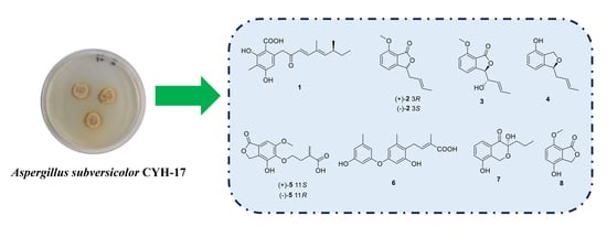
:Seven new phenol derivatives, subversins A–E (1–5), subversic acid A (6) and epi-wortmannine G (7); one new natural product, 4-hydroxy-7-methoxyphthalide (8); and five known compounds (9–13) were isolated from the fungus Aspergillus subversicolor CYH-17 collected from the Haima cold seep. The structures and absolute configurations of these compounds were determined via NMR, MS, optical rotation, electronic circular dichroism (ECD) calculation, X-ray diffraction analysis and comparison with the literature. Compounds 2 and 5 were two pairs of enantiomers. All compounds were tested for their α-glucosidase and acetylcholinesterase (AChE) inhibitory activity, antioxidant activity and antibacterial activity, but no obvious activity was observed among these studied compounds.
1. Introduction
Cold seeps have attracted increasing amounts of research interest since they were first discovered in 1983 [1]. In cold seeps, hydrocarbons such as methane, hydrogen sulfide and carbon dioxide are carried to the ocean floor due to geological activity, which leads to abundant chemosynthetic ecosystems [2]. The biological resources found in cold seeps are abundant and include archaea, bacteria, fungi, tubeworms, clams and mussels [3]. Over the past 40 years, most related research has focused on the taxonomy of species [4,5] and the ecological role of microorganisms [6,7]. However, few studies have reported that new secondary metabolites are produced by cold seep-derived creatures [8].
In fact, cold seep organisms possess the potential to produce intriguing natural products as they survive in extreme environments [9]. Under extreme conditions, cold seep organisms have evolved unique pathways to produce structurally diverse and biologically active secondary metabolites. According to previous reports, there were a great deal of new compounds that were found in the microorganisms derived from cold seeps, involving alkaloids [10], polyketides [11], terpenoids [12], glycosides [13], macrolides [14] and so forth. The new secondary metabolites displayed significant biological activity, including antimicrobial activity, cytotoxic activity and antioxidant activity. Therefore, cold seeps are a new and significant source for the discovery of active natural products.
With the aim of uncovering new secondary metabolites from cold seep-derived fungi, our team carried out a series of works. As a result, we isolated some fungi from the Haima cold seep and discovered several new diketopiperazine alkaloids from the fungi Aspergillus fumigatus CYH-5 [15] and Toxicocladosporium sp. CYH-18 [16]. Recently, the fungus Aspergillus subversicolor CYH-17, isolated from the sediment of the Haima cold seep at a depth of 1363 m in 2021, attracted our attention. Seven new phenol derivatives, subversins A–E (1–5), subversic acid A (6) and epi-wortmannine G (7); one new natural product, 4-hydroxy-7-methoxyphthalide (8); and five known products, diorcinol (9) [17], 3,7-dihydroxy-1,9-dimethyldibenzofuran (10) [18], 2-methoxyl cordyol C (11) [19], farnesylemefuranone E (12) [12] and citreorosein (13) [20] (Figure 1), were separated from the fungus A. subversicolor CYH-17. NMR, MS, optical rotation, electronic circular dichroism calculation and X-ray diffraction analysis were used to confirm the planar structures and absolute configurations of these compounds. In this study, the separation, structural elucidation and biological activity of those secondary metabolites are reported.
2. Results and Discussion
2.1. Structural Elucidation
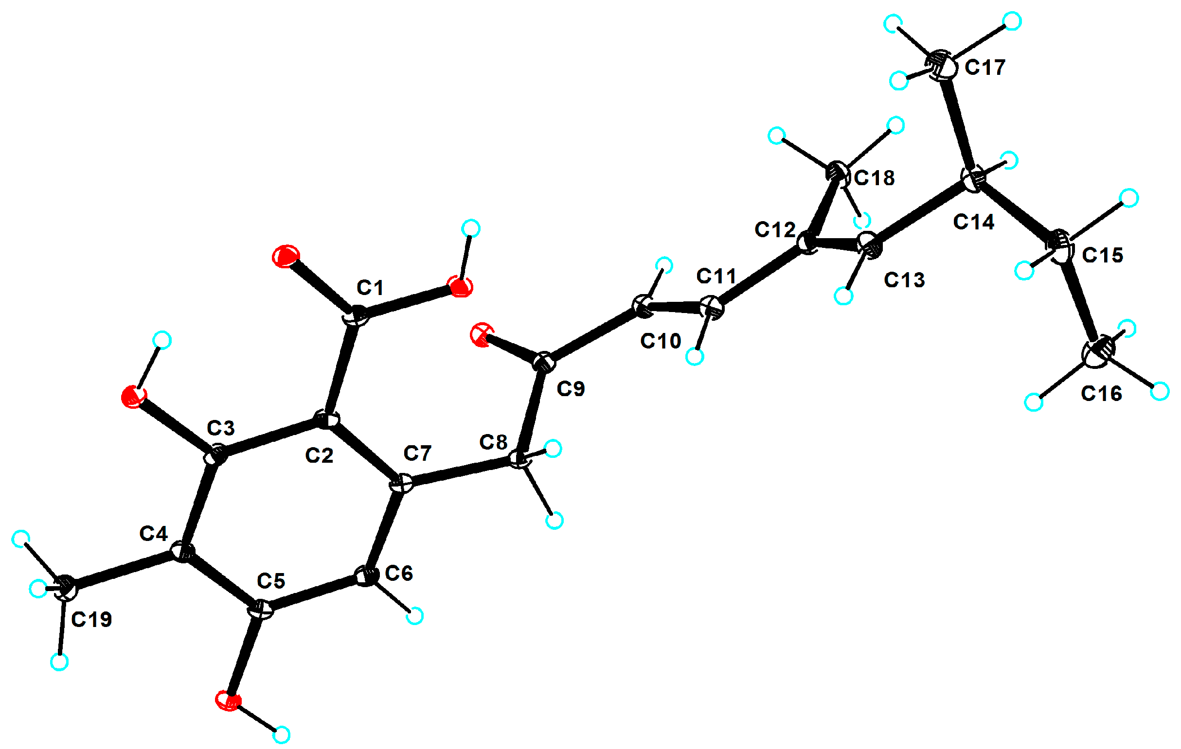
Compound 1 was obtained as a colorless crystal with the molecular formula of C19H24O5 based on the molecular ion peak at m/z 333.1696 [M + H]+ (calculated for C19H25O5, 333.1697), suggesting eight degrees of unsaturation. After analyzing detailed NMR data, 1 was found to have a benzoic acid skeleton similar to that of 2,4-dihydroxy-6-(5,7-dimethyl-2-oxo-trans-3-trans-5-nonadienyl)-3-methylbenzaldehyde [21], except for the aldehyde group being a carboxyl group in 1. The downshift of C-1 (δC 173.23) in 1 and the mass data revealed the difference. The coupling constant (J10,11 = 15.9 Hz) and the NOESY correlations between H-10 (δH 6.12, d, J = 15.9 Hz) and H3-18 (δH 1.75, s) and between H-11 (δH 7.22, d, J = 15.9 Hz) and H-13 (δH 5.81, d, J = 9.7 Hz) suggested that the geometric configurations of the double bonds were determined to be 10E and 12E. Compared with the optical rotation data of 2,4-dihydroxy-6-(5,7-dimethyl-2-oxo-trans-3-trans-5-nonadienyl)-3-methylbenzaldehyde ( = +51.0 (c 1.0, CHCl3)), the absolute configuration of 1 ( = +31.5 (c 0.02, CHCl3)) was determined as 14S. After repeated crystallization, the crystal of compound 1 was obtained, and the absolute configuration of compound 1 was unambiguously confirmed as 14S based on the Cu Kα radiation data with a good flack parameter (−0.01 (8)) (Figure 2). Compound 1 was named subversin A.
Compounds 2–3 were obtained as oils with the molecular formulas of C13H14O3 and C13H14O4 on the basis of molecular ion peaks at m/z 219.1018 [M + H]+ (calculated for C13H15O3, 219.1016) and 235.0966 [M + H]+ (calculated for C13H15O4, 235.0965), respectively. NMR data showed that 2-3 shared the same isobenzofuran-1(3H)-one molecular framework. 2 was close to the known compound (S)-3-allyl-7-methoxyisobenzofuran-1(3H)-one [22], with the exception of the presence of one methyl (δC 18.12/δH 1.61) at the C-10 position in 2. The above change was proven by the 1H-1H COSY correlations between H-10/H-11 and the HMBC correlations from H3-11 (δH 1.61) to C-9 (δC 124.92) and C-10 (δC 131.17). Compared with 2, there was a hydroxyl group at the C-8 position in 3. This was confirmed by the detailed HMBC and 1H-1H COSY correlations (Figure 3). Thus, the planar structures of 2 and 3 were determined, and 2-3 were named subversins B–C.
The geometric configuration of the double bond of 2–3 was confirmed to be 9E via the NOESY correlations between H-9 and H3-11. Compound 2 was a racemic mixture ( = 0 (c 0.1, CHCl3)), which was separated via chiral HPLC into two optically pure enantiomers: (+)-2 and (−)-2 (Figure S72). Based on optical rotation data of the (S)-3-allyl-7-methoxyisobenzofuran-1(3H)-one ( = −43.8 (c 1.0, CHCl3)) [22], the absolute structures of (+)-2 ( = +18.5 (c 0.04, CHCl3)) and (−)-2 ( = −26.2 (c 0.05, CHCl3)) were defined as 3R and 3S, respectively. Additionally, the absolute configurations of (+)-2 and (−)-2 were confirmed further by ECD calculations, and the calculated ECD spectra for (3R)-2 and (3S)-2 agreed with the experimental curves (Figure S73). The relative configuration of 3 was confirmed as 3R*, 8S* (3b) by DP4+ probability analysis using GIAO NMR chemical shift calculations [23] (Table S31). The absolute configuration of 3 was determined through ECD calculation. The calculated ECD spectrum for (3R,8S)-3 agreed with the experimental curve (Figure S81).
Compound 4 was yielded as yellow oil with molecular formula of C12H14O2 according to the molecular ion peak at m/z 191.1074 [M + H]+ (calculated for C12H15O2, 191.1067), implying six degrees of unsaturation. According to the NMR data, 4 possessed an isobenzofuran skeleton and was similar to riboxylarinol B [24], and the differences were the presence of one double bond between C-9 and C-10 and the absence of the two hydroxyl groups on C-9 and C-10 in 4. The 1H-1H COSY correlations between H-3/H-8/H-9/H-10/H-11, the HMBC correlations from H-9 (δH 5.37–5.52, m) and H-10 (δH 5.59–5.64, m) to C-8 (δC 41.85) and C-11 (δC 18.23) and the mass data indicated the differences. The geometric configuration of the double bond was determined in the same way as 2. Compared with optical rotation data of the (S)-3-deoxyisoochracinic acid [25] = −13.0 (c 0.08, MeOH)), the configuration of 4 ( = −24.7 (c 0.08, MeOH)) was defined as 3S. Compound 4 was named subversin D.

Table 1.
1H NMR and 13C NMR data of compounds 1, 5 and 6 (700, 176 MHz, δ in ppm, J in Hz).
| Position | 1 a | 5 b | 6 b | |||
|---|---|---|---|---|---|---|
| δC | δH | δC | δH | δC | δH | |
| 1 | 173.23, C | 173.84, C | 159.53, C | |||
| 2 | 104.89, C | 103.91, CH | 6.17 (t, 2.1) | |||
| 3 | 162.93, C | 69.19, CH2 | 5.22 (s) | 160, C | ||
| 3a | 129.65, C | |||||
| 4 | 108.61, C | 146.91, C | 111.46, CH | 6.26 (s) | ||
| 5 | 159.10, C | 142.04, C | 141.57, C | |||
| 6 | 110.94, CH | 6.15 (s) | 156.63, C | 111.71, CH | 6.33 (s) | |
| 7 | 136.70, C | 99.61, CH | 6.94 (s) | 157.12, C | ||
| 7a | 121.58, C | |||||
| 8 | 47.47, CH2 | 4.09–4.16 (m) | 104.77, CH | 6.30 (s) | ||
| 9 | 197.23, C | 72.84, CH2 | 4.12 (m) 4.03–4.09 (m) | 157.35, C | ||
| 10 | 124.45, CH | 6.12 (d, 15.9) | 35.75, CH2 | 2.00–2.08 (m) 1.81–1.89 (m) | 120.99, C | |
| 11 | 146.61, CH | 7.22 (d, 15.9) | 39.92, CH | 2.68 (m) | 139.9, C | |
| 12 | 131.76, C | 184.64, C | 113.02, CH | 6.30 (s) | ||
| 13 | 148.35, CH | 5.81 (d, 9.7) | 18.46, CH3 | 1.19 (d, 6.8) | 26.46, CH2 | 3.48 (d, 7.0) |
| 14 | 34.37, CH | 2.44–2.48 (m) | 56.66, CH3 | 3.88 (s) | 140.17, CH | 6.60 (t, 6.8) |
| 15 | 29.48, CH2 | 1.36–1.42 (m) 1.24–1.30 (m) | 130.37, C | |||
| 16 | 11.84, CH3 | 0.81 (t, 7.4) | 175.62, C | |||
| 17 | 20.09, CH3 | 0.96 (d, 6.6) | 13.09, CH3 | 1.96 (s) | ||
| 18 | 12.29, CH3 | 1.75 (s) | 19.99, CH3 | 2.20 (s) | ||
| 19 | 8.14, CH3 | 1.94 (s) | 21.55, CH3 | 2.21 (s) | ||
a Spectra were measured in DMSO-d6; b spectra were measured in methanol-d4.
Table 2.
1H NMR and 13C NMR data of compounds 2–4 (700, 176 MHz, δ in ppm, J in Hz).
| Position | 2 a | 3 a | 4 b | |||
|---|---|---|---|---|---|---|
| δC | δH | δC | δH | δC | δH | |
| 1 | 171.03, C | 171.06, C | 58.97, CH2 | 4.95 (s) | ||
| 3 | 81.58, CH | 5.46–5.53 (m) | 84.49, CH | 5.42 (d, 4.0) | 70.89, CH | 4.82 (dd, 7.9, 5.3) |
| 3a | 154.00, C | 151.26, C | 142.03, C | |||
| 4 | 115.06, CH | 7.10 (d, 7.3) | 116.16, CH | 7.15 (d, 7.6) | 118.13, CH | 6.98 (d, 7.7) |
| 5 | 137.92, CH | 7.69 (dd, 8.1, 7.7) | 137.57, CH | 7.67 (t, 8.0) | 129.32, CH | 7.20 (t, 7.9) |
| 6 | 112.03, CH | 7.08 (d, 8.0) | 112.25, CH | 7.09 (d, 8.3) | 116.16, CH | 6.82 (d, 8.0) |
| 7 | 159.92, C | 159.90, C | 156.63, C | |||
| 7a | 114.35, C | 114.97, C | 122.95, C | |||
| 8 | 38.31, CH2 | 2.68–2.75 (m) 2.46–2.59 (m) | 74.23, CH | 4.42–4.45 (m) | 41.85, CH2 | 2.38–2.45 (m) |
| 9 | 124.92, CH | 5.25–5.35 (m) | 129.15, CH | 5.51 (m) | 126.79, CH | 5.37–5.52 (m) |
| 10 | 131.17, CH | 5.58 (m) | 130.49, CH | 5.73 (m) | 130.04, CH | 5.59–5.64 (m) |
| 11 | 18.12, CH3 | 1.61 (dd, 6.5, 1.4) | 17.94, CH3 | 1.68 (m) | 18.23, CH3 | 1.70 (d, 6.3) |
| 12 | 56.33, CH3 | 3.96 (s, 3H) | 56.30, CH3 | 3.96 (s) | ||
a Spectra were measured in methanol-d4; b spectra were measured in chloroform-d.
Compound 5 was obtained as a yellow oil with the molecular formula of C14H16O7 on the basis of the molecular ion peak at m/z 295.0829 [M − H]− (calculated for C14H15O7, 295.0823), suggesting seven degrees of unsaturation. The 1H and 13C NMR data indicated that 5 also had a isobenzofuran-1(3H)-one unit, similar to the known compound (+)-5-(3-carboxy-butoxy)-7-hydroxy-4,6-dimethylphthalide [26,27], with the exception of the different substituents on C-4, C-6 and C-7 of the benzene ring in 5. The differences were proven by the HMBC correlations from H2-3 (δH 5.22, s) to C-4 (δC 146.91, s), from H3-14 (δH 3.88, s) to C-6 (δC 156.63) and from H-7 (δH 6.94, s) to C-1 (δC 173.84) and C-7a (δC 121.58). Compound 5 was also a racemic mixture ( = 0 (c 0.1, MeOH)), which was separated via chiral HPLC into two optically pure enantiomers: (+)-5 and (−)-5 (Figure S74). Based on optical rotation data of the (+)-5-(3-carboxy-butoxy)-7-hydroxy-4,6-dimethylphthalide ( = +6.2 (c 0.36, MeOH)), the absolute structures of (+)-5 ( = +7.1 (c 0.1, MeOH)) and (−)-5 ( = −7.7 (c 0.08, MeOH)) were defined as 11S and 11R, respectively. Compound 5 was named subversin E.
Compound 6 was purified as a yellow oil with the molecular formula of C19H20O5 according to the molecular ion peak at m/z 329.1393 [M + H]+ (calculated for C19H21O5, 329.1384), indicating 10 degrees of unsaturation. The NMR data of 6 were close to those of the known compound verticilatin [28] and the changes were the presence of a carboxyl (δC 175.62) and the absence of one methyl group in 6. This suggested that the carboxyl group might replace the methyl group in 6. The above deduction was supported via the HMBC correlations from H-14 (δH 6.60, t, J = 6.8 Hz) and H3-17 (δH 1.96, s) to C-16 (δC 175.62) and the MS data. The NOESY correlations between H2-13 (δH 3.48, d, J = 7.0 Hz) and H3-17 indicated that the geometric configuration of the double bond was determined as 14E. Compound 6 was named subversic acid A.
Compound 7 was a yellow oil with the molecular formula of C12H14O4 based on the molecular ion peak at m/z 245.0796 [M + Na]+ (calculated for C12H14NaO4, 245.0784), implying six degrees of unsaturation. The NMR and the mass data of 7 proved that 7 had the same planar structure as the known compound wortmannine G [29]. However, based on the optical rotation data of wortmannine G ( = +4.0 (c 4 mM, CHCl3)), the absolute structure of 7 ( = −5.9 (c 0.1, CHCl3)) was defined as 3R. Compound 7 was named epi-wortmannine G.
Compound 8 was obtained as a white powder with the molecular formula of C9H8O4 on the basis of the molecular ion peak at m/z 181.0498 [M + H]+ (calculated for C9H9O4, 181.0495), suggesting six degrees of unsaturation. The 1H-1H COSY correlations between H-5/H-6 and the HMBC correlations from H2-3 (δH 5.22, s) to C-1 (δC 172.00), C-3a (δC 136.44), C-4 (δC 147.22) and C-7a (δC 114.58), from H-5 (δH 7.05, d, J = 8.7 Hz) to C-3a (δC 136.44) and C-7 (δC 152.87), and from H-6 (δH 6.92, d, J = 8.7 Hz) to C-4 (δC 147.22) and C-7a (δC 114.58), confirmed the planar structure of compound 8. Compound 8 was first synthesized by Keay [30] in 1984 and this was the first time that compound 8 had been reported from nature.
Table 3.
1H NMR and 13C NMR data of compounds 7–8 (700, 176 MHz, δ in ppm, J in Hz).
| Position | 7 a | 8 a | ||
|---|---|---|---|---|
| δC | δH | δC | δH | |
| 1 | 58.42, CH2 | 5.03 (d, 16.2) 4.84 (d, 16.2) | 172.00, C | |
| 3 | 97.69, C | 68.88, CH2 | 5.22 (s) | |
| 3a | 136.44, C | |||
| 4 | 193.27, C | 147.22, C | ||
| 4a | 130.64, C | |||
| 5 | 118.59, CH | 7.46 (d, 7.6) | 123.37, CH | 7.05 (d, 8.7) |
| 6 | 128.87, CH | 7.23 (t, 7.9) | 113.43, CH | 6.92 (d, 8.7) |
| 7 | 120.56, CH | 7.00 (dd, 8.0, 0.7) | 152.87, C | |
| 7a | 114.58, C | |||
| 8 | 154.19, C | 56.53, CH3 | 3.87 (s) | |
| 8a | 130.29, C | |||
| 9 | 39.75, CH2 | 2.02 (ddd, 13.5, 11.7, 4.8) 1.77 (ddd, 13.6, 11.7, 4.8) | ||
| 10 | 17.61, CH2 | 1.51 (m) 1.31–1.39 (m) | ||
| 11 | 14.76, CH3 | 0.93 (t, 7.4) | ||
a Spectra were measured in methanol-d4.
2.2. Biological Test
All compounds were tested for antibacterial activity, antioxidant activity, α-glucosidase inhibitory activity and acetylcholinesterase inhibitory activity. Compound 10 displayed inhibitory activity against five Gram-positive bacteria (B. subtilis, E. profundum, E. faecalis, S. aureus and MRSA) and one Gram-negative bacterium (A. baumannii). Specifically, compound 10 potently inhibited B. subtilis with an MIC value of 0.1 μM. No obvious activity of the compounds was observed in terms of antioxidant activity and enzyme inhibitory activity. The IC50 and MIC values of the compounds larger than 200 μM were not included in the results of the bioassays (Tables S32 and S33). The structure and activity analysis of 10 and its analogues 6, 9 and 11 indicated that the dibenzofuran skeleton played an essential role in the antibacterial activity, which was consistent with the literature [31,32].
By the end of 2023, over 575 isobenzofuran derivatives had been reported, mainly in Umbelliferae plants and fungi [33,34,35,36,37,38]. Among the isobenzofuran derivatives, 97 originate from marine fungi. Based on the literature [36,37], it was reported that isobenzofuran derivatives exhibited effects on neuroprotective, anti-inflammatory, hepatoprotective and cytotoxicity assays.
3. Materials and Methods
3.1. Fungal Materials
The fungus was separated from the sediment (−1363 m) obtained from the Haima cold seep in 2021. The DNA of the fungus was extracted according to the instructions of the DNA extraction kit. Then, a polymerase chain reaction instrument was used to amplify the purified DNA of the fungus with ITS primers (ITS1:5’-CTTGGTCATTTAGAGGAAGTAA-3’; ITS4: 5’-TCCTCCGCTTATTGATATGC-3’). According to the ITS region sequence in the NCBI database, the strain was 99.59% identical to A. subversicolor (accession No. NR_135446.1). In terms of the results of the morphological features and the ITS region sequence, the strain was determined to be A. subversicolor and was named A. subversicolor CYH-17. The fungus was stored in the Research Network for Applied Microbiology (RNAM) Center for Marine Microbiology, South China Sea Institute of Oceanology, Chinese Academy of Sciences.
3.2. General
Sephadex LH-20 (GE Healthcare, Stockholm, Sweden) and 100–200 and 200–300 mesh Silica gel (Qingdao Marine Chemical Group Co., Qingdao, China) were used for column chromatography (CC), while an Agilent 1260 HPLC (Agilent Technologies, Santa Clara, CA, USA) equipped with an ODS C-18 column (5 μm, 10 × 250 mm) was used for HPLC separation. In addition, all the data for structural elucidation were collected from an MCP500 automatic polarimeter (AntonPaar, Graz, Austria), UV-2600 spectrometer (Shimadzu, Kyoto, Japan), IR Affinity-1 spectrometer (Shimadzu, Kyoto, Japan), AVANCE IIIHD 700 MHz Digital NMR Spectrometer (Bruker, Billerica, MA, USA), MaXis quadrupole time-of-flight mass spectrometer (Bruker, Mannheim, Germany), Rigaku XtaLAB AFC12 single-crystal diffractometer (Rigaku, Tokyo, Japan) and chirascan circular dichroism spectrometer (Applied Photophysics, Leatherhead, UK).
3.3. Fermentation, Extraction and Purification
The fungus was fermented on PDA plates and then transferred to 200 flasks containing medium (rice 100.0 g, artificial sea salt 3.0 g, distilled water 0.1 L). The strain was fermented statically at 25 °C for a month. The rice was extracted with ethyl acetate three times to gather 77.0 g of the ethyl acetate extract.
The ethyl acetate extract of the fungus was fractionated by silica gel column chromatography (CC) eluted with petroleum ether and ethyl acetate gradient (100:0 to 0:100) to obtain seven fractions (Frs.1–7). Fr.4 was purified via HPLC with MeOH-H2O gradient (5:95 to 100:0) to obtain six fractions (Frs.4.1–Fr.4.6). Fr.4.3 was fractionated via silica gel CC eluted with CH2Cl2-MeOH (100:0 to 90:10) to yield two parts (Frs.4.3.1–Fr.4.3.2). Fr.4.3.1 was purified by HPLC (65% MeOH/H2O) to obtain 7 (3.1 mg). Fr.4.3.2 was subjected to Sephadex LH-20 (CH2Cl2/MeOH = 1:1) to give 4 (1.5 mg). Fr.4.4 was fractionated via silica gel CC eluted with CH2Cl2-MeOH (100:0 to 90:10) to yield three subfractions Frs.4.4.1–Fr.4.4.3. Fr.4.4.2 was subjected to Sephadex LH-20 (CH2Cl2/MeOH = 1:1) to give 9 (46.7 mg) and three fractions (Frs.4.4.2.1–4.4.2.3). Fr.4.4.2.1 was purified by HPLC (60% ACN/H2O) to obtain 2 (1.1 mg). Fr.4.4.2.2 was separated by HPLC (60% ACN/H2O) to yield 11 (1.8 mg). Fr.4.4.2.3 was fractionated by HPLC (60% ACN/H2O) to give 10 (3.9 mg). Fr.4.4.3 was subjected to Sephadex LH-20 (CH2Cl2/MeOH = 1:1) to obtain Fr.4.4.3.1. Fr.4.4.3.1 was separated via silica gel CC eluted with CH2Cl2-MeOH (100:0 to 90:10) to yield 13 (2.2 mg) and 6 (3.1 mg). Fr.4.5 was fractionated via silica gel CC eluted with CH2Cl2-MeOH (100:0 to 95:5) to give the two parts Fr.4.5.1–Fr.4.5.2. Fr.4.5.1 was purified by HPLC (70% ACN/H2O) to obtain 12 (3.0 mg). Fr.4.5.2 was fractionated via Sephadex LH-20 (CH2Cl2/MeOH = 1:1) to yield 1 (6.0 mg). Fr.5 was separated via HPLC with MeOH-H2O gradient (5:95 to 100:0) to obtain two fractions (Frs.5.1–Fr.5.2). Fr.5.2 was subjected to Sephadex LH-20 (CH2Cl2/MeOH = 1:1) to give Fr.5.2.1–Fr.5.2.2. Fr.5.2.1 was purified by HPLC (43% MeOH/H2O) to obtain 3 (2.2 mg). Fr.5.2.2 was fractionated by HPLC (55% MeOH/H2O) to obtain 8 (1.2 mg). Fr.6 was separated via HPLC with MeOH-H2O gradient (5:95 to 100:0) to yield three fractions (Frs.6.1–Fr.6.3). Fr.6.2 was subjected to Sephadex LH-20 (MeOH) to give Fr.6.2.1. Fr.6.2.1 was purified by HPLC (37% ACN/H2O) to obtain 5 (1.5 mg).
3.3.1. Subversin A (1)
Colorless crystal; = +41.1 (c 0.1, MeOH), = +31.5 (c 0.02, CHCl3); HR-ESI-MS at m/z 333.1696 [M + H]+ (calculated for C19H25O5, 333.1697); UV (MeOH) λmax (log ε) 219 (4.38), 279 (4.33) nm; IR (film) νmax 3340, 2949, 1647, 1018, 671 cm−1; 1H NMR and 13C NMR data, see Table 1.
3.3.2. Subversin B (2)
Colorless oil; = 0 (c 0.1, CHCl3); HR-ESI-MS at m/z 219.1018 [M + H]+ (calculated for C13H15O3, 219.1016); UV (MeOH) λmax (log ε) 215 (4.12), 235 (3.94) 298 (3.74) nm; IR (film) νmax 2926, 1759, 1607,1298,1038,787 cm−1; 1H NMR and 13C NMR data, see Table 2.
(+)-2. Colorless oil; = +18.5 (c 0.04, CHCl3); CD (MeOH): 209 nm (Δε = 2.94), 240 nm (Δε = −1.11), 296 nm (Δε = −0.65).
(−)-2. Colorless oil; = −26.2 (c 0.05, CHCl3); CD (MeOH): 210 nm (Δε = −2.33), 241 nm (Δε = 1.06), 296 nm (Δε = 0.66).
3.3.3. Subversin C (3)
Colorless oil; = +6.4 (c 0.1, MeOH); HR-ESI-MS at m/z 235.0966 [M + H]+ (calculated for C13H15O4, 235.0965); UV (MeOH) λmax (log ε) 213 (4.13), 236 (3.84) 299 (3.64) nm; IR (film) νmax 3385, 2954, 1744, 1026, 689 cm−1; CD (MeOH): 209 nm (Δε = −9.85), 243 nm (Δε = 2.31), 298 nm (Δε = 1.25); 1H NMR and 13C NMR data, see Table 2.
3.3.4. Subversin D (4)
Yellow oil; = −17 (c 0.1, CHCl3), = −24.7 (c 0.08, MeOH); HR-ESI-MS at m/z 191.1074 [M + H]+ (calculated for C12H15O2, 191.1067); UV (MeOH) λmax (log ε) 220 (3.72), 281 (3.31) nm; IR (film) νmax 3327, 2945,1018, 671 cm−1; 1H NMR and 13C NMR data, see Table 2.
3.3.5. Subversin E (5)
Yellow oil; = 0 (c 0.1, MeOH); HR-ESI-MS at m/z 295.0829 [M − H]− (calculated for C14H15O7, 295.0823); UV (MeOH) λmax (log ε) 216 (4.21), 266 (3.65) nm; IR (film) νmax 3319, 2947, 1651, 1018, 675 cm−1; 1H NMR and 13C NMR data, see Table 1.
(+)-5. Yellow oil; = +7.1 (c 0.1, MeOH); CD (MeOH): 223 nm (Δε = 0.30).
(−)-5. Yellow oil; = −7.7 (c 0.08, MeOH); CD (MeOH): 224 nm (Δε = −0.39).
3.3.6. Subversic Acid A (6)
Yellow oil; HR-ESI-MS at m/z 329.1393 [M + H]+ (calculated for C19H21O5, 329.1384); UV (MeOH) λmax (log ε) 219 (4.61), 282 (3.73) nm; IR (film) νmax 3364, 1595, 1522, 1153, 837, 677 cm−1; 1H NMR and 13C NMR data, see Table 1.
3.3.7. epi-Wortmannine G (7)
Yellow oil; = −5.9 (c 0.1, CHCl3); HR-ESI-MS at m/z 245.0796 [M + Na]+ (calculated for C12H14NaO4, 245.0784); UV (MeOH) λmax (log ε) 223 (4.18), 260 (3.86) 318 (3.41) nm; IR (film) νmax 3334, 2954, 1690,1020, 754, 677 cm−1; 1H NMR and 13C NMR data, see Table 3.
3.3.8. 4-Hydroxy-7-methoxyphthalide (8)
White powder; HR-ESI-MS at m/z 181.0498 [M + H]+ (calculated for C9H9O4, 181.0495); UV (MeOH) λmax (log ε) 217 (3.96), 237 (3.57) 324 (3.37) nm; IR (film) νmax 3343, 2947, 1649, 1018, 669 cm−1; 1H NMR and 13C NMR data, see Table 3.
3.4. X-ray Crystal Structure Analysis
Crystallographic data of compound 1 were yielded on a Rigaku XtaLAB AFC12 single-crystal diffractometer (Rigaku, Japan) via Cu Kα radiation. The crystal was kept at 100.5 (9) K during the data collection. Using Olex2, the structure was solved with the SHELXT structure solution program using Intrinsic Phasing and refined with the SHELXL refinement package using Least Squares minimization. The crystallographic data of compound 1 were stored in the Cambridge Crystallographic Data Centre database (deposition numbers 2324024). Copies of the data are available free of charge from the CCDC at www.ccdc.cam.ac.uk, accessed on 6 March 2022.
Crystal data for compound 1: C38H48O10, M = 664.76, triclinic, space group P1 (no.1), a = 6.32630 (10) Å, b = 10.8354 (2) Å, c = 13.0039 (2) Å, α = 91.6160(10)°, β = 103.8220 (10)°, γ = 92.5990 (10)°, V = 863.99 (3) Å3, Z = 1, T = 100.5 (9) K, μ (Cu Kα) = 0.751 mm−1, Dcalc = 1.278 g/cm3, 17397 reflections measured (7.006° ≤ 2Θ ≤ 148.54°), 6466 unique (Rint = 0.0298, Rsigma = 0.0318) which were used in all calculations. The final R1 was 0.0340 (I > 2σ (I)) and wR2 was 0.0928 (all data). The goodness of fit on F2 was 1.079. Flack parameter = −0.01 (8), melting point: 184.0–185.0 °C.
3.5. Bioassays
3.5.1. Antibacterial Assay
The bacteria Vibrio alginolyticus XSBZ14, Enterococcus faecalis ATCC 29212, Acinetobacter baumannii ATCC 19606, Escherichia coli ATCC 25922, Bacillus subtilis BS01, Klebsiella pneumoniae ATCC 13883, Exiguobacterium profundum Staphylococcus aureus ATCC 29213 and MRSA 107352 were used to measure antibacterial activity. The procedures of the antibacterial test were the same as Zhang [39]. Ciprofloxacin was used as the positive control.
3.5.2. AChE Inhibitory Assay
The procedures of the AChE inhibitory test were the same as Yang [40]. Tacrine was used as the positive control.
3.5.3. α-Glucosidase Inhibitory Assay
The procedures of the α-glucosidase inhibitory test were the same as Ding [41]. Acarbose was used as the positive control.
3.5.4. DPPH Radical Scavenging Assay
The procedures of the DPPH radical scavenging test were the same as Zhong [42]. Ascorbic acid was used as the positive control.
3.6. Chiral HPLC Separation of Compounds 2 and 5
Chiral HPLC separations of the compounds were recorded on the HPLC (Agilent 1260) equipped with CHIRALPAK® IA (250 × 4.6 mm, 5 μm). A phase: hexane with 0.1% formic acid; B phase: isopropanol.
Compound 2: gradient program: 0 min (93%A–7%B) to 25 min (93%A–7%B); flow rate: 1 mL/min; detection: UV 215 nm.
Compound 5: gradient program: 0 min (80%A–20%B) to 12 min (80%A–20%B); flow rate: 1 mL/min; detection: UV 215 nm.
4. Conclusions
Chemical exploration of the fungus Aspergillus subversicolor CYH-17 resulted in the isolation and elucidation of seven new phenol derivatives, subversins A–E (1–5), subversic acid A (6) and epi-wortmannine G (7); one new natural product, 4-hydroxy-7-methoxyphthalide (8); and five known secondary metabolites (9–13). The structural frameworks of the compounds included benzoic acid, isobenzofuran-1(3H)-one, isobenzofuran and isochroman-4-one. Compound 10 inhibited six bacteria with MIC values ranging from 0.1 to 50 μM. No obvious activity of the compounds was seen in the enzyme inhibitory activity and antioxidant activity. Future research should focus on exploring the diverse structures of dibenzofuran and isobenzofuran derivatives through OSMAC strategies and elucidating the structure–activity relationship of the compounds in this fungus.
Supplementary Materials
The following supporting information can be downloaded at: https://www.mdpi.com/article/10.3390/md22030117/s1, HR-ESI-MS, 1H NMR, 13C NMR and DEPT 135°, HSQC, HMBC, 1H-1H COSY, NOESY, UV, IR spectra of compounds 1–8 (Figures S1–S71); the chiral HPLC separation profile 2 and 5 and the ECD calculation and results of 2 (Figures S72–S75, Table S1); the computational methods and results of 3 (Figures S76–S81 and Tables S2–S31); the ITS sequence of A. subversicolor CYH-17 and the antibacterial activity, α-glucosidase inhibitory activity and antioxidant activity of the compounds (Tables S32–S33) [43,44,45].
Author Contributions
Y.-H.C. contributed to separation, purification and identification and writing the manuscript. W.-P.D. and H.Y. contributed to identification. Z.-H.X. and J.-M.W. contributed to the bioassays. F.-Z.W. and S.Z. contributed to designing and supervising the article and revising the manuscript. All authors have read and agreed to the published version of the manuscript.
Funding
This work was financially supported by the National Natural Science Foundation of China (No. 41890853) and the Finance Science and Technology Project of Hainan Province (No. ZDKJ202018).
Data Availability Statement
Data is contained within the article or Supplementary Material.
Acknowledgments
We sincerely appreciate the help from Ai-Jun Sun, Yun Zhang, Xuan Ma and Xiao-Hong Zheng from the Equipment Public Service Center of South China Sea Institute of Oceanology, Chinese Academy of Sciences for measuring spectroscopic data.
Conflicts of Interest
The authors declare no conflicts of interest.
References
- Paull, C.K.; Hecker, B.; Commeau, R.; Freeman-Lynde, R.P.; Neumann, C.; Corso, W.P.; Golubic, S.; Hook, J.E.; Sikes, E.; Curray, J. Biological communities at the Florida escarpment resemble hydrothermal vent taxa. Science 1984, 226, 965–967. [Google Scholar] [CrossRef] [PubMed]
- Zhong, S.; Feng, J.; Kong, J.; Huang, Y.; Chen, X.; Zhang, S. Differences in bacterial co-occurrence networks and ecological niches at the surface sediments and bottom seawater in the Haima cold seep. Microorganisms 2023, 11, 3001. [Google Scholar] [CrossRef] [PubMed]
- Ling, J.; Guan, H.; Liu, L.; Tao, J.; Li, J.; Dong, J.; Zhang, S. The diversity, composition, and putative functions of gill-associated bacteria of bathymodiolin mussel and vesicomyid clam from haima cold seep, South China Sea. Microorganisms 2020, 8, 1699. [Google Scholar] [CrossRef] [PubMed]
- Nethupul, H.; Stöhr, S.; Zhang, H. Review of Ophioplinthaca Verrill, 1899 (Echinodermata, Ophiuroidea, Ophiacanthidae), description of new species in Ophioplinthaca and Ophiophthalmus, and new records from the Northwest Pacific and the South China Sea. Zookeys 2022, 1099, 155–202. [Google Scholar] [CrossRef]
- Leduc, D. Six new species of free-living nematodes (Nematoda: Enoplida) from deep-sea cold seeps on Hikurangi Margin, New Zealand. PeerJ 2023, 11, e14867. [Google Scholar] [CrossRef]
- Shekarriz, E.; Chen, J.; Xu, Z.; Liu, H. Disentangling the functional role of fungi in cold seep sediment. Microbiol. Spectr. 2023, 11, e0197822. [Google Scholar] [CrossRef] [PubMed]
- Dong, X.; Zhang, C.; Peng, Y.; Zhang, H.X.; Shi, L.D.; Wei, G.; Hubert, C.R.J.; Wang, Y.; Greening, C. Phylogenetically and catabolically diverse diazotrophs reside in deep-sea cold seep sediments. Nat. Commun. 2022, 13, 4885. [Google Scholar] [CrossRef]
- Huang, X.; Wang, Y.; Zhou, L.; Wang, W.; Anjum, K.; Zhang, J.; Zhang, G.; Zhu, T.; Li, D.; Che, Q. Glycosylated 24-membered lactones and unsaturated fatty acids from cold-seep-derived Bacillus sp. HDN 20-1259. Tetrahedron Lett. 2023, 121, 154477. [Google Scholar] [CrossRef]
- Cong, M.; Pang, X.; Zhao, K.; Song, Y.; Liu, Y.; Wang, J. Deep-sea natural products from extreme environments: Cold seeps and hydrothermal vents. Mar. Drugs 2022, 20, 404. [Google Scholar] [CrossRef]
- Yan, L.H.; Du, F.Y.; Li, X.M.; Yang, S.Q.; Wang, B.G.; Li, X. Antibacterial indole diketopiperazine alkaloids from the deep-sea cold seep-derived fungus Aspergillus chevalieri. Mar. Drugs 2023, 21, 195. [Google Scholar] [CrossRef]
- Song, Q.; Yang, S.Q.; Li, X.M.; Hu, X.Y.; Li, X.; Wang, B.G. Aromatic polyketides from the deep-sea cold-seep mussel associated endozoic fungus Talaromyces minioluteus CS-138. Mar. Drugs 2022, 20, 529. [Google Scholar] [CrossRef]
- Chi, L.P.; Li, X.M.; Wan, Y.P.; Li, X.; Wang, B.G. Ophiobolin sesterterpenoids and farnesylated phthalide derivatives from the deep sea cold-seep-derived fungus Aspergillus insuetus SD-512. J. Nat. Prod. 2020, 83, 3652–3660. [Google Scholar] [CrossRef]
- Yang, S.Q.; Song, Q.; Li, X.M.; Li, X.; Li, H.L.; Meng, L.H.; Wang, B.G. Antimicrobial polyketides and sesquiterpene lactones from the deep-sea cold-seep-derived fungus Talaromyces minioluteus CS-113 triggered by the histone deacetylase inhibitor SAHA. Org. Biomol. Chem. 2023, 21, 2575–2585. [Google Scholar] [CrossRef]
- Li, C.P.; Song, Y.P.; Wang, B.G.; Ji, N.Y. Sulfurated and iodinated metabolites from the cold-seep fungus Cladosporium cladosporioides 8-1. Tetrahedron Lett. 2022, 93, 153689. [Google Scholar] [CrossRef]
- Che, Y.H.; Wang, J.F.; Shi, X.F.; Ding, W.P.; Xiao, Z.H.; Wu, J.M.; Wang, F.Z.; Zhang, S. 8R-methoxy-9R-hydroxyl-fumitremorgin C, a new diketopiperazine alkaloid from Haima cold seep-derived fungus Aspergillus fumigatus CYH-5. Nat. Prod. Res. 2023. [Google Scholar] [CrossRef]
- Che, Y.H.; Wang, J.F.; Ding, W.P.; Xiao, Z.H.; Shi, X.F.; Wu, J.M.; Wang, F.Z.; Zhang, S. New diketopiperazine alkaloids from the Haima cold seep-derived fungus Toxicocladosporium sp. CYH-18. Phytochem. Lett. 2024, 60, 96–100. [Google Scholar] [CrossRef]
- Weng, H.Z.; Zhu, J.Y.; Yuan, F.Y.; Tang, Z.Y.; Tian, X.Q.; Chen, Y.; Fan, C.Q.; Tang, G.H.; Yin, S. Homo/hetero-dimers of aromatic bisabolane sesquiterpenoids with neuroprotective activity from the fungus Aspergillus versicolor A18 from south China sea. Mar. Drugs 2022, 20, 322. [Google Scholar] [CrossRef] [PubMed]
- Tanahashi, T.; Takenaka, Y.; Nagakura, N.; Hamada, N. Dibenzofurans from the cultured lichen mycobionts of Lecanora cinereocarnea. Phytochemistry 2001, 58, 1129–1134. [Google Scholar] [CrossRef] [PubMed]
- Yao, Q.; Wang, J.; Zhang, X.; Nong, X.; Xu, X.; Qi, S. Cytotoxic polyketides from the deep-sea-derived fungus Engyodontium album DFFSCS021. Mar. Drugs 2014, 12, 5902–5915. [Google Scholar] [CrossRef] [PubMed]
- Fredimoses, M.; Zhou, X.; Ai, W.; Tian, X.; Yang, B.; Lin, X.; Liu, J.; Liu, Y. Emerixanthone E, a new xanthone derivative from deep sea fungus Emericella sp. SCSIO 05240. Nat. Prod. Res. 2019, 33, 2088–2094. [Google Scholar] [CrossRef] [PubMed]
- Matsuzaki, K.; Tahara, H.; Inokoshi, J.; Tanaka, H.; Masuma, R.; Omura, S. New brominated and halogen-less derivatives and structure-activity relationship of azaphilones inhibiting gp120-CD4 binding. J. Antibiot. 1998, 51, 1004–1011. [Google Scholar] [CrossRef] [PubMed]
- Cabrera, J.M.; Tauber, J.; Krische, M.J. Enantioselective iridium-catalyzed phthalide formation through internal redox allylation of phthalaldehydes. Angew. Chem. Int. Ed. Engl. 2018, 57, 1390–1393. [Google Scholar] [CrossRef]
- Zanardi, M.M.; Sarotti, A.M. Sensitivity analysis of DP4+ with the probability distribution terms: Development of a universal and customizable method. J. Org. Chem. 2021, 86, 8544–8548. [Google Scholar] [CrossRef] [PubMed]
- Rebollar-Ramos, D.; Macías-Ruvalcaba, M.L.; Figueroa, M.; Raja, H.A.; González-Andrade, M.; Mata, R. Additional α-glucosidase inhibitors from Malbranchea flavorosea (Leotiomycetes, Ascomycota). J. Antibiot. 2018, 71, 862–871. [Google Scholar] [CrossRef]
- Höller, U.; Gloer, J.B.; Wicklow, D.T. Biologically active polyketide metabolites from an undetermined fungicolous hyphomycete resembling Cladosporium. J. Nat. Prod. 2002, 65, 876–882. [Google Scholar] [CrossRef]
- Katoh, N.; Nakahata, T.; Kuwahara, S. Synthesis of novel antifungal phthalides produced by a wheat rhizosphere fungus. Tetrahedron 2008, 64, 9073–9077. [Google Scholar] [CrossRef]
- Takahashi, K.; Koshino, H.; Narita, Y.; Yoshihara, T. Novel antifungal compounds produced by Sterile Dark, an unidentified wheat rhizosphere fungus. Biosci. Biotechnol. Biochem. 2005, 69, 1018–1020. [Google Scholar] [CrossRef] [PubMed]
- Wei, P.Y.; Li, L.; Yang, C.G.; Luo, D.Q.; Zheng, Z.H.; Lu, X.H.; Shi, B.Z. A novel oxybis cresol verticilatin with highly varying degrees of biological activities from the insect pathogenic fungus Paecilomyces verticillatus. J. Asian Nat. Prod. Res. 2014, 16, 1153–1157. [Google Scholar] [CrossRef]
- Zhao, J.W.; Yang, Z.D.; Zhou, S.Y.; Yang, L.J.; Sun, J.H.; Yao, X.J.; Shu, Z.M.; Li, S. Wortmannine F and G, two new pyranones from Talaromyces wortmannii LGT-4, the endophytic fungus of Tripterygium wilfordii. Phytochem. Lett. 2019, 29, 115–118. [Google Scholar] [CrossRef]
- Keay, B.A.; Rodrigo, R. A convergent synthesis of (±)daunomycinoni. Tetrahedron 1984, 40, 4597–4607. [Google Scholar] [CrossRef]
- Shou, Q.; Banbury, L.K.; Renshaw, D.E.; Lambley, E.H.; Mon, H.; Macfarlane, G.A.; Griesser, H.J.; Heinrich, M.M.; Wohlmuth, H. Biologically active dibenzofurans from Pilidiostigma glabrum, an endemic Australian Myrtaceae. J. Nat. Prod. 2012, 75, 1612–1617. [Google Scholar] [CrossRef] [PubMed]
- Oramas-Royo, S.; Pantoja, K.D.; Amesty, Á.; Romero, C.; Lorenzo-Castrillejo, I.; Machín, F.; Estévez-Braun, A. Synthesis and antibacterial activity of new symmetric polyoxygenated dibenzofurans. Eur. J. Med. Chem. 2017, 141, 178–187. [Google Scholar] [CrossRef] [PubMed]
- Huang, L.J.; Li, X.A.; Jin, M.Y.; Guo, W.X.; Lei, L.R.; Liu, R.; Zhang, M.Z.; Guo, D.L.; Wang, D.; Zhou, Y.; et al. Two previously undescribed phthalides from Talaromyces amestolkiae, a symbiotic fungus of Syngnathus acus. J. Asian Nat. Prod. Res. 2023, 25, 147–155. [Google Scholar] [CrossRef] [PubMed]
- Wan, S.J.; Ren, H.G.; Jiang, J.M.; Xu, G.; Xu, Y.; Chen, S.M.; Chen, G.; Zheng, D.; Yuan, M.; Zhang, H.; et al. Two novel phenylpropanoid trimers from Ligusticum chuanxiong Hort with inhibitory activities on alpha-hemolysin secreted by Staphylococcus aureus. Front. Chem. 2022, 10, 877469. [Google Scholar] [CrossRef]
- Huang, L.; Peng, C.; Guo, L.; Feng, R.; Shu, H.Z.; Tian, Y.C.; Zhou, Q.M.; Xiong, L. Six pairs of enantiomeric phthalide dimers from the rhizomes of Ligusticum chuanxiong and their absolute configurations and anti-inflammatory activities. Bioorg. Chem. 2022, 127, 105970. [Google Scholar] [CrossRef]
- Wei, X.; Zeng, Y.; Sun, C.; Meng, F.; Wang, Y. Recent advances in natural phthalides: Distribution, chemistry, and biological activities. Fitoterapia 2022, 160, 105223. [Google Scholar] [CrossRef]
- León, A.; Del-Ángel, M.; Ávila, J.L.; Delgado, G. Phthalides: Distribution in nature, chemical reactivity, synthesis, and biological activity. Prog. Chem. Org. Nat. Prod. 2017, 104, 127–246. [Google Scholar]
- Liao, H.X.; Li, X.B.; Shao, T.M.; Yu, Z.X. A new phthalide derivative from the mangrove-derived fungus Eupenicillium sp. HJ002. Chem. Nat. Compd. 2023, 59, 441–443. [Google Scholar] [CrossRef]
- Zhang, X.; Chen, S.; Zhang, L.; Zhang, Q.; Zhang, W.; Chen, Y.; Zhang, W.; Zhang, H.; Zhang, C. Dassonmycins A and B, polycyclic thioalkaloids from a marine sponge-derived Nocardiopsis dassonvillei SCSIO 40065. Org. Lett. 2021, 23, 2858–2862. [Google Scholar] [CrossRef]
- Yang, B.; Qi, C.; Yao, Z.; Lin, S.; Li, F.; Sun, W.; Hu, Z.; Zhang, Y. Hybeanones A and B, two highly modified polycyclic polyprenylated acylphloroglucinols from Hypericum beanii. Chin. J. Chem. 2022, 40, 53–58. [Google Scholar] [CrossRef]
- Ding, W.; Li, Y.; Tian, X.; Xiao, Z.; Li, R.; Zhang, S.; Yin, H. Investigation on metabolites in structure and biosynthesis from the deep-sea sediment-derived Actinomycete Janibacter sp. SCSIO 52865. Molecules 2023, 28, 2133. [Google Scholar] [CrossRef] [PubMed]
- Zhong, W.M.; Wang, J.F.; Shi, X.F.; Wei, X.Y.; Chen, Y.C.; Zeng, Q.; Xiang, Y.; Chen, X.Y.; Tian, X.P.; Xiao, Z.H.; et al. Eurotiumins A⁻E, five new alkaloids from the marine-derived fungus Eurotium sp. SCSIO F452. Mar. Drugs 2018, 16, 136. [Google Scholar] [CrossRef] [PubMed]
- Grimme, S. Exploration of chemical compound, conformer, and reaction space with meta-dynamics simulations based on tight-binding quantum chemical calculations. J. Chem. Theory Comput. 2019, 15, 2847–2862. [Google Scholar]
- Lu, T. Molclus Program, Version 1.9.9.9. Available online: http://www.keinsci.com/research/molclus.html (accessed on 6 March 2022).
- Bruhn, T.; Schaumlöffel, A.; Hemberger, Y.; Pecitelli, G. SpecDis Version 1.71, Berlin, Germany. 2017. Available online: https://specdis-software.jimdo.co (accessed on 6 March 2022).
Figure 1.
Structures of compounds 1–13.

Figure 2.
The crystal structure for compound 1.

Figure 3.
Key 1H-1H COSY, HMBC and NOESY correlations for compounds 1–8.

Disclaimer/Publisher’s Note: The statements, opinions and data contained in all publications are solely those of the individual author(s) and contributor(s) and not of MDPI and/or the editor(s). MDPI and/or the editor(s) disclaim responsibility for any injury to people or property resulting from any ideas, methods, instructions or products referred to in the content. |
© 2024 by the authors. Licensee MDPI, Basel, Switzerland. This article is an open access article distributed under the terms and conditions of the Creative Commons Attribution (CC BY) license (https://creativecommons.org/licenses/by/4.0/).
Share and Cite
MDPI and ACS Style
Che, Y.-H.; Ding, W.-P.; Xiao, Z.-H.; Wu, J.-M.; Yin, H.; Wang, F.-Z.; Zhang, S. New Phenol Derivatives from the Haima Cold Seep-Derived Fungus Aspergillus subversicolor CYH-17. Mar. Drugs 2024, 22, 117. https://doi.org/10.3390/md22030117
AMA Style
Che Y-H, Ding W-P, Xiao Z-H, Wu J-M, Yin H, Wang F-Z, Zhang S. New Phenol Derivatives from the Haima Cold Seep-Derived Fungus Aspergillus subversicolor CYH-17. Marine Drugs. 2024; 22(3):117. https://doi.org/10.3390/md22030117
Chicago/Turabian StyleChe, Yi-Hao, Wen-Ping Ding, Zhi-Hui Xiao, Jia-Min Wu, Hao Yin, Fa-Zuo Wang, and Si Zhang. 2024. "New Phenol Derivatives from the Haima Cold Seep-Derived Fungus Aspergillus subversicolor CYH-17" Marine Drugs 22, no. 3: 117. https://doi.org/10.3390/md22030117
Note that from the first issue of 2016, this journal uses article numbers instead of page numbers. See further details here.