
A Review of Cyclic Imines in Shellfish: Worldwide Occurrence, Toxicity and Assessment of the Risk to Consumers

1
AgResearch, Ruakura Research Centre, Private Bag 3123, Hamilton 3240, New Zealand
2
Cawthron Institute, Private Bag 2, Nelson 7042, New Zealand
*
Author to whom correspondence should be addressed.
Mar. Drugs 2024, 22(3), 129; https://doi.org/10.3390/md22030129
Submission received: 14 February 2024
/
Revised: 7 March 2024
/
Accepted: 8 March 2024
/
Published: 11 March 2024
(This article belongs to the Special Issue Emerging Toxins Accumulation in Shellfish)
Abstract
:Cyclic imines are a class of lipophilic shellfish toxins comprising gymnodimines, spirolides, pinnatoxins, portimines, pteriatoxins, prorocentrolides, spiro-prorocentrimine, symbiomines and kabirimine. They are structurally diverse, but all share an imine moiety as part of a bicyclic ring system. These compounds are produced by marine microalgal species and are characterized by the rapid death that they induce when injected into mice. Cyclic imines have been detected in a range of shellfish species collected from all over the world, which raises the question as to whether they present a food safety risk. The European Food Safety Authority (EFSA) considers them to be an emerging food safety issue, and in this review, the risk posed by these toxins to shellfish consumers is assessed by collating all available occurrence and toxicity data. Except for pinnatoxins, the risk posed to human health by the cyclic imines appears low, although this is based on only a limited dataset. For pinnatoxins, two different health-based guidance values have been proposed at which the concentration should not be exceeded in shellfish (268 and 23 µg PnTX/kg shellfish flesh), with the discrepancy caused by the application of different uncertainty factors. Pinnatoxins have been recorded globally in multiple shellfish species at concentrations of up to 54 times higher than the lower guidance figure. Despite this observation, pinnatoxins have not been associated with recorded human illness, so it appears that the lower guidance value may be conservative. However, there is insufficient data to generate a more robust guidance value, so additional occurrence data and toxicity information are needed.
1. Introduction
Some phytoplankton and benthic microalgae produce marine biotoxins which can accumulate in the flesh of filter-feeding shellfish species. These toxins can pose a health risk to humans, and illness due to their presence has been documented throughout history. Illnesses include paralytic shellfish poisoning (PSP) induced by the saxitoxin class of toxin, amnesic shellfish poisoning (ASP) caused by domoic acid, neurotoxic shellfish poisoning (NSP) caused by brevetoxins, diarrhetic shellfish poisoning (DSP) caused by okadaic acid and dinophysis toxins, and azaspiracid shellfish poisoning (AZP) caused by azaspiracids. The causative agents of these illnesses are well described, such that regulatory limits can be applied to protect the health of consumers and to facilitate trade [1,2]. These toxins have traditionally been detected and quantified using a mouse bioassay (MBA) [3]. The testing of some shellfish extracts on the NSP/DSP MBA, used for monitoring purposes, resulted in a potent response in mice with unusually short death times which was not consistent with NSP/DSP toxins. Analysis of the extracts showed no known toxins and further investigation resulted in the discovery of cyclic imines [4]. The quantification of shellfish toxins using the MBA is flawed, as it is based on the death time of mice which incorrectly assumes that the relationship between the death time and concentration is the same for each toxin type [5,6]. In addition, there are ethical concerns regarding the use of animals for routine monitoring of shellfish toxins [7], and for these reasons the MBA continues to be replaced by analytical chemical test methods. Development of LC-MS (liquid chromatography–mass spectrometry) methods now allows the detection of CIs alongside the ASP, DSP, NSP and AZP toxin groups [8].
2. Hazard Identification
The CI class of lipophilic shellfish toxins is comprised of gymnodimines (GYM), spirolides (SPX), pinnatoxins (PnTX), portimines, pteriatoxins, prorocentrolide, spiro-prorocentrimines, symbiomines and kabirimine (Figure 1). These compounds are structurally diverse but share a cyclic imine moiety (C-N double bond) as part of a bicyclic ring system. Extensive reviews of the chemistry and structures of CIs are available [9,10,11,12]. The mode of action of CIs is through the muscle and neuronal nicotinic acetylcholine receptors [9] and, through the use of cell and tissue assays, CIs have been shown to have high affinity and broad specificity for these receptor types [12,13]. CIs have been detected in multiple shellfish species from all around the world.
In 1980, a large-scale shellfish poisoning event in Japan occurred from the consumption of Taragi scallops. Initially, this was attributed to CIs, but contamination of the scallops with the pathogenic bacteria Vibrio parahaemolyticus was later found to be the cause [14]. Since then, despite the regular detection of CIs in shellfish, there have been no reported human intoxications. For this reason, CIs are currently not regulated, although the toxicology working group of the EU Community Reference Laboratory for Marine Biotoxins (CRLMB) have set a guidance level for SPXs [15], and an assessment by the French Agency for Food, Environment and Occupational Health and Safety (ANSES) has proposed a provisional acute reference dose for PnTXs [16].
CIs are considered by EFSA to be an emerging threat, and in this review, current knowledge on CIs will be summarized and the potential risk posed by this class of compound discussed. In addition, data gaps will be identified.
3. Hazard Characterization
The following section summarizes the discovery and toxicity of the various CIs.
3.1. Gymnodimines
In 1994, dredge oysters (Tiostrea chilensis) from Foveaux Strait (New Zealand) showed unusual toxicity in the routine MBA for lipophilic toxins. This resulted in the isolation and characterization of GYM A [4,17,18]. A later survey of historical New Zealand shellfish samples showed GYM contamination in eight species of shellfish from areas all around the New Zealand coast [19]. GYMs were found to be produced by the dinoflagellate Gymnodinium cf. mikimotoi (later reassigned as Karenia selliformis) and Alexandrium peruvianum. GYM B [20], C [21], D [22] and E [23] were isolated from these organisms along with 12-methyl GYM [24] and 16-desmethyl GYM D [23].
In addition to New Zealand, shellfish contaminated with GYMs have been detected in shellfish collected from Bosnia and Herzegovina [25], Croatia [25,26], France [27], Italy [28], Morocco [29], South Africa [30], Australia [31], China [32,33,34], Lebanon [35], Tunisia [36], Spain [37], New Caledonia [38] and Greece [39]. GYM A has been the dominant compound observed (Figure 1), but GYM B has been detected in shellfish from Lebanon and GYM D has been detected in shellfish originating from Spain. In addition, fatty acid esters of GYM A have been detected in shellfish from Tunisia [40] and China [41].
The characteristic feature of the toxicity induced by CIs is rapid mouse death. As enough pure GYMs became available, they were tested on mice. Mice dosed at lethal doses of GYM A were affected within 1 min, showing a rolling gait. Paralysis of the hind legs was then observed before respiratory distress and abdominal breathing. The respiratory rate of affected mice then became slower until death, typically 15 min post-dosing. Mice dosed with sub-lethal doses of GYM A showed paralysis and abnormal respiration but recovered within 30 min post-dosing. Pre-treatment of mice with the acetylcholine inhibitors physostigmine or neostigmine protected mice from the toxic effect of GYM A, which is consistent with their mode of action being on the nicotinic acetylcholine receptors [42]. This was confirmed in later work utilizing electrophysiological studies and binding assays [13]. Results of the available toxicity data for GYMs administered by intraperitoneal (i.p.) injection are presented in Table 1. The two LD50s reported for GYM A were consistent, but surprisingly the minimum lethal dose (MLD) and the “lethality” figures were considerably higher. However, these two results were presented purely as a value with no experiment details. It is therefore difficult to assess the validity of these figures. When tested in the same study, GYM B was shown to be considerably less toxic than GYM A by i.p. injection [13].
As expected, GYM A was less toxic when dosed orally compared to i.p. injection (Table 2). When dosed by gavage, symptoms of toxicity were the same as those observed with i.p. injection with death times of up to 12 min. This rapid death is unusual for orally administered compounds, suggesting that the method of dosing may be an issue. Unlike humans, the stomach contents of mice are paste-like, such that when a liquid is introduced it can flow around the solid mass to be rapidly absorbed by the duodenum [44]. This would result in an overestimation of toxicity, a phenomenon observed for many shellfish toxins [5]. In contrast, when administered with a solid matrix, the toxin will efficiently mix with the stomach contents of mice. Consistent with this hypothesis, mice dosed with GYM A in a solid matrix had longer death times and toxicity was greatly reduced compared to when the toxin was administered by gavage. Incorporation of GYM A consumed by mice resulted in no toxicity at a dose rate of 7500 µg/kg. Therefore, although highly toxic when injected by i.p., GYM A is of low toxicity when administered orally, making it unlikely to pose a food safety threat. Concentrations of GYMs in shellfish were regularly reported to be greater than 1 mg/kg shellfish flesh, but despite these very high concentrations there have been no reports of human illness [4].
3.2. Spirolides
Consistent with the discovery and isolation of GYMs, the SPXs were also identified due to the very rapid death of mice observed during routine monitoring of shellfish using the MBA. Shellfish collected from Nova Scotia, Canada in 1992 led to the isolation of SPXs B and D from the digestive glands of mussels and scallops [45]. In 1996, the same research group isolated SPXs E and F, again from Canadian shellfish [46], and in 2001, SPXs A and C as well as 13-desmethyl SPX C were isolated from the same source [47]. SPXs were found to be produced by the dinoflagellate Alexandrium ostenfeldii [48] and A. peruvianum [24] and further SPX analogues were isolated from these algal species. These analogues include 13-desmethyl SPX D [49], 20-methyl SPX G [50], 13,19-didesmethyl SPX C [51], SPX G [51], 27-hydroxy 13,19-didesmethyl SPX C [52], 27-hydroxy 13-desmethyl SPX C [52] 20-hydroxy 13,19-didesmethyl SPXs C and D [23] and SPXs H and I [53]. As observed for other CI classes, fatty acid esters of SPXs were detected [54]. SPXs were found in shellfish from Argentina [55], Croatia [26], France [39], Italy [28], Norway [56], Portugal [57], Slovenia [58], Spain [59], China [60], Lebanon [35], New Zealand [61], Greece [39] and the Netherlands [15]. The dominant SPX detected was 13-desmethyl SPX C, but SPX A, 13-desmethyl SPX D, 13,19-didesmethyl SPX C, SPX C, isoSPX C, 20-methyl SPX G, SPX D, 13,19-didesmethyl SPX C and iso13,19-desmethyl SPX C were also found.
The available information on the toxicity of SPXs administered to mice by i.p. injection is presented in Table 3. The symptoms of SPX toxicity were consistent with those observed for GYM, including the characteristic rapid death (3 to 20 min post-dosing). Any mice that survived for 20 min fully recovered. Some of the studies completed in different laboratories gave conflicting toxicities. For example, Munday et al. [62] reported the LD50s for 13-desmethyl SPX C and methyl SPX G to be 6.9 and 8.0 µg/kg, respectively, whereas Otero et al. [63] reported the LD50 of 13-desmethyl SPX C to be 27.9 µg/kg and the MLD of 20-methyl SPX G to be >63.5 µg/kg. The latter study is therefore reporting considerably less toxicity. However, comparison of results is not possible due to the lack of details regarding the mice used (strain, gender, state of alimentation) in the Otero et al. study [63]. This discrepancy could also have been due to the purity of the SPXs used. The study by Hu et al. [45] gave no experimental details, meaning that the validity of the data cannot be assessed. Based on the data presented in Table 3, 13-desmethyl SPX C, SPX C and 20-methyl SPX G are the most toxic, followed by SPX A, 13,19-didesmethyl SPX C, 27-hydroxy-13-desmethyl SPX C and 27-oxo-13,19-didesmethyl SPX C. SPXs E, F and H were of low toxicity to mice by i.p. injection.
There are less data available on the oral toxicity of SPX analogues, presumably due to the larger amount of compound required to perform the testing. The only study is that of Munday et al. [62], who tested SPXs using several different experimental protocols (Table 4). As expected, the oral toxicity of SPXs was less than that observed by i.p. injection and although clinical signs were the same as those previously observed, longer death times of up to 35 min were reported. SPX was more toxic to mice that had been fasted prior to dosing, thus highlighting the influence of mouse stomach contents. This phenomenon is seen regularly for shellfish toxins and the influence of stomach contents on toxicity has recently been thoroughly investigated [64]. Toxicity of SPXs was higher in mice that were dosed by gavage as opposed to those fed a matrix containing the toxin. This is not surprising, and as discussed in Section 3.1, the toxicity observed in mice voluntary fed is of greater relevance. None of the SPX analogues tested were associated with high toxicity when voluntarily fed to mice.
3.3. Pinnatoxins
The first pinnatoxin, PnTX A, was isolated in 1995 from the bivalve Pinna muricata originating from Okinawa, Japan [65]. PnTXs B, C and D were later isolated from the same Japanese bivalves [66,67]. In 2007, pacific oysters (Crassostrea gigas) from South Australia induced rapid deaths of mice in routine biotoxin monitoring using the MBA. This led to the isolation of PnTXs E, F and G [68]. At this point, the causative organism was unknown, but Rhodes et al. [69] determined it to be the dinoflagellate Vulcanodinium rugosum and PnTX H was isolated from this algal species [70]. As observed with other CIs, fatty acid esters were detected. In this case, it was fatty acid esters of PnTXs A and G [71]. PnTXs have been found to contaminate shellfish from Canada [71], Chile [72], Croatia [26], France [73], Ireland [58], Netherlands [58], New Caledonia [38] Norway [74], Spain [75], Mozambique [76], New Zealand [61], Slovenia [58], Italy [58] and Greece [39]. PnTX G is the most abundant analogue observed, but PnTXs A, D, E and F have also been detected in shellfish (Section 4). Although no human illness has been associated with PnTXs in shellfish, an outbreak of acute dermatitis in Cuba was linked to a bloom of V. rugosum. This affected 60 swimmers who required medical attention, but all fully recovered within 7–10 days [77]. Furthermore, artisanal fishermen suffered similar symptoms in the presence of V. rugosum [78]. It is clear that the V.rugosum bloom was the cause of the skin irritations and this bloom produced PnTX and portimine, but further work is required to determine whether these CIs are causal [77].
The acute toxicities of PnTXs are presented in Table 5 and Table 6. Early work in 1995 showed that natural PnTX A had an acute toxicity of 135–180 µg/kg by i.p. in contrast to the synthetic enantiomer which showed no effect even at 5000 µg/kg. The mixture of PnTXs B and C also showed high toxicity. However, as acknowledged in Table 5, these studies give no experimental details, so it is not possible to assess their validity. PnTx F was of higher toxicity than PnTXs E, G and H. PnTX D appears to be of lower toxicity than PnTX A (Table 5).
Only limited oral toxicity data are available for the PnTXs. By gavage, PnTX E had an LD50 of 2800 µg/kg. As discussed earlier, gavage gives an overestimation of oral toxicity and therefore the toxicity of PnTX E appears to be low. The acute toxicity of PnTXs F and G were 2.0 and 2.7 times lower by gavage than by voluntary feeding. Since voluntary feeding is the route of administration of most relevance to humans, these data are more informative than that determined by gavage. There was no difference between the toxicity of PnTX F to mice that had been fasted or fed prior to dosing. This is unusual and was not the case for SPXs or GYMs. The two independent determinations of the toxicity of PnTX G were reasonably consistent (150 and 208 µg, for the studies by Munday et al. [80] and Sosa et al. [81], respectively). The clinical signs of PnTX toxicity were the same as those observed for GYMs and SPXs. No observable adverse effect levels (NOAEL) for PnTXs F and G were determined to be 16 and 153 µg/kg, respectively [80]. It is interesting to compare the relative toxicities between the i.p. and oral routes of administration. GYM A is 78 times less toxic orally than by i.p. injection. Similarly, the relative toxicity of the SPXs were 32–73 times less toxic. In contrast, the oral toxicities of the PnTXs are only 3.2–9.4 times less than that determined by i.p. injection. Since toxicity via oral administration has the most relevance to human health, this difference is important. By i.p. injection, the SPXs are more toxic (6.9–37 µg/kg) than the PnTXs (14.9–400 µg/kg), but by oral administration PnTXs (50–400 µg/kg) are more toxic than the SPXs (500–1200 µg/kg). Based on these data, it appears that PnTXs are the sub-group of CIs that present the greater food safety risk.
3.4. Other Cyclic Imine Classes
Pteriatoxins (PtTX) A-C were isolated from the Okinawan bivalve Pteria penguin (Japan) and are thought to be derived from the metabolism of PnTX G in shellfish [68]. When tested on mice by i.p. injection, clinical signs were said to “resemble those of pinnatoxins” [82]. Acute toxicity data by i.p. injection are presented in Table 7. There are currently no oral toxicity data available.
A further class of CI, the prorocentrolides, were first isolated in 1988 from a culture of the dinoflagellate Prorocentrum lima collected from Okinawa, Japan [83]. A further prorocentrolide, prorocentrolide B, was later isolated from a P. maculosum culture and was described as inducing rapid death in mice by i.p. injection [84]. This rapid death is consistent with that observed for other CIs. A structurally related compound, spiro-prorocentrimine, was isolated from a P. lima strain collected from Taiwan [85]. All available toxicity data are presented in Table 7. As detailed in the table, a variable quantity of experimental details are published for each study. Prorocentrolide appears to be of moderate toxicity, although toxicity is merely described as “lethality”. In comparison, spiro-prorocentrimine was of low toxicity. Research into prorocentrolide analogues continues as they have been discovered to have in vitro antitumor activity and are therefore of interest as cancer therapeutic agents [86]. To date, eight analogues have been described in this sub-group of CIs. This includes the three detailed above, as well as prorocentrolide C, 4-hydroxy prorocentrolide, 9,51-dihydro prorocentrolide, 30-sulphate prorocentrolide and 14-0-acetyl-4-hydroxy prorocentrolide. These compounds were all isolated from P. lima, with prorocentrolide also being isolated from P. caipirignum [87].

Table 7.
Toxicity of pteriatoxin, prorocentrolide and portimine analogues (µg/kg BW) to mice by i.p. injection.
Table 7.
Toxicity of pteriatoxin, prorocentrolide and portimine analogues (µg/kg BW) to mice by i.p. injection.
| Compound | Parameter | Acute Toxicity | Ref |
|---|---|---|---|
| Pteriatoxin A c | LD99 | 100 | [82] |
| Pteriatoxins B and C (1:1) c | LD99 | 8 | [82] |
| Prorocentrolide c | “lethality” | 400 | [83] |
| Prorocentrolide B a | Fast acting | ND | [84] |
| Spiro-prorocentrimine c | LD99 | 2500 | [85] |
| Portimine b | LD50 | 1570 (1269–3080) | [88] |
Figures in brackets indicate 95% confidence intervals; a mice were female CD-1; b details regarding the mice used were not available; c no experimental details were provided; ND = no data available.
Portimine, later named portimine A, was isolated from the benthic dinoflagellate Vulcanodinium rugosum collected from Northland, New Zealand [88]. Acute toxicity testing using mice showed it to be of low toxicity in comparison to other CIs (Table 7) [88] although it was highly toxic to mammalian cells in vitro. These in vitro effects include activity against cancer cells, which makes portimine an attractive target as a cancer therapeutic agent [89,90]. Portimine B was identified from V. rugosum isolated from Florida, USA. No in vivo toxicity data are available, but it also showed in vitro effects on mammalian cell lines, although with less potency than portimine A [91]. Portimine A has been detected in the digestive glands of shellfish collected from Ingril Lagoon, France, at concentrations of up to 69.3 µg/kg. These shellfish samples also contained GYMs (617.3 µg/kg), 13-desmethyl SPX C (25.9 µg/kg), PnTX A (6.6 µg/kg) and PnTx G (273.1 µg/kg) [92].
Another CI compound, kabirimine, was discovered from V. rugosum isolated from Okinawa, Japan. This compound, structurally related to portimine, was reported to have anti-respiratory syncytial virus activity [93]. No in vivo toxicity data are available [93].
Symbioimine and neosymbioimine were isolated from a symbiotic marine dinoflagellate, Symbiodinium sp., which is found in a wide variety of marine invertebrates [94]. These compounds showed in vitro activity, sparking interest in them as possible leads in the development of novel nonsteroid anti-inflammatory drugs [94].
4. Exposure Assessment
Worldwide occurrence data for CIs in multiple shellfish species have been collated (Table 8).
4.1. Gymnodimines
As described in Section 3.1, gymnodimines have been detected in a range of shellfish species from around the world (Table 8). However, compared to levels observed in other countries (max 103 µg/kg), flat oysters (Tiostrea chilensis) from New Zealand had remarkably high concentrations of GYMs (23,437 µg/kg).
4.2. Spirolides
The highest concentration of SPXs came from blue mussels (Mytilus edulis) collected from Norway in 2009, with 13 desmethyl SPX C observed at a level of 226 µg/kg shellfish flesh [56] (Table 8). SPXs have also been detected in processed shellfish samples. In 2020, 13-desmethyl SPX C was observed in the powder of mussels originating from New Zealand at concentrations of up to 98 µg/kg [110]. This level would be higher than what was observed in the original “wet” shellfish due to the removal of water during the powder drying process. Mussel powders are typically used as a dietary supplement, so only small amounts are consumed as a dose. In Portugal, “mussels in pickle sauce” were found to contain 66 µg/kg shellfish flesh of 13-desmethyl SPX C [58].
4.3. Pinnatoxins
The Ingril lagoon in France is a hot spot for PnTXs, with concentrations of up to 1244 µg total PnTXs per kg shellfish flesh being found in mussels, a concentration much higher than that seen in shellfish from anywhere else in the world [73]. PnTXs D, E and F have been found in oysters collected from the Rangaunu Harbour in New Zealand, with a total PnTX level of 198 µg/kg being reported. PnTX G was observed in mussels collected from Norway at a concentration of 115 µg/kg. Other than these examples, the concentration of PnTXs in shellfish was consistently <100 µg/kg irrespective of both the shellfish species and country of origin. PnTXs have also been detected in processed shellfish samples. Cooked mussels from Chile (5.2 µg/kg) [110] and frozen/canned mussels from Italy (4 µg/kg), Slovenia (3 µg/kg) and Spain (4 µg/kg) were all found to contain low levels of PnTX G. Furthermore, “mussels in brine” from Spain (6 µg/kg) and “mussels in tomato” from Slovenia (12 µg/kg) contained PnTX G [58].
5. Risk Characterization
To assess the risk posed by CIs, a safe concentration of each toxin in shellfish needs to be compared to the occurrence data presented in Table 8. Using animal toxicity data, an acute reference dose (ARfD) can by determined by taking the NOAEL and applying uncertainty factors (safety factors). The default uncertainty factor recommended and used by the EFSA is 100, which comprises a 10-fold uncertainty factor for inter-species variability and a 10-fold uncertainty factor for inter-human variability [111,112]. This is also the approach used by the UK government [113], but some organizations apply additional uncertainty factors due to limitations in the dataset. From the ARfD, the concentration in seafood that would not be exceeded by an average person (70 kg) eating a large portion size of shellfish (400 g) can be determined (safe concentration). The average bodyweight and large portion size are defined by EFSA [114], although the FAO/IOC/WHO (2004) Committee noted that a smaller portion size of 250 g would cover 97.5% of consumers [115].
The food safety risk posed by pteriatoxins, prorocentrolides, portimines, symbioimines and kabirimine can be regarded as low. Of these compounds, only portimine A has been detected in shellfish (69.3 µg/kg), and the acute toxicity of this compound is low (LD50 by i.p of 1570 µg/kg in mice). It should also be noted that this toxicity figure was generated by i.p. injection rather than by the more relevant oral route, meaning that toxicity was overestimated. Furthermore, the concentration given was µg/kg of digestive glands, where the toxin will be concentrated, rather than in whole shellfish flesh. These compounds therefore represent little risk to consumers and indeed they are under investigation as therapeutic agents. For the remaining CIs, concentrations considered safe in seafood were calculated using the above rationale (Table 9).
For GYM A, the highest dose tested on mice had no effect (7500 µg/kg), so although this figure was used in the calculation in Table 9, the actual “safe concentration” would be higher. This can be calculated when a true NOAEL is generated. Comparing the derived safe concentration of GYMs in shellfish (13,125 µg/kg) alongside the occurrence data shows that the amount of GYMs detected in most countries is only a fraction of this figure. The exception is the historic shellfish samples from Foveaux Strait, New Zealand, which contained GYM concentrations up to 23,437 µg/kg in shellfish flesh. However, in 1994, since oral toxicity was demonstrated to be low and no human illness had been detected, it was decided not to regulate GYMs despite the very high concentrations observed [4].
For SPXs, only oral LD50 data have been presented. An unpublished NOAEL of 320 µg/kg for 13-desmethyl SPX C can be used, yielding a guidance figure of 560 µg SPXs/kg shellfish flesh (Table 9). Previously, the toxicology working group of the EU Community Reference Laboratory for Marine Toxins (CRLMB) proposed a guidance level of 400 µg total SPXs/kg shellfish flesh [15]. However, the rationale for this limit was not mentioned, so it is hard to critique this objectively. There are no reports of SPXs exceeding the guidance figure in shellfish collected from anywhere in the world. The CRLMB specifies a figure for total SPXs and some shellfish samples have been shown to contain a mixture of SPX analogues. For example, Norwegian mussel samples contained 226 µg/kg 13-demethyl SPX C, 63 µg/kg SPX C, 49 µg/kg iso SPX C and 34 µg/kg 20-methyl SPX G. It is not valid to simply total the concentrations to estimate toxicity because the analogues have different toxicities. To be able to sum the concentrations of the different analogues, toxicity equivalence factors (TEFs) must be determined and applied. From the data presented to date, it is unlikely that SPXs pose any threat to human health.
PnTX G is the dominant PnTX species detected in shellfish and there are three studies that report an oral NOAEL or MLD (75–153 µg/kg). The lower figures of 75 and 120 µg/kg were generated by dosing PnTX G by gavage, whereas the higher figure of 153 µg/kg was obtained when mice were dosed with PnTX G by voluntary feeding. Since gavage is known to overestimate the toxicity of shellfish toxins, the figure determined by feeding is regarded as the most relevant, and using this figure yields a safe concentration of 268 µg/kg PnTXs in shellfish (Table 9). This figure is substantially higher than the 23 µg/kg proposed by Arnich et al. [95]. This study used the NOAEL determined by gavage (120 µg/kg) rather than that determined by feeding (153 µg/kg), but it was the application of additional uncertainty factors that was the major driver of the large discrepancy. In addition to the standard uncertainty factor of 100, Arnich et al. [95] also applied an extra uncertainty factor of 3 to account for insufficient data and a further uncertainty factor of 3 due to the severity and pattern of the dose–response curve, giving an overall safety factor of 900. Comparing our 268 µg PnTX/kg shellfish flesh value to the occurrence data reported in Table 8 shows that shellfish samples collected from France would at times exceed this figure. Using the 23 µg/kg value means that in addition to the samples from France, shellfish from Norway, New Zealand, Canada, Chile and Greece would also exceed this guidance figure on occasion. Given that shellfish samples from Ingril Lagoon in France have been reported to be contaminated with PnTX G at concentrations of up to 1244 µg/kg, which is 4.6 and 54 times the two proposed safe limits, it is perhaps surprising that no illness attributable to PnTX has been reported. It could be argued that illness is going undetected, but an investigation into PnTX contamination of shellfish collected from Rangaunu Harbour, New Zealand suggests that this may not be the case. Oysters collected from this harbour were analysed in 1993–2008 and found to contain PnTXs D, E and F at concentrations of 3.9, 126 and 68 µg/kg, respectively. Although these concentrations well exceed Arnich et al.’s guidance figure of 23 µg/kg for PnTXs, interviews conducted with 22 consumers of shellfish from this region in 2008, when the highest PnTX concentrations were detected, reported no adverse effects. These consumers reported their consumption of shellfish from this region to be 2.6 times/week for their entire lives. Consistent with the interviews, neither the local public health agency (Northland District Health Board, Te Runanga o Ngati Kahu) nor the national food safety regulatory agency (New Zealand Food Safety Authority) recorded any incidents of illness in Rangaunu Harbour residents over this time period [61].
Due to the lack of human illness from areas where PnTX concentrations in shellfish far exceed the safe level of 23 µg/kg, it can be concluded that this figure appears to be very conservative. In fact, the concentrations of PnTX in shellfish from some countries, such as Ingril Lagoon, France (1244 µg/kg), also exceed the higher guidance value of 268 µg/kg.
6. Discussion
In 2010, the EFSA produced a Scientific Opinion on Marine Biotoxins in Shellfish-Cyclic Imines [15]. In this report, the available knowledge was collated, and the risk posed by CIs discussed. The conclusion of this report was that “estimated exposure to SPXs does not raise concern for the health of the consumer”, although it was highlighted that this was based on limited data. The risk posed by the other CIs could also not be estimated due to lack of data. Since the 2010 Scientific Opinion, CI analysis methods have improved, more occurrence data have become available and additional toxicology has been performed.
However, despite this additional information, the data on CIs are still limited and the two proposed safe levels of PnTXs in shellfish (23 µg/kg by French researchers and 268 µg/kg by New Zealand researchers) should be considered provisional. To improve the accuracy and robustness of the CI risk assessment, additional data are required. This includes continuing to collect occurrence data in shellfish from around the world. New analysis methods for the CIs are also required, as many of these toxins exist as fatty acid esters which are not detected using current methods and their analysis poses a considerable technical challenge. In some cases, it has been found that these fatty acid esters can contribute >90% of the total CI concentration [36,71]. Current data illustrate that different analogues of the same CI sub-groups have different oral toxicities (e.g., PnTX G vs. PnTX F). Since the total concentration would therefore not be correlated to total toxicity, an adjustment must be made. This can be achieved by using TEFs, which compare the toxicity of each analogue to the parent compound of that toxin class. Appling TEFs allows a total concentration, in terms of parent toxin equivalents, to be calculated which contains the adjustment for toxicity differences. To be able to relate CI concentrations in shellfish to toxicity, TEFs must be determined and applied, which would require well characterized reference material to be isolated. To give greater certainty on the risk that CIs could pose to human health, better toxicity data need to be generated using pure compounds. In addition to further acute toxicity determinations, a sub-chronic study whereby the test compound is dosed for ≥21 days is required for the toxicological assessment of CIs. The results of a sub-chronic study would also allow a tolerable daily intake (TDI) to be generated. While these data gaps are relevant for each of the CI groups, it is the PnTXs that should be prioritised.
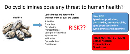
7. Conclusions
A review of all available data suggests that spirolides, portimines, pteriatoxins, prorocentrolide, spiro-prorocentrimine, symbiomines and kabirimine pose little risk to human health through consumption of shellfish. Despite occasional very high concentrations of GYMs and PnTXs in some shellfish collected from hot spots around the world, no illnesses have been reported, even when consumers have been directly questioned. Therefore, the risk of GYM and PnTXs to humans does not appear to be high. However, further occurrence and toxicity data are required to better define the risk posed by these compounds.
Author Contributions
Conceptualization, S.C.F. and D.T.H.; investigation, S.C.F., M.J.B. and D.T.H.; writing—original draft preparation, S.C.F., D.T.H., M.J.B. and A.I.S.; writing—review and editing, S.C.F., D.T.H., M.J.B. and A.I.S. All authors have read and agreed to the published version of the manuscript.
Funding
This work was funded by the New Zealand Ministry of Business, Innovation and Employment-Seafood Safety research programme (Contract CAWX1801).
Data Availability Statement
No new data were created or analysed in this study. Data sharing is not applicable to this article.
Conflicts of Interest
The authors declare no conflicts of interest.
References
- EFSA. Scientific opinion of the panel on contaminants in the food chain on a request from the European Commission on Marine Biotoxins in Shellfish-summary on regulated marine biotoxins. EFSA J. 2009, 1306, 1–23. [Google Scholar]
- Mafra, L.L.; de Souza, D.A.; Menezes, M.; Schramm, M.A.; Hoff, R. Marine biotoxins: Latest advances and challenges toward seafood safety, using Brazil as a case study. Curr. Opin. Food Sci. 2023, 53, 101078. [Google Scholar] [CrossRef]
- Hannah, D.J.; Till, D.G.; Deverall, T.; Jones, P.D.; Fry, J.M. Extraction of lipid-soluble marine biotoxins. J. AOAC Int. 1995, 78, 480–483. [Google Scholar] [CrossRef]
- MacKenzie, A.L.; Haywood, A.; Adamson, J.; Truman, P.; Till, D.; Seki, T.; Satake, M.; Yasumoto, K. Gymnodimine contamination of shellfish in New Zealand. In Harmful and Algal Blooms; Yasumoto, K., Oshima, Y., Fukuyo, Y., Eds.; Intergovernmental Oceanographic Commission of UNESCO: Paris, France, 1996; pp. 97–100. [Google Scholar]
- Finch, S.C.; Boundy, M.J.; Harwood, D.T. The acute toxicity of tetrodotoxin and tetrodotoxin–saxitoxin mixtures to mice by various routes of administration. Toxins 2018, 10, 423. [Google Scholar] [CrossRef]
- Munday, R.; Thomas, K.; Gibbs, R.; Murphy, C.; Quilliam, M.A. Acute toxicities of saxitoxin, neosaxitoxin, decarbamoyl saxitoxin and gonyautoxins 1&4 and 2&3 to mice by various routes of administration. Toxicon 2013, 76, 77–83. [Google Scholar] [CrossRef] [PubMed]
- Combes, R.D. The mouse bioassay for diarrhetic shellfish poisoning: A gross misuse of laboratory animals and of scientific methodology. Altern. Lab. Anim. 2003, 31, 595–610. [Google Scholar] [CrossRef] [PubMed]
- McNabb, P.; Selwood, A.I.; Holland, P.T.; Collaborators. Multiresidue Method for Determination of Algal Toxins in Shellfish: Single-Laboratory Validation and Interlaboratory Study. J. AOAC Int. 2019, 88, 761–772. [Google Scholar] [CrossRef]
- Otero, A.; Chapela, M.-J.; Atanassova, M.; Vieites, J.M.; Cabado, A.G. Cyclic imines: Chemistry and mechanism of action: A review. Chem. Res. Toxicol. 2011, 24, 1817–1829. [Google Scholar] [CrossRef]
- Molgó, J.; Aráoz, R.; Benoit, E.; Iorga, B. Cyclic imine toxins: Chemistry, origin, metabolism, pharmacology, toxicology, and detection. In Seafood and Freshwater Toxins, 3rd ed.; Botana, L.M., Ed.; CRC Press: Boca Raton, FL, USA, 2014. [Google Scholar]
- Davidson, K.; Baker, C.; Higgins, C.; Higman, W.; Swan, S.; Veszelovszki, A.; Turner, A.D. Potential threats posed by new or emerging marine biotoxins in UK waters and examination of detection methodologies used for their control: Cyclic imines. Mar. Drugs 2015, 13, 7087–7112. [Google Scholar] [CrossRef]
- Molgó, J.; Marchot, P.; Aráoz, R.; Benoit, E.; Iorga, B.I.; Zakarian, A.; Taylor, P.; Bourne, Y.; Servent, D. Cyclic imine toxins from dinoflagellates: A growing family of potent antagonists of the nicotinic acetylcholine receptors. J. Neurochem. 2017, 142, 41–51. [Google Scholar] [CrossRef]
- Kharrat, R.; Servent, D.; Girard, E.; Ouanounou, G.; Amar, M.; Marrouchi, R.; Benoit, E.; Molgó, J. The marine phycotoxin gymnodimine targets muscular and neuronal nicotinic acetylcholine receptor subtypes with high affinity. J. Neurochem. 2008, 107, 952–963. [Google Scholar] [CrossRef]
- Otofuji, T.; Ogo, A.; Koisjo, J.; Matsuo, K.; Tokiwa, H.; Yasumoto, T.; Nishihara, K.; Yamamoto, E.; Saisho, M.; Kurihara, Y.; et al. Food poisoning caused by Atrina pectinata in the Ariake Sea. Food Santitation Res. 1981, 31, 76–83. [Google Scholar]
- EFSA Panel on Contaminants in the Food Chain. Scientific Opinion on marine biotoxins in shellfish—Cyclic imines (spirolides, gymnodimines, pinnatoxins and pteriatoxins). EFSA J. 2010, 8, 1628. [Google Scholar] [CrossRef]
- ANSES. Opinion of the French Agency for Food, Environmental and Occupational Health and Safety on the Assessment of the Health Risks Associated with Pinnatoxins in Shellfish; ANSES Opinion Request No 2016-SA-0013; French Agency for Food, Environmental and Occupational Health and Safety: Maisons-Alfort, France, 2019.
- Seki, T.; Satake, M.; Mackenzie, L.; Kaspar, H.F.; Yasumoto, T. Gymnodimine, a new marine toxin of unprecedented structure isolated from New Zealand oysters and the dinoflagellate, Gymnodinium sp. Tetrahedron Lett. 1995, 36, 7093–7096. [Google Scholar] [CrossRef]
- Seki, T.; Satake, M.; MacKenzie, A.L.; Faspar, H.F.; Yasumoto, T. Gymnodimine, a novel toxic imine isolated from the Foveaux Strait oysters and Gymnodinium sp. In Harmful and Toxic Algal Blooms; Yasumoto, T., Oshima, Y., Fukuyo, Y., Eds.; Intergovernmental Oceanographic Commission of UNESCO: Paris, France, 1996; pp. 495–498. [Google Scholar]
- Stirling, D.J. Survey of historical New Zealand shellfish samples for accumulation of gymnodimine. N. Z. J. Mar. Freshw. Res. 2001, 35, 851–857. [Google Scholar] [CrossRef]
- Miles, C.O.; Wilkins, A.L.; Stirling, D.J.; MacKenzie, A.L. New analogue of gymnodimine from a Gymnodinium Species. J. Agric. Food Chem. 2000, 48, 1373–1376. [Google Scholar] [CrossRef] [PubMed]
- Miles, C.; Wilkins, A.; Stirling, D.; Mackenzie, L. Gymnodimine C, an isomer of gymnodimine B, from Karenia selliformis. J. Agric. Food Chem. 2003, 51, 4838–4840. [Google Scholar] [CrossRef] [PubMed]
- Harju, K.; Koskela, H.; Kremp, A.; Suikkanen, S.; de la Iglesia, P.; Miles, C.O.; Krock, B.; Vanninen, P. Identification of gymnodimine D and presence of gymnodimine variants in the dinoflagellate Alexandrium ostenfeldii from the Baltic Sea. Toxicon 2016, 112, 68–76. [Google Scholar] [CrossRef]
- Zurhelle, C.; Nieva, J.; Tillmann, U.; Harder, T.; Krock, B.; Tebben, J. Identification of novel gymnodimines and spirolides from the marine dinoflagellate Alexandrium ostenfeldii. Mar. Drugs 2018, 16, 446. [Google Scholar] [CrossRef]
- Van Wagoner, R.M.; Misner, I.; Tomas, C.R.; Wright, J.L.C. Occurrence of 12-methylgymnodimine in a spirolide-producing dinoflagellate Alexandrium peruvianum and the biogenetic implications. Tetrahedron Lett. 2011, 52, 4243–4246. [Google Scholar] [CrossRef]
- Talić, S.; Škobić, D.; Dedić, A.; Nazlić, N.; Ujević, I.; Ivanković, A.; Pavela-Vrančić, M. The occurrence of lipophilic toxins in shellfish from the Middle Adriatic Sea. Toxicon 2020, 186, 19–25. [Google Scholar] [CrossRef]
- Kvrgić, K.; Lešić, T.; Aysal, A.I.; Džafić, N.; Pleadin, J. Cyclic imines in shellfish and ascidians in the northern Adriatic Sea. Food Addit. Contam. Part B 2021, 14, 12–22. [Google Scholar] [CrossRef]
- Amzil, Z.; Sibat, M.; Royer, F.; Masson, N.; Abadie, E. Report on the first detection of pectenotoxin-2, spirolide-A and their derivatives in French shellfish. Mar. Drugs 2007, 5, 168–179. [Google Scholar] [CrossRef]
- Bacchiocchi, S.; Siracusa, M.; Campacci, D.; Ciriaci, M.; Dubbini, A.; Tavoloni, T.; Stramenga, A.; Gorbi, S.; Piersanti, A. Cyclic Imines (CIs) in mussels from North-Central Adriatic Sea: First evidence of gymnodimine A in Italy. Toxins 2020, 12, 370. [Google Scholar] [CrossRef]
- Haddouch, A.B.; Amanhi, R.; Amzil, Z.; Taleb, H.; Rovillon, G.; Adly, F.; Loutfi, M. Lipophilic toxin profile in Mytilus galloprovincialis from the north atlantic coast of Morocco: LC-MS/MS and mouse bioassay analyses. Int. J. Sci. Res. 2017, 6, 187–195. [Google Scholar] [CrossRef]
- Krock, B.; Pitcher, G.; Ntuli, J.; Cembella, A. Confirmed identification of gymnodimine in oysters from the west coast of South Africa by liquid chromatography–tandem mass spectrometry. Afr. J. Mar. Sci. 2009, 31, 113–118. [Google Scholar] [CrossRef]
- Takahashi, E.; Yu, Q.; Eaglesham, G.; Connell, D.W.; McBroom, J.; Costanzo, S.; Shaw, G.R. Occurrence and seasonal variations of algal toxins in water, phytoplankton and shellfish from North Stradbroke Island, Queensland, Australia. Mar. Environ. Res. 2007, 64, 429–442. [Google Scholar] [CrossRef] [PubMed]
- Liu, R.; Liang, Y.; Wu, X.; Xu, D.; Liu, Y.; Liu, L. First report on the detection of pectenotoxin groups in Chinese shellfish by LC–MS/MS. Toxicon 2011, 57, 1000–1007. [Google Scholar] [CrossRef] [PubMed]
- Ji, Y.; Yan, G.; Wang, G.; Liu, J.; Tang, Z.; Yan, Y.; Qiu, J.; Zhang, L.; Pan, W.; Fu, Y.; et al. Prevalence and distribution of domoic acid and cyclic imines in bivalve mollusks from Beibu Gulf, China. J. Hazard. Mater. 2022, 423, 127078. [Google Scholar] [CrossRef] [PubMed]
- Jiang, T.; Liu, L.; Li, Y.; Zhang, J.; Tan, Z.; Wu, H.; Jiang, T.; Lu, S. Occurrence of marine algal toxins in oyster and phytoplankton samples in Daya Bay, South China Sea. Chemosphere 2017, 183, 80–88. [Google Scholar] [CrossRef] [PubMed]
- Hassoun, A.E.R.; Ujević, I.; Mahfouz, C.; Fakhri, M.; Roje-Busatto, R.; Jemaa, S.; Nazlić, N. Occurrence of domoic acid and cyclic imines in marine biota from Lebanon-Eastern Mediterranean Sea. Sci. Total Environ. 2021, 755, 142542. [Google Scholar] [CrossRef]
- Marrouchi, R.; Dziri, F.; Belayouni, N.; Hamza, A.; Benoit, E.; Molgó, J.; Kharrat, R. Quantitative determination of gymnodimine-A by high performance liquid chromatography in contaminated clams from Tunisia coastline. Mar. Biotechnol. 2010, 12, 579–585. [Google Scholar] [CrossRef]
- Lamas, J.P.; Arévalo, F.; Moroño, Á.; Correa, J.; Rossignoli, A.E.; Blanco, J. Gymnodimine A in mollusks from the north Atlantic Coast of Spain: Prevalence, concentration, and relationship with spirolides. Environ. Pollut. 2021, 279, 116919. [Google Scholar] [CrossRef]
- Sibat, M.; Mai, T.; Tanniou, S.; Biegala, I.; Hess, P.; Jauffrais, T. Seasonal single-site sampling reveals large diversity of marine algal toxins in coastal waters and shellfish of New Caledonia (southwestern pacific). Toxins 2023, 15, 642. [Google Scholar] [CrossRef] [PubMed]
- Amzil, Z.; Derrien, A.; Terre Terrillon, A.; Savar, V.; Bertin, T.; Peyrat, M.; Duval, A.; Lhaute, K.; Arnich, N.; Hort, V.; et al. Five years monitoring the emergence of unregulated toxins in shellfish in France (EMERGTOX 2018–2022). Mar. Drugs 2023, 21, 435. [Google Scholar] [CrossRef] [PubMed]
- de la Iglesia, P.; McCarron, P.; Diogène, J.; Quilliam, M.A. Discovery of gymnodimine fatty acid ester metabolites in shellfish using liquid chromatography/mass spectrometry. Rapid Commun. Mass Spectrom. 2013, 27, 643–653. [Google Scholar] [CrossRef] [PubMed]
- Ji, Y.; Che, Y.; Wright, E.J.; McCarron, P.; Hess, P.; Li, A. Fatty acid ester metabolites of gymnodimine in shellfish collected from China and in mussels (Mytilus galloprovincialis) exposed to Karenia selliformis. Harmful Algae 2020, 92, 101774. [Google Scholar] [CrossRef]
- Munday, R.; Towers, N.R.; Mackenzie, L.; Beuzenberg, V.; Holland, P.T.; Miles, C.O. Acute toxicity of gymnodimine to mice. Toxicon 2004, 44, 173–178. [Google Scholar] [CrossRef] [PubMed]
- Stewart, M.; Blunt, J.W.; Munro, M.H.; Robinson, W.T.; Hannah, D.J. The absolute stereochemistry of the New Zealand shellfish toxin gymnodimine. Tetrahedron Lett. 1997, 38, 4889–4890. [Google Scholar] [CrossRef]
- Munday, R.; Reeve, J. Risk assessment of shellfish toxins. Toxins 2013, 5, 2109. [Google Scholar] [CrossRef]
- Hu, T.; Curtis, J.M.; Oshima, Y.; Quilliam, M.A.; Walter, J.A.; Watson-Wright, W.M.; Wright, J.L.C. Spirolides B and D, two novel macrocycles isolated from the digestive glands of shellfish. J. Chem. Soc. Chem. Commun. 1995, 20, 2159–2161. [Google Scholar] [CrossRef]
- Hu, T.; Curtis, J.M.; Walter, J.A.; Wright, J.L.C. Characterization of biologically inactive spirolides E and F: Identification of the spirolide pharmacophore. Tetrahedron Lett. 1996, 37, 7671–7674. [Google Scholar] [CrossRef]
- Hu, T.; Burton, I.W.; Cembella, A.D.; Curtis, J.M.; Quilliam, M.A.; Walter, J.A.; Wright, J.L.C. Characterization of spirolides A, C, and 13-desmethyl C, new marine toxins isolated from toxic plankton and contaminated shellfish. J. Nat. Prod. 2001, 64, 308–312. [Google Scholar] [CrossRef]
- Cembella, A.D.; Lewis, N.I.; Quilliam, M.A. The marine dinoflagellate Alexandrium ostenfeldii (Dinophyceae) as the causative organism of spirolide shellfish toxins. Phycologia 2000, 39, 67–74. [Google Scholar] [CrossRef]
- Cembella, A.D.; Lewis, N.I.; Quilliam, M.A. Spirolide composition of micro-extracted pooled cells isolated from natural plankton assemblages and from cultures of the dinoflagellate Alexandrium ostenfeldii. Nat. Toxins 1999, 7, 197–206. [Google Scholar] [CrossRef] [PubMed]
- Aasen, J.; MacKinnon, S.L.; LeBlanc, P.; Walter, J.A.; Hovgaard, P.; Aune, T.; Quilliam, M.A. Detection and identification of spirolides in norwegian shellfish and plankton. Chem. Res. Toxicol. 2005, 18, 509–515. [Google Scholar] [CrossRef]
- MacKinnon, S.L.; Walter, J.A.; Quilliam, M.A.; Cembella, A.D.; LeBlanc, P.; Burton, I.W.; Hardstaff, W.R.; Lewis, N.I. Spirolides isolated from danish strains of the toxigenic dinoflagellate Alexandrium ostenfeldii. J. Nat. Prod. 2006, 69, 983–987. [Google Scholar] [CrossRef]
- Ciminiello, P.; Dell’Aversano, C.; Iacovo, E.D.; Fattorusso, E.; Forino, M.; Grauso, L.; Tartaglione, L.; Guerrini, F.; Pezzolesi, L.; Pistocchi, R. Characterization of 27-hydroxy-13-desmethyl spirolide C and 27-oxo-13,19-didesmethyl spirolide C. Further insights into the complex Adriatic Alexandrium ostenfeldii toxin profile. Toxicon 2010, 56, 1327–1333. [Google Scholar] [CrossRef]
- Roach, J.S.; LeBlanc, P.; Lewis, N.I.; Munday, R.; Quilliam, M.A.; MacKinnon, S.L. Characterization of a dispiroketal spirolide subclass from Alexandrium ostenfeldii. J. Nat. Prod. 2009, 72, 1237–1240. [Google Scholar] [CrossRef]
- Aasen, J.A.; Hardstaff, W.; Aune, T.; Quilliam, M.A. Discovery of fatty acid ester metabolites of spirolide toxins in mussels from Norway using liquid chromatography/tandem mass spectrometry. Rapid Commun. Mass Spectrom. 2006, 20, 1531–1537. [Google Scholar] [CrossRef]
- Turner, A.D.; Goya, A.B. Occurrence and profiles of lipophilic toxins in shellfish harvested from Argentina. Toxicon 2015, 102, 32–42. [Google Scholar] [CrossRef]
- Rundberget, T.; Aasen, J.A.B.; Selwood, A.I.; Miles, C.O. Pinnatoxins and spirolides in Norwegian blue mussels and seawater. Toxicon 2011, 58, 700–711. [Google Scholar] [CrossRef]
- Li, A.; Chen, H.; Qiu, J.; Lin, H.; Gu, H. Determination of multiple toxins in whelk and clam samples collected from the Chukchi and Bering seas. Toxicon 2016, 109, 84–93. [Google Scholar] [CrossRef]
- Rambla-Alegre, M.; Miles, C.O.; de la Iglesia, P.; Fernandez-Tejedor, M.; Jacobs, S.; Sioen, I.; Verbeke, W.; Samdal, I.A.; Sandvik, M.; Barbosa, V.; et al. Occurrence of cyclic imines in European commercial seafood and consumers risk assessment. Environ. Res. 2018, 161, 392–398. [Google Scholar] [CrossRef]
- Villar González, A.; Rodríguez-Velasco, M.L.; Ben-Gigirey, B.; Botana, L.M. First evidence of spirolides in Spanish shellfish. Toxicon 2006, 48, 1068–1074. [Google Scholar] [CrossRef] [PubMed]
- Liang, Y.; Li, A.; Chen, J.; Tan, Z.; Tong, M.; Liu, Z.; Qiu, J.; Yu, R. Progress on the investigation and monitoring of marine phycotoxins in China. Harmful Algae 2022, 111, 102152. [Google Scholar] [CrossRef] [PubMed]
- McNabb, P.; McCoubrey, D.; Rhodes, L.; Smith, K.; Selwood, A.; Van Ginkel, R.; MacKenzie, A.; Munday, R.; Holland, P. New perspectives on biotoxin detection in Rangaunu Harbour, New Zealand arising from the discovery of pinnatoxins. Harmful Algae 2012, 13, 34–39. [Google Scholar] [CrossRef]
- Munday, R.; Quilliam, M.A.; LeBlanc, P.; Lewis, N.; Gallant, P.; Sperker, S.A.; Ewart, H.S.; MacKinnon, S.L. Investigations into the toxicology of spirolides, a group of marine phycotoxins. Toxins 2012, 4, 1–14. [Google Scholar] [CrossRef]
- Otero, P.; Alfonso, A.; Rodríguez, P.; Rubiolo, J.A.; Cifuentes, J.M.; Bermúdez, R.; Vieytes, M.R.; Botana, L.M. Pharmacokinetic and toxicological data of spirolides after oral and intraperitoneal administration. Food Chem. Toxicol. 2012, 50, 232–237. [Google Scholar] [CrossRef]
- Finch, S.C.; Boundy, M.J.; Webb, N.G.; Harwood, D.T. The effect of experimental protocol on the toxicity of saxitoxin in mice. Toxins 2023, 15, 290. [Google Scholar] [CrossRef]
- Uemura, D.; Chou, T.; Haino, T.; Nagatsu, A.; Fukuzawa, S.; Zheng, S.-Z.; Chen, H.-S. Pinnatoxin A: A toxic amphoteric macrocycle from the Okinawan bivalve Pinna muricata. J. Am. Chem. Soc. 1995, 117, 1155–1156. [Google Scholar] [CrossRef]
- Takada, N.; Umemura, N.; Suenaga, K.; Chou, T.; Nagatsu, A.; Haino, T.; Yamada, K.; Uemura, D. Pinnatoxins B and C, the most toxic components in the pinnatoxin series from the Okinawan bivalve Pinna muricata. Tetrahedron Lett. 2001, 42, 3491–3494. [Google Scholar] [CrossRef]
- Chou, T.; Haino, T.; Kuramoto, M.; Uemura, D. Isolation and structure of pinnatoxin D, a new shellfish poison from the Okinawan bivalve Pinna muricata. Tetrahedron Lett. 1996, 37, 4027–4030. [Google Scholar] [CrossRef]
- Selwood, A.I.; Miles, C.O.; Wilkins, A.L.; van Ginkel, R.; Munday, R.; Rise, F.; McNabb, P. Isolation, structural determination and acute toxicity of pinnatoxins E., F and G. J. Agric. Food Chem. 2010, 58, 6532–6542. [Google Scholar] [CrossRef]
- Rhodes, L.; Smith, K.; Selwood, A.; McNabb, P.; Munday, R.; Suda, S.; Molenaar, S.; Hallegraeff, G. Dinoflagellate Vulcanodinium rugosum identified as the causative organism of pinnatoxins in Australia, New Zealand and Japan. Phycologia 2011, 50, 624–628. [Google Scholar] [CrossRef] [PubMed]
- Selwood, A.I.; Wilkins, A.L.; Munday, R.; Gu, H.; Smith, K.F.; Rhodes, L.L.; Rise, F. Pinnatoxin H: A new pinnatoxin analogue from a South China Sea Vulcanodinium rugosum isolate. Tetrahedron Lett. 2014, 55, 5508–5510. [Google Scholar] [CrossRef]
- McCarron, P.; Rourke, W.A.; Hardstaff, W.; Pooley, B.; Quilliam, M.A. Identification of pinnatoxins and discovery of their fatty acid ester metabolites in mussels (Mytilus edulis) from Eastern Canada. J. Agric. Food Chem. 2012, 60, 1437–1446. [Google Scholar] [CrossRef] [PubMed]
- Norambuena, L.; Mardones, J.I. Emerging phycotoxins in the Chilean coast: First localized detection of the neurotoxic cyclic imine Pinnatoxin-G in shellfish banks. Mar. Pollut. Bull. 2023, 190, 114878. [Google Scholar] [CrossRef] [PubMed]
- Hess, P.; Abadie, E.; Hervé, F.; Berteaux, T.; Séchet, V.; Aráoz, R.; Molgó, J.; Zakarian, A.; Sibat, M.; Rundberget, T.; et al. Pinnatoxin G is responsible for atypical toxicity in mussels (Mytilus galloprovincialis) and clams (Venerupis decussata) from Ingril, a French Mediterranean lagoon. Toxicon 2013, 75, 16–26. [Google Scholar] [CrossRef]
- Miles, C.; Rundberget, T.; Sandvik, M.; Aasen, J.; Selwood, A. The Presence of Pinnatoxins in Norwegian Mussels; National Veterinary Institute’s Report Series 7b-2010; National Veterinary Institute: Oslo, Norway, 2009. [Google Scholar]
- García-Altares, M.; Casanova, A.; Bane, V.; Diogène, J.; Furey, A.; De la Iglesia, P. Confirmation of pinnatoxins and spirolides in shellfish and passive samplers from Catalonia (Spain) by liquid chromatography coupled with triple quadrupole and high-resolution hybrid tandem mass spectrometry. Mar. Drugs 2014, 12, 3706–3732. [Google Scholar] [CrossRef]
- Tamele, I.J.; Timba, I.; Vasconcelos, V.; Costa, P.R. First report of pinnatoxins in bivalve molluscs from Inhaca Island (South of Mozambique)—South of the Indian Ocean. J. Mar. Sci. Eng. 2022, 10, 1215. [Google Scholar] [CrossRef]
- Moreira-González, A.R.; Comas-González, A.; Valle-Pombrol, A.; Seisdedo-Losa, M.; Hernández-Leyva, O.; Fernandes, L.F.; Chomérat, N.; Bilien, G.; Hervé, F.; Rovillon, G.A.; et al. Summer bloom of Vulcanodinium rugosum in Cienfuegos Bay (Cuba) associated to dermatitis in swimmers. Sci. Total Environ. 2021, 757, 143782. [Google Scholar] [CrossRef] [PubMed]
- Hess, P.; Mertens, K.; Chomérat, N.; Sechet, V.; Hervé, F.; Plessis, L.; Reveillon, D.; Brehmer, P. Vulcanodinium rugosum—A potent and ubiquitous genus affecting mice and man. In Dinophyte Seminars; Ludwig-Maximilians-Universität München: Munich, Germany, 2022. [Google Scholar]
- McCauley, J.A.; Nagasawa, K.; Lander, P.A.; Mischke, S.G.; Semones, M.A.; Kishi, Y. Total synthesis of pinnatoxin A. J. Am. Chem. Soc. 1998, 120, 7647–7648. [Google Scholar] [CrossRef]
- Munday, R.; Selwood, A.I.; Rhodes, L. Acute toxicity of pinnatoxins E., F and G to mice. Toxicon 2012, 60, 995–999. [Google Scholar] [CrossRef]
- Sosa, S.; Pelin, M.; Cavion, F.; Hervé, F.; Hess, P.; Tubaro, A. Acute oral toxicity of pinnatoxin G in mice. Toxins 2020, 12, 87. [Google Scholar] [CrossRef] [PubMed]
- Takada, N.; Umemura, N.; Suenaga, K.; Uemura, D. Structural determination of pteriatoxins A, B and C, extremely potent toxins from the bivalve Pteria penguin. Tetrahedron Lett. 2001, 42, 3495–3497. [Google Scholar] [CrossRef]
- Torigoe, K.; Murata, M.; Yasumoto, T.; Iwashita, T. Prorocentrolide, a toxic nitrogenous macrocycle from a marine dinoflagellate, Prorocentrum lima. J. Am. Chem. Soc. 1988, 110, 7876–7877. [Google Scholar] [CrossRef]
- Hu, T.; deFreitas, A.S.W.; Curtis, J.M.; Oshima, Y.; Walter, J.A.; Wright, J.L.C. Isolation and structure of prorocentrolide B, a fast-acting toxin from Prorocentrum maculosum. J. Nat. Prod. 1996, 59, 1010–1014. [Google Scholar] [CrossRef]
- Lu, C.-K.; Lee, G.-H.; Huang, R.; Chou, H.-N. Spiro-prorocentrimine, a novel macrocyclic lactone from a benthic Prorocentrum sp. of Taiwan. Tetrahedron Lett. 2001, 42, 1713–1716. [Google Scholar] [CrossRef]
- Lee, S.M.; Kim, N.-H.; Jeong, E.J.; Rho, J.-R. Cytotoxic 4-hydroxyprorocentrolide and prorocentrolide C from cultured dinoflagellate Prorocentrum lima induce human cancer cell death through apoptosis and cell cycle arrest. Toxins 2020, 12, 304. [Google Scholar] [CrossRef]
- Amar, M.; Aráoz, R.; Iorga, B.I.; Yasumoto, T.; Servent, D.; Molgó, J. Prorocentrolide-A from cultured Prorocentrum lima dinoflagellates collected in Japan blocks sub-types of nicotinic acetylcholine receptors. Toxins 2018, 10, 97. [Google Scholar] [CrossRef]
- Selwood, A.I.; Wilkins, A.L.; Munday, R.; Shi, F.; Rhodes, L.L.; Holland, P.T. Portimine: A bioactive metabolite from the benthic dinoflagellate Vulcanodinium rugosum. Tetrahedron Lett. 2013, 54, 4705–4707. [Google Scholar] [CrossRef]
- Cuddihy, S.L.; Drake, S.; Harwood, D.T.; Selwood, A.I.; McNabb, P.S.; Hampton, M.B. The marine cytotoxin portimine is a potent and selective inducer of apoptosis. Apoptosis 2016, 21, 1447–1452. [Google Scholar] [CrossRef]
- Tang, J.; Li, W.; Chiu, T.-Y.; Martínez-Peña, F.; Luo, Z.; Chong, C.T.; Wei, Q.; Gazaniga, N.; West, T.J.; See, Y.Y.; et al. Synthesis of portimines reveals the basis of their anti-cancer activity. Nature 2023, 622, 507–513. [Google Scholar] [CrossRef]
- Fribley, A.M.; Xi, Y.; Makris, C.; Alves-de-Souza, C.; York, R.; Tomas, C.; Wright, J.L.C.; Strangman, W.K. Identification of portimine B, a new cell permeable spiroimine that induces apoptosis in oral squamous cell carcinoma. ACS Med. Chem. Lett. 2019, 10, 175–179. [Google Scholar] [CrossRef]
- Aráoz, R.; Barnes, P.; Séchet, V.; Delepierre, M.; Zinn-Justin, S.; Molgó, J.; Zakarian, A.; Hess, P.; Servent, D. Cyclic imine toxins survey in coastal european shellfish samples: Bioaccumulation and mode of action of 28-O-palmitoyl ester of pinnatoxin-G. first report of portimine-A bioaccumulation. Harmful Algae 2020, 98, 101887. [Google Scholar] [CrossRef] [PubMed]
- Hermawan, I.; Higa, M.; Hutabarat, P.U.B.; Fujiwara, T.; Akiyama, K.; Kanamoto, A.; Haruyama, T.; Kobayashi, N.; Higashi, M.; Suda, S.; et al. Kabirimine, a new cyclic imine from an Okinawan dinoflagellate. Mar. Drugs 2019, 17, 353. [Google Scholar] [CrossRef] [PubMed]
- Kita, M.; Ohishi, N.; Washida, K.; Kondo, M.; Koyama, T.; Yamada, K.; Uemura, D. Symbioimine and neosymbioimine, amphoteric iminium metabolites from the symbiotic marine dinoflagellate Symbiodinium sp. Bioorganic Med. Chem. 2005, 13, 5253–5258. [Google Scholar] [CrossRef] [PubMed]
- Arnich, N.; Abadie, E.; Delcourt, N.; Fessard, V.; Fremy, J.M.; Hort, V.; Lagrange, E.; Maignien, T.; Molgó, J.; Peyrat, M.B.; et al. Health risk assessment related to pinnatoxins in French shellfish. Toxicon 2020, 180, 1–10. [Google Scholar] [CrossRef]
- Bouquet, A.; Thébault, A.; Arnich, N.; Foucault, E.; Caillard, E.; Gianaroli, C.; Bellamy, E.; Rolland, J.L.; Laabir, M.; Abadie, E. Modelling spatiotemporal distributions of Vulcanodinium rugosum and pinnatoxin G in French Mediterranean lagoons: Application to human health risk characterisation. Harmful Algae 2023, 129, 102500. [Google Scholar] [CrossRef] [PubMed]
- Katikou, P.; Aligizaki, K.; Zacharaki, T.; Iossifidis, D.; Nikolaidis, G. First report of spirolides in Greek shellfish assocociated with causative Alexandrium species. In Proceedings of the 14th International Conference on Harmful Algal Blooms, Crete, Greece, 1–5 November 2010; pp. 197–199. [Google Scholar]
- Pigozzi, S.; Bianchi, L.; Boschetti, L.; Cangini, M.; Ceredi, A.; Magnani, F.; Milandri, A.; Montanari, S.; Pompei, M.; Riccardi, E.; et al. First evidence of spirolide accumulation in northwestern Adriatic shellfish. In Proceedings of the 12th International Conference on Harmful Algae, Copenhagen, Denmark, 4–8 September 2006; pp. 319–322. [Google Scholar]
- Paredes-Banda, P.; García-Mendoza, E.; Ponce-Rivas, E.; Blanco, J.; Almazán-Becerril, A.; Galindo-Sánchez, C.; Cembella, A. Association of the toxigenic dinoflagellate Alexandrium ostenfeldii with spirolide accumulation in cultured mussels (Mytilus galloprovincialis) from Northwest Mexico. Front. Mar. Sci. 2018, 5, 491. [Google Scholar] [CrossRef]
- Silva, M.; Barreiro, A.; Rodriguez, P.; Otero, P.; Azevedo, J.; Alfonso, A.; Botana, L.M.; Vasconcelos, V. New invertebrate vectors for PST, spirolides and okadaic acid in the North Atlantic. Mar. Drugs 2013, 11, 1936–1960. [Google Scholar] [CrossRef]
- Blanco, J.; Arévalo, F.; Moroño, Á.; Correa, J.; Rossignoli, A.E.; Lamas, J.P. Spirolides in bivalve mollusk of the Galician (NW Spain) coast: Interspecific, spatial, temporal variation and presence of an isomer of 13-desmethyl spirolide C. Toxins 2023, 15, 13. [Google Scholar] [CrossRef] [PubMed]
- Moreiras, G.; Leão, J.M.; Gago-Martínez, A. Analysis of cyclic imines in mussels (Mytilus galloprovincialis) from Galicia (NW Spain) by LC-MS/MS. Int. J. Environ. Res. Public Health 2020, 17, 281. [Google Scholar] [CrossRef]
- Barreiro-Crespo, L.; Fernández-Tejedor, M.; Diogène, J.; Rambla-Alegre, M. The temporal distribution of cyclic imines in shellfish in the bays of Fangar and Alfacs, Northwestern Mediterranean region. Toxins 2024, 16, 10. [Google Scholar] [CrossRef] [PubMed]
- Rossignoli, A.E.; Mariño, C.; Martín, H.; Blanco, J. First report of two gymnodimines and two tetrodotoxin analogues in invertebrates from the North Atlantic Coast of Spain. Mar. Drugs 2023, 21, 232. [Google Scholar] [CrossRef] [PubMed]
- Rossignoli, A.E.; Ben-Gigirey, B.; Cid, M.; Mariño, C.; Martín, H.; Garrido, S.; Rodríguez, F.; Blanco, J. Lipophilic shellfish poisoning toxins in marine invertebrates from the Galician coast. Toxins 2023, 15, 631. [Google Scholar] [CrossRef]
- Geng, H.; Sun, H.; Liu, C.; Kong, F.; Zhang, Q.; Yan, T.; Yu, R. Screening for lipophilic marine toxins and their potential producers in coastal waters of Weihai in autumn, 2020. J. Oceanol. Limnol. 2022, 40, 2218–2230. [Google Scholar] [CrossRef]
- Wang, X.-Z.; Cheng, Y.; Li, N.; Wen, H.-M.; Liu, R.; Shan, C.-X.; Chai, C.; Wu, H. Occurrence and seasonal variations of lipophilic marine toxins in commercial clam species along the coast of Jiangsu, China. Toxins 2016, 8, 8. [Google Scholar] [CrossRef]
- Ujević, I.; Roje-Busatto, R.; Ezgeta-Balić, D. Comparison of amnesic, paralytic and lipophilic toxins profiles in cockle (Acanthocardia tuberculata) and smooth clam (Callista chione) from the central Adriatic Sea (Croatia). Toxicon 2019, 159, 32–37. [Google Scholar] [CrossRef]
- Villar-González, A.; Rodríguez-Velasco, M.L.; Ben-Gigirey, B.; Botana, L.M. Lipophilic toxin profile in Galicia (Spain): 2005 toxic episode. Toxicon 2007, 49, 1129–1134. [Google Scholar] [CrossRef] [PubMed]
- Otero, P.; Vale, C.; Boente-Juncal, A.; Costas, C.; Louzao, M.C.; Botana, L.M. Detection of cyclic imine toxins in dietary supplements of sreen lipped mussels (Perna canaliculus) and in shellfish Mytilus chilensis. Toxins 2020, 12, 613. [Google Scholar] [CrossRef] [PubMed]
- EFSA CONTAM Panel (EFSA Panel on Contaminants in the Food Chain); Knutsen, H.K.; Alexander, J.; Barregård, L.; Bignami, M.; Brüschweiler, B.; Ceccatelli, S.; Cottrill, B.; Dinovi, M.; Edler, L.; et al. Scientific opinion on the risks for public health related to the presence of tetrodotoxin (TTX) and TTX analogues in marine bivalves and gastropods. EFSA J. 2017, 15, 4752. [Google Scholar] [CrossRef]
- EFSA Scientific Committee. Guidance on selected default values to be used by the EFSA Scientific Committee, Scientific Panels and Units in the absence of actual measured data. EFSA J. 2012, 10, 2579. [Google Scholar] [CrossRef]
- Interdepartmental Group on Health Risks from Chemicals. Uncertainty Factors: Their Use in Human Health Risk Assessment by UK Government; MRC Institute for Environment and Health: Leicester, UK, 2003; p. 69. [Google Scholar]
- EFSA. Scientific opinion of the panel on contaminants in the food chain on a request from the european commission on marine biotoxins in shellfish–saxitoxin group. EFSA J. 2009, 1019, 1–76. [Google Scholar] [CrossRef]
- FAO/IOC/WHO. Report of the Joint FAO/IOC/WHO Ad Hoc Expert Consultation on Biotoxins in Bivalve Molluscs; FAO: Oslo, Norway, 2004; p. 31. [Google Scholar]
Figure 1.
Chemical structures of Gymnodimine A, Spirolide A, Pinnatoxin A and Portimine.

Table 1.
Toxicity of GYMs to mice (µg/kg BW) by intraperitoneal injection.
| Compound | Mouse Strain | Gender | State of Alimentation | Parameter | Acute Toxicity | Ref |
|---|---|---|---|---|---|---|
| GYM A | ? | ? | ? | “lethality” | 450 | [17] |
| GYM A | ? | ? | ? | MLD | 700 | [43] |
| GYM A | Swiss albino | F | Fed | LD50 | 96 (79–118) | [42] |
| GYM A | Swiss Webster | M | ? | LD50 | 80 | [13] |
| GYM B | Swiss Webster | M | ? | LD50 | 800 | [13] |
Figures in brackets indicate 95% confidence intervals; LD50 = median lethal dose; MLD = minimum lethal dose; ? = these experimental details are unknown.
Table 2.
Oral LD50s of GYM A (µg/kg BW) using Swiss albino female mice.
| Compound | Method of Administration | State of Alimentation | Acute Toxicity | Ref |
|---|---|---|---|---|
| GYM A | Gavage | Fed | 750 (600–945) | [42] |
| GYM A | Over the tongue a | ? | 4057 (3750–4390) | [42] |
| GYM A | Feeding b | Fasted | >7500 | [42] |
Figures in brackets indicate 95% confidence intervals; a GYM A was mixed with ground mouse food to form a paste which was then administered over the tongue of the mouse; b GYM A was mixed with cream cheese which was eaten by mice within 30 s; ? = these experimental details are unknown.
Table 3.
Toxicity of SPX analogues (µg/kg BW) administered to mice by i.p. injection.
| Compound | Parameter | Acute Toxicity | Ref |
|---|---|---|---|
| Spirolide A a | LD50 | 37 (35–44) | [62] |
| Spirolide B a | LD50 | 99 | [62] |
| Spirolide B c | LD100 | 250 | [45] |
| Dihydrospirolide B b | MLD | >1000 | [46] |
| Spirolide C a | LD50 | 8.0 (4.6–16.0) | [62] |
| 13-desmethyl spirolide C a | LD50 | 6.9 (5.0–8.0) | [62] |
| 13-desmethyl spirolide C | LD50 | 27.9 | [63] |
| 27-hydroxy-13-desmethyl spirolide C b | MLD | >27 | [52] |
| 27-oxo-13,19-didesmethyl spirolide C b | MLD | >35 | [52] |
| 13,19-didesmethyl spirolide C b | LD50 | 32 | [63] |
| 13,19-didesmethyl spirolide C b | MLD | 30 | [51] |
| Spirolide D c | LD100 | 250 | [45] |
| Spirolide E b | MLD | >1000 | [46] |
| Spirolide F b | MLD | >1000 | [46] |
| 20-methyl spirolide G a | LD50 | 8.0 (3.9–14.0) | [62] |
| 20-methyl spirolide G | MLD | >63.5 | [63] |
| Spirolide H a | MLD | >2000 | [53] |
Figures in brackets indicate 95% confidence intervals. a Studies used fed, female, Swiss albino mice; b details of the mice used were not specified; c no experimental details were given.
Table 4.
Oral toxicity of SPX analogues (µg/kg BW) to mice (from Munday et al. [62]).
Table 4.
Oral toxicity of SPX analogues (µg/kg BW) to mice (from Munday et al. [62]).
| Compound | Gavage | Voluntary Feeding | ||
|---|---|---|---|---|
| Fed | Fasted | Fed | Fasted | |
| Spirolide A | 550 (436–690) | 240 (188–298) | 1300 (1250–1580) | 1200 (1047–3690) |
| Spirolide B | ND | 440 (320–500) | ND | ND |
| Spirolide C | ND | 53 (50–63) | 780 | 500 (353–657) |
| 13-desmethyl spirolide C | 160 (123–198) | 130 (87–166) | 1000 (861–1290) | 500 (381–707) |
| 20-methyl spirolide G | 160 | 88 (27–120) | ND | 500 (381–707) |
Figures in brackets indicate 95% confidence intervals; female, Swiss albino mice were used in all experiments; ND = no data available.
Table 5.
Toxicity of PnTX analogues (µg/kg BW) in mice by i.p. injection.
| Compound | Parameter | Fed Acute Toxicity | Fasted Acute Toxicity | Acute Toxicity | Ref |
|---|---|---|---|---|---|
| (+)-Pinnatoxin A b | LD99 | ND | ND | 180 | [65] |
| (+)-Pinnatoxin A b | LD99 | ND | ND | 135 | [79] |
| (−)-Pinnatoxin A b | MLD | ND | ND | >5000 | [79] |
| Pinnatoxin B/C 1:1 b | LD99 | ND | ND | 22 | [66] |
| Pinnatoxin D b | LD99 | ND | ND | 400 | [67] |
| Pinnatoxin E a | LD50 | 45 (32–58) | ND | ND | [68] |
| Pinnatoxin E a | LD50 | 57 (39.7–75.3) | 48.0 (33.5–63.5) | ND | [80] |
| Pinnatoxin F a | LD50 | 16 (12–23) | ND | ND | [68] |
| Pinnatoxin F a | LD50 | 12.7 (9.5–14.6) | 14.9 (12.6–15.8) | ND | [80] |
| Pinnatoxin G a | LD50 | 50 (35–66) | ND | ND | [68] |
| Pinnatoxin G a | LD50 | 48.0 (36.3–68.1) | 42.7 (40.0–50.0) | ND | [80] |
| Pinnatoxin H a | LD50 | 67 (63–79) | ND | ND | [70] |
Figures in brackets indicate 95% confidence intervals; a Swiss albino, female mice were used; b no experimental details were provided; ND = no data available.
Table 6.
Toxicity of PnTX analogues (µg/kg BW) to mice by oral administration.
| Compound | Gavage | Voluntary Feeding | Ref | ||
|---|---|---|---|---|---|
| Fed | Fasted | Fed | Fasted | ||
| Pinnatoxin E | 2800 (2380–3000) | ND | ND | ND | [80] |
| Pinnatoxin F | 25.0 (19.1–35.1) | 29.9 (25.0–32.0) | 50.0 (39.4–62.8) | 77 | [80] |
| Pinnatoxin G | 150 (105–199) | ND | 400 (380–470) | ND | [80] |
| Pinnatoxin G | ND | 208 (155–281) | ND | ND | [81] |
| Pinnatoxin H | 163 (139–175) | ND | ND | ND | [70] |
Figures in brackets indicate 95% confidence intervals; all used female Swiss albino mice, apart from the study by Sosa et al. [81] which used female CD-1s; ND = no data available.
Table 8.
The maximum concentrations of GYMs, SPXs or PnTXs (µg/kg) detected in the whole flesh (unless otherwise specified) of different species of shellfish from around the world.
Table 8.
The maximum concentrations of GYMs, SPXs or PnTXs (µg/kg) detected in the whole flesh (unless otherwise specified) of different species of shellfish from around the world.
| Country | Area | Year | GYM a | SPX b | PnTX c | Ref |
|---|---|---|---|---|---|---|
| Mussels (various species) | ||||||
| Argentina | Beagle Channel | 2005–2007 | <LOD | 68 (1) | NT | [55] |
| Bosnia and Herzegovina | Bay of Neum | 2017 | 11.4 | NT | NT | [25] |
| Canada | Eastern Canada | 2010–2011 | NT | NT | 83 (G), 1.5 (A) | [71] |
| Chile | Beagle Channel | 2021–2022 | NT | NT | 100 | [72] |
| Croatia | Istrian Peninsula | 2018–2019 | 17.2 | 17.0 (1) | 6.9 | [26] |
| Makarska City Bay | 2017 | 7.4 | NT | NT | [25] | |
| France | Corsica | 2021 | 3.5 | Low | Low | [39] |
| South Brittany | 2005 | <LOD | 14 (1), 7 (2), 2 (3) | NT | [27] | |
| Atlantic Coast | 2005 | <LOD | 68 (2), 19 (1) | NT | [27] | |
| Ingril Lagoon | 2018 | <LOD | 12 (total) | 473 | [39] | |
| Ingril Lagoon | 2010 | NT | NT | 1244 | [73] | |
| Survey data | 2013 | NT | NT | 89 | [95] | |
| Ingril Lagoon | 2021–2022 | NT | NT | 129 | [96] | |
| Prevost Lagoon | 2021–2022 | NT | NT | 129 | [96] | |
| Thau Lagoon | 2021–2022 | NT | NT | 58 | [96] | |
| Vic Lagoon | 2021–2022 | NT | NT | 318 | [96] | |
| Greece | Thermaikos Gulf | 2008–2009 | NT | 26 (1) | NT | [97] |
| Ireland | Dublin | 2015 | <LOD | <LOD | 4.6 | [58] |
| Italy | 2014–2015 | 12.1 | 29.2 (1) + (4) | <LOD | [28] | |
| Emilia Romagna Coast | 2003 | NT | 13 (1) | NT | [98] | |
| Mexico | Todos Santos Bay | NT | 1.05 (1) | NT | [99] | |
| Morocco | Essaouira | 2014–2015 | 5.6 | <LOD | NT | [29] |
| Netherlands | Ijmuiden | 2015 | <LOD | <LOD | 5.1 | [58] |
| New Zealand | Survey | 1993–1999 | 2773 | NT | NT | [19] |
| Norway | 2009 | NT | 226 (1), 63 (5), 49 (6) 34 (7) | <LOD | [56] | |
| Hvaler | 2009 | NT | 3 (5), 5 (7) | <LOD | [56] | |
| Nordreisa | 2009 | NT | 52 (1), 34 (7) | 7 | [56] | |
| Nærøy | 2009 | NT | 226 (1), 5 (3), 6 (6), 37 (7) | 20 | [56] | |
| Vadsø | 2009 | NT | 42 (1), 63 (3), 16 (7) | 10 | [56] | |
| Vestvågøy | 2009 | NT | 2 (1), 5 (7) | 115 | [56] | |
| Skjer | 2002–2003 | NT | 44 (7), 103 total | NT | [50] | |
| Portugal | Atlantic Coast | 2009–2010 | NT | 2.2 (1) | NT | [100] |
| Slovenia | Marobor | 2015 | <LOD | 33 (1) | <LOD | [58] |
| South Africa | Lambert’s Bay | 2008 | 0.15 | NT | NT | [30] |
| Spain | Galicia | NT | 78 (1) | NT | [101] | |
| Galicia | 2015 | <LOD | 6.9 (1) | 3.1 | [102] | |
| Catalonia | 2012 | NT | 16 (1) | 59 | [75] | |
| Fangar Bay | 2015–2021 | Trace | Low | 38 | [103] | |
| Sant Carles de la Rapita | 2018 | <LOD | 28 (1) | 4 | [58] | |
| Oysters (various species) | ||||||
| Australia | SE Queensland | 2003–2005 | 43 | NT | NT | [31] |
| China | Beibu Gulf | 2017–2018 | 40.9 | <LOD | NT | [32] |
| Beibu Gulf | 2018–2022 | 10.1 | 19.6 (1) | <LOD | [33] | |
| Daya Bay | 2013–2014 | 2.64 | NT | NT | [34] | |
| Croatia | Istrian Peninsula | 2018–2019 | 40.2 | 38 (1) | 3.59 | [26] |
| France | Atlantic Coast | 2005 | <LOD | 47 (1) | NT | [27] |
| Lebanon | Tripoli, Beirut, Tyre | 102.9 (B) | 15.1 (1) | <LOD | [35] | |
| Mozabique | Ihaca Island | 2020 | NT | NT | 1.6 | [76] |
| New Zealand | Foveaux Strait | 1996 | 23,437 | NT | NT | [19] |
| Rangaunu Harbour | 2008 | <LOD | 4.7 (1) | 3.9 (D), 126 (E) 68 (F) | [61] | |
| Spain | Galicia | 2022 | 10 (DMD) | NT | NT | [104] |
| Galicia | 2021–2022 | <LOD | 21 (1) | NT | [105] | |
| Catalonia | 2012 | NT | 6.6 | <LOD | [75] | |
| Slovenia | Marobor | 2014–2015 | <LOD | 27 | 4 | [58] |
| South Africa | Lambert’s Bay | 2008 | 0.65 | NT | NT | [30] |
| Clams (various species) | ||||||
| China | Weihai, Shandong | 2020 | 3.77 | <LOD | NT | [106] |
| Ganyu Harbour | 2014–2015 | 5.96 | <LOD | NT | [107] | |
| Croatia | Cetina Estuary | 2009–2010 | 6.14 | 2.1 | NT | [108] |
| France | Ingril Lagoon | 2010 | NT | NT | 95 | [73] |
| Brittany | 2005 | NT | 8 (1) | NT | [27] | |
| Italy | Goro, Caleri and La Spezia | 2014 | <LOD | <LOD | 4 | [58] |
| Lebanon | Tripoli, Beirut, Tyre | 2019–2020 | 15.8 (B) | 5.9 (1) | NT | [35] |
| New Caledonia | Noumea | 2021–2022 | 22.6 | <LOD | 22.6 | [38] |
| Chukchi Sea | 2014 | <LOD | 0.78 (1) | <LOD | [57] | |
| Lisbon | 2015 | <LOD | 63 (1) | <LOD | [58] | |
| Spain | Galicia | 2005 | NT | 13 (1) | NT | [109] |
| Tunisia | Boughrara Lagoon | 2000–2007 | 1290 (DG) | NT | NT | [36] |
| Mozambique | Inhaca Island | 2020 | NT | NT | 4.5 | [76] |
| Cockles (Acanthocardia turberculata or Cerastoderma edule) | ||||||
| Croatia | Cetina Estuary | 2009–2010 | 15.8 | 5.9 (1) | NT | [108] |
| Portugal | Lisbon | 2015 | <LOD | 57 (1) | <LOD | [58] |
| Spain | Galicia | 2022 | 8.8 (D) | NT | NT | [104] |
| Scallops (Aequipecten opercularis or Pecten novaezelandiae) | ||||||
| Croatia | Istrian Peninsula | 2018–2019 | 3.66 | <LOD | <LOD | [26] |
| New Zealand | Survey | 1993–1999 | 66.2 | NT | NT | [19] |
| Pipi (Donax deltoides or Paphies australis) | ||||||
| New Zealand | Survey | 1993–1999 | 17.7 | NT | NT | [19] |
| Australia | SE Queensland | 2003–2005 | 220 | NT | NT | [31] |
| Abalone (Haliotis iris) | ||||||
| New Zealand | Survey | 1993–1999 | 81.7 | NT | NT | [19] |
| Limpet (Patella rustica complex or Patella intermedia) | ||||||
| Lebanon | Tripoli, Beirut, Tyre | 2019–2020 | 26.9 (B) | <LOD | <LOD | [35] |
| Portugal | Atlantic Coast | 2009–2010 | NT | 1.9 (1) | NT | [100] |
| Whelk (Nucella lapillus or Neptunea varicifera) | ||||||
| Portugal | Atlantic Coast | 2009–2010 | NT | 1.1 (1) | NT | [100] |
| Chukchi Sea | 2014 | <LOD | 20 (1), 7.6 (5), 2.2 (8), 3.3 (9) (DG) | <LOD | [57] | |
| Bering Sea | 2014 | <LOD | 1.4 (1), 1.6 (9) (DG) | <LOD | [57] | |
| Pen Shell (Atrina vexillum) | ||||||
| Mozambique | Inhaca Island | 2020 | NT | NT | 7.7 | [76] |
| Tellina donacina | ||||||
| Spain | Galicia | 2021–2022 | <LOD | NT | 63 | [105] |
| “Shellfish” | ||||||
| China | Beibu Gulf | 2016 | 211 | Low | NT | [33] |
| France | 2005–2008 | <LOD | 90 total | <LOD | [15] | |
| Greece | 74 | 69 (1) | 64 | [39] | ||
| Italy | 2002–2008 | <LOD | 105 total | <LOD | [15] | |
| Netherlands | Ijmuiden | 2007 | <LOD | 9.6 total | <LOD | [15] |
| Spain | Galicia | 2017–2019 | 23.9 | NT | NT | [37] |
a GYM A unless otherwise specified. DMD = 16-desmethyl GYM D. DG = digestive glands; b SPX analogues are labelled (1) to (9). (1) 13 desmethyl SPX C, (2) SPX A, (3) 13 desmethyl SPX D, (4) 13,19-didesmethyl SPX C, (5) SPX C, (6) isoSPX C, (7) 20-methyl SPX G, (8) SPX D, (9) 13,19-didesmethyl SPX C. DG = digestive glands; c PnTX G unless otherwise specified; NT = no data available; <LOD = below the limit of detection.
Table 9.
Determination of a “safe” concentration of cyclic imines in seafood utilizing the oral mouse NOAEL and assuming a 70 kg person eats 400 g of shellfish.
Table 9.
Determination of a “safe” concentration of cyclic imines in seafood utilizing the oral mouse NOAEL and assuming a 70 kg person eats 400 g of shellfish.
| GYM | SPX | PnTX | |
|---|---|---|---|
| NOAEL (µg/kg bodyweight) | 7500 | 320 | 153 |
| Apply safety factors (100×) (µg/kg bodyweight) | 75 | 3.2 | 1.53 |
| “Safe” concentration in seafood (µg/kg shellfish flesh) | 13,125 | 560 | 268 |
See below for the origin of the NOAEL data.
Disclaimer/Publisher’s Note: The statements, opinions and data contained in all publications are solely those of the individual author(s) and contributor(s) and not of MDPI and/or the editor(s). MDPI and/or the editor(s) disclaim responsibility for any injury to people or property resulting from any ideas, methods, instructions or products referred to in the content. |
© 2024 by the authors. Licensee MDPI, Basel, Switzerland. This article is an open access article distributed under the terms and conditions of the Creative Commons Attribution (CC BY) license (https://creativecommons.org/licenses/by/4.0/).
Share and Cite
MDPI and ACS Style
Finch, S.C.; Harwood, D.T.; Boundy, M.J.; Selwood, A.I. A Review of Cyclic Imines in Shellfish: Worldwide Occurrence, Toxicity and Assessment of the Risk to Consumers. Mar. Drugs 2024, 22, 129. https://doi.org/10.3390/md22030129
AMA Style
Finch SC, Harwood DT, Boundy MJ, Selwood AI. A Review of Cyclic Imines in Shellfish: Worldwide Occurrence, Toxicity and Assessment of the Risk to Consumers. Marine Drugs. 2024; 22(3):129. https://doi.org/10.3390/md22030129
Chicago/Turabian StyleFinch, Sarah C., D. Tim Harwood, Michael J. Boundy, and Andrew I. Selwood. 2024. "A Review of Cyclic Imines in Shellfish: Worldwide Occurrence, Toxicity and Assessment of the Risk to Consumers" Marine Drugs 22, no. 3: 129. https://doi.org/10.3390/md22030129
Note that from the first issue of 2016, this journal uses article numbers instead of page numbers. See further details here.